Embed Size (px)
Citation preview

Journal of Microencapsulation, November 2005; 22(7): 773–785
Production of haloperidol-loaded PLGA nanoparticles forextended controlled drug release of haloperidol
AVINASH BUDHIAN1, STEVEN J. SIEGEL2, & KAREN I. WINEY3
1Department of Chemical and Biomolecular Engineering, 2Department of Psychiatry, and3Department of Material Science and Engineering, University of Pennsylvania, 3231 Walnut St.,
Philadelphia, PA 19104-6272, USA
(Received 29 November 2004; revised 12 May 2005; accepted 30 May 2005)
AbstractThis study developed an emulsion-solvent evaporation method for producing haloperidol-loadedPLGA nanoparticles with up to 2% (wt/wt. of polymer) drug content, in vitro release duration ofover 13 days and less than 20% burst release. The free haloperidol is removed from the nanoparticlesuspension using a novel solid phase extraction technique. This leads to a more accurate determinationof drug incorporation efficiency than the typical washing methods. It was discovered that PLGA endgroups have a strong influence on haloperidol incorporation efficiency and its release from PLGAnanoparticles. The hydroxyl-terminated PLGA (uncapped) nanoparticles have a drug incorporationefficiency of more than 30% as compared to only 10% with methyl-terminated PLGA (capped) nano-particles. The in vitro release profile of nanoparticles with uncapped PLGA has a longer release periodand a lower initial burst as compared to capped PLGA. By varying other processing and materialsparameters, the size, haloperidol incorporation and haloperidol release of the haloperidol-loadedPLGA nanoparticles were controlled.
Keywords: Controlled release, haloperidol, nanoparticles, PLGA end groups, drug-delivery
Introduction
Biodegradable microparticles and nanoparticles are promising candidates for controlled
drug delivery and can deliver small molecular weight drugs, peptides or genes to the tissue
of interest. The therapeutic agent of interest is encapsulated within the polymer matrix of
biodegradable particles to achieve extended release (Allemann et al. 1996; Soppimath and
Aminabhavi 2002). The drug is released slowly over an extended time interval; the polymer
degrades and is metabolized by the body. The polymers used most extensively for long-term
Correspondence: Karen I. Winey, Department of Material Science and Engineering, University of Pennsylvania, 3231 Walnut St.,
Philadelphia, PA 19104-6272, USA. Tel: 215 898 0593. Fax: 215 573 2128. E-mail: [email protected]
ISSN 0265-2048 print/ISSN 1464-5246 online # 2005 Taylor & Francis
DOI: 10.1080/02652040500273753
Jour
nal o
f M
icro
enca
psul
atio
n D
ownl
oade
d fr
om in
form
ahea
lthca
re.c
om b
y U
nive
rsity
of
Bri
tish
Col
umbi
a on
04/
21/1
3Fo
r pe
rson
al u
se o
nly.

drug delivery are poly(lactic acid) (PLA), poly(glycolic acid) (PGA) and their copolymer
poly(lactide-co-glycolide acid) (PLGA) (Jain et al. 1998).
Nanoparticles have been successfully used for systemic, oral, pulmonary and transdermal
routes for various purposes (Cappel 1991). A nanoparticulate drug delivery system, once
designed, can be evaluated on the basis of four important performance metrics. These are
particle size, drug incorporation efficiency, drug content and the drug release characteristics.
Hence, a basic understanding of all the factors controlling the above mentioned perfor-
mance metrics is of paramount importance in designing a nanoparticulate drug delivery
system for a particular drug.
Haloperidol is an extensively used, highly potent anti-psychotic drug. An uninterrupted
supply of anti-psychotic medication therapy is vital for patient health. A long-term drug
delivery system would be an ideal candidate to improve drug adherence and to ensure a
continuous supply of optimum dosage levels of the drug. The aim of the research is to
understand the various factors that affect the crucial performance metrics of haloperidol-
loaded nanoparticles including size, drug incorporation, loading and release. Haloperidol-
loaded PLGA nanoparticles were produced using an emulsification-solvent evaporation
method a novel solid phase extraction technique was developed to remove the non-
encapsulated haloperidol (free haloperidol) from the formulation. Subsequent to produc-
tion, the particles were extensively studied for factors influencing haloperidol incorporation,
particle size and haloperidol release from the particles. It was determined that the PLGA end
groups, the haloperidol-PLGA interaction and the PLGA hydrophobicity most strongly
influence the haloperidol incorporation and its release from nanoparticles.
Materials and methods
Materials
Poly (D,L-lactic-co-glycolic acid) (PLGA) 50 : 50 DL 3A (inherent viscosity, 0.37 dL g�1),
50 : 50 DL 3M (inherent viscosity, 0.36 dL g�1), 95 : 5 (inherent viscosity, 0.68 dL g�1) were
purchased from Alkermes (USA). Polyvinyl alcohol (PVA) (Mw 25 000, 88% hydrolysed)
was purchased from Polysciences Inc. (USA). Haloperidol, PBS, ammonium acetate and
HEPES were purchased from Sigma (USA). Acetonitrile, DCM and acetone were
purchased from Fisher scientific. All the solvents were HPLC grade.
Nanoparticle preparation
An emulsification-solvent evaporation method was used to prepare haloperidol-loaded
PLGA nanoparticles. Haloperidol and 100 mg of PLGA was dissolved in 3 ml of DCM.
Fifty millilitres of surfactant solution (250 mg of PVA dissolved in 50 ml of pH 10 HEPES
buffer) was added to the organic phase and an O/W emulsion was prepared by
homogenizing at 12 000 rpm for 7 min (Kinematica Polytron Benchtop Homogenizer,
Brinkmann Instruments). The nanodroplets were then stirred at 400 rpm under
atmospheric conditions for 2–3 h to evaporate the DCM and form polymer nanoparticles.
Unless otherwise noted, the following set of parameters was chosen to prepare the
nanoparticles: PLGA 50 : 50 uncapped, molecular weight 51 kD at a concentration of
33.3 mg ml�1 in DCM; initial haloperidol concentration of 0.83 mg ml�1 in DCM; aqueous
phase of pH 10; PVA as surfactant at a concentration of 1% wt/vol.; homogenization
at 12 000 rpm for 7 min.
774 A. Budhian et al.
Jour
nal o
f M
icro
enca
psul
atio
n D
ownl
oade
d fr
om in
form
ahea
lthca
re.c
om b
y U
nive
rsity
of
Bri
tish
Col
umbi
a on
04/
21/1
3Fo
r pe
rson
al u
se o
nly.

Free drug extraction
This method consists of passing the nanoparticulate suspension through a cartridge
packed with porous particles of a polymeric sorbent that selectively captures basic analytes,
while allowing the nanoparticles (with encapsulated drug) to pass through as effluent. The
sorbent captures haloperidol, a basic drug, through a combined reversed-phase and mixed-
cation-exchange chromatographic mechanisms. Specifically, the nanoparticulate suspen-
sions were passed through Oasis Mixed-Cation-Exchange (MCX) cartridges (Waters, USA)
pre-conditioned with methanol and water to solvate the sorbent. The effluent nanoparticle
suspension was taken for drug incorporation studies and in vitro release studies.
Nanoparticle characterization
The size and size distribution of the nanoparticles were measured by laser dynamic light
scattering (DLS, 90 plus Particle Size Analyser, Brookhaven Instruments, USA). Scanning
electron microscopy (SEM, JEOL 6300F FEG HRSEM, USA; 5 kV) was used to determine
the shape and surface texture of the nanoparticles. One millilitre of the nanoparticulate
suspension was dried under vacuum, coated with platinum in a sputter coater (Cressington
108 Sputter Coater) and examined by SEM.
The haloperidol content and incorporation efficiency were measured using HPLC
(Waters, USA) with a reversed phase Symmetry C18 5.0 micrometre column (4.6�
150 mm). The mobile phase used for the column was 38% acetonitrile and 62% 10 mM,
pH 4.8 ammonium acetate solution. After being passed through the MCX cartridge, 1 ml
of nanoparticulate suspension was dissolved in 40 ml of mobile phase and a 50 ml aliquot
of this sample was injected in HPLC machine with an auto injector (Waters 717plus
Autosampler). The column effluent was detected at 254 nm by UV spectrophotometry
(Waters 2487 Dual wavelength absorbance detector). A calibration curve for haloperidol
was obtained using a series of haloperidol standards prepared in the mobile phase. The
calibration curve was linear in the range of concentrations measured. The encapsulation
efficiency was obtained as the ratio of the amount of haloperidol incorporated in the
nanoparticles to the total amount of haloperidol used. Drug content was calculated as
the ratio of the mass of drug inside the nanoparticles to the total initial mass amount of
the polymer. Since the polymer recovery was close to 90%, this method of drug content
calculation gave similar results as the usual method of taking it as the ratio of the drug
amount inside the particles to the total mass recovered. Thus, this method of calculating
drug content provides a lower limit of drug content and the true drug content could be
�10–12% higher.
In vitro release study
The in vitro release study of the haloperidol-loaded PLGA nanoparticles was carried out in
stirred dissolution cells at 37.4�C by suspending the nanoparticulate suspension in a large
quantity of pH 7.4 PBS solution such that the total amount of haloperidol inside the
suspended nanoparticles is less than 10% of its solubility limit in PBS buffer. This ensures
the correct in vitro conditions to study the release behaviour of a hydrophobic drug (Chorny
et al. 2002b). One millilitre aliquots were taken out of the dissolution cells at pre-
determined time intervals, replaced by fresh PBS buffer and analysed for released
haloperidol.
Haloperidol-loaded PLGA nanoparticles 775
Jour
nal o
f M
icro
enca
psul
atio
n D
ownl
oade
d fr
om in
form
ahea
lthca
re.c
om b
y U
nive
rsity
of
Bri
tish
Col
umbi
a on
04/
21/1
3Fo
r pe
rson
al u
se o
nly.
Results and discussion
A novel method to eliminate free drug and determine drug incorporation
The haloperidol incorporation in PLGA nanoparticles determined after using the standard
method of multiple washings and the method of solid phase extraction were compared. Two
batches of haloperidol-loaded nanoparticle suspensions were each divided into three groups:
(i) raw nanoparticle suspension, (ii) washed nanoparticle suspension and (iii) stripped
nanoparticle suspension. The raw nanoparticle suspension refers to the nanoparticulate
suspension obtained just after solvent evaporation. The washed nanoparticle suspensions
refer to raw nanoparticles that were centrifuged and washed three times with distilled water.
The stripped nanoparticle suspensions refer to the raw suspension passed through MCX
cartridges.
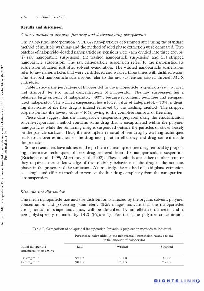
Table I shows the percentage of haloperidol in the nanoparticle suspension (raw, washed
and stripped) for two initial concentrations of haloperidol. The raw suspension has a
relatively large amount of haloperidol, �90%, because it contains both free and encapsu-
lated haloperidol. The washed suspension has a lower value of haloperidol, �70%, indicat-
ing that some of the free drug is indeed removed by the washing method. The stripped
suspension has the lowest value, <40%, owing to the complete removal of free drug.
These data suggest that the nanoparticle suspension prepared using the emulsification
solvent-evaporation method contains some drug that is encapsulated within the polymer
nanoparticles while the remaining drug is suspended outside the particles or sticks loosely
on the particle surfaces. Thus, the incomplete removal of free drug by washing techniques
leads to an over-estimation of the drug incorporation efficiency and drug content inside
the particles.
Some researchers have addressed the problem of incomplete free drug removal by propos-
ing alternative techniques of free drug removal from the nanoparticulate suspension
(Baichello et al. 1999; Aberturas et al. 2002). These methods are either cumbersome or
they require an exact knowledge of the solubility behaviour of the drug in the aqueous
phase, in the presence of the surfactant. Alternatively, the method of solid phase extraction
is a simple and efficient method to remove the free drug completely from the nanoparticu-
late suspension.
Size and size distribution
The mean nanoparticle size and size distribution is affected by the organic solvent, polymer
concentration and processing parameters. SEM images indicate that the nanoparticles
are spherical in shape and, thus, will be described by an effective diameter and a
size polydispersity obtained by DLS (Figure 1). For the same polymer concentration
Table I. Comparison of haloperidol incorporation for various preparation methods as indicated.
Percentage haloperidol in the nanoparticle suspension relative to the
initial amount of haloperidol
Initial haloperidol
concentration in DCM
Raw Washed Stripped
0.83 mg ml�1 92�3 70�8 37�6
1.67 mg ml�1 90�5 75�3 23�5
776 A. Budhian et al.
Jour
nal o
f M
icro
enca
psul
atio
n D
ownl
oade
d fr
om in
form
ahea
lthca
re.c
om b
y U
nive
rsity
of
Bri
tish
Col
umbi
a on
04/
21/1
3Fo
r pe
rson
al u
se o
nly.

0
20
40
60
80
100
120
200
231
262
300
407
526
680
811
920
Diameter (nm)
Inte
nsity
0
20
40
60
80
100
120
200
231
262
300
407
526
680
811
920
Inte
nsity
(a)
(c)
(b)
1 µm
0
20
40
60
80
100
120
200
231
262
300
407
526
680
811
920
Inte
nsity 1 µm
1 µm
Figure 1. Nanoparticle size distribution provided by dynamic light scattering histograms and theSEM images of nanoparticles prepared using PLGA 50 : 50 at (a) 33.3 mg ml�1 initial concentrationin acetone, (b) 20 mg ml�1 initial concentration in DCM and (c) 33.3 mg ml�1 initial concentrationin DCM. The nanoparticles in (a) and (b) are unimodal in size while those in (c) are bimodal.
Haloperidol-loaded PLGA nanoparticles 777
Jour
nal o
f M
icro
enca
psul
atio
n D
ownl
oade
d fr
om in
form
ahea
lthca
re.c
om b
y U
nive
rsity
of
Bri
tish
Col
umbi
a on
04/
21/1
3Fo
r pe
rson
al u
se o
nly.

of 33.3 mg ml�1 PLGA, the effective diameter for particles prepared with acetone is
275� 25 nm, while it is 524� 220 nm for particles prepared with DCM (Figure 1(a, c)).
The particles obtained from acetone have a unimodal population distribution with a
polydispersity of 0.04 (Figure 1(a)). With DCM, the particles have a bimodal population
distribution and, consequently, a high polydispersity of �0.29 (Figure 1(c)). Decreasing the
PLGA concentration in DCM from 33.3 mg ml�1 to 20 mg ml�1 changes the particle size
distribution from bimodal to unimodal (Figure 1(c, b)). Figure 2 shows that the effective
particle diameters for a range of PLGA concentration in both acetone and DCM. Bimodal
distributions of particles were obtained with DCM at higher polymer concentrations,
while acetone gives unimodal distributions of particles at all polymer concentrations.
The diameters of the two populations obtained from DCM are shown by dotted lines while
the mean diameter is shown by a full line.
The polymer concentration and organic solvent selection are critical in producing
unimodal nanoparticles. DCM, a water immiscible solvent, forms nanoparticles by a true
emulsification mechanism in which the larger emulsion droplets are broken into smaller
droplets by the application of external energy (Bodmeier and McGinity 1987). At higher
polymer concentrations, the energy applied through homogenization is insufficient to over-
come the resistive viscous forces provided by the dissolved PLGA in the organic phase and
the dissolved surfactant (PVA) in the aqueous phase, leading to heterogeneous droplets and
a bimodal size distribution.
In contrast, acetone, a water-soluble solvent, gives a smaller sized, more uniform,
unimodal population even in the absence of homogenization through a nanoprecipitation
mechanism (Fessi et al. 1989; Quintanar-Guerrero et al. 1998; Chorny et al. 2002a).
Acetone rapidly diffuses into the aqueous phase resulting in the precipitation of the polymer
(PLGA) that forms nanoparticles. Thus, the preparation of nanoparticles using acetone is
less sensitive to the polymer concentration and the homogenization parameters than when
using DCM. Birnbaum et al. (2000) have reported similar trends with DCM and acetone
for their PLGA nanoparticles.
Drug incorporation efficiency and drug content
Effect of initial haloperidol concentration. Figure 3(a) shows the haloperidol incorporation
efficiency values for various initial concentrations of PLGA 50 : 50 (33.3, 25 and
16.6 mg ml�1) in DCM. There are two trends evident in Figure 3(a). First, the haloperidol
incorporation efficiency decreases upon increasing the initial haloperidol concentration for a
fixed initial polymer concentration. Secondly, for a fixed initial drug concentration, the
haloperidol incorporation efficiency increases upon increasing the initial PLGA concentra-
tion. However, the haloperidol content in the nanoparticles has a constant value of �1% for
PLGA 50 : 50 particles, irrespective of the initial polymer or drug concentrations for the
range tested (Figure 3(b)). A similar trend was observed for capped PLGA 95 : 5 particles
(data not shown).
The initial drug-to-polymer ratio in the organic phase is critical in determining the drug
incorporation, although the larger values of this ratio lead to smaller values of drug
incorporation. This unexpected finding is furthered by the observation that the haloperidol
content in the nanoparticles is independent of the initial haloperidol concentration. These
results combine to suggest that the final haloperidol content in these PLGA nanoparticles
has an upper limit, which cannot be increased by simply increasing the initial haloperidol-
to-polymer ratio in the emulsion. Rather, increasing the initial haloperidol-to-polymer
ratio in the emulsion leads to an increase in the amount of free drug, while the amount
778 A. Budhian et al.
Jour
nal o
f M
icro
enca
psul
atio
n D
ownl
oade
d fr
om in
form
ahea
lthca
re.c
om b
y U
nive
rsity
of
Bri
tish
Col
umbi
a on
04/
21/1
3Fo
r pe
rson
al u
se o
nly.

of encapsulated drug remains constant. Chorny et al. (2002b) also reported an upper limit
on their drug (tyrphostin AG-1295) amount that can be incorporated in a fixed amount of
PLGA. Baichello et al. (1999) suggested that a low affinity between PLGA and their drug
(valproic acid) may be responsible for extremely low incorporation efficiency of 5.6%.
Hence, the physical encapsulation of haloperidol in PLGA is limited by the
haloperidol-PLGA interaction.
Effect of PLGA end groups. It was found that the end groups have a significant effect on the
haloperidol incorporation and its release behaviour from the nanoparticles. Two types of
PLGA polymers were used to produce haloperidol-loaded nanoparticles: uncapped
(carboxyl acid end group) and capped (methyl ester end group) PLGA (Figure 4). The
haloperidol incorporation efficiency with uncapped PLGA 50 : 50 is 32� 15%, which is
three times higher than with capped PLGA 50 : 50, 8� 5%. Also, the haloperidol
incorporation values for capped PLGA 50 : 50 (�8%) and capped PLGA 95 : 5 (�12%)
are comparable, thus indicating that the L:G ratio is less important than the end-groups.
The importance of end-groups has previously been suggested for PLGA microspheres
loaded with the drugs gentamicin (Nagata et al. 1994) and leuprorelin (Takada 1998).
There is also a report for PLGA nanoparticles in which the end group doubles the protein
loading (Gaspar et al. 1998).
Based on these results, we propose that the presence of carboxylic acid groups (�COOH)
increases the hydrogen bonding between the PLGA chains and the haloperidol molecules,
which hinders the drug diffusion out of the polymer nanoparticle during solvent evapora-
tion. Thus, the amount of haloperidol incorporated in the uncapped PLGA matrix
is higher due to the tendency for hydrogen bonding between �COOH end groups and
haloperidol (Figure 4(b)). The �COCH3 end group of capped PLGA and the �CH2OH
end group of both capped and uncapped PLGA are much less capable of forming a
hydrogen bond with haloperidol. This is further verified if one considers two nanoparticle
samples: uncapped and capped PLGA nanoparticles, prepared with 2.5 mg of haloperidol
0
200
400
600
800
1000
1200
1400
0 20 40 60 80
PLGA 50:50 concentration in organic solvent, mg/ml
Effe
ctiv
e di
amet
er, n
m
DCM; Mean
Acetone
DCM; Population 1
DCM; Population 2
Figure 2. Effect of PLGA 50 : 50 concentration in the organic phase on the nanoparticleeffective diameter for particles prepared with DCM and acetone as the organic solvents. The dottedlines indicate the diameters of the two populations (œ) in the case of DCM at higher concentrations.The full lines indicate the mean effective diameters for particles prepared with DCM (g) andwith acetone (m).
Haloperidol-loaded PLGA nanoparticles 779
Jour
nal o
f M
icro
enca
psul
atio
n D
ownl
oade
d fr
om in
form
ahea
lthca
re.c
om b
y U
nive
rsity
of
Bri
tish
Col
umbi
a on
04/
21/1
3Fo
r pe
rson
al u
se o
nly.

and 100 mg of polymer of molecular weight �50 kD corresponding to �1.2�1018 –COOH
end groups in the uncapped PLGA. If each –COOH group corresponds to one
additional haloperidol molecule (by virtue of hydrogen bonding), then the uncapped
PLGA nanoparticles would contain �0.75 mg more haloperidol than the capped
PLGA nanoparticles. The initial haloperidol amount was 2.5 mg, so this corresponds to
an increase of �30% in haloperidol incorporation, which is comparable to the observed
increase of �24%. Hence, the polymer end groups exercise significant effect on haloperidol
incorporation and content in the nanoparticles.
In vitro release study
The release rate is strongly influenced by the PLGA end groups and the PLGA copolymer
composition (L:G ratio). Figure 5 shows the cumulative percentage of haloperidol released
as a function of time for nanoparticles made from PLGA 50 : 50, uncapped and capped.
It can be seen that the particles from uncapped PLGA have a lower initial burst of �40%
0
20
40
Initial haloperidol concentration, mg/ml
% In
corp
orat
ion
efic
ienc
y
0
0.5
1
1.5
2
0 1 2 3 4
0 1 2 3 4
Initial haloperidol concentration, mg/ml
Dru
g co
nten
t (w
t.%)
(a)
(b)
Figure 3. (a) Haloperidol incorporation efficiency as a function of initial haloperidol loading forvarious initial concentrations in DCM of uncapped PLGA 50 : 50 (of Mw 51 kD). (b) Finalhaloperidol content in the nanoparticles as a function of initial haloperidol concentration forvarious initial concentrations of PLGA 50 : 50. Legends: ^: 33.3 mg ml�1 of PLGA 50 : 50;s: 25 mg ml�1 of PLGA 50 : 50; m: 16.6 mg ml�1 of PLGA 50 : 50.
780 A. Budhian et al.
Jour
nal o
f M
icro
enca
psul
atio
n D
ownl
oade
d fr
om in
form
ahea
lthca
re.c
om b
y U
nive
rsity
of
Bri
tish
Col
umbi
a on
04/
21/1
3Fo
r pe
rson
al u
se o
nly.

(a)
Capped PLGA
(b)
Uncapped PLGA
OH
N
O
F
Cl
O
C OCH HC O
O
m n
OH
Haloperidol
O
C OCH CH2 H
CH3
CH3
CH2
C O
O
m n
CH3
Figure 4. (a) Chemical structure of capped (–COCH3 terminated) PLGA. (b) Schematic showingthe hydrogen bonding between uncapped (–COOH terminated) PLGA and haloperidol.
0
20
40
60
80
100
120
0 5 10 15
Time (days)
% H
alop
erid
ol r
elea
sed
Capped PLGA 50:50
UncappedPLGA 50:50
Figure 5. In vitro haloperidol release from PLGA 50 : 50 nanoparticles for œ uncapped (�COOHterminated) and g capped (�COCH3 terminated) PLGA. Nanoparticles prepared with uncappedPLGA exhibit a lower initial burst release and a longer release period as compared to nanoparticlesprepared with capped PLGA.
Haloperidol-loaded PLGA nanoparticles 781
Jour
nal o
f M
icro
enca
psul
atio
n D
ownl
oade
d fr
om in
form
ahea
lthca
re.c
om b
y U
nive
rsity
of
Bri
tish
Col
umbi
a on
04/
21/1
3Fo
r pe
rson
al u
se o
nly.

as compared to that of �70% from capped PLGA. Also, �95% of the haloperidol is released
in 4 days from uncapped PLGA nanoparticles, while it takes only 2 days with capped PLGA.
There are two opposing factors that contribute to the drug release from PLGA nano-
particles. The rate of hydrolysis is higher for uncapped PLGA, corresponding to a faster
release. Conversely, the strength of haloperidol-PLGA interactions is greater for the
uncapped PLGA due to the –COOH groups, yielding slower drug release. In the
haloperidol-PLGA nanoparticles, the slower release observed in uncapped PLGA 50 : 50
(relative to capped 50 : 50) indicates that the haloperidol-PLGA interactions dominate the
release profile. The prolonged drug release from nanoparticles prepared using uncapped
PLGA is consistent with the extended release response reported for nanospheres of
L-asparginase and PLGA (Gaspar et al. 1998). In contrast, other researchers have reported
faster drug release when using uncapped PLGA (Lam et al. 2000; Soppimath and
Aminabhavi 2002). These opposing observations can be reconciled by considering the
balance between the rate of polymer hydrolysis and the strength of drug-polymer inter-
actions for specific drug-polymer combinations. In the haloperidol-PLGA system and the
L-asparginase-PLGA system (Gaspar et al. 1998) there are strong interactions between
the drug molecules and the carboxylic acid end groups of the uncapped PLGA chains.
This is evident from the fact that the drug incorporation in uncapped vs. capped PLGA
nanoparticles increases by thrice and twice, respectively, in the above two systems. These
strong interactions clearly overwhelm the faster hydrolysis typical of uncapped PLGA,
causing an overall slower release for uncapped PLGA. On the other hand, the rhIGF-
I-PLGA system (Lam et al. 2000) and the nifedipine-PLGA system (Soppimath and
Aminabhavi 2002) have the same drug incorporation for uncapped and capped PLGA,
indicating the absence of any favourable interaction between the drug and polymer end
groups. So, in these cases, the faster hydrolysis of the uncapped PLGA dominates and
the overall release is faster for the uncapped PLGA particles. Clearly, the balance between
faster hydrolysis and stronger interactions is specific to the number and type of drug-PLGA
interactions and, thus, will be drug dependent.
In addition to the influence of end groups, the PLGA composition strongly influences
release. Figure 6(a) shows the cumulative percentage haloperidol release for nanoparticles
made from capped PLGA 95 : 5 and uncapped PLGA 50 : 50 as a function of time. Both
sets of particles have a drug content of �0.3% and a bimodal size distribution with popula-
tions of �250 nm and �970 nm in size. It can be seen that the capped PLGA 95 : 5 particles
exhibit low burst release of �15% as compared to that of �35% with uncapped
PLGA 50 : 50. The drug release period is �13 days with capped PLGA 95 : 5 as opposed
to �2 days with uncapped PLGA 50 : 50.
Another release study was performed for unimodal particles made from uncapped PLGA
50 : 50 and capped PLGA 95 : 5 and the particle diameter was monitored. These particles
from both polymers have an initial effective diameter of �630 nm and the drug loading
is �1.9% for uncapped PLGA 50 : 50 particles and �0.4% for capped PLGA 95 : 5 particles.
As expected, the haloperidol release is slower from capped PLGA 95 : 5 nanoparticles
(Figure 6(b)). The particle size, as measured by dynamic light scattering, remains approxi-
mately constant for both sets of particles during the in vitro release study (Figure 6(c)).
Comparison of drug release profiles for the same polymer in Figure 6(a) and (b) suggests
that the particle size, size distribution and drug loading also influence drug release rate.
Drug can be released from polymer nanoparticles by the mechanism of diffusion and/or
polymer erosion. It was observed that haloperidol release from bulk PLGA requires many
weeks as it proceeds by both diffusion and polymer erosion (Siegel et al. 2002). In contrast,
the nanoparticles studied here release the haloperidol without a detectable change
782 A. Budhian et al.
Jour
nal o
f M
icro
enca
psul
atio
n D
ownl
oade
d fr
om in
form
ahea
lthca
re.c
om b
y U
nive
rsity
of
Bri
tish
Col
umbi
a on
04/
21/1
3Fo
r pe
rson
al u
se o
nly.

0
20
40
60
80
100
120
Time (days)
Time (days)
Time (days)
% H
alop
erid
ol r
elea
sed
0
20
40
60
80
100
120
% H
alop
erid
ol r
elea
sed
0
100
200
300
400
500
600
700
800
0 5 10 15
0 5 10 15
0 5 10 15
Effe
ctiv
e di
amet
er, n
m
UncappedPLGA 50:50 Capped
PLGA 95:5
UncappedPLGA 50:50
CappedPLGA 95:5
Capped PLGA 95:5
UncappedPLGA 50:50
(a)
(b)
(c)
Figure 6. (a) In vitro haloperidol release from PLGA nanoparticles with bimodal size distribution.Nanoparticles prepared from capped PLGA 95 : 5 (g) show an extended drug release periodand a lower initial burst release as compared to nanoparticles prepared from uncapped PLGA50 : 50 (œ). (b) In vitro haloperidol release profile from PLGA nanoparticles with unimodalsize distribution and prepared from uncapped PLGA 50 : 50 and capped PLGA 95 : 5. (c) Variationof the effective diameter of the particles in (b), as measured by dynamic light scattering. The error barsrepresent the standard deviation of the effective diameter from triplicate light scattering data.
Haloperidol-loaded PLGA nanoparticles 783
Jour
nal o
f M
icro
enca
psul
atio
n D
ownl
oade
d fr
om in
form
ahea
lthca
re.c
om b
y U
nive
rsity
of
Bri
tish
Col
umbi
a on
04/
21/1
3Fo
r pe
rson
al u
se o
nly.

in effective diameter. Thus, the mechanism governing release from these nanoparticles is
predominantly drug diffusion. Furthermore, the slower release from capped PLGA 95 : 5
can be understood in terms of drug diffusion, because the increased hydrophobicity of
capped PLGA 95 : 5 as compared to uncapped PLGA 50 : 50 corresponds to less hydration
and swelling and consequently slower drug diffusion out of the polymer matrix. The
dramatic differences in drug release characteristics between bulk and nanoparticle PLGA
systems highlights the importance of size when designing materials smaller than 1 mm,
as is typical in the field of nanotechnology.
Conclusions
Haloperidol-loaded PLGA particles were produced using an emulsification-solvent
evaporation method. The size of the particles can be varied from 200–2000 nm and is
most strongly affected by solvent miscibility and polymer concentration in the organic phase.
Solid phase extraction was used to remove free drug from the nanoparticle suspension.
PLGA with a –COOH end group (uncapped) substantially increases the drug incorporation
in these nanoparticles, perhaps through the mechanism of hydrogen bonding. Haloperidol
incorporation in uncapped PLGA nanoparticles is three times that in capped PLGA
nanoparticles. Including –COOH end-groups on PLGA (uncapped) and increasing the
PLA content in the PLGA reduces the initial burst and extends the duration of drug release
from the nanoparticles. Uncapped PLGA shows a lower initial burst release (�40%) and a
longer period of haloperidol release (�4 days) as compared to capped PLGA (�70% burst
release and �2 day release period). Nanoparticles prepared from uncapped PLGA 95 : 5
show a lower burst release of �15% and have a drug release period of �13 days. While
release in bulk mixtures of PLGA-drug is dominated by the degradation rate of the PLGA,
the drug release from PLGA-haloperidol nanoparticles is dominated by the diffusion rate
of the drug as controlled by the swelling of the PLGA, which is related to the PLA content
and the interactions between the drug and the PLGA. The distinctions between bulk and
nanoparticle drug-loaded PLGA systems highlight the importance of size on the production
and properties of materials, a cornerstone of nanotechnology and suggest a general approach
to engineering nanoparticles for extended drug delivery.
Acknowledgements
This research was supported by financial assistance from the Commonwealth of
Pennsylvania through the Nanotechnology Institute. We are grateful to Professor Anthony
Lowman (Drexel University) and Professor Nily Dan (Drexel University) for their valuable
insights. We are thankful to Professor Anthony Lowman for providing laboratory facilities
and to Meredith Hans (Drexel University) and Yuling Liang (University of Pennsylvania)
for their invaluable help with the experiments.
References
Aberturas MR, Molpeceres J, Guzman M, Garcia F. 2002. Development of a new cyclosporine formulation based
on poly(caprolactone) microspheres. Journal of Microencapsulation 19:61–72.
Allemann E, Gurny R, Leroux JC. 1996. Boidegradable nanoparticles of poly(lactic-co-glycolic acid) for parenteral
administration. In: Liberman HA, Reiger MM, Banker GS, editors. Pharmaceutical dosage forms: Disperse
systems. New York: Marcel Dekker. pp 163–194.
784 A. Budhian et al.
Jour
nal o
f M
icro
enca
psul
atio
n D
ownl
oade
d fr
om in
form
ahea
lthca
re.c
om b
y U
nive
rsity
of
Bri
tish
Col
umbi
a on
04/
21/1
3Fo
r pe
rson
al u
se o
nly.

Baichello JM, Morishita M, Takayama K, Nagai T. 1999. Encapsulation of hydrophilic and lipophilic
drugs in PLGA nanoparticles by the nanoprecipitation method. Drug Development and Industrial Pharmacy
25:471–476.
Birnbaum DT, Kosmala JD, Brannon-Peppas L. 2000. Optimization of preparation techniques for poly(lactic acid-
co-glycolic acid) nanoparticles. Journal of Nanoparticle Research 2:173–181.
Bodmeier R, McGinity JW. 1987a. Polylactic acid microspheres containing quinidine base and quinidine sulphate
prepared by the solvent evaporation technique. I. Methods and morphology. Journal of Microencapsulation
4:279–288.
Cappel MJJK. 1991. Effect of nanoparticles on transdermal drug delivery. Journal of Microencapsulation
8:369–374.
Chorny M, Fishbein I, Danenberg HD, Golomb G. 2002a. Lipophilic drug loaded nanospheres prepared by
nanoprecipitation: Effect of formulation variables on size, drug recovery and release kinetics. Journal of
Controlled Release 83:389–400.
Chorny M, Fishbein I, Danenberg HD, Golomb G. 2002b. Study of the drug release mechanism from tyrphostin
AG-1295 loaded nanospheres by in situ and external sink methods. Journal of Controlled Release 83:401–414.
Fessi H, Puisieux F, Devissaguet JP, Ammoury N, Benita S. 1989. Nanocapsules formulation by interfacial polymer
deposition following solvent displacement. International Journal of Pharmaceutics 55:R1–R4.
Gaspar MMBD, Cruz MEM, Alonso MJ. 1998. Formulation of L-asparaginase-loaded poly(lactic-co-glycolide)
nanoparticles: Influence of polymer properties on enzyme loading, acitvity and in vitro release. Journal of
Controlled Release 52:53–62.
Jain R, Shah NH, Malick AW, Rhodes CT. 1998. Controlled drug delivery by biodegradable poly(ester) devices:
Different preparative approaches. Drug Development and Industrial Pharmacy 24:703–727.
Lam XMDET, Daugherty AL, Levin N, Cleland JL. 2000. Sustained release of recombinant human insulin-like
growth factor-I for treatment of diabetes. Journal of Controlled Release 67:281–292.
Nagata STK, Hirano K, Takagishi Y. 1994. Pharmaceutical dosage form design of copoly (lactic/glycolic acid)
microspheres. Mechanism of in vitro release of gentamicin. Yakugaku Zasshi 114:1005–1014.
Quintanar-Guerrero D, Allemann E, Doelker E, Fessi H. 1998. Preparation and characterization of nanocapsules
from preformed polymers by a new process based on emulsification-diffusion technique. Pharmaceutical
Research 15:1056–1062.
Siegel SJKIW, Gur RE, Lenox RH, Bilker WB, Ikeda D, Gandhi N, Zhang W-X. 2002. Surgically implantable
long-term antipsychotic delivery systems for the treatment of schizophrenia. Neuropsychopharmacology
26:817–823.
Soppimath KS, Aminabhavi TM. 2002. Ethyl acetate as a dispersing solvent in the production of poly(DL-lactide-
co-glycolide) microspheres: Effect of process parameters and polymer type. Journal of Microencapsulation
19:281–292.
Takada SOY. 1998. Design and development of controlled release of drugs from injectable microcapsules.
Nippon Rinsho 56:675–679.
Haloperidol-loaded PLGA nanoparticles 785
Jour
nal o
f M
icro
enca
psul
atio
n D
ownl
oade
d fr
om in
form
ahea
lthca
re.c
om b
y U
nive
rsity
of
Bri
tish
Col
umbi
a on
04/
21/1
3Fo
r pe
rson
al u
se o
nly.