Embed Size (px)
Citation preview
Journal of Advanced Research Design ISSN (online): 2289-7984 | Vol. 6, No.1. Pages 21-36, 2015
21
Penerbit
Akademia Baru
Production of Anti-Cancer Compound, Physalin B from Callus Cultures of Physalis Minima (Linn.)
G. I. Jualang Azlan*,a and M. Marziahb
aFaculty of Science and Natural Resources, Universiti Malaysia Sabah, UMS Road, 88400 Kota Kinabalu, Sabah, Malaysia.
bDepartment of Biochemistry and Microbiology, Faculty of Biotechnology and Biomolecular Science, Universiti Putra Malaysia, 43400 Serdang, Selangor, Malaysia.
Abstract – This study was conducted to establish callus cultures from leaf, stem and root explants of
Physalis minima using different combinations of 2,4-D and kinetin. Callus growth and anti-cancer
compound, physalin B production were further enhanced by optimising the cell explants and media
compositions such as basal media, salts concentration, carbon sources and plant growth regulators.
The results indicated that callus cultures derived from leaf, stem and root explants were best initiated
using a combination of 9.0 µM 2,4-D and 4.5 µM kinetin. Callus growth and synthesis of physalin B
were peaked at the late exponential growth phase over 25 d of culture. Callus growth did not vary
between explants, but physalin B was observed higher in leaf (0.78 mg g-1 dry wt.), followed by root
(0.71 mg g-1 dry wt.) and stem (0.64 mg g-1 dry wt.). MS basal medium was found superior to B5, SH
and WH basal media in supporting growth and physalin B production. Further tests on the media
compositions obtained a half strength of MS salts (½MS), 2.5% (w/v) sucrose and 9.0:4.5 µM of 2,4-
D:kinetin combination, which were the preferred salts strength, carbon sources and plant growth
regulators for optimum growth (0.23 g dry wt.) and physalin B production (1.75 mg g-1 dry wt.) of
callus cultures derived from leaf. Copyright © 2015 Penerbit Akademia Baru - All rights reserved.
Keywords: Basal medium, Carbon source, Callus cultures, Explants, Plant Growth Regulators, Secondary metabolites, Physalin B
1.0 INTRODUCTION
Physalin B is a steroid derivative that possesses a novel 13,14-seco-16,24-cycloergostane skeleton discovered from the Physalis alkekengi var. Franchetii by Matsuura et al. [1] and was later used as a taxonomic maker for the genus [2]. Physalin B has a wide array of bioactivities including for anti-inflammatory [3–4], anti-mycobacterial [5–6], antimicrobial [7], anti-malarial [8], antileishmanial [9], antinociceptive [10] and anticancer [11–17]. Physalin B represents a novel therapeutic option for the treatment of inflammatory diseases through the activation of glucocorticoid receptors [3]. Physalin B has also been found to display a non-selective cytotoxic effect on human CORL23 lung and MCF-7 breast carcinoma cells [18] and possess suppressive effects on human leukemia (K562 and APM1840 HL-60, KG-1, CTV1 and B cell), cervix (HeLa), hepatoma (HA22T), nasopharynx (KB), colon (Colo-205), lung (Calu-1), sarcoma cell lines [12–13],[19] and other cancerous cell lines such as A431, HCT-8, PC-3, and ZR751 [20]. Physalin B induces apoptosis of
Journal of Advanced Research Design ISSN (online): 2289-7984 | Vol. 6, No.1. Pages 21-36, 2015
22
Penerbit
Akademia Baru
melanoma cancer cells by inducing the pro-apoptotic protein NOXA expression and also triggers the expression of Bax and caspase-3 [16]. Recently, physalin B was reported to inhibit Gli in Hedgehog signalling pathways [21–22], which leads to their significant function in controlling cancerous cells.
Physalin B can be obtained from various Physalis sp. including Physalis minima [1], [23–25], which is the only species found in Malaysia. Physalis minima plants are tetraploid with globular fruits enclosed in an inflated bladder-like calyx and belong to the Solanaceae family [26]. The decoction of the entire plant is traditionally consumed by Malay communities in Peninsular Malaysia to treat cancer [27]. The plants have also been used to remedy headaches, earaches, fevers, ulcers, spleen disorder, wound pustule, intestinal pains, purgative, diuretic, gonorrhoea, as a tonic, and to restore flaccid breasts [26], [28–30].
Sipahimalani et al. [31] initially reported the presence of physalin B in tissue culture systems, and were further investigated by Jualang et al. (2005) [25] to compare the production of physalin in cell cultures and intact plants. Physalin production in hairy-roots was also reported by Jualang et al. (2002) [24] and Jualang et al. (2013) [32]. Our continuous effort to study the enhancement of physalin production in cell cultures is further elaborated in this report. Plant cell cultures hold great promises for a controlled production of useful secondary metabolites on demand. Cell cultures are capable of producing specific bioactives at a rate similar or superior to that of intact plants under controlled conditions, independent of climatic changes and soil conditions, free from microbes and insects, and feasible for automated control of cell growth and regulation of metabolites to reduce labor costs and improve productivity [33–35]. Toward these, the primary task is to optimize cell sources (cells line) from specific explant and media compositions such as basal media, salts strength, carbon sources and plant growth regulators to enhance biomass yields and also to improve production of bioactive compounds [35]. Therefore, the objective of this work is to establish callus cultures from the leaf, stem and root explants of P. minima and to optimise their growth and physalin B production in callus cultures by manipulating the media components.
2.0 MATERIALS AND METHODS
2.1 Establishment of In-vitro Plant Cultures
The mature seeds of P. minima obtained from plants growing wild in the vegetable farm at Universiti Putra Malaysia, Malaysia were sterilised by soaking them in 70% (v/v) of ethanol for 5 minutes and then agitated by dipping in 25% (v/v) of commercial Clorox® with 3 drops of Tween 20 for 30 minutes and finally, rinsed for five times with sterile distilled water. The surface-sterilised seeds were germinated at room temperature in the dark on petri dishes lined with sterile wet towel paper. The germinated seedlings were aseptically transferred into flasks containing MS basal medium [36] supplemented with B5 vitamins [37], 3% (w/v) of sucrose, 1% (w/v) of casein hydrolysate and solidified with 0.25% (w/v) of Gelrite agar. The pH of the media was 5.7. The cultures were grown at 25 ± 2 °C with a photoperiod of 16 h fluorescent light and subcultured for every three weeks in medium containing 0.5 mg l-1 BAP.
2.2 Initiation of Callus
Callus cultures were established from leaf, stem and root explants of three week old seedlings germinated in vitro. About 30 pieces of leaves (5x5 mm), stems (5 mm) and roots (10 mm)
Journal of Advanced Research Design ISSN (online): 2289-7984 | Vol. 6, No.1. Pages 21-36, 2015
23
Penerbit
Akademia Baru
were each subjected to 2,4-D (2.25 – 22.5 µM) and kinetin (0 – 9 µM) treatments on petri dishes containing MS salts, B5 vitamins, 3% (w/v) sucrose and 0.25% (w/v) Gelrite. The study was conducted in 7 replicates and cultures were grown at 25±2 0C with a photoperiod of 16 h with fluorescent light. Cultures were monitored daily to observe callus initiation. The percentage of callus induction (Cip) was observed after 3 weeks and measured according to Holme and Petersen [38]. Callus cultures were maintained in a fresh MS medium supplemented with 9.0:4.5 µM of 2,4-D: kinetin and subcultured for every 20 days.
2.3 Growth Curves, the Cell Explant and Media Optimization
The curves of growth and physalin B production in cells derived from leaf explant were monitored every 5 days for 35 days on a MS medium supplemented with 9.0:4.5 µM of 2,4-D: kinetin. Callus growth and physalin B production were compared between the leaf, stem and root explants of similar culture conditions. To further optimise the production of physalin B, media compositions such as basal media and salt strength, carbon sources and plant growth regulators were subsequently determined. The four basal media tested were MS [36], B5 [37], SH [39] and WH [40]. Iron was supplied as FeNaEDTA. The best basal medium was further tested for different salts strength. The ¼, ½, 1, and 2 salts strength represents quarter, half, full (normal) and double strength of the salts concentration (macronutrients) from standard formulation of basal medium, respectively. Effect of sucrose (Merck) was determined at the concentrations between 2.0 to 3.5% (w/v). Optimum sucrose concentration was later compared with their monomers, glucose and fructose using a relatively similar carbon ratio. Auxins such as 2,4-dichlorophenoxyacetic acid (2,4-D), 1-naphthaleneacetic acid (NAA), 4-amino-2,5,6-trichloropicolinic acid (Picloram), and 2-metoxy-3,6-dichlorobenzoic acid (Dicamba) were tested between 2.25 – 13.5 µM, and the best performance was further combined with 0 – 4.5 µM of 6-furfurylaminopurine (Kinetin), to optimize the callus biomass and physalin B production. All treatments were set-up in transparent glass tubes containing 10 mL basal medium at pH 5.7, supplemented with B5 vitamins and solidified with 0.25% (w/v) Gelrite agar. A 0.5±0.1 g fresh weight calluses obtained from 20 days old culture was used as inoculum. The treatments in 7 replicates were incubated at 25 ± 2 0C with 16 hours of fluorescent light.
2.4 Extraction Procedures and High Performance Liquid Chromatography Analysis
Two-gram samples of dry powdered samples were extracted with methanol at room temperature under dark conditions. The crude methanolic extracts diluted with the same volume of distilled water were partitioned twice by hexane and chloroform. The chloroform partition was evaporated and dissolved in 65% (v/v) methanol (spectra grade) and finally filtered through a Sep-Pak Classic Cartridge (Waters Corp.; Milford, MA, USA) for analysis using high performance liquid chromatography (HPLC) [24]. HPLC was performed on a PerkinElmer Series 200 system equipped with Series 200 auto sampler, Series 200 column oven, PerkinElmer LC-250B pump and Series 200 UV/Vis detector. A 3.9 × 150 mm I.D. Nova Pak C18 60Å steel cartridge column, fitted at 4 µm (Waters Associates Inc.; Milford, MA, USA) containing dimethyloctadecylsilyl- bounded amorphous silica and methylalcohol was used as eluents in the mixtures of methanol-water (65:35, v/v) at a flow rate of 1.0 mL.min-1. The mixtures consisted of methanol aqueous isocratic solvent (Uvasol, spectroscopy grade, Merck Group; Darmstadt, Germany) and double reverse-phase distilled water. Chromatogram of physalin B was detected at 220 nm and referred to as the authentic compound.
Journal of Advanced Research Design ISSN (online): 2289-7984 | Vol. 6, No.1. Pages 21-36, 2015
24
Penerbit
Akademia Baru
2.5 Statistical Analysis
The results were analyzed using one-way analysis of variance and the mean values were compared at P < 0.05 via Duncan’s multiple range test (DMRT) to find the significant difference using the General Linear Model Procedure of the SAS version 9.1 (SAS Institute, NC, USA).
3.0 RESULTS AND DISCUSSION
3.1 Establishment of Callus Cultures
Table 1 shows the initiation of callus from leaf, stem and root segments in different combinations of 2,4-D (2.25 to 22.5 µM) and kinetin (0 to 9.0 µM) on a MS basal medium. Treatment with 2,4-D alone is capable to induce callus within 10-22 days for leaf; 10-23 days for stem; and 13-21 days for root explants. The fastest callus induction (10-12 days) for all tested explants was observed in 22.5 µM of 2,4-D. The same concentration was required for higher Cip (41.5%) in leaf explant. However, stem (48.6%) and root (45.9%) explants produce higher Cip at concentrations of 13.5 and 18.0 µM 2,4-D, respectively. Meanwhile, the highest biomass dry wt. production for leaf (0.14 g per tube), stem (0.11 g per tube) and root (0.16 g per tube) explants were observed at 4.5 µM 2,4-D, respectively. Callus morphology obtained from 2,4-D treatments is commonly friable and yellowish in colour, except for the green-yellow colour in stem at 22.5 µM 2,4-D.
The addition of kinetin in 2,4-D-containing treatment improved the Cip and callus growth, but appears to delay the callus formation, particularly at lower concentrations of 2,4-D combined with higher concentrations of kinetin. Cip and growth of the culture were reduced significantly in higher kinetin, (9.0 µM) concentration. The highest Cip (88.6-96.6%) and biomass dry wt. production (0.15-0.20 g per tube) for leaf, stem and root explants were observed at 9.00 µM 2,4-D and 4.50 µM kinetin combination, which gave better performance as compared to single 2,4-D and other 2,4-D – kinetin combinations. The time required for callus formation in this treatment was 12 days in leaf, 12 days in stem, and 15 days in root explant. The callus proliferated into an unorganised yellowish mass of cells with friable textures. Greenish or white friable callus derived from leaf and stem explants were also obtained in treatments with kinetin and lower concentration of 2,4-D (2.25-4.5 µM). However, higher concentrations of 2,4-D (22.5 µM) and kinetin (9.0 µM) combination tends to induce browning in all explants, which is in agreement with Murthy et al. [35]. Other PGRs such as 2,4-D (4.5 µM) and BA (4.4 µM), or NAA (2.7 µM), 2,4-D (2.3 µM) and BAP (2.2 µM) combinations have also successfully promoted callus induction in P. minima [31, 41]. Both cytokinin and auxin play positive roles in regulating cell division, but the detailed mechanism is yet to be understood. However, both hormones can affect cell cycle progression into the S-phase through the cyclin-dependent kinase A/D-type cyclin (CYCD) pathway [42]. The specific response also depends on plant organ and the plant species due to its interaction between endogenous and exogenous hormones [35, 43].
3.2 Callus Growth and Physalin B Production Curves
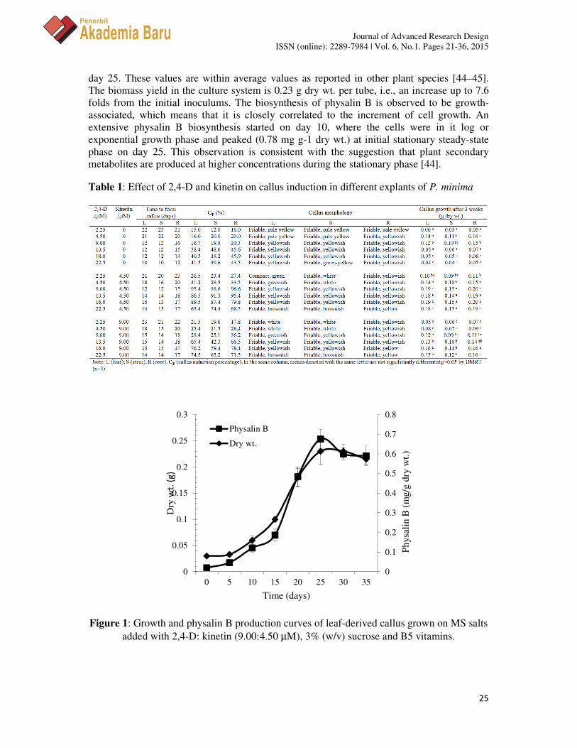
The growth and physalin B production curves of P. minima leaf-derived callus cultures are shown in Figure 1. The lag phase of the cultures is about 5 days before entering the exponential stage, where the cells actively proliferate and reach a stationary growth phase on
Journal of Advanced Research Design ISSN (online): 2289-7984 | Vol. 6, No.1. Pages 21-36, 2015
25
Penerbit
Akademia Baru
day 25. These values are within average values as reported in other plant species [44–45]. The biomass yield in the culture system is 0.23 g dry wt. per tube, i.e., an increase up to 7.6 folds from the initial inoculums. The biosynthesis of physalin B is observed to be growth-associated, which means that it is closely correlated to the increment of cell growth. An extensive physalin B biosynthesis started on day 10, where the cells were in it log or exponential growth phase and peaked (0.78 mg g-1 dry wt.) at initial stationary steady-state phase on day 25. This observation is consistent with the suggestion that plant secondary metabolites are produced at higher concentrations during the stationary phase [44].
Table 1: Effect of 2,4-D and kinetin on callus induction in different explants of P. minima
Figure 1: Growth and physalin B production curves of leaf-derived callus grown on MS salts added with 2,4-D: kinetin (9.00:4.50 µM), 3% (w/v) sucrose and B5 vitamins.
0
0.05
0.1
0.15
0.2
0.25
0.3
0
0.1
0.2
0.3
0.4
0.5
0.6
0.7
0.8
0 5 10 15 20 25 30 35
Dry
wt.
(g
)
Phys
alin
B (
mg/
g dr
y w
t.)
Time (days)
Physalin B
Dry wt.
Journal of Advanced Research Design ISSN (online): 2289-7984 | Vol. 6, No.1. Pages 21-36, 2015
26
Penerbit
Akademia Baru
3.3 Effect of Callus Explant on Growth and Physalin B Production
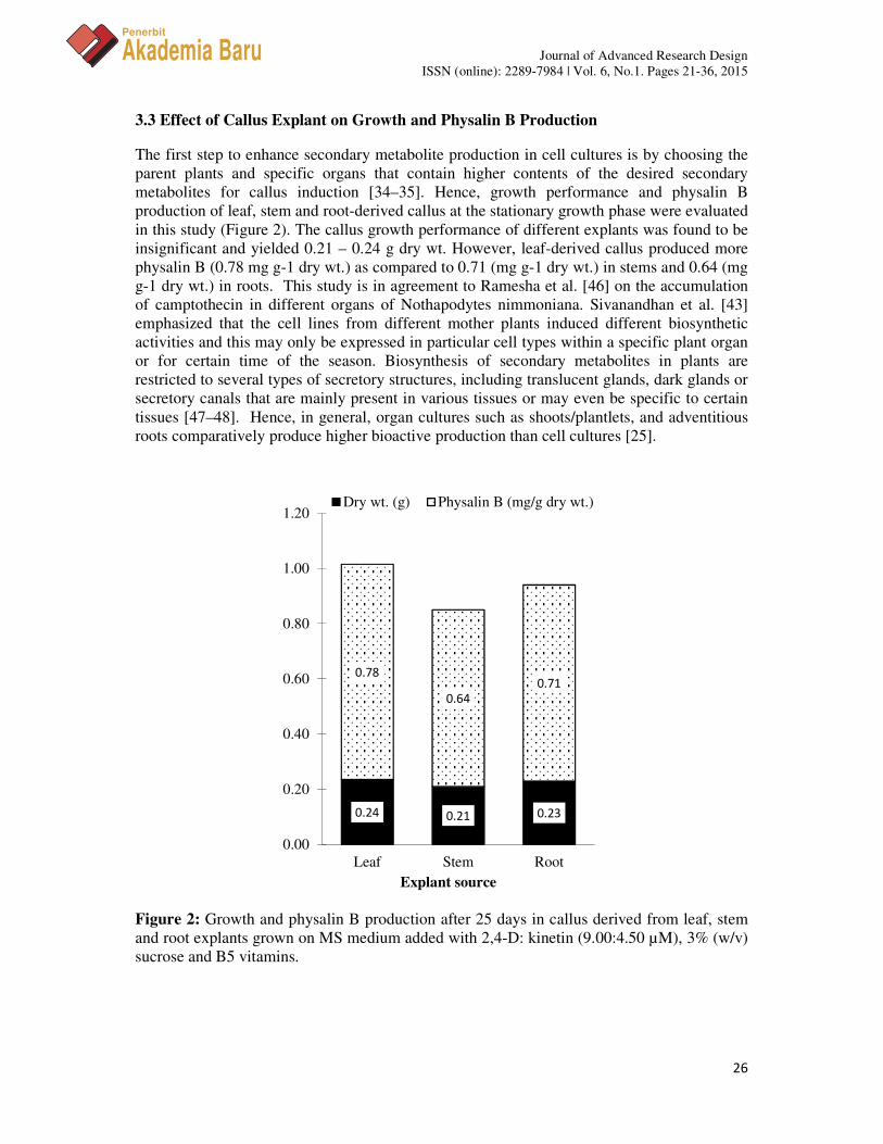
The first step to enhance secondary metabolite production in cell cultures is by choosing the parent plants and specific organs that contain higher contents of the desired secondary metabolites for callus induction [34–35]. Hence, growth performance and physalin B production of leaf, stem and root-derived callus at the stationary growth phase were evaluated in this study (Figure 2). The callus growth performance of different explants was found to be insignificant and yielded 0.21 – 0.24 g dry wt. However, leaf-derived callus produced more physalin B (0.78 mg g-1 dry wt.) as compared to 0.71 (mg g-1 dry wt.) in stems and 0.64 (mg g-1 dry wt.) in roots. This study is in agreement to Ramesha et al. [46] on the accumulation of camptothecin in different organs of Nothapodytes nimmoniana. Sivanandhan et al. [43] emphasized that the cell lines from different mother plants induced different biosynthetic activities and this may only be expressed in particular cell types within a specific plant organ or for certain time of the season. Biosynthesis of secondary metabolites in plants are restricted to several types of secretory structures, including translucent glands, dark glands or secretory canals that are mainly present in various tissues or may even be specific to certain tissues [47–48]. Hence, in general, organ cultures such as shoots/plantlets, and adventitious roots comparatively produce higher bioactive production than cell cultures [25].
Figure 2: Growth and physalin B production after 25 days in callus derived from leaf, stem and root explants grown on MS medium added with 2,4-D: kinetin (9.00:4.50 µM), 3% (w/v) sucrose and B5 vitamins.
0.24 0.21 0.23
0.78
0.64
0.71
0.00
0.20
0.40
0.60
0.80
1.00
1.20
Leaf Stem Root
Dry wt. (g) Physalin B (mg/g dry wt.)
Explant source
Journal of Advanced Research Design ISSN (online): 2289-7984 | Vol. 6, No.1. Pages 21-36, 2015
27
Penerbit
Akademia Baru
3.4 Effect of Basal Media on Growth and Physalin B Production
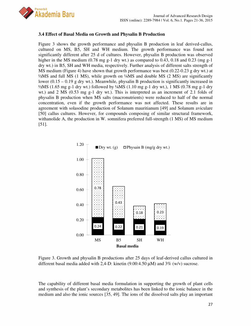
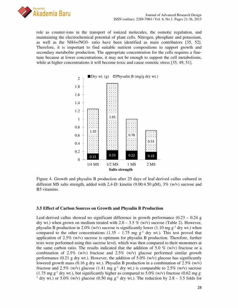
Figure 3 shows the growth performance and physalin B production in leaf derived-callus, cultured on MS, B5, SH and WH medium. The growth performance was found not significantly different after 25 d of cultures. However, physalin B production was observed higher in the MS medium (0.78 mg g-1 dry wt.) as compared to 0.43, 0.18 and 0.23 (mg g-1 dry wt.) in B5, SH and WH media, respectively. Further analysis of different salts strength of MS medium (Figure 4) have shown that growth performance was best (0.22-0.23 g dry wt.) at ½MS and full MS (1 MS), while growth on ¼MS and double MS (2 MS) are significantly lower (0.15 – 0.19 g dry wt.). Meanwhile, physalin B production is significantly increased in ½MS (1.65 mg g-1 dry wt.) followed by ¼MS (1.10 mg g-1 dry wt.), 1 MS (0.78 mg g-1 dry wt.) and 2 MS (0.53 mg g-1 dry wt.). This is interpreted as an increment of 2.1 folds of physalin B production when MS salts (macronutrients) were reduced to half of the normal concentration, even if the growth performance was not affected. These results are in agreement with solasodine production of Solanum mauritianum [49] and Solanum aviculare [50] callus cultures. However, for compounds composing of similar structural framework, withanolide A, the production in W. somnifera preferred full-strength (1 MS) of MS medium [51].
Figure 3. Growth and physalin B productions after 25 days of leaf-derived callus cultured in different basal media added with 2,4-D: kinetin (9.00:4.50 µM) and 3% (w/v) sucrose.
The capability of different basal media formulation in supporting the growth of plant cells and synthesis of the plant’s secondary metabolites has been linked to the ionic balance in the medium and also the ionic sources [35, 49]. The ions of the dissolved salts play an important
0.24 0.22 0.21 0.19
0.78
0.43
0.18 0.23
0.00
0.20
0.40
0.60
0.80
1.00
1.20
MS B5 SH WH
Dry wt. (g) Physain B (mg/g dry wt.)
Basal media
Journal of Advanced Research Design ISSN (online): 2289-7984 | Vol. 6, No.1. Pages 21-36, 2015
28
Penerbit
Akademia Baru
role as counter-ions in the transport of ionized molecules, the osmotic regulation, and maintaining the electrochemical potential of plant cells. Nitrogen, phosphate and potassium, as well as the NH4+/NO3- ratio have been identified as main contributors [35, 52]. Therefore, it is important to find suitable nutrient compositions to support growth and secondary metabolite production. The appropriate concentration for the cells requires a fine-tune because at lower concentrations, it may not be enough to support the cell metabolisms, while at higher concentrations it will become toxic and cause osmotic stress [35, 49, 51].
Figure 4. Growth and physalin B production after 25 days of leaf-derived callus cultured in different MS salts strength, added with 2,4-D: kinetin (9.00:4.50 µM), 3% (w/v) sucrose and B5 vitamins.
3.5 Effect of Carbon Sources on Growth and Physalin B Production
Leaf-derived callus showed no significant difference in growth performance (0.23 – 0.24 g dry wt.) when grown on medium treated with 2.0 – 3.5 % (w/v) sucrose (Table 2). However, physalin B production in 2.0% (w/v) sucrose is significantly lower (1.10 mg g-1 dry wt.) when compared to the other concentrations (1.35 – 1.75 mg g-1 dry wt.). This test proved that application of 2.5% (w/v) sucrose is optimum for physalin B production. Therefore, further tests were performed using this sucrose level, which was then compared to their monomers at the same carbon ratio. The results indicated that the addition of 5.0 % (w/v) fructose or a combination of 2.5% (w/v) fructose and 2.5% (w/v) glucose performed similar growth performance (0.21 g dry wt.). However, the addition of 5.0% (w/v) glucose has significantly lowered growth mass (0.16 g dry wt.). Physalin B production in a combination of 2.5% (w/v) fructose and 2.5% (w/v) glucose (1.41 mg g-1 dry wt.) is comparable to 2.5% (w/v) sucrose (1.75 mg g-1 dry wt.), but significantly higher as compared to 5.0% (w/v) fructose (0.62 mg g-
1 dry wt.) or 5.0% (w/v) glucose (0.50 mg g-1 dry wt.). The reduction by 2.8 – 3.5 folds for
0.15 0.23 0.22 0.19
1.10
1.65
0.78
0.53
0
0.2
0.4
0.6
0.8
1
1.2
1.4
1.6
1.8
2
1/4 MS 1/2 MS 1 MS 2 MS
Dry wt. (g) Physalin B (mg/g dry wt.)
Salts strength
Journal of Advanced Research Design ISSN (online): 2289-7984 | Vol. 6, No.1. Pages 21-36, 2015
29
Penerbit
Akademia Baru
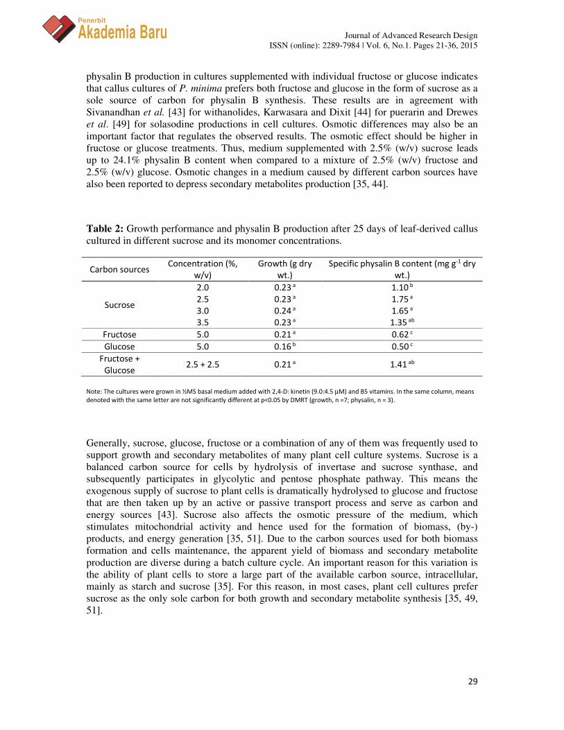
physalin B production in cultures supplemented with individual fructose or glucose indicates that callus cultures of P. minima prefers both fructose and glucose in the form of sucrose as a sole source of carbon for physalin B synthesis. These results are in agreement with Sivanandhan et al. [43] for withanolides, Karwasara and Dixit [44] for puerarin and Drewes et al. [49] for solasodine productions in cell cultures. Osmotic differences may also be an important factor that regulates the observed results. The osmotic effect should be higher in fructose or glucose treatments. Thus, medium supplemented with 2.5% (w/v) sucrose leads up to 24.1% physalin B content when compared to a mixture of 2.5% (w/v) fructose and 2.5% (w/v) glucose. Osmotic changes in a medium caused by different carbon sources have also been reported to depress secondary metabolites production [35, 44].
Table 2: Growth performance and physalin B production after 25 days of leaf-derived callus cultured in different sucrose and its monomer concentrations.
Carbon sources Concentration (%,
w/v)
Growth (g dry
wt.)
Specific physalin B content (mg g-1 dry
wt.)
Sucrose
2.0 0.23 a 1.10 b
2.5 0.23 a 1.75 a
3.0 0.24 a 1.65 a
3.5 0.23 a 1.35 ab
Fructose 5.0 0.21 a 0.62 c
Glucose 5.0 0.16 b 0.50 c
Fructose +
Glucose 2.5 + 2.5 0.21 a 1.41 ab
Note: The cultures were grown in ½MS basal medium added with 2,4-D: kinetin (9.0:4.5 µM) and B5 vitamins. In the same column, means
denoted with the same letter are not significantly different at p<0.05 by DMRT (growth, n =7; physalin, n = 3).
Generally, sucrose, glucose, fructose or a combination of any of them was frequently used to support growth and secondary metabolites of many plant cell culture systems. Sucrose is a balanced carbon source for cells by hydrolysis of invertase and sucrose synthase, and subsequently participates in glycolytic and pentose phosphate pathway. This means the exogenous supply of sucrose to plant cells is dramatically hydrolysed to glucose and fructose that are then taken up by an active or passive transport process and serve as carbon and energy sources [43]. Sucrose also affects the osmotic pressure of the medium, which stimulates mitochondrial activity and hence used for the formation of biomass, (by-) products, and energy generation [35, 51]. Due to the carbon sources used for both biomass formation and cells maintenance, the apparent yield of biomass and secondary metabolite production are diverse during a batch culture cycle. An important reason for this variation is the ability of plant cells to store a large part of the available carbon source, intracellular, mainly as starch and sucrose [35]. For this reason, in most cases, plant cell cultures prefer sucrose as the only sole carbon for both growth and secondary metabolite synthesis [35, 49, 51].
Journal of Advanced Research Design ISSN (online): 2289-7984 | Vol. 6, No.1. Pages 21-36, 2015
30
Penerbit
Akademia Baru
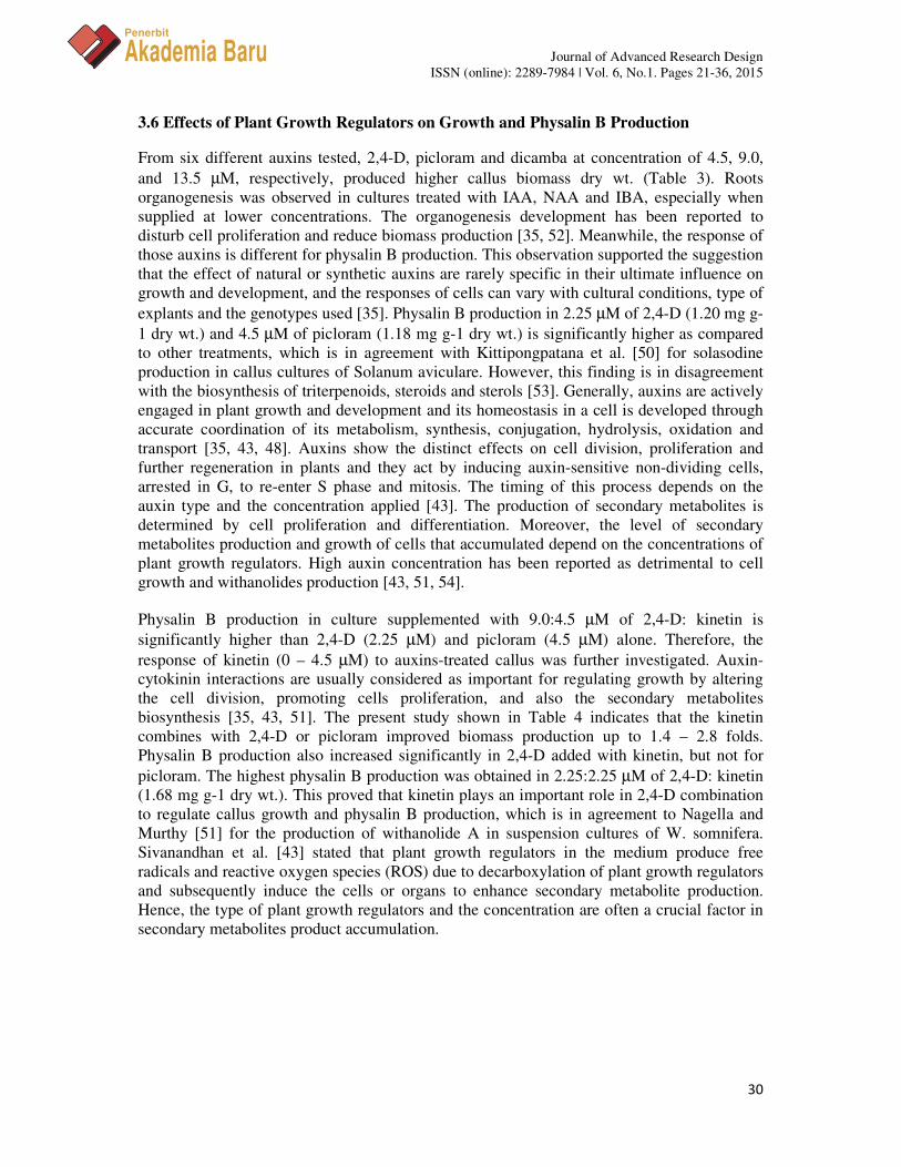
3.6 Effects of Plant Growth Regulators on Growth and Physalin B Production
From six different auxins tested, 2,4-D, picloram and dicamba at concentration of 4.5, 9.0, and 13.5 µM, respectively, produced higher callus biomass dry wt. (Table 3). Roots organogenesis was observed in cultures treated with IAA, NAA and IBA, especially when supplied at lower concentrations. The organogenesis development has been reported to disturb cell proliferation and reduce biomass production [35, 52]. Meanwhile, the response of those auxins is different for physalin B production. This observation supported the suggestion that the effect of natural or synthetic auxins are rarely specific in their ultimate influence on growth and development, and the responses of cells can vary with cultural conditions, type of explants and the genotypes used [35]. Physalin B production in 2.25 µM of 2,4-D (1.20 mg g-1 dry wt.) and 4.5 µM of picloram (1.18 mg g-1 dry wt.) is significantly higher as compared to other treatments, which is in agreement with Kittipongpatana et al. [50] for solasodine production in callus cultures of Solanum aviculare. However, this finding is in disagreement with the biosynthesis of triterpenoids, steroids and sterols [53]. Generally, auxins are actively engaged in plant growth and development and its homeostasis in a cell is developed through accurate coordination of its metabolism, synthesis, conjugation, hydrolysis, oxidation and transport [35, 43, 48]. Auxins show the distinct effects on cell division, proliferation and further regeneration in plants and they act by inducing auxin-sensitive non-dividing cells, arrested in G, to re-enter S phase and mitosis. The timing of this process depends on the auxin type and the concentration applied [43]. The production of secondary metabolites is determined by cell proliferation and differentiation. Moreover, the level of secondary metabolites production and growth of cells that accumulated depend on the concentrations of plant growth regulators. High auxin concentration has been reported as detrimental to cell growth and withanolides production [43, 51, 54].
Physalin B production in culture supplemented with 9.0:4.5 µM of 2,4-D: kinetin is significantly higher than 2,4-D (2.25 µM) and picloram (4.5 µM) alone. Therefore, the response of kinetin (0 – 4.5 µM) to auxins-treated callus was further investigated. Auxin-cytokinin interactions are usually considered as important for regulating growth by altering the cell division, promoting cells proliferation, and also the secondary metabolites biosynthesis [35, 43, 51]. The present study shown in Table 4 indicates that the kinetin combines with 2,4-D or picloram improved biomass production up to 1.4 – 2.8 folds. Physalin B production also increased significantly in 2,4-D added with kinetin, but not for picloram. The highest physalin B production was obtained in 2.25:2.25 µM of 2,4-D: kinetin (1.68 mg g-1 dry wt.). This proved that kinetin plays an important role in 2,4-D combination to regulate callus growth and physalin B production, which is in agreement to Nagella and Murthy [51] for the production of withanolide A in suspension cultures of W. somnifera. Sivanandhan et al. [43] stated that plant growth regulators in the medium produce free radicals and reactive oxygen species (ROS) due to decarboxylation of plant growth regulators and subsequently induce the cells or organs to enhance secondary metabolite production. Hence, the type of plant growth regulators and the concentration are often a crucial factor in secondary metabolites product accumulation.
Journal of Advanced Research Design ISSN (online): 2289-7984 | Vol. 6, No.1. Pages 21-36, 2015
31
Penerbit
Akademia Baru
Table 3. Growth performance and physalin B production after 25 days of leaf-derived callus cultured in different auxins concentration on a ½MS medium added with 2.5% (w/v) sucrose and B5 vitamins.
Auxins Concentration (µM) Growth (g dry wt.) Specific physalin B content (mg g-1 dry wt.)
2,4-D 2.25 0.09 b 1.20 a
4.50 0.16 a 0.90 b
9.00 0.14 a 0.55 c
13.5 0.08 b 0.26 d
IBA 2.25 0.03 c 0.10 d
4.50 0.05 c 0.11 d
9.00 0.07 b 0.23 d
13.5 0.07 b 0.04 e
NAA 2.25 0.02 c 0.06 e
4.50 0.04 c 0.10 d
9.00 0.04 c 0.13 d
13.5 0.04 c 0.14 d
Picloram 2.25 0.05 c 0.60 c
4.50 0.06 b 1.18 a
9.00 0.15 a 0.61 c
13.5 0.10 ab 0.38 cd
IAA 2.25 0.03 c 0.23 d
4.50 0.04 c 0.39 cd
9.00 0.04 c 0.25 d
13.5 0.06 b 0.21 d
Dicamba 2.25 0.06 b 0.43 cd
4.50 0.08 b 0.93 b
9.00 0.10 ab 0.73 bc
13.5 0.18 a 0.05 e
Note: In the same column, means denoted with the same letter are not significantly different at p<0.05 by DMRT (growth, n =7; physalins,
n = 3). Each tube contains 10 mL of basal medium.
Table 4. Growth performance and physalin B production after 25 days of leaf-derived callus cultured in different combination of auxins and kinetin on a ½MS medium added with 2.5% (w/v) sucrose and B5 vitamins.
Auxins Kinetin (µM) Growth (g dry
wt.)
Specific physalin B content (mg g-1 dry
wt.)
2,4-D (2.25 µM) 0 0.16 b 1.20 b
2.25 0.22 a 1.68 a
4.5 0.20 a 1.23 b
Picloram (4.5 µM) 0 0.06 c 1.18 b
2.25 0.15 b 0.92 bc
4.5 0.17 b 0.52 c
Control (2,4-D: kinetin (9.0:4.5µM)) 0.23 a 1.75 a
Note: In the same column, means denoted with the same letter are not significantly different at p<0.05 by DMRT (growth, n =7; physalins,
n = 3). Each tube contains 10 mL of basal medium.
Journal of Advanced Research Design ISSN (online): 2289-7984 | Vol. 6, No.1. Pages 21-36, 2015
32
Penerbit
Akademia Baru
4.0 CONCLUSION
This study concludes that optimum growth and physalin B production of P. minima callus cultures have been obtained for callus derived from leaf; grown on half strength of MS salts (½MS) and supplemented with 2.5% (w/v) sucrose and 9.0:4.5 µM of 2,4-D: kinetin.
REFERENCES
[1] T. Matsuura, M. Kawai, R. Nakashima, Y. Butsugan, Structures of physalin A and physalin B, 13,14-seco-16,24-cyclo-steroids from Physalis alkekengi var. Francheti, Journal of the Chemical Society, Perkin Transactions 1 (1970) 664-670.
[2] A.L. Pérez-Castorena, M. García, E. Martínez, E. Maldonado, Physalins from Physalis solanaceus, Biochemical Systematics and Ecology 32 (2004) 1231-1234.
[3] A.T. Vieira, V. Pinho, L.B. Lepsch, C. Scavone, I.M Ribeiro, T. Tomassini, R. Ribeiro-dos-Santos, M.B.P. Soares, M. M. Teixeira, D.G Souza, Mechanisms of the anti-inflammatory effects of the natural secosteroids physalins in a model of intestinal ischaemia and reperfusion injury, British Journal of Pharmacology 146 (2005) 244-251.
[4] J.M. Hong, O.K. Kwon, I.S. Shin, H.H. Song, N.R. Shin, C.M. Jeon, S.R. Oh, S.B. Han, and K. S., Anti-inflammatory activities of Physalis alkekengi var. franchetii extract through the inhibition of MMP-9 and AP-1 activation, Immunobiology 220 (2005) 1-9.
[5] R.C. Pietro, S. Kashima, D.N. Sato, A.H. Januario, S.C. Franca, In vitro antimycobacterial activities of Physalis angulata L, Phytomedicine 7 (2000) 355-358.
[6] A.H. Januario, E. Rodrigues, R.C.L.R. Pietro, S. Kashima, D.N. Sato, S.C. Franca, Antimycobacterial physalins from Physalis angulata L. (Solanaceae), Phytotherapy Research 16 (2002) 445-448.
[7] M.T. G. Silva, S.M. Simas, T.G.F.M. Batista, P. Cardarelli, T.C.B. Tomassini, Studies on antimicrobial activity, in vitro, of Physalis angulata L. (Solanaceae) fraction and physalin B bringing out the importance of assay determination, Memórias do Instituto Oswaldo Cruz, Rio de Janeiro, 100 (2005) 779-782.
[8] M.S Sá, M.N. de Menezes, A.U. Krettli, I.M. Ribeiro, T.C. Tomassini, R. Ribeiro dos Santos, F. Walter de Azevedo Jr. M.B. Soares, Antimalarial activity of physalins B, D, F, and G, Journal of Natural Products 74 (2011) 2269-2272.
[9] T.G. Elisalva, S.L. Milena, A.S. Luana, M.R. Ivone, B.C.T. Therezinha, R. Ribeiro dos Santos, L.C. Washington dos Santos and B.P.S. Milena, Activity of physalins purified from Physalis angulata in in vitro and in vivo models of cutaneous leishmaniasis, Journal of Antimicrobial Chemotherapy 64 (2009) 84-87.
[10] M. D. S. Lima, A.F. Evangelista, G.G.L.D. Santos, I.M. Ribeiro, T.C. B. Tomassini, M.B. Pereira Soares, C.F. Villarreal, Antinociceptive Properties of Physalins from Physalis angulate, Journal of Natural Products 77 (2014) 2397-2403.
Journal of Advanced Research Design ISSN (online): 2289-7984 | Vol. 6, No.1. Pages 21-36, 2015
33
Penerbit
Akademia Baru
[11] M.D. Antoun, D. Abramson, R.L. Tyson, C.J. Chang, J.L. McLaughlin, G. Peck, J.M. Cassady, Potential antitumor agents XVII physalin B and 25,26-epidihydrophysalin C from Witheringia coccoloboides, Journal of Natural Products 44 (1981) 579-585.
[12] H.C. Chiang, S.M. Jaw, C. F. Chen, W.S. Kan, Antitumor agent, physalin F from Physalis angulata L, Anticancer Research 12 (1992a) 837- 844.
[13] H.C. Chiang, S.M. Jaw, P.M. Chen, Inhibitory effects of physalin B and physalin F on various human Leukemia cells in vitro, Anticancer Research 12 (1992b) 1155-1162.
[14] I.F.M. Hemerson R.T. Márcia, L.V. Costa-Lotufo, M.O. De Moraes, C. Pessoa, M.L. Veras, O.D. Loiola Pessoa, E.R. Silveira, A.P.N. Nunes Alves, In-vitro and in-vivo antitumour activity of physalins B and D from Physalis angulata L, Journal of Pharmacy and Pharmacology 58 (2006) 235-241.
[15] K.L. Ooi T.M. Tengku Sifzizul, S. Shaida Fariza, Growth arrest and induction of apoptotic and non-apoptotic programmed cell death by, Physalis minima L. chloroform extract in human ovarian carcinoma Caov-3 cells, Journal of Ethnopharmacology 128 (2010) 92-99.
[16] C.C. Hsu, L. Wu, Y.C. Farh, Y.C., Du, W.K. Tseng, C.C. Wu, F.R.Chang, Physalin B from Physalis angulata triggers the NOXA-related apoptosis pathway of human melanoma A375 cells, Food and Chemical Toxicology 50 (2012) 619-624.
[17] S.Y. Wu, Y.L. Leu, Y. L., Chang, T.S. Wu, P.C. Kuo, Y.R. Liao, C.E. Teng, S.L. Pan, Physalin F induces cell apoptosis in human renal carcinoma cells by targeting NF-kappaB and generating reactive oxygen species, PloS one 7(2012) e40727.
[18] C.C. Lee, P. Houghton, Cytotoxicity of plants from Malaysia and Thailand used traditionally to treat cancer, Journal of Ethnopharmacology 100 (2005) 237-243.
[19] H.I.F. Magalhães, M.L. Veras, O.D.L. Pessoa, E.R. Silveira, M.O. Moraes, C. Pessoa, L.V. Costa-Lotufo, Preliminary investigation of structure-activity relationship of cytotoxic physalins, Letters in Drug Design & Discovery 3 (2006) 9-13.
[20] P.C. Kuo, T.H. Kuo, A.G. Damu, C.R. Su, E.J. Lee, T.S. Wu, R.X. Shu, C.M. Chen, K.F. Bastow, T.H. Chen, K.H. Lee, Physanolide A, a novel skeleton steroid, and other cytotoxic principles from Physalis angulate, Organic Letters 14 (2006) 2953-2956.
[21] S. Peukert, K. Miller-Moslin, Small-molecule inhibitors of the Hedgehog signaling pathway as cancer therapeutics, ChemMedChem 5 (2010) 500-512.
[22] T.N. Trinh, E.A. McLaughlin, C.P. Gordon, A. McCluskey, Hedgehog signalling pathway inhibitors as cancer suppressing agents, ChemMedChem 5 (2014) 117-133.
[23] G. Sen H.D. Pathak ,Physalin L, a 13,14-seco-16,24 cyclosteroid from Physalis minima, Phytochemistry, 39 (1995) 1245-1246.
[24] G. Jualang Azlan, M. Marziah, M. Radzali, R. Johari, Establishment of Physalis minima hairy roots culture for the production of physalins, Plant Cell, Tissue and Organ Culture., 69 (2002) 271-278.
Journal of Advanced Research Design ISSN (online): 2289-7984 | Vol. 6, No.1. Pages 21-36, 2015
34
Penerbit
Akademia Baru
[25] G. Jualang Azlan, M. Marziah, M. Radzali, R. Johari, Accumulation of physalin in cells and tissues of Physalis minima (Linn.), Acta Horticulturae 676 (2005) 53-59.
[26] I.H. Burkill, A Dictionary of the Economic Product of Malay Peninsular, Vol II. Ministry of Agriculture and Cooperatives, Kuala Lumpur, Malaysia (1996) 1750-1751.
[27] M. Zakaria, M. A. Mohamad, (1994). Traditional Malay Medicinal Plants, Penerbit Fajar Bakti Sdn. Bhd, Kuala Lumpur.
[28] K.R. Kirtikar B.D. Basu and An ICS (1975) Indian Medicinal Plants, Vol, 3 (2nd Ed) (pp 1766–1769), Bishen Singh Mahandra Pal Singh and Periodical Experts, New Delhi.
[29] V. Sethuraman. N. Sulochana, The anti-inflammatory activity of Physalis minima, Fitoterapia 59 (1988) 335-336.
[30] G. Ashok Kumar, K.P. Shivalinge Gowda, C.M. Mahesh, Antiulcer effect of aqueous extract of Physalis minima in ethanol induced acute gastric ulcer in rats, Journal of Pharmacy Research 3 (2010) 671-674.
[31] A.T. Sipahimalani, V.A. Bapat, P.S. Rao, M.S. Chanda, Biosynthesis potential of cultured tissues and regenerated plants of Physalis minima, Journal of Natural Products 44 (1981) 114-118.
[32] G. Jualang Azlan, M. Marziah. (2013). Growth characteristics and production of physalins from Physalis minima hairy roots in shake flasks, Kasetsart Journal: Natural Science 47(2013) 1-12.
[33] M.S. Hussain, S. Fareed, M. Saba Ansari, A. Rahman, I.Z. Ahmad, M. Saeed,Current approaches toward production of secondary plant metabolites, Journal of Pharmacy & Bioallied Sciences 4 (2012) 10-20.
[34] K.M. Davies, S.C. Deroles, Prospects for the use of plant cell cultures in food biotechnology, Current Opinion in Biotechnology 26 (2014)133-140.
[35] H.N. Murthy, E.J. Lee, K.Y. Paek, Production of secondary metabolites from cell and organ cultures: strategies and approaches for biomass improvement and metabolite accumulation, Plant Cell, Tissue and Organ Culture 118 (2014a)1-16.
[36] T. Murashige, F. Skoog, A revised medium for rapid growth and bioassays with tobacco tissue cultures, Physiologia Plantarum 15 (1962) 473-497.
[37] O.L. Gamborg, R.A. Miller, K. Ojima, Nutrient requirement of suspension cultures of soybean root cells, Experimental Cell Research 50 (1968) 151-158.
[38] I.B. Holme, K.K. Petersen, Callus induction and plant regeneration from different explant types of Miscanthus x ogiformis Honda 'Ginganteus', Plant Cell, Tissue and Organ Culture 45 (1996) 43-52.
Journal of Advanced Research Design ISSN (online): 2289-7984 | Vol. 6, No.1. Pages 21-36, 2015
35
Penerbit
Akademia Baru
[39] R.V Schenk, A.C. Hildebrandt, Medium and techniques for induction and growth of monocotyledonous and dicotyledonous plant cell cultures, Canadian Journal of Botany 50 (1972) 199-204.
[40] P.R. White, The cultivation of animal and plant cells, 2nd ed New York, Ronald Press The present formulation is based on the correction given by Owen, H. R. and Miller, A. R. (1992). An examination and correction of plant tissue culture basal medium formulations, Plant Cell, Tissue and Organ Culture 28 (1963) 147-150.
[41] G.K. Patel, V.A. Bapat, P.S. Rao, Protoplast culture and genetic transformation in Physalis minima L. Proceedings of the Indian Academy of Sciences, Plant Sciences 97 (1987) 333-335.
[42] G.E. Schaller, A. Bishop, J.J. Kieber, The ying-yang of hormones: cytokinin and auxin interaction in plant development. The Plant Cell, 27 (2015) 44-63.
[43] G. Sivanandhan, G. K. Dev, M. Jeyaraj, M. Rajesh, M. Muthuselvam, N. Selvaraj, M. Manickavasagam, A. Ganapathi, A promising approach on biomass accumulation and withanolides production in cell suspension culture of Withania somnifera (L.), Dunal Protoplasma, 250 (2013) 885-898.
[44] V.S. Karwasara, V.K. Dixit, Culture medium optimization for improved puerarin production by cell suspension cultures of Pueraria tuberosa (Roxb. ex Willd.) DC, In Vitro Cellular & Developmental Biology 48 (2012) 189-199.
[45] V. Sayadi, A.A. Mehrabi, M. Saidi, K. Nourollahi, In vitro culture and callus induction of chamomile (Matricaria chamomilla L.) explants under different concentrations of plant growth regulators, International Journal of Biosciences 4 (2014) 206-211.
[46] B.T. Ramesha, T. Amna, G. Ravikanth, R.P. Gunaga, R. Vasudeva, K.N. Ganeshaiah, R. Uma Shaanker, R.K. Khajuria, S.C. Puri, G.N. Qazi, Prospecting for camptothecines from Nothapodytes nimmoniana in the Western Ghats, South India: identification of high-yielding sources of camptothecin and new families of camptothecines, Journal of Chromatographic Science 46 (2008) 362-368.
[47] S.M.A. Zobayed, F. Afreen, E. Goto, T. Kozai, Plant-environment interactions: accumulation of hypericin in dark glands of Hypericum perforatum, Annals of Botany 98 (2006) 793-804
[48] H.N. Murthy, Y.S. Kim, S.Y. Park, K.Y. Paek, Biotechnological production of caffeic acid derivatives from cell and organ cultures of Echinacea species, Applied Microbiology and Biotechnology 98 (2014b) 7707-7717.
[49] F.E. Drewes, J. van Staden, J. van Staden, Attempts to produce solasodine in callus and suspension cultures of Solanum mauritianum Scop, Plant Growth Regulation 17 (1995) 21-25.
[50] N. Kittipongpatana, R.S. Hock, J.R. Porter Production of solasodine by hairy root, callus, and cell suspension cultures of Solanum aviculare Forst, Plant Cell, Tissue and Organ Culture 52 (1998) 133-143.
Journal of Advanced Research Design ISSN (online): 2289-7984 | Vol. 6, No.1. Pages 21-36, 2015
36
Penerbit
Akademia Baru
[51] P. Nagella, H.N. Murthy, Establishment of cell suspension cultures of Withania somnifera for the production of withanolide A, Bioresource Technology 101 (2010) 6735-6739.
[52] A. Coste, L. Vlase, A. Halmagyi, C. Deliu, A. Coldea Effect of plant growth regulators and elicitors on production of secondary metabolites in shoot cultures of Hyperium hirsutum and Hyperium maculatum, Plant Cell, Tissue and Organ Culture 106 (2011) 279-288.
[53] M. Fernandes-Ferreira, J.M. Novais, M.S. Pais, Hormonal control of Triterpenols synthesis in Euphorbia characias calli, Bioresoure Technology 39 (1992) 31-37.
[54] M.H. Zenk, H. El-Shagi, H. Arnes, J. Stöckigt, E.W. Weiler, B. Deus, Formation of indole alkaloids serpentine and ajmalicine in cell suspension cultures of Catharanthus roseus. In: Barz W, Reinhard E and Zenk MH (Eds), Plant Tissue Culture and its Bio-Technological Application, Springer Verlag, Berlin (1977) 27-43.