Embed Size (px)
Citation preview

120
https://doi.org/10.22319/rmcp.v12i1.5445
Article
Production and evaluation of probiotic milk inocula obtained from the
digestive tract of piglets (Sus scrofa domesticus) proposed for pig feed
Carmen Rojas Mogollón a
Gloria Ochoa Mogollón b
Rubén Alfaro Aguilera c
Javier Querevalú Ortiz d
Héctor Sánchez Suárez e*
a Universidad Nacional de Tumbes. Facultad de Agronomía, Escuela de Agroindustria.
Perú.
b Universidad Nacional de Tumbes. Facultad de Ciencias de la Salud. Laboratorio de
Biología Molecular. Perú.
c Facultad de Ciencias de la Salud. Laboratorio de Biotecnología. Biodes- Jefatura de
práctica. Perú.
d Universidad Nacional de Tumbes. Facultad de Agronomía. Escuela de Agroindustria.
Laboratorio de procesamiento de alimentos. Perú.
e Universidad Nacional de Tumbes. Facultad Ciencias Agrarias. Escuela de Medicina
Veterinaria y Zootecnia. Perú.
* Corresponding author: [email protected]
Abstract:
In order to evaluate a probiotic milk inoculum (MI), from lactic acid bacteria (LAB)
native to the piglet for use as feed (yogurt) in piglets, samples were taken from the final
part of the digestive tract (excreta) of ten piglets raised in the backyard and sown in
selective medium (MRS agar with aniline blue). To verify its purity, biochemically
characterized and probiotic capacity, tests were performed (low pH tolerance, high bile
salts, and NaCl, oxidase, catalase, gas production, and antagonism tests), molecular

Rev Mex Cienc Pecu 2021;12(1):120-137
121
identification by standard method CTAB-DTAB. For the elaboration of MI (yogurt), with
selected, reactivated and homogenized probiotic strains (OD from 1 to 600 nm), each
LAB was activated in pasteurized milk (1 ml/100 ml) obtaining mixture one and two with
the strains 1, 5, 2 and 1, 5, 6 respectively. They were evaluated every 5 days for 15 days
in refrigeration. The following bacteria were molecularly identified: 01 Lactobacillus
reuteri, 04 Enterococcus faecium (LAB), and Escherichia fergusonii, Shigella flexneri
(pathogenic). The LAB were selected by tolerance as probiotics: 2.3x10⁴ CFU/ml in pH
3.5, 7.00x10³ CFU/ml in 5% bile salt, and 2.80x10⁴ CFU/ml in 13% NaCl. In viability of
the milk inoculum (yogurt) was obtained according to the Peruvian technical norm NTP
202.092:2014 and to the norm INEN 710: 1996, stored in refrigeration for 15 days;
mixture one turned out to be better, and mixture two, acceptable, with counts of 106
CFU/ml and 107 CFU/ml of probiotic cells. Therefore, they are both suitable as probiotic
milk inocula to be provided orally to piglets.
Key words: Piglets, LAB, Probiotics, Lactic Inoculum, Safe food, Antagonism.
Received: 11/07/2019
Accepted: 21/11//2019
Introduction
Pigs are born without bacterial flora in their digestive tract; they become infected as
maternal antibodies disappear, installing a pattern of microorganisms and production of
digestive enzymes that adapt to each stage of digestion, thus avoiding microbiological
imbalance. The intestinal bacterial flora native to the piglet is changing(1,2), colonizes, is
replaced or lost according to age, type of feed and changes in the environment. When the
microbiological balance is broken, the diarrhea syndrome related to weaning is
generated(3-6).
Lactic acid bacteria (LAB), are part of the normal intestinal microbiota of many animals
and act as probiotics. They share common morphological, physiological and metabolic
characteristics; they are cocci or Gram-positive, non-sporulated, immobile, anaerobic,
microaerophilic or aerotolerant bacilli and they are oxidase and catalase negative.
Likewise, as the main product of carbohydrate fermentation, they generate lactic acid(2,7);
they grow at different temperatures and high salt concentration; they tolerate acid or
alkaline pH, and they are the main microorganisms used as probiotics(3,8,9).
Probiotics are live microorganisms that, when supplied in the diet, benefit the
development of microbial flora in the intestine, stimulate the protective functions of the

Rev Mex Cienc Pecu 2021;12(1):120-137
122
digestive system, and are biotherapeutic, bioprotective or bioprophylactic(7,8,10), capable
of producing antimicrobial compounds. Their reproduction time is short; they have the
ability to cross the gastric barrier (secretions from the stomach and duodenum), and they
must be stable during the manufacturing and marketing process, so that they can reach
the intestine alive. They also act by preventing the adhesion of pathogenic bacteria in the
receptors of the intestinal epithelium(10), neutralizing toxic metabolites(7,9,11) and they
adapt to a particular region of the intestine according to the age of the piglet(3,12,13).
In pig nutrition, probiotics help establish beneficial microbiota and inhibit the
enteropathogens Escherichia coli and Salmonella typhimurium(3). Also, Lactobacillum
plantarum has probiotic potential in piglets(14), and Rhodopseudomonas spp,
Lactobacillus spp and Saccharomyces spp inhibit the growth of Salmonella tiphymurium,
L. acidophilus SS80 and Streptococcus thermophilus(15,16,17). In addition, probiotics are
also present in saliva, and it is recommended that they be used in the same host species
from which they were isolated (9,10).
These probiotics can be supplied as MI (yogurt), which is a product without excessive
acidification, where the LAB are viable to incubation and storage; there are time tolerant
strains such as L. acidophilus and others that deteriorate rapidly in refrigeration when
Lactobacillus bulgaricus is used(18,19,20). Most of the microorganisms used to manufacture
yogurt are commercial. Cow's and goat's milk are used to prepare yogurt, which has good
syneresis, viability of LAB and probiotic characteristics(17,18,20); its consumption improves
food efficiency and avoids contracting gastrointestinal diseases(5,19,20), being of little use
for animal nutrition.
In this context, it was proposed to produce a dairy inoculum with native LAB isolated
from the pig's digestive tract, phenotypically and genotypically characterized, innocuous
and with probiotic properties for the feeding of piglets.
Material and methods
Population and sample
Ten lactating backyard piglets aged 35 days fed with antibiotic-free diets, coming from
the El Limón farm, located in the Pampas district of Hospital, department of Tumbes
(3°43'35" S, 80°26'38" W), were used. The sample included lactic acid bacteria isolated
from the final part of the digestive tract (excrements).

Rev Mex Cienc Pecu 2021;12(1):120-137
123
Sample collection
From each piglet, excrements were collected with sterile swabs (scraping) and placed
individually in sterile and airtight plastic tubes, to be transported in a cold chain(19) to the
Molecular Biology Laboratory located at the Faculty of Health Sciences of the National
University of Tumbes.
Microbiological analysis
Each sample was immersed in a sterile physiological saline solution at 0.85% (diluent);
the dilutions were homogenized for 5 to 10 seconds, and 1 ml was transferred for the
following tubes with 9 ml of MRS Broth, 10-1 to 10-5 dilutions for bacterial colony
counting. The seeding was carried out by the surface method, taking 25 µl of the decimal
dilutions in plates with selective agar for LAB (MRS agar + Aniline Blue 0. 13%);
selecting the colonies stained with blue (from the surface of the agar), purified in MRS
agar by the striae method, and verifying the population of LAB. The macroscopic
characterization was carried out according to size, shape, color, density, consistency, and
Gram staining for identification(21,22). The LAB were preserved in tubes with MRS agar
tilted at an angle of 20º, sown by the striae method, kept at 4 ºC, and cryopreserved in
TSB medium with 30% glycerol at -20 ºC, after refrigeration(21,22).
For the pathogenic samples, the sowing was done by the exhaustion method, in specific
media such as SS agar (salmonella, shiguella) and EMB agar (methylene blue eosin)(22).
The strains of the samples, obtained from the fluid excretions of piglets, were isolated,
purified, identified and preserved.
Biochemical analysis and tolerance tests
The isolated and purified LAB strains were tested for selection as probiotics: oxidase test,
using paper strips impregnated with the reagent para-amino-N-dimethylaniline, which in
the presence of the cytochrome enzyme C-oxidase changes its color, considered as
positive or negative. Catalase test: the capacity was observed to split H2O2 at 30%, in
water and oxygen; it was verified with the intense bubbling that can be determined as
positive or negative (attributed to the catalase enzyme)(16,23). Gas (CO2) was generated by
the metabolic fermentative process. For the tolerance tests, the selected LAB strains were
used, and cultivated in MRS Broth at 37 °C for 24 h; their growth was measured by optical
density (OD=1) at 600 nm in a UV spectrophotometer, and one ml of LAB was used for
each test. Viability was evaluated by counting bacteria on MRS agar before and after

Rev Mex Cienc Pecu 2021;12(1):120-137
124
incubation(15,16,23). Tolerance to low pH: in 15 ml falcon tubes: 10 ml of MRS broth
adjusted to pH 2.5, 3.5, 4.5 with HCl were added. Tolerance to bile salts: in 15 ml capacity
falcon tubes, 10 ml of MRS broth enriched with 1 g (1% w/v), 5 g (5% w/v) and 10 g
(10% w/v) of Ox-Bilis(15,16,23) were added. Tolerance to NaCl concentrations, in 15 ml
falcon tubes, 10 ml of MRS Broth enriched with 5 g (5% w/v), 9 g (9% w/v) and 13 g
(13% w/v) of NaCl were added(15,16,23).
Inhibitory activity against pathogenic microorganisms of the piglets
It consists in the confrontation of each of the selected LAB strains against each pathogenic
strain of the piglets (E. fergusonii and S. flexneri). Cells and supernatants were used
according to the proposed method; the observation of the halo was considered as a
positive inhibitory activity(2,15,16). LAB and pathogenic bacteria (homogenized DO=1, at
600 nm) were preserved in tubes with PCA agar slants, activated at 37 °C during 24 h for
their use.
Direct or contact method. The LAB strain was sown in Petri dishes, on MRS agar, using
the swab technique; at the same time, 25 µl of the pathogenic strain were sown in Mueller
Hinton agar, by surface technique. Circular bits with a diameter of 6 mm were extracted
from the plate with LAB and placed on the plate with the pathogen(15,16).
Non-neutralizing dish method. The LAB strain was sown in MRS broth at 37 °C during
24 h, the pH was determined, and 1 ml of the broth was added in 1.5 microtubes for
centrifugation at 16,800 xg during 10 min, in order to obtain the supernatant to perform
the antagonism tests. Sowing in parallel 25 µl of the pathogenic strain in Mueller Hinton
agar by the surface method, on which cylindrical perforations of a 6 mm diameter were
made, where 35 to 40 µl of the LAB supernatant were added(15,16).
Neutralizing dish method. The procedure was the same as for the non-neutralizing dish
method; the supernatant changed, having been neutralized by adjusting it to pH 7 with a
1N NaOH solution(15,16).
Molecular analysis
LAB bacteria from healthy piglets and pathogenic bacteria from piglets with diarrhea
were molecularly identified, adapting the DNA extraction by Gustincich’s standard
CTAB-DTAB method for bacterial cells(15,24). For PCR (Polymerase Chain Reaction), the
amplification of the 16S rRNA gene, the universal primers 8F (5' AGA GTT TGA TCC
TGG CTC AG 3') and 1510R (5' GGT TAC CTT GTT ACG ACT T 3') described by

Rev Mex Cienc Pecu 2021;12(1):120-137
125
Weisburg for bacterial phylogenetic studies were used; the electrophoresis was performed
in 1% agarose gel. For the sequencing of the PCR products, 10 μl were used; 5 μl portions
of each universal primer for the 16S rRNA gene were deposited in 0.2 ml microtubes,
which were then packaged and sent for sequencing to Macrogen company in Korea(15).
The DNA sequences were aligned using the free software MEGA 7 and compared with
the 16S rRNA sequences, which are in the GenBank public access database using the free
software BLAST (Basic Local Alignment Search Tool)(15,24).
Preparation of the milk inoculum (MI)
The elaboration and evaluation of the MI were carried out according to the norms of the
INEN(25). The stages were: reception of fresh milk; organoleptic inspection; sieving;
mechanical homogenization(21,26,27); pasteurization, carried out at 75 °C during 10 min;
cooling, and incubation, at a temperature ranging between 40 and 45 °C(26,27).
Milk inoculum mixture. Selected LABs with probiotic capacity were activated(28-29) on
MRS agar and incubated at 37 ºC for 48 to 72 h, depending on the strain; an aliquot was
sown in 10 ml MRS broth and incubated at 37 ºC for 48 h, to be used it was homogenized
at OD= 1; at 600 nm(26,27,28). 100 ml of pasteurized milk and 1 ml of MRS broth with a
selected LAB strain were placed in 250 ml sterile flasks and incubated at 32 ºC during 12
h. For the preparation of the final mixture of MI (yogurt), 50 ml of each previous
inoculum (per strain) were extracted and mixed (strains 1, 2 and 5 and strains 1, 5 and 6)
in half a liter of pasteurized milk, incubated at 32 ºC during 12 h and kept at 4 ºC(27, 28,).
Chemical analysis of the milk inoculum. The pH, titratable acidity, syneresis and
colony count were evaluated at 0, 5, 10 and 15 days in refrigeration at 4 °C. The pH value
of the MI was measured according to method 981.12 (AOAC, 1990), using the digital
potentiometer, calibrated. 40 to 45 ml of the MI were placed in a container; the pH
electrode was introduced, and the reading was recorded. In order to determine the
titratable acidity, 5 g of sample were taken, and three drops of phenolphthalein were
homogenized and titrated with NaOH 0.1N, until a persistent pale pink color was obtained
(lactic acid formula factor 0.09)(29,30,31). For the evaluation of syneresis, 10 g of sample
were used, placed in falcon tubes, and centrifuged for 20 min at 4,200 xg; after
centrifugation, the weight of the supernatant was obtained, and the percentage of
syneresis (w/w) was calculated based on the relationship between the weight of the
supernatant and the weight of the sample multiplied by 100(31,32,33).
Microbiological analysis of the milk inoculum. This analysis was carried out taking
into account the bacterial identity for yogurt, utilized by NTP 202.092:2014. The ISO
7889:2003 method (IDF 117:2003) was used according to the enumeration of
characteristic microorganisms with the technique of counting colonies at 37 °C(27,28,30,34).

Rev Mex Cienc Pecu 2021;12(1):120-137
126
Results and discussion
Evaluation of microbiological analysis
Ten strains with LAB characteristics were found after discarding in MRS agar + aniline
blue and purification on MRS agar and confirmed by method validation(16,23,29). LAB are
stained an intense blue in the selective medium by the presence of colony metabolites
reacting with aniline blue(23,24,29); the literature also confirms that LAB are Gram positive
and can include different forms of bacilli, coconut and cocobacilli(16,19,29), as shown in
Table 1, where the growth of LAB is also exhibited, being greater the 05 strain(2). 80 x
104 CFU/ml), followed by the 01 strain (2.60 x 10⁴ CFU/ml). The 04, 09, 07 and 02 strains
had similar values; however, the 03, 06, 08 and 10 strains presented less growth (1.00 x
104 to 1.20 x 10⁴ CFU/ml), understanding that the reproduction capacity of the LAB
strains is variable according to the temperature and environment conditions(29,35,36).
Table 1: Initial evaluation of isolated strains for determining the characteristics of LAB
LAB Size
(mm) Sh Ele Mar Col Den Con Group Shape
Size
CFU/ml
01
Strain P 1.72 C Convex w W O Viscous Gram + Bacilli 2.60x10⁴
02
Strain P 1.82 C Convex w W O Viscous Gram +
Coccobacil
lus 1.70x10⁴
03
Strain
M
2.44 C Flat w W O Viscous Gram + Bacilli 1.10x10⁴
04
Strain
05
P 1.28 C Flat w W O Viscous Gram + Bacilli 2.20x10⁴
Strain P 1.51 C Convex w W O Viscous Gram + Cocci 2.80x10⁴
06
Strain
07
M
3.54 C Convex w W O Viscous Gram +
Coccobacil
lus 1.10x10⁴
Strain
08
M
3.54 C Convex w W O Viscous Gram + Cocci 1.70x10⁴
Strain
09
D
0.48 C Convex w W O Viscous Gram + Bacilli 1.20x10⁴
Strain
D 0.4
8 C Flat w W O Viscous Gram + Bacilli 1.90x10⁴
10
Strain
D
0.48 C Flat w W O Viscous Gram + Bacilli 1.00x10⁴
LAB= lactic acid bacteria; Sh= shape; Ele= elevation; Mar= margin; Col= color; Den= density; Con=
consistency.
C= circular, w= whole, W= white, o= opaque.

Rev Mex Cienc Pecu 2021;12(1):120-137
127
Assessment of the biochemical analysis of LAB
Test for oxidase, hydrogen peroxide, gas generation and tolerance to pH, NaCl and
bile salts. Table 2 shows that the 10 strains were negative oxidase (they do not produce
the cytochrome enzyme C-oxidase in their breathing process). They are not aerobic;
therefore, they do not need oxygen in their cell membrane. Furthermore, they exhibited
negative catalase reaction (not reacting with H2O2) and did not produce CO2(7,16,24) (except
04, 08 and 10). In the experiment, strains 03, 04, 08, 09 and 10 did not achieve tolerance
to the concentrations of pH, NaCl, or bile salts, which are characteristic of probiotic
cells(19,23,29); therefore, they were definitely discarded.
Table 2: Biochemical and tolerance evaluation of LAB strains as a probiotic
Strain Oxi pH GG
Tolerance to
pH
Tolerance to
NaCl, %
Tolerance to bile
Salt, %
2.5 3.5 4.5 5 9 13 1 5 10
01 - - - - + + + + + + + -
02 - - - - + + + - - + + -
03 - + - - - - - - - - - -
04 - - + - - - - - - - - -
05 - - - - + + + + + + + -
06 - - - - + + + + + + + -
07 - - - - + + + + - + + -
08 - - + - - - - - - - - -
09 - - - - - - - - - - - -
10 - - + - - - - - - - - -
Oxi= oxidasa; pH= hydrogen peroxide; GG= gas generation;
Positive test: + Negative test: -
Table 3 shows the initial and final amount, in CFU/ml, of the strains submitted to different
tolerance concentrations for selection purposes. The 1st strain was the one that presented
the highest final growth, followed by strains 2, 5 and 6 in pH 4.5 and 3.5, which are
sufficient for selection(3,16,35); however, they were all susceptible to the highly acidic
culture medium (pH 2.5). The same table shows the tolerance to NaCl, where stumps 5,
1 and 6 evidenced greater tolerance in all the concentrations, while stumps 2 and 7 were
susceptible to the highest concentration (w/v). Also, tolerance to bile salts is observed in
all the stumps at a maximum concentration of 5%, which is a reason for their
selection(24,29,35); of these, stump 5 exhibited the greatest growth –7.0 x 10³ CFU/ml–,
followed by the stumps 6, 1, 2 and 7.

Rev Mex Cienc Pecu 2021;12(1):120-137
128
Table 3: Evaluation of the final count of LAB colonies (CFU/ml), according to the
tolerance of strains as probiotics
Strain pH tolerance NaCl tolerance, % Bile salt tolerance, %
2.5 3.5 4.5 5 9 13 1 5 10
1 Base
line
6.80x1
03
7.90x1
03
1.30x1
04
2.85x1
03
3.75x1
03
3.25x1
03
2.10x1
04
9.40x1
03
6.50x1
03
Final - 2.30x1
04
3.80x1
04
1.05x1
04
1.19x1
04
5.25x1
03
6.40x1
03
4.90x1
03 -
2 Base
line
6.30x1
03
7.20x1
03
9.20x1
03
2.50x1
03
2.55x1
03
2.90x1
03
1.00x1
04
8.60x1
03
2.90x1
03
Final - 1.90x1
04
2.90x1
04
9.73x1
03 - -
6.30x1
03
5.00x1
03 -
5 Base
line
6,40x1
03
7.50x1
03
1.30x1
04
5.03x1
04
1.50x1
04
7.56x1
03
1.70x1
04
1.40x1
04
1.70x1
04
Final - 1.80x1
04
2.30x1
04
1.13x1
04
2.80x1
04
2.80x1
04
1.10x1
04
7.00x1
03 -
6 Base
line
4.70x1
03
6.50x1
03
1.30x1
04
4.67x1
03
5.20x1
03
3.25x1
03
1.20x1
04
7.10x1
03
6.20x1
03
Final - 1,80x1
04
2.30x1
04
9.80x1
03
1,00x1
04
5.25x1
03
7.50x1
03
5.80x1
03 -
7 Base
line
4.50x1
03
5.50x1
03
1.00x1
04
4.50x1
03
4.80x1
03
3.00x1
03
7.10x1
03
4.50x1
03
5.00x1
03
Final - 9.50x1
03
2.00x1
04
7.50x1
03
5.50x1
03 -
5.30x1
03
3.80x1
03 -
Negative test: - ; LAB colony forming unit = CFU/ml.
The LABs found in the study exhibited probiotic characteristics evaluated according to
tolerance to low pH concentrations, as stated by most authors, who consider 3 to 3.4 as
survival pH values, and 3.5 as an optimal pH(14,35,36). They also exhibited tolerance to high
concentrations of bile salts and NaCl similar to those of other researches(7,35,36) –
conditions considered to be mandatory as probiotics–; thus, the LAB strains (5, 1, 2 and
6) were found to exhibit viability for their selection as probiotics according to the
methodology carried out by other researchers(16,37,38).
Evaluation of the molecular analysis. DNA sequencing
Table 4 presents the molecular identification of the LAB strains and pathogenic bacteria
of the work with a high percentage of identity (99 %).
Rev Mex Cienc Pecu 2021;12(1):120-137
129
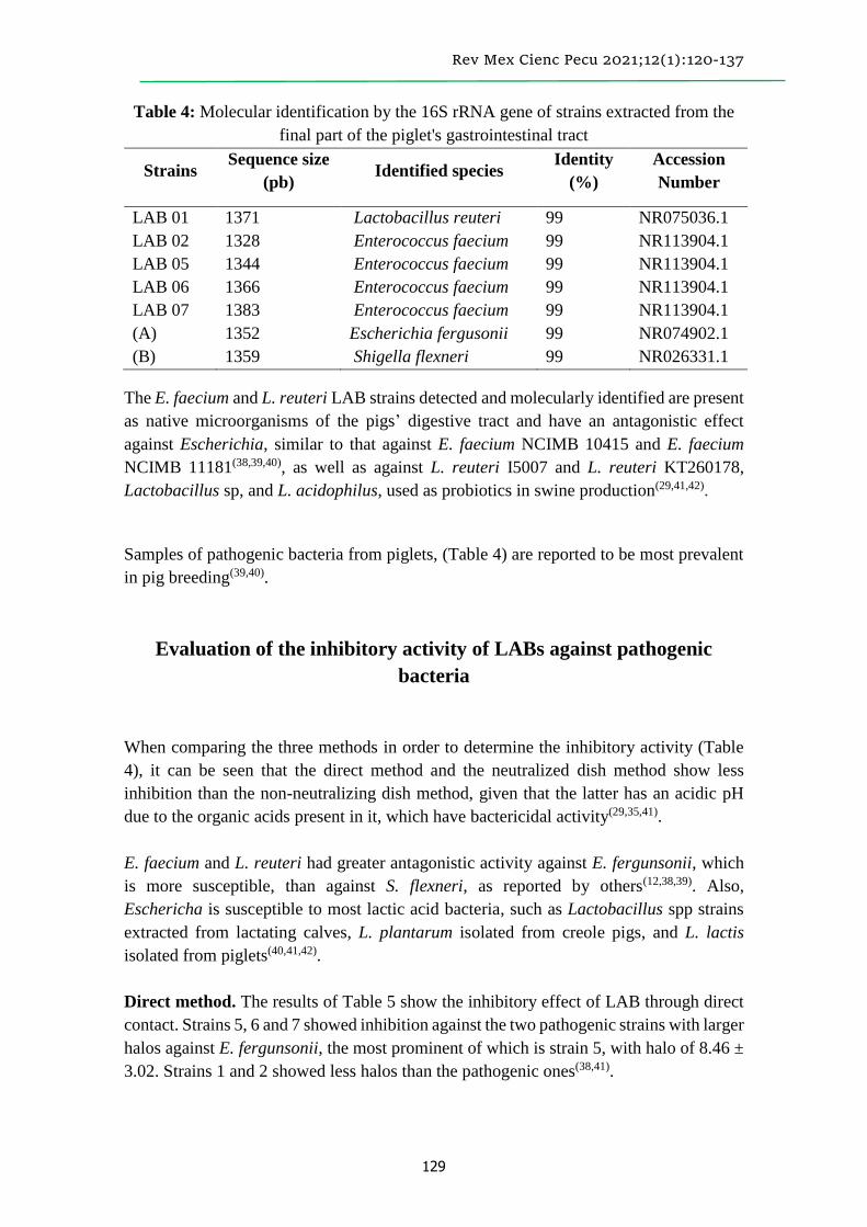
Table 4: Molecular identification by the 16S rRNA gene of strains extracted from the
final part of the piglet's gastrointestinal tract
Strains Sequence size
(pb) Identified species
Identity
(%)
Accession
Number
LAB 01 1371 Lactobacillus reuteri 99 NR075036.1
LAB 02
LAB 05
LAB 06
LAB 07
1328
1344
1366
1383
Enterococcus faecium
Enterococcus faecium
Enterococcus faecium
Enterococcus faecium
99
99
99
99
NR113904.1
NR113904.1
NR113904.1
NR113904.1
(A) 1352 Escherichia fergusonii 99 NR074902.1
(B) 1359 Shigella flexneri 99 NR026331.1
The E. faecium and L. reuteri LAB strains detected and molecularly identified are present
as native microorganisms of the pigs’ digestive tract and have an antagonistic effect
against Escherichia, similar to that against E. faecium NCIMB 10415 and E. faecium
NCIMB 11181(38,39,40), as well as against L. reuteri I5007 and L. reuteri KT260178,
Lactobacillus sp, and L. acidophilus, used as probiotics in swine production(29,41,42).
Samples of pathogenic bacteria from piglets, (Table 4) are reported to be most prevalent
in pig breeding(39,40).
Evaluation of the inhibitory activity of LABs against pathogenic
bacteria
When comparing the three methods in order to determine the inhibitory activity (Table
4), it can be seen that the direct method and the neutralized dish method show less
inhibition than the non-neutralizing dish method, given that the latter has an acidic pH
due to the organic acids present in it, which have bactericidal activity(29,35,41).
E. faecium and L. reuteri had greater antagonistic activity against E. fergunsonii, which
is more susceptible, than against S. flexneri, as reported by others(12,38,39). Also,
Eschericha is susceptible to most lactic acid bacteria, such as Lactobacillus spp strains
extracted from lactating calves, L. plantarum isolated from creole pigs, and L. lactis
isolated from piglets(40,41,42).
Direct method. The results of Table 5 show the inhibitory effect of LAB through direct
contact. Strains 5, 6 and 7 showed inhibition against the two pathogenic strains with larger
halos against E. fergunsonii, the most prominent of which is strain 5, with halo of 8.46 ±
3.02. Strains 1 and 2 showed less halos than the pathogenic ones(38,41).

Rev Mex Cienc Pecu 2021;12(1):120-137
130
Table 5: Halo size of LAB inhibition tests against the pathogens of Shigella flexneri
and Escherichia fergusonii
Strains
Escherichia fergusonii Shigella flexneri
Direct Un-
neutralized Neutralized Direct
Un-
neutralized Neutralized
01 6.52 ± 0.132 7.54 ± 0.43 6.85 ± 036 6.00 6.30 ± 0.56 6.00
02 6.94 ± 0.44 7.90 ± 0.078 7.10 ± 0.60 6.00 7.34 ± 0.05 6.00
05 8.46 ± 3.02 8.86 ± 0.62 8.76 ± 3.80 6.9 ± 0.45 10.34 ± 0.13 7.34 ± 0.89
06 8.33 ± 2.70 9.72 ± 1.88 8.52 ± 3.17 6.72 ± 0.25 9.84 ± 0.40 6.84 ± 0.35
07 8.25 ± 2.53 9.24 ± 0.60 8.65 ± 3.51 6.24 ± 0.02 10.10 ± 2.0 7.10 ± 0.60
Negative test: 6.00
Non-neutralizing dish method. The test was performed using the supernatant of the
LAB culture, with an average pH of 4.486 ± 0.001. Table 5 shows that all the stumps
exhibit inhibition halos in the confrontation against E. fergusonii; the most prominent
stumps were Nos. 6 and 7, followed by stump 5 and, finally, by stumps 2 and 1. The halos
formed in the presence of S. flexneri were of a larger size than those formed with the other
pathogen, the stumps (in order of size from the largest to the smallest) were 5, 7, 6, 2, and
1 respectively.
LAB 5, 7 and 6 (E. faecium) exhibited larger halos in the presence of S. flexneri, and
similar and smaller halos to those obtained in the test with Lactobacillus spp, compared
to pathogens of the pig; halos ranging between 11.24 ± 0.03 and 32.62 ± 0.04 have been
reported in the presence of Salmonella sp(19,36,41). In tests using the bacterial supernatant
without neutralization, it has exhibited a greater inhibition action, due to the effect of the
organic acids, according to the antagonism tests(29,35,40).
Neutralizing dish method. In this case, the supernatant of the LAB culture was adjusted
to pH 7.00 (neutralized with sodium hydroxide) in order to exclude the inhibition of
organic acids. All the strains exhibited halos (Table 5) against E. fergusonii, but of a
smaller size than in the test without neutralizing, the largest halos being for strains 5, 7
and 6 in the test with S. flexneri. In this method, since there is no acid action, the
antimicrobial action is attributed to the presence of non-acid metabolites. Reportedly,
LAB produce peptide substances that have a bactericidal or bacteriostatic mode of
action(16,42), which is also referred to the activity of protein metabolites or complex lipid
molecules or carbohydrates(11).
With all three methods, the assessed strains 5, 6 and 7 (E. faecium) exhibited the largest
halos against E. fergunsonii, the non-neutralizing dish method being the one that
generated the largest halos, as previously reported(11,16). The antagonism of LAB is
influenced by several factors, such as the type of bacterium, the place where it was
obtained, the host species, the temperature, and the incubation time(14,15,41). The LAB with

Rev Mex Cienc Pecu 2021;12(1):120-137
131
probiotic activity exhibited antagonism against pig pathogens, and its action is compared
with the majority of probiotics obtained from bacteria Lactobacillus ssp. L. acidophilus,
L. plantarum, (L. casei and L. brevis)(14,19,29), which act against the pathogenic bacteria E.
coli ATCC 25922 and S. typhimurium(14).
Evaluation of the milk inoculum
Physical-chemical evaluation (pH, acidity and syneresis). Strains 1, 2, 5 and 6 were
selected from the evaluated LAB as probiotics. Two mixtures were prepared with them
as MI (yogurt); the first mixture utilized strains 1, 2, and 5, and the second one, strains 1,
5 and 6 (1 ml of activated strain in 100 ml of milk). 50 ml of each strain were used,
according to the mixture, in order to evaluate its viability, at 0, 5, 10 and 15 days of
storage until its use as MI (yogurt). Table 6 shows that mixture one exhibited better
stability; in it, the pH values were inversely proportional to the acidity, which decreases
according to the number of days of refrigeration. The pH at 15 days was pH of 4.53 for
mixture one, and pH 4.78 for mixture two; these values are within the acceptable
parameters of stability and useful life, related to the time of degradation of lactose to lactic
acid. The results obtained are acceptable, comparable to those obtained with pH 4.65 in
the manufacture of yogurt with goat's or cow's milk using commercial fermenting
microorganisms and symbiotic yogurts(26,27); besides, they comply with the Codex
standard STAN 243-2003(43), which states that all yogurts must have a pH of ≤ 4. 6 to
4.90 –values similar to those of yogurts and non-traditional milk products(25,27,30). The pH
obtained was similar to that of milk ferments for pigs, ensilaged with milk products that
maintain pH values of 3 to 4.9(18,35,44). Despite the fact that mixture two presented slightly
higher pH, this is also within the technical norms, NTP 202.092:2014 and Norm INEN
710 of 1996(20); the pH is modified to cover a greater range when incorporating wheat
fiber and other grains into Mexican artisanal yogurts(18,32,33). Although the yogurt is
refrigerated, the growth of LAB strains ceases. However, the acidity proceeds slowly, due
to its residual activity(26,30,31); its shelf life is increased by incorporating acid fruits and
pectin shakes(27,34), and its quality and flavor are also improved by adding fruits (lucuma,
banana, mango, and others) with functional components(34,45); therefore, the pH in the
incubation and storage of MI determines its acceptability for use(31).

Rev Mex Cienc Pecu 2021;12(1):120-137
132
Table 6: Evaluation of pH, % acidity, degree of syneresis and viable LAB count of MI
(yogurt) at different days of storage at 4 °C
Mixture 01 Mixture 02
Days pH Acidity
%
Syneresis
% CFU/g Ph
Acidity
% Syneresis % CFU /g
0 4.65 0.80 0.36 4.8x10⁶ 4.94 0.47 0.39 2.8x10⁴
5 4.61 0.86 0.49 1.5x10⁷ 4.88 0.50 0.45 7.4x10⁴
10 4.57 0.90 0.55 2.7x10⁷ 4.82 0.55 0.53 9.6x10⁴
15 4.53 0.93 0.61 3.9x10⁷ 4.78 0.58 0.58 1.1x10⁶
The acidity value (%) is a function of the content of lactic acid, reaching up to 0.93 and
0.58 %, considered acceptable in a dairy product according to the standard for the
preparation of yogurt as established by Codex STAN 243-2003(44), to the Standard INEN
710 of 1996. It has a final range of 0.6 to 1.5 % during refrigeration(20), and the percentage
of acidity obtained is accepted in fermented foods and silage(28,35,44). The degree of
measured syneresis of MI (yogurt) increased slowly during storage, an effect caused by
loss of stability, water retention, and its components. Table 6 shows the values of the
evaluation of the degree of syneresis during storage; at 15 d of refrigeration, it exhibited
an acceptable range of 0.36 to 0. 61 % (mixture one and two) –results similar to those
obtained in yogurt and goat milk shake with fruits(26,27,34), but higher than those obtained
for modified yogurts, because of the addition of commercial stabilizers, microcapsules of
gum Arabic and maltodextrin (0.12 to 0.1 %)(27,30), and fiber that helps to prevent the
separation of whey(32,33).
Microbiological evaluation. Table 6 shows the viability of LAB as probiotics in the MI
(yogurt). Mixture one shows greater increase of probiotic microorganisms than mixture
two, during its evolution in refrigeration. The LAB count in both mixtures is similar to
that recommended by NTP 202.092:2014 for the preparation of yogurt with the ISO
7889:2003 (IDF 117:2003) method(33), which considers – at least for total number of lactic
bacteria microorganisms in yogurt during its useful life– a concentration of 107 CFU/g.
Mixtures one and two attained this concentration (3. 9 x 10⁷ CFU/g and 1.1 x 10⁶ CFU/g,
respectively) at 15 d of refrigeration. Thereby, the processed product was guaranteed to
contain and preserve its viability and probiotic activity, as in the manufacture of the
different yogurts, in which goat or cow milk, commercial fermenting bacteria, flavorings,
fruits, and fiber are utilized, with a concentration of 107 CFU/g to 106 CFU/g of viable
probiotic cells in the first 16 d(30,34,46). Currently the consumer demands less processed
and more natural, functional foods (antioxidant fruits)(33,45); therefore, it also seeks to
improve their life span by providing antimicrobial qualities using beneficial native
bacteria(17,18,42). Reuterine, produced by L. reuteri, is an antibacterial that can be used as
a biopreservative with potential probiotic controller of Salmonella spp. and E. coli in food
for people or animals. The trend is the use of native LAB and its bacterial extracts as
isolated probiotic potentials used in the same animal species(12,38,39).

Rev Mex Cienc Pecu 2021;12(1):120-137
133
Conclusions and implications
The biochemical characterization, the tolerance tests, and the antagonistic effect of the
selected LAB were of great importance for the growth and survival of four probiotic
strains (three Enterococcus faecium and one Lactobacillus reuteri strain). The production
of organic acids presents in the non-neutralized supernatants stood out for their
antimicrobial activity, and there is a non-acid action by metabolic modifiers in neutralized
supernatants with bactericidal effect. It was possible to elaborate a lactic inoculum
(yogurt) with acceptable, viable and innocuous characteristics with the isolated stumps
for it to be considered as a potential probiotic for oral administration, with 15 d of useful
life.
Acknowledgements
The authors thank the National University of Tumbes (Universidad Nacional de Tumbes),
the unconditional support provided for the research, as well as the workers' staff of the
Faculty of Agricultural Sciences (FCA).
Literature cited:
1. Hevia RM. Microbiota digestiva del cerdo: determinación del patrón en condiciones
de salud y enfermedad [tesis doctoral]. León, España: Universidad de León; 2018.
2. Balasingham K, Valli C, Radhakrishnan L. Balasuramanyam D. Caracterización
probiótica de bacterias del ácido láctico aisladas de intestino porcino. Mundo Vet
2017;10(7):825.
3. Gámez HJ, Aguirre D, Ramírez CJRMC. Caracterización de bacterias probióticas
aisladas del intestino grueso de cerdos como alternativa al uso de antibióticos. Rev
MVZ Córd 2009;14(2):1723-1735.
4. Miranda-Yuquilema JE, Marin-Cárdenas A, Oliva-Bello H, Baño-Ayala D, Barros-
Rodríguez M, Jácome-Vargas H, Villamarín-Barragán D. Influence of a microbial
additive on the productive behavior of pregnant sows, as well as, hematochemical
and diarrheal incidence in their offspring. Trop Subtrop Agroecosyst 2018;21(1):39-
45.
5. Reis de Souza TC, Mariscal Landín G, Escobar García K, Aguilera Barreyro A,
Magné Barrón A. Cambios nutrimentales en el lechón y desarrollo morfoisiológico
de su aparato digestivo. Vet Méx 2012;43(2):155-173.

Rev Mex Cienc Pecu 2021;12(1):120-137
134
6. Pérez FA. Prácticas de manejo del lechón en maternidad: estrategias para mejorar su
sobrevida y aumentar la productividad. Rev Electr Vet 2010;11(1):177-190.
7. Cossio DS, Hernández YG, Mendoza JD. Development of probiotics for animal
production. experience in Cuba. Cuba J Agric Sci 2018;52(4):357-373.
8. Quintero MJV. Bacterias del ácido láctico un potencial para la producción de
alimentos probióticos fermentados en la industria láctea de Panamá. KnE Eng
2018;3(1):38-47.
9. Ávila J, Ávila M, Tovar B, Brizuela M, Perazzo Y, Hernández H. Capacidad
probiótica de cepas del género Lactobacillus extraídas del tracto intestinal de
animales de granja. Rev Cienc 2010;20(2):161-169.
10. Martín C, Escobedo S, Suárez JE, Quirós LM, Pérez-Martínez G, Coll-Marqués JM,
et al. Two alkaline motifs in the lactobacillus salivarius Lv72 OppA surface are
important to its adhesion function. Benefic Microbiol 2019;10(1):101-109.
11. Lu X, Zhang M, Zhao L, Ge K, Wang Z, Jun L, et al. Growth performance and post-
weaning diarrhea in piglets fed a diet supplemented with probiotic complexes. J
Microbiol Biotechnol 2018;28(11):1791-1799.
12. Chengjun H, Weigang X, Xiaohua L, Xiuzhu Z, Ke L, Jia L, et al. Effects of dietary
supplementation of probiotic Enterococcus faecium on growth performance and gut
microbiota in weaned piglets. AMB Express. 2019;9(1):11-12.
13. Fengjuan Y, Aina W, Xiangfang Z, Chengli H, Hong L, Shiyan Q. Lactobacillus
reuteri I5007 modulates tight junction protein expression in IPEC-J2 cells with LPS
stimulation and in newborn piglets under normal conditions. BMC Microbiology
2015;15(1):10-11.
14. Vera-Mejía R, Ormaza-Donoso J, Muñoz-Cedeño J, Arteaga-Chávez F, Sánchez-
Miranda L. Cepas de Lactobacillus plantarum con potencialidades probióticas
aisladas de cerdos criollos. Rev Salud Anim 2018;40(2):11-12.
15. Sánchez SH, Ochoa MG, Rojas MC, Peralta OT, Ordinola ZA. Aislamiento de
péptidos inhibidores de bacterias a partir de bacterias ácido lácticas del tracto
digestivo del lechón e identificación mediante prueba proteómica. Sci Agropec
2017;8(4):437-443.
16. Vélez Zea JM. Evaluación de la actividad antimicrobiana de bacterias probióticas
extraídas del calostro de cerdas de granjas del Aburrá sur [tesis doctoral]. Medellín,
Colombia: Universidad Nacional de Colombia-Sede Medellín; 2015.
17. Barón LVC. Probióticos y prebióticos como alimentos funcionales en nutrición
animal. Zoocienc 2016;3(2):15-21.

Rev Mex Cienc Pecu 2021;12(1):120-137
135
18. Medina PJG. Propiedades tecnológicas de bacterias ácido lácticas aisladas de
productos lácteos fermentados artesanales expendidos en la región Ciénega de
Jalisco. Adv Invest Food Saf 2018;1(1):230-234.
19. Ming-Lun C, Hsi-Chia C, Kun-Nan C, Yu-Chun L, Ya-Ting L, Ming-Ju C.
Optimizing production of two potential probiotic Lactobacilli strains isolated from
piglet feces as feed additives for weaned piglets. Asian-Austral J Anim Sci
2015;28(8):1163-1170.
20. Landa-Salgado P, Caballero-Cervantes Y, Ramírez-Bribiesca E, Hernández-
Anguiano AM, Ramírez-Hernández LM, Espinosa-Victoria D, Hernández-Sánchez
D. Aislamiento e identificación de bacterias ácido lácticas con potencial probiótico
para becerros del altiplano mexicano. Rev Mex Cienc Pecu 2019;10(1):68-83.
21. Flores Fuentes GD. Propuesta de un manual de procedimientos operativos estándar
sobre la toma de muestras de alimentos para uso de la Defensoría del Consumidor
[tesis doctoral]. El Salvador: Universidad de El Salvador; 2015.
22. Botero Ospina MJ, Castaño DRT, Castaño Zapata J. Manual práctico de
microbiología general. No. Doc. 26157. CO-BAC, Bogotá. 2011.
23. MacFaddin JF. Pruebas bioquímicas para la identificación de bacterias de
importancia clínica: Métodos microbiológicos. Capítulo 17. 18a. ed: Editorial
Panamericana; 2002.
24. Arteaga-Chávez F, López-Vera M, Laurencio-Silva M, Rondón-Castillo A, Milián-
Florido G, Barrios-González V. Selección e identificación de aislados de Bacillus
spp. del tracto digestivo de pollos de traspatio, con potencial probiótico. Pastos y
Forrajes 2017:155-164.
25. ECUADOR NTE INEN IJR, Quito, Ecuador. Instituto Ecuatoriano de
Normalización Norma técnica Ecuatoriana del Yogurt INEN
2395:2011.https://www.normalizacion.gob.ec/buzon/normas/nte-inen-2395-2r.pdf.
consultado nov 15, 2018.
26. Zapata IC, Sepúlveda-Valencia U, Rojano BA. Efecto del tiempo de almacenamiento
sobre las propiedades fisicoquímicas, probióticas y antioxidantes de yogurt
saborizado con mortiño (Vaccinium meridionale Sw). Inf Tech 2015;26(2):17-28.
27. Ortega LAJ, Ramírez LB, Rodríguez EA. Desarrollo y evaluación de un yogurt
bebible adicionado de extracto liofilizado de Justicia spicigera como colorante
natural. e-CUBA 2018;(9):25-34.
28. Miranda MO, Espinosa REN, Ponce PI. Características físico-químicas y
propiedades nutricionales del suero resultante del proceso de obtención del yogurt
Griego. Rev Cubana Alim Nutri 2016;26(1):172-174.

Rev Mex Cienc Pecu 2021;12(1):120-137
136
29. Hernández-García JE, Sebastián-Frizzo L, Rodríguez-Fernández JC, Valdez-Paneca
G, Virginia-Zbrun M, Calero-Herrera I. Evaluación in vitro del potencial probiótico
de Lactobacillus acidophilus SS80 y Streptococcus thermophilus SS77. Rev Salud
Anim 2019;41(1):1-13.
30. Huertas RAP. Efecto del té verde (Camellia Sinensis L.) en las características
fisicoquímicas, microbiológicas, proximales y sensoriales de yogurt durante el
almacenamiento bajo refrigeración. @ limentech, Cient Tech food 2013;11(1):56-
64.
31. Sánchez J, Enríquez D, Castro PJAS. Efecto de la concentración de sólidos totales
de la leche entera y tipo de cultivo comercial en las características reológicas del
yogurt natural tipo batido Agroind Sci 2012;2(2):173-80.
32. Díaz Jiménez B, Sosa Morales M, Vélez Ruiz J. Efecto de la adición de fibra y la
disminución de grasa en las propiedades fisicoquímicas del yogur. Rev Mex Ing
Quím 2004;3(3):287-305.
33. Simanca MM, Andrade RD, Arteaga MR. Efecto del salvado de trigo en las
propiedades fisicoquímicas y sensoriales del yogurt de leche de búfala. Inf Tech
2013;24(1):79-86.
34. Vásquez-Villalobos V, Aredo V, Velásquez L, Lázaro M. Propiedades
fisicoquímicas y aceptabilidad sensorial de yogur de leche descremada de cabra
frutado con mango y plátano en pruebas aceleradas. Sci Agropecu 2015;6(3):177-
189.
35. Sánchez SH, Fabián DF, Ochoa MG, Alfaro AR. Sucesión bacteriana del tracto
digestivo del lechón alimentado con ensilado biológico. Rev Inv Vet Perú
2019;30(1):214-223.
36. Ng SY, Koon SS, Padam BS, Chye FY. Evaluation of probiotic potential of lactic
acid bacteria isolated from traditional Malaysian fermented Bambangan (Mangifera
pajang). CyTA: J Food 2015;13(4):563-572.
37. Jurado GH, Aguirre FD, Ramírez TC. Caracterización de bacterias probióticas
aisladas del intestino grueso de cerdos como alternativa al uso de antibióticos. Rev
MVZ Córdova 2009;4(2):1723-1735.
38. Kern M, Aschenbach JR, Tedin K, Pieper R, Loss H, Lodemann U. Characterization
of inflammasome components in pig intestine and analysis of the influence of
probiotic Enterococcus faecium during and Escherichia coli challenge. Immunology
Res 2017;46(7):742-757.
39. Chae JP, Pajarillo EAB, Oh JK, Kim H, Kang DK. Revealing the combined effects
of lactulose and probiotic enterococci on the swine faecal microbiota using 454
pyrosequencing. Microb Biotechnol 2016;9(4):486-495.

Rev Mex Cienc Pecu 2021;12(1):120-137
137
40. Dowarah R, Verma AK, Agarwal N, Singh P, Singh BR. Selection and
characterization of probiotic lactic acid bacteria and its impact on growth, nutrient
digestibility, health and antioxidant status in weaned piglets. PLoS One
2018;13(3):1-24.
41. Estrada MAC, Gutiérrez RLA, Montoya COI. Evaluación in vitro del efecto
bactericida de cepas nativas de Lactobacillus sp. contra Salmonella sp. y Escherichia
coli in vitro. Rev FNAgron Medellín 2005;58(1):2601-2609.
42. Andrea M. Probióticos y su mecanismo de acción en alimentación animal. Agron
Mesoamericana 2019;30(2):601-611.
43. Codex para leches fermentadas. Leche y productos lácteos segunda edición. STAN
243-2003. www.fao.org/3/a-i2085s.pdf. Consultado nov 20, 2018.
44. Caicedo WO, Moyano JC, Valle SB, Díaz LA, Caicedo ME. Calidad fermentativa
de ensilajes líquidos de chontaduro (Bactris gasipaes) tratados con yogur natural,
suero de leche y melaza. Rev Inv Vet Perú 2019;30(1):167-177.
45. Paucar-Menacho LM. Lúcuma (Pouteria lucuma): Composición, componentes
bioactivos, actividad antioxidante, usos y propiedades beneficiosas para la salud. Sci
Agropec 2020;11(1):135-142.
46. Benítez-de la Torre A, Montejo-Sierra IL, Morales-García YE, Muñoz-Rojas J,
Díaz-Ruíz R, López PA. Adición de fuentes energéticas e inoculantes en la
elaboración de yogurt de yuca. Pastos Forrajes 2018;4(1):30-34.