Embed Size (px)
Citation preview
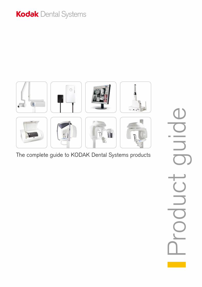
Pro
duct
gui
deThe complete guide to KODAK Dental Systems products
2
Why have we created this product guide?
This brochure aims to help you choose the right KODAK Dental Systems products for the dental practice.
With this in mind, we have summarised the most important advantages and selling points of our digital radiography and imaging systems.
For further information please contact your local authorised dealer:
Register your product online and get an additional year of warranty!
http://register.kodakdental.com/eamer
Visit our website: www.kodakdental.com
E-mail: [email protected]
2
KO
DA
K 9
00
0/9
50
0K
OD
AK
80
00
/80
00
CK
OD
AK
RV
G
65
00
/61
00
/51
00
KD
IS/L
OG
ICO
NK
OD
AK
22
00
/21
00
KO
DA
K C
R 7
40
0
KO
DA
K 1
50
0
Dig
ital e
xtra
oral
rad
iogr
aphy
Com
pute
d ra
diog
raph
yIn
trao
ral c
amer
aS
oftw
are
Dig
ital i
ntra
oral
ra
diog
raph
yX
-ray
uni
ts
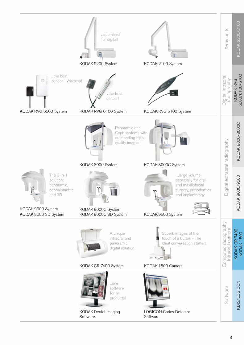
...optimised for digital!
Panoramic and Ceph systems with outstanding high quality images
...large volume, especially for oral and maxillofacial surgery, orthodontics and implantology
The 3-in-1solution: panoramic, cephalometric and 3D
A unique intraoral and panoramic digital solution
Superb images at the touch of a button - The ideal conversation starter!
KODAK 2100 System
KODAK RVG 5100 SystemKODAK RVG 6100 System
KODAK 2200 System
KODAK RVG 6500 System
LOGICON Caries Detector Software
KODAK 1500 CameraKODAK CR 7400 System
KODAK Dental Imaging Software
...one software for all products!
KODAK 8000C System
KODAK 9500 SystemKODAK 9000C 3D SystemKODAK 9000 System KODAK 9000C SystemKODAK 9000 3D System
KODAK 8000 System
...the best sensor!
...the best sensor - Wireless!
3
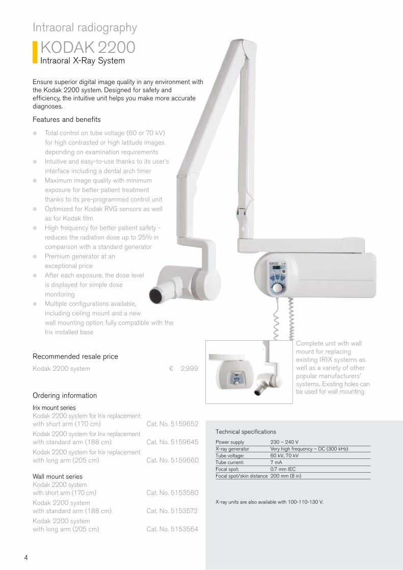
Complete unit with wall mount for replacing existing IRIX systems as well as a variety of other popular manufacturers’ systems. Existing holes can be used for wall mounting
Intraoral radiography
KODAK 2200Intraoral X-Ray System
Ensure superior digital image quality in any environment with the Kodak 2200 system. Designed for safety and efficiency, the intuitive unit helps you make more accurate diagnoses.
Features and benefits
Total control on tube voltage (60 or 70 kV) ●
for high contrasted or high latitude images depending on examination requirementsIntuitive and easy-to-use thanks to its user’s ●
interface including a dental arch timerMaximum image quality with minimum ●
exposure for better patient treatment thanks to its pre-programmed control unitOptimized for Kodak RVG sensors as well ●
as for Kodak filmHigh frequency for better patient safety - ●
reduces the radiation dose up to 25% in comparison with a standard generator Premium generator at an ●
exceptional priceAfter each exposure, the dose level ●
is displayed for simple dose monitoringMultiple configurations available, ●
including ceiling mount and a new wall mounting option fully compatible with the Irix installed base
Ordering information
Irix mount seriesKodak 2200 system for Irix replacement with short arm (170 cm) Cat. No. 5159652
Kodak 2200 system for Irix replacement with standard arm (188 cm) Cat. No. 5159645
Kodak 2200 system for Irix replacement with long arm (205 cm) Cat. No. 5159660
Wall mount series Kodak 2200 system with short arm (170 cm) Cat. No. 5153580
Kodak 2200 system with standard arm (188 cm) Cat. No. 5153572
Kodak 2200 system with long arm (205 cm) Cat. No. 5153564
Recommended resale price
Kodak 2200 system ¤ 2,999
Technical specifications
Power supply 230 – 240 VX-ray generator Very high frequency – DC (300 kHz) Tube voltage: 60 kV, 70 kVTube current: 7 mAFocal spot: 0.7 mm IECFocal spot/skin distance 200 mm (8 in)
X-ray units are also available with 100-110-130 V.
4
KO
DA
K 2
20
0/2
10
0K
OD
AK
22
00
/21
00
Intraoral radiography
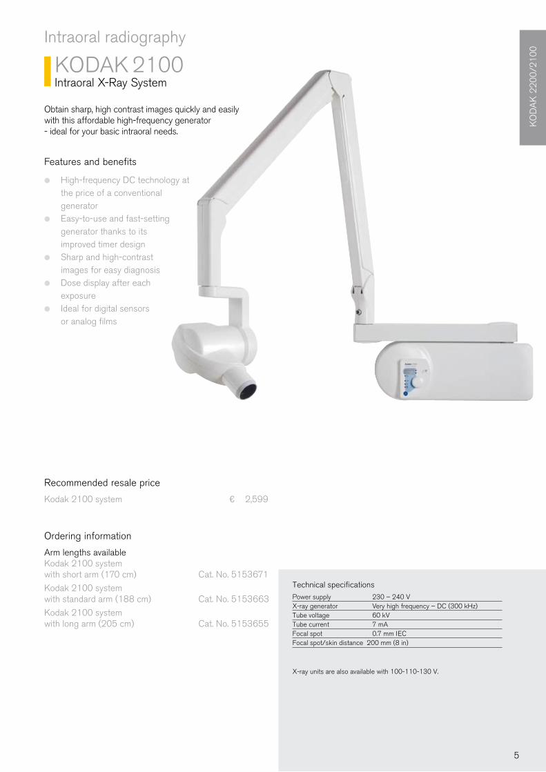
KODAK 2100Intraoral X-Ray System
Obtain sharp, high contrast images quickly and easily with this affordable high-frequency generator - ideal for your basic intraoral needs.
Ordering information
Arm lengths availableKodak 2100 system with short arm (170 cm) Cat. No. 5153671
Kodak 2100 system with standard arm (188 cm) Cat. No. 5153663
Kodak 2100 system with long arm (205 cm) Cat. No. 5153655
Features and benefits
High-frequency DC technology at ●
the price of a conventional generator Easy-to-use and fast-setting ●
generator thanks to its improved timer designSharp and high-contrast ●
images for easy diagnosisDose display after each ●
exposureIdeal for digital sensors ●
or analog films
Recommended resale price
Kodak 2100 system ¤ 2,599
Technical specificationsPower supply 230 – 240 VX-ray generator Very high frequency – DC (300 kHz) Tube voltage 60 kV Tube current 7 mAFocal spot 0.7 mm IECFocal spot/skin distance 200 mm (8 in)
X-ray units are also available with 100-110-130 V.
5
Digital intraoral radiography
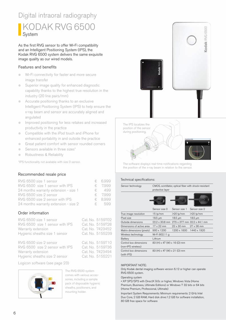
KODAK RVG 6500System
As the first RVG sensor to offer Wi-Fi compatibility and an Intelligent Positioning System (IPS), the Kodak RVG 6500 system delivers the same exquisite image quality as our wired models.
Features and benefits
Wi-Fi connectivity for faster and more secure ●
image transferSuperior image quality for enhanced diagnostic ●
capability thanks to the highest true resolution in the industry (20 line pairs/mm)Accurate positioning thanks to an exclusive ●
Intelligent Positioning System (IPS) to help ensure the x-ray beam and sensor are accurately aligned and angulatedImproved positioning for less retakes and increased ●
productivity in the practiceCompatible with the iPod touch and iPhone for ●
enhanced portability in and outside the practiceGreat patient comfort with sensor rounded corners ●
Sensors available in three sizes* ●
Robustness & Reliability ●
*IPS functionality not available with size 0 sensor.
The IPS localizes the position of the sensor during positioning.
The software displays real-time notifications regarding the position of the x-ray beam in relation to the sensor.
The RVG 6500 system comes with various acces-sories, including a sample pack of disposable hygienic sheaths, positioners, and mounting holder.
IMPORTANT NOTE:Only Kodak dental imaging software version 6.12 or higher can operate RVG 6500 system.
Operating system• XP SP2/SP3 with DirectX 9.0c or higher, Windows Vista (Home Premium, Business, Ultimate Editions) or Windows 7 32 bits or 64 bits (Home Premium, Professional, Ultimate)
Important System Requirements: Minimum requirements: 2 GHz Intel Duo Core, 2 GB RAM, Hard disk drive:1.2 GB for software installation, 80 GB free space for software
Recommended resale price
RVG 6500 size 1 sensor ¤ 6.999RVG 6500 size 1 sensor with IPS ¤ 7.99924 months warranty extension - size 1 ¤ 499RVG 6500 size 2 sensor ¤ 7.999RVG 6500 size 2 sensor with IPS ¤ 8.99924 months warranty extension - size 2 ¤ 599
Order information
RVG 6500 size 1 sensor Cat. No. 5159702 RVG 6500 size 1 sensor with IPS Cat. No. 5159728Warranty extension Cat. No. 7423452 Hygienic sheaths size 1 sensor Cat. No. 5155239
RVG 6500 size 2 sensor Cat. No. 5159710RVG 6500 size 2 sensor with IPS Cat. No. 5159736Warranty extension Cat. No. 7423494Hygienic sheaths size 2 sensor Cat. No. 5155221
Logicon software (see page 23)
Sensor technology CMOS, scintillator, optical fiber with shock-resistant protective layer
Sensor size 0 Sensor size 1 Sensor size 2
True image resolution 15 lp/mm >20 lp/mm >20 lp/mm
Pixel size 18.5 µm 18.5 µm 18.5 µm
Outside dimensions 22.2 x 30.8 mm 27.5 x 37.7 mm 32.2 x 44.1 mm
Dimensions of active area 17 x 22 mm 22 x 30 mm 27 x 36 mm
Matrix dimensions (pixels) 900 x 1200 1200 x 1600 1440 x 1920
Wireless technology Wi-Fi 802.11 g
Battery Lithium
Control box dimensions (non-IPS wireless)
83 (H) x 47 (W) x 16 (D) mm
Control box dimensions (with IPS)
83 (H) x 47 (W) x 21 (D) mm
Technical specifications:
6
KO
DA
K R
VG
6
50
0/6
10
0/5
10
0
Digital intraoral radiography
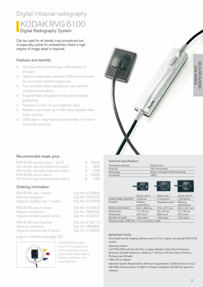
KODAK RVG 6100Digital Radiography System
Can be used for all dental x-ray procedures but is especially suited for endodontics where a high degree of image detail is required.
Features and benefits
The best sensor technology in the market for ●
25 yearsHighest measurable resolution (20 line pairs/mm) ●
for quick and confident diagnosesThe unrivalled detail reproduction you need for ●
complex examinationsErgonomically designed for easy and accurate ●
positioningRounded corners for your patients’ ease ●
Pediatric size 0 with up to 40% less radiation than ●
larger sensorsUSB cable – easy handling and flexible - for use in ●
multi chair practices
The RVG 6100 system comes with various acces-sories, including a sample pack of disposable hygienic sheaths, positioners, and mounting holder.
IMPORTANT NOTE:Only Kodak dental imaging software version 6.12 or higher can operate RVG 6100 system.
Operating system• XP SP2/SP3 with DirectX 9.0c or higher, Windows Vista (Home Premium, Business, Ultimate Editions) or Windows 7 32 bits or 64 bits (Home Premium, Professional, Ultimate) • Mac OS on request
Important System Requirements: Minimum requirements: 2 GHz Intel Duo Core, 2 GB RAM, Hard disk drive:1.2 GB for software installation, 80 GB free space for software
Actual image resolutionPurpose
External dimensionsDimensionsActive areaNumber of pixelsDelivery scope USB hub + 5 m connection cable
Technical specificationsTheoretical resolution 27.03 lp/mm Pixel size 18.5 micrometersTechnology Sensor with optical fiber technology Connection USB 2
Recommended resale price
RVG 6100 sensors sizes 1 and 0 ¤ 5,99924-months warranty extension size 0 ¤ 89924-months warranty extension sizes 1 ¤ 499RVG 6100 sensor size 2 ¤ 6,99924-months warranty extension size 2 ¤ 599
Ordering information
RVG 6100 size 1 sensor Cat. No. 5154828Warranty extension Cat. No. 7423452Hygienic sheaths size 1 sensor Cat. No. 5155239 RVG 6100 size 2 sensor Cat. No. 5154810Warranty extension Cat. No. 7423494Hygienic sheaths size 2 sensor Cat. No. 5155221
RVG 6100 size 0 sensor Cat. No. 5155114Warranty extension Cat. No. 7423825Hygienic sheaths size 0 sensor Cat. No. 5155205
Logicon software (see page 23)
Sensor size 1> 20 lp/mmAll-purpose sensor
27.5 x 37.7 mm22 x 30 mm (660 mm2)1.92 million
Sensor size 015 lp/mmPediatric examinations22.2 x 30.8 mm17 x 22 mm(374 mm2)1.08 million
Sensor size 2> 20 lp/mmBitewingexaminations32.2 x 44.1 mm27 x 36 mm (972 mm2)2.76 million
7
Digital intraoral radiography
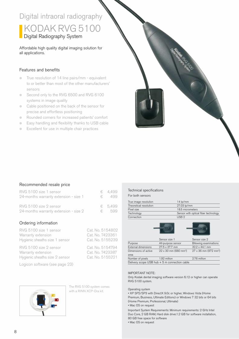
KODAK RVG 5100Digital Radiography System
Affordable high quality digital imaging solution for all applications.
Features and benefits
True resolution of 14 line pairs/mm - equivalent ●
to or better than most of the other manufacturers’ sensorsSecond only to the RVG 6500 and RVG 6100 ●
systems in image qualityCable positioned on the back of the sensor for ●
precise and effortless positioning Rounded corners for increased patients’ comfort ●
Easy handling and flexibility thanks to USB cable ●
Excellent for use in multiple chair practices ●
Recommended resale price
RVG 5100 size 1 sensor ¤ 4,49924-months warranty extension - size 1 ¤ 499
RVG 5100 size 2 sensor ¤ 5,49924-months warranty extension - size 2 ¤ 599
Ordering information
RVG 5100 size 1 sensor Cat. No. 5154802 Warranty extension Cat. No. 7423361Hygienic sheaths size 1 sensor Cat. No. 5155239
RVG 5100 size 2 sensor Cat. No. 5154794 Warranty extension Cat. No. 7423387Hygienic sheaths size 2 sensor Cat. No. 5155221
Logicon software (see page 23)
The RVG 5100 system comes with a RINN XCP-Ora kit.
PurposeExternal dimensionsDimensions of active areaNumber of pixelsDelivery scope USB hub + 5 m connection cable
IMPORTANT NOTE:Only Kodak dental imaging software version 6.12 or higher can operate RVG 5100 system.
Operating system• XP SP2/SP3 with DirectX 9.0c or higher, Windows Vista (Home Premium, Business, Ultimate Editions) or Windows 7 32 bits or 64 bits (Home Premium, Professional, Ultimate) • Mac OS on request
Important System Requirements: Minimum requirements: 2 GHz Intel Duo Core, 2 GB RAM, Hard disk drive:1.2 GB for software installation, 80 GB free space for software• Mac OS on request
Technical specificationsFor both sensors
True image resolution 14 lp/mm Theoretical resolution 27.03 lp/mm Pixel size 18.5 micrometersTechnology Sensor with optical fiber technology Connection USB 2
Sensor size 1 All-purpose sensor 27.5 x 37.7 mm22 x 30 mm (660 mm2)
1.92 million
Sensor size 2 Bitewing examinations32.2 x 44.1 mm27 x 36 mm (972 mm2)
2.76 million
8
Com
para
tive
Mat
rix f
or R
VG
Ran
ge
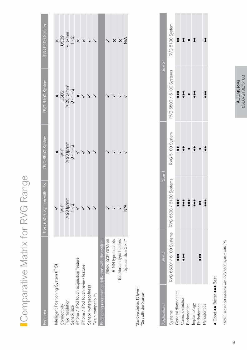
KO
DA
K R
VG
6
50
0/6
10
0/5
10
0
Feat
ures
RV
G 6
50
0 S
yste
m w
ith IP
SR
VG
65
00
Sys
tem
RV
G 6
10
0 S
yste
mR
VG
51
00
Sys
tem
Inte
llige
nt P
ositi
onin
g S
yste
m (
IPS
)ü
××
×C
onne
ctiv
ityW
i-Fi
Wi-F
iU
SB
2U
SB
2Tr
ue r
esol
utio
n>
20
lp/m
m>
20
lp/m
m>
20
lp/m
m*
14
lp/m
mS
enso
r si
ze1
- 2
0 -
1 -
20
- 1
- 2
1 -
2iP
hone
/ iP
od to
uch
acqu
isiti
on fe
atur
eü
ü×
×iP
hone
/ iP
od to
uch
revi
ew fe
atur
eü
üü
üS
enso
r w
ater
proo
fnes
sü
üü
üTw
ain
com
patib
ility
üü
üü
Pos
ition
ing
acce
ssor
ies
deliv
ered
with
the
syst
em:
RIN
N X
CP
-OR
A k
itü
üü
üR
INN
type
bas
kets
üü
ü×
Toot
hbru
sh ty
pe h
olde
rs
üü
ü×
Spe
cial
Siz
e 0
kit*
*N
/Aü
üN
/A
*Siz
e 0
res
olut
ion:
15
lp/m
m
**O
nly
with
siz
e 0
sen
sor
App
licat
ions
Siz
e 0
Siz
e 1
Siz
e 2
Sys
tem
sR
VG
65
00
* / 6
10
0 S
yste
ms
RV
G 6
50
0 /
61
00
Sys
tem
sR
VG
51
00
Sys
tem
RV
G 6
50
0 /
61
00
Sys
tem
sR
VG
51
00
Sys
tem
Gen
eral
dia
gnos
tics
lll
lll
ll
lll
ll
Car
ies
dete
ctio
nlll
lll
ll
lll
ll
End
odon
tics
lll
ll
lIm
plan
tolo
gylll
ll
lll
ll
Ped
odon
tics
lll
ll
lP
erio
dont
ics
lll
ll
lll
ll
l G
ood ll
Bet
ter lll
Bes
t
* Siz
e 0
sen
sor
not a
vaila
ble
with
RV
G 6
50
0 s
yste
m w
ith IP
S
9
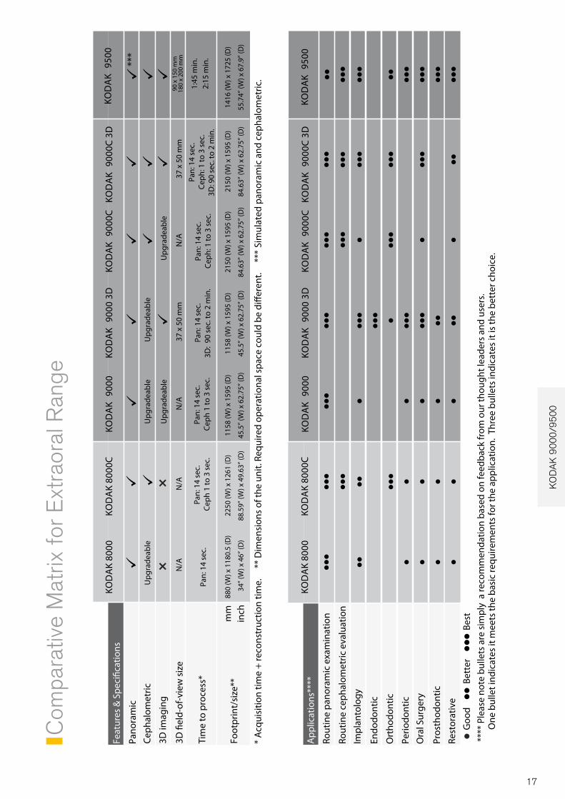
Digital extraoral radiography
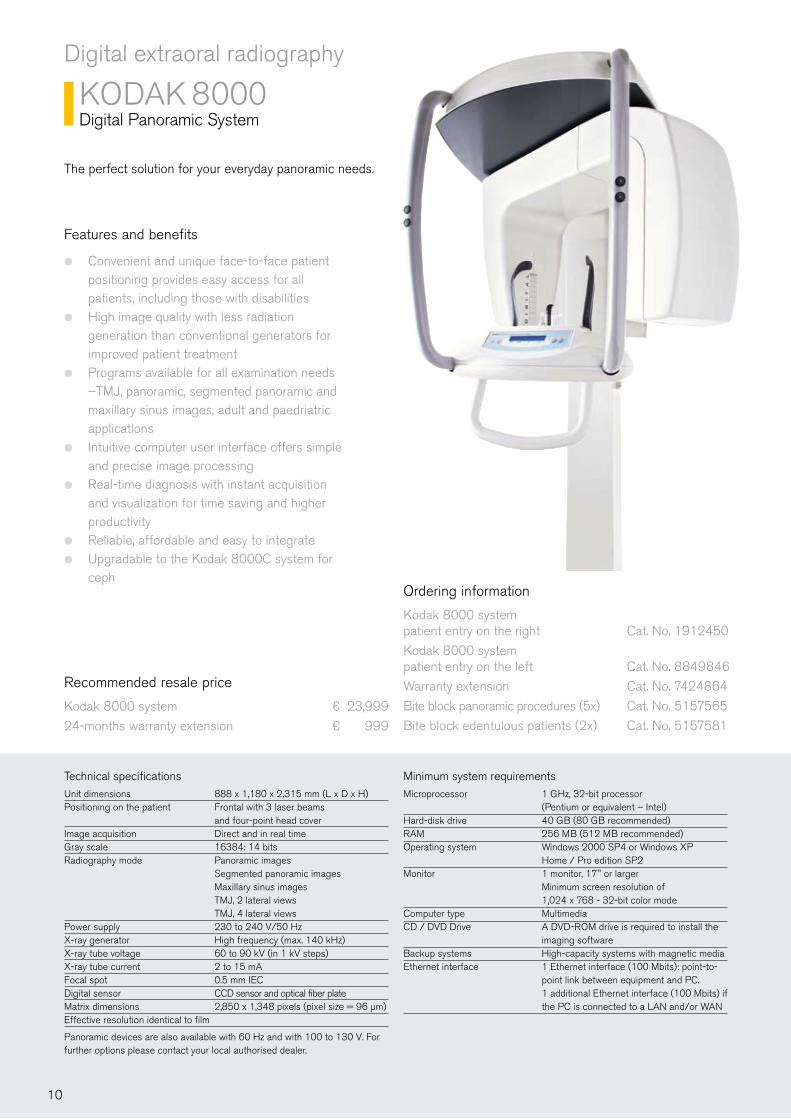
KODAK 8000Digital Panoramic System
Technical specificationsUnit dimensions 888 x 1,180 x 2,315 mm (L x D x H)Positioning on the patient Frontal with 3 laser beams and four-point head cover Image acquisition Direct and in real time Gray scale 16384: 14 bitsRadiography mode Panoramic images Segmented panoramic images Maxillary sinus images TMJ, 2 lateral views TMJ, 4 lateral viewsPower supply 230 to 240 V/50 Hz X-ray generator High frequency (max. 140 kHz) X-ray tube voltage 60 to 90 kV (in 1 kV steps) X-ray tube current 2 to 15 mA Focal spot 0.5 mm IECDigital sensor CCD sensor and optical fiber plateMatrix dimensions 2,850 x 1,348 pixels (pixel size = 96 µm)Effective resolution identical to film
Panoramic devices are also available with 60 Hz and with 100 to 130 V. For further options please contact your local authorised dealer.
The perfect solution for your everyday panoramic needs.
Features and benefits
Convenient and unique face-to-face patient ●
positioning provides easy access for all patients, including those with disabilitiesHigh image quality with less radiation ●
generation than conventional generators for improved patient treatmentPrograms available for all examination needs ●
–TMJ, panoramic, segmented panoramic and maxillary sinus images, adult and paedriatric applicatlonsIntuitive computer user interface offers simple ●
and precise image processingReal-time diagnosis with instant acquisition ●
and visualization for time saving and higher productivityReliable, affordable and easy to integrate ●
Upgradable to the Kodak 8000C system for ●
cephOrdering information
Kodak 8000 system patient entry on the right Cat. No. 1912450
Kodak 8000 system patient entry on the left Cat. No. 8849846
Warranty extension Cat. No. 7424864
Bite block panoramic procedures (5x) Cat. No. 5157565
Bite block edentulous patients (2x) Cat. No. 5157581
Minimum system requirementsMicroprocessor 1 GHz, 32-bit processor (Pentium or equivalent – Intel)Hard-disk drive 40 GB (80 GB recommended)RAM 256 MB (512 MB recommended)Operating system Windows 2000 SP4 or Windows XP Home / Pro edition SP2Monitor 1 monitor, 17" or larger Minimum screen resolution of 1,024 x 768 - 32-bit color modeComputer type MultimediaCD / DVD Drive A DVD-ROM drive is required to install the imaging softwareBackup systems High-capacity systems with magnetic mediaEthernet interface 1 Ethernet interface (100 Mbits): point-to- point link between equipment and PC. 1 additional Ethernet interface (100 Mbits) if the PC is connected to a LAN and/or WAN
Recommended resale price
Kodak 8000 system ¤ 23,999
24-months warranty extension ¤ 999
10
KO
DA
K 8
00
0/8
00
0C
Digital extraoral radiography
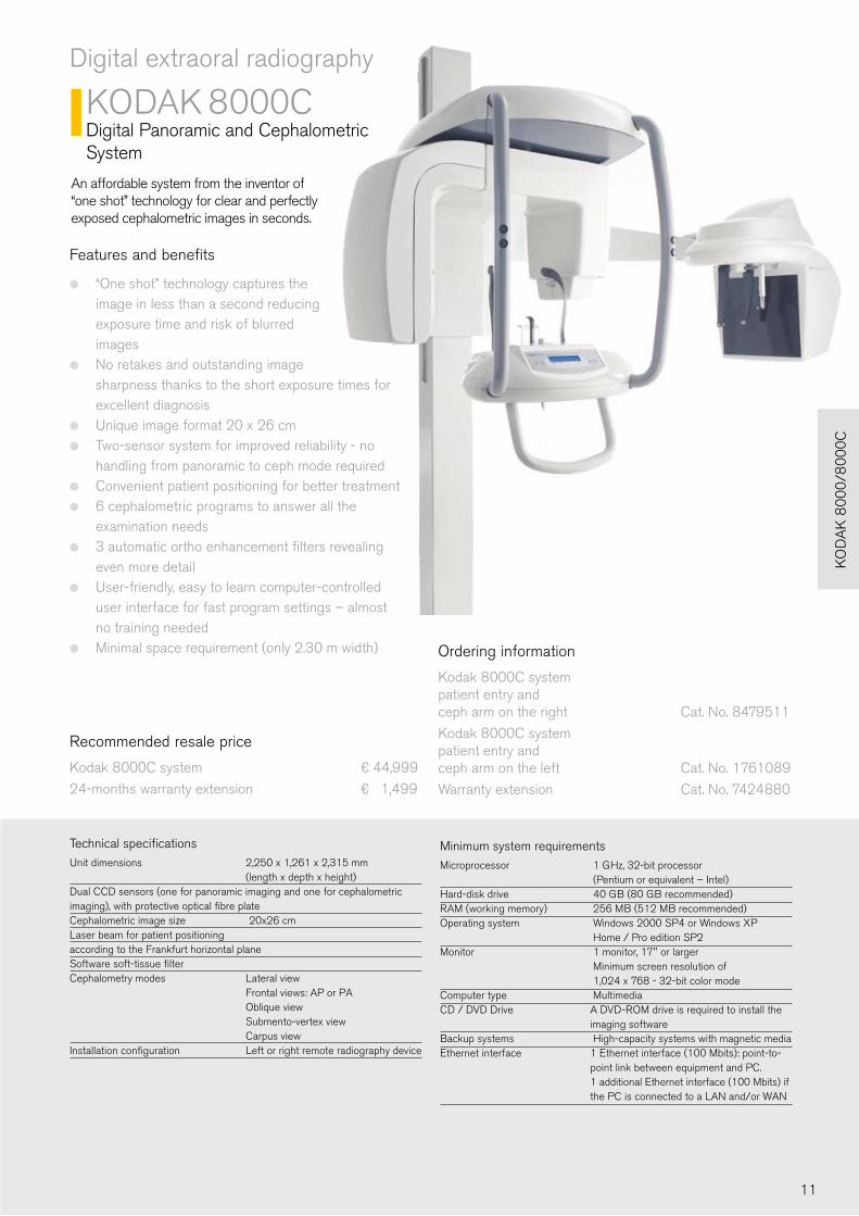
KODAK 8000CDigital Panoramic and Cephalometric System
Technical specificationsUnit dimensions 2,250 x 1,261 x 2,315 mm (length x depth x height)Dual CCD sensors (one for panoramic imaging and one for cephalometric imaging), with protective optical fibre plateCephalometric image size 20x26 cmLaser beam for patient positioning according to the Frankfurt horizontal planeSoftware soft-tissue filterCephalometry modes Lateral view Frontal views: AP or PA Oblique view Submento-vertex view Carpus viewInstallation configuration Left or right remote radiography device
Features and benefits
“One shot” technology captures the ●
image in less than a second reducing exposure time and risk of blurred images No retakes and outstanding image ●
sharpness thanks to the short exposure times for excellent diagnosisUnique image format 20 x 26 cm ●
Two-sensor system for improved reliability - no ●
handling from panoramic to ceph mode requiredConvenient patient positioning for better treatment ●
6 cephalometric programs to answer all the ●
examination needs3 automatic ortho enhancement filters revealing ●
even more detailUser-friendly, easy to learn computer-controlled ●
user interface for fast program settings – almost no training neededMinimal space requirement (only 2.30 m width) ● Ordering information
Kodak 8000C system patient entry and ceph arm on the right Cat. No. 8479511
Kodak 8000C system patient entry and ceph arm on the left Cat. No. 1761089
Warranty extension Cat. No. 7424880
An affordable system from the inventor of “one shot” technology for clear and perfectly exposed cephalometric images in seconds.
Minimum system requirementsMicroprocessor 1 GHz, 32-bit processor (Pentium or equivalent – Intel)Hard-disk drive 40 GB (80 GB recommended)RAM (working memory) 256 MB (512 MB recommended)Operating system Windows 2000 SP4 or Windows XP Home / Pro edition SP2Monitor 1 monitor, 17" or larger Minimum screen resolution of 1,024 x 768 - 32-bit color modeComputer type MultimediaCD / DVD Drive A DVD-ROM drive is required to install the imaging softwareBackup systems High-capacity systems with magnetic mediaEthernet interface 1 Ethernet interface (100 Mbits): point-to- point link between equipment and PC. 1 additional Ethernet interface (100 Mbits) if the PC is connected to a LAN and/or WAN
Recommended resale price
Kodak 8000C system ¤ 44,999
24-months warranty extension ¤ 1,499
11
Digital extraoral radiography
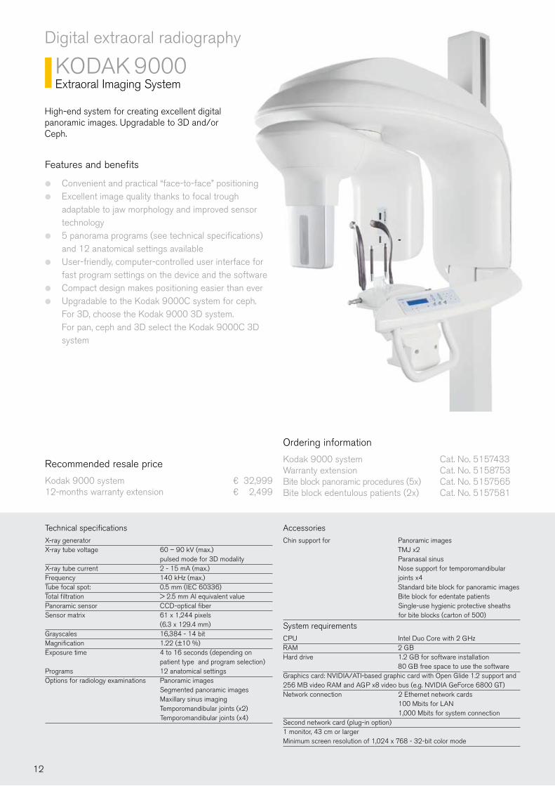
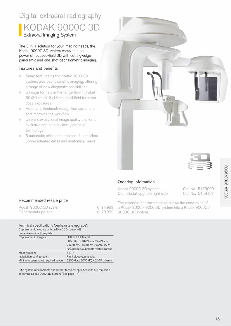
KODAK 9000Extraoral Imaging System
High-end system for creating excellent digital panoramic images. Upgradable to 3D and/or Ceph...
Technical specificationsX-ray generatorX-ray tube voltage 60 – 90 kV (max.) pulsed mode for 3D modalityX-ray tube current 2 - 15 mA (max.)Frequency 140 kHz (max.)Tube focal spot: 0.5 mm (IEC 60336)Total filtration > 2.5 mm Al equivalent valuePanoramic sensor CCD-optical fiber Sensor matrix 61 x 1,244 pixels (6.3 x 129.4 mm)Grayscales 16,384 - 14 bitMagnification 1.22 (±10 %)Exposure time 4 to 16 seconds (depending on patient type and program selection)Programs 12 anatomical settingsOptions for radiology examinations Panoramic images Segmented panoramic images Maxillary sinus imaging Temporomandibular joints (x2) Temporomandibular joints (x4)
Ordering information
Kodak 9000 system Cat. No. 5157433Warranty extension Cat. No. 5158753Bite block panoramic procedures (5x) Cat. No. 5157565Bite block edentulous patients (2x) Cat. No. 5157581
Accessories
Chin support for Panoramic images TMJ x2 Paranasal sinus Nose support for temporomandibular joints x4 Standard bite block for panoramic images Bite block for edentate patients Single-use hygienic protective sheaths for bite blocks (carton of 500)
System requirementsCPU Intel Duo Core with 2 GHz RAM 2 GB Hard drive 1.2 GB for software installation 80 GB free space to use the softwareGraphics card: NVIDIA/ATI-based graphic card with Open Glide 1.2 support and 256 MB video RAM and AGP x8 video bus (e.g. NVIDIA GeForce 6800 GT)Network connection 2 Ethernet network cards 100 Mbits for LAN 1,000 Mbits for system connection Second network card (plug-in option)1 monitor, 43 cm or largerMinimum screen resolution of 1,024 x 768 - 32-bit color mode
Recommended resale price
Kodak 9000 system ¤ 32,99912-months warranty extension ¤ 2,499
Features and benefits
Convenient and practical “face-to-face” positioning ●
Excellent image quality thanks to focal trough ●
adaptable to jaw morphology and improved sensor technology5 panorama programs (see technical specifications) ●
and 12 anatomical settings availableUser-friendly, computer-controlled user interface for ●
fast program settings on the device and the softwareCompact design makes positioning easier than ever ●
Upgradable to the Kodak 9000C system for ceph. ●
For 3D, choose the Kodak 9000 3D system. For pan, ceph and 3D select the Kodak 9000C 3D system
12
KO
DA
K 9
00
0/9
50
0
Digital extraoral radiography
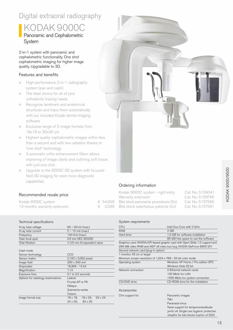
KODAK 9000CPanoramic and Cephalometric System
2-in-1 system with panoramic and cephalometric functionality. One shot cephalometric imaging for higher image quality. Upgradable to 3D.
Features and benefits
High-performance 2-in-1 radiography ●
system (pan and ceph)The ideal choice for all of your ●
orthodontic tracing needs Recognize landmark and anatomical ●
structures and trace them automatically with our included Kodak dental imaging softwareExclusive range of 5 image formats from ●
18x18 to 30x30 cmHighest quality cephalometric images within less ●
than a second and with low radiation thanks to “one shot” technology3 automatic ortho enhancement filters allows ●
improving of image clarity and outlining soft tissue with just one clickUpgrade to the 9000C 3D system with focused- ●
field 3D imaging for even more diagnostic capabilities
Technical specificationsX-ray tube voltage 60 – 90 kV (max.)X-ray tube current 2 – 15 mA (max.)Frequency 140 kHz (max.)Tube focal spot: 0.5 mm (IEC 60336)Total filtration > 2.5 mm Al equivalent value
Ceph modeSensor technology CCD Sensor matrix 2,100 x 2,092 pixels Image field 300 x 300 mmGrayscales 16,384 - 14 bitMagnification 1.14Exposure time 0.1 to 3.2 secondsOptions for radiology examinations Lateral Frontal AP or PA Oblique Submento-vertex CarpusImage format size 18 x 18, 18 x 24, 24 x 24 24 x 30, 30 x 30
Ordering information
Kodak 9000C system - right entry Cat. No. 5159041Warranty extension Cat. No. 5158746Bite block panoramic procedures (5x) Cat. No. 5157565Bite block edentulous patients (2x) Cat. No. 5157581
System requirementsCPU Intel Duo Core with 2 GHz RAM 2 GB Hard drive 1.2 GB for software installation 80 GB free space to use the softwareGraphics card: NVIDIA/ATI-based graphic card with Open Glide 1.2 support and 256 MB video RAM and AGP x8 video bus (e.g. NVIDIA GeForce 6800 GT)Second network card (plug-in option)1 monitor, 43 cm or largerMinimum screen resolution of 1,024 x 768 - 32-bit color modeOperating system Windows XP Home / Pro edition SP2 Windows Vista 32-bitNetwork connection 2 Ethernet network cards 100 Mbits for LAN 1000 Mbits for system connection CD/DVD drive CD-ROM drive for the installation
Accessories
Chin support for Panoramic images TMJ Paranasal sinus Nose support for temporomandibular joints x4, Single-use hygienic protective sheaths for bite blocks (carton of 500)
Recommended resale price
Kodak 9000C system ¤ 54,99912-months warranty extension ¤ 3,599
13
KO
DA
K 2
20
0/2
10
0
Recommended resale price
Kodak 9000 3D system ¤ 64,99912 months warranty extension ¤ 3,999
Digital extraoral radiography
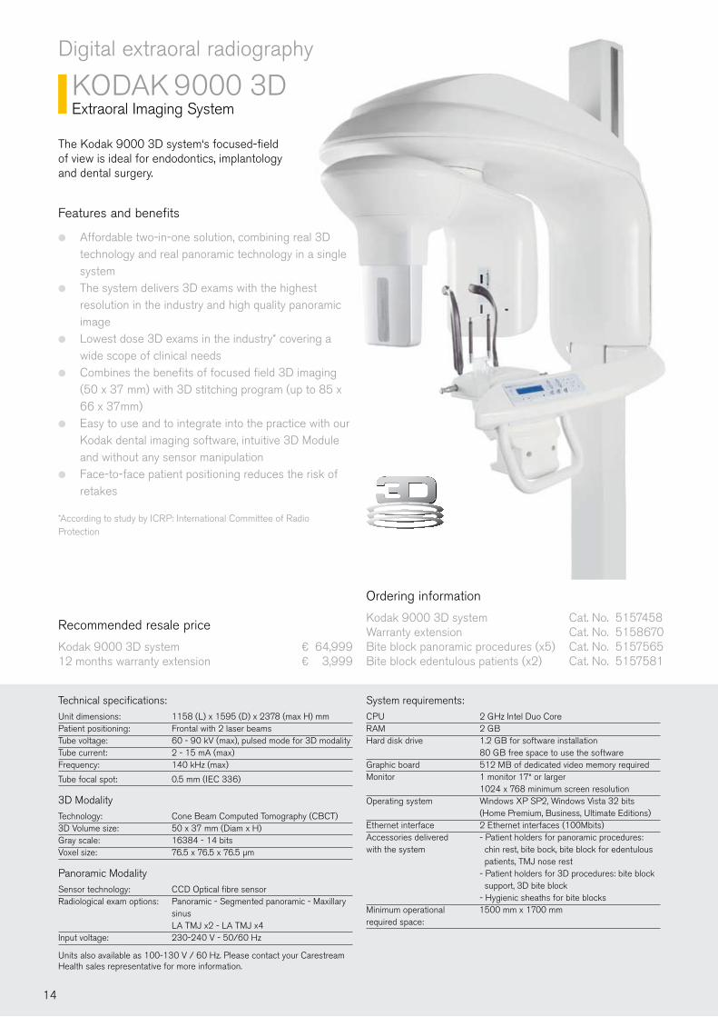
KODAK 9000 3DExtraoral Imaging System
Features and benefits
Affordable two-in-one solution, combining real 3D ●
technology and real panoramic technology in a single systemThe system delivers 3D exams with the highest ●
resolution in the industry and high quality panoramic imageLowest dose 3D exams in the industry* covering a ●
wide scope of clinical needsCombines the benefits of focused field 3D imaging ●
(50 x 37 mm) with 3D stitching program (up to 85 x 66 x 37mm)Easy to use and to integrate into the practice with our ●
Kodak dental imaging software, intuitive 3D Module and without any sensor manipulationFace-to-face patient positioning reduces the risk of ●
retakes
*According to study by ICRP: International Committee of Radio Protection
The Kodak 9000 3D system‘s focused-fieldof view is ideal for endodontics, implantology and dental surgery.
Ordering information
Kodak 9000 3D system Cat. No. 5157458 Warranty extension Cat. No. 5158670 Bite block panoramic procedures (x5) Cat. No. 5157565Bite block edentulous patients (x2) Cat. No. 5157581
Technical specifications:Unit dimensions: 1158 (L) x 1595 (D) x 2378 (max H) mmPatient positioning: Frontal with 2 laser beamsTube voltage: 60 - 90 kV (max), pulsed mode for 3D modalityTube current: 2 - 15 mA (max)Frequency: 140 kHz (max)
Tube focal spot: 0.5 mm (IEC 336)
3D Modality Technology: Cone Beam Computed Tomography (CBCT)3D Volume size: 50 x 37 mm (Diam x H)Gray scale: 16384 - 14 bitsVoxel size: 76.5 x 76.5 x 76.5 µm
Panoramic Modality Sensor technology: CCD Optical fibre sensorRadiological exam options: Panoramic - Segmented panoramic - Maxillary sinus LA TMJ x2 - LA TMJ x4Input voltage: 230-240 V - 50/60 Hz
Units also available as 100-130 V / 60 Hz. Please contact your Carestream Health sales representative for more information.
System requirements:CPU 2 GHz Intel Duo CoreRAM 2 GBHard disk drive 1.2 GB for software installation 80 GB free space to use the softwareGraphic board 512 MB of dedicated video memory requiredMonitor 1 monitor 17“ or larger 1024 x 768 minimum screen resolutionOperating system Windows XP SP2, Windows Vista 32 bits (Home Premium, Business, Ultimate Editions)Ethernet interface 2 Ethernet interfaces (100Mbits)Accessories delivered - Patient holders for panoramic procedures: with the system chin rest, bite bock, bite block for edentulous patients, TMJ nose rest - Patient holders for 3D procedures: bite block support, 3D bite block - Hygienic sheaths for bite blocksMinimum operational 1500 mm x 1700 mm required space:
14
KO
DA
K 9
00
0/9
50
0
Digital extraoral radiography
KODAK 9000C 3D Extraoral Imaging System
Technical specifications Cephalostate upgrade*:Cephalometric module with built-in CCD sensor with protective optical fibre plateCephalometric images: Half and full lateral (18x18 cm, 18x24 cm, 24x24 cm, 24x30 cm, 30x30 cm), frontal (AP/ PA), oblique, submento-vertex, carpusMagnification: x 1.14Installation configuration: Right sided-cephalostatMinimum operational required space 2230 (L) x 2000 (D) x 2400 (H) mm
*The system requirements and further technical specifications are the same as for the Kodak 9000 3D System (See page 14)
Features and benefits
Same features as the Kodak 9000 3D ●
system, plus cephalometric imaging, offering a range of new diagnostic possibilities5 image formats in the range from full skull ●
30x30 cm to18x18 cm small field for lower dose exposuresAutomatic landmark recognition saves time ●
and improves the workflowDelivers exceptional image quality thanks to ●
exclusive and best in class „one shot“ technology3 automatic ortho enhancement filters offers ●
unprecedented detail and anatomical views
The 3-in-1 solution for your imaging needs, the Kodak 9000C 3D system combines the power of focused-field 3D with cutting-edge panoramic and one-shot cephalometric imaging.
Recommended resale price
Kodak 9000C 3D system ¤ 94,999Cephalostat upgrade ¤ 29,999
Ordering information
Kodak 9000C 3D system Cat. No. 5159009Cephalostat upgrade right side Cat. No. 5159157
The cephalostat attachment kit allows the conversion of a Kodak 9000 / 9000 3D system into a Kodak 9000C / 9000C 3D system.
15
Digital extraoral radiography
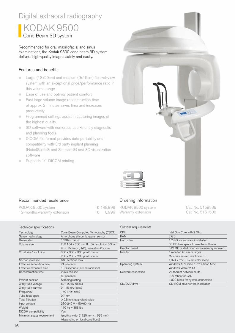
KODAK 9500Cone Beam 3D system
System requirementsCPU Intel Duo Core with 2 GHz RAM 2 GB Hard drive 1.2 GB for software installation 80 GB free space to use the softwareGraphic board 512 MB of dedicated video memory requiredMonitor 1 monitor, 43 cm or larger Minimum screen resolution of 1,024 x 768 - 32-bit color modeOperating system Windows XP Home / Pro edition SP2 Windows Vista 32-bitNetwork connection 2 Ethernet network cards 100 Mbits for LAN 1,000 Mbits for system connection CD/DVD drive CD-ROM drive for the installation
Technical specificationsTechnology Cone Beam Computed Tomography (CBCT)Sensor technology Amorphous silicon flat-panel sensorGrayscales 16384 - 14 bitVolume size FoV 184 x 206 mm (HxD), resolution 0.3 mm 90 x 150 mm (HxD), resolution 0.2 mmVoxel size/resolution 300 x 300 x 300 µm/0.3 mm 200 x 200 x 200 µm/0.2 mmSections/volume 618 sections max.Effective acquisition time 24 secondsEffective exposure time 10.8 seconds (pulsed radiation)Reconstruction time 2 min. 20 sec. 80 secondsPatient position Standing/sittingX-ray tube voltage 60 - 90 kV (max.)X-ray tube current 2 - 15 mA (max.)Frequency 140 kHz (max.)Tube focal spot: 0.7 mm Total filtration > 2.5 mm. equivalent valueInput voltage 230-240 V – 50/60 HzWeight 176 kg – 388 lbs.DICOM compatibility YesMinimum space requirement length x width (1725 mm x 1635 mm) (depending on local conditions)
Features and benefits
Large (18x20cm) and medium (9x15cm) field-of-view ●
system with an exceptional price/performance ratio in this volume rangeEase of use and optimal patient comfort ●
Fast large volume image reconstruction time ●
of approx. 2 minutes saves time and increases productivityProgrammed settings assist in capturing images of ●
the highest quality3D software with numerous user-friendly diagnostic ●
and planning toolsDICOM file format provides data portability and ●
compatibility with 3rd party implant planning (NobelGuide® and Simplant®) and 3D visualization softwareSupports 1:1 DICOM printing ●
Recommended for oral, maxillofacial and sinus examinations, the Kodak 9500 cone beam 3D system delivers high-quality images safely and easily.
Ordering information
KODAK 9500 system Cat. No. 5159538Warranty extension Cat. No. 5161500
Recommended resale price
KODAK 9500 system ¤ 149,99912-months warranty extension ¤ 8,999
16
KO
DA
K 9
00
0/9
50
0
Com
para
tive
Mat
rix f
or E
xtra
oral
Ran
geC
ompa
rativ
e M
atrix
for
Ext
raor
al R
ange
App
licat
ions
****
Rout
ine
pano
ram
ic e
xam
inat
ion
Rout
ine
ceph
alom
etric
eva
luat
ion
Impl
anto
logy
Endo
dont
ic
Ort
hodo
ntic
Perio
dont
icO
ral S
urge
ry
Pros
thod
ontic
Rest
orat
ive
****
Ple
ase
note
bul
lets
are
sim
ply
a re
com
men
datio
n ba
sed
on fe
edba
ck fr
om o
ur th
ough
t lea
ders
and
use
rs.
One
bul
let i
ndic
ates
it m
eets
the
basi
c re
quire
men
ts fo
r the
app
licat
ion.
Thr
ee b
ulle
ts in
dica
tes
it is
the
bett
er c
hoic
e.
Goo
dBe
tter
Best
KOD
AK
8000
KOD
AK
8000
C
KOD
AK
8000
KOD
AK
8000
C
KOD
AK
900
0KO
DA
K 9
000
3DKO
DA
K 9
000C
KOD
AK
900
0C 3
D
KOD
AK
900
0KO
DA
K 9
000
3DKO
DA
K 9
000C
KOD
AK
900
0C 3
D
Ceph
alom
etric
Feat
ures
& S
peci
�cat
ions
Pano
ram
ic
3D im
agin
g
3D �
eld-
of-v
iew
siz
e
Pan:
14
sec.
Ceph
1 to
3 s
ec.
Pan:
14
sec.
Ceph
1 to
3 s
ec.
Pan:
14
sec.
3D:
90 s
ec. t
o 2
min
.Pa
n: 1
4 se
c.Ce
ph: 1
to 3
sec
.
Pan:
14
sec.
Ceph
: 1 to
3 s
ec.
3D: 9
0 se
c. to
2 m
in.
* A
cqui
sitio
n tim
e +
reco
nstr
uctio
n tim
e.
**
Dim
ensi
ons
of th
e un
it. R
equi
red
oper
atio
nal s
pace
cou
ld b
e di
�ere
nt.
**
*Si
mul
ated
pan
oram
ic a
nd c
epha
lom
etric
.
Foot
prin
t/si
ze**
KOD
AK
950
0
KOD
AK
950
0
Tim
e to
pro
cess
*
Upg
rade
able
Upg
rade
able
Upg
rade
able
Upg
rade
able
Upg
rade
able
180
x 20
0 m
m90
x 1
50 m
m37
x 5
0 m
m37
x 5
0 m
mN
/AN
/AN
/AN
/A
2:15
min
.1:
45 m
in.
mm
inch
880
(W) x
118
0.5
(D)
34” (
W) x
46”
(D)
2250
(W) x
126
1 (D
)
88.5
9” (W
) x 4
9.63
” (D
)
1158
(W) x
159
5 (D
)
45.5
” (W
) x 6
2.75
” (D
)
1158
(W) x
159
5 (D
)
45.5
” (W
) x 6
2.75
” (D
)
2150
(W) x
159
5 (D
)
84.6
3” (W
) x 6
2.75
” (D
)
2150
(W) x
159
5 (D
)
84.6
3” (W
) x 6
2.75
” (D
)
1416
(W) x
172
5 (D
)
55.7
4” (W
) x 6
7.9”
(D)
Pan:
14
sec.
***
17
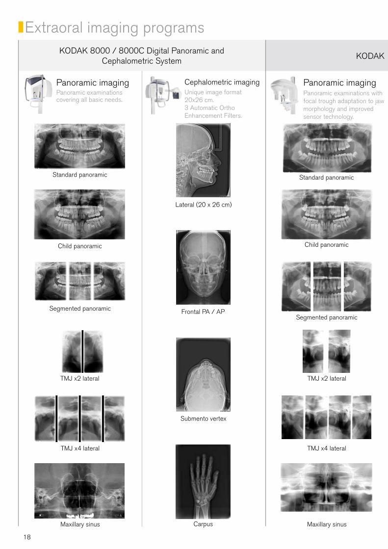
Extraoral imaging programsKODAK 8000 / 8000C Digital Panoramic and
Cephalometric System KODAK 9000 Extraoral Imaging System
Panoramic imagingPanoramic examinations covering all basic needs.
Panoramic imagingPanoramic examinations with focal trough adaptation to jaw morphology and improved sensor technology.
Cephalometric imagingUnique image format 20x26 cm. 3 Automatic Ortho Enhancement Filters.
Standard panoramic Standard panoramic
Child panoramic Child panoramic
Segmented panoramicSegmented panoramic
Lateral (20 x 26 cm)
Frontal PA / AP
Carpus
Submento vertex
TMJ x2 lateral TMJ x2 lateral
TMJ x4 lateral TMJ x4 lateral
Maxillary sinus Maxillary sinus
18
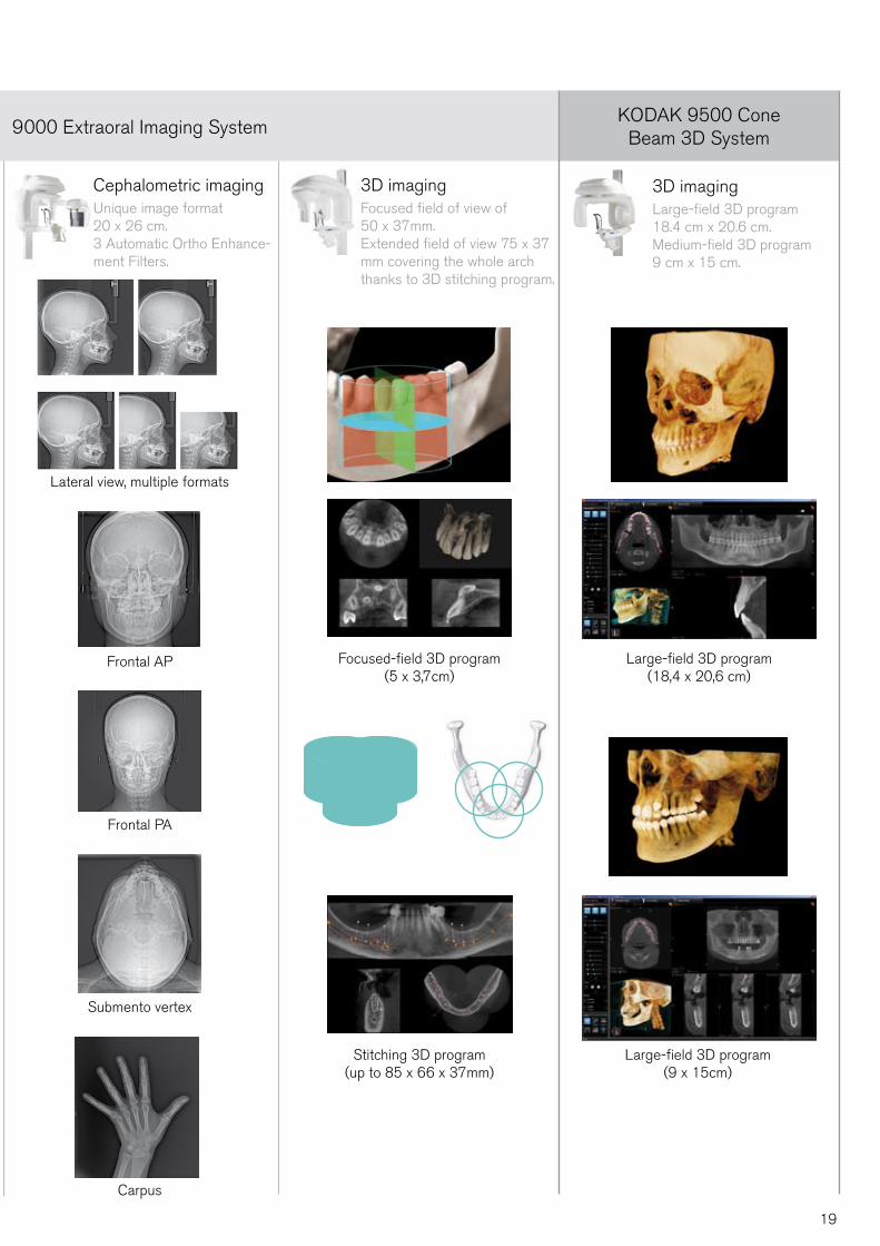
KODAK 9000 Extraoral Imaging SystemKODAK 9500 Cone
Beam 3D System
Cephalometric imagingUnique image format 20 x 26 cm.3 Automatic Ortho Enhance-ment Filters.
3D imagingFocused field of view of 50 x 37mm. Extended field of view 75 x 37 mm covering the whole arch thanks to 3D stitching program.
3D imagingLarge-field 3D program 18.4 cm x 20.6 cm.Medium-field 3D program 9 cm x 15 cm.
Lateral view, multiple formats
Frontal AP
Frontal PA
Carpus
Submento vertex
Focused-field 3D program (5 x 3,7cm)
Stitching 3D program (up to 85 x 66 x 37mm)
Large-field 3D program (18,4 x 20,6 cm)
Large-field 3D program (9 x 15cm)
19
Computed radiography system
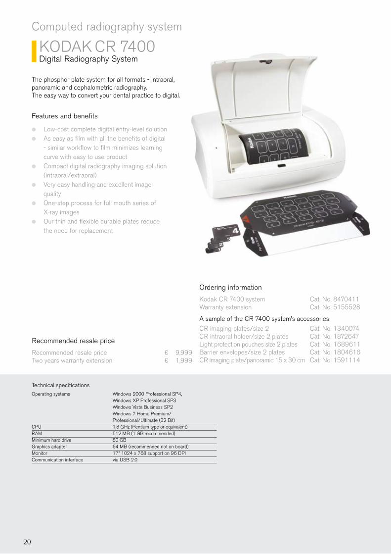
KODAK CR 7400Digital Radiography System
The phosphor plate system for all formats - intraoral, panoramic and cephalometric radiography. The easy way to convert your dental practice to digital.
Features and benefits
Low-cost complete digital entry-level solution ●
As easy as film with all the benefits of digital ●
- similar workflow to film minimizes learning curve with easy to use productCompact digital radiography imaging solution ●
(intraoral/extraoral)Very easy handling and excellent image ●
qualityOne-step process for full mouth series of ●
X-ray imagesOur thin and flexible durable plates reduce ●
the need for replacement
Ordering information
Kodak CR 7400 system Cat. No. 8470411Warranty extension Cat. No. 5155528
A sample of the CR 7400 system’s accessories:
CR imaging plates/size 2 Cat. No. 1340074CR intraoral holder/size 2 plates Cat. No. 1872647Light protection pouches size 2 plates Cat. No. 1689611Barrier envelopes/size 2 plates Cat. No. 1804616CR imaging plate/panoramic 15 x 30 cm Cat. No. 1591114
Technical specifications
Operating systems Windows 2000 Professional SP4, Windows XP Professional SP3 Windows Vista Business SP2 Windows 7 Home Premium/ Professional/Ultimate (32 Bit)CPU 1.8 GHz (Pentium type or equivalent)RAM 512 MB (1 GB recommended)Minimum hard drive 80 GBGraphics adapter 64 MB (recommended not on board)Monitor 17” 1024 x 768 support on 96 DPICommunication interface via USB 2.0
Recommended resale price
Recommended resale price ¤ 9,999Two years warranty extension ¤ 1,999
20
K
OD
AK
CR
74
00
K
OD
AK
15
00
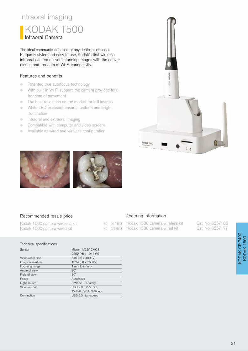
Intraoral imaging
KODAK 1500Intraoral Camera
Features and benefits
Patented true autofocus technology ●
With built-in Wi-Fi support, the camera provides total ●
freedom of movementThe best resolution on the market for still images ●
White LED exposure ensures uniform and bright ●
illuminationIntraoral and extraoral imaging ●
Compatible with computer and video screens ●
Available as wired and wireless configuration ●
Ordering information
Kodak 1500 camera wireless kit Cat. No. 6557185Kodak 1500 camera wired kit Cat. No. 6557177
Technical specificationsSensor Micron 1/2.5’’ CMOS 2592 (H) x 1944 (V)Video resolution 640 (H) x 480 (V)Image resolution 1024 (H) x 768 (V)Focusing range 1 mm to infinityAngle of view 90°Field of view 80°Focus AutofocusLight source 8 White LED arrayVideo output USB 2.0; TV-NTSC; TV-PAL; VGA; S-VideoConnection USB 2.0 high-speed
Recommended resale price
Kodak 1500 camera wireless kit ¤ 3,499Kodak 1500 camera wired kit ¤ 2,999
The ideal communication tool for any dental practitioner.Elegantly styled and easy to use, Kodak’s first wireless intraoral camera delivers stunning images with the conve-nience and freedom of Wi-Fi connectivity.
21

Software
KODAK Dental Imaging Software
The master software for all systems.Integrated 3D module.
Features and benefits
Top-quality and easy to use software ●
Included as standard with all Kodak dental ●
systems digital imaging productsServes as the control panel for all our ●
systemsDesigned specifically for dental radiological ●
diagnosisCan be used as a standalone program ●
or integrated with practice management softwareNo need to purchase additional licenses ●
Ordering information
Kodak dental imaging software Cat. No. 8557530
The software controls all KODAK Dental Systems products
Particularly easy to use radiography software ●
All features are accessible with just a few clicks ●
100% designed for user-friendliness ●
Also ideal for novice PC users ●
High image quality ●
Network-ready ●
Optional: DICOM support ●
22
KD
IS/L
OG
ICO
N
Software
LOGICON Caries Detector Software
Software for detecting caries even at an early stage.
Features and benefits
A unique tool for detecting approximal ●
caries at an early stageA software option for Kodak RVG digital ●
radiography systemsThe software automatically highlights pos- ●
sible abnormalities on digital dental radiographsE-mail function allows sending of result ●
screens to third partiesResults can be saved to patient file ●
Fully integrated with Kodak dental imaging ●
software
Ordering information
Logicon caries detector software Cat. No. 7417967
Recommended resale price
Logicon caries detector software ¤ 999
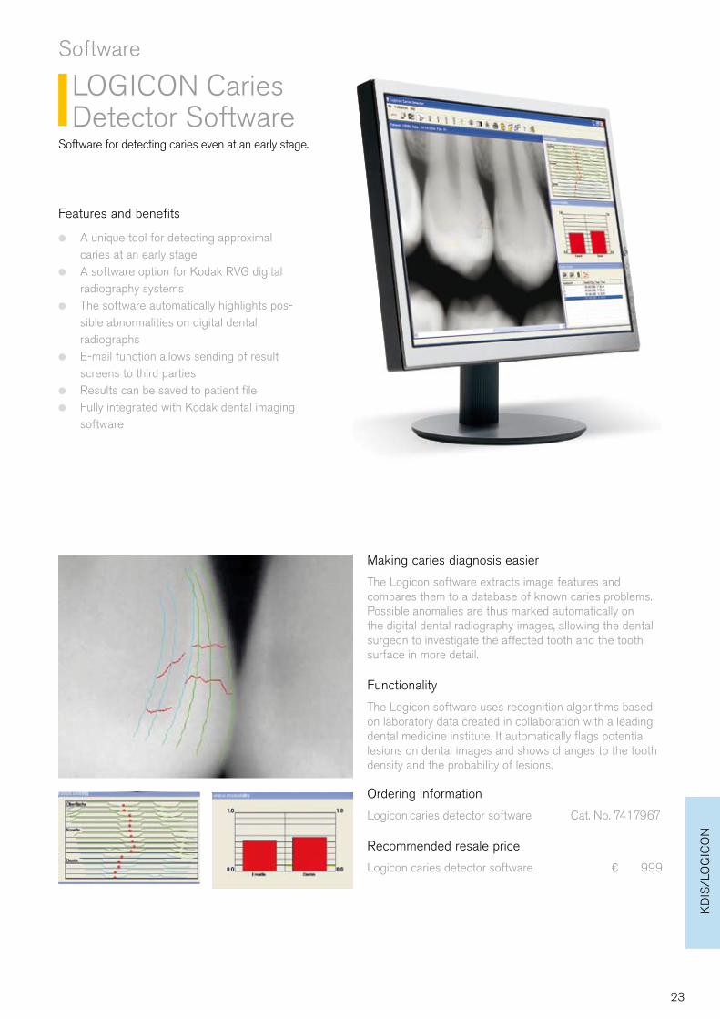
Making caries diagnosis easier
The Logicon software extracts image features and compares them to a database of known caries problems. Possible anomalies are thus marked automatically on the digital dental radiography images, allowing the dental surgeon to investigate the affected tooth and the tooth surface in more detail.
Functionality
The Logicon software uses recognition algorithms based on laboratory data created in collaboration with a leading dental medicine institute. It automatically flags potential lesions on dental images and shows changes to the tooth density and the probability of lesions.
23
Carestream Health
Would you like to know more?
www.kodakdental.comAlternatively, contact your local authorised dealer.
© Carestream Health, Inc. 2010.The Kodak brand the brand elements/colors of Kodak are used under license. RVG is a trademark of Carestream Health, Inc.
We're confident we have the ideal solution for every practice!
A full range of dental radiography and imaging systems ●
100 years' experience in dental imaging ●
In-house product development and manufacturing ●
Worldwide dealer and service network ●
The inventor of intraoral sensor technology ●
High quality standards ●
Looking for an imaging solution?