Embed Size (px)
Citation preview

Procollagen C-endopeptidase Enhancer Protein 2 (PCPE2)Reduces Atherosclerosis in Mice by Enhancing ScavengerReceptor Class B1 (SR-BI)-mediated High-density Lipoprotein(HDL)-Cholesteryl Ester Uptake*
Received for publication, February 17, 2015, and in revised form, April 7, 2015 Published, JBC Papers in Press, May 6, 2015, DOI 10.1074/jbc.M115.646240
Ricquita D. Pollard‡1, Christopher N. Blesso§2, Manal Zabalawi‡, Brian Fulp‡, Mark Gerelus‡, Xuewei Zhu‡,Erica W. Lyons‡, Nebil Nuradin¶, Omar L. Francone�, Xiang-An Li**, Daisy Sahoo¶3, Michael J. Thomas¶,and Mary G. Sorci-Thomas¶4
From the ¶Department of Medicine and the Department of Pharmacology and Toxicology, Medical College of Wisconsin,Milwaukee, Wisconsin 53226, the ‡Section of Molecular Medicine, Department of Internal Medicine and the Department ofBiochemistry, Wake Forest University School of Medicine, Winston-Salem, North Carolina 27101, the §Department of NutritionalSciences, University of Connecticut, Storrs, Connecticut 06268, �Shire Human Genetic Therapies, Lexington, Massachusetts 02421,and the **Department of Pediatrics, University of Kentucky, Lexington, Kentucky 40506
Background: Extracellular matrix protein PCPE2 is linked to alterations in HDL size and concentration.Results: PCPE2 protects against diet-induced atherosclerosis by promoting HDL catabolism, reverse cholesterol transport, andSR-BI-mediated uptake of HDL-cholesteryl ester.Conclusion: PCPE2 mediates HDL function by reducing lipid and immune cell accumulation in the artery.Significance: These findings establish a role for the extracellular matrix glycoprotein PCPE2 in SR-BI-mediated HDL functionand the prevention of atherosclerosis.
Studies in human populations have shown a significant corre-lation between procollagen C-endopeptidase enhancer protein2 (PCPE2) single nucleotide polymorphisms and plasma HDLcholesterol concentrations. PCPE2, a 52-kDa glycoproteinlocated in the extracellular matrix, enhances the cleavage ofC-terminal procollagen by bone morphogenetic protein 1(BMP1). Our studies here focused on investigating the basis forthe elevated concentration of enlarged plasma HDL in PCPE2-deficient mice to determine whether they protected againstdiet-induced atherosclerosis. PCPE2-deficient mice werecrossed with LDL receptor-deficient mice to obtain LDLr�/�,PCPE2�/� mice, which had elevated HDL levels compared withLDLr�/� mice with similar LDL concentrations. We found thatLDLr�/�, PCPE2�/� mice had significantly more neutral lipidand CD68� infiltration in the aortic root than LDLr�/� mice.Surprisingly, in light of their elevated HDL levels, the extent ofaortic lipid deposition in LDLr�/�, PCPE2�/� mice was similarto that reported for LDLr�/�, apoA-I�/� mice, which lack anyapoA-I/HDL. Furthermore, LDLr�/�, PCPE2�/� mice hadreduced HDL apoA-I fractional clearance and macrophage to
fecal reverse cholesterol transport rates compared withLDLr�/� mice, despite a 2-fold increase in liver SR-BI expres-sion. PCPE2 was shown to enhance SR-BI function by increasingthe rate of HDL-associated cholesteryl ester uptake, possibly byoptimizing SR-BI localization and/or conformation. We con-clude that PCPE2 is atheroprotective and an important compo-nent of the reverse cholesterol transport HDL system.
In humans there is an inverse correlation between the con-centration of plasma HDL and the relative risk of developingcardiovascular disease (CVD)5 (1– 4). To explain this associa-tion, many studies have suggested that HDL plays a central rolein the pathway called reverse cholesterol transport (RCT), aprocess that returns cholesterol to the liver for excretion (5, 6).In the first step, ATP-binding cassette transporter A1 (ABCA1)lipidates apoA-I with phospholipid and cholesterol to form na-scent HDL (nHDL) (5, 7–9). These nHDL are rapidly convertedto mature HDL by plasma lecithin-cholesterol acyltransferase,an enzyme that esterifies free cholesterol yielding maturespherical HDL particles with a cholesteryl ester (CE)-rich core.The RCT pathway is complete when the scavenger receptorclass B1 (SR-BI) in the liver removes CE from mature HDL,leaving lipid-poor apoA-I to be recycled or filtered by the kid-ney (10). Because the liver is the primary site of HDL metabo-
* This work was supported, in whole or in part, by National Institutes of HealthGrants NHLBI HL-112270, HL 112276, and HL 127649 (to M. G. S.-T.). Thiswork was also supported by American Heart Association Grants09GRNT2280053 and 14GRNT20500029 (to M. J. T.). The authors declarethat they have no conflicts of interest with the contents of this article.
1 Supported by National Institutes of Health Diversity Supplement GrantHL112270A1S1.
2 Supported by post-doctoral American Heart Association Grant13POST17000005.
3 Supported by National Institutes of Health Grant RO1-HL058012.4 To whom correspondence should be addressed: Dept. of Medicine, Div. of
Endocrinology, Metabolism, and Clinical Nutrition, Medical College of Wis-consin, 8701 W. Watertown Plank Rd., Milwaukee, WI 53226. Tel.: 414-955-5728; Fax: 414-456-6312; E-mail: [email protected].
5 The abbreviations used are: CVD, cardiovascular disease; RCT, reverse cho-lesterol transport; ABCA1, ATP-binding cassette transporter A1; nHDL, na-scent HDL; CE, cholesteryl ester; SR-BI, scavenger receptor class B1; PCPE2,procollagen C-endopeptidase enhancer protein 2; BMP1, bone morpho-genetic protein 1; NTR, netrin-like; OCT, optimal cutting temperature(medium); 3H-COE, cholesteryl [1a,2a-3H]oleoyl ether; FCR, fractional cata-bolic rate.
THE JOURNAL OF BIOLOGICAL CHEMISTRY VOL. 290, NO. 25, pp. 15496 –15511, June 19, 2015© 2015 by The American Society for Biochemistry and Molecular Biology, Inc. Published in the U.S.A.
15496 JOURNAL OF BIOLOGICAL CHEMISTRY VOLUME 290 • NUMBER 25 • JUNE 19, 2015
by guest on September 9, 2019
http://ww
w.jbc.org/
Dow
nloaded from

lism, the steady-state concentration of plasma HDL dependslargely on the rate of nHDL formation balanced by the catabo-lism of HDL.
For some time, epidemiology studies used plasma HDL con-centration to help calculate the relative risk of developing CVD.That approach has been criticized because plasma HDL con-centration does not adequately predict an individual’s CVD risk(11, 12). Recent studies have shown that an individual’s choles-terol efflux capacity is a more accurate measure of assessingCVD risk (13–15). Efflux capacity measures the potential of anindividual’s apoB-depleted plasma to mobilize cellular choles-terol, suggesting that the inherent properties of one’s plasmaHDL are responsible for driving this process.
Cholesterol efflux is an essential physiological process thatproduces nHDL particles. It is now recognized that the compo-sition and structure of nHDL, before modification in plasma,differ significantly from plasma or mature HDL. These nHDLparticles contain three apoA-I monomers and are highlyenriched in free cholesterol and sphingomyelin, having a com-position similar to lipid rafts (16).
nHDL formation is not a spontaneous process occurringwhen apoA-I and phospholipid come in contact. Rather,nHDL formation requires carefully orchestrated conforma-tional changes in the apoprotein structure that permit shielded,hydrophobic residues within multiple monomers of apoA-I tobecome accessible and bind phospholipid (16). This processyields �11-nm diameter nHDL particles carrying over 100molecules of free cholesterol/particle (17), an efficient processto efflux cholesterol from the cell.
Several mechanisms have been proposed to explain hownHDL is assembled via ABCA1 on the membrane surface (7, 18,19). One hypothesis has binding of lipid-poor apoA-I to ABCA1as the first step in the process. However, other studies suggestthat low affinity, high capacity membrane binding sites areinvolved before apoA-I binds to ABCA1 (20 –23). Interactionbetween apoA-I and membrane-bound accessory protein(s)could potentially promote conformational changes within lip-id-poor apoA-I and in so doing enhance the rate of nHDL par-ticle formation (12, 24, 25).
Recent reports suggest that procollagen C-endopeptidaseenhancer protein 2 (PCPE2) participates in promoting choles-terol efflux. A connection between PCPE2 and HDL wasinferred when significant correlations were observed betweenPCPE2 single nucleotide polymorphisms and plasma HDL cho-lesterol concentrations (26). PCPE2 is a 52-kDa glycoproteinencoded by the PCOLCE2 gene (27, 28). It shares 43% aminoacid sequence identity with PCPE1 (29). Both PCPE1 andPCPE2 are located in the extracellular matrix, where they facil-itate bone morphogenetic protein 1 (BMP1) cleavage of C-ter-minal procollagen propeptides. PCPE2 and PCPE1 have dif-ferent tissue distributions and heparin-binding affinities,suggesting a functional divergence (29). PCPE2 is heavilyexpressed in heart tissue in contrast to PCPE1. Both PCPE1 andPCPE2 have two CUB (Complement C1r/C1s, Uegf, Bmp1)domains separated by a short linker region (30 –32), with eachdomain consisting of about 110 residues containing a �-sand-wich fold that mediates a variety of protein-protein interac-tions. The CUB domains also contain a homologous Ca2� bind-
ing site that mediates ionic interactions between proteinpartners, similar to that described in the low-density lipopro-tein receptor family (33). The tandem CUB domains are fol-lowed by a C-terminal netrin-like (NTR) domain (27, 29). CUBdomains are commonly found in extracellular membrane pro-teins that mediate protein-protein interactions (33–36),whereas NTR domains bind to cell surface glycosaminoglycans(37, 38), presumably anchoring PCPE2 to the extracellularmatrix.
Interestingly, BMP1, in addition to catalyzing procollagenprocessing, removes the apoA-I six-amino acid propeptide (39).Studies by Francone and others (26, 40, 41) show that PCPE2stabilizes a ternary catalytic complex with apoA-I and BMP1.These findings suggest that BMP1-PCPE2 processing of theapoA-I propeptide might stimulate nHDL assembly (42).
Studies in PCPE2�-deficient mice provide the first directexperimental evidence linking PCPE2 to HDL metabolism (43).PCPE2 knock-out mice had elevated concentrations of en-larged plasma HDL (44), despite displaying defective ABCA1-mediated cholesterol efflux capacity. Thus, reduced cholesterolefflux is paradoxically associated with elevated plasma HDLcholesterol in PCPE2-deficient mice. A recent genome-widesiRNA screen also suggests that PCPE2 affects the efficiency ofHDL apoA-I secretion (45).
Because elevated plasma HDL levels are associated withreduced atherosclerosis, we sought to investigate whether ele-vated concentrations of enlarged HDL in PCPE2-deficient micewere atheroprotective or atherogenic. We have suggestedmechanisms to explain the role of PCPE2 and provided evi-dence demonstrating the participation of PCPE2 in HDL cho-lesterol catabolism and reverse cholesterol transport.
Experimental Procedures
Animals and Diets—PCPE2-deficient mice in the C57BL/6background were obtained from the Max Planck Institute (43)and then crossed with LDLr�/� mice to generate LDLr�/�,PCPE2�/� mice. Both LDLr�/� and LDLr�/�, apoA-I�/� micehave been fully crossed onto the C57BL/6 background asdescribed previously (46, 47). Starting at 6 weeks of age, micewere fed for 12 weeks either a standard chow diet or an athero-genic diet containing 0.1% cholesterol and 10% calories frompalm oil (47). Mice were fasted for 3 h prior to being anesthe-tized with ketamine/xylazine (200 mg/kg ketamine and 10mg/kg xylazine) followed by euthanasia and blood collectionby cardiac puncture. The Animal Care and Use Committeeof the Wake Forest University School of Medicine approvedall procedures used in the current study. All mice werehoused in a temperature-controlled room and maintained ina 12-h light/12-h dark cycle at the Wake Forest School ofMedicine vivarium.
Plasma Lipoprotein Isolation and Characterization—Bloodwas collected in EDTA containing tubes then centrifuged at15 °C for 10 min at 7000 rpm. The plasma fraction was collectedand stored at �80 °C. Total plasma cholesterol (Wako Diagnos-tics Cholesterol E) was determined using an enzymatic assaykit. For total lipoprotein cholesterol distribution, 15 �g of totalplasma cholesterol was applied to a Superose 6 10/300GL col-umn (GE Healthcare) with online mixing of enzymatic reagent
PCPE2 Modulates SR-BI Function and Atherosclerosis
JUNE 19, 2015 • VOLUME 290 • NUMBER 25 JOURNAL OF BIOLOGICAL CHEMISTRY 15497
by guest on September 9, 2019
http://ww
w.jbc.org/
Dow
nloaded from
(Cholesterol (Liquid) Reagents Set, Pointe Scientific Inc.) asdescribed previously (48). Isolation of the total lipoprotein frac-tion for compositional analysis was carried out by first obtain-ing the d � 1.225 g/ml fraction after density gradient ultracen-trifugation using KBr as described previously (46). The d �1.225 g/ml fractions were separated into lipoprotein classesusing size-exclusion fast protein liquid chromatography(FPLC) (46). Aliquots corresponding to VLDL, LDL, and HDLclasses were pooled and stored at �80 °C for composition anal-ysis or used immediately for particle size determination. TheHDL particle size was determined by a comparison with stan-dards of known Stokes diameter following separation usingNovex (Life Technologies) 4 –20% non-denaturing gels andthen stained with Coomassie Blue G-250 dye as described pre-viously (49). Mouse plasma apoA-I concentration was mea-sured using an enzyme-linked immunosorbent assay asdescribed previously (49).
Quantification of Pro-apolipoprotein A-I by Mass Spec-trometry—The abundance of pro-apoA-I and apoA-I wasassessed from purified HDL and whole plasma from PCPE2�/�
and C57BL/6 mice using liquid chromatography-tandem massspectrometry. Whole plasma aliquots were separated on 12%SDS-PAGE as described previously (16, 50 –52). The gel wasstained with SimplyBlue (Life Technologies), and the proteinband corresponding to 28,000 Da was excised from the gel,minced, and repeatedly dehydrated with acetonitrile. The gelpieces were rehydrated with a cold, freshly prepared solutioncontaining 20 ng/�l trypsin in 10 mM ammonium bicarbonate,pH 7.8, 0.1% (w/v) RapiGest SFTM, and 1 mM CaCl2. The finaltrypsin to apoA-I mass ratio was �1:20. After incubation on icefor 10 min, the digests were incubated for 18 h at 37 °C. Thedigestion solution was removed, and gel pieces were coveredwith 200 �l of acetonitrile/formic acid/water (v/v/v, 50:5:45).After sitting for 10 min the solvent was transferred to a freshtube. The extraction was repeated and the combined aliquotsacidified to an HCl:apoA-I ratio of 1:10 (v/v) using 500 mM HCl.After the acidified solution was incubated for 35 min at 37 °C,the sample was centrifuged for 10 min at 13,000 rpm. Thesupernatant was transferred to a fresh tube before mass spec-trometry. Survey scans were performed on each peptide mix-ture using a Waters Q-TOF API-US mass spectrometerequipped with a Waters CapLC and Advion Nanomate source.Acquisition was controlled using Mass-LynxTM 4.0 software.Peptides were loaded onto a PLRP-S trapping column, 0.5-mmdiameter � 2.0-mm length, containing 3-�m diameter parti-cles with a pore diameter of 100 Å. Peptides were loaded ontothe trapping column in water/acetonitrile/formic acid (97:3:0.2) at 500 nl/min. After switching of the trapping column, in-line separation was accomplished on a 0.3 � 150-mm PCRP-Scolumn packed with 3-�m diameter particles with 100 Å poresat 470 nl/min. The following gradient elution was used: solventA (25 mM formic acid in 97% water and 3% acetonitrile) andsolvent B solvent B (25 mM formic acid in 3% water 97% aceto-nitrile). The gradient profile was: 2% solvent B for 3 min with alinear increase to 40% B at 90 min and then to 80% B in 5 min. At95 min, the composition was ramped to 2% B over 5 min andequilibrated with 2% B for 30 min. Positive ion survey scanswere recorded in the continuum mode with a scan window of
300 to 1500 m/z for 2 s. The source temperature was 80 °C andcone voltage was 45 V. Experimental m/z was corrected usingmouse apoA-I tryptic fragment 7, m/z � 533.7462. Ions within�0.051 m/z of the theoretical ions were sequenced. Product ionMS/MS spectra were acquired in the continuum mode from 50to 1500 m/z using a data-directed charge state-selective colli-sion energy and an accumulation time of 2 s. Sequence analysisof the MS/MS spectra was performed with a fragment ion tol-erance of �0.05 m/z.
For quantification of the ratio of mature and pro-apoA-I, theN-terminal tryptic peptides for mouse pro-apoA-I (WHV-WQQDEPQSQWDK, protonated m/z � 1996.8942) andmature mouse apoA-I (DEPQSQWDK, protonated m/z �1132.4911) were synthesized (GenScript), and the m/z, purity,and sequence were verified by mass spectrometry. The reten-tion time, mass, and sequence were used to identify the N-ter-minal sequence from all samples analyzed.
For the analysis of intact pro- or mature mouse apoA-I frompurified HDL, mass spectrometric analysis was performedusing a Waters Q-TOF equipped with an Advion TriversaNanomate source as described previously (53). Samples (0.4�M) were prepared in 1:1 (v:v) acetonitrile-water containing0.2% formic acid and then introduced into the mass spectrom-eter at 500 nl/min. Acquisition parameters were adjusted tomaximize resolution.
Lipid Composition—Total and free cholesterol determina-tion were performed on aliquots from FPLC-separated andpooled HDL that had been spiked with the internal standard,cholesterol-3,4-13C2 (Sigma-Aldrich), and subjected to lipidextraction as previously described (16). To measure free cho-lesterol an aliquot was removed, evaporated under argon, dis-solved in hexane and then injected on a Finnigan TSQ Quan-tum XLS tandem mass spectrometer interfaced to a TRACE gaschromatograph (GC-MS/MS), as described previously (16, 47),by select reaction monitoring (SRM) with the following param-eters in the positive ion mode: scan time, 0.1 s; collision energy,10 V; emission current, 25 �A; electron energy, �42 eV; sourceand transfer line temperature, 260 °C; and flow rate, 1 ml/min.Analysis was carried out using a DB-1 column (10.4 m, 250 �minner diameter) with a 0.25-�M film thickness. For quantifyingtotal cholesterol, the remaining sample was dried under astream of nitrogen, redissolved in 1 ml of ethanol, mixed with100 �l of 50% (w/w) aqueous potassium hydroxide, and thensaponified for 1 h at 60 °C. After extraction, total cholesterolwas measured on the Quantum XLS. Cholesteryl ester was cal-culated as the difference between free cholesterol and totalcholesterol.
Plasma triglycerides were analyzed using mass spectrometryfrom mouse plasma after preliminary separation from otherpolar and nonpolar lipids using aminopropyl solid-phaseextraction columns using the procedure of Kaluzny et al. (54)with minor modifications. The nonpolar fraction was driedunder nitrogen and loaded onto the second solid-phase extrac-tion column dissolved in 500 �l of hexane. Cholesteryl esterswere removed from the column with 3– 4-ml washes of 4%CH2Cl2 in hexane. Then triglycerides were eluted with 3– 8-mlwashes of CH2Cl2:diethyl ether:hexane (14:1:84). Quantitationwas by gas chromatography-mass spectrometry after saponifi-
PCPE2 Modulates SR-BI Function and Atherosclerosis
15498 JOURNAL OF BIOLOGICAL CHEMISTRY VOLUME 290 • NUMBER 25 • JUNE 19, 2015
by guest on September 9, 2019
http://ww
w.jbc.org/
Dow
nloaded from

cation to generate methylated fatty acids (55, 56). The fattyacids were quantified to obtain the total fatty acid composition.The moles of fatty acids were summed and divided by 3 toobtain the number of moles of triglyceride. C17 triglyceride wasused as the internal standard.
RNA Isolation, cDNA Synthesis, and QuantitativeReal-Time-PCR—Tissues harvested at necropsy were flash-fro-zen with liquid nitrogen and kept at �80 °C until needed.Briefly, tissue was quickly thawed, weighed, and then added to a14-ml screw cap polypropylene tube containing 1 ml of pre-chilled TRIzol (Life Technologies). The tissue was immediatelyhomogenized using a Polytron PT 1200C homogenizer, andRNA was isolated following the manufacturer’s instructions.Purified RNA pellets were thoroughly resuspended in Ultra-Pure water (Sigma-Aldrich) on ice. RNA concentrationswere determined at 260 nm using a Beckman DU800spectrophotometer.
Using 1–2 �g of RNA, cDNA was synthesized using theqScript cDNA Supermix (Quant BioSciences) according to themanufacturer’s instructions. Quantitative real-time PCR wasperformed using the Applied Biosystems 7500 Fast Real TimePCR system with Fast Start Universal SYBR Green Master Mix(Roche Applied Science) according to the manufacturer’sinstructions. Reactions were set up in triplicate using a 1-�g/�lcDNA template. Following amplification, Ct values wereobtained from Applied Biosystems sequence detection soft-ware, and the Ct values were determined for the gene of inter-est compared with that for mouse GAPDH for each animal runas a control. For each tissue the percent relative abundance ofmouse PCPE2 and PCPE1 was derived from the Ctmax � Ct/Ctmax) � 100. Primers designed using the Universal ProbeLibrary were purchased from Integrated DNA Technologies.The following are the mouse sequences used: SR-BI forward,GCCCATCATCATCTGCCAACT, and reverse, TCCTGGG-AGCCCTTTTTACT; PCPE1 forward, TTACGTGGCAAGT-GAGGGTTT, and reverse, TGTCCAGATGCACTTCTTGT-TTG; and PCPE2 forward, TCACATGTGGCGGCATTCT,and reverse, CAGGAAAACCTTCACTGCCAAT.
Immunoblotting—Immunoblotting was performed subse-quent to total tissue protein isolation following extraction usinga radioimmune precipitation assay buffer (RIPA, Cell SignalingTechnologies) or using detergent-free lipid raft isolation asdescribed. For tissues extracted with radioimmune precipita-tion assay buffer, aliquots of equal protein content were run on12% SDS-PAGE. All sample aliquots were diluted with 4� LDSbuffer (Novex) to which solid DTT had been added to achieve a100 mM final concentration. Samples were heated to 70 °C for10 min. Transfer to PVDF membrane (PerkinElmer Life Sci-ences) was accomplished after treatment with Tris-glycine, pH8.3, buffer using a semi-dry blot (Bio-Rad) apparatus for 30 minat 10 V. Membranes were blocked with 5% nonfat dry milk in 10mM Tris-glycine, pH 7.4, buffer and processed. The primaryantibodies used (PCPE1 (Novus Biologicals), PCPE2 (Abcam),GAPDH (Ambion), and SR-BI (Novus Biologicals)) werediluted 1:1000 and incubated with the membrane at 4 °C over-night. The blots were washed and then incubated with an HRP-conjugated anti-mouse IgG secondary antibody (GE Health-care) at a 1:10,000 dilution for 1 h at room temperature. Blots
were washed again and then incubated with SuperSignal WestPico chemiluminescence substrate (Pierce) and visualized witha FujiFilm LAS-3000 camera. Band intensities were comparedusing MultiGauge software (FujiFilm).
HDL ApoA-I Turnover—Diet-fed mouse HDL was purifiedusing a combination of ultracentrifugation and FPLC. The puri-fied HDL apoA-I was then labeled with 125I using the Mc-Farlane method (57) as described previously (58, 59). SufficientICl was used to have 0.5 mol of radiolabeled iodine for everymole of HDL apoA-I and prevent damage to the protein orlabeling lipid. Labeled HDL apoA-I was purified from freeiodine using a desalting column (Bio-Rad), and the eluate wasdialyzed exhaustively against 0.15 M NaCl, pH 7.4. On the day ofthe study, anesthetized mice were retro-orbitally injected with125I-radiolabeled HDL (10 � 106 cpm) after isoflurane admin-istration. Approximately 30 �l of blood was collected from thecontralateral retro-orbital sinus at 0.08, 0.17, 0.5, 1, 3, 6, 22, 28,and 48 h. Radioactivity was measured at each time point using aBeckman Gamma 4000 counter. The fractional catabolic ratesand half-lives of the 125I-labeled HDL were determined fromplasma decay curves assuming a one-pool model (58). Bloodvolumes were estimated to be 5.85 ml/100 g of body weight.
Histology and Immunofluorescence Microscopy—At the timeof sacrifice, a 21-gauge butterfly needle was inserted into theleft ventricle; the atrium was clipped, and the heart and aortawere perfused with saline for 5 min and then removed andcleaned of fat and adventitia. Mouse heart and aorta wereembedded in optimal cutting temperature (OCT) medium andthen sectioned for staining and immunohistochemistry. Forquantification of total and percent lesion area, OCT-embeddedaortas were cut into sequential 6-�m sections using a Leicacryostat at �50 °C. Tissue sections were stained overnight in0.5% Oil Red O dissolved in propylene glycol and then counter-stained with hemotoxylin. Masson’s trichrome stain was usedto assess the connective tissue content of the aortic root. Sec-tions were digitized using a Nikon microscope and Image-ProPlus 6.2 software and then quantified using NIS-Elements soft-ware (Nikon Instruments Inc.). Eight to 10 sections at intervalsof 30 �m were used for morphological analyses and were aver-aged to obtain a value for each animal. The results are expressedas both the percentage of lesion area and absolute lesion area.
For immunofluorescence microscopy, purified rat anti-mouse CD68 (AbD FA-11-Serotec) at a 1:100 dilution was usedas the primary antibody. Slides were incubated for 1 h in PBSbuffer containing 2% fetal calf plasma, washed with PBS, andthen incubated with a 1:300 dilution of Cy3-conjugated goatanti-rat IgG antibody (Rockland) for 30 min at room tempera-ture. After the slides were washed briefly with PBS, they werestained with DAPI to visualize the nuclei and then mountedwith Fluoro-Gel (Electron Microscopy Sciences). Immunofluo-rescence was visualized using a Nikon Eclipse TE2000-S micro-scope, and the total and percentage of CD68 staining was quan-tified using NIS-Elements software.
Picrosirius red staining of mouse tissues was performed onformalin-fixed tissue embedded in paraffin and then cut seriallyin 6-�m sections. Collagen fibers were stained using picrosiriusred (Polysciences Inc.) and viewed with polarized light asdescribed (60).
PCPE2 Modulates SR-BI Function and Atherosclerosis
JUNE 19, 2015 • VOLUME 290 • NUMBER 25 JOURNAL OF BIOLOGICAL CHEMISTRY 15499
by guest on September 9, 2019
http://ww
w.jbc.org/
Dow
nloaded from
In Vivo Reverse Cholesterol Transport Studies—Diet-fed micereceived an intraperitoneal injection of 0.6 � 106 dpm of[3H]cholesterol-labeled J774 cells as described previously (61).Blood was collected at 6, 24, and 48 h, and the plasma wascounted for radioactive content (62). Feces collected from 0 to48 h and subjected to lipid extraction were then counted forradioactive content of neutral and acidic sterols as describedpreviously (63).
Tissue Culture and Transfection—CHO cells were grown inDMEM/F-12 medium (1:1) supplemented with 10% (v/v) FBS,100 units/ml penicillin, 100 �g/ml streptomycin, and 2 mM
L-glutamine. Transient transfections were performed usingLipofectamine 2000 according to the manufacturer’s protocol(Life Technologies) with 2.5 �g of DNA and 12 �l of Lipo-fectamine 2000/well of a confluent 6-well plate. The humanPCPE2-pCMV6 plasmid used in the transfection was obtainedfrom OriGene (Rockville, MD) and contains a Myc-DDK tag atthe N terminus, which adds 10 amino acids.
Selective Uptake of HDL Labeled with [3H]Cholesteryl OleylEther—HDL isolated as described above, using a combinationof ultracentrifugation and FPLC, was labeled with cholesteryl[1a,2a-3H]oleoyl ether (3H-COE) (American RadiolabeledChemicals Inc.) using recombinant cholesterol ester transferprotein (CETP, Roar Biomedical Inc.) as described (64). An ali-quot containing 10 �g of labeled HDL (based on protein)/ml ofplasma-free DMEM medium plus 0.5% BSA was incubated withconfluent CHO cells in a 6-well plate at 37 °C for 1.5 h. Afterincubation the medium was removed, and the cells werewashed extensively to remove surface-bound labeled HDL. Thecell monolayer was lysed with 1 ml of 0.1 N NaOH, and aliquotsof the lysed cells were counted for their 3H radioactivity contentand normalized to total cell protein as determined by the Lowryassay (65).
Surface Staining of CHO Cells—Transfected CHO cells wereharvested from 6-well plates and stained for SR-BI and PCPE2using fluorescence-activated cell sorting (FACS) as describedpreviously (66). Briefly, 3 � 105 cells were stained with poly-clonal rabbit anti-SR-BI-APC conjugate (Alexa Fluor 647)(Bioss Antibodies) and mouse anti-MycDDK-FITC conjugate(Alexa Fluor 488) (AbD Serotec) for 30 min at 4 °C. Sampleswere washed and resuspended in 2% paraformaldehyde in PBSbefore acquisition of samples using a system (BD Biosciences).
Statistical Analysis—GraphPad Prism version 5 was used forstatistical analysis, and the data are reported as mean � S.D.Differences between groups were evaluated by independent ttests and by analysis of variance with Tukey’s post hoc test.
Results
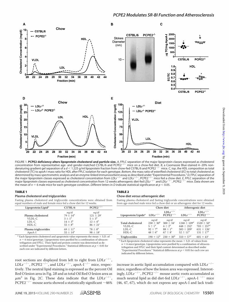
The Absence of PCPE2 Affects Lipoprotein Distribution andConcentration—PCPE2-deficient mice in the C57BL/6 back-ground (43) were characterized with respect to HDL size andconcentration for comparison with the previously publishedphenotype (44). Fig. 1A shows the cholesterol distribution fol-lowing FPLC separation of the major lipoprotein classes fromthe d � 1.225 g/ml density fraction. We see that PCPE2-defi-cient mice possess a greater concentration of enlarged HDLparticles (Fig. 1A, dashed line) compared with control C57BL/6mice that express PCPE2 (solid line). The increase in the HDL
cholesterol concentration in PCPE2-deficient mice was accom-panied by an elevation in plasma apoA-I concentrations asshown in Table 1. These changes in HDL levels were indepen-dent of statistically significant changes in LDL or VLDL choles-terol levels between genotypes. A trend to higher plasma trig-lycerides was observed for chow-fed PCPE2-deficient mice, asexemplified by the increased VLDL peak in Fig. 1A, but thetrend was not statistically significant. Non-denaturing gradientgel separation confirmed that PCPE2�/� mice had larger HDLparticle sizes and diameters compared with HDL from controlmice, as shown in Fig. 1B and consistent with the previous study(44). The cholesterol to apoA-I composition was determined inHDL particles purified by a combination of density ultracentri-fugation and FPLC. Fig. 1C shows that HDL particles fromPCPE2�/� mice were enriched in total cholesterol when nor-malized to apoA-I content, likely accounting for the increase inparticle size. However, HDL from both genotypes had similarratios of esterified to total cholesterol (Fig. 1C, EC/TC), sug-gesting that plasma lecithin-cholesterol acyltransferase activitywas similar for the two genotypes and that conversion of freecholesterol to esterified cholesterol was not involved in particleenlargement.
To investigate the atherosclerotic properties of PCPE2 HDL,PCPE2-deficient mice were crossed with the hypercholester-olemic LDL receptor-deficient (LDLr�/�) mouse, to createLDLr�/�, PCPE2�/� mice. Aliquots of plasma from 12 weekschow-fed LDLr�/�, PCPE2�/� mice were centrifuged to obtainthe d � 1.225 g/ml lipoprotein fraction, which was then sepa-rated into lipoprotein classes by FPLC. Fig. 1D shows the lipo-protein cholesterol distribution for LDLr�/�, PCPE2�/�
(dashed line) and LDLr�/� mice (solid line). As anticipated,HDL from LDLr�/�, PCPE2�/� mice contained larger sizedparticles, indicated by elution in earlier eluting fractions as seenin PCPE2�/� mouse plasma (Fig. 1A). The HDL cholesterolconcentration in LDLr�/�, PCPE2�/� mice was also signifi-cantly increased compared with LDLr�/� mice, as shown inTable 2.
Lipoprotein cholesterol distribution was then evaluated afterfeeding an atherogenic diet (46, 47, 67) for 12 weeks (resultsshown in Fig. 1E). As expected, both LDLr�/�, PCPE2�/� andLDLr�/� mice had substantial increases in VLDL and LDL cho-lesterol levels. Table 2 shows a comparison of these values withtheir chow-fed counterparts. Interestingly, although diet-fedLDLr�/�, PCPE2�/� mice did not show a significant differencein LDL cholesterol concentrations between genotypes, therewas a statistically significant increase (�90%) in VLDL andHDL cholesterol concentration. These results suggest thatfeeding an atherogenic diet to hypercholesterolemic mice thatlack PCPE2 appears to also affect VLDL in addition to HDLmetabolism.
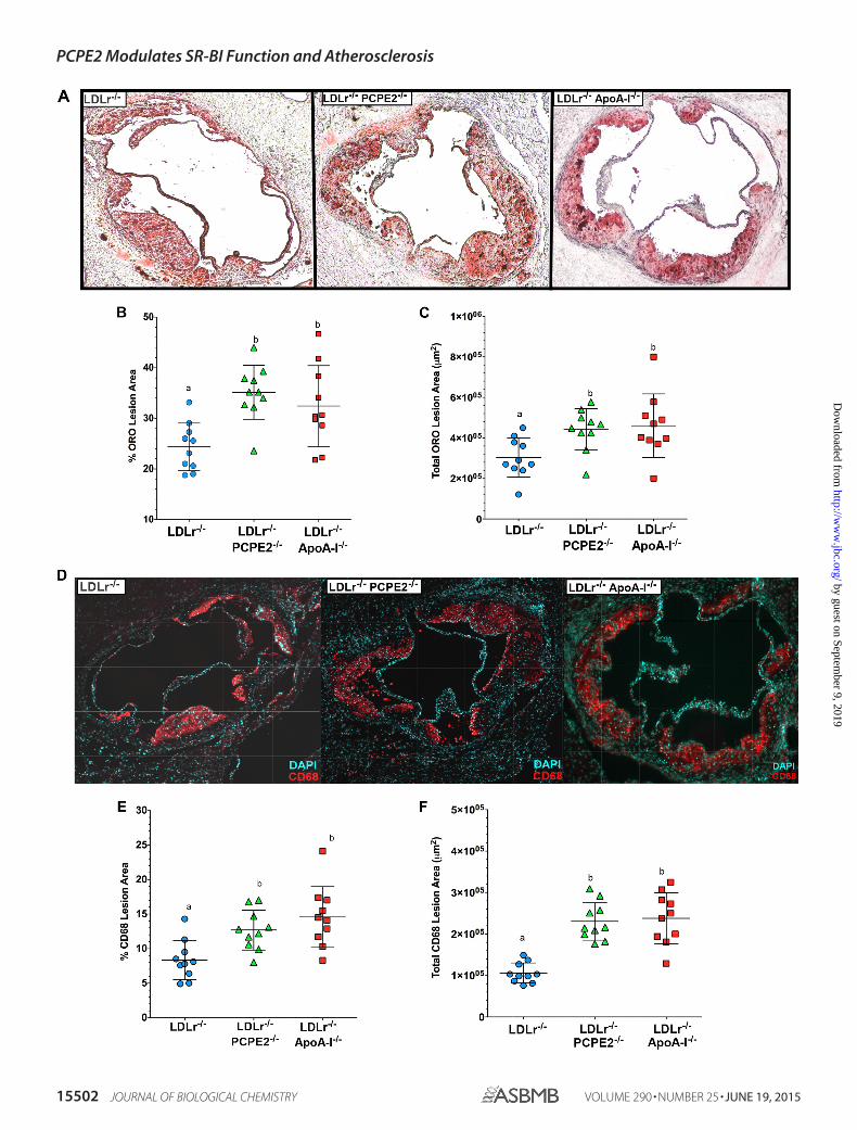
PCPE2 Protects against Diet-induced Atherosclerosis—Groups of male LDLr�/�, PCPE2�/�, LDLr�/�, and LDLr�/�,apoA-I�/� mice were fed an atherogenic diet. After 12 weeks ofdiet feeding their hearts and aortas were embedded into OCT,sectioned, and stained with Oil Red O. The extent of athero-sclerotic lipid accumulation was assessed by staining aortic rootsections with the neutral lipid stain followed by quantification,as shown in Fig. 2A. Representative Oil Red O-stained aortic
PCPE2 Modulates SR-BI Function and Atherosclerosis
15500 JOURNAL OF BIOLOGICAL CHEMISTRY VOLUME 290 • NUMBER 25 • JUNE 19, 2015
by guest on September 9, 2019
http://ww
w.jbc.org/
Dow
nloaded from
root sections are displayed from left to right from LDLr�/�,LDLr�/�, PCPE2�/�, and LDLr�/�, apoA-I�/� mice, respec-tively. The neutral lipid staining is expressed as the percent OilRed O lesion area in Fig. 2B and as total Oil Red O lesion area as�m2 in Fig. 2C. These data indicate that the LDLr�/�,PCPE2�/� mouse aorta showed a statistically significant �46%
increase in aortic lipid accumulation compared with LDLr�/�
mice, regardless of how the lesion area was expressed. Interest-ingly, LDLr�/�, PCPE2�/� mouse aortic roots accumulated asmuch neutral lipid as did diet-fed LDLr�/�, apoA-I �/� mice(46, 47, 67), which do not express any apoA-I and lack tradi-
FIGURE 1. PCPE2 deficiency alters lipoprotein cholesterol and particle size. A, FPLC separation of the major lipoprotein classes expressed as cholesterolconcentration from representative age- and gender-matched C57BL/6 and PCPE2�/� mice on a chow-fed diet. B, a Coomassie Blue-stained 4 –20% non-denaturing gradient gel separation of a d � 1.225 g/ml lipoprotein fraction from chow-fed C57BL/6 and PCPE2�/� mice. C, top, the HDL composition as totalcholesterol (TC) to apoA-I mass ratio for HDL after FPLC isolation for each genotype. Bottom, the mass ratio of esterified cholesterol (EC) to total cholesterol asdetermined by mass spectrometric analysis and an enzyme-linked immunosorbent assay as described under “Experimental Procedures.” D, FPLC separation ofthe major lipoprotein classes expressed as cholesterol concentration from LDLr�/� and LDLr�/�, PCPE2�/� mice fed a chow diet. E, FPLC separation of themajor lipoprotein classes expressed as cholesterol concentration from 12 weeks atherogenic diet-fed LDLr�/� and LDLr�/�, PCPE2�/� mice. Data shown arethe mean of n � 6 male mice for each genotype condition. Different letters in D indicate statistical significance at p � 0.05.
TABLE 1Plasma cholesterol and triglyceridesFasting plasma cholesterol and triglyceride concentrations were obtained fromequal numbers of male and female mice fed a chow diet for 12 weeks.
Lipoprotein/Lipida C57BL/6 PCPE2�/�
mg/dl mg/dlPlasma cholesterol 79 � 14b 121 � 29c
VLDL-C 3 � 1b 5 � 3b
LDL-C 14 � 3b 12 � 4b
HDL-C 55 � 8b 86 � 21c
Plasma triglycerides 69 � 11b 78 � 8b
ApoA-I 55 � 14b 98 � 18c
a Each lipoprotein cholesterol and apoprotein value represents the mean � S.D. ofn � 6 mice/genotype. Lipoproteins were purified by a combination of ultracen-trifugation and FPLC. Their lipid and protein content was determined as de-scribed under “Experimental Procedures.” Statistical differences at p � 0.05 foreach row are indicated by different letters.
TABLE 2Chow diet versus atherogenic dietFasting plasma cholesterol and fasting triglyceride concentrations were obtainedfrom age-matched male mice fed a chow diet or an atherogenic diet for 12 weeks.
Lipoprotein/Lipidsa
Chow diet Atherogenic diet
LDLr�/�LDLr�/�
PCPE2�/� LDLr�/�LDLr�/�
PCPE2�/�
mg/dl mg/dl mg/dl mg/dlTotal cholesterol 230 � 30b 300 � 34c 1248 � 109d 1520 � 50e
VLDL-C 5 � 1b 4 � 2b 448 � 252c 703 � 257d
LDL-C 92 � 7b 88 � 1b 583 � 209c 631 � 126c
HDL-C 48 � 6b 67 � 8c 52 � 11b 131 � 17d
Triglycerides 190 � 12b 230 � 30b 310 � 57d 601 � 43e
a Each lipoprotein cholesterol value represents the mean � S.D. of values fromn � 6 mice/genotype. Lipoproteins were purified by a combination of ultracen-trifugation and FPLC and their lipid content determined as described under“Experimental Procedures.” Statistical differences at p � 0.05 for each row areindicated by different letters.
PCPE2 Modulates SR-BI Function and Atherosclerosis
JUNE 19, 2015 • VOLUME 290 • NUMBER 25 JOURNAL OF BIOLOGICAL CHEMISTRY 15501
by guest on September 9, 2019
http://ww
w.jbc.org/
Dow
nloaded from
PCPE2 Modulates SR-BI Function and Atherosclerosis
15502 JOURNAL OF BIOLOGICAL CHEMISTRY VOLUME 290 • NUMBER 25 • JUNE 19, 2015
by guest on September 9, 2019
http://ww
w.jbc.org/
Dow
nloaded from
tionally defined HDL particles in their plasma. Taken together,these results show that higher concentrations of enlarged HDLin LDLr�/�, PCPE2�/� mice do not offer significant protectionagainst the progression of atherosclerosis. Furthermore, aorticlipid deposition in LDLr�/�, PCPE2�/� mice could not be dis-tinguished from that seen in mice having no apoA-I-associatedHDL, suggesting that the HDL particles in LDLr�/�, PCPE2�/�
mice are dysfunctional in nature.Staining for the presence of CD68� cells in the aortic root is
commonly used as a marker for macrophage infiltration. Rep-resentative stained aortic root sections are shown in Fig. 2D:LDLr�/�, LDLr�/�, PCPE2�/�, and LDLr�/�, apoA-I�/� micedisplayed from left to right, respectively. CD68� staining isexpressed as the percent of CD68� area in Fig. 2E and as totalCD68� area in �m2 in Fig. 2F. As observed with neutral lipid
staining, LDLr�/�, PCPE2�/� and LDLr�/�, apoA-I�/� mouseaortas had about a �45% increase in CD68� staining cells com-pared with aortas from LDLr�/� mice.
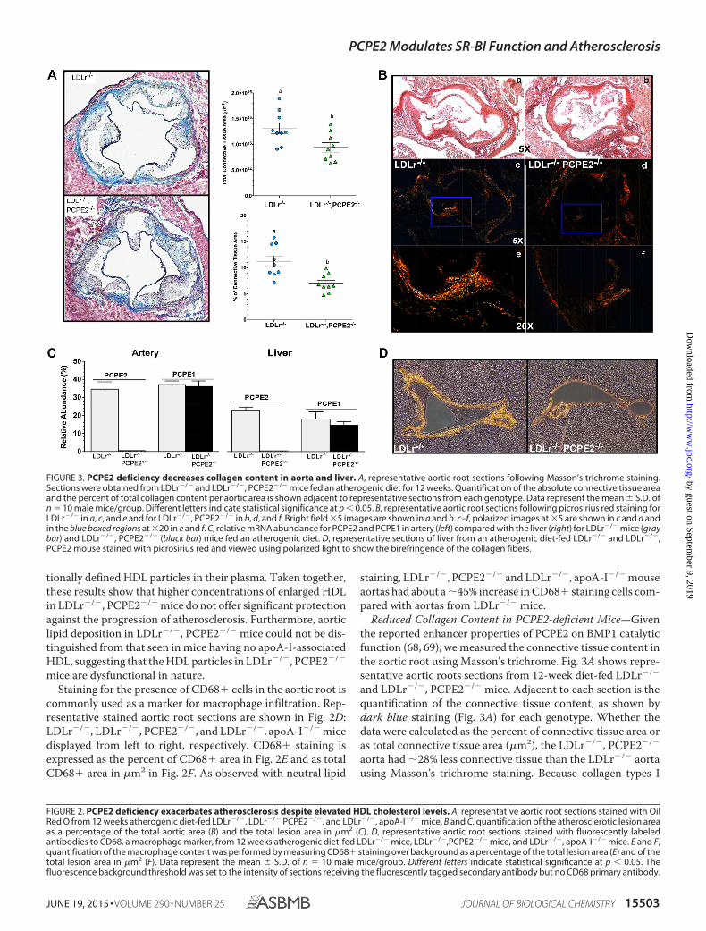
Reduced Collagen Content in PCPE2-deficient Mice—Giventhe reported enhancer properties of PCPE2 on BMP1 catalyticfunction (68, 69), we measured the connective tissue content inthe aortic root using Masson’s trichrome. Fig. 3A shows repre-sentative aortic roots sections from 12-week diet-fed LDLr�/�
and LDLr�/�, PCPE2�/� mice. Adjacent to each section is thequantification of the connective tissue content, as shown bydark blue staining (Fig. 3A) for each genotype. Whether thedata were calculated as the percent of connective tissue area oras total connective tissue area (�m2), the LDLr�/�, PCPE2�/�
aorta had �28% less connective tissue than the LDLr�/� aortausing Masson’s trichrome staining. Because collagen types I
FIGURE 2. PCPE2 deficiency exacerbates atherosclerosis despite elevated HDL cholesterol levels. A, representative aortic root sections stained with OilRed O from 12 weeks atherogenic diet-fed LDLr�/�, LDLr�/� PCPE2�/�, and LDLr�/�, apoA-I�/� mice. B and C, quantification of the atherosclerotic lesion areaas a percentage of the total aortic area (B) and the total lesion area in �m2 (C). D, representative aortic root sections stained with fluorescently labeledantibodies to CD68, a macrophage marker, from 12 weeks atherogenic diet-fed LDLr�/� mice, LDLr�/�,PCPE2�/� mice, and LDLr�/�, apoA-I�/� mice. E and F,quantification of the macrophage content was performed by measuring CD68� staining over background as a percentage of the total lesion area (E) and of thetotal lesion area in �m2 (F). Data represent the mean � S.D. of n � 10 male mice/group. Different letters indicate statistical significance at p � 0.05. Thefluorescence background threshold was set to the intensity of sections receiving the fluorescently tagged secondary antibody but no CD68 primary antibody.
FIGURE 3. PCPE2 deficiency decreases collagen content in aorta and liver. A, representative aortic root sections following Masson’s trichrome staining.Sections were obtained from LDLr�/� and LDLr�/�, PCPE2�/� mice fed an atherogenic diet for 12 weeks. Quantification of the absolute connective tissue areaand the percent of total collagen content per aortic area is shown adjacent to representative sections from each genotype. Data represent the mean � S.D. ofn � 10 male mice/group. Different letters indicate statistical significance at p � 0.05. B, representative aortic root sections following picrosirius red staining forLDLr�/� in a, c, and e and for LDLr�/�, PCPE2�/� in b, d, and f. Bright field �5 images are shown in a and b. c–f, polarized images at �5 are shown in c and d andin the blue boxed regions at �20 in e and f. C, relative mRNA abundance for PCPE2 and PCPE1 in artery (left) compared with the liver (right) for LDLr�/� mice (graybar) and LDLr�/�, PCPE2�/� (black bar) mice fed an atherogenic diet. D, representative sections of liver from an atherogenic diet-fed LDLr�/� and LDLr�/�,PCPE2 mouse stained with picrosirius red and viewed using polarized light to show the birefringence of the collagen fibers.
PCPE2 Modulates SR-BI Function and Atherosclerosis
JUNE 19, 2015 • VOLUME 290 • NUMBER 25 JOURNAL OF BIOLOGICAL CHEMISTRY 15503
by guest on September 9, 2019
http://ww
w.jbc.org/
Dow
nloaded from

and III are prevalent in mouse heart and aorta, it follows thatthe regulation of procollagen processing must play an impor-tant role in normal tissue maintenance during disease progres-sion (70). The reduction in LDLr�/�, PCPE2�/� aortic rootconnective tissue was confirmed by the picrosirius red stainingof paraffin-embedded aortic root sections show in Fig. 3B, a–f.In Fig. 3B, a, c, and e, different views are offered of a represen-tative slide taken from LDLr�/� mouse aortic root, whereas theviews in b, d, and f are from LDLr�/�, PCPE2�/� mouse aorticroot. Fig. 3B, a and b, shows stained aortic root at �5 magnifi-cation using light microscopy, and c and d panels show the sameslide but under polarizing filters. Fig. 3B, e and f, shows the sameslides as above but at a �20 magnification under polarizingfilters, as indicated by the boxed areas in c and d. The birefrin-gence of collagen fibers under polarized light shows a shift incolor between the various components, red, orange, yellow, andgreen, and represents the order of decreasing thickness of thefibers. In this case, the LDLr�/�, PCPE2�/� mouse aortic rootappears to have significantly less birefringence of all thicknessesof collagen fibers when compared with the LDLr�/� aortic root,again confirming the results of the Masson’s trichrome stain-ing. Because PCPE1 is highly similar in structure and functionto PCPE2 with respect to procollagen processing, we examinedwhether the expression of PCPE1 might be elevated in thePCPE2-deficient state. Fig. 3C compares PCPE1 and PCPE2mRNA expression in the artery and liver from the two geno-types of mice. No elevation of PCPE1 was seen in either tissue inlight of the absence of PCPE2, although these results do showsignificant expression of both PCPE1 and PCPE2 in the liver ofLDLr�/� mice.
Based on the mRNA expression data we then examined thepicrosirius staining of collagen fibers in the hepatic triad oflivers from LDLr�/�, PCPE2�/� and LDLr �/� mice (shown inFig. 3D). Here, under polarizing light the picrosirius-stainedhepatic portal triad from LDLr�/�, PCPE2�/� mice was com-pared with that from LDLr�/� mice and showed that both thethickness and amount of collagen are significantly reduced inLDLr�/�, PCPE2�/� mice.
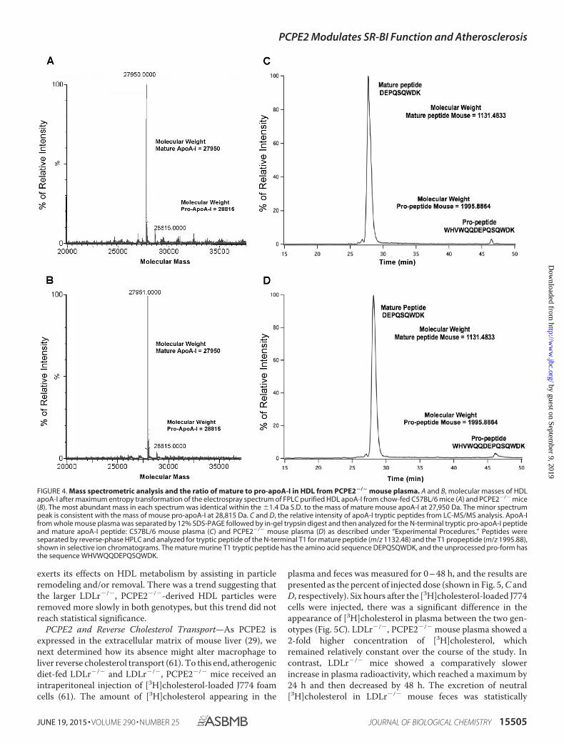
PCPE2 Was Not Essential for in Vivo Pro-apoA-I Processing—PCPE2 had been reported to enhance the BMP1-mediatedcleavage of propeptides from procollagen (27, 29, 71) duringassembly in the extracellular matrix. These studies suggest thatcleavage of apoA-I’s propeptide is the rate-limiting step to formnHDL (39, 71, 72) and the primary defect in PCPE2-deficientmice. Investigators have hypothesized that if PCPE2 and BMP1work together to remove the pro-segment of pro-apoA-I, thenHDL concentrations in mice lacking PCPE2 expression mightbe affected. Francone et al. (44), having measured HDL apoA-Ifrom PCPE2-deficient mice by isoelectric focusing and massspectrometry, report an elevation in the amount of plasma pro-apoA-I over mature apoA-I.
To investigate this effect further, we first measured the ratioof intact pro-apoA-I to mature apoA-I in plasma HDL fromPCPE2-deficient mice by mass spectrometric analyses. Fig. 4, Aand B, shows the masses of directly infused delipidated apoA-Ifrom the plasma of C57BL/6 (A) and PCPE2�/� mice (B),respectively, after transformation of the electrospray spectrum.These results demonstrate that in both samples the most abun-
dant mass was consistent with mature mouse apoA-I (27,950 �1.4 Da (73)), with only a small amount of mouse pro-apoA-I(28,815 � 1.4 Da), which includes the mass of the pro-sequenceWHVWQQ.
To quantify the amount of pro-apoA-I in plasma, the uniqueT1 peptide for the mature apoA-I (DEPQSQWDK) tryptic pep-tide and T1 for pro-apoA-I (WHVWQQDEPQSQWDK),which includes the additional six pro-amino acids, were syn-thesized and used as standards. The sequence of each syn-thetic peptide was verified by MS/MS sequencing, and thenLC/MS was used to establish the retention times and ionizabil-ity of the two different T1 peptides. The ion-current ratio ofequal molar amounts of the two synthetic T1 peptides,T1apoA-I/T1pro-apoA-I, was found to be 1.07.
Aliquots of mouse plasma were separated on 12% SDS-PAGE. The gel band corresponding to apoA-I at �28,000 Dawas excised and subjected to in-gel digestion using trypsin (51).Following digestion, peptides were exhaustively extracted fromthe gel slice, separated by reverse-phase HPLC, and identifiedby monitoring the �2 charge state of each peptide (566.75 m/zfor mature apoA-I and 998.95 m/z for pro-apoA-I). Fig. 4 showsthe summed selective ion electrospray mass chromatogramsfor apoA-I from C57BL/6 (C) and PCPE2�/� mouse plasma(D). Note that the distribution of both pro- and mature apoA-Iwas virtually identical for both genotypes. The critical factor forthis analysis is that the molar ion intensities of each peptidewere nearly identical; therefore, the measured ion intensitiescould be used to quantify the relative amount of each peptide.This interpretation of our data completely changes the inter-pretation based on a previous study (44) that reported an ele-vation in the amount of pro-apoA-I. Thus, our studies suggestthat the role of PCPE2 in HDL enlargement is not due to theconversion of pro-apoA-I to mature apoA-I. Overall, we foundthat both genotypes had very little (�4%) pro-apoA-I, and theseresults suggest that although PCPE2 may enhance pro-apoA-Iprocessing in vitro, in vivo the absence of PCPE2 does notappear to be rate-limiting.
PCPE2 Affects HDL ApoA-I Catabolism Rates—To investi-gate the possibility that increased concentrations of enlargedHDL in plasma were related to the reduced catabolism ofplasma HDL in PCPE2-deficient mice, HDL was purified fromthe plasma of diet-fed LDLr�/� and LDLr�/�, PCPE2�/� miceusing a combination of ultracentrifugation and FPLC and thenradiolabeled with 125I. Purified 125I-labeld HDL particles wereinjected into diet-fed recipient mice, and the disappearance ofradioactivity from plasma was followed over time, as shown inFig. 5, A and B. 125I-labeled HDL from LDLr�/� mice injectedinto LDLr�/� mice had a fractional catabolic rate (FCR) of0.106 � 0.010 pools/h as compared with a FCR of 0.076 � 0.011when the same HDL was injected into LDLr�/�, PCPE2�/�
mice (mean � S.D. of n � 3 mice). A similar trend was observedwhen 125I-labeled HDL from LDLr�/�, PCPE2�/� mice wasinjected into LDLr�/� versus LDLr�/�, PCPE2�/� mice,resulting in an FCR of 0.097 � 0.030 versus 0.062 � 0.008,respectively. From these data it appears the rate of turnover forHDL isolated from LDLr�/� or LDLr�/�, PCPE2�/� mice wasa function of the recipient genotype and not a result of HDLparticle origin. In conclusion, these results suggest that PCPE2
PCPE2 Modulates SR-BI Function and Atherosclerosis
15504 JOURNAL OF BIOLOGICAL CHEMISTRY VOLUME 290 • NUMBER 25 • JUNE 19, 2015
by guest on September 9, 2019
http://ww
w.jbc.org/
Dow
nloaded from
exerts its effects on HDL metabolism by assisting in particleremodeling and/or removal. There was a trend suggesting thatthe larger LDLr�/�, PCPE2�/�-derived HDL particles wereremoved more slowly in both genotypes, but this trend did notreach statistical significance.
PCPE2 and Reverse Cholesterol Transport—As PCPE2 isexpressed in the extracellular matrix of mouse liver (29), wenext determined how its absence might alter macrophage toliver reverse cholesterol transport (61). To this end, atherogenicdiet-fed LDLr�/� and LDLr�/�, PCPE2�/� mice received anintraperitoneal injection of [3H]cholesterol-loaded J774 foamcells (61). The amount of [3H]cholesterol appearing in the
plasma and feces was measured for 0 – 48 h, and the results arepresented as the percent of injected dose (shown in Fig. 5, C andD, respectively). Six hours after the [3H]cholesterol-loaded J774cells were injected, there was a significant difference in theappearance of [3H]cholesterol in plasma between the two gen-otypes (Fig. 5C). LDLr�/�, PCPE2�/� mouse plasma showed a2-fold higher concentration of [3H]cholesterol, whichremained relatively constant over the course of the study. Incontrast, LDLr�/� mice showed a comparatively slowerincrease in plasma radioactivity, which reached a maximum by24 h and then decreased by 48 h. The excretion of neutral[3H]cholesterol in LDLr�/� mouse feces was statistically
FIGURE 4. Mass spectrometric analysis and the ratio of mature to pro-apoA-I in HDL from PCPE2�/� mouse plasma. A and B, molecular masses of HDLapoA-I after maximum entropy transformation of the electrospray spectrum of FPLC purified HDL apoA-I from chow-fed C57BL/6 mice (A) and PCPE2�/� mice(B). The most abundant mass in each spectrum was identical within the �1.4 Da S.D. to the mass of mature mouse apoA-I at 27,950 Da. The minor spectrumpeak is consistent with the mass of mouse pro-apoA-I at 28,815 Da. C and D, the relative intensity of apoA-I tryptic peptides from LC-MS/MS analysis. ApoA-Ifrom whole mouse plasma was separated by 12% SDS-PAGE followed by in-gel trypsin digest and then analyzed for the N-terminal tryptic pro-apoA-I peptideand mature apoA-I peptide: C57BL/6 mouse plasma (C) and PCPE2�/� mouse plasma (D) as described under “Experimental Procedures.” Peptides wereseparated by reverse-phase HPLC and analyzed for tryptic peptide of the N-terminal T1 for mature peptide (m/z 1132.48) and the T1 propeptide (m/z 1995.88),shown in selective ion chromatograms. The mature murine T1 tryptic peptide has the amino acid sequence DEPQSQWDK, and the unprocessed pro-form hasthe sequence WHVWQQDEPQSQWDK.
PCPE2 Modulates SR-BI Function and Atherosclerosis
JUNE 19, 2015 • VOLUME 290 • NUMBER 25 JOURNAL OF BIOLOGICAL CHEMISTRY 15505
by guest on September 9, 2019
http://ww
w.jbc.org/
Dow
nloaded from

greater than that in LDLr�/�, PCPE2�/� mouse feces (Fig. 5D).Although a trend for greater 3H acidic sterol excretion was seenfor LDLr�/� compared with LDLr�/�, PCPE2�/� mice, it wasnot statistically significant. Together, the plasma and fecal anal-yses indicate that in the absence of PCPE2 less macrophagecholesterol was removed by the RCT pathway to the liver.
Although RCT consists of multiple steps, it is largely depen-dent on the expression of hepatic SR-BI (74). It is also knownthat attenuation of SR-B1 in mice results in a substantialincrease in HDL plasma levels (10). Based on these facts, wenext examined the expression of SR-BI in the liver of LDLr�/�,PCPE2�/� mice by both RT-PCR and Western blot analysis(shown in Fig. 5, E and F). Here an immunoblot probing for mouseSR-B1 using liver extracts from two different mice per genotypeshows that SR-B1 levels in LDLr�/�, PCPE2�/� mice are approx-imately twice that in LDLr�/� mouse livers, whereas GAPDH lev-els are constant. These findings were further supported by RT-PCR analysis showing a greater abundance of SR-BI mRNA in liverfrom LDLr�/�, PCPE2�/� mice compared with controls (Fig. 5F),suggesting that both SR-B1 mRNA, as well as protein, is up-regu-lated in the LDLr�/�, PCPE2�/� mouse liver. This outcome wasunexpected because our earlier results showed a reduction in RCTand HDL catabolism, consistent with a reduction in SR-BI. Theseresults led us to focus next on measuring the effect of PCPE2expression on SR-BI function.
PCPE2 Enhances SR-BI Cholesteryl Ester Uptake Function—To determine how PCPE2 expression might affect SR-BI func-tion, we first considered testing primary hepatocytes fromLDLr�/�, PCPE2�/� and control mice. However, the presence ofPCPE2 in the extracellular matrix was not compatible with the useof collagenase during hepatocyte isolation. In addition, reportscautioning against the use of primary hepatocytes when studyingSR-BI regulation (75) led us to perform a gain of function study inCHO cells (76). To test the effect of PCPE2 on SR-BI-mediatedHDL-cholesteryl ester uptake, we first determined the surfaceexpression of PCPE2 in CHO cells that were either mock-trans-fected or transfected with a plasmid expressing PCPE2 for 24 h.Fig. 6A shows a histogram of PCPE2 surface expression 24 hafter transfection measured by flow cytometry. The dotted linedepicts the expression of PCPE2 in control cells, and the shadedsolid line depicts the shift in PCPE2 surface expression inPCPE2 transiently transfected CHO cells (Fig. 6A). The 2-foldincrease (47926 � 1025 versus 21336 � 525, mean � S.D., n �3 independent experiments) indicates that PCPE2 transfectionsignificantly increased its expression on the cell surface, consis-tent with the known function of PCPE2 in the extracellularmatrix.
Next, to test the effect of PCPE2 on SR-BI function, CHOcells were mock-transfected or transfected with a constructencoding PCPE2 and then assayed for their ability to take up
FIGURE 5. Absence of liver PCPE2 affects HDL-mediated reverse cholesterol transport and SR-BI expression. A, HDL from diet-fed LDLr�/� and LDLr�/�,PCPE2�/� mice was purified and then radiolabeled with 125I as described under “Experimental Procedures.” Approximately 10 � 106 cpm of 125I-labeled HDLwas retro-orbitally injected into atherogenic diet-fed recipient mice of the indicated genotype. Blood was collected from the contralateral retro-orbital sinusat the indicated times. B, the FCR of 125I-labeled HDL were determined from plasma decay curves assuming a one-pool model as described under ”ExperimentalProcedures.” Data represent the mean � S.D. of n � 3 mice/group. C, [3H]cholesterol levels in plasma during macrophage reverse cholesterol transport studyin LDLr�/� (gray circles) and LDLr�/�, PCPE2�/� (black squares) mice fed an atherogenic diet. Mice were injected intraperitoneally with [3H]cholesterol-labeledJ774 foam cells as described under “Experimental Procedures.” D, [3H]cholesterol in feces after 0 – 48 h collection. The numerical data shown is the mean � S.D.of n � 5– 6 male mice for each genotype. The asterisk indicates statistical significance at p � 0.001. ns, indicates that the difference was not significant. E, animmunoblot analysis of liver SR-BI levels in atherogenic diet-fed mice. F, relative mRNA abundance of liver SR-BI in atherogenic diet-fed mice. Data representthe results from n � 6 –10 male mice/genotype. The liver mRNA abundance of SR-BI in LDLr�/� mouse liver was set at 100%.
PCPE2 Modulates SR-BI Function and Atherosclerosis
15506 JOURNAL OF BIOLOGICAL CHEMISTRY VOLUME 290 • NUMBER 25 • JUNE 19, 2015
by guest on September 9, 2019
http://ww
w.jbc.org/
Dow
nloaded from

HDL-cholesteryl ester. The day after mock or transient trans-fection with PCPE2, confluent cell monolayers were washedand then incubated with 10 �g of labeled 3H-COE HDL for 1.5 hin triplicate. Following incubation, the monolayer was washedextensively and the cell pellet subjected to radioactivity andprotein quantification. Fig. 6B shows the results of these studiesand suggests that the overexpression of PCPE2 enhanced theuptake of 3H-COE HDL. This increase in SR-BI functionappeared to be independent of HDL origin, because HDL par-ticles derived from either LDLr�/� or LDLr�/�, PCPE2�/�
mice showed a similar extent of 3H-COE uptake.We next examined the protein extracts from cells that were
either mock-transfected or transfected with PCPE2 usingimmunoblot analysis, as shown in Fig. 6C. In this study, the totalcellular SR-BI protein was unchanged by PCPE2 overexpres-sion. PCPE2 protein levels were increased with overexpression,as identified by the 1.2-kDa increase in the endogenous molec-ular mass due to the MycDDK tag on the exogenouslyexpressed PCPE2.
To further explore the mechanism behind PCPE2 enhance-ment of SR-BI function, mock- and PCPE2-transfected CHO
cells were stained and analyzed for SR-BI surface expressionusing flow cytometry. Fig. 6D shows a histogram of surfaceSR-BI expression in control cells, indicated by the dotted line,and in cells overexpressing PCPE2, denoted by a solid line.Interestingly, the control cells contain two populations of cells,one characterized by a low surface frequency of SR-BI/cell anda second population with a significantly higher frequency ofSR-BI/cell. Upon overexpression of PCPE2, a shift to one pop-ulation with a significantly higher frequency of SR-BI/cell isobserved (50992 � 1070 versus 36244 � 905, mean � S.D., n �3 independent experiments). These results suggest that PCPE2may act to enhance SR-BI function by promoting or maintain-ing SR-BI expression on the cell surface.
Discussion
The current studies show for the first time that PCPE2, anextracellular matrix-associated protein, confers an atheropro-tective function to HDL in vivo. The implication is that in theabsence of PCPE2, HDL particles are dysfunctional and cannotprotect against the progression of atherosclerosis. In addition,these studies show that the apparent HDL dysfunction results
FIGURE 6. SR-BI functionality is enhanced in the presence of PCPE2. A, a histogram of PCPE2 cell surface expression by flow cytometry. CHO cells were eithermock-transfected or control CHO cells (dotted line) or transiently transfected with PCPE2 (shaded solid line). The next day the cells were stained for surface PCPE2expression as described under “Experimental Procedures.” A 2-fold increase (47926 � 1025 versus 21336 � 525, mean � S.D., n � 3 independent experiments)in PCPE2 surface expression in transiently transfected cells is consistent with the PCPE2 known location in the extracellular matrix. B, the uptake of 3H-COE-labeled HDL by control and PCPE2-overexpressing CHO cells expressed as ng of HDL taken up/mg of cell protein. Confluent wells of CHO cells were eithermock-transfected or transfected with a plasmid encoding PCPE2 as described under “Experimental Procedures.” The next day the cells were washed and thenincubated for 1.5 h with 10 �g of 3H-COE-labeled HDL obtained from either diet-fed LDLr�/� or LDLr�/�, PCPE2�/� mouse plasma as described under“Experimental Procedures.” The cells were washed extensively, and then the cell pellet was digested in 0.1 N NaOH and its protein and radioactivity contentquantified as described under “Experimental Procedures.” Each value represents the mean � S.D. of n � 3 wells/condition from three independent experi-ments. C, an immunoblot of SR-BI and PCPE2 protein expression from control and PCPE2-overexpressing CHO cells. Each lane contains 25 �g of total protein.D, a histogram of SR-BI surface expression in mock (dotted line)- and PCPE2-transfected CHO (shaded solid line) cells by flow cytometry. Overexpression of PCPE2leads to a shift to a single population, with a significantly higher frequency of SR-BI per cell (50992 � 1070 versus 36244 � 905, mean � S.D., n � 3 independentexperiments).
PCPE2 Modulates SR-BI Function and Atherosclerosis
JUNE 19, 2015 • VOLUME 290 • NUMBER 25 JOURNAL OF BIOLOGICAL CHEMISTRY 15507
by guest on September 9, 2019
http://ww
w.jbc.org/
Dow
nloaded from

from reduced HDL-CE catabolism via SR-BI and is not actuallydue to changes within the particle or its components itself (Fig.5, A–D). Surprisingly, the extent of atherosclerosis in LDLr�/�,PCPE2�/� mice was indistinguishable from that in LDLr�/�,apoA-I�/� mice, which lack all apoA-I-containing HDL parti-cles (47). Although both LDLr�/�, PCPE2�/� and LDLr�/�,apoA-I�/� mice had similar extents of atherosclerosis, therewas an uncoupling of its progression as it relates to total plasmacholesterol levels (77). LDLr�/�, apoA-I�/� mice had no HDLand half the circulating LDL cholesterol levels as diet-fedLDLr�/� mice (46). In contrast, LDLr�/�, PCPE2�/� mice hadhigher HDL cholesterol concentrations and similar LDL cho-lesterol concentrations relative to LDLr�/� mice but increasedatherosclerosis. These findings suggest that the term “dysfunc-tional HDL,” e.g. HDL that is not atheroprotective, does nottechnically apply in this situation. Rather, HDL-related dys-functionality was caused by obstruction of the RCT at one ormore of its individual steps.
There are several mechanistic scenarios for the role ofPCPE2. The first is that PCPE2 confers atheroprotection toapoA-I by enhancing BMP1-mediated catalytic cleavage, con-verting pro-apoA-I to mature apoA-I, and stimulating ABCA1-mediated cholesterol flux (26, 44). However, our studies showconclusively that pro-apoA-I processing was not altered in thecase of PCPE2 deficiency (Fig. 4). The absence of PCPE2 did notaffect the circulating levels of pro-apoA-I in vivo, whichremained around 3– 6% of total apoA-I for both PCPE2-repleteand PCPE2 knock-out mice. Although PCPE2 enhanced pro-apoA-I processing in vitro and was shown to have a high affinityfor apoA-I using both surface plasmon resonance and selectiveco-immuno-precipitation (26), its absence in vivo did affect theratio of pro- to mature apoA-I. This was not unexpected,because PCPE2 is not essential for catalytic cleavage of pro-apoA-I by BMP1 (39, 42). Theoretically, PCPE2 may limitBMP1-mediated processing of pro-apoA-I under high sub-strate conditions, as reported for the processing of procollagenin the heart following chronic pressure overload (70).
Insight into the molecular mechanism of the protective func-tion of PCPE2 comes from our HDL clearance studies (Fig. 5, Aand B), where the fractional clearance of HDL was delayed inPCPE2-deficient mice, regardless of the source of the HDL. Aslower catabolic turnover resulted in increased HDL concen-tration, which ultimately caused HDL particle enlargement(Fig. 1). In mice, increased HDL size and concentration is ahallmark of SR-B1 deficiency (78). Hepatic specific SR-B1-de-ficient mice were reported to have higher plasma HDL levelsthan the controls, with greater aortic lesion formation. Becausehepatic SR-B1 mediates selective uptake of HDL-cholesterylesters and their removal from circulation for excretion into bile(74), it was concluded that in mice SR-BI is a positive regulatorof macrophage RCT. As seen in genetically modified mice,humans with genetic variants in SR-BI show a reduced capacityto efflux CE, which leads to greater HDL plasma concentrations(79 – 82) and a greater risk for CVD. Our study showed amacrophage to fecal RCT rate significantly lower in LDLr�/�,PCPE2�/� mice and similar to that reported for SR-BI knock-out mice. Interestingly, this lower rate occurred despite a 2-fold
higher level of SR-BI protein in the livers of LDLr�/�,PCPE2�/� mice (Fig. 5, C–F).
The impact of PCPE2 on liver SR-BI functionality was dem-onstrated by slower plasma HDL-CE turnover, reduced macro-phage cholesterol to fecal deliver, and increased plasma HDLsize and concentration. Despite elevations in plasma HDL, theaortic lipid and immune cell infiltration was greater in theabsence of PCPE2, highlighting the importance of aortic SR-BIin preventing the progression of atherosclerosis. These resultsprompted us to consider how PCPE2 might influence hepaticSR-BI function.
Transient transfection of CHO cells with PCPE2 gave a2-fold increase in PCPE2 surface expression (Fig. 6, A and C)and an almost 2-fold increase in the uptake of 3H-COE fromlabeled HDL (Fig. 6B). Overexpression of PCPE2 in CHO cellsdid not change total SR-BI protein levels but did increase theamount of SR-B1 protein measured on the surface of trans-fected cells (Fig. 6D). In contrast, liver SR-BI expression inPCPE2 knock-out mice was increased 2-fold. The absence of anincrease in SR-B1 protein following transfection may beascribed to the transient nature of the in vitro experiment.
Our studies also provide evidence for a profound reductionin connective tissue content in both the aortic root and thehepatic triad in mice lacking PCPE2 (Fig. 3, A–D). PCPE2enhances procollagen processing and is abundantly expressedin the heart, aorta, adipose tissue, and trabecular meshwork (27,29, 38). The expression of PCPE1, which is related to PCPE2,was not increased as a result of PCPE2 deletion (Fig. 3, C and D)and did not substitute for PCPE2 in arterial tissue. The conse-quences of these changes in connective tissue content are notknown at this time but may be related to the positioning and orstabilization of SR-B1 on the cell surface.
The mechanistic steps involved in SR-BI-mediated aorticcholesterol homeostasis have been studied for years, but ques-tions still remain. How SR-B1 mediates selective CE removalfrom HDL at the cell surface, leaving lipid-poor apoA-I aftertransferring CE into the cell, remains unresolved. Neverthelessour results strongly suggest that SR-BI-mediated 3H-COEremoval from HDL is facilitated by surface-bound PCPE2. Themechanism of the influence of PCPE2 on SR-BI uptake ofHDL-CE may involve promoting the oligomerization of SR-BIwith itself, with PCPE2, or with other proteins. Previous studieshave demonstrated that SR-BI forms dimers and tetramers onthe cell surface necessary for its function (83– 85). Oligomeri-zation has been postulated to form a hydrophobic channel thatfacilitates the selective delivery of CE from HDL to the cell.Although putative SR-BI oligomerization domains are thoughtto localize to the transmembrane segment, it has been sug-gested that the extracellular domains of SR-BI may also con-tribute to oligomerization (86). The shift in SR-BI surface stainintensity (Fig. 6D) suggests a significant change in antibodybinding properties and, therefore, a change in the conformationof amino acid region 338 – 440.
Alternative explanations for the action of PCPE2 on SR-BIinclude the postulation that PCPE2 binds and sequesters SR-B1at the cell surface. Previous studies have reported that the intra-cellular adaptor protein PDZK1 binds the C terminus of SR-BIand is required for receptor function (87, 88). In an analogous
PCPE2 Modulates SR-BI Function and Atherosclerosis
15508 JOURNAL OF BIOLOGICAL CHEMISTRY VOLUME 290 • NUMBER 25 • JUNE 19, 2015
by guest on September 9, 2019
http://ww
w.jbc.org/
Dow
nloaded from

way, PCPE2 may provide scaffolding or tethering at the cellsurface, promoting association of SR-B1 with HDL-CE.Another possibility would be that PCPE2 is involved in SR-BIendocytosis (89 –92). Yet another plausible mechanism is basedon the previously shown ability of PCPE2 to bind apoA-I (26);here PCPE2 would interact with apoA-I bound to HDL, therebyaltering the protein conformation and in turn enhancing CEremoval. As the current report provides a solid foundation forour understanding of PCPE2 function, additional studies will beneeded to examine the structures of and potential interactionsbetween PCPE2 and SR-BI, allowing insights into the mecha-nisms through which PCPE2 controls SR-BI-mediatedHDL-CE uptake.
In conclusion, our studies show for the first time that anextracellular matrix protein, PCPE2, is essential for the integ-rity of the HDL-mediated CE transport system. These studiesalso show that simply measuring plasma HDL concentration orits associated components in order to assess one’s risk of CVDcan be misleading. Rather, a combination of HDL production,catabolism, and concentration is essential for a precise analysisof HDL function. The novel finding is that PCPE2 contributesto SR-BI function by enhancing the rate of HDL-CE uptake,thereby stimulating the RCT pathway. Because the molecularevents that drive the selective uptake of CE from HDL particlesvia SR-BI have yet to be completely delineated, the questionsgenerated by this work will lead to new studies designed todefine those mechanisms. Overall, these studies will ultimatelylead to a precise understanding of how SR-BI mediates selectiveCE uptake and provide new insights into how extracellularmatrix-associated proteins, such as PCPE2, protect against thedevelopment of atherosclerosis.
Acknowledgments—The mass spectrometers used for these studieswere acquired with funds from several agencies: TSQ Discovery MaxLC-MS/MS and Advion Nanomate from North Carolina Biotechnol-ogy Grant 2007-IDG-1021, TSQ Quantum XLS GC-MS/MS fromNational Institutes of Health (NIH) Shared Instrumentation Grant1S10RR027940, and Waters Q-TOF mass spectrometer from NIHShared Instrumentation Grant 1S10RR17846. The MS analyses wereperformed in the Mass Spectrometer Facility of the ComprehensiveCancer Center of Wake Forest University School of Medicine, sup-ported in part by NIH Grant 5P30CA12197 from NCI.
References1. Kwiterovich, P. O., Jr. (1998) The antiatherogenic role of high-density
lipoprotein cholesterol. Am. J. Cardiol. 82, 13Q–21Q2. Boden, W. E. (2000) High-density lipoprotein cholesterol as an indepen-
dent risk factor in cardiovascular disease: assessing the data fromFramingham to the Veterans Affairs High-density Lipoprotein Interven-tion Trial. Am. J. Cardiol. 86, 19L–22L
3. Castelli, W. P., Doyle, J. T., Gordon, T., Hames, C. G., Hjortland, M. C.,Hulley, S. B., Kagan, A., and Zukel, W. J. (1977) HDL cholesterol and otherlipids in coronary heart disease: the cooperative lipoprotein phenotypingstudy. Circulation 55, 767–772
4. Patel, S., Drew, B. G., Nakhla, S., Duffy, S. J., Murphy, A. J., Barter, P. J., Rye,K. A., Chin-Dusting, J., Hoang, A., Sviridov, D., Celermajer, D. S., andKingwell, B. A. (2009) Reconstituted high-density lipoprotein increasesplasma high-density lipoprotein anti-inflammatory properties and choles-terol efflux capacity in patients with type 2 diabetes. J. Am. Coll. Cardiol.53, 962–971
5. Oram, J. F., and Vaughan, A. M. (2000) ABCA1-mediated transport ofcellular cholesterol and phospholipids to HDL apolipoproteins. Curr.Opin. Lipidol. 11, 253–260
6. Iatan, I., Bailey, D., Ruel, I., Hafiane, A., Campbell, S., Krimbou, L., andGenest, J. (2011) Membrane microdomains modulate oligomeric ABCA1function: impact on apoAI-mediated lipid removal and phosphatidylcho-line biosynthesis. J. Lipid Res. 52, 2043–2055
7. Denis, M., Haidar, B., Marcil, M., Bouvier, M., Krimbou, L., and Genest, J.,Jr. (2004) Molecular and cellular physiology of apolipoprotein A-I lipida-tion by the ATP-binding cassette transporter A1 (ABCA1). J. Biol. Chem.279, 7384 –7394
8. Wang, S., Gulshan, K., Brubaker, G., Hazen, S. L., and Smith, J. D. (2013)ABCA1 mediates unfolding of apolipoprotein AI N terminus on the cellsurface before lipidation and release of nascent high-density lipoprotein.Arterioscler. Thromb. Vasc. Biol. 33, 1197–1205
9. Nagata, K. O., Nakada, C., Kasai, R. S., Kusumi, A., and Ueda, K. (2013)ABCA1 dimer-monomer interconversion during HDL generation re-vealed by single-molecule imaging. Proc. Natl. Acad. Sci. U.S.A. 110,5034 –5039
10. Trigatti, B., Rayburn, H., Viñals, M., Braun, A., Miettinen, H., Penman, M.,Hertz, M., Schrenzel, M., Amigo, L., Rigotti, A., and Krieger, M. (1999)Influence of the high density lipoprotein receptor SR-BI on reproductiveand cardiovascular pathophysiology. Proc. Natl. Acad. Sci. U.S.A. 96,9322–9327
11. Cuchel, M., Lund-Katz, S., de la Llera-Moya, M., Millar, J. S., Chang, D.,Fuki, I., Rothblat, G. H., Phillips, M. C., and Rader, D. J. (2010) Pathways bywhich reconstituted high-density lipoprotein mobilizes free cholesterolfrom whole body and from macrophages. Arterioscler. Thromb. Vasc. Biol.30, 526 –532
12. Rye, K. A., and Barter, P. J. (2004) Formation and metabolism of pre�-migrating, lipid-poor apolipoprotein A-I. Arterioscler. Thromb. Vasc. Biol.24, 421– 428
13. Khera, A. V., Cuchel, M., de la Llera-Moya, M., Rodrigues, A., Burke, M. F.,Jafri, K., French, B. C., Phillips, J. A., Mucksavage, M. L., Wilensky, R. L.,Mohler, E. R., Rothblat, G. H., and Rader, D. J. (2011) Cholesterol effluxcapacity, high-density lipoprotein function, and atherosclerosis. N. Engl.J. Med. 364, 127–135
14. de la Llera-Moya, M., Drazul-Schrader, D., Asztalos, B. F., Cuchel, M.,Rader, D. J., and Rothblat, G. H. (2010) The ability to promote efflux viaABCA1 determines the capacity of serum specimens with similar high-density lipoprotein cholesterol to remove cholesterol from macrophages.Arterioscler. Thromb. Vasc. Biol. 30, 796 – 801
15. Rohatgi, A., Khera, A., Berry, J. D., Givens, E. G., Ayers, C. R., Wedin, K. E.,Neeland, I. J., Yuhanna, I. S., Rader, D. R., de Lemos, J. A., and Shaul, P. W.(2014) HDL cholesterol efflux capacity and incident cardiovascularevents. N. Engl. J. Med. 371, 2383–2393
16. Sorci-Thomas, M. G., Owen, J. S., Fulp, B., Bhat, S., Zhu, X., Parks, J. S.,Shah, D., Jerome, W. G., Gerelus, M., Zabalawi, M., and Thomas, M. J.(2012) Nascent high density lipoproteins formed by ABCA1 resemblelipid rafts and are structurally organized By three ApoA-I monomers. J.Lipid Res. 53, 1890 –1909
17. Sorci-Thomas, M. G., and Thomas, M. J. (2013) Why targeting HDLshould work as a therapeutic tool, but has not. J. Cardiovasc. Pharmacol.62, 239 –246
18. Vedhachalam, C., Duong, P. T., Nickel, M., Nguyen, D., Dhanasekaran, P.,Saito, H., Rothblat, G. H., Lund-Katz, S., and Phillips, M. C. (2007) Mech-anism of ATP-binding cassette transporter A1-mediated cellular lipid ef-flux to apolipoprotein A-I and formation of high density lipoprotein par-ticles. J. Biol. Chem. 282, 25123–25130
19. Sorci-Thomas, M. G., and Thomas, M. J. (2012) High density lipoproteinbiogenesis, cholesterol efflux, and immune cell function. Arterioscler.Thromb. Vasc. Biol. 32, 2561–2565
20. Vedhachalam, C., Ghering, A. B., Davidson, W. S., Lund-Katz, S., Rothblat,G. H., and Phillips, M. C. (2007) ABCA1-induced cell surface binding sitesfor ApoA-I. Arterioscler. Thromb. Vasc. Biol. 27, 1603–1609
21. Hozoji, M., Kimura, Y., Kioka, N., and Ueda, K. (2009) Formation of twointramolecular disulfide bonds is necessary for ApoA-I-dependent cho-lesterol efflux mediated by ABCA1. J. Biol. Chem. 284, 11293–11300
PCPE2 Modulates SR-BI Function and Atherosclerosis
JUNE 19, 2015 • VOLUME 290 • NUMBER 25 JOURNAL OF BIOLOGICAL CHEMISTRY 15509
by guest on September 9, 2019
http://ww
w.jbc.org/
Dow
nloaded from

22. Wang, N., Chen, W., Linsel-Nitschke, P., Martinez, L. O., Agerholm-Larsen, B., Silver, D. L., and Tall, A. R. (2003) A PEST sequence in ABCA1regulates degradation by calpain protease and stabilization of ABCA1 byapoA-I. J. Clin. Invest. 111, 99 –107
23. Wang, N., Silver, D. L., Costet, P., and Tall, A. R. (2000) Specific binding ofApoA-I, enhanced cholesterol efflux, and altered plasma membrane mor-phology in cells expressing ABC1. J. Biol. Chem. 275, 33053–33058
24. Curtiss, L. K., Valenta, D. T., Hime, N. J., and Rye, K. A. (2006) What is sospecial about apolipoprotein AI in reverse cholesterol transport? Arterio-scler. Thromb. Vasc. Biol. 26, 12–19
25. Chirackal Manavalan, A. P., Kober, A., Metso, J., Lang, I., Becker, T., Has-slitzer, K., Zandl, M., Fanaee-Danesh, E., Pippal, J. B., Sachdev, V., Kratky,D., Stefulj, J., Jauhiainen, M., and Panzenboeck, U. (2014) Phospholipidtransfer protein is expressed in cerebrovascular endothelial cells and in-volved in high density lipoprotein biogenesis and remodeling at the blood-brain barrier. J. Biol. Chem. 289, 4683– 4698
26. Zhu, J., Gardner, J., Pullinger, C. R., Kane, J. P., Thompson, J. F., andFrancone, O. L. (2009) Regulation of apoAI processing by procollagenC-proteinase enhancer-2 and bone morphogenetic protein-1. J. Lipid Res.50, 1330 –1339
27. Xu, H., Acott, T. S., and Wirtz, M. K. (2000) Identification and expressionof a novel type I procollagen C-proteinase enhancer protein gene from theglaucoma candidate region on 3q21-q24. Genomics 66, 264 –273
28. Steiglitz, B. M., and Greenspan, D. S. (2001) Assignment of the mousePcolce2 gene, which encodes procollagen C-proteinase enhancer protein2, to chromosome 9 and localization of PCOLCE2 to human chromosome3q23. Cytogenet. Cell Genet. 95, 244 –245
29. Steiglitz, B. M., Keene, D. R., and Greenspan, D. S. (2002) PCOLCE2 en-codes a functional procollagen C-proteinase enhancer (PCPE2) that is acollagen-binding protein differing in distribution of expression and post-translational modification from the previously described PCPE1. J. Biol.Chem. 277, 49820 – 49830
30. Vadon-Le Goff, S., Kronenberg, D., Bourhis, J. M., Bijakowski, C., Raynal,N., Ruggiero, F., Farndale, R. W., Stöcker, W., Hulmes, D. J., and Moali, C.(2011) Procollagen C-proteinase enhancer stimulates procollagen pro-cessing by binding to the C-propeptide region only. J. Biol. Chem. 286,38932–38938
31. Kronenberg, D., Vadon-Le Goff, S., Bourhis, J. M., Font, B., Eichenberger,D., Hulmes, D. J., and Moali, C. (2009) Strong cooperativity and loosegeometry between CUB domains are the basis for procollagen c-protein-ase enhancer activity. J. Biol. Chem. 284, 33437–33446
32. Blanc, G., Font, B., Eichenberger, D., Moreau, C., Ricard-Blum, S., Hulmes,D. J., and Moali, C. (2007) Insights into how CUB domains can exertspecific functions while sharing a common fold: conserved and specificfeatures of the CUB1 domain contribute to the molecular basis of procol-lagen C-proteinase enhancer-1 activity. J. Biol. Chem. 282, 16924 –16933
33. Gaboriaud, C., Gregory-Pauron, L., Teillet, F., Thielens, N. M., Bally, I.,and Arlaud, G. J. (2011) Structure and properties of the Ca(2�)-bindingCUB domain, a widespread ligand-recognition unit involved in major bi-ological functions. Biochem. J. 439, 185–193
34. Teillet, F., Gaboriaud, C., Lacroix, M., Martin, L., Arlaud, G. J., and Thiel-ens, N. M. (2008) Crystal structure of the CUB1-EGF-CUB2 domain ofhuman MASP-1/3 and identification of its interaction sites with mannan-binding lectin and ficolins. J. Biol. Chem. 283, 25715–25724
35. Brier, S., Pflieger, D., Le Mignon, M., Bally, I., Gaboriaud, C., Arlaud, G. J.,and Daniel, R. (2010) Mapping surface accessibility of the C1r/C1stetramer by chemical modification and mass spectrometry provides newinsights into assembly of the human C1 complex. J. Biol. Chem. 285,32251–32263
36. Bally, I., Rossi, V., Lunardi, T., Thielens, N. M., Gaboriaud, C., and Arlaud,G. J. (2009) Identification of the C1q-binding Sites of Human C1r and C1s:a refined three-dimensional model of the C1 complex of complement.J. Biol. Chem. 284, 19340 –19348
37. Bekhouche, M., Kronenberg, D., Vadon-Le Goff, S., Bijakowski, C., Lim,N. H., Font, B., Kessler, E., Colige, A., Nagase, H., Murphy, G., Hulmes,D. J., and Moali, C. (2010) Role of the netrin-like domain of procollagenC-proteinase enhancer-1 in the control of metalloproteinase activity.J. Biol. Chem. 285, 15950 –15959
38. Weiss, T., Ricard-Blum, S., Moschcovich, L., Wineman, E., Mesilaty, S.,and Kessler, E. (2010) Binding of procollagen C-proteinase enhancer-1(PCPE-1) to heparin/heparan sulfate: properties and role in PCPE-1 inter-action with cells. J. Biol. Chem. 285, 33867–33874
39. Chau, P., Fielding, P. E., and Fielding, C. J. (2007) Bone morphogeneticprotein-1 (BMP-1) cleaves human proapolipoprotein A1 and regulates itsactivation for lipid binding. Biochemistry 46, 8445– 8450
40. Sviridov, D. (2009) Maturation of apolipoprotein A-I: unrecognizedhealth benefit or a forgotten rudiment? J. Lipid Res. 50, 1257–1258
41. Getz, G. S., and Reardon, C. A. (2011) Cubbing in proapolipoprotein mat-uration. J. Lipid Res. 52, 1861–1864
42. Chau, P., Nakamura, Y., Fielding, C. J., and Fielding, P. E. (2006) Mecha-nism of pre�-HDL formation and activation. Biochemistry 45, 3981–3987
43. Heinzel, K., and Bleul, C. C. (2007) The Foxn1-dependent transcriptsPCOLCE2 and mPPP1R16B are not required for normal thymopoiesis.Eur. J. Immunol. 37, 2562–2571
44. Francone, O. L., Ishida, B. Y., de la Llera-Moya, M., Royer, L., Happe, C.,Zhu, J., Chalkey, R. J., Schaefer, P., Cox, C., Burlingame, A., Kane, J. P., andRothblat, G. H. (2011) Disruption of the murine procollagen C-proteinaseenhancer 2 gene causes accumulation of pro-apoA-I and increased HDLlevels. J. Lipid Res. 52, 1974 –1983
45. Miles, R. R., Perry, W., Haas, J. V., Mosior, M. K., N’Cho, M., Wang, J. W.,Yu, P., Calley, J., Yue, Y., Carter, Q., Han, B., Foxworthy, P., Kowala, M. C.,Ryan, T. P., Solenberg, P. J., and Michael, L. F. (2013) Genome-wide screenfor modulation of hepatic apolipoprotein A-I (ApoA-I) secretion. J. Biol.Chem. 288, 6386 – 6396
46. Zabalawi, M., Bhat, S., Loughlin, T., Thomas, M. J., Alexander, E., Cline,M., Bullock, B., Willingham, M., and Sorci-Thomas, M. G. (2003) Induc-tion of fatal inflammation in LDL receptor and ApoA-I double-knockoutmice fed dietary fat and cholesterol. Am. J. Pathol. 163, 1201–1213
47. Wilhelm, A. J., Zabalawi, M., Grayson, J. M., Weant, A. E., Major, A. S.,Owen, J., Bharadwaj, M., Walzem, R., Chan, L., Oka, K., Thomas, M. J., andSorci-Thomas, M. G. (2009) Apolipoprotein A-I and its role in lympho-cyte cholesterol homeostasis and autoimmunity. Arterioscler. Thromb.Vasc. Biol. 29, 843– 849
48. Potteaux, S., Gautier, E. L., Hutchison, S. B., van Rooijen, N., Rader, D. J.,Thomas, M. J., Sorci-Thomas, M. G., and Randolph, G. J. (2011) Sup-pressed monocyte recruitment drives macrophage removal from athero-sclerotic plaques of Apoe�/� mice during disease regression. J. Clin. In-vest. 121, 2025–2036
49. Sorci-Thomas, M. G., Zabalawi, M., Bharadwaj, M. S., Wilhelm, A. J.,Owen, J. S., Asztalos, B. F., Bhat, S., and Thomas, M. J. (2012) Dysfunc-tional HDL containing L159R ApoA-I leads to exacerbation of atheroscle-rosis in hyperlipidemic mice. Biochim. Biophys. Acta 1821, 502–512
50. Bhat, S., Sorci-Thomas, M. G., Alexander, E. T., Samuel, M. P., andThomas, M. J. (2005) Intermolecular contact between globular N-termi-nal fold and C-terminal domain of ApoA-I stabilizes its lipid-bound con-formation: studies employing chemical cross-linking and mass spectrom-etry. J. Biol. Chem. 280, 33015–33025
51. Bhat, S., Sorci-Thomas, M. G., Tuladhar, R., Samuel, M. P., and Thomas,M. J. (2007) Conformational adaptation of apolipoprotein A-I to discretelysized phospholipid complexes. Biochemistry 46, 7811–7821
52. Bhat, S., Sorci-Thomas, M. G., Calabresi, L., Samuel, M. P., and Thomas,M. J. (2010) Conformation of dimeric apolipoprotein A-I Milano on re-combinant lipoprotein particles. Biochemistry 49, 5213–5224
53. Sorci-Thomas, M. G., Parks, J. S., Kearns, M. W., Pate, G. N., Zhang, C.,and Thomas, M. J. (1996) High level secretion of wild-type and mutantforms of human proapoA-I using baculovirus-mediated Sf-9 cell expres-sion. J. Lipid Res. 37, 673– 683
54. Kaluzny, M. A., Duncan, L. A., Merritt, M. V., and Epps, D. E. (1985) Rapidseparation of lipid classes in high yield and purity using bonded phasecolumns. J. Lipid Res. 26, 135–140
55. Schwenke, D. C., Rudel, L. L., Sorci-Thomas, M. G., and Thomas, M. J.(2002) �-Tocopherol protects against diet induced atherosclerosis in NewZealand white rabbits. J. Lipid Res. 43, 1927–1938
56. Berquin, I. M., Min, Y., Wu, R., Wu, J., Perry, D., Cline, J. M., Thomas,M. J., Thornburg, T., Kulik, G., Smith, A., Edwards, I. J., D’Agostino, R.,Zhang, H., Wu, H., Kang, J. X., and Chen, Y. Q. (2007) Modulation of
PCPE2 Modulates SR-BI Function and Atherosclerosis
15510 JOURNAL OF BIOLOGICAL CHEMISTRY VOLUME 290 • NUMBER 25 • JUNE 19, 2015
by guest on September 9, 2019
http://ww
w.jbc.org/
Dow
nloaded from
prostate cancer genetic risk by omega-3 and omega-6 fatty acids.J. Clin. Invest. 117, 1866 –1875
57. McFarlane, A. (1958) Efficient trace-labelling of proteins with iodine. Na-ture 182, 53
58. Sorci-Thomas, M. G., Thomas, M., Curtiss, L., and Landrum, M. (2000)Single repeat deletion in ApoA-I blocks cholesterol esterification and re-sults in rapid catabolism of delta6 and wild-type ApoA-I in transgenicmice. J. Biol. Chem. 275, 12156 –12163
59. Huggins, K. W., Colvin, P. L., Burleson, E. R., Kelley, K., Sawyer, J. K.,Barrett, P. H., Rudel, L. L., and Parks, J. S. (2001) Dietary n-3 polyunsatu-rated fat increases the fractional catabolic rate of medium-sized HDLparticles in African green monkeys. J. Lipid Res. 42, 1457–1466
60. Junqueira, L. C., Bignolas, G., and Brentani, R. R. (1979) Picrosirius stain-ing plus polarization microscopy, a specific method for collagen detectionin tissue sections. Histochem. J. 11, 447– 455
61. Zhang, Y., Zanotti, I., Reilly, M. P., Glick, J. M., Rothblat, G. H., and Rader,D. J. (2003) Overexpression of apolipoprotein A-I promotes reverse trans-port of cholesterol from macrophages to feces in vivo. Circulation 108,661– 663
62. Alexander, E. T., Vedhachalam, C., Sankaranarayanan, S., de la Llera-Moya, M., Rothblat, G. H., Rader, D. J., and Phillips, M. C. (2014) Influenceof apolipoprotein A-I domain structure on macrophage reverse choles-terol transport in mice. Arterioscler. Thromb. Vasc. Biol. 31, 320 –327
63. Batta, A. K., Salen, G., Rapole, K. R., Batta, M., Batta, P., Alberts, D., andEarnest, D. (1999) Highly simplified method for gas-liquid chromato-graphic quantitation of bile acids and sterols in human stool. J. Lipid Res.40, 1148 –1154
64. Connelly, M. A., Klein, S. M., Azhar, S., Abumrad, N. A., and Williams,D. L. (1999) Comparison of class B scavenger receptors, CD36 and scav-enger receptor BI (SR-BI), shows that both receptors mediate high densitylipoprotein-cholesteryl ester selective uptake but SR-BI exhibits a uniqueenhancement of cholesteryl ester uptake. J. Biol. Chem. 274, 41– 47
65. Lowry, O. H., Rosebrough, N. J., Farr, A. L., and Randall, R. J. (1951)Protein measurement with the Folin phenol reagent. J. Biol. Chem. 193,265–275
66. Wilhelm, A. J., Zabalawi, M., Owen, J. S., Shah, D., Grayson, J. M., Major,A. S., Bhat, S., Gibbs, D. P., Jr., Thomas, M. J., and Sorci-Thomas, M. G.(2010) Apolipoprotein A-I modulates regulatory T cells in autoimmuneLDLr�/�, ApoA-I�/� mice. J. Biol. Chem. 285, 36158 –36169
67. Zabalawi, M., Bharadwaj, M., Horton, H., Cline, M., Willingham, M.,Thomas, M. J., and Sorci-Thomas, M. G. (2007) Inflammation and skincholesterol in LDLr�/�, ApoA-I�/� mice: link between cholesterolhomeostasis and self-tolerance? J. Lipid Res. 48, 52– 65
68. Goldsmith, E. C., Bradshaw, A. D., and Spinale, F. G. (2013) Cellular mech-anisms of tissue fibrosis. 2. Contributory pathways leading to myocardialfibrosis: moving beyond collagen expression. Am. J. Physiol. Cell Physiol.304, C393–C402
69. Canty, E. G., and Kadler, K. E. (2005) Procollagen trafficking, processingand fibrillogenesis. J. Cell Sci. 118, 1341–1353
70. Baicu, C. F., Zhang, Y., Van Laer, A. O., Renaud, L., Zile, M. R., and Brad-shaw, A. D. (2012) Effects of the absence of procollagen C-endopeptidaseenhancer-2 on myocardial collagen accumulation in chronic pressureoverload. Am. J. Physiol. Heart Circ. Physiol. 303, H234 –H240
71. Moali, C., Font, B., Ruggiero, F., Eichenberger, D., Rousselle, P., François,V., Oldberg, A., Bruckner-Tuderman, L., and Hulmes, D. J. (2005) Sub-strate-specific modulation of a multisubstrate proteinase: C-terminal pro-cessing of fibrillar procollagens is the only BMP-1-dependent activity to beenhanced by PCPE-1. J. Biol. Chem. 280, 24188 –24194
72. Ricard-Blum, S., Bernocco, S., Font, B., Moali, C., Eichenberger, D., Far-janel, J., Burchardt, E. R., van der Rest, M., Kessler, E., and Hulmes, D. J.(2002) Interaction properties of the procollagen C-proteinase enhancerprotein shed light on the mechanism of stimulation of BMP-1. J. Biol.Chem. 277, 33864 –33869
73. Puppione, D. L., Yam, L. M., Bassilian, S., Souda, P., Castellani, L. W.,Schumaker, V. N., and Whitelegge, J. P. (2006) Mass spectral analysis ofthe apolipoproteins on mouse high density lipoproteins: detection of post-
translational modifications. Biochim. Biophys. Acta 1764, 1363–137174. Zhang, Y., Da Silva, J. R., Reilly, M., Billheimer, J. T., Rothblat, G. H., and
Rader, D. J. (2005) Hepatic expression of scavenger receptor class B type I(SR-BI) is a positive regulator of macrophage reverse cholesterol transportin vivo. J. Clin. Invest. 115, 2870 –2874
75. Tsukamoto, K., Buck, L., Inman, W., Griffith, L., Kocher, O., and Krieger,M. (2013) Challenges in using cultured primary rodent hepatocytes or celllines to study hepatic HDL receptor SR-BI regulation by its cytoplasmicadaptor PDZK1. PLoS One 8, e69725
76. Harder, C. J., Vassiliou, G., McBride, H. M., and McPherson, R. (2006)Hepatic SR-BI-mediated cholesteryl ester selective uptake occurs withunaltered efficiency in the absence of cellular energy. J. Lipid Res. 47,492–503
77. Tavori, H., Su, Y. R., Yancey, P. G., Giunzioni, I., Wilhelm, A. J., Blakemore,J. L., Zabalawi, M., Linton, M. F., Sorci-Thomas, M. G., and Fazio, S. (2015)Macrophage apoAI protects against dyslipidemia-induced dermatitis andatherosclerosis without affecting HDL. J. Lipid Res. 56, 635– 643
78. Krieger, M., and Kozarsky, K. (1999) Influence of the HDL receptor SR-BIon atherosclerosis. Curr. Opin. Lipidol. 10, 491– 497
79. Vergeer, M., Holleboom, A. G., Kastelein, J. J., and Kuivenhoven, J. A.(2010) The HDL hypothesis: does high-density lipoprotein protect fromatherosclerosis? J. Lipid Res. 51, 2058 –2073
80. West, M., Greason, E., Kolmakova, A., Jahangiri, A., Asztalos, B., Pollin,T. I., and Rodriguez, A. (2009) Scavenger receptor class B type I protein asan independent predictor of high-density lipoprotein cholesterol levels insubjects with hyperalphalipoproteinemia. J. Clin. Endocrinol. Metab. 94,1451–1457
81. Chadwick, A. C., and Sahoo, D. (2012) Functional characterization of new-ly-discovered mutations in human SR-BI. PLoS One 7, e45660
82. Chadwick, A. C., and Sahoo, D. (2013) Functional genomics of the humanhigh-density lipoprotein receptor scavenger receptor BI: an old dog withnew tricks. Curr. Opin. Endocrinol. Diabetes Obes. 20, 124 –131
83. Reaven, E., Cortez, Y., Leers-Sucheta, S., Nomoto, A., and Azhar, S. (2004)Dimerization of the scavenger receptor class B type I: formation, function,and localization in diverse cells and tissues. J. Lipid Res. 45, 513–528
84. Sahoo, D., Darlington, Y. F., Pop, D., Williams, D. L., and Connelly, M. A.(2007) Scavenger receptor class B type I (SR-BI) assembles into detergent-sensitive dimers and tetramers. Biochim. Biophys. Acta 1771, 807– 817
85. Sahoo, D., Peng, Y., Smith, J. R., Darlington, Y. F., and Connelly, M. A.(2007) Scavenger receptor class B, type I (SR-BI) homo-dimerizes via itsC-terminal region: fluorescence resonance energy transfer analysis.Biochim. Biophys. Acta 1771, 818 – 829
86. Kartz, G. A., Holme, R. L., Nicholson, K., and Sahoo, D. (2014) SR-BI/CD36 chimeric receptors define extracellular subdomains of SR-BI criticalfor cholesterol transport. Biochemistry 53, 6173– 6182
87. Kocher, O., and Krieger, M. (2009) Role of the adaptor protein PDZK1 incontrolling the HDL receptor SR-BI. Curr. Opin. Lipidol. 20, 236 –241
88. Saddar, S., Mineo, C., and Shaul, P. W. (2010) Signaling by the high-affinityHDL receptor scavenger receptor B type I. Arterioscler. Thromb. Vasc.Biol. 30, 144 –150
89. Pittman, R. C., Knecht, T. P., Rosenbaum, M. S., and Taylor, C. A., Jr.(1987) A nonendocytotic mechanism for the selective uptake of highdensity lipoprotein-associated cholesterol esters. J. Biol. Chem. 262,2443–2450
90. Silver, D. L., Wang, N., Xiao, X., and Tall, A. R. (2001) High density lipo-protein (HDL) particle uptake mediated by scavenger receptor class B type1 results in selective sorting of HDL cholesterol from protein and polar-ized cholesterol secretion. J. Biol. Chem. 276, 25287–25293
91. Pagler, T. A., Rhode, S., Neuhofer, A., Laggner, H., Strobl, W., Hintern-dorfer, C., Volf, I., Pavelka, M., Eckhardt, E. R., van der Westhuyzen, D. R.,Schütz, G. J., and Stangl, H. (2006) SR-BI-mediated high density lipopro-tein (HDL) endocytosis leads to HDL resecretion facilitating cholesterolefflux. J. Biol. Chem. 281, 11193–11204
92. Röhrl, C., and Stangl, H. (2013) HDL endocytosis and resecretion.Biochim. Biophys. Acta 1831, 1626 –1633
PCPE2 Modulates SR-BI Function and Atherosclerosis
JUNE 19, 2015 • VOLUME 290 • NUMBER 25 JOURNAL OF BIOLOGICAL CHEMISTRY 15511
by guest on September 9, 2019
http://ww
w.jbc.org/
Dow
nloaded from
Sahoo, Michael J. Thomas and Mary G. Sorci-ThomasXuewei Zhu, Erica W. Lyons, Nebil Nuradin, Omar L. Francone, Xiang-An Li, Daisy
Ricquita D. Pollard, Christopher N. Blesso, Manal Zabalawi, Brian Fulp, Mark Gerelus,(SR-BI)-mediated High-density Lipoprotein (HDL)-Cholesteryl Ester Uptake
Atherosclerosis in Mice by Enhancing Scavenger Receptor Class B1 Procollagen C-endopeptidase Enhancer Protein 2 (PCPE2) Reduces
doi: 10.1074/jbc.M115.646240 originally published online May 6, 20152015, 290:15496-15511.J. Biol. Chem.
10.1074/jbc.M115.646240Access the most updated version of this article at doi:
Alerts:
When a correction for this article is posted•
When this article is cited•
to choose from all of JBC's e-mail alertsClick here
http://www.jbc.org/content/290/25/15496.full.html#ref-list-1
This article cites 92 references, 56 of which can be accessed free at
by guest on September 9, 2019
http://ww
w.jbc.org/
Dow
nloaded from