Embed Size (px)
Citation preview
Processing of PLP in a Model of Pelizaeus-MerzbacherDisease/SPG2 due to the rumpshaker MutationMARK McLAUGHLIN, JENNIFER A. BARRIE, SAADIA KARIM, PAUL MONTAGUE, JULIA M. EDGAR,DOUGLAS KIRKHAM, CHRISTINE E. THOMSON, AND IAN R GRIFFITHS*
Applied Neurobiology Group, Institute of Comparative Medicine, University of Glasgow, Bearsden Glasgow G61 1QH, Scotland
KEY WORDSoligodendrocyte; myelin; misfolded protein; dysmyelination;spastic paraplegia type 2
ABSTRACTThe rumpshaker mutation of the X-linked myelin proteo-lipid protein (PLP1) gene causes spastic paraplegia type 2or a mild form of Pelizaeus-Merzbacher disease in man.The identical mutation occurs spontaneously in mice. Bothhuman and murine diseases are associated with dysmyeli-nation. Using the mouse model, we show that the lowsteady state levels of PLP result from accelerated proteaso-mal degradation rather than decreased synthesis. The T1/2
for degradation of rumpshaker PLP is 11 h compared with23 h for wild type. A minority of newly synthesized PLP isincorporated into myelin in the correct orientation but at areduced rate compared with wild type. However, inhibitionof proteasomal degradation does not increase the level ofPLP incorporated into myelin. As Plp null mice do not havea similar myelin deficiency, it is unlikely that the reducedPLP levels are the main cause of the dysmyelination.Rumpshaker oligodendrocytes also have a reduced level ofother myelin proteins, such as MBP, although the mecha-nisms are not yet defined but are likely to operate at atranslational or post-translational level. VVC 2006 Wiley-Liss, Inc.
INTRODUCTION
Proteolipid protein (PLP) and the smaller DM20 iso-form constitute the major proteins of CNS myelin. Thefunctions of PLP/DM20 have been partly clarified throughthe study of PLP-deficient mice and humans. Despite itsabundance, the protein is not essential for myelin forma-tion but is required for the formation of a normal intra-period line (IPL) and maintenance of axonal integrity(Garbern et al., 2002; Griffiths et al., 1998; Klugmannet al., 1997). Both PLP and DM20 seem necessary forcompletely normal function, although each is capable,individually, of insertion into myelin (McLaughlin et al.,2002).
Mutations of the X-linked myelin PLP1/Plp1 gene causePelizaeus-Merzbacher disease (PMD) and spastic paraple-gia type 2 (SPG2) in man and dysmyelinating disorders ina range of animal species (Hudson, 2003; Inoue, 2005). Inman, gene duplication is the most frequent cause of PMD,with a range of missense mutations accounting for themajority of the remaining cases. Typically, missense mu-tations are associated with dysmyelination characterizedby hypomyelination, increased apoptosis of oligodendro-
cytes, and gliosis (Al-Saktawi et al., 2003; Skoff, 1995),although the severity of each parameter varies betweendifferent mutations. The lack of a similar phenotype inPLP-deficient mice and humans (Garbern et al., 2002;Klugmann et al., 1997) suggests that missense mutationscause disease through a ‘‘gain of function’’ effect.
The rumpshaker mutation of the PLP1/Plp1 gene(Plpjp-rsh) causes disease in both man and mouse and pro-vides an excellent model of mild forms of PMD and ofSPG2. The distinction between mild PMD and SPG2 isblurred depending on the clinical presentation, whichmay change over time and also on individual clinicians;both syndromes have been ascribed to the rumpshakermutation, with spastic paraparesis as a major sign (John-ston and McKusick, 1962; Kobayashi et al., 1994; Naiduet al., 1997). The mutation, an amino acid substitution(Ile186Thr), was described first in mice of the C3H.HeHstrain, in which it causes a relatively benign phenotypewith normal longevity and fertility and is characterizedby mild to moderate dysmyelination with oligodendrocytenumbers being maintained (Al-Saktawi et al., 2003; Grif-fiths et al., 1990). Many larger axons are either naked orsurrounded by thin sheaths, whereas the smaller axonsobtain sheaths of appropriate thickness by 12 to 18months (Edgar et al., 2004). An outstanding question iswhy the surviving oligodendrocytes fail to assemble nor-mal amounts of myelin, particularly around larger axons.
Misfolding of proteins is a common mechanism bywhich mutations cause disease (Sanders and Myers,2004), and cells have evolved the unfolded protein re-sponse (UPR) and endoplasmic reticulum-associated deg-radation (ERAD) to deal with this event (Ellgaard andHelenius, 2003; Jarosch et al., 2003; Rutkowski and Kauf-man, 2004). Evidence for a UPR is present in rumpshakermice (Southwood and Gow, 2001; Southwood et al., 2002),suggesting that PLP/DM20 is misfolded. As oligoden-drocytes generate massive amounts of membrane duringmyelination and PLP accounts for �50% of proteinin myelin, the rumpshaker mutation provides an idealmodel to dissect how the cell deals with a misfolded, non-
Grant sponsor: The Wellcome Trust; Grant sponsor: Birth Defects Foundation.
*Correspondence to: I.R. Griffiths, Applied Neurobiology Group, Division of CellSciences, University of Glasgow, Bearsden, Glasgow G61 1QH, Scotland.E-mail: [email protected].
Received 26 October 2005; Accepted 10 January 2006
DOI 10.1002/glia.20325
Published online 27 February 2006 in Wiley InterScience (www.interscience.wiley.com).
GLIA 53:715–722 (2006)
VVC 2005 Wiley-Liss, Inc.
glycosylated, polytopic membrane protein. Very little isknown about the dynamics of PLP/DM20 encoded by mis-sense mutations or how this may influence the processingof other myelin components. Evidence, largely from trans-fected heterologous cells, has implicated impaired proteintrafficking in that the products are retained within therough endoplasmic reticulum (RER) (Gow and Lazzarini,1996). Variations in trafficking between different PLP1/Plp1 mutations have been demonstrated in such systemsand, in general, suggest that the impairment correlateswith phenotypic severity of the natural disease (Thomsonet al., 1997). The purpose of this study was to establishhow the oligodendrocyte handles misfolded rumpshakerPLP.
We find that rumpshaker oligodendrocytes synthesizePLP/DM20 as efficiently as their wild type counterpartsbut, once formed, much of the protein is rapidly degraded,most probably by the proteasome, with a minority insert-ing correctly into the myelin membrane.
MATERIALS AND METHODSAnimals
The rumpshaker (Plpjp-rsh) mutation arose originally inC3H.HeH mice and was maintained on a hybrid C3H/101background. Mutant mice were genotyped by PCR ampli-fication of genomic DNA and restriction digest of thenovel AccI site (Schneider et al., 1992). All animal studieswere approved by the Ethical Committee of the Univer-sity of Glasgow and licensed by the UK Home Office.
Quantitative Real Time PCR (qRT-PCR)
Total RNA was extracted from oligodendrocyte culturesusing RNAzol Bee reagent (Tel-Test Inc, Friendswood,TX) following the manufacturer’s instructions. RNA wasextracted from wild type and rumpshaker cultures andreconstituted in DEPC water. RNA quality and integritywere determined using spectrophotometry and gel elec-trophoresis. The ABI prism 7500 sequence detection sys-tem (Applied Biosystems, Foster City, CA) using Taqmantechnologies (PE Biosystems, Foster City, CA, USA) wasemployed following the manufacturer’s instructions. Realtime PCR was preformed using the Platinum Quantita-tive RT-PCR Thermoscript One-Step System (Invitrogen,Carlsbad, CA). Full details of the primers, probes, andmethod are in Supplementary Information. The relativeamount of rumpshaker Plp and Plp/Dm20 mRNA wasexpressed as a percentage of wild type.
Antibodies and Immunostaining
Antibodies
PLP/DM20 was detected with a rabbit polyclonal antibodyrecognizing the common C-terminal (gift from Prof. N.P.Groome) or a rat monoclonal (AA3, gift from Dr. S. Pfeif-
fer). MBP was detected with a mouse monoclonal (clone12, gift from Prof. N.P. Groome). CNP was detected with amouse monoclonal (Chemicon Europe, Chandlers Ford,UK). Monoclonal and polyclonal antibodies to MAG wereobtained from Chemicon (clone 513) and as gifts fromProf. N.P. Groome and Dr. R.H. Quarles. Aspartoacylase(ASPA) was detected with a rabbit polyclonal (gift fromDr. J. Garbern). Anti ubiquitin monoclonal was obtainedfrom Santa Cruz (sc 8017, Autogen Bioclear UK Ltd,Calne, UK) and a rabbit polyclonal against p27kip1 fromUpstate (Lake Placid, NY). A rabbit polyclonal againstGRP78 was obtained from Stressgen (Bioquote Ltd., York,UK) and a mouse monoclonal GM130 antibody for Golgimembranes from Chemicon.
Immunostaining
Immunostaining of cells or tissue sections by indirect im-munofluorescence was performed as described previously(Al-Saktawi et al., 2003; Edgar et al., 2002; Thomsonet al., 1999). Fluorescent images were obtained using anOlympus IX70 microscope and a Photonic Sciences ColourCoolView camera. Contrast and brightness were adjustedusing Adobe Photoshop 6.
Primary Oligodendrocyte Cultures
Oligodendrocytes were isolated and cultured from thespinal cords of P5 mice as previously described (Montagueet al., 1998). Females homozygous for the rumpshakermutation were mated with hemizygous males to generatelitters in which all pups were affected. Cells from spinalcords were pooled and plated onto two 30mm poly-L-ly-sine coated plates per mouse. The resultant cultures con-tained a mixed population—70–80% O41 cells togetherwith astrocytes, occasional neurons, and other cells.
At 7 days in culture, the medium was removed, cellswashed twice with Hank’s buffered salt (HBS), then in-cubated for 30 min in HBS. The medium was replacedwith 0.5 ml HBS containing [35S]-Pro-Mix (Amersham) at100 lCi/ml. For analysis of protein synthesis, dishes wereremoved at the indicated times, rinsed twice in chilledPBS, and lysed with Buffer A (10mM Tris pH 7.4, 150 mMNaCl, 1% triton X-100, and inhibitors of protease andphosphatase activity). For pulse chase experiments, cellswere labeled for 1.5 h and chased by replacing the HBSwith SATO medium supplemented with 2.5mM cysteine/2.5mM methionine. At the appropriate time point, cellswere washed twice with chilled PBS, then lysed with75 ll buffer A. The lysates were rotated at 4�C for 30 minand cell debris pelleted by centrifugation at 5,000 rpm for5 min. Proteins were quantified using the BCA assay sys-tem (Perbio Science UK Ltd, Tattenhall, UK).
Brain Slices, Myelin Extraction, andFractionation Procedures
A brain slice system was used to investigate the incor-poration of newly synthesized, radiolabeled PLP/DM20
716 McLAUGHLIN ET AL.
GLIA DOI 10.1002/glia
into myelin. Full details of the methods are presented inSupplementary Information.
Western Blotting and Immunoprecipitation
Western blotting and immunoprecipitation analysis wereconducted as described in detail elsewhere (McLaughlinet al., 2002; Yool et al., 2001), except that in the presentstudy the immunoprecipitation reactions were conductedwith buffer A, which contains 1% triton X-100 as themajor solubilization detergent. Following SDS PAGEseparation, the proteins were transferred to PVDF andthe membrane exposed to a low energy, high sensitivityphosphorimage screen (Amersham) for a period of 1–2weeks and the image captured on a Storm phosphorimagesystem. Quantitation was performed using ImageQuantdensitometry software. The PVDF membranes were thenprocessed for western blotting.
Statistical Analysis
Statistical significance of differences between valueswas assessed by ANOVA followed by Bonferonni’s posthoc comparison test or by Student’s t-test, as appropriate,with significance P < 0.05. Analyses and curve fittingwere performed using Graphpad Prism4 software (Graph-Pad Software Inc., San Diego, CA). In the bar charts,significance is indicated as <0.05 (*), <0.01 (**), and<0.001 (***).
RESULTSMyelin Protein Levels Are Disproportionately
Low Compared with mRNA
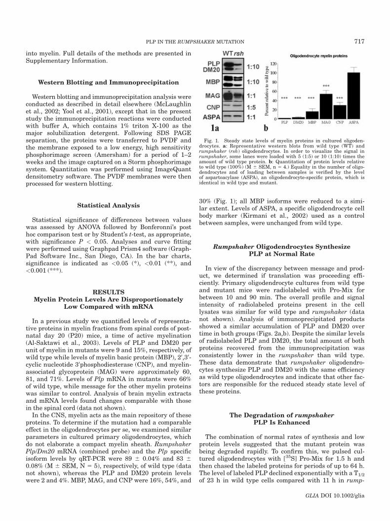
In a previous study we quantified levels of representa-tive proteins in myelin fractions from spinal cords of post-natal day 20 (P20) mice, a time of active myelination(Al-Saktawi et al., 2003). Levels of PLP and DM20 perunit of myelin in mutants were 9 and 15%, respectively, ofwild type while levels of myelin basic protein (MBP), 20,30-cyclic nucleotide 30phosphodiesterase (CNP), and myelin-associated glycoprotein (MAG) were approximately 60,81, and 71%. Levels of Plp mRNA in mutants were 66%of wild type, while message for the other myelin proteinswas similar to control. Analysis of brain myelin extractsand mRNA levels found changes comparable with thosein the spinal cord (data not shown).
In the CNS, myelin acts as the main repository of theseproteins. To determine if the mutation had a comparableeffect in the oligodendrocytes per se, we examined similarparameters in cultured primary oligodendrocytes, whichdo not elaborate a compact myelin sheath. RumpshakerPlp/Dm20 mRNA (combined probe) and the Plp specificisoform levels by qRT-PCR were 89 6 0.04% and 83 60.08% (M 6 SEM, N 5 5), respectively, of wild type (datanot shown), whereas the PLP and DM20 protein levelswere 2 and 4%. MBP, MAG, and CNP were 16%, 54%, and
30% (Fig. 1); all MBP isoforms were reduced to a simi-lar extent. Levels of ASPA, a specific oligodendrocyte cellbody marker (Kirmani et al., 2002) used as a controlbetween samples, were unchanged from wild type.
Rumpshaker Oligodendrocytes SynthesizePLP at Normal Rate
In view of the discrepancy between message and prod-uct, we determined if translation was proceeding effi-ciently. Primary oligodendrocyte cultures from wild typeand mutant mice were radiolabeled with Pro-Mix forbetween 10 and 90 min. The overall profile and signalintensity of radiolabeled proteins present in the celllysates was similar for wild type and rumpshaker (datanot shown). Analysis of immunoprecipitated productsshowed a similar accumulation of PLP and DM20 overtime in both groups (Figs. 2a,b). Despite the similar levelsof radiolabeled PLP and DM20, the total amount of bothproteins recovered from the immunoprecipitation wasconsistently lower in the rumpshaker than wild type.These data demonstrate that rumpshaker oligodendro-cytes synthesize PLP and DM20 with the same efficiencyas wild type oligodendrocytes and indicate that other fac-tors are responsible for the reduced steady state level ofthese proteins.
The Degradation of rumpshakerPLP Is Enhanced
The combination of normal rates of synthesis and lowprotein levels suggested that the mutant protein wasbeing degraded rapidly. To confirm this, we pulsed cul-tured oligodendrocytes with [35S] Pro-Mix for 1.5 h andthen chased the labeled proteins for periods of up to 64 h.The level of labeled PLP declined exponentially with a T1/2
of 23 h in wild type cells compared with 11 h in rump-
Fig. 1. Steady state levels of myelin proteins in cultured oligoden-drocytes. a: Representative western blots from wild type (WT) andrumpshaker (rsh) oligodendrocytes. In order to visualize the signal inrumpshaker, some lanes were loaded with 5 (1:5) or 10 (1:10) times theamount of wild type protein. b: Quantitation of protein levels relativeto wild type (100%) (M 6 SEM, n 5 4.) Equality in the number of oligo-dendrocytes and of loading between samples is verified by the levelof aspartoacylase (ASPA), an oligodendrocyte-specific protein, which isidentical in wild type and mutant.
717PLP IN THE RUMPSHAKER MUTATION
GLIA DOI 10.1002/glia
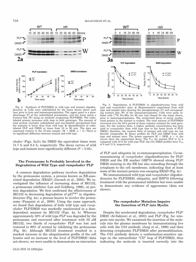
shaker (Figs. 3a,b); for DM20 the equivalent times were11.7 h and 8.4 h, respectively. The decay curves of wildtype and mutant were significantly different (P < 0.05).
The Proteasome Is Probably Involved in theDegradation of Wild Type and rumpshaker PLP
A common degradation pathway involves degradationby the proteasome system, a process known as ER-asso-ciated degradation (ERAD) (Jarosch et al., 2003). We in-vestigated the influence of increasing doses of MG132,a proteasome inhibitor (Lee and Goldberg, 1996), on pro-tein degradation. We first confirmed the effectiveness ofMG132 in decreasing degradation of p27kip1 in oligoden-drocytes (Fig. 4a), a process known to involve the protea-some (Pasquini et al., 2000). Using the same approach,we found that degradation of both wild type and rump-shaker PLP/DM20 was markedly ameliorated in a dose-dependant manner by MG132 (Figs. 4a, b). Whereasapproximately 30% of wild type PLP was degraded by theproteasome and recovered after treatment with 50 lMMG132, two thirds of rumpshaker PLP was lost andrestored to 90% of normal by inhibiting the proteasome(Fig. 4b). Although MG132 treatment resulted in amarked increase in the ubiquitination profile of the celllysates and an increase in the level of PLP/DM20 (datanot shown), we were unable to demonstrate an interaction
of PLP and ubiquitin by co-immunoprecipitation. Co-im-munostaining of rumpshaker oligodendrocytes for PLP/DM20 and the ER marker GRP78 showed strong PLP/DM20 staining in the ER but also extending through thecytoplasm to the cell membrane, indicating that at leastsome of the mutant protein was escaping ERAD (Fig. 4c).
We immunostained wild type and rumpshaker oligoden-drocytes for PLP/DM20, ubiquitin, and HSP70 followingtreatment with the proteasomal inhibitor but were unableto demonstrate any evidence of aggresomes (data notshown).
The rumpshaker Mutation Impairsthe Insertion of PLP into Myelin
Despite the accelerated degradation, rumpshaker PLP/DM20 (Al-Saktawi et al., 2003) and PLP (Fig. 5a) inte-grate into myelin. We examined the insertion of the mole-cule into the plasma membrane by immunostaining livecells with the O10 antibody (Jung et al., 1996) and thendetecting cytoplasmic PLP/DM20 after permeabilization.The O10 antibody detects a conformation-sensitive epi-tope on the extracellular ‘‘CD’’ loop of PLP/DM20, thusindicating the molecule is inserted correctly into the
Fig. 2. Synthesis of PLP/DM20 in wild type and mutant oligoden-drocytes. a: Cells were radiolabeled for the times shown above eachlane prior to lysis and immunoprecipitation. The upper panel is a phos-phorimage (P) of the radiolabeled precipitates, and the lower panel awestern blot (W) using an antibody recognizing PLP/DM20. The radio-labeled protein increases with time in both genotypes. The amount oftotal protein (includes radiolabeled and non-labeled) precipitated fromthe mutants is lower than from wild type. b: Quantification of radio-labeled PLP and DM20 at times from 10 to 90 min. The data areexpressed relative to the 10-min sample. (M 6 SEM, n 5 6.) There isno significant difference between mutant and wild type.
Fig. 3. Degradation of PLP/DM20 in oligodendrocytes from wildtype and rumpshaker mice. a: Representative experiment from wildtype and mutant mice showing the phosphorimage (P) and correspond-ing western blot (W) of the immunoprecipitates. Cells were pulse la-beled with [35S] Pro-Mix for 90 min and chased for the times shown,prior to immunoprecipitation. The accelerated decay of newly synthe-sized protein in the mutant is evident. The total amount of PLP/DM20recovered over the 64-h period of chase remains constant for each geno-type. For the western blots, a higher antibody concentration has beenused in rumpshaker than wild type (due to the lower levels of PLP/DM20); therefore, the western blots of mutant and wild type are notdirectly comparable. b: Decay profiles for PLP and DM20 from wildtype and mutant mice. The points represent M 6 SEM, n 5 4; thecurves show the exponential decay. The T1/2 for mutant PLP is 11 hcompared with 23 h for wild type PLP; the two DM20 profiles have T1/2
of 8 and 11 h, respectively.
718 McLAUGHLIN ET AL.
GLIA DOI 10.1002/glia
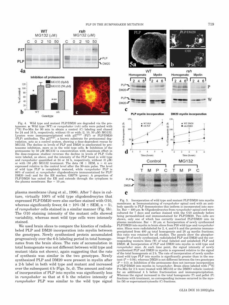
plasma membrane (Jung et al., 1996). After 7 days in cul-ture, virtually 100% of wild type oligodendrocytes thatexpressed PLP/DM20 were also surface stained with O10,whereas significantly fewer, 64 6 10% (M 6 SEM, n 5 5),of rumpshaker cells stained in a similar manner (Fig. 5b).The O10 staining intensity of the mutant cells showedvariability, whereas most wild type cells were intenselystained.
We used brain slices to compare the kinetics of radiola-beled PLP and DM20 incorporation into myelin betweenthe genotypes. Newly synthesized protein accumulatedprogressively over the 6-h labeling period in total homoge-nates from the brain slices. The rate of accumulation intotal homogenate was not different between wild type andmutant (data not shown), again suggesting that the rateof synthesis was similar in the two genotypes. Newlysynthesized PLP and DM20 were present in myelin aftera 2-h label in both wild type and mutant and increasedover the subsequent 4 h (Figs. 5c, d). The amount and rateof incorporation of PLP into myelin was significantly lessin rumpshaker so that at 6 h the relative intensity ofrumpshaker PLP was similar to the wild type signal
Fig. 4. Wild type and mutant PLP/DM20 are degraded via the pro-teasome. a: Wild type (WT) or rumpshaker (rsh) cells were pulsed with[35S] Pro-Mix for 90 min to obtain a control (C) labeling and chasedfor 24 and 16 h, respectively, without (0) or with (2, 10, 50 lM) MG132.Lysates were immunoprecipitated with p27kip1 (P27) or PLP/DM20(PLP) antibodies. The p27kip1, a known substrate for proteasomal deg-radation, acts as a control system, showing a dose-dependent rescue byMG132. The decline in levels of PLP and DM20 is ameliorated by pro-teasome inhibition, more so in the wild type cells. b: Inhibition of theproteasome by 50 lM MG132 (a concentration with maximum effect inthe dose-response studies) reverses the decline in levels of PLP. Cellswere labeled, as above, and the intensity of the PLP band in wild typeand rumpshaker quantified at 24 or 16 h, respectively, without (0 lM)or with (50 lM) MG132 treatment. The data (M 6 SEM, n 5 4) areexpressed relative to the control level after the 90-min pulse. The levelof wild type PLP is completely restored, while rumpshaker PLP is86% of control. c: rumpshaker oligodendrocyte immunostained for PLP/DM20 (red) and for the ER marker, GRP78 (green). A proportion ofPLP/DM20 has exited the ER and extends through the cytoplasm tothe plasma membrane. Bar 5 10 lm.
Fig. 5. Incorporation of wild type and mutant PLP/DM20 into myelinmembrane. a: Immunostaining of rumpshaker spinal cord with an anti-body specific to PLP demonstrates this isoform is incorporated into mye-lin. Bar5 400 lm. b: Oligodendrocytes from rumpshaker spinal cord werecultured for 7 days and surface stained with the O10 antibody beforebeing permeabilized and immunostained for PLP/DM20. Two cells areshown, only one of which has correctly inserted PLP/DM20 into itsplasma membrane. Bar 5 20 lm. c: Incorporation of newly synthesizedPLP/DM20 into myelin in brain slices from P20 wild type and rumpshakermice. Slices were radiolabeled for 2, 4, and 6 h and the proteins immuno-precipitated from 400 lg total homogenate and 20 lg myelin fractions;this ratio was retained for all studies. The panels show the phosphor-image (P) of newly synthesized, radiolabeled PLP and DM20 and the cor-responding western blots (W) of total (labeled and unlabeled) PLP andDM20. d: Incorporation of PLP and DM20 into myelin in wild type andrumpshaker mice. At each time point, the signal intensity of newlysynthesized PLP and DM20 in myelin is expressed relative to the signalof the total homogenate at 2 h. The rate of incorporation of newly synthe-sized wild type PLP into myelin is significantly greater than in the mu-tant (P5 0.04), whereas DM20 is not different between the two genotypes(P 5 0.5). e: Inhibition of the proteasome does not increase incorporationof PLP/DM20 into myelin in rumpshaker. Brain slices labeled with [35S]Pro-Mix for 2 h were treated with MG132 or the DMSO vehicle (control)for an additional 4 h before fractionation and immunoprecipitation.Whereas the signal increased in the total homogenate (T) and pellet (P)fractions with treatment, no corresponding increase occurred in the mye-lin (M) or supernatant/cytosolic (C) fractions.
719PLP IN THE RUMPSHAKER MUTATION
GLIA DOI 10.1002/glia
observed after 2 h, indicating a significant delay in incor-poration. The rate at which rumpshaker DM20 incorpo-rated into myelin was similar to wild type, although theamounts were considerably lower. Western blot analysisof the immunoprecipitated products (Fig. 5c) showed thatthe ratio of PLP recovered from the homogenate com-pared with the myelin fraction was similar for wild typeand rumpshaker. The difference in myelin incorporationrates is not simply a reflection of the amount of PLPrecovered from the myelin fraction in rumpshaker.
As inhibition of ERAD resulted in a significant eleva-tion of the level of PLP in cultured rumpshaker cells, weasked whether there would be a similar increase in mye-lin incorporation. However, treatment of brain slices with50 lM MG132 failed to increase PLP/DM20 incorporationinto the myelin fraction, although signal in both the totalhomogenate and pellet fractions was elevated (Fig. 5e).
DISCUSSION
Missense mutations of the PLP1/Plp1 gene resulting inan amino acid substitution are a cause of PMD in manand account for the majority of spontaneous disorders inanimals. Dysmyelination is a major feature of the pathol-ogy and probably accounts for many of the clinical signs,yet its basis is not clear. Indeed, how the abnormal PLP/DM20 is handled by the oligodendrocyte is only partlyresolved and is the focus of the present study. The rump-shaker mutation encodes an isoleucine to threonine sub-stitution in the putative extracellular CD loop of the PLP/DM20 molecule that results in mild or lethal phenotypesdependent on genetic background, suggesting the influ-ence of other modifying genes (Al-Saktawi et al., 2003).Using a model of the mild form of PMD/SPG2, we showthat the reduced steady state level of PLP/DM20 is causedprincipally by enhanced degradation, most probably viathe proteasome, rather than a reduced rate of translation.The majority of rumpshaker PLP/DM20 that escapesERAD is targeted to the developing myelin sheath, whereit inserts correctly into the membrane, albeit more slowlythan in wild type.
Translational Efficiency of PLP Is NotAffected by the rumpshaker Mutation
The level of Plp/Dm20mRNA is only minimally reducedcompared with PLP/DM20 protein, suggesting that atranslational or post-translational defect is responsiblefor the low protein level. Misfolded proteins in the RERcan inhibit translation by activating PERK, leading tophosphorylation of the eIF2a factor (Harding et al., 1999),although this is often a transient effect and is not nec-essarily operative. For example, in the Akita diabeticmouse, misfolded proinsulin accumulates in the RER yettranslation proceeds normally (Izumi et al., 2003). Simi-larly, translational efficiency of PLP/DM20 in rumpshakeroligodendrocytes is comparable with wild type, indicatingthat the low protein levels are due to post-translationalmechanisms.
The Fate of the rumpshaker PLP/DM20
The exit of most misfolded membrane proteins from theRER tends to be perturbed (Ellgaard and Helenius, 2003).Transfection and in vivo studies have shown that pro-ducts encoded by many missense mutations of the PLP1/Plp1 gene accumulate in the RER rather than reach theplasma membrane (Gow and Lazzarini, 1996). The major-ity of information has been gathered from microscopicimages, which create the impression of a static proteinmass in the RER cisternae and provide little kinetic infor-mation. We show that rumpshaker PLP is a dynamic pro-tein, the majority of which is degraded by an MG132-sen-sitive pathway at twice the rate of the wild type product,causing the low steady state levels. The rate of DM20breakdown is also accelerated by the mutation. Only a mi-nority of PLP/DM20 reaches its correct destination in themyelin sheath.
The majority of proteins destined for proteasomal deg-radation are polyubiquitinated, although there are exam-ples of non-ubiquitinated proteins being degraded by thispathway (Asher et al., 2002; Jarosch et al., 2003; Shrin-garpure et al., 2003). Despite numerous attempts, wewere unable to demonstrate polyubiquitination of PLP/DM20. We cannot exclude the possibility that the putativepool of ubiquitin-conjugated PLP is rapidly deubiquiti-nated by the activity of isopeptidases (Kim et al., 2003).There are also examples of wild type or misfolded RERproteins being degraded by non-proteasomal pathways(Donoso et al., 2005; Schmitz and Herzog, 2004). Sensitiv-ity to MG132 is a well-accepted indicator of proteasomalinvolvement; and despite the failure to demonstrate poly-ubiquitination, it seems most probable that the protea-some is the main degradative pathway for rumpshakerPLP/DM20.
Rumpshaker PLP Is Incorporated into Myelin LessEfficiently than Wild Type
The amount and rate of incorporation of newly synthe-sized PLP into myelin, relative to the total pool of labeledPLP, were reduced in rumpshaker consistent with a delayin the processing pathway at an earlier stage. Evidencefor the insertion of PLP and/or DM20 into the myelinmembrane was obtained by surface staining with the O10antibody, which recognizes an epitope on the extracellularCD loop (Jung et al., 1996), a region that also contains therumpshaker mutation. While the amino acid substitutioncompromises the function of PLP/DM20, it does not ap-pear sufficient to prevent the membrane insertion of thefraction that escapes ERAD.
The presence of misfolded PLP influences the structureof myelin. In common with other Plp1 mutants (Duncanet al., 1987), rumpshaker mice have thin myelin sheathswith abnormal, condensed, intraperiod lines (IPL), com-pared with the closely apposed double IPL of normal mye-lin (Griffiths et al., 1990). In contrast, Plp1 null mice havewidened or absent IPL (Yool et al., 2002). Although PMDis classed as a dysmyelinating rather than a demyelinat-
720 McLAUGHLIN ET AL.
GLIA DOI 10.1002/glia
ing disorder, it is possible that individual components,such as misfolded PLP, have an enhanced rate of turnoverin myelin, thus making the myelin unstable and unable toachieve the appropriate thickness.
Increasing the amount of rumpshaker PLP/DM20potentially available to myelin by inhibiting proteasomaldegradation does not lead to an enhanced incorporation,suggesting the system is handling the maximum amountof mutated product or that other mechanisms of degrada-tion also operate (Schmitz and Herzog, 2004). One candi-date is the endosomes/lysosome system, which is involvedin clearing excess wild type PLP generated by increasedPlp1 gene dosage (Simons et al., 2002). It is possible thatthis pathway also degrades a minority of the rumpshakerPLP/DM20.
Relevance to the Dysmyelinating Phenotype
Most forms of PMD and its spontaneous animal modelsare characterized by the inability of surviving oligoden-drocytes to assemble and maintain normal amounts ofmyelin. Although the level of PLP/DM20 is markedlyreduced in rumpshaker myelin, this is unlikely to be thecause of the hypomyelination, as a similar phenotype doesnot occur in Plp null mice or humans (Garbern et al.,2002; Klugmann et al., 1997). Also, introducing wild typePLP/DM20 into mutant myelin through transgenic com-plementation does not rescue the phenotype (Schneideret al., 1995). One potential mechanism is the effect of therumpshaker mutation on other myelin components, parti-cularly MBP, which is critical for myelin formation (Shineet al., 1992). Levels of MBP are markedly reduced inrumpshaker oligodendrocytes (present study), and parti-cularly when the mutation is expressed on the C57BL/6background (Al-Saktawi et al., 2003). In ongoing studies(Barrie et al., unpublished) we have complemented theC57 rumpshaker mice with a wild type Plp transgene,increasing their PLP/DM20 levels to normal. However,MBP levels remain low and severe dysmyelination is stillpresent. It is, therefore, possible that the low levels ofMBP in rumpshaker contribute to the hypomyelination.MbpmRNA levels are only minimally reduced, suggestingthe rumpshaker mutation operates at a translational orpost-translational level to perturb MBP, presumablythrough the action of misfolded PLP/DM20 through an, asyet, unknown mechanism.
ACKNOWLEDGMENTS
This work was supported by The Wellcome Trust andBirth Defects Foundation. We are grateful to Prof. N.P.Groome and Drs. J. Garbern, S. Pfeiffer, and R.H. Quarlesfor the gift of antibodies.
REFERENCES
Al-Saktawi K, McLaughlin M, Klugmann M, Schneider A, Barrie JA,McCulloch MC, Montague P, Kirkham D, Nave K-A, Griffiths IR.2003. Genetic background determines phenotypic severity of the Plprumpshaker mutation. J Neurosci Res 72:12–24.
Asher G, Lotem J, Sachs L, Kahana C, Shaul Y. 2002. Mdm-2 and ubi-quitin-independent p53 proteasomal degradation regulated by NQO1.Proc Natl Acad Sci USA 99:13125–13130.
Donoso G, Herzog V, Schmitz A. 2005. Misfolded BiP is degraded by aproteasome-independent endoplasmic-reticulum-associated degrada-tion pathway. Biochem J 387:897–903.
Duncan ID, Hammang JP, Trapp BD. 1987. Abnormal compact myelinin the myelin-deficient rat: absence of proteolipid protein correlateswith a defect in the intraperiod line. Proc Natl Acad Sci USA 84:6287–6291.
Edgar JM, Anderson TJ, Dickinson PJ, Barrie JA, McCulloch MC,Nave K-A, Griffiths IR. 2002. Survival of, and competition between,oligodendrocytes expressing different alleles of the Plp gene. J CellBiol 158:719–729.
Edgar JM, McLaughlin M, Barrie JA, McCulloch MC, Garbern J, Grif-fiths IR. 2004. Age-related axonal and myelin changes in the rump-shaker mutation of the Plp gene. Acta Neuropath (Berl) 107:331–335.
Ellgaard L, Helenius A. 2003. Quality control in the endoplasmic reti-culum. Nat Rev Mol Cell Biol 4:181–191.
Garbern J, Yool DA, Moore GJ, Wilds I, Faulk M, Klugmann M, NaveK-A, Sistermans EA, van der Knaap MS, Bird TD, et al. 2002.Patients lacking the major CNS myelin protein, proteolipid protein 1,develop length-dependent axonal degeneration in the absence of de-myelination and inflammation. Brain 125:551–561.
Gow A, Lazzarini RA. 1996. A cellular mechanism governing the sever-ity of Pelizaeus-Merzbacher disease. Nat Genet 13:422–428.
Griffiths IR, Klugmann M, Anderson TJ, Yool D, Thomson CE, SchwabMH, Schneider A, Zimmermann F, McCulloch MC, Nadon NL, et al.1998. Axonal swellings and degeneration in mice lacking the majorproteolipid of myelin. Science 280:1610–1613.
Griffiths IR, Scott I, McCulloch MC, Barrie JA, McPhilemy K, Catta-nach BM. 1990. Rumpshaker mouse: a new X-linked mutation affect-ing myelination: evidence for a defect in PLP expression. J Neurocy-tol 19:273–283.
Harding HP, Zhang YH, Ron D. 1999. Protein translation and foldingare coupled by an endoplasmic-reticulum-resident kinase. Nature397:271–274.
Hudson LD. 2003. Pelizaeus-Merzbacher disease and spastic paraplegiatype 2: two faces of myelin loss from mutations in the same gene.J Child Neurol 18:616–624.
Inoue K. 2005. PLP1-related inherited dysmyelinating disorders: Peli-zaeus-Merzbacher disease and spastic paraplegia type 2. Neuroge-netics 6:1–16.
Izumi T, Yokota-Hashimoto H, Zhao S, Wang J, Halban PA, Takeuchi T.2003. Dominant negative pathogenesis by mutant proinsulin in theAkita diabetic mouse. Diabetes 52:409–416.
Jarosch E, Lenk U, Sommer T. 2003. Endoplasmic reticulum-associatedprotein degradation. Int Rev Cytol 223:39–81.
Johnston AW, McKusick VA. 1962. A sex-linked recessive form of spas-tic paraplegia. Am J Hum Genet 14:83–94.
Jung M, Sommer I, Schachner M, Nave K-A. 1996. Monoclonal anti-body O10 defines a conformationally sensitive cell-surface epitope ofproteolipid protein (PLP): evidence that PLP misfolding underliesdysmyelination in mutant mice. J Neurosci 16:7920–7929.
Kim JH, Park KC, Chung SS, Bang O, Chung CH. 2003. Deubiquitinat-ing enzymes as cellular regulators. J Biochem (Tokyo) 134:9–18.
Kirmani BF, Jacobowitz DM, Kallarakal AT, Namboodiri MAA. 2002.Aspartoacylase is restricted primarily to myelin synthesizing cells inthe CNS: therapeutic implications for Canavan disease. Mol BrainRes 107:176–182.
Klugmann M, Schwab MH, P€uhlhofer A, Schneider A, Zimmermann F,Griffiths IR, Nave K-A. 1997. Assembly of CNS myelin in the absenceof proteolipid protein. Neuron 18:59–70.
Kobayashi H, Hoffman EP, Marks HG. 1994. The rumpshaker mutationin spastic paraplegia. Nat Genet 7:351–352.
Lee DH, Goldberg AL. 1996. Selective inhibitors of the proteasome-dependent and vacuolar pathways of protein degradation in Saccharo-myces cerevisiae. J Biol Chem 271:27280–27284.
McLaughlin M, Hunter DJB, Thomson CE, Yool D, Kirkham D, FreerAA, Griffiths IR. 2002. Evidence for possible interactions betweenPLP, DM20 within the myelin sheath. Glia 39:31–36.
Montague P, Barrie JA, Thomson CE, Kirkham D, McCallion AS,Davies RW, Kennedy PGE, Griffiths IR. 1998. Cytoskeletal and nu-clear localization of MOBP polypeptides. Europ J Neurosci 10:1321–1328.
Naidu S, Dlouhy SR, Geraghty MT, Hodes ME. 1997. A male child withthe rumpshaker mutation, X-linked spastic paraplegia Pelizaeus-Merzbacher disease and lysinuria. J Inherited Metab Dis 20:811–816.
Pasquini LA, Moreno MB, Adamo AM, Pasquini JM, Soto EF. 2000.Lactacystin, a specific inhibitor of the proteasome, induces apoptosisand activates caspase-3 in cultured cerebellar granule cells. J Neu-rosci Res 59:601–611.
721PLP IN THE RUMPSHAKER MUTATION
GLIA DOI 10.1002/glia
Rutkowski DT, Kaufman RJ. 2004. A trip to the ER: coping with stress.Trends Cell Biol 14:20–28.
Sanders CR, Myers JK. 2004. Disease-related misassembly of mem-brane proteins. Annu Rev Biophys Biomol Struct 33:25–51.
Schmitz A, Herzog V. 2004. Endoplasmic reticulum-associated degrada-tion: exceptions to the rule. Eur J Cell Biol 83:501–509.
Schneider A, Griffiths IR, Readhead C, Nave K-A. 1995. Dominant-negative action of the jimpy mutation in mice complemented with anautosomal transgene for myelin proteolipid protein. Proc Natl AcadSci USA 92:4447–4451.
Schneider A, Montague P, Griffiths IR, Fanarraga ML, Kennedy PGE,Brophy PJ, Nave K-A. 1992. Uncoupling of hypomyelination and glialcell death by a mutation in the proteolipid protein gene. Nature 358:758–761.
Shine HD, Readhead C, Popko B, Hood L, Sidman RL. 1992. Morpho-metric analysis of normal, mutant and transgenic CNS: correlation ofmyelin basic protein expression to myelinogenesis. J Neurochem58:342–349.
Shringarpure R, Grune T, Mehlhase J, Davies KJ. 2003. Ubiquitin con-jugation is not required for the degradation of oxidized proteins byproteasome. J Biol Chem 278:311–318.
Simons M, Kr€amer EM, Macchi P, Rathke-Hartlieb S, Trotter J, NaveK-A, Schulz JB. 2002. Overexpression of the myelin proteolipid pro-tein leads to accumulation of cholesterol and proteolipid protein in
endosomes/lysosomes: implications for Pelizaeus-Merzbacher disease.J Cell Biol 157:327–336.
Skoff RP. 1995. Programmed cell death in the dysmyelinating mutants.Brain Pathol 5:283–288.
Southwood C, Gow A. 2001. Molecular pathways of oligodendrocyte apo-ptosis revealed by mutations in the proteolipid protein gene. MicroscRes Tech 52:700–708.
Southwood CM, Garbern J, Jiang W, Gow A. 2002. The unfoldedprotein response modulates disease severity in Pelizaeus-Merzbacherdisease. Neuron 36:585–596.
Thomson CE, Anderson TJ, McCulloch MC, Dickinson PJ, VouyiouklisDA, Griffiths IR. 1999. The early phenotype associated with thejimpy mutation of the proteolipid protein gene. J Neurocytol 28:207–221.
Thomson CE, Montague P, Jung M, Nave K-A, Griffiths IR, Nave KA.1997. Phenotypic severity of murine Plp mutants reflects in vivo andin vitro variations in transport of PLP isoproteins. Glia 20:322–332.
Yool D, Klugmann M, Barrie JA, McCulloch MC, Nave K-A, GriffithsIR. 2002. Observations on the structure of myelin lacking the majorproteolipid protein. Neuropath Appl Neurobiol 28:75–78.
Yool DA, Klugmann M, McLaughlin M, Vouyiouklis DA, Dimou L, Bar-rie JA, McCulloch MC, Nave K-A, Griffiths IR. 2001. Myelin proteoli-pid proteins promote the interaction of oligodendrocytes and axons.J Neurosci Res 63:151–164.
722 McLAUGHLIN ET AL.
GLIA DOI 10.1002/glia


![RAMSAUER - TOWNSEND EFFECTprepost/407/ramsauer/ramsauer.pdflor, [3] Quantum Mechanics, 3rd Ed. (Wiley, 1998), E. Merzbacher. [4] Modern Physics and Quantum Mechanics (Saunders, 1971),](https://img.pdfslide.us/doc/110x75/5f9a66bf0cb4eb101f31e287/ramsauer-townsend-effect-prepost407ramsauer-lor-3-quantum-mechanics-3rd.jpg)

![G. VITALI - Optimization OnlineWe recall the SPGM introduced in [3, alg. SPG2] for the minimization of di erentiable functions on nonempty closed and convex sets. When SPGM is applied](https://img.pdfslide.us/doc/110x75/5e615de1cdc89540ff44a2bf/g-vitali-optimization-we-recall-the-spgm-introduced-in-3-alg-spg2-for-the.jpg)