Embed Size (px)
Citation preview
Rui Manuel Lucas Gameiro Domingues Tostões
Licenciado
Process Engineering of Liver Cells for Drug Testing Applications
Dissertação para obtenção do Grau de Doutor em Bioengenharia
Orientador: Paula Marques Alves, PhD, ITQB-UNL Co-orientador: Manuel Carrondo, Professor, FCT-UNL
Co-orientador: Daniel Wang, Professor, MIT
2012
II
Process Engineering of Liver Cells for Drug Testing Applications
Copyright
Rui Manuel Lucas Gameiro Domingues Tostões
Faculdade de Ciências e Tecnologia – Universidade Nova de
Lisboa
A Faculdade de Ciências e Tecnologia e a Universidade Nova de Lisboa têm o direito, perpétuo e sem limites geográficos, de arquivar e publicar esta dissertação através de exemplares impressos reproduzidos em papel ou de forma digital, ou por qualquer outro meio conhecido ou que venha a ser inventado, e de a divulgar atraves de repositórios científicos e de admitir a sua copia e distribuição com objectivos educacionais ou de investigação, não comerciais, desde que seja dado crédito ao autor e editor.
III
ACKNOWLEDGEMENTS
I would like to acknowledge everyone who, directly or indirectly, supported and helped me during these 3 years of experimental work. To both my supervisors at the Animal Cell Technology (ACT) unit, Doctor Paula Alves and Professor Manuel Carrondo, for giving me the opportunity to work on the hepatocyte project, in a lab with conditions much beyond the standard in our country. To my supervisor, Doctor Paula Alves, for her pacience, support, strength and the scientific
knowledge conveyed to me.
To my co-supervisor, Professor Manuel Carrondo, for helping me to disentangle some of the
complex problems I have run into during this work, namely by helping me to focus on the
physical meaning of my hypothesis and results.
To my co-supervisor at the MIT, Danny Wang, who will always be an example in the creation of
value from scientific knowledge.
To the Fundação para a Ciência e Tecnologia, FCT, for funding my fellowship SFRH / BD /
35296 / 2007, under the scope of the MIT-Portugal programme.
To the MIT-Portugal programme, for a wonderful year during which I have been introduced to
(or almost drowned in) the Bioengineering field and several concepts and tools that helped me
to endure the following three years of research at the ACT. In particular, I would like to thank
Professor Manuel Nunes da Ponte, Professor Isabel Rocha and Professor Eugénio Ferreira, for
their scientific and personal support, and to my collegues and friends from the 2007
Bioengineering focus area.
To the ACT, namely the bioprocess (Marcos, Carina Silva and Ana Teixeira) hepatocyte
(Catarina Brito, Sofia Leite and Joana Miranda) and stem cell teams (Catarina Brito, Margarida
Serra, Claudia Correia). I would also like to mention the collective commitment of the entire ACT
unit to bioprocess research; it is easy to work when surrounded by such committed people.
Finally and again, to Catarina Brito, not only to write her name for the third time, but also to say
that she has been the greatest colleague and friend along this hepatic journey. And before I
forget, to Marta, for her willingness to be a part of a project born between Catarina, Gonçalo
and myself; it was certainly not easy and it is my sincere hope that this project reaches its
maturity.
To Cellartis AB, Sweden, namely to Doctor Petter Bjorquist, Jenny Lindquist and our most
frequent partner in crime, Janne Jensen, with whom we have created the synergy essential to
the work in the Chapter 4 of this thesis.
Pessoalmente, gostaria de agradecer:
Aos meus amigos de sempre (Chefe, DC, DN, Frade, Fred, Lois e Vasco) e à minha família
(Mana, Sobrinhos, Cunhado, Mãe e Pai) por terem contribuído decisivamente para ser como
sou.
Aos meus avós Luélia e Joaquim Serafim. Porque já não se fazem pessoas nem nomes assim.
À Filipa, porque sem a nossa história o Capítulo 4, tal como é, não existiria. E pelo seu apoio
em todos os momentos.

IV
V
Resumo
As culturas primárias de hepatócitos são indispensáveis no processo de desenvolvimento de fármacos. Esta aplicação tem duas limitações: a reduzida proliferação dos hepatócitos e a rápida perda das funcionalidades hepáticas, quando cultivados in vitro. O objectivo desta tese foi a minimização deste último problema através da cultura de hepatócitos, como esferóides multicelulares, em bioreactores totalmente controlados. No Capítulo 1 é feita uma revisão do estado da arte em culturas primárias de hepatócitos, precedida de uma introdução à fisiologia do fígado e ao processo de desenvolvimento de fármacos. Inicialmente, a melhoria das culturas primárias de esferóides de hepatócitos, em bioreactor, foi feita com hepatócitos de ratos; Nos Capítulos 2 e 3 foram analisados os efeitos da microencapsulação em alginato, da cultura em perfusão e das duas estratégias em simultâneo na manutenção do fenótipo hepático dos esferóides; a aplicação simultânea das duas estratégias tem um efeito sinérgico positivo neste fenótipo hepático. No Capítulo 4 é feito o estudo da cultura, a longo prazo, de esferóides de hepatócitos isolados de 3 dadores humanos. Estes esferóides de hepatócitos responderam à administração repetida de fármacos, como esperado em hepatócitos maduros e diferenciados in vivo, até quatro semanas de cultura. No Capítulo 5, a cultura de esferóides multicelulares foi aplicada à maturação hepática de progenitores de hepatócitos, derivados de células estaminais embrionárias humanas; esta cultura de esferóides, quando comparada com a cultura em monocamada, levou a um aumento na expressão de genes hepáticos. No Capítulo 6 foram discutidas as melhorias na bioengenharia da cultura de hepatócitos possibilitadas por esta tese, bem como o possível trabalho futuro nesta área. Esta tese permitiu estabelecer e validar um sistema de bioreactores de perfusão que permite testar os efeitos de um novo fármaco, a longo prazo e com administração repetida, sobre esferóides de hepatócitos humanos. Termos chave: CYP450, hepatócitos, 3D, longo prazo.

VI
VII
Abstract
The primary culture of human hepatocytes is a requirement in drug development tests. This application is currently hampered by two problems: the limited proliferation of the hepatocytes and the rapid loss of liver-specific phenotype of these cells, when cultured in vitro. This thesis aimed at minimizing this latter issue by cultivating hepatocytes, as spheroids, in fully controlled bioreactors. The state of the art of the primary cultures of hepatocytes is reviewed in Chapter 1, after a brief introduction to the liver physiology the drug development process. The improvement of the bioreactor cultures of hepatocyte spheroids was initially done using freshly isolated rat hepatocytes; the effects of alginate microencapsulation, perfusion culture and their synergy on the maintenance of the hepatocyte spheroids liver-specific phenotype were assessed in Chapters 2 and 3; it was concluded that the perfusion culture and alginate-encapsulation had a positive synergic effect on such hepatic phenotype. The perfusion bioreactor developed in Chapter 3 was used in Chapter 4 for the extended culture of freshly isolated human hepatocytes, as spheroids, from three different donors. These cultures responded to repeated dose drug treatments as expected from mature and differentiated hepatocytes, in up to 4 weeks culture time. In Chapter 5, human embryonic stem cell-derived hepatic progenitors were cultured as spheroids and further differentiated into hepatocyte-like cells; the differential expression of hepatic genes between this spheroid population and a monolayer differentiated hepatocyte-like cell population showed a more efficient differentiation under spheroid culture. The bioengineering improvements of this thesis, as well as the future work, were discussed in Chapter 6. This thesis has led to the establishment and validation of primary cultures of hepatocyte spheroids, in perfusion bioreactors, which can be used for long-term, repeated dose tests in drug development. Keywords: CYP450, hepatocytes, 3D, long term.

VIII
IX
Table of Contents
CHAPTER 1 - INTRODUCTION ................................................................................................... 1
CHAPTER 2 - ALGINATE-ENCAPSULATED THREE-DIMENSIONAL CULTURES OF
HEPATOCYTES IN BIOREACTORS ........................................................................................ 21
CHAPTER 3 - BIOREACTOR PERFUSION OF 3D ENCAPSULATED HEPATOCYTES ....... 39
CHAPTER 4 - HUMAN LIVER CELL SPHEROIDS IN EXTENDED PERFUSION
BIOREACTOR CULTURE FOR REPEATED DOSE DRUG TESTING .................................... 56
CHAPTER 5 - SPHEROID FORMATION OF HESC-DERIVED HEPATIC PROGENITORS FOR
IMPROVED HEPATIC DIFFERENTIATION .............................................................................. 73
CHAPTER 6 - DISCUSSION AND CONCLUSIONS ................................................................. 85

X
XI
LIST OF FIGURES
FIGURE 1.1: THE ARCHITECTURE OF THE LIVER ................................................................................. 4
FIGURE 1.2: BILE CANALICULI PHYSIOLOGY ....................................................................................... 5
FIGURE 1.3: METABOLIC LIVER ZONATION ......................................................................................... 6
FIGURE 1.4: DRUG DEVELOPMENT WORKFLOW .................................................................................. 7
FIGURE 1.5: GENERAL MECHANISM FOR DRUG METABOLISM AND DRUG-DRUG INTERACTIONS .............. 9
FIGURE 1.6: SIMMETRY OF THE EPITHELIAL INTERACTIONS ............................................................... 13
FIGURE 2.1: PHASE-CONTRAST MICROSCOPY OF ENCAPSULATED HEPATOCYTE AGGREGATES ........... 29
FIGURE 2.2: FUNCTIONAL CAPACITY OF HEPATOCYTES .................................................................... 30
FIGURE 2.3: SARTORIUS-STEDIM BIOSTAT Q-PLUS BIOREACTOR SYSTEM ........................................ 32
FIGURE 2.4: ECOD AND UGT ACTIVITIES IN RAT HEPATOCYTES CULTURED AS ENCAPSULATED
AGGREGATES ........................................................................................................................ 33
FIGURE 2.5: 3-METHYLCHOLANTHRENE INDUCTION OF CYP450-DEPENDENT ACTIVITY IN PRIMARY
CULTURE OF RAT HEPATOCYTES ............................................................................................. 34
FIGURE 2.6: DEXAMETHASONE INDUCTION OF CYP450-DEPENDENT ACTIVITY IN PRIMARY CULTURE OF
RAT HEPATOCYTES ................................................................................................................. 35
FIGURE 3.1: PERFUSION BIOREACTOR SYSTEM WITH GRAVIMETRIC CONTROL ................................... 46
FIGURE 3.2: EFFECT OF PERFUSION IN THE LIVER-SPECIFIC FUNCTIONS OF PRIMARY BIOREACTOR
CULTURES OF NON ENCAPSULATED HEPATOCYTE SPHEROIDS ................................................... 47
FIGURE 3.3: CHARACTERIZATION OF ULTRA HIGH VISCOUS ALGINATE ENCAPSULATED HEPATOCYTE
SPHEROIDS ............................................................................................................................ 48
FIGURE 3.4: SPECIFIC ALBUMIN AND UREA SYNTHESIS RATES IN PERFUSION AND 50% MEDIUM
EXCHANGE BIOREACTOR CULTURE .......................................................................................... 49
FIGURE 3.5: SPECIFIC OXYGEN CONSUMPTION RATE IN PERFUSION AND 50% MEDIUM EXCHANGE
BIOREACTOR CULTURE ........................................................................................................... 50
FIGURE 3.6: CYP450 ACTIVITY IN PERFUSION AND 50% MEDIUM EXCHANGE BIOREACTOR CULTURE .. 50
FIGURE 4.1: EXPERIMENTAL DESIGN OF THE INDUCTION OF THE CYP450 ENZYMES IN PRIMARY
CULTURES OF HEPATOCYTE SPHEROIDS IN THE BIOREACTOR .................................................... 61
FIGURE 4.2: HEPATOCYTE SPHEROID DIAMETER DISTRIBUTION AND CELL VIABILITY ........................... 63
FIGURE 4.3: UREA AND ALBUMIN SYNTHESIS IN BIOREACTOR HEPATOCYTE SPHEROID CULTURES ....... 64
FIGURE 4.4: INTER-DONOR VARIABILITY AND TIME COURSE PROFILES OF PHASE I AND II ENZYMES IN
HUMAN HEPATOCYTE SPHEROIDS BIOREACTOR CULTURES ....................................................... 65
FIGURE 4.5: INDUCTION OF ECOD ACTIVITY IN BIOREACTOR ............................................................ 66
FIGURE 4.6: IMMUNOFLUORESCENCE MICROSCOPY OF LIVER-SPECIFIC ANTIGENS IN HUMAN
HEPATOCYTE SPHEROIDS AFTER 2 WEEKS OF BIOREACTOR CULTURE ....................................... 66
FIGURE 4.7: FLUORESCENCE MICROSCOPY OF STRUCTURAL AND POLARITY MARKERS AND BILE
CANALICULI FUNCTION IN HUMAN HEPATOCYTE SPHEROIDS AFTER 2 WEEKS OF BIOREACTOR
CULTURE ............................................................................................................................... 68
FIGURE 5.1: EFFECT OF ROCKI ON THE AGGREGATION AND VIABILITY OF HEPATIC PROGENITOR CELLS
UNDER MICROGRAVITY CONDITIONS ........................................................................................ 79
FIGURE 5.2: HARDWARE (FIRST COLUMN) AND CULTURE PARAMETERS TESTED FOR THE OPTIMIZATION
OF THE AGGREGATION PROCESS OF THE HEPATIC PROGENITORS .............................................. 80
FIGURE 5.3: WORKFLOW FOR THE MATURATION OF HEPATIC PROGENITORS TO HEPATOCYTE-LIKE
CELLS, IN 2D AND 3D ............................................................................................................. 81
FIGURE 5.4: PHASE CONTRAST AND FLUORESCENCE MICROSCOPY IMAGES OF THE MEMBRANE
INTEGRITY AND METABOLIC ACTIVITY OF ALGINATE ENCAPSULATED HEPATIC PROGENITOR
SPHEROIDS ............................................................................................................................ 82
FIGURE 6.1: WORKING HYPOTHESIS FOR THE SYNERGISTIC EFFECT OF PERFUSION AND ALGINATE
ENCAPSULATION IN PRIMARY CULTURES OF RAT HEPATOCYTE SPHEROIDS ................................ 89
FIGURE 6.2: POSSIBLE HEPATIC CELL SOURCES .............................................................................. 92

XII
XIII
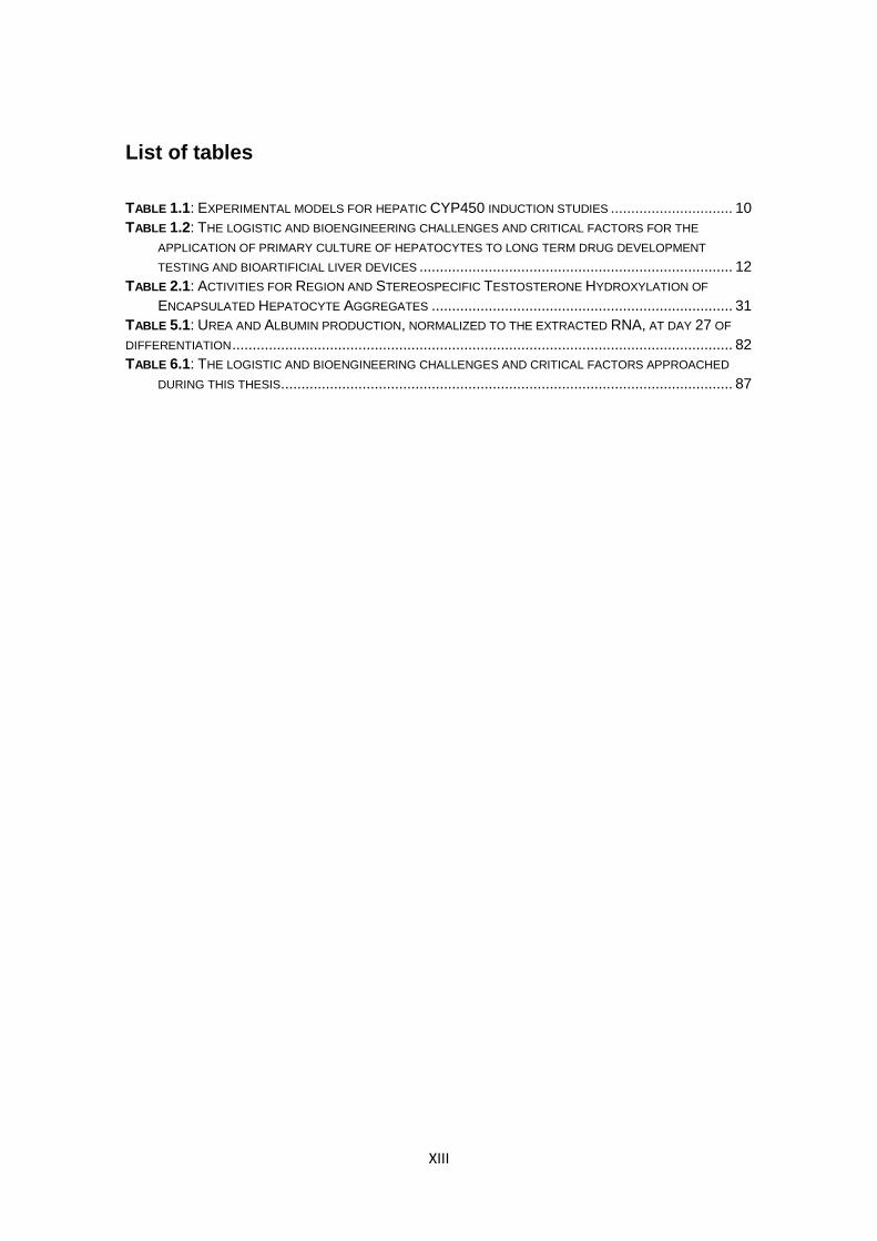
List of tables
TABLE 1.1: EXPERIMENTAL MODELS FOR HEPATIC CYP450 INDUCTION STUDIES .............................. 10
TABLE 1.2: THE LOGISTIC AND BIOENGINEERING CHALLENGES AND CRITICAL FACTORS FOR THE
APPLICATION OF PRIMARY CULTURE OF HEPATOCYTES TO LONG TERM DRUG DEVELOPMENT
TESTING AND BIOARTIFICIAL LIVER DEVICES ............................................................................. 12
TABLE 2.1: ACTIVITIES FOR REGION AND STEREOSPECIFIC TESTOSTERONE HYDROXYLATION OF
ENCAPSULATED HEPATOCYTE AGGREGATES .......................................................................... 31
TABLE 5.1: UREA AND ALBUMIN PRODUCTION, NORMALIZED TO THE EXTRACTED RNA, AT DAY 27 OF
DIFFERENTIATION ........................................................................................................................... 82
TABLE 6.1: THE LOGISTIC AND BIOENGINEERING CHALLENGES AND CRITICAL FACTORS APPROACHED
DURING THIS THESIS. .............................................................................................................. 87

XIV
XV
Abbreviations
Abbreviation Full form SECs Sinusoidal endothelial cells
HSCs Hepatic stellate cells
ECM Extracellular Matrix
NK Natural Killer
Km Michaelis Menten enzyme kinetics constant
ALF Acute liver failure
OLT Orthotopic liver transplant
AL or BAL artificial or bioartificial liver
NCE New chemical entity
PK Pharmacokinetics
OATP Organic anion transporting polypeptide
CYP450 Cytochrome P450
CYP1A2 Cytochrome P450, isoform 1A2
CYP2C9 Cytochrome P450, isoform 2C9
CYP3A4 Cytochrome P450, isoform 3A4
CYP7A1 Cytochrome P450, isoform 7A1
PXR Pregnane X Receptor
CAR Constitutive Androstan Receptor
AhR Aromatic Hydrocarbon Receptor
DDI Drug- drug interactions
mRNA Messenger Ribonucleic Acid
FDA Food and Drug Administration
TGF- Transforming Growth Factor Beta
ELISA enzyme- linked immunosorbent assay
3D Three-dimensional
2D Two-dimensional
RGD Arginine- Glycine - Aspartic Acid
DO Dissolved oxygen
hESC Human embryonic stem cells
UGT Uridine diphosphate glucuronosyltransferases
UGT2B7 Uridine diphosphate glucuronosyltransferase, isoform 2B7
ECOD 7- Ethoxycoumarin -O- Dealkilase
3-MC 3- Methylcholantherene
4-MU 4- Methylumbelliferone
OHT Hydroxytestosterone
Da Dämkohler number
OCR Oxygen Consumption Rate
FBS Foetal Bovine Serum
Rif Rifampicin
BNF -Naphtoflavone
dave average diameter
cDNA complementary Deoxyribonucleic acid
PCR Polymerase Chain Reaction
GAPDH Glyceraldehyde 3- phosphate dehydrogenase
PFA Paraformaldehyde
DAPI 4',6- diamidino -2- phenylindole
CDFDA 5-(and-6)-carboxy-2',7'-dichlorofluorescein diacetate
GSTA1 Glutathione S- transferase A1
HNF4 Hepatocyte Nuclear Factor 4 alfa
aPKC atypical Protein Kinase C
MRP2 associated protein 2
HLC hepatocyte-like cell
EBs embryoid bodies
DE Definitive Endoderm
HGF Hepatocyte Growth Factor
KO-DMEM knock out Dulbecco's modified Eagle medium
KO-SR knock out serum replacement
HD hanging drop
Rocki Rock inhibitor
EGF Epidermal Growth Factor
BMPs Bone Morphogenetic Proteins
MSC mesenchymal stem cells

XVI
1
Chapter 1
Introduction – Liver physiology, drug development and state of the art in primary cultures of hepatocytes
Chapter 1 – Introduction
2
Table of Contents
1. Introduction .......................................................................................................... 3
1.1. Liver architecture and zonation ....................................................................... 3
1.2. Liver pathologies ............................................................................................ 7
1.3. Relevance of the liver metabolism for drug development ................................ 7
1.4. Models for hepatic CYP450 enzyme induction .............................................. 10
1.5. Critical factors in long-term primary cultures of hepatocytes ......................... 12
1.6. State of the art in long term primary cultures of hepatocytes ......................... 14
2. Scope of the Thesis ........................................................................................... 15
3. References ......................................................................................................... 16
Process engineering of liver cells for drug testing applications
3
1. Introduction
The liver has a major role in maintaining physiological homeostasis and in detoxifying blood. In mammals, among other functions, it is responsible for nitrogen excretion in the form of urea, albumin biosynthesis (1), glucose storage in the form of glycogen, gluconeogenesis (2), glutamine homeostasis (3) and xenobiotic detoxification (4-7).
1.1. Liver architecture and zonation
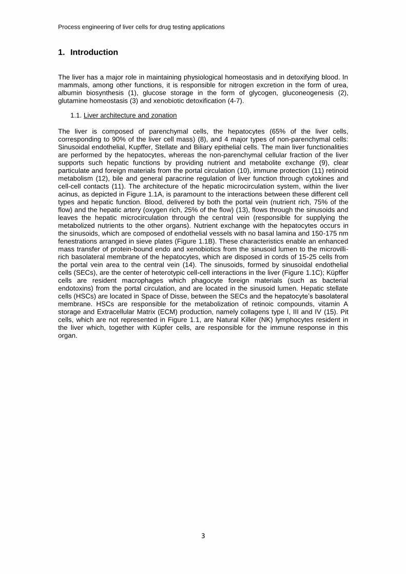
The liver is composed of parenchymal cells, the hepatocytes (65% of the liver cells, corresponding to 90% of the liver cell mass) (8), and 4 major types of non-parenchymal cells: Sinusoidal endothelial, Kupffer, Stellate and Biliary epithelial cells. The main liver functionalities are performed by the hepatocytes, whereas the non-parenchymal cellular fraction of the liver supports such hepatic functions by providing nutrient and metabolite exchange (9), clear particulate and foreign materials from the portal circulation (10), immune protection (11) retinoid metabolism (12), bile and general paracrine regulation of liver function through cytokines and cell-cell contacts (11). The architecture of the hepatic microcirculation system, within the liver acinus, as depicted in Figure 1.1A, is paramount to the interactions between these different cell types and hepatic function. Blood, delivered by both the portal vein (nutrient rich, 75% of the flow) and the hepatic artery (oxygen rich, 25% of the flow) (13), flows through the sinusoids and leaves the hepatic microcirculation through the central vein (responsible for supplying the metabolized nutrients to the other organs). Nutrient exchange with the hepatocytes occurs in the sinusoids, which are composed of endothelial vessels with no basal lamina and 150-175 nm fenestrations arranged in sieve plates (Figure 1.1B). These characteristics enable an enhanced mass transfer of protein-bound endo and xenobiotics from the sinusoid lumen to the microvilli-rich basolateral membrane of the hepatocytes, which are disposed in cords of 15-25 cells from the portal vein area to the central vein (14). The sinusoids, formed by sinusoidal endothelial cells (SECs), are the center of heterotypic cell-cell interactions in the liver (Figure 1.1C); Küpffer cells are resident macrophages which phagocyte foreign materials (such as bacterial endotoxins) from the portal circulation, and are located in the sinusoid lumen. Hepatic stellate cells (HSCs) are located in Space of Disse, between the SECs and the hepatocyte’s basolateral membrane. HSCs are responsible for the metabolization of retinoic compounds, vitamin A storage and Extracellular Matrix (ECM) production, namely collagens type I, III and IV (15). Pit cells, which are not represented in Figure 1.1, are Natural Killer (NK) lymphocytes resident in the liver which, together with Küpfer cells, are responsible for the immune response in this organ.
Chapter 1 – Introduction
4
Figure 1.1 – The architecture of the liver: acinus (A), sinusoids (B) and the location of the different liver cell types in the sinusoid (C). Adapted from (13, 16)
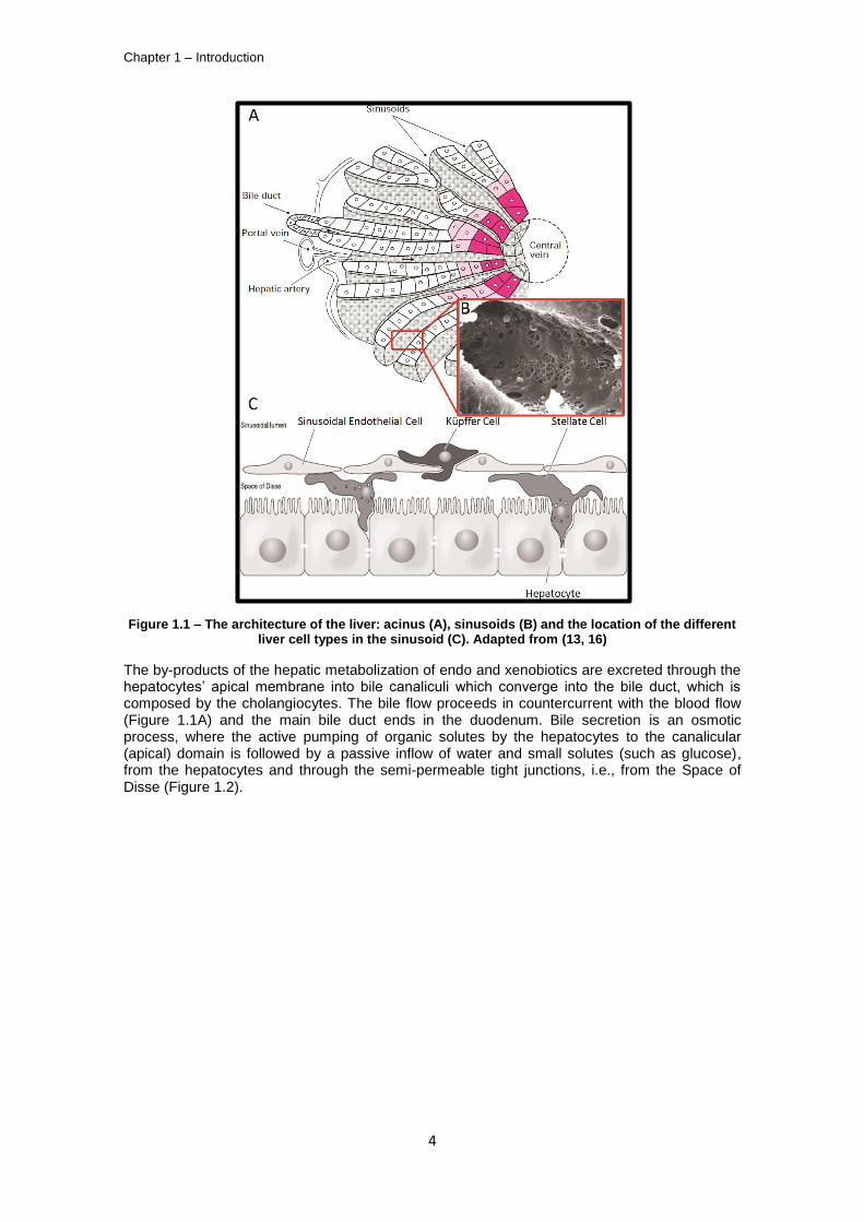
The by-products of the hepatic metabolization of endo and xenobiotics are excreted through the hepatocytes’ apical membrane into bile canaliculi which converge into the bile duct, which is composed by the cholangiocytes. The bile flow proceeds in countercurrent with the blood flow (Figure 1.1A) and the main bile duct ends in the duodenum. Bile secretion is an osmotic process, where the active pumping of organic solutes by the hepatocytes to the canalicular (apical) domain is followed by a passive inflow of water and small solutes (such as glucose), from the hepatocytes and through the semi-permeable tight junctions, i.e., from the Space of Disse (Figure 1.2).
Process engineering of liver cells for drug testing applications
5
Figure 1.2 – Bile canaliculi physiology; a transversal section of the hepatic sinusoid (lower figure) depicts the pumping of organic anions from the hepatocytes’ cytosol to the bile canaliculi. This
solute accumulation is the driving force for paracellular water osmosis, i.e., via the semi-permeable tight junctions (green) between the hepatocytes. TJ – Tight junctions; BC – Bile canaliculus. Upper
image adapted from (15).
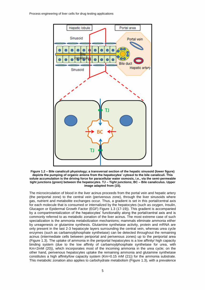
The microcirculation of blood in the liver acinus proceeds from the portal vein and hepatic artery (the periportal zone) to the central vein (perivenous zone), through the liver sinusoids where gas, nutrient and metabolite exchanges occur. Thus, a gradient is set in this portal/central axis for each molecule that is consumed or internalized by the hepatocytes (such as oxygen, Insulin, Glucagon or Epidermal Growth Factor (EGF) Figure 1.3 (17-19)). This gradient is accompanied by a compartmentalization of the hepatocytes’ functionality along the portal/central axis and is commonly referred to as metabolic zonation of the liver acinus. The most extreme case of such specialization is the ammonia metabolization mechanisms; mammals eliminate ammonia either by ureagenesis or glutamine synthesis. Glutamine synthetase activity, protein and mRNA are only present in the last 2-3 hepatocyte layers surrounding the central vein, whereas urea cycle enzymes (such as carbamoylphosphate synthetase) can be detected throughout the remaining acinus (intermediate cells between periportal and perivenous zones) up to the periportal area (Figure 1.3). The uptake of ammonia in the periportal hepatocytes is a low affinity/ high capacity binding system (due to the low affinity of carbamoylphosphate synthetase for urea, with Km=2mM (20)), which incorporates most of the incoming ammonia in the urea cycle; on the other hand, perivenous hepatocytes uptake the remaining ammonia and glutamine synthetase constitutes a high affinity/low capacity system (Km=0.15 mM (21)) for the ammonia substrate. This metabolic zonation also applies to carbohydrate metabolism (Figure 1.3), with a prevalence
Chapter 1 – Introduction
6
of oxidative metabolism in the oxygen-rich periportal zone (70 mmHg/90 M O2) and an enhancement of the glycolytic metabolism in the oxygen poor perivenous zone (35 mmHg/45
M O2) (18).
Figure 1.3 – Metabolic liver zonation as a function of the (variable) zonation of oxygen, hormones
and growth factors and of the (stable) zonation of -catenin signalling, adapted from (22).
Interestingly, the metabolic zonation of carbohydrates has shown to be partially reversed upon retrograde liver perfusion (i.e., nutrient flow is reversed to start from the perivenous to the periportal zone), indicating that the hepatocytes can “sense” the oxygen concentration (for a review on the role of oxygen in liver zonation see (19))`. The fact that metabolic zonation is only partially reversed in these retrograde perfusion experiments lead to the discovery of a stable
zonation mechanism based on -catenin signaling (23). In fact, stable zonation can also be seen in the non-parenchymal cell distribution and phenotype as well as in the extracellular matrix composition (17).
Thus, oxygen and nutrient gradients are critical factors in the dynamic component of the hepatic acinus metabolic zonation, including the metabolism of xenobiotics (such as drugs) and must be accounted for in the design criteria of any system for primary cultures of hepatocytes.
Process engineering of liver cells for drug testing applications
7
1.2. Liver pathologies
Liver tissue damage, irrespective of its etiology (viral or non-viral, autoimmune, alcoholic or non-alcoholic hepatitis (11, 15, 24)), leads to an increased synthesis of collagen fibers by HSC and other fibroblast-like cell types in the liver, constituting the basic liver wound-healing process. If the injury is continuous, this fibrotic response typically leads to the clogging of the liver vasculature (namely in the sinusoids) which results in necrosis, scarring of the affected area and the formation of nodules of regenerating hepatocytes; the final stage of progressive liver fibrosis is cirrhosis (see (25) for a comprehensive review), which can also lead to the onset of primary hepatocellular carcinoma. These chronic clinical conditions, as well as acute liver failure (ALF), can result in hepatic function impairment, multi-organ failure and subsequent death, unless an orthotopic liver transplant (OLT) is performed (26). However, the scarcity of liver donors creates a logistics problem for clinical situations where OLT is recommended. The need to keep patients with ALF alive from the onset of the clinical symptoms until a donated liver is available has lead to the development of artificial (27) and bioartificial liver (28) (AL or BAL, respectively) devices to perform the minimal hepatic functions which are necessary for patient survival; while the AL devices depend mainly on plasma filtration and purification operations (remnant of kidney dialysis), the BAL devices typically couple plasma filtration steps to a cell-based purification unit (i.e., a bioreactor) which can perform residual hepatic functions to sustain the patients lives. Irrespective of being AL or BAL devices, these solutions have not yet proved to be effective enough in improving ALF outcomes. In the specific case of BAL devices, the lack of an appropriate cellular component with significant volumetric hepatic activity is considered to be the major bottleneck for a clinically efficient and robust product.
1.3. Relevance of the liver metabolism for drug development
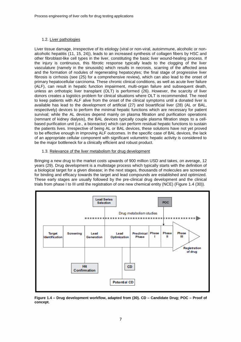
Bringing a new drug to the market costs upwards of 900 million USD and takes, on average, 12 years (29). Drug development is a multistage process which typically starts with the definition of a biological target for a given disease; in the next stages, thousands of molecules are screened for binding and efficacy towards the target and lead compounds are established and optimized. These early stages are usually followed by the pre-clinical drug development and the clinical trials from phase I to III until the registration of one new chemical entity (NCE) (Figure 1.4 (30)).
Figure 1.4 – Drug development workflow, adapted from (30). CD – Candidate Drug; POC – Proof of concept.
Chapter 1 – Introduction
8
The progression along this process phases leads to a decrease in the compounds tested and an increase in the operation costs (i.e., clinical trials are more expensive than the pre-clinical or screening phases). The probability of a given candidate drug to progress from phase II to phase III clinical trials was about 28% from 2006 to 2007 and 18% in the period from 2008-2009; given that the attrition rate for a drug entering phase III is about 50% (31, 32), it has been suggested that the success rates of drug development are too low to cope with the increasing R&D budget (33). While more global and systemic analysis about the underlying reasons of this issue have been performed by several authors (29, 34), the concrete problems that lead to failure in phase II clinical trials are well documented (31): 51% of these failures were due to insufficient efficacy, 29% to strategic reasons, 19% to pre-clinical or clinical safety issues and 1% due to inadequate pharmacokinetics (PK). PK can be defined as a methodology to determine a drug’s (and its metabolites) concentration profile along time in the human body fluids; the critical parameters for a PK profiling are the administration, distribution, metabolization and excretion (ADME) of the drug (35). The drug concentration profile in the human body is a critical variable for its therapeutical efficiency and since pharmaceutical companies started investing in PK pre-clinical research, in the early 1990’s, the failure rate in drug development owed to inadequate PK has fallen by 10% (30). However, the effects of a drug and it’s metabolites in the patients’ physiology is more difficult to assess in pre-clinical trials and such effects can lead either to enhanced toxicology (36, 37) or to insufficient efficacy (38). Central to these two causes of attrition in drug development is the biotransformation (i.e., metabolization) of the drug itself. In fact, according to data from Brystol-Myers Squibb from 1993 to 2006, 27% of the toxicology drug attrition were metabolism-related.
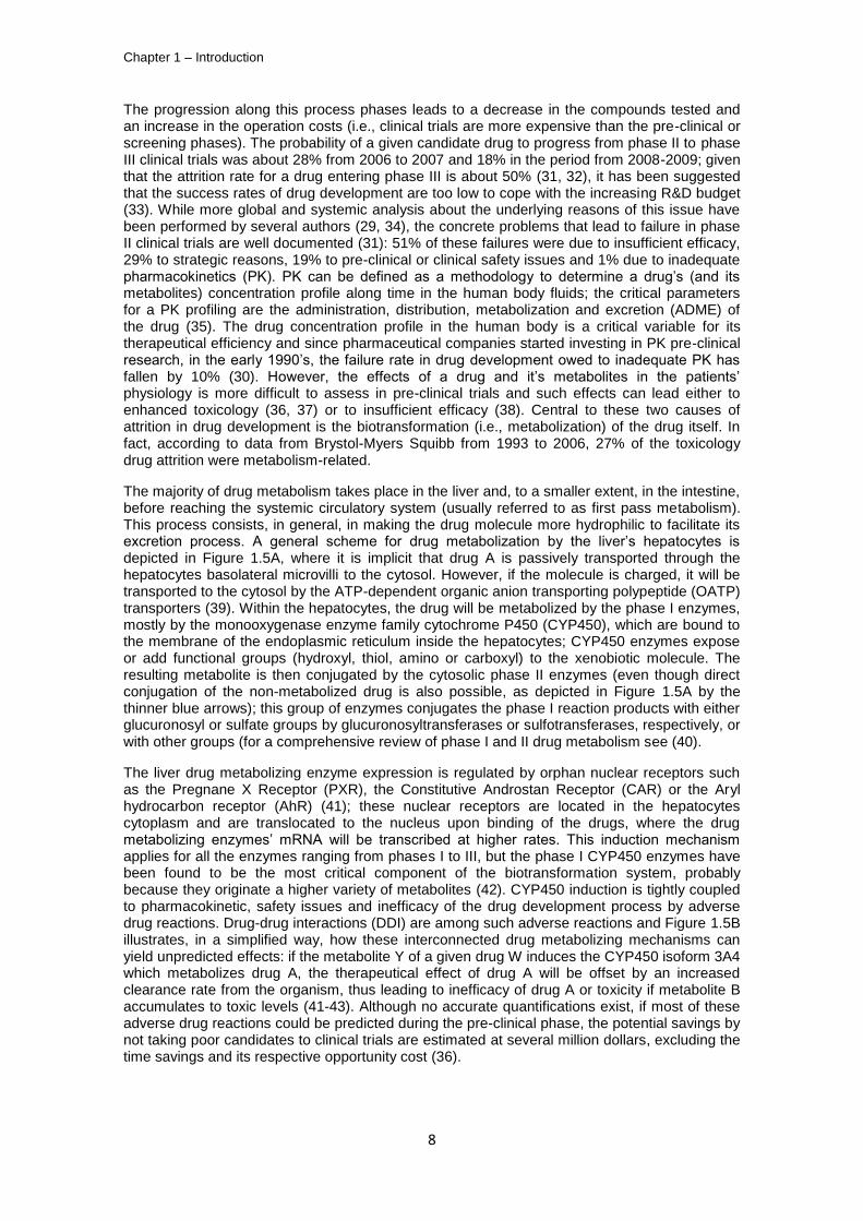
The majority of drug metabolism takes place in the liver and, to a smaller extent, in the intestine, before reaching the systemic circulatory system (usually referred to as first pass metabolism). This process consists, in general, in making the drug molecule more hydrophilic to facilitate its excretion process. A general scheme for drug metabolization by the liver’s hepatocytes is depicted in Figure 1.5A, where it is implicit that drug A is passively transported through the hepatocytes basolateral microvilli to the cytosol. However, if the molecule is charged, it will be transported to the cytosol by the ATP-dependent organic anion transporting polypeptide (OATP) transporters (39). Within the hepatocytes, the drug will be metabolized by the phase I enzymes, mostly by the monooxygenase enzyme family cytochrome P450 (CYP450), which are bound to the membrane of the endoplasmic reticulum inside the hepatocytes; CYP450 enzymes expose or add functional groups (hydroxyl, thiol, amino or carboxyl) to the xenobiotic molecule. The resulting metabolite is then conjugated by the cytosolic phase II enzymes (even though direct conjugation of the non-metabolized drug is also possible, as depicted in Figure 1.5A by the thinner blue arrows); this group of enzymes conjugates the phase I reaction products with either glucuronosyl or sulfate groups by glucuronosyltransferases or sulfotransferases, respectively, or with other groups (for a comprehensive review of phase I and II drug metabolism see (40).
The liver drug metabolizing enzyme expression is regulated by orphan nuclear receptors such as the Pregnane X Receptor (PXR), the Constitutive Androstan Receptor (CAR) or the Aryl hydrocarbon receptor (AhR) (41); these nuclear receptors are located in the hepatocytes cytoplasm and are translocated to the nucleus upon binding of the drugs, where the drug metabolizing enzymes’ mRNA will be transcribed at higher rates. This induction mechanism applies for all the enzymes ranging from phases I to III, but the phase I CYP450 enzymes have been found to be the most critical component of the biotransformation system, probably because they originate a higher variety of metabolites (42). CYP450 induction is tightly coupled to pharmacokinetic, safety issues and inefficacy of the drug development process by adverse drug reactions. Drug-drug interactions (DDI) are among such adverse reactions and Figure 1.5B illustrates, in a simplified way, how these interconnected drug metabolizing mechanisms can yield unpredicted effects: if the metabolite Y of a given drug W induces the CYP450 isoform 3A4 which metabolizes drug A, the therapeutical effect of drug A will be offset by an increased clearance rate from the organism, thus leading to inefficacy of drug A or toxicity if metabolite B accumulates to toxic levels (41-43). Although no accurate quantifications exist, if most of these adverse drug reactions could be predicted during the pre-clinical phase, the potential savings by not taking poor candidates to clinical trials are estimated at several million dollars, excluding the time savings and its respective opportunity cost (36).
Process engineering of liver cells for drug testing applications
9
Figure 1.5 – General mechanism for drug metabolism (A) and possible drug-drug interactions (B). MV-microvilli.
The studies of the NCE’s metabolization can be performed using different approaches; the incubation of human hepatocyte microsomes (which are lipid vesicles containing bound CYP450 enzymes) with the NCE yields information about the metabolites originated from the drug; these microsome assays are also useful to determine if the NCE or any of its metabolites are inhibitors of CYP450 activity (44, 45). However, the induction of CYP450 enzymes implies de novo mRNA synthesis, which requires intact cellular machinery and the models for these studies range from cells (5) to entire organisms for in vivo studies (46). These models may be ranked according to three main categories: throughput, predictability (relative to human subjects) and long term studies.
MV
PhaseIII
PhaseII
PhaseI
ABCDDrug
A
BC
Hydrophilic
Hydrophobic
Phase II(conjugation)
CYP450isozyme 3A4
Drug A B C
Phase II(conjugation)
CYP450Isozyme 1A1
Drug W Y Z
INDUCTION
INHIBITION
B
A
Chapter 1 – Introduction
10
1.4. Models for hepatic CYP450 enzyme induction
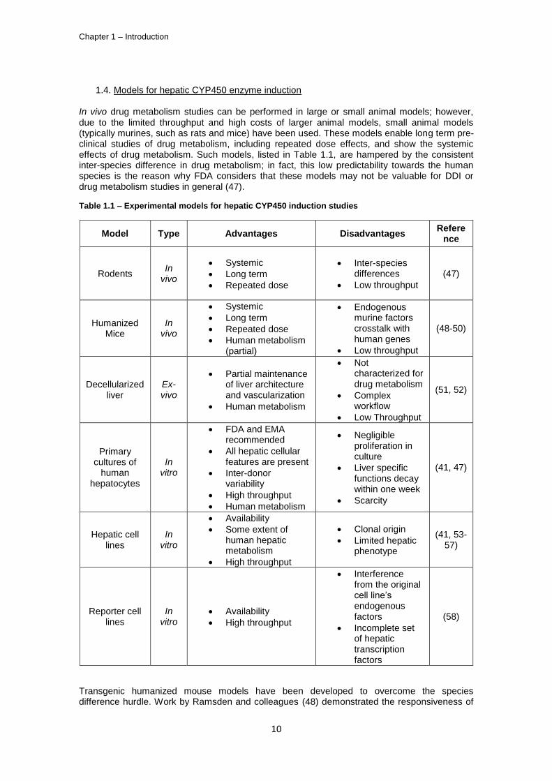
In vivo drug metabolism studies can be performed in large or small animal models; however, due to the limited throughput and high costs of larger animal models, small animal models (typically murines, such as rats and mice) have been used. These models enable long term pre-clinical studies of drug metabolism, including repeated dose effects, and show the systemic effects of drug metabolism. Such models, listed in Table 1.1, are hampered by the consistent inter-species difference in drug metabolism; in fact, this low predictability towards the human species is the reason why FDA considers that these models may not be valuable for DDI or drug metabolism studies in general (47).
Table 1.1 – Experimental models for hepatic CYP450 induction studies
Model Type Advantages Disadvantages Refere
nce
Rodents In
vivo
Systemic
Long term
Repeated dose
Inter-species differences
Low throughput
(47)
Humanized Mice
In vivo
Systemic
Long term
Repeated dose
Human metabolism (partial)
Endogenous murine factors crosstalk with human genes
Low throughput
(48-50)
Decellularized liver
Ex-vivo
Partial maintenance of liver architecture and vascularization
Human metabolism
Not characterized for drug metabolism
Complex workflow
Low Throughput
(51, 52)
Primary cultures of
human hepatocytes
In vitro
FDA and EMA recommended
All hepatic cellular features are present
Inter-donor variability
High throughput
Human metabolism
Negligible proliferation in culture
Liver specific functions decay within one week
Scarcity
(41, 47)
Hepatic cell lines
In vitro
Availability
Some extent of human hepatic metabolism
High throughput
Clonal origin
Limited hepatic phenotype
(41, 53-57)
Reporter cell lines
In vitro
Availability
High throughput
Interference from the original cell line’s endogenous factors
Incomplete set of hepatic transcription factors
(58)
Transgenic humanized mouse models have been developed to overcome the species difference hurdle. Work by Ramsden and colleagues (48) demonstrated the responsiveness of
Process engineering of liver cells for drug testing applications
11
transgenic mice carrying a copy of the human CYP2B2 gene to phenobarbital; more recently, double transgenic mice carrying one copy of the nuclear receptor PXR and its downstream target CYP3A4 were shown to be inducible by Rifampicin, a prototypical CYP3A4 inducer in humans, unlike the wild-type animals (49). The current limitation of these models is the interaction of murine endogenous factors with the human transgenic proteins. Recently, modified Polyethilene Glycol hydrogels containing a primary culture of human hepatocytes, mouse 3T3 fibroblasts and a human liver endothelial cell line (TMNK-1) were implanted in mice for drug metabolism studies (50). Such ectopic bioartificial livers enable the host mice to have a typically human metabolism of known drugs (namely Acetaminophen cytotoxicity); with the use of the correct control animals, this approach may be useful for long term drug metabolism studies. Still, the synergy of the host’s murine metabolism with the human hepatocytes can yield confounding results in drugs with unknown metabolism.
The recellularization of previously acellularized organs is a current trend in tissue engineering (59); this technique, which consists in the removal of cells from an organ with a detergent, yields the organ scaffold with a preserved ECM and vasculature. Decellularized livers from several species have been successfully prepared and efficiently reseeded either with adult rat hepatocytes (52) or with human endothelial cells and fetal liver cells (51), respectively. Uygun and colleagues (52) have shown that this strategy allows to maintain primary cultures of rat hepatocytes for up to 2 weeks, namely drug metabolizing enzymes mRNA expression. However, these expression levels did not outperform those obtained by the corresponding collagen sandwich cultures; further improvements can be made to this system by the addition of non-parenchymal liver cells. In fact, the presence of SECs may be critical to re-organize the liver sinusoids, which are lost during the decellularization process. From a practical point of view, this technique is not hampered by the scarcity of livers, since cadaveric livers can be used.
1
Cell-based assays for hepatic drug induction can be divided in 3 classes: reporter cell lines for nuclear receptor activation, immortalized hepatocytes/ hepatoma cell lines and primary cultures of human hepatocytes.
Primary cultures of human hepatocytes, either freshly isolated or cryopreserved, are FDA’s preferred human liver tissue for CYP450 induction tests (47). These cultures retain nuclear receptor, CYP450 isoforms as well as phase II and III mRNA and protein expression. This expression of all the major sensors and effectors of drug metabolism enables primary cultures of human hepatocytes to demonstrate CYP450 induction and hepatoxicity resulting from DDI. Primary cultures of hepatocytes have 3 main drawbacks: the hepatocytes have a negligible proliferation in culture, when cultured in monolayer, they lose their liver-specific activities (including CYP450) in less than one week (42) and these cells are scarce (even more since these hepatocytes do not proliferate ex vivo) and expensive, making increased throughput
assays expensive.
Nuclear receptor activation is the common upstream event in CYP450 de novo mRNA synthesis. Such a reporter system has been described by Luo and colleagues (58) based on the co-transfection of a hepatoma cell line, HepG2, with a plasmid with the cDNA for PXR under the control of a constitutive promoter and another plasmid containing the CYP3A4 regulatory region which controlled the expression of a reporter luciferase gene. Upon activation by a drug, the PXR nuclear receptor binds the regulatory CYP3A4 regions and increases the expression of Luciferase. This assay, which can be translated to high throughput settings, showed a fair correlation between PXR activation in the transfected HepG2 cells and the increase of CYP3A4 activity in primary cultures of human hepatocytes, when exposed to prototypical CYP3A4 inducers. However, such reporter systems are prone to interference from the endogenous factors from the transfected cell line and lack other transcription factors which are important for CYP450 mRNA synthesis. In fact, this incomplete hepatic cellular machinery is also an issue when the use of immortalized or hepatoma cell lines such as HepG2 or Fa2N-4 is considered (41). Still, there are substantial differences between immortalized cell lines: while the HepG2 hepatoma cell line exhibits low expression levels of adult human CYP450 enzymes and high
1 In the future, recellularized livers may be transplanted to hepatic failure patients, but this possibility will only become
real if the host’s immune system does not reject the whole organ scaffold graft. In addition, it still has to be assessed if it
is possible to reseed human liver scaffolds with a minimal functional amount of hepatocytes, for BAL applications.
Chapter 1 – Introduction
12
levels of fetal the CYP450 (53, 54) the HepaRG cell line holds promise for hepatic drug induction tests. This latter hepatoma cell line was shown to express nuclear receptors and CYP450 transcripts at levels comparable with primary human hepatocytes (55) and its CYP450 activity induction has been proven for the CYP450 isoforms 1A2, 2C9 and 3A4, among others, as well as the presence of phase II and III drug metabolizing enzymes (57, 60); such results make this cell line an alternative to larger throughput drug metabolism studies, since it can proliferate up to several passages. The main disadvantages of using the HepaRG cell line are its clonal origin (it originated from a hepatocellular carcinoma from one single individual (55)) and the co-culture nature of this system, since HepaRG cell culture contain both hepatocytes and cholangiocytes, making it difficult to quantify the metabolic activity per hepatocyte. The former clonality issue limits the assessment of the naturally occurring inter-individual variability in CYP450 metabolism (42, 61) when using the HepaRG model. For a mid to long term perspective, the differentiation of human pluripotent stem cell lines to hepatocytes is a promising strategy which can potentially deliver an unlimited number of terminally differentiated hepatocytes. Hepatic differentiation protocols have already been established (62, 63) and adapted to large scale culture (64), but the obtained hESC derived hepatocyte-like cells have a strong fetal liver phenotype, namely the expression of Alfa-Fetoprotein and CYP7A1. Further maturation of these cells has been a subject of several publications (65-69) showing the expression of mature mRNAs and protein, such as CYP3A4 (70), thus proving the potential of pluripotent stem cell hepatic differentiation for providing hepatocytes for drug development tests.
1.5. Critical factors in long-term primary cultures of hepatocytes
Each of the models described above has its set of strengths and weaknesses. Primary cultures of hepatocytes have been the subject of several works (28, 71-77) that aim at overcoming its short lifespan, because these cultures are still FDA’s gold standard. While some of the published works aim at maintaining hepatocyte viability (78), the most pursued goal of these works is to maintain the liver specific functionality of the hepatocytes for extended time in culture (79-81).
Long term primary cultures of human hepatocytes are thus required for both drug development pre-clinical tests and as a biotransformation component in BAL devices; the scarcity and in vitro dedifferentiation of these cells are common logistic and engineering challenges for both applications, as depicted in Table 2:
Table 1.2 – The logistic and engineering challenges and critical factors for the application of primary culture of hepatocytes to long term drug development testing and bioartificial liver devices.
Logistic Engineering
Challenge Cell type/line Hepatic functionality
Critical biological factors Availability
Storage/cryopreservation
Cell-cell interactions Cell-ECM interactions
Soluble factors Dimensionality (2D or 3D)
The engineering challenge for long term primary cultures of hepatocytes, i.e., the maintenance of hepatic functionality throughout culture time, is dependent upon critical biological factors which are, in fact, general barriers to obtain an in vivo-like environment when performing in vitro cultures. Thus, the design of the bioprocess system associated with these cultures must be based liver physiology principles.
The hepatocytes in the liver are connected by tight junctions and integrate their apical surfaces into bile canaliculi, while sharing the same microenvironment with SECs, Kupfer and stellate cells; these close contacts represent the physical basis to induce effective cell-cell interactions, whether these are autocrine (homotypic cell interactions) or paracrine (heterotypic
cell interactions), such as the cholesterol 7-hydroxylase (CYP7A1)-mediated bile acid
synthesis (82) or the Transforming Growth Factor Beta (TGF-) signaling pathways (15), respectively. Cell-ECM interactions have a dual role in tissue engineering: they provide both
Process engineering of liver cells for drug testing applications
13
the appropriate substrate mechanical stiffness (83) and the ligands for cell signaling; these can be part of the ECM itself, such as the Fibronectin’s RGD motif, or they can simply dock at the ECM to be presented to the cell’s surface receptors, as is the case with Fibroblast Growth Factors (84, 85). Soluble factors are typically identified with the blood-dissolved nutrients (such as glucose, aminoacids or oxygen), metabolite products (lactate or carbon dioxide) and endocrine factors, such as insulin, glucagon and the glucocorticoids dexamethasone and hydrocortisone; the design of an appropriate culture medium is thus essential for the primary culture of hepatocytes. In addition, it is also necessary to ensure an efficient mass transfer to the hepatocytes; this is a complex challenge, since the liver is one of the most irrigated organs and the fenestrated hepatic sinusoid enhances the mass transfer between the blood and the hepatocytes beyond that of most capillary delivery systems. Moreover, since some of the liver’s major homeostatic functions such as the regulation of glucose levels in the blood (2) or glutamine synthesis (86) are based on the hepatocytes’ ability to continuously sense and act upon the blood’s nutrient and hormone levels, it is essential that these concentrations are kept fairly constant throughout culture time.
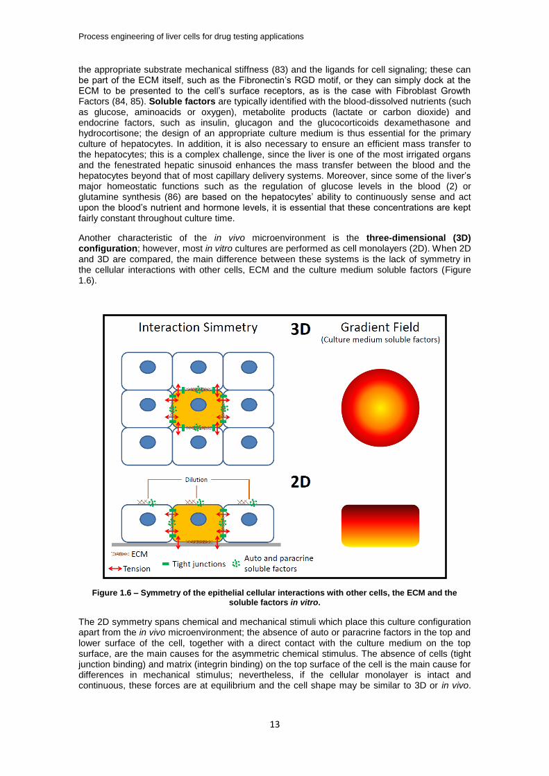
Another characteristic of the in vivo microenvironment is the three-dimensional (3D) configuration; however, most in vitro cultures are performed as cell monolayers (2D). When 2D and 3D are compared, the main difference between these systems is the lack of symmetry in the cellular interactions with other cells, ECM and the culture medium soluble factors (Figure 1.6).
Figure 1.6 – Symmetry of the epithelial cellular interactions with other cells, the ECM and the soluble factors in vitro.
The 2D symmetry spans chemical and mechanical stimuli which place this culture configuration apart from the in vivo microenvironment; the absence of auto or paracrine factors in the top and lower surface of the cell, together with a direct contact with the culture medium on the top surface, are the main causes for the asymmetric chemical stimulus. The absence of cells (tight junction binding) and matrix (integrin binding) on the top surface of the cell is the main cause for differences in mechanical stimulus; nevertheless, if the cellular monolayer is intact and continuous, these forces are at equilibrium and the cell shape may be similar to 3D or in vivo.
Chapter 1 – Introduction
14
However, if there is a gap in the cellular monolayer, the nearby cells will tend to spread on the culture surface.
1.6. State of the art in long term primary cultures of hepatocytes
One of the major breakthroughs for achieving a long term primary culture of hepatocytes has been the collagen sandwich system (87). In these cultures the hepatocyte monolayer is coated with a collagen layer and the albumin secretion of these primary cultures of hepatocytes can be kept up to 6 weeks with the formation of bile-canaliculi like structures between the cells (88). The formation of the canalicular structures was suggested to be due to a reduction in cell spreading (due to the attachment of the cells to the top collagen layer) that increased the surface area of the lateral membranes of the hepatocytes available for the formation of bile canaliculi; this hypothesis is further reinforced be the absence of stress fibers (typical of cell spreading) in the actin cytoskeleton of the collagen sandwich cultured rat hepatocytes and by the fact that matrigel (a basement membrane-like ECM) can also be used in the sandwich configuration, yielding a similar performance (89). In addition, the top collagen layer also reduces the diffusion rate of the autocrine factors and ECM to the culture medium. Nevertheless, the chemical gradient between the lower surface and the top surface of the hepatocytes is still maintained. This limitation has been recently mitigated by the development of a sandwich culture system with an oxygen permeable membrane for cell attachment; while the lack of other nutrients has not been solved, the enhancement in oxygen availability on the bottom surface of the cells has lead to increased CYP450 1A activity for 4 days, when compared to a non-permeable surface (90).
The perfusion bioreactor developed by Gerlach and co-workers (91) for BAL applications consists of three interwoven permeable capillary systems and a culture chamber; two of these capillary systems are for medium perfusion and each system flows countercurrently to the other to minimize the gradients from the fiber inlet to outlet (nutrient addition and clearance of the metabolic by-products, respectively), whereas the third is meant for gas exchange (O2 delivery and CO2 removal). This perfusion configuration is an attempt to recapitulate the intensive vascularization of the liver. The central culture chamber contains the liver cells (hepatocytes and non-parenchymal fraction), which are cultured in a compact 3D structure. This bioreactor system addresses all the critical biological factors for these cultures and has currently been scaled down from the original 800 mL working volume to 2 mL (92, 93), enabling the use of these devices for long-term drug development tests. However, this bioreactor does not allow sampling of the cells during the culture, only at its end, thus, even though it is a long term system for the primary culture of hepatocytes, these cells cannot be imaged throughout culture time nor withdrawn from the reactor to measure mRNA or protein expression. In addition, the hollow fiber system does not allow for accurate control of pH nor dissolved oxygen (DO) concentration within the fibers.
The application of micropatterning and microfluidic techniques to design miniaturized cell culture devices has also been applied to primary cultures of hepatocytes, but few of these devices span extended culture times. The Bhatia group has designed one of the most successful microscale culture devices for liver cells (80); the most important design criteria for this device were the homo and heterotypic cell-cell interactions. Such interactions were based in hepatocyte-3T3 fibroblast co-culture, which is known to improve the activity and maintenance of the hepatocytes’ liver-specific functions (94). This device was designed in a 24 well plate format to
ensure it was user friendly; each well was micropatterned with 500 m collagen I islands and subsequently seeded with hepatocytes, which formed colonies on top of the collagen islands. Finally, 3T3 fibroblasts were seeded across the entire well to achieve the co-culture system with
a 300 L culture volume. This system enabled the maintenance of liver-specific drug metabolizing activity for up to 2 weeks. This system also showed an impressive robustness, which may be due to the automated spotting of hepatocyte islands; nevertheless, the lack of control of pH and DO, both of them soluble factors, a static culture system and a limited 3D configuration may be the cause for the 2 week limit of these culture systems.
Another promising microscale device for the primary culture of hepatocytes has been developed by the Griffith lab (95). This device has a 12 well plate format, for the above mentioned mentioned practical purposes. In this system, rat hepatocytes were co-cultured with liver non-parenchymal cells enriched in SECs, thus ensuring the presence of homo and heterotypic cell-
Process engineering of liver cells for drug testing applications
15
cell interactions. When comparing with the Bhatia group microscale device, this one has the additional advantage of having a dynamic perfusion system and some extent of oxygen control, which, being based on Computational Fluid Dynamics calculations and oxygen sensors located at the inlet and outlet, does not act upon an actually measured oxygen concentration sensed by the hepatocytes. The dynamic perfusion minimizes the gradients of culture medium soluble factors at the surface of the cells, which improves the transport of nutrients and products of metabolism but may also wash out the cell-secreted autocrine and paracrine factors, as well as ECM components such as fibronectin. Thus, the dilution rate set by the perfusion system must balance these 2 factors. In the device herein described, the dilution rate is approximately 1.4 day
-1,i.e., similar to that of the Bhatia group device (1 medium exchange per day), with the
significant difference that the former device avoids sudden changes in the concentration of these factors (i.e., full medium exchanges). Regarding the shear stress elicited by the culture medium perfusion, its effect has been shown to be negligible in this specific microfluidic device (81), but other published works on such devices typically report flow velocity optimization to minimize shear stress (96). More recently, Sonntag and colleagues (97) have described a microfluidic reactor which has connected culture chambers for liver, brain and bone marrow cells; such a device may potentially allow the assessment of a more systemic response of the human organism to xenobiotics.
The collagen sandwich cultures, hollow fiber bioreactors and microfludic devices have one limitation for long term primary culture of hepatocytes: the control of pH and DO. The main cause for this limitation is the difference between the oxygen concentrations in the inlets of fibers or microchannels (or incubator atmosphere) and the DO in the culture medium bulk. The use of stirred tank bioreactors with good mixing as well as pH and DO control will maintain steady-state concentrations of these two soluble factors in the culture medium. Our group has shown that this controlled system improves the culture of human hepatocyte spheroids when compared to stirred spinner vessels (73), which may be due to the maintenance of a
physiological DO level in the bioreactor (approximately 60 M) (98). Primary cultures of hepatocyte spheroids also retain the in vivo-like 3D configuration that retains ECM and autocrine factors, while creating gradients from the surface to the center of the spheroid. These gradients have been shown not to be detrimental for the hepatocyte viability if the spheroid
diameter is below 200 m (99), whereas spheroid diameters of 100 m enable the maximum specific albumin production (100). Moreover, such bioreactors have a user friendly sampling system (while the system set-up may require specialized bioengineering knowledge), which can be used for specific cell-based assays and analysis and to feed higher throughput devices (such as well plates). This flexibility raises the possibility of using these bioreactors for long term primary cultures of human hepatocytes coupled with a higher throughput analysis approach at each time point.
2. Scope of the Thesis
The main goal of this thesis was to extend primary cultures of hepatocytes for long term drug testing applications; while the current state of the art enables a 2 week culture of functional hepatocytes, their ability to synthesize CYP450 mRNA, de novo, is typically limited to the first week of culture. Thus, this hepatocyte potential to induce the CYP450 enzymes is the most demanding test used in drug development and it has been one of the most important milestones of this work.
The work herein presented is the optimization of a stirred tank bioreactor system for primary cultures of hepatocyte spheroids (73); these bioreactors, with low shear impellers and control of pH, DO and temperature within the culture bulk, provide most of the above mentioned critical factors for the hepatocyte cultures: cell-cell interactions, cell-matrix interactions (matrix synthesized by the cells) and the maintenance of constant concentrations of some soluble factors, namely pH and oxygen (besides the 3D architecture provided by the spheroid culture).
In chapter 2 the effect of alginate microencapsulation on rat hepatocyte spheroid cultures is compared to the non-encapsulated cultures; such these alginate hydrogels have shown to
Chapter 1 – Introduction
16
increase the liver-specific functionality of hepatocyte spheroids, enabling the repeated induction of CYP1A and CYP3A isoforms during 15 and 26 days of culture, respectively.
The bioreactor system used in rat hepatocyte spheroid cultures was further optimized by adding an automated, gravimetrically controlled perfusion system, which allowed the maintenance of stable concentrations of soluble factors. In chapter 3 the effects of this perfusion system will be compared with manual medium exchange, for both non-encapsulated and alginate encapsulated rat hepatocyte spheroid cultures.
This perfusion bioreactor system has been applied to the primary cultures of human hepatocytes. In chapter 4 such cultures will be characterized and validated for long term, repeated dose, drug tests; the results show that this culture system maintains hepatocyte functionality for up to 2 weeks, and the hepatocytes CYP450 enzymes have been induced after 7, 14, 21 and 28 days using hepatocytes from 3 different donors. In addition, the 3D architecture of these human hepatocyte spheroids has been investigated by confocal immunofluorescence microscopy, revealing a liver acinus-like polarization of the hepatocytes and the presence of functional bile canaliculi.
In chapter 5 this thesis will approach the logistics problem concerning the scarcity of isolated human hepatocytes. The effects of both spheroid culture and alginate encapsulation in the hepatic differentiation of human embryonic stem cells (hESC) will be studied by comparison with a standard 2D differentiation protocol.
The results described in chapters 2-5 will be thoroughly discussed in chapter 6, as well as the
future research in this area.
3. References
1. Quinlan GJ, Martin GS, Evans TW. Albumin: Biochemical properties and therapeutic potential. Hepatology 2005;41:1211-1219. 2. Pilkis SJ, Granner DK. MOLECULAR PHYSIOLOGY OF THE REGULATION OF HEPATIC GLUCONEOGENESIS AND GLYCOLYSIS. Annual Review of Physiology 1992;54:885-909. 3. Watford M, Chellaraj V, Ismat A, Brown P, Raman P. Hepatic glutamine metabolism. Nutrition 2002;18:301-303. 4. Gebhardt R, Hengstler JG, Muller D, Glockner R, Buenning P, Laube B, Schmelzer E, et al. New hepatocyte in vitro systems for drug metabolism: Metabolic capacity and recommendations for application in basic research and drug development, standard operation procedures. Drug Metabolism Reviews 2003;35:145-213. 5. Gomez-Lechon MJ, Donato MT, Castell JV, Jover R. Human hepatocytes as a tool for studying toxicity and drug metabolism. Current Drug Metabolism 2003;4:292-312. 6. LeCluyse EL. Human hepatocyte culture systems for the in vitro evaluation of cytochrome P450 expression and regulation. European Journal of Pharmaceutical Sciences 2001;13:343-368. 7. Maurel P. The use of adult human hepatocytes in primary culture and other in vitro systems to investigate drug metabolism in man. Advanced Drug Delivery Reviews 1996;22:105-132. 8. Altin JG, Bygrave FL. Non-parenchymal cells as mediators of physiological responses in liver. Mol Cell Biochem 1988;83:3-14. 9. Wisse E, Braet F, Luo DZ, DeZanger R, Jans D, Crabbe E, Vermoesen A. Structure and function of sinusoidal lining cells in the liver. Toxicologic Pathology 1996;24:100-111. 10. Bilzer M, Roggel F, Gerbes AL. Role of Kupffer cells in host defense and liver disease. Liver International 2006;26:1175-1186. 11. Kmieć Z. Cooperation of liver cells in health and disease. Adv Anat Embryol Cell Biol 2001;161:III-XIII, 1-151. 12. Friedman SL. Hepatic stellate cells: Protean, multifunctional, and enigmatic cells of the liver. Physiological Reviews 2008;88:125-172.
Process engineering of liver cells for drug testing applications
17
13. Vollmar B, Menger MD. The Hepatic Microcirculation: Mechanistic Contributions and Therapeutic Targets in Liver Injury and Repair. Physiological Reviews 2009;89:1269-1339. 14. Monga SPS. Molecular pathology of liver diseases. In: Molecular pathology library. New York: Springer,; 2011. p. xxiii, 931 p. 15. Ishibashi H, Nakamura M, Komori A, Migita K, Shimoda S. Liver architecture, cell function, and disease. Semin Immunopathol 2009;31:399-409. 16. Lindros KO. Zonation of cytochrome P450 expression, drug metabolism and toxicity in liver. Gen Pharmacol 1997;28:191-196. 17. Jungermann K, Kietzmann T. Zonation of parenchymal and nonparenchymal metabolism in liver. Annu Rev Nutr 1996;16:179-203. 18. Jungermann K, Kietzmann T. Role of oxygen in the zonation of carbohydrate metabolism and gene expression in liver. Kidney Int 1997;51:402-412. 19. Jungermann K, Kietzmann T. Oxygen: modulator of metabolic zonation and disease of the liver. Hepatology 2000;31:255-260. 20. Katz NR. Metabolic heterogeneity of hepatocytes across the liver acinus. J Nutr 1992;122:843-849. 21. Listrom CD, Morizono H, Rajagopal BS, McCann MT, Tuchman M, Allewell NM. Expression, purification, and characterization of recombinant human glutamine synthetase. Biochem J 1997;328 ( Pt 1):159-163. 22. Braeuning A, Ittrich C, Köhle C, Hailfinger S, Bonin M, Buchmann A, Schwarz M. Differential gene expression in periportal and perivenous mouse hepatocytes. FEBS J 2006;273:5051-5061. 23. Benhamouche S, Decaens T, Godard C, Chambrey R, Rickman DS, Moinard C, Vasseur-Cognet M, et al. Apc tumor suppressor gene is the "zonation-keeper" of mouse liver. Dev Cell 2006;10:759-770. 24. Hernandez-Gea V, Friedman SL. Pathogenesis of liver fibrosis. Annu Rev Pathol 2011;6:425-456. 25. Svegliati-Baroni G, De Minicis S, Marzioni M. Hepatic fibrogenesis in response to chronic liver injury: novel insights on the role of cell-to-cell interaction and transition. Liver Int 2008;28:1052-1064. 26. Gerlach JC, Zeilinger K, Patzer Ii JF. Bioartificial liver systems: why, what, whither? Regen Med 2008;3:575-595. 27. Carpentier B, Gautier A, Legallais C. Artificial and bioartificial liver devices: present and future. Gut 2009;58:1690-1702. 28. Schmelzer E, Mutig K, Schrade P, Bachmann S, Gerlach JC, Zeilinger K. Effect of Human Patient Plasma Ex Vivo Treatment on Gene Expression and Progenitor Cell Activation of Primary Human Liver Cells in Multi-Compartment 3D Perfusion Bioreactors for Extra-Corporeal Liver Support. Biotechnology and Bioengineering 2009;103:817-827. 29. Kola I, Landis J. Can the pharmaceutical industry reduce attrition rates? Nat Rev Drug Discov 2004;3:711-715. 30. Baranczewski P, Stańczak A, Sundberg K, Svensson R, Wallin A, Jansson J, Garberg P, et al. Introduction to in vitro estimation of metabolic stability and drug interactions of new chemical entities in drug discovery and development. Pharmacol Rep 2006;58:453-472. 31. Arrowsmith J. Trial watch: Phase II failures: 2008-2010. Nat Rev Drug Discov 2011;10:328-329. 32. Arrowsmith J. Trial watch: phase III and submission failures: 2007-2010. Nat Rev Drug Discov 2011;10:87. 33. Pisano GP. Can science be a business? Lessons from biotech. Harv Bus Rev 2006;84:114-124, 150. 34. Garnier JP. Rebuilding the R&D engine in big pharma. Harv Bus Rev 2008;86:68-70, 72-66, 128. 35. Mager DE, Jusko WJ. General pharmacokinetic model for drugs exhibiting target-mediated drug disposition. J Pharmacokinet Pharmacodyn 2001;28:507-532. 36. Guengerich FP. Mechanisms of drug toxicity and relevance to pharmaceutical development. Drug Metab Pharmacokinet 2011;26:3-14. 37. Baillie TA, Rettie AE. Role of biotransformation in drug-induced toxicity: influence of intra- and inter-species differences in drug metabolism. Drug Metab Pharmacokinet 2011;26:15-29.
Chapter 1 – Introduction
18
38. Shuldiner AR, O'Connell JR, Bliden KP, Gandhi A, Ryan K, Horenstein RB, Damcott CM, et al. Association of cytochrome P450 2C19 genotype with the antiplatelet effect and clinical efficacy of clopidogrel therapy. JAMA 2009;302:849-857. 39. Diaz GJ. Basolateral and canalicular transport of xenobiotics in the hepatocyte: A review. Cytotechnology 2000;34:225-235. 40. Klaassen CD. Casarett and Doull's toxicology: the basic science of poisons: McGraw-Hill, 2007. 41. Sinz M, Wallace G, Sahi J. Current industrial practices in assessing CYP450 enzyme induction: Preclinical and clinical. Aaps Journal 2008;10:391-400. 42. Hewitt NJ, Lechon MJG, Houston JB, Hallifax D, Brown HS, Maurel P, Kenna JG, et al. Primary hepatocytes: Current understanding of the regulation of metabolic enzymes and transporter proteins, and pharmaceutical practice for the use of hepatocytes in metabolism, enzyme induction, transporter, clearance, and hepatotoxicity studies. In: 1st Medicon Valley Hepatocyte User Form Symposium; 2006 Jan 26-27; Copenhagen, DENMARK: Taylor & Francis Inc; 2006. p. 159-234. 43. Pritchard JF, Jurima-Romet M, Reimer ML, Mortimer E, Rolfe B, Cayen MN. Making better drugs: Decision gates in non-clinical drug development. Nat Rev Drug Discov 2003;2:542-553. 44. Mouly S, Meune C, Bergmann JF. Mini-series: I. Basic science. Uncertainty and inaccuracy of predicting CYP-mediated in vivo drug interactions in the ICU from in vitro models: focus on CYP3A4. Intensive Care Med 2009;35:417-429. 45. Hosea NA, Collard WT, Cole S, Maurer TS, Fang RX, Jones H, Kakar SM, et al. Prediction of human pharmacokinetics from preclinical information: comparative accuracy of quantitative prediction approaches. J Clin Pharmacol 2009;49:513-533. 46. Tang C, Prueksaritanont T. Use of in vivo animal models to assess pharmacokinetic drug-drug interactions. Pharm Res 2010;27:1772-1787. 47. Huang SM, Strong JM, Zhang L, Reynolds KS, Nallani S, Temple R, Abraham S, et al. New era in drug interaction evaluation: US Food and Drug Administration update on CYP enzymes, transporters, and the guidance process. J Clin Pharmacol 2008;48:662-670. 48. Ramsden R, Beck NB, Sommer KM, Omiecinski CJ. Phenobarbital responsiveness conferred by the 5'-flanking region of the rat CYP2B2 gene in transgenic mice. Gene 1999;228:169-179. 49. Ma X, Cheung C, Krausz KW, Shah YM, Wang T, Idle JR, Gonzalez FJ. A double transgenic mouse model expressing human pregnane X receptor and cytochrome P450 3A4. Drug Metab Dispos 2008;36:2506-2512. 50. Chen AA, Thomas DK, Ong LL, Schwartz RE, Golub TR, Bhatia SN. Humanized mice with ectopic artificial liver tissues. Proc Natl Acad Sci U S A 2011;108:11842-11847. 51. Baptista PM, Siddiqui MM, Lozier G, Rodriguez SR, Atala A, Soker S. The use of whole organ decellularization for the generation of a vascularized liver organoid. Hepatology 2011;53:604-617. 52. Uygun BE, Soto-Gutierrez A, Yagi H, Izamis ML, Guzzardi MA, Shulman C, Milwid J, et al. Organ reengineering through development of a transplantable recellularized liver graft using decellularized liver matrix. Nat Med 2010;16:814-820. 53. Ogino M, Nagata K, Yamazoe Y. Selective suppressions of human CYP3A forms, CYP3A5 and CYP3A7, by troglitazone in HepG2 cells. Drug Metab Pharmacokinet 2002;17:42-46. 54. Wilkening S, Stahl F, Bader A. Comparison of primary human hepatocytes and hepatoma cell line Hepg2 with regard to their biotransformation properties. Drug Metab Dispos 2003;31:1035-1042. 55. Aninat C, Piton A, Glaise D, Le Charpentier T, Langouët S, Morel F, Guguen-Guillouzo C, et al. Expression of cytochromes P450, conjugating enzymes and nuclear receptors in human hepatoma HepaRG cells. Drug Metab Dispos 2006;34:75-83. 56. Josse R, Aninat C, Glaise D, Dumont J, Fessard V, Morel F, Poul JM, et al. Long-term functional stability of human HepaRG hepatocytes and use for chronic toxicity and genotoxicity studies. Drug Metabolism and Disposition 2008;36:1111-1118. 57. Kanebratt KP, Andersson TB. HepaRG cells as an in vitro model for evaluation of cytochrome P450 induction in humans. Drug Metab Dispos 2008;36:137-145. 58. Luo G, Cunningham M, Kim S, Burn T, Lin J, Sinz M, Hamilton G, et al. CYP3A4 induction by drugs: correlation between a pregnane X receptor reporter gene assay and CYP3A4 expression in human hepatocytes. Drug Metab Dispos 2002;30:795-804.
Process engineering of liver cells for drug testing applications
19
59. Gilbert TW, Sellaro TL, Badylak SF. Decellularization of tissues and organs. Biomaterials 2006;27:3675-3683. 60. Darnell M, Schreiter T, Zeilinger K, Urbaniak T, Söderdahl T, Rossberg I, Dillnér B, et al. Cytochrome P450-dependent metabolism in HepaRG cells cultured in a dynamic three-dimensional bioreactor. Drug Metab Dispos 2011;39:1131-1138. 61. Xu Y, Zhou Y, Hayashi M, Shou M, Skiles GL. Simulation of clinical drug-drug interactions from hepatocyte CYP3A4 induction data and its potential utility in trial designs. Drug Metab Dispos 2011;39:1139-1148. 62. D'Amour KA, Agulnick AD, Eliazer S, Kelly OG, Kroon E, Baetge EE. Efficient differentiation of human embryonic stem cells to definitive endoderm. Nature Biotechnology 2005;23:1534-1541. 63. Brolen G, Sivertsson L, Bjorquist P, Eriksson G, Ek M, Semb H, Johansson I, et al. Hepatocyte-like cells derived from human embryonic stem cells specifically via definitive endoderm and a progenitor stage. Journal of Biotechnology 2010;145:284-294. 64. Lock L, Tzanakakis E. Expansion and Differentiation of Human Embryonic Stem Cells to Endoderm Progeny in a Microcarrier Stirred-Suspension Culture. Tissue Engineering Part a 2009:2051-2063. 65. Agarwal S, Holton KL, Lanza R. Efficient differentiation of functional hepatocytes from human embryonic stem cells. Stem Cells 2008;26:1117-1127. 66. Baharvand H, Hashemi SM, Ashtian SK, Farrokhi A. Differentiation of human embryonic stem cells into hepatocytes in 2D and 3D culture systems in vitro. International Journal of Developmental Biology 2006;50:645-652. 67. Cai J, Zhao Y, Liu YX, Ye F, Song ZH, Qin H, Meng S, et al. Directed differentiation of human embryonic stem cells into functional hepatic cells. Hepatology 2007;45:1229-1239. 68. Duan YY, Ma XC, Zou W, Wang C, Bahbahan IS, Ahuja TP, Tolstikov V, et al. Differentiation and Characterization of Metabolically Functioning Hepatocytes from Human Embryonic Stem Cells. Stem Cells 2010;28:674-686. 69. Hay DC, Zhao D, Ross A, Mandalam R, Lebkowski J, Cui W. Direct differentiation of human embryonic stem cells to hepatocyte-like cells exhibiting functional activities. Cloning and Stem Cells 2007;9:51-62. 70. Ishii T, Yasuchika K, Fukumitsu K, Kawamoto T, Kawamura-Saitoh M, Amagai Y, Ikai I, et al. In vitro hepatic maturation of human embryonic stem cells by using a mesenchymal cell line derived from murine fetal livers. Cell and Tissue Research 2010:505-512. 71. Goral VN, Hsieh YC, Petzold ON, Clark JS, Yuen PK, Faris RA. Perfusion-based microfluidic device for three-dimensional dynamic primary human hepatocyte cell culture in the absence of biological or synthetic matrices or coagulants. Lab on a Chip 2010;10:3380-3386. 72. Kidambi S, Yarmush RS, Novik E, Chao P, Yarmush ML, Nahmias Y. Oxygen-mediated enhancement of primary hepatocyte metabolism, functional polarization, gene expression, and drug clearance. Proceedings of the National Academy of Sciences of the United States of America 2009;106:15714-15719. 73. Miranda JP, Leite SB, Muller-Vieira U, Rodrigues A, Carrondo MJT, Alves PM. Towards an Extended Functional Hepatocyte In Vitro Culture. Tissue Engineering Part C-Methods 2009;15:157-167. 74. Nishimura M, Hagi M, Ejiri Y, Kishimoto S, Horie T, Narimatsu S, Naito S. Secretion of Albumin and Induction of CYP1A2 and CYP3A4 in Novel Three-dimensional Culture System for Human Hepatocytes using Micro-space Plate. Drug Metabolism and Pharmacokinetics 2010;25:236-242. 75. Sellaro TL, Ranade A, Faulk DM, McCabe GP, Dorko K, Badylak SF, Strom SC. Maintenance of Human Hepatocyte Function In Vitro by Liver-Derived Extracellular Matrix Gels. Tissue Engineering Part A 2010;16:1075-1082. 76. Yeon JH, Na D, Park JK. Hepatotoxicity assay using human hepatocytes trapped in microholes of a microfluidic device. Electrophoresis 2010;31:3167-3174. 77. Zeisberg M, Kramer K, Sindhi N, Sarkar P, Upton M, Kalluri R. De-differentiation of primary human hepatocytes depends on the composition of specialized liver basement membrane. Mol Cell Biochem 2006;283:181-189. 78. Alvarez SD, Derfus AM, Schwartz MP, Bhatia SN, Sailor MJ. The compatibility of hepatocytes with chemically modified porous silicon with reference to in vitro biosensors. Biomaterials 2009;30:26-34.
Chapter 1 – Introduction
20
79. Yagi H, Parekkadan B, Suganuma K, Soto-Gutierrez A, Tompkins RG, Tilles AW, Yarmush ML. Long-term superior performance of a stem cell/hepatocyte device for the treatment of acute liver failure. Tissue Eng Part A 2009;15:3377-3388. 80. Khetani SR, Bhatia SN. Microscale culture of human liver cells for drug development. Nat Biotechnol 2008;26:120-126. 81. Powers MJ, Domansky K, Kaazempur-Mofrad MR, Kalezi A, Capitano A, Upadhyaya A, Kurzawski P, et al. A microfabricated array bioreactor for perfused 3D liver culture. Biotechnology and Bioengineering 2002;78:257-269. 82. Chiang JY. Bile acids: regulation of synthesis. J Lipid Res 2009;50:1955-1966. 83. Zhao G, Cui J, Qin Q, Zhang J, Liu L, Deng S, Wu C, et al. Mechanical stiffness of liver tissues in relation to integrin β1 expression may influence the development of hepatic cirrhosis and hepatocellular carcinoma. J Surg Oncol 2010;102:482-489. 84. Gullberg D, Terracio L, Borg TK, Rubin K. Identification of integrin-like matrix receptors with affinity for interstitial collagens. J Biol Chem 1989;264:12686-12694. 85. Rahman S, Patel Y, Murray J, Patel KV, Sumathipala R, Sobel M, Wijelath ES. Novel hepatocyte growth factor (HGF) binding domains on fibronectin and vitronectin coordinate a distinct and amplified Met-integrin induced signalling pathway in endothelial cells. BMC Cell Biol 2005;6:8. 86. Haussinger D, Lamers WH, Moorman AFM. HEPATOCYTE HETEROGENEITY IN THE METABOLISM OF AMINO-ACIDS AND AMMONIA. Enzyme 1992;46:72-93. 87. Dunn JCY, Tompkins RG, Yarmush ML. Long-term in vitro function of adult hepatocytes in a collagen sandwich configuration. Biotechnology Progress 1991;7:237-245. 88. LeCluyse EL, Audus KL, Hochman JH. Formation of extensive canalicular networks by rat hepatocytes cultured in collagen-sandwich configuration. Am J Physiol 1994;266:C1764-1774. 89. Cosgrove BD, Alexopoulos LG, Hang TC, Hendriks BS, Sorger PK, Griffith LG, Lauffenburger DA. Cytokine-associated drug toxicity in human hepatocytes is associated with signaling network dysregulation. Mol Biosyst 2010;6:1195-1206. 90. Matsui H, Osada T, Moroshita Y, Sekijima M, Fujii T, Takeuchi S, Sakai Y. Rapid and enhanced repolarization in sandwich-cultured hepatocytes on an oxygen-permeable membrane. Biochemical Engineering Journal 2010;52:255-262. 91. Gerlach JC, Encke J, Hole O, Müller C, Ryan CJ, Neuhaus P. Bioreactor for a larger scale hepatocyte in vitro perfusion. Transplantation 1994;58:984-988. 92. Zeilinger K, Schreiter T, Darnell M, Söderdahl T, Lübberstedt M, Dillner B, Knobeloch D, et al. Scaling down of a clinical 3D perfusion multi-compartment hollow fiber liver bioreactor developed for extracorporeal liver support to an analytical scale device useful for hepatic pharmacological in vitro studies. Tissue Eng Part C Methods 2011. 93. Mueller D, Tascher G, Müller-Vieira U, Knobeloch D, Nuessler AK, Zeilinger K, Heinzle E, et al. In-depth physiological characterization of primary human hepatocytes in a 3D hollow-fiber bioreactor. J Tissue Eng Regen Med 2011;5:e207-218. 94. Bhatia SN, Balis UJ, Yarmush ML, Toner M. Effect of cell-cell interactions in preservation of cellular phenotype: cocultivation of hepatocytes and nonparenchymal cells. Faseb Journal 1999;13:1883-1900. 95. Domansky K, Inman W, Serdy J, Dash A, Lim MHM, Griffith LG. Perfused multiwell plate for 3D liver tissue engineering. Lab on a Chip 2010;10:51-58. 96. Korin N, Bransky A, Khoury M, Dinnar U, Levenberg S. Design of Well and Groove Microchannel Bioreactors for Cell Culture. Biotechnology and Bioengineering 2009;102:1222-1230. 97. Sonntag F, Schilling N, Mader K, Gruchow M, Klotzbach U, Lindner G, Horland R, et al. Design and prototyping of a chip-based multi-micro-organoid culture system for substance testing, predictive to human (substance) exposure. J Biotechnol 2010;148:70-75. 98. Carreau A, El Hafny-Rahbi B, Matejuk A, Grillon C, Kieda C. Why is the partial oxygen pressure of human tissues a crucial parameter? Small molecules and hypoxia. J Cell Mol Med 2011;15:1239-1253. 99. Curcio E, Salerno S, Barbieri G, De Bartolo L, Drioli E, Bader A. Mass transfer and metabolic reactions in hepatocyte spheroids cultured in rotating wall gas-permeable membrane system. Biomaterials 2007;28:5487-5497. 100. Glicklis R, Merchuk JC, Cohen S. Modeling mass transfer in hepatocyte spheroids via cell viability, spheroid size, and hepatocellular functions. Biotechnology and Bioengineering 2004;86:672-680.
21
Chapter 2
Alginate-Encapsulated Three-Dimensional Cultures of
Hepatocytes in Bioreactors
This Chapter was based on the manuscript: Miranda J, Rodrigues A, Tostoes R, Leite S, Zimmermann H, Carrondo M, Alves P. 2010. Extending hepatocyte functionality for drug testing applications using high viscosity alginate encapsulated 3D cultures in bioreactors. Tissue Eng Part C Methods
Chapter 2 –Alginate-Encapsulated Three-Dimensional Cultures of hepatocytes in Bioreactors
22
Abstract
The maintenance of differentiated hepatocyte phenotype in vitro depends on several factors—in particular, on extracellular matrix interactions, for example, with three-dimensional (3D) matrices. Alginate hydrogel provides the cells with a good extracellular matrix due to the formation of a massive capsule with semi-permeable properties that allows for diffusion of the medium components into the cells as well as efficient waste product elimination. Simultaneously, alginate protects the cells from shear stress caused by the hydrodynamics when cultured in stirred systems such as bioreactors. We have previously developed a hepatocyte aggregate 3D culture system in a bioreactor where improved hepatocyte functionality could be maintained over longer periods (21 days). In this work, ultra-high-viscosity alginate was used for hepatocyte aggregates entrapment. Hepatocyte biotransformation (phase I and II enzymes), CYP450 inducibility, and secretory capacity (albumin and urea production) were monitored. The analyses were performed in both spinner vessels and bioreactors to test the effect of the DO control, unavailable in the spinners. Performance of alginate-encapsulated hepatocyte aggregates in culture was compared with nonencapsulated aggregate cultures in both bioreactor (controlled environment) and spinner vessels. For both culture systems, hepatocytes' metabolic and biotransformation capacities were maintained for up to 1 month, and encapsulated cells in bioreactors showed the best performance. In particular, albumin production rate increased 2- and 1.5-fold in encapsulated aggregates compared with nonencapsulated aggregates in bioreactor and spinner vessels, respectively. Urea production rate increased twofold in encapsulated cultures compared with nonencapsulated cells, in both bioreactor and spinner vessels. Similarly, in both the bioreactor and the spinner system, cell encapsulation resulted in a 1.5- and 2.8-fold improvement of hepatocyte 7-ethoxycoumarin and uridine diphosphate glucuronosyltransferases (UGT) activities, respectively. For all parameters, but for UGT activity, the bioreactor system resulted better than the spinner vessels; for UGT activity no difference was observed between the two. Furthermore, both encapsulated and nonencapsulated 3D culture systems were inducible by 3-methylcholanthrene and dexamethasone. The encapsulated systems consistently showed improved performance over the nonencapsulated cells, indicating that the protection conferred by the alginate matrix plays a relevant role in maintaining the hepatocyte functionalities in vitro.
Process engineering of liver cells for drug testing applications
23
Table of Contents
1. Introduction ........................................................................................................ 24
2. Material and Methods ........................................................................................ 25
2.1. Rat hepatocyte isolation ................................................................................... 25
2.2. Cell culture ....................................................................................................... 25
2.3. Entrapment of hepatocyte aggregates in alginate ............................................. 26
2.4. Cultures in the bioreactor ................................................................................. 26
2.5. Cell counting .................................................................................................... 26
2.6. Determination of albumin secretion and urea synthesis .................................... 26
2.7. CYP450 measurement ..................................................................................... 26
2.8. UGT activity ...................................................................................................... 27
2.9. Induction assay ................................................................................................ 27
2.10. Statistics ......................................................................................................... 27
3. Results ............................................................................................................... 28
3.1. Effect of cell inoculum and alginate concentration on hepatocytes functionality 28
3.2. Scale-up: Encapsulated aggregates cultures in spinner vessels and bioreactor 31
3.3. Proof of concept and validation ........................................................................ 34
4. Discussion ......................................................................................................... 35
5. References ......................................................................................................... 37
Chapter 2 –Alginate-Encapsulated Three-Dimensional Cultures of hepatocytes in Bioreactors
24
1. Introduction
Primary cultures of hepatocytes constitute a well-accepted in vitro model system for studies of drug metabolism, enzyme induction, cell transplantation, viral hepatitis studies, and hepatocyte regeneration. However, such cells grown in culture undergo rapid loss of differentiated functions and do not proliferate swiftly (1, 2).
Innumerous attempts have been made to culture hepatocytes in an environment that supports functional longevity and where cells more closely reflect characteristics of the liver in vivo. The difficulty has been in maintaining hepatocyte cultures that show differentiated characteristics and maintain metabolic rates comparable with physiological liver functions over long periods. Approaches that have been explored include choice of hormonally defined media for the maintenance of differentiated functions (3-5), cultured on different types of extracellular matrix (ECM) molecules (6, 7), use of cocultures with nonparenchymal liver cells(8), the addition of molecules that are able to improve expression of a differentiated phenotype(9), and a combination of several of these conditions to realize in vitro hepatic organoids (10). Such techniques are of great help; however, for the development of bioartificial organs(11), besides the need of optimal hepatocyte functional and structural integrity and efficiency, a large amount of liver-like tissue is necessary(12).
Since organisms have a three-dimensional (3D) spatial organization and cells in organs are exposed to nonuniform oxygen and nutrient distributions as well as physical and chemical stresses, it is expectable that hepatocytes grown as two-dimensional (2D) monolayers have less complex intercellular and cell–matrix interactions compared to their real physiological state. Thus, in the last two decades efforts were made to develop 3D cell culture models bridging the gap between cell-based assays and animal studies(13). Among the several types of 3D cell models that have been developed, the hepatocyte aggregate model, consisting on cell aggregates of 100–500 μm, has been increasingly gaining recognition in the biomedical research field(14-17).
One concern when using aggregates is that they can experience strong shear stresses, which may affect cell physiology, and the system could hardly be used in clinical applications due to cell aggregate unprotected nature. Within the human body, cells like hepatocytes interact with neighboring cells and ECM components to establish a complex communication network of biochemical and mechanical signals. On the basis of these facts, hydrogel matrices that provide physical support for cells' self-assembly have been adopted. Among the biopolymers used in biotechnological and medical applications, alginate has been one of the most successful. Distinct but interrelated factors substantiate the cells' enhanced performance in alginate: ECM, coculture with nonparenchymal cells, maintenance of cell morphology, cell-to-cell contact with expression of intercellular contact zones and polarity, and aggregation into multi-layers may all enhance survival and performance of encapsulated hepatocytes compared to monolayer cultures on simple substrata(18).
Presently, selection of the hydrogel scaffold methods has been largely based on material availability. However, properties like biodegradability and biocompatibility became a major concern due to the need to immobilize hepatocyte aggregates for tissue engineering and to generate bioartificial organs(19). Therefore, in this work, a clinical-grade, biocompatible, immunologically inert, highly purified alginate of extremely high viscosity (>30 mPa · s; 0.1% w/v solution) extracted by a novel process in a good manufacturing practices (GMP) amenable facility and uniformly cross-linked with Ba2Cl (19) has been adopted. Simultaneously, this alginate provides the cells with a good, thermostable ECM forming a semi-permeable membrane that allows for diffusion of the medium contents to the cells and for product elimination.
Finally, aggregate culture requirements are substantially higher than those of 2D cultures grown in static conditions since diffusion of nutrients and oxygen should not be limited. Bioreactors were primarily developed to optimize mass transfer, a crucial element for guaranteeing gas/nutrient supply and waste elimination, essential factors for maintaining cell viability within 3D cell tissue cultures(20).
Process engineering of liver cells for drug testing applications
25
On the basis of this prior concept, we herein describe a novel 3D culture strategy based on the cultivation of hepatocyte aggregates encapsulated in such alginate matrices under a totally controlled environment bioreactor. This system combines the advantages of a 3D culture system in alginate gel, controlled oxygen supply, and homogeneous and well-defined culture conditions with the possibility of high throughput and automation. Within this context, for culture characterization, hepatocyte biotransformation and metabolic capacity were assessed. In particular, phase I (7-Ethoxycoumarin [ECOD] and testosterone hydroxilation) and phase II (uridine diphosphate glucuronosyltransferases [UGT]) activities and induction measurements will be performed as well as albumin and urea quantifications.
2. Material and Methods
2.1. Rat hepatocyte isolation
Male Wistar rats (6–9 weeks old, 200–300 g body weight) kept on a standard diet with free access to water were obtained from Instituto de Higiene e Medicina Tropical Animal House, Lisbon, Portugal. For surgery the animals were anesthetized with an intraperitoneal injection of ketamine (90 mg/kg body weight) and xylazine (10 mg/kg body weight) solution.
Hepatocytes were isolated by the two-step collagenase perfusion method described by Seglen(21), with slight modifications. Briefly, the liver was perfused in situ, via portal vein, for 7 min with a Perfusion Buffer I (0.14 M NaCl, 6.7 mM KCl, and 10 mM HEPES with pH 7.5 adjusted with 2.4 M EGTA) at 39°C. Subsequently, perfusion was continued with a collagenase (10 mg/100 g body weight) containing buffer (67 mM NaCl, 6.7 mM KCl, 100 mM HEPES, albumin (0.5%), and 4.8 mM CaCl2, adjusted to pH 7.6) at 39°C for 4 min. The flow rate for the perfusion buffers was 10 mL/min. After perfusion the liver was removed from the animal and dissociated in cold Perfusion Buffer I with 10 g/L of albumin. The resultant cell suspension was filtered through gauze, centrifuged for 10 min at 50 g, washed once with the medium, centrifuged again, and resuspended in the cold medium for a final concentration less than 3.5 × 10
6 cell/mL. For the enrichment of the final hepatocyte population, an additional Percoll-
step was included. Five milliliters of cell suspension was layered over a 25% Percoll solution and centrifuged at 1300 g at 4°C for 20 min, and the hepatocytes were obtained as a pellet. The cell pellet was diluted in phosphate-buffered saline (PBS), centrifuged for 10 min at 50 g, and washed twice with PBS for removing the Percoll solution. Finally, the cells were harvested in supplemented Williams'E medium for culturing. Cell viability was assessed by the trypan blue exclusion test and was, routinely, within an 85%–95%.
2.2. Cell culture
Freshly isolated hepatocytes were cultured with Williams'E medium supplemented with 10% fetal bovine serum (FBS) (v/v), 1.4 μM hydrocortisone, 0.032 U/mL insulin, 15 mM HEPES, 1 mM sodium pyruvate, 1 mM NEAA, antibiotics 100 U/mL penicillin/100 μg/mL streptomycin, and 40 μg/mL gentamicin—designated as Williams'E complete medium.
In 2D cultures (static monolayer cultures) cells were seeded onto Matrigel®-precoated culture
plates at a density of 5 × 10⁴ cells/cm
2. Hepatocytes were left untreated for at least 12 h at 37°C
in a humidified atmosphere with 5% CO2 in air, allowing cell attachment. The medium was changed the following day to remove unattached cells. The culture medium was renewed every 24 h and the cells were routinely examined under phase-contrast microscopy before every culture medium renewal.
In 3D cultures (stirred tank cultures), single-cell suspensions were seeded in 125 mL spinner vessels or in a 250 mL stirred tank bioreactor. To promote cell aggregation, 15 × 10
6 cells or
30 × 106 cells were inoculated in 70 or 200 mL in the spinner vessels or in the bioreactor,
respectively, with supplemented medium at 15% FBS. Stirred tanks were agitated at 80 rotations per minute (RPM), and kept at 37°C in a humidified atmosphere of 5% CO2 in air for 24 h. After 24 h, 50% culture medium was changed, added to a final culture volume of 125 mL or
Chapter 2 –Alginate-Encapsulated Three-Dimensional Cultures of hepatocytes in Bioreactors
26
250 mL in the spinner vessels or in the bioreactor, respectively, and the FBS concentration was adjusted to 10% (v/v). To maintain the aggregates, the operational mode applied was a 50% medium substitution (refeed mode) every 4 days, for nutrient availability and to decrease the accumulation of bioproducts of cellular metabolism that can be toxic to the cells.
The supernatants and cells were collected according to the protocol at the indicated time points and stored at −20°C for further assays.
2.3. Entrapment of hepatocyte aggregates in alginate
Ultra-high-viscosity, clinical-grade alginates (viscosity of a 0.1% [w/v] solution in distilled water was 20–30 mPa · s) were collected in Coquimbo, Chile, and extracted in a GMP-amenable facility at the Fraunhofer IBMT (Sulzbach, Germany) separately from stripes of Lessonia nigrescens and Lessonia trabeculata as described by Zimmermann et al (19). A 1:1 mixture of those alginates was used at 0.4% or 0.7% (w/v) in 0.9% NaCl solution.
For alginate entrapment, hepatocyte aggregates were suspended in the mixture described above to obtain the desired cell density per mL of alginate. Cell suspension was dropped through a syringe-based apparatus into an isotonic polymerization solution containing 20 mM BaCl2 (pH 7.3). After 15–20 min of incubation, the resulting spherical beads (mean size 400 ± 100 μm) with entrapped hepatocyte aggregates were washed in 0.9% NaCl solution, suspended in Williams'E complete medium, and transferred to the stirring vessel (spinner or bioreactor).
2.4. Cultures in the bioreactor
To ensure a fully controlled cell culture environment, a commercially available bioreactor, 500 mL Sartorius-Stedim Biostat Q-Plus system, with a three-blade segment stirrer was used. The internal geometry of each vessel is similar to the commercially available spinner vessels, although it has a marine impeller, in contrast to the paddle impeller of the spinners. The bioreactors have multiple (up to 12) upper cap ports for different applications, such as pH and DO meters, that allow online measurement and control of these parameters, easy sampling, and the addition or removal of medium or other supplements or solutions. The pH and DO were controlled via surface aeration with air, N2 (for DO), and CO2 (for pH). The pH was kept at 7.4 and the DO at 30%. The temperature was kept at 37°C by water recirculation in the vessel jacket controlled by a thermocirculator bath. The bioreactor controller unit was used to monitor and control pH, DO, and temperature. Data acquisition and process control were performed using MFCS/Win Supervisory Control and Data Acquisition (SCADA) software (B-Braun Biotech International GmbH).
2.5. Cell counting
Alginate beads were dissolved by incubating in Na2SO4 1 M, pH 7, at 37°C for 20 min. Released aggregates were digested with trypsin, and single cells' viability was assessed using trypan blue exclusion method. Cell were counted using a Neubauer counting chamber, and data calculated as percentage of cell survival considering cell number at day 1 as 100%.
2.6. Determination of albumin secretion and urea synthesis
The secretion of albumin from hepatocytes was measured by an enzyme-linked immunosorbent assay using NEPHRAT albumin test kit (ref.NR002; Exocell). The assay was performed according to the manufacturer's description. The results were expressed as μmol/10
6 cells/day
at the indicated time point.
The urea synthesis rate was determined using a quantitative colorimetric urea kit (QuantiChrom™ Urea Assay Kit, DIUR-500, ref DIUR-500; BioAssay Systems), according to the manufacturer's instructions. The results were expressed as μmol/10
6 cells/day at the indicated
time points.
2.7. CYP450 measurement
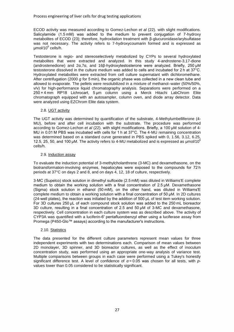
Process engineering of liver cells for drug testing applications
27
ECOD activity was measured according to Gomez-Lechon et al (22). with slight modifications. Salicylamide (1.5 mM) was added to the medium to prevent conjugation of 7-hydroxy metabolites of ECOD (23); therefore, hydroxilation treatment with β-glucuronidase/arylsulfatase was not necessary. The activity refers to 7-hydroxycoumarin formed and is expressed as μmol/10
6 cells/h.
Testosterone is regio- and stereoselectively metabolized by CYPs to several hydroxylated metabolites that were extracted and analyzed. In this study 4-androstene-3,17-dione (androstenedione) and 2α,7α, and 16β-hydroxytestosterone were analyzed. Briefly, 250 μM testosterone dissolved in the culture medium was added to cells and incubated for 2 h at 37°C. Hydroxylated metabolites were extracted from cell culture supernatant with dichloromethane. After centrifugation (2000 g for 5 min), the organic phase was collected in a new clean tube and allowed to evaporate. The pellets were resolubilized in a mixture of methanol–water (50%/50%, v/v) for high-performance liquid chromatography analysis. Separations were performed on a 250 × 4 mm RP18 Lichrocart, 5 μm column using a Merck Hitachi LabChrom Elite chromatograph equipped with an autosampler, column oven, and diode array detector. Data were analyzed using EZChrom Elite data system.
2.8. UGT activity
The UGT activity was determined by quantification of the substrate, 4-Methylumbelliferone (4-MU), before and after cell incubation with the substrate. The procedure was performed according to Gomez-Lechon et al (22). with slight modifications. Briefly, a 100 μM solution of 4-MU in 0.01 M PBS was incubated with cells for 1 h at 37°C. The 4-MU remaining concentration was determined based on a standard curve generated in PBS spiked with 0, 1.56, 3.12, 6.25, 12.5, 25, 50, and 100 μM. The activity refers to 4-MU metabolized and is expressed as μmol/10
6
cells/h.
2.9. Induction assay
To evaluate the induction potential of 3-methylcholanthrene (3-MC) and dexamethasone, on the biotransformation-involving enzymes, hepatocytes were exposed to the compounds for 72 h periods at 37°C on days 2 and 8, and on days 4, 12, 18 of culture, respectively.
3-MC (Supelco) stock solution in dimethyl sulfoxide (2.5 mM) was diluted in Williams'E complete medium to obtain the working solution with a final concentration of 2.5 μM. Dexamethasone (Sigma) stock solution in ethanol (50 mM), on the other hand, was diluted in Williams'E complete medium to obtain a working solution with a final concentration of 50 μM. In 2D cultures (24-well plates), the reaction was initiated by the addition of 500 μL of test item working solution. For 3D cultures 250 μL of each compound stock solution was added to the 250 mL bioreactor 3D culture, resulting in a final concentration of 2.5 and 50 μM of 3-MC and dexamethasone, respectively. Cell concentration in each culture system was as described above. The activity of CYP3A was quantified with a luciferin-6′ pentafluorobenzyl ether using a luciferase assay from Promega (P450-Glo™ assays) according to the manufacturer's instructions.
2.10. Statistics
The data presented for the different culture parameters represent mean values for three independent experiments with two determinations each. Comparison of mean values between 2D monolayer, 3D spinner, and 3D bioreactor cultures, as well as the effect of inoculum concentration study, was performed using an appropriate one-way analysis of variance test. Multiple comparisons between groups in each case were performed using a Tukey's honestly significant difference test. A level of confidence of α = 0.05 was chosen for all tests, with p-
values lower than 0.05 considered to be statistically significant.
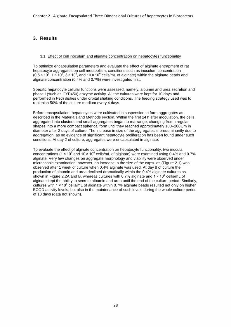
Chapter 2 –Alginate-Encapsulated Three-Dimensional Cultures of hepatocytes in Bioreactors
28
3. Results
3.1. Effect of cell inoculum and alginate concentration on hepatocytes functionality
To optimize encapsulation parameters and evaluate the effect of alginate entrapment of rat hepatocyte aggregates on cell metabolism, conditions such as inoculum concentration (0.5 × 10
6, 1 × 10
6, 3 × 10
6, and 10 × 10
6 cells/mL of alginate) within the alginate beads and
alginate concentration (0.4% and 0.7%) were investigated first.
Specific hepatocyte cellular functions were assessed, namely, albumin and urea secretion and phase I (such as CYP450) enzyme activity. All the cultures were kept for 10 days and performed in Petri dishes under orbital shaking conditions. The feeding strategy used was to replenish 50% of the culture medium every 4 days.
Before encapsulation, hepatocytes were cultivated in suspension to form aggregates as described in the Materials and Methods section. Within the first 24 h after inoculation, the cells aggregated into clusters and small aggregates began to rearrange, changing from irregular shapes into a more compact spherical form until they reached approximately 100–200 μm in diameter after 2 days of culture. The increase in size of the aggregates is predominantly due to aggregation, as no evidence of significant hepatocyte proliferation has been found under such conditions. At day 2 of culture, aggregates were encapsulated in alginate.
To evaluate the effect of alginate concentration on hepatocyte functionality, two inocula concentrations (1 × 10
6 and 10 × 10
6 cells/mL of alginate) were examined using 0.4% and 0.7%
alginate. Very few changes on aggregate morphology and viability were observed under microscopic examination; however, an increase in the size of the capsules (Figure 2.1) was observed after 1 week of culture when 0.4% alginate was used. At day 8 of culture the production of albumin and urea declined dramatically within the 0.4% alginate cultures as shown in Figure 2.2A and B, whereas cultures with 0.7% alginate and 1 × 10
6 cells/mL of
alginate kept the ability to secrete albumin and urea until the end of the culture period. Similarly, cultures with 1 × 10
6 cells/mL of alginate within 0.7% alginate beads resulted not only on higher
ECOD activity levels, but also in the maintenance of such levels during the whole culture period of 10 days (data not shown).
Process engineering of liver cells for drug testing applications
29
Figure 2.1– Phase-contrast microscopy (amplification of 100 × ) of encapsulated hepatocyte
aggregates in 0.7% alginate using an inoculum of (A) 10 × 106 or (B) 1 × 10
6 cells/mL alginate, and
0.4% alginate using an inoculum of (C) 10 × 106 or (D) 1 × 10
6 cells/mL alginate. Samples collected at
day 8 of culture.
Chapter 2 –Alginate-Encapsulated Three-Dimensional Cultures of hepatocytes in Bioreactors
30
Figure 2.2 – Functional capacity of hepatocytes assessed by determining (A) urea production and (B) albumin secretion when cultured as encapsulated aggregates in 0.4% alginate with an inoculum of 1 × 10
6 cells/mL of
alginate (white bars) or 10 × 106 cells/mL of alginate (hatched bars), and 0.7% alginate with an inoculum of 1 × 10
6
cells/mL of alginate (black bars) or 10 × 106 cells/mL of alginate (gray bars).
The quantity of hydroxytestosterone (OHT) metabolites produced by cultured hepatocytes was monitored as well. It varied depending on the metabolite (2α-OHT, 7α-OHT, and 16β-OHT and androstenedione) and the culture conditions. Metabolite production during cultivation is presented for the four culture systems at days 3 and 6 of culture (Table 2.1), showing higher
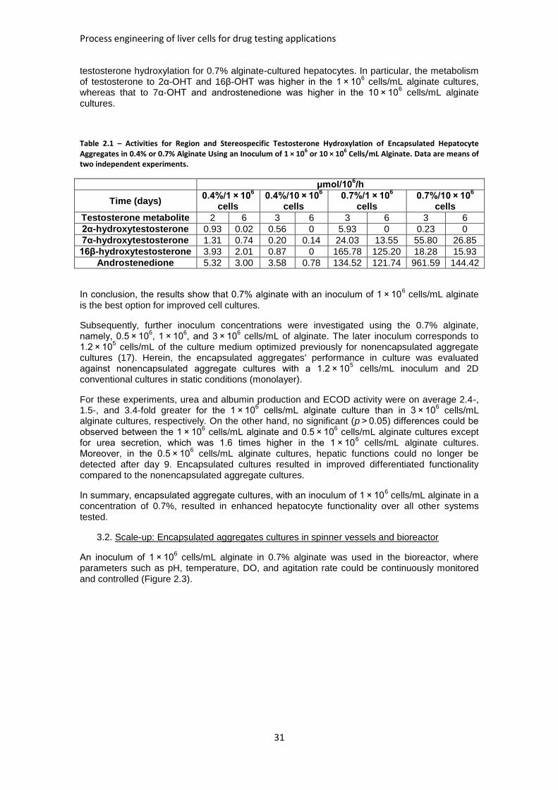
Process engineering of liver cells for drug testing applications
31
testosterone hydroxylation for 0.7% alginate-cultured hepatocytes. In particular, the metabolism of testosterone to 2α-OHT and 16β-OHT was higher in the 1 × 10
6 cells/mL alginate cultures,
whereas that to 7α-OHT and androstenedione was higher in the 10 × 106 cells/mL alginate
cultures.
Table 2.1 – Activities for Region and Stereospecific Testosterone Hydroxylation of Encapsulated Hepatocyte Aggregates in 0.4% or 0.7% Alginate Using an Inoculum of 1 × 10
6 or 10 × 10
6 Cells/mL Alginate. Data are means of
two independent experiments.
μmol/106/h
Time (days) 0.4%/1 × 10
6
cells 0.4%/10 × 10
6
cells 0.7%/1 × 10
6
cells 0.7%/10 × 10
6
cells
Testosterone metabolite 2 6 3 6 3 6 3 6
2α-hydroxytestosterone 0.93 0.02 0.56 0 5.93 0 0.23 0
7α-hydroxytestosterone 1.31 0.74 0.20 0.14 24.03 13.55 55.80 26.85
16β-hydroxytestosterone 3.93 2.01 0.87 0 165.78 125.20 18.28 15.93
Androstenedione 5.32 3.00 3.58 0.78 134.52 121.74 961.59 144.42
In conclusion, the results show that 0.7% alginate with an inoculum of 1 × 10⁶ cells/mL alginate
is the best option for improved cell cultures.
Subsequently, further inoculum concentrations were investigated using the 0.7% alginate, namely, 0.5 × 10
6, 1 × 10
6, and 3 × 10
6 cells/mL of alginate. The later inoculum corresponds to
1.2 × 10⁵ cells/mL of the culture medium optimized previously for nonencapsulated aggregate
cultures (17). Herein, the encapsulated aggregates' performance in culture was evaluated against nonencapsulated aggregate cultures with a 1.2 × 10
5 cells/mL inoculum and 2D
conventional cultures in static conditions (monolayer).
For these experiments, urea and albumin production and ECOD activity were on average 2.4-, 1.5-, and 3.4-fold greater for the 1 × 10
6 cells/mL alginate culture than in 3 × 10
6 cells/mL
alginate cultures, respectively. On the other hand, no significant (p > 0.05) differences could be observed between the 1 × 10
6 cells/mL alginate and 0.5 × 10
6 cells/mL alginate cultures except
for urea secretion, which was 1.6 times higher in the 1 × 10⁶ cells/mL alginate cultures.
Moreover, in the 0.5 × 10⁶ cells/mL alginate cultures, hepatic functions could no longer be
detected after day 9. Encapsulated cultures resulted in improved differentiated functionality compared to the nonencapsulated aggregate cultures.
In summary, encapsulated aggregate cultures, with an inoculum of 1 × 10⁶ cells/mL alginate in a
concentration of 0.7%, resulted in enhanced hepatocyte functionality over all other systems tested.
3.2. Scale-up: Encapsulated aggregates cultures in spinner vessels and bioreactor
An inoculum of 1 × 10⁶ cells/mL alginate in 0.7% alginate was used in the bioreactor, where
parameters such as pH, temperature, DO, and agitation rate could be continuously monitored and controlled (Figure 2.3).
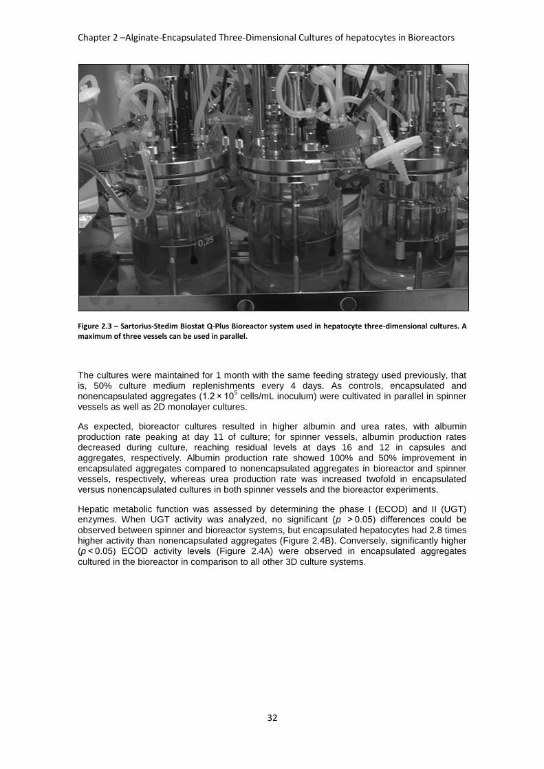
Chapter 2 –Alginate-Encapsulated Three-Dimensional Cultures of hepatocytes in Bioreactors
32
Figure 2.3 – Sartorius-Stedim Biostat Q-Plus Bioreactor system used in hepatocyte three-dimensional cultures. A maximum of three vessels can be used in parallel.
The cultures were maintained for 1 month with the same feeding strategy used previously, that is, 50% culture medium replenishments every 4 days. As controls, encapsulated and nonencapsulated aggregates (1.2 × 10
5 cells/mL inoculum) were cultivated in parallel in spinner
vessels as well as 2D monolayer cultures.
As expected, bioreactor cultures resulted in higher albumin and urea rates, with albumin production rate peaking at day 11 of culture; for spinner vessels, albumin production rates decreased during culture, reaching residual levels at days 16 and 12 in capsules and aggregates, respectively. Albumin production rate showed 100% and 50% improvement in encapsulated aggregates compared to nonencapsulated aggregates in bioreactor and spinner vessels, respectively, whereas urea production rate was increased twofold in encapsulated versus nonencapsulated cultures in both spinner vessels and the bioreactor experiments.
Hepatic metabolic function was assessed by determining the phase I (ECOD) and II (UGT) enzymes. When UGT activity was analyzed, no significant (p > 0.05) differences could be observed between spinner and bioreactor systems, but encapsulated hepatocytes had 2.8 times higher activity than nonencapsulated aggregates (Figure 2.4B). Conversely, significantly higher (p < 0.05) ECOD activity levels (Figure 2.4A) were observed in encapsulated aggregates
cultured in the bioreactor in comparison to all other 3D culture systems.
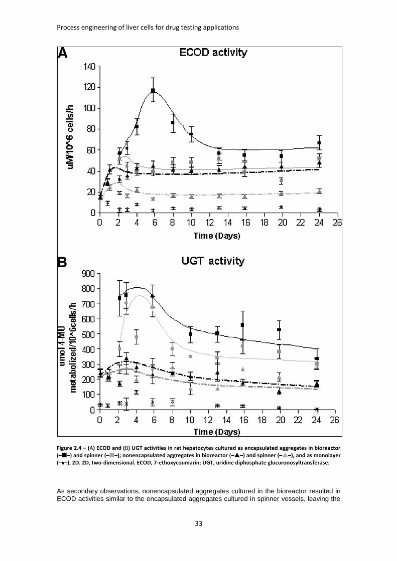
Process engineering of liver cells for drug testing applications
33
Figure 2.4 – (A) ECOD and (B) UGT activities in rat hepatocytes cultured as encapsulated aggregates in bioreactor (––) and spinner (––); nonencapsulated aggregates in bioreactor (––) and spinner (––), and as monolayer (–x–), 2D. 2D, two-dimensional. ECOD, 7-ethoxycoumarin; UGT, uridine diphosphate glucuronosyltransferase.
As secondary observations, nonencapsulated aggregates cultured in the bioreactor resulted in ECOD activities similar to the encapsulated aggregates cultured in spinner vessels, leaving the
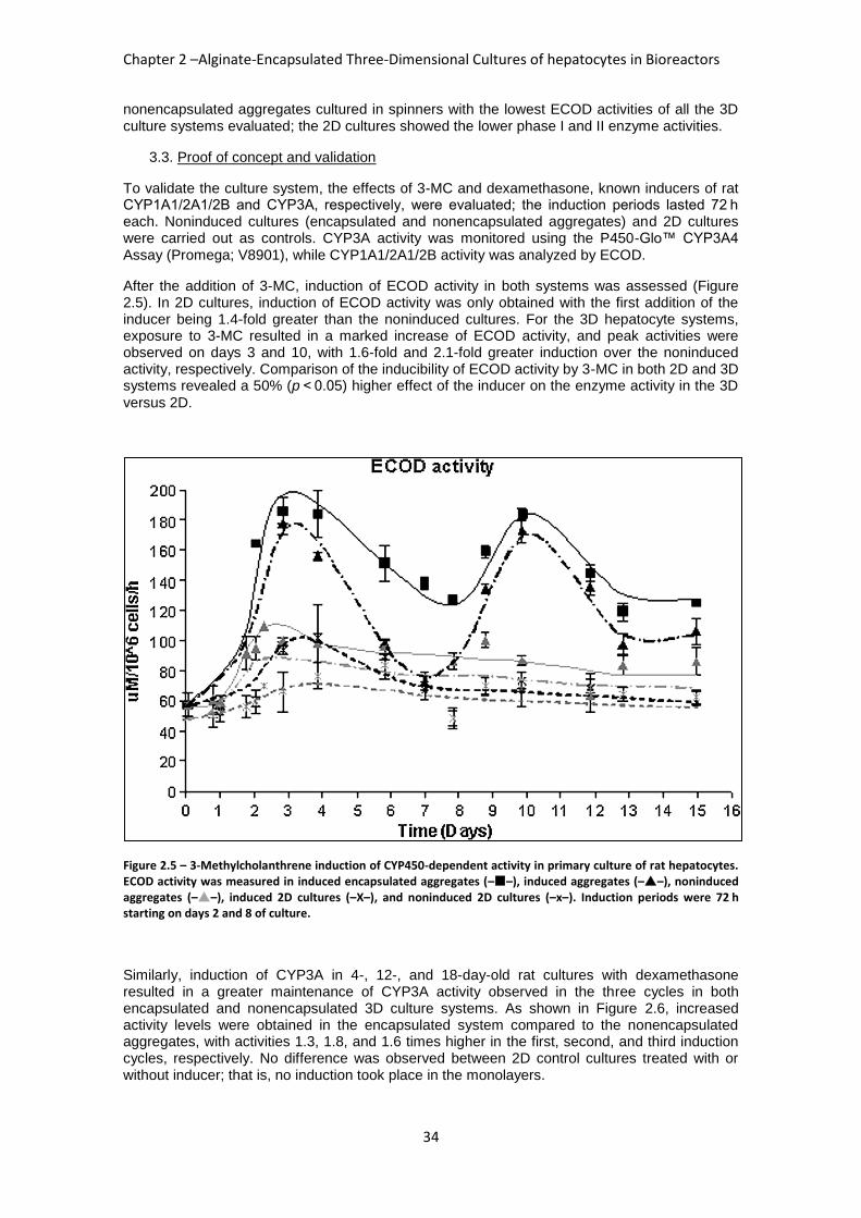
Chapter 2 –Alginate-Encapsulated Three-Dimensional Cultures of hepatocytes in Bioreactors
34
nonencapsulated aggregates cultured in spinners with the lowest ECOD activities of all the 3D culture systems evaluated; the 2D cultures showed the lower phase I and II enzyme activities.
3.3. Proof of concept and validation
To validate the culture system, the effects of 3-MC and dexamethasone, known inducers of rat CYP1A1/2A1/2B and CYP3A, respectively, were evaluated; the induction periods lasted 72 h each. Noninduced cultures (encapsulated and nonencapsulated aggregates) and 2D cultures were carried out as controls. CYP3A activity was monitored using the P450-Glo™ CYP3A4 Assay (Promega; V8901), while CYP1A1/2A1/2B activity was analyzed by ECOD.
After the addition of 3-MC, induction of ECOD activity in both systems was assessed (Figure 2.5). In 2D cultures, induction of ECOD activity was only obtained with the first addition of the inducer being 1.4-fold greater than the noninduced cultures. For the 3D hepatocyte systems, exposure to 3-MC resulted in a marked increase of ECOD activity, and peak activities were observed on days 3 and 10, with 1.6-fold and 2.1-fold greater induction over the noninduced activity, respectively. Comparison of the inducibility of ECOD activity by 3-MC in both 2D and 3D systems revealed a 50% (p < 0.05) higher effect of the inducer on the enzyme activity in the 3D
versus 2D.
Figure 2.5 – 3-Methylcholanthrene induction of CYP450-dependent activity in primary culture of rat hepatocytes. ECOD activity was measured in induced encapsulated aggregates (––), induced aggregates (––), noninduced aggregates (––), induced 2D cultures (–X–), and noninduced 2D cultures (–x–). Induction periods were 72 h starting on days 2 and 8 of culture.
Similarly, induction of CYP3A in 4-, 12-, and 18-day-old rat cultures with dexamethasone resulted in a greater maintenance of CYP3A activity observed in the three cycles in both encapsulated and nonencapsulated 3D culture systems. As shown in Figure 2.6, increased activity levels were obtained in the encapsulated system compared to the nonencapsulated aggregates, with activities 1.3, 1.8, and 1.6 times higher in the first, second, and third induction cycles, respectively. No difference was observed between 2D control cultures treated with or without inducer; that is, no induction took place in the monolayers.
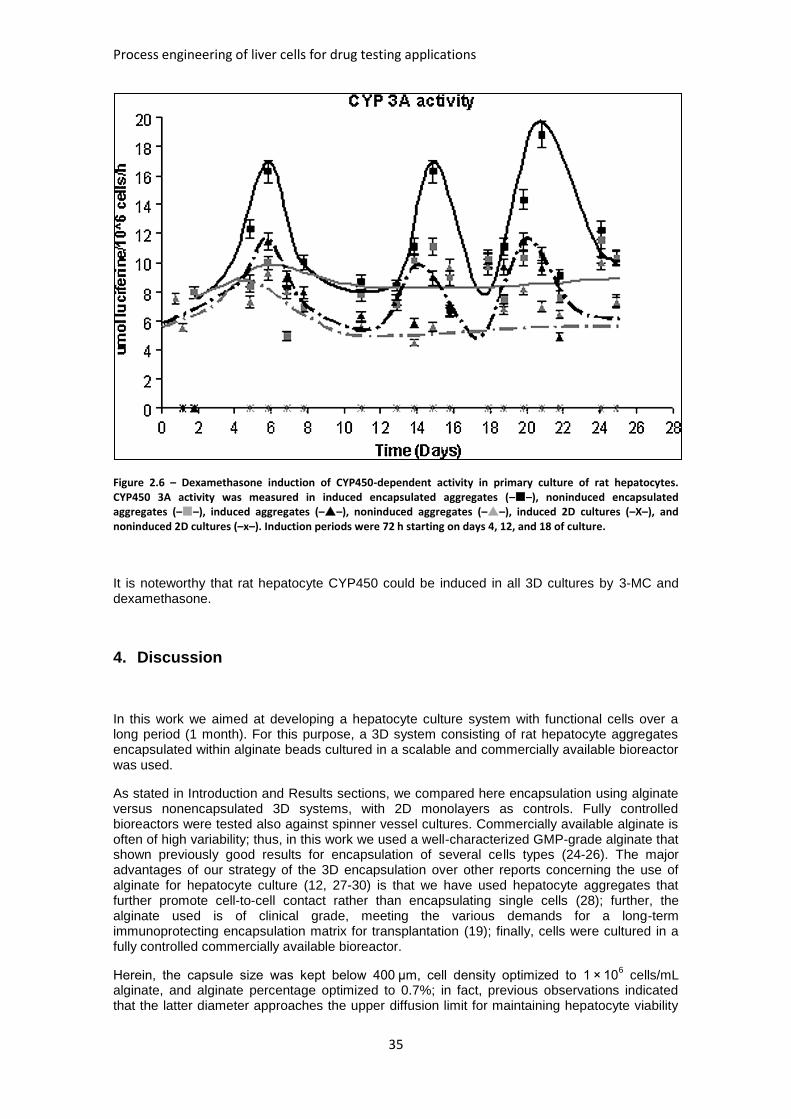
Process engineering of liver cells for drug testing applications
35
Figure 2.6 – Dexamethasone induction of CYP450-dependent activity in primary culture of rat hepatocytes. CYP450 3A activity was measured in induced encapsulated aggregates (––), noninduced encapsulated aggregates (––), induced aggregates (––), noninduced aggregates (––), induced 2D cultures (–X–), and noninduced 2D cultures (–x–). Induction periods were 72 h starting on days 4, 12, and 18 of culture.
It is noteworthy that rat hepatocyte CYP450 could be induced in all 3D cultures by 3-MC and dexamethasone.
4. Discussion
In this work we aimed at developing a hepatocyte culture system with functional cells over a long period (1 month). For this purpose, a 3D system consisting of rat hepatocyte aggregates encapsulated within alginate beads cultured in a scalable and commercially available bioreactor was used.
As stated in Introduction and Results sections, we compared here encapsulation using alginate versus nonencapsulated 3D systems, with 2D monolayers as controls. Fully controlled bioreactors were tested also against spinner vessel cultures. Commercially available alginate is often of high variability; thus, in this work we used a well-characterized GMP-grade alginate that shown previously good results for encapsulation of several cells types (24-26). The major advantages of our strategy of the 3D encapsulation over other reports concerning the use of alginate for hepatocyte culture (12, 27-30) is that we have used hepatocyte aggregates that further promote cell-to-cell contact rather than encapsulating single cells (28); further, the alginate used is of clinical grade, meeting the various demands for a long-term immunoprotecting encapsulation matrix for transplantation (19); finally, cells were cultured in a fully controlled commercially available bioreactor.
Herein, the capsule size was kept below 400 μm, cell density optimized to 1 × 10⁶ cells/mL
alginate, and alginate percentage optimized to 0.7%; in fact, previous observations indicated that the latter diameter approaches the upper diffusion limit for maintaining hepatocyte viability
Chapter 2 –Alginate-Encapsulated Three-Dimensional Cultures of hepatocytes in Bioreactors
36
(18). The uncontrolled swelling of 0.4% alginate capsules may be due to the weaker alginate network compared with 0.7% alginate matrices, which can withstand the osmotic pressure occurring between gel and the surrounding solution (25).
The use of complex extracellular substrates has been reported to play a key role in the maintenance of biotransformation and metabolic system of hepatocytes. In this work, monitoring of albumin and urea production (31) and ECOD and UGT activities(32) was performed to evaluate cell function and biotransformation, respectively. As shown here rat hepatocyte aggregates encapsulated within an alginate matrix exhibited remarkably enhanced expression of all the parameters analyzed, mainly when cultivated in fully controlled bioreactors. Since oxygen supply is critical for the adaptation of hepatocytes to culture and it decreases within alginate beads depending on size and entrapped cell density (12), the continuous oxygen flow enabled by the bioreactor revealed to be a decisive factor in maintaining hepatocytes functionality. However, UGT activity was improved in the encapsulated cultures, regardless of the culture vessel used (bioreactor or spinner), meaning that, in this case, the key factor was the encapsulation.
In the case of urea production or ECOD activity, similar performance was observed on encapsulated cells cultured in spinner vessel and on nonencapsulated cells cultured in bioreactor. Thus, the data obtained in this study suggest that hepatocyte cultivation within alginate microbeads not only resulted in a clear increase of hepatocyte-specific functions tested in vitro, but also significantly improved spinner effectiveness in urea production and ECOD
activity. These results are in accordance with previously reported data(9, 18, 28).
Another concern when developing new in vitro hepatocyte cultures is that hepatic metabolism of drugs is frequently the cause of adverse drug interactions. Induction of CYP450 enzymes is clinically important because it can modify the metabolism of drugs. It can potentiate their pharmacological action, diminish their efficacy, or produce unexpected hepatic side effects (33). The response of hepatic biotransformation enzymes to the action of classical in vivo inducers can easily be studied in vitro by exposing cultured hepatocytes to chemicals. In this study, treatments were initiated once the cells stabilized, that is, 24 h after encapsulation, and were prolonged for 72 h. This exposure time was sufficient to bring about the effects of moderate to strong drug-metabolizing enzyme inducers. The presence of dexamethasone in culture media increases CYP3A activity in rat hepatocytes selectively, namely, CYP3A1 and 3A23, the ortholog of the human CYP3A4 (34). In contrast, ECOD activity depends on different isozymes, including CYP1A1, 2A1, 2B1, and 2B2, and it is induced by 3-MC or Phenobarbital (35). Using this in vitro model, an increase in total CYP450 levels produced by both dexamethasone and 3-MC was observed. ECOD and CYP3A activity studies demonstrated that the 3D culture systems studied responded to all the induction insult. For CYP3A inducibility, alginate entrapment of hepatocytes aggregates significantly enhanced CYP induction compared to the nonencapsulated system. 2D cultures were not inducible. In in vitro cultures, the metabolism of testosterone to different hydroxyl metabolites is reduced by 90% in human hepatocytes after 2 days in culture(36). Thus, it is expectable that no activity is detected at day 5 of culture, that is, already at the first induction period. Regarding 3-MC induction, it should also be highlighted that although both encapsulated and nonencapsulated aggregates had similar induction levels, higher ECOD activities during the noninduced periods were observed in encapsulated aggregates, suggesting that the ECM provided by the alginate entrapment conferred the hepatocytes an advantageous culture system. Regarding 2D cultures, they only responded to the first induction by 3-MC, whereas no induction was observed with dexamethasone. Here, the importance of an adequate oxygenation for the phase I metabolism is emphasized by the lack of an effective induction of phase I enzymatic activities in the conventional 2D cultures, which have been shown to force hepatocyte cultures into more anaerobic metabolic states (37). Moreover, the use of a complex ECM had a beneficial effect on cell survival and on maintenance of rat hepatocytes differentiated functions, and cultivation under controlled environment also prevented the loss of CYP450 inducibility as it occurred in monolayer.
Process engineering of liver cells for drug testing applications
37
5. References
1. Cheng, N., E. Wauthier, and L.M. Reid, Mature human hepatocytes from ex vivo differentiation of alginate-encapsulated hepatoblasts. Tissue Eng Part A, 2008. 14(1): p. 1-7. 2. LeCluyse, E.L., Bullock, P. L., Parkinson, A., Hochman, J. H., Cultured rat hepatocytes. Pharm Biotechnol, 1996. 8: p. 121-59. 3. Tateno, C. and K. Yoshizato, Long-term cultivation of adult rat hepatocytes that undergo multiple cell divisions and express normal parenchymal phenotypes. Am J Pathol, 1996. 148(2): p. 383-92. 4. Isom, H.C., Secott, T., Georgoff, I., Woodworth, C., Mummaw, J., Maintenance of differentiated rat hepatocytes in primary culture. Proc Natl Acad Sci U S A, 1985. 82(10): p. 3252-6. 5. Eschbach, E., Chatterjee, S. S., Noldner, M., Gottwald, E., Dertinger, H., Weibezahn, K. F., Knedlitschek, G., Microstructured scaffolds for liver tissue cultures of high cell density: morphological and biochemical characterization of tissue aggregates. J Cell Biochem, 2005. 95(2): p. 243-55. 6. Dunn, J.C., Yarmush, M. L., Koebe, H. G., Tompkins, R. G., Hepatocyte function and extracellular matrix geometry: long-term culture in a sandwich configuration. Faseb J, 1989. 3(2): p. 174-7. 7. Moghe, P.V., Berthiaume, F., Ezzell, R. M., Toner, M., Tompkins, R. G., Yarmush, M. L., Culture matrix configuration and composition in the maintenance of hepatocyte polarity and function. Biomaterials, 1996. 17(3): p. 373-85. 8. Guguen-Guillouzo, C., M. Bourel, and A. Guillouzo, Human hepatocyte cultures. Prog Liver Dis, 1986. 8: p. 33-50. 9. Falasca, L., et al., The effect of retinoic acid on the re-establishment of differentiated hepatocyte phenotype in primary culture. Cell Tissue Res, 1998. 293(2): p. 337-47. 10. Mitaka, T., Sato, F., Mizuguchi, T., Yokono, T., Mochizuki, Y., Reconstruction of hepatic organoid by rat small hepatocytes and hepatic nonparenchymal cells. Hepatology, 1999. 29(1): p. 111-25. 11. Park, J.K. and D.H. Lee, Bioartificial liver systems: current status and future perspective. J Biosci Bioeng, 2005. 99(4): p. 311-9. 12. Falasca, L., Miccheli, A., Sartori, E., Tomassini, A., Conti Devirgiliis, L., Hepatocytes entrapped in alginate gel beads and cultured in bioreactor: rapid repolarization and reconstitution of adhesion areas. Cells Tissues Organs, 2001. 168(3): p. 126-36. 13. Lin, R.Z. and H.Y. Chang, Recent advances in three-dimensional multicellular spheroid culture for biomedical research. Biotechnol J, 2008. 3(9-10): p. 1172-84. 14. Bader, A., Knop, E., Boker, K., Fruhauf, N., Schuttler, W., Oldhafer, K., Burkhard, R., Pichlmayr, R., Sewing, K. F., A novel bioreactor design for in vitro reconstruction of in vivo liver characteristics. Artif Organs, 1995. 19(4): p. 368-74. 15. Berthiaume, F., Moghe, P. V., Toner, M., Yarmush, M. L., Effect of extracellular matrix topology on cell structure, function, and physiological responsiveness: hepatocytes cultured in a sandwich configuration. Faseb J, 1996. 10(13): p. 1471-84. 16. Talamini, M.A., B. Kappus, and A. Hubbard, Repolarization of hepatocytes in culture. Hepatology, 1997. 25(1): p. 167-72. 17. Miranda, J.P., Leite, S. B., Muller-Vieira, U., Rodrigues, A., Carrondo, M. J., Alves, P. M., Towards an extended functional hepatocyte in vitro culture. Tissue Eng Part C Methods, 2009. 15(2): p. 157-67. 18. Khalil, M. and A. Shariat-Panahi, Tootle, R., Ryder, T., McCloskey, P., Roberts, E., Hodgson, H., Selden, C., Human hepatocyte cell lines proliferating as cohesive spheroid colonies in alginate markedly upregulate both synthetic and detoxificatory liver function. J Hepatol, 2001. 34(1): p. 68-77. 19. Zimmermann, H., Zimmermann, D., Reuss, R., Feilen, P.J., Manz, B., Katsen, A., Weber, M., Ihmig, F.R., Ehrhart, F., Gessner, P., Behringer, M., Steinbach, A., Wegner, L.H., Sukhorukov, V.L., Va´squez, J.A., Schneider, S., Weber, M.M., Volke, F., Wolf, R., Zimmermann, U., Towards a medically approved technology for alginate-based microcapsules
Chapter 2 –Alginate-Encapsulated Three-Dimensional Cultures of hepatocytes in Bioreactors
38
allowing long-term immunoisolated transplantation. J Mater Sci Mater Med 2005. 16: p. 491-501. 20. Mazzoleni, G., D. Di Lorenzo, and N. Steimberg, Modelling tissues in 3D: the next future of pharmaco-toxicology and food research? Genes Nutr, 2009. 4(1): p. 13-22. 21. Seglen, P.O., Preparation of isolated rat liver cells. Methods Cell Biol, 1976. 13: p. 29-83. 22. Gomez-Lechon, M.J., Donato, M. T., Ponsoda, X., Fabra, R., Trullenque, R., Castell, J. V., In vitro Methods in Pharmaceutical Research,, in Isolation, culture and use of human hepatocytes in drug research, C.J.G.-L.M. (eds), Editor. 1997, Academic Press: San Diego, London, Boston. p. 129-153. 23. Burke, M.D. and S. Orrenius, The effect of albumin on metabolism of ethoxyresorufin through O-deethylation and sulphate-conjugation using isolated rat hepatocytes. Biochem Pharmacol, 1978. 27: p. 1533-1538. 24. Malpique, R., et al., Cryopreservation of adherent cells: strategies to improve cell viability and function after thawing. Tissue Eng Part C Methods, 2009. 15(3): p. 373-86. 25. Zimmermann, H., S.G. Shirley, and U. Zimmermann, Alginate-based encapsulation of cells: past, present, and future. Curr Diab Rep, 2007. 7(4): p. 314-20. 26. Zimmermann, H., F. Ehrhart, D. Zimmermann, K. Müller, A. Katsen-Globa, M. Behringer, P.J. Feilen, P. Gessner, G. Zimmermann, S.G. Shirley, M.M. Weber, J. Metze, U. Zimmermann, Hydrogel-based encapsulation of biological, functional tissue: fundamentals, technologies and applications. Applied Physics A: Materials Science & Processing 2007. 89 p. 909-922. 27. Miura, Y., Akimoto, T., Fuke, Y., Yamazaki, S., Yagi, K., In vitro maintenance of terminal-differentiated state in hepatocytes entrapped within calcium alginate. Artif Organs, 1987. 11(5): p. 361-5. 28. Ringel, M., von Mach, M. A., Santos, R., Feilen, P. J., Brulport, M., Hermes, M., Bauer, A. W., Schormann, W., Tanner, B., Schon, M. R., Oesch, F., Hengstler, J. G., Hepatocytes cultured in alginate microspheres: an optimized technique to study enzyme induction. Toxicology, 2005. 206(1): p. 153-67. 29. Maguire, T., Novik, E., Schloss, R., Yarmush, M., Alginate-PLL microencapsulation: effect on the differentiation of embryonic stem cells into hepatocytes. Biotechnol Bioeng, 2006. 93(3): p. 581-91. 30. Maguire, T., Davidovich, A. E., Wallenstein, E. J., Novik, E., Sharma, N., Pedersen, H., Androulakis, I. P., Schloss, R., Yarmush, M., Control of hepatic differentiation via cellular aggregation in an alginate microenvironment. Biotechnol Bioeng, 2007. 98: p. 631-44. 31. Gebhardt, R., Hengstler, J. G., Muller, D., Glockner, R., Buenning, P., Laube, B., Schmelzer, E., Ullrich, M., Utesch, D., Hewitt, N., Ringel, M., Hilz, B. R., Bader, A., Langsch, A., Koose, T., Burger, H. J., Maas, J., Oesch, F., New hepatocyte in vitro systems for drug metabolism: metabolic capacity and recommendations for application in basic research and drug development, standard operation procedures. Drug Metab Rev, 2003. 35(2-3): p. 145-213. 32. Funaki, N., Tanaka, J., Sugiyama, T., Ohshio, G., Nonaka, A., Yotsumoto, F., Sugie, T., Imamura, M., Successive cultures of mature hepatocytes for hepatocyte autotransplantation to assist liver function after liver resection for cancer. Oncol Rep, 2002. 9(4): p. 713-21. 33. Tsiaoussis, J., Newsome, P. N., Nelson, L. J., Hayes, P. C., Plevris, J. N., Which hepatocyte will it be? Hepatocyte choice for bioartificial liver support systems. Liver Transpl, 2001. 7(1): p. 2-10. 34. Ronis, M.J., Butura, A., Sampey, B. P., Shankar, K., Prior, R. L., Korourian, S., Albano, E., Ingelman-Sundberg, M., Petersen, D. R., Badger, T. M., Effects of N-acetylcysteine on ethanol-induced hepatotoxicity in rats fed via total enteral nutrition. Free Radic Biol Med, 2005. 39(5): p. 619-30. 35. Xu, C., C.Y. Li, and A.N. Kong, Induction of phase I, II and III drug metabolism/transport by xenobiotics. Arch Pharm Res, 2005. 28(3): p. 249-68. 36. Thomas, R.J., Bhandari, R., Barrett, D. A., Bennett, A. J., Fry, J. R., Powe, D., Thomson, B. J., Shakesheff, K. M., The effect of three-dimensional co-culture of hepatocytes and hepatic stellate cells on key hepatocyte functions in vitro. Cells Tissues Organs, 2005. 181(2): p. 67-79. 37. Jensen, M.D., Wallach D. F., Sherwood, P., Diffusion in tissue cultures on gas-permeable and impermeable supports. J Theor Biol, 1976. 56: p. 443-458.
39
Chapter 3
Bioreactor perfusion of 3D encapsulated hepatocytes
This Chapter was based on the manuscript: Tostoes R, Leite S, Miranda J, Sousa M, Wang D, Carrondo M, Alves P. 2011. Perfusion of 3D Encapsulated Hepatocytes-A Synergistic Effect Enhancing Long-Term Functionality in Bioreactors. Biotechnology and Bioengineering:41-49.
Chapter 3 – Bioreactor perfusion of 3D encapsulated hepatocytes
40
Abstract
Long term primary cultures of hepatocytes are essential for bioartificial liver (BAL) devices and to reduce and replace animal tests in lead candidate optimization in drug discovery and toxicology tests. The aim of this work was to improve bioreactor cultures of hepatocyte spheroids by adding a more physiological perfusion feeding regime to these bioreactor systems. A continuous perfusion feeding was compared with 50% medium replacement (routinely used for in vitro tests) at the same dilution rate, 0.125 day
-1, for 3 operative weeks. Perfusion feeding
led to a 10 fold improvement in albumin synthesis in bioreactors containing non-encapsulated hepatocyte spheroids; no significant improvement was observed in phase I drug metabolizing activity. When ultra high viscous alginate encapsulated spheroids were cultured in perfusion, urea synthesis, phase I drug metabolizing activity and oxygen consumption had a 3-fold improvement over the 50% medium replacement regime; albumin production was the same for both feeding regimes. The effective diffusion of albumin in the alginate capsules was 7.75.10
-9
cm2.s
-1 and no diffusion limitation for this protein was observed using these alginate capsules
under our operational conditions. In conclusion, perfusion feeding coupled with alginate encapsulation of hepatocyte spheroids showed a synergistic effect with a 3-fold improvement in three independent liver-specific functions of long-term hepatocyte spheroid cultures.
Process engineering of liver cells for drug testing applications
41
Table of Contents
1. Introduction ........................................................................................................ 42
2. Material and Methods ........................................................................................ 43
2.1. Rat Hepatocyte Isolation and Cell Culture .................................................... 43
2.2. Cell concentration determination ................................................................... 44
2.3. Determination of albumin and urea synthesis ............................................... 44
2.4. Glucose concentration measurement............................................................ 44
2.5. CYP450 Measurement ................................................................................. 44
2.6. Entrapment of hepatocyte spheroids in alginate ........................................... 44
2.7. Albumin encapsulation in alginate for diffusion experiments ......................... 44
2.8. Modified Dämkohler number ......................................................................... 45
2.9. Statistical analysis ........................................................................................ 45
3. Results ............................................................................................................... 45
3.1. Perfusion bioreactor set-up ........................................................................... 45
3.2. Perfusion bioreactor cultures of hepatocyte spheroids .................................. 46
3.3. Perfusion bioreactor cultures of alginate entrapped hepatocyte spheroids .... 47
4. Discussion ......................................................................................................... 51
5. References ......................................................................................................... 52
Chapter 3 – Bioreactor perfusion of 3D encapsulated hepatocytes
42
1. Introduction
The liver has a major role in maintaining physiological homeostasis and in detoxifying blood. In mammals, it is responsible for nitrogen excretion in the form of urea, albumin biosynthesis (1), glucose storage in the form of glycogen, gluconeogenesis (2), glutamine homeostasis (3) and xenobiotic detoxification (4-7).
Acute liver failure (ALF), at an approximate clinical rate of 2000 cases per year in the US (8), leads to death in 33% of cases. Due to liver donor scarcity, bioartificial liver (BAL) devices and immunoisolated xenografts of hepatocytes have been used to support these patients while waiting for a liver donor; nevertheless, the liver-specific functions of the hepatocytes are rapidly downregulated during support operation. This same problem affects primary cultured hepatocytes used in lead candidate optimization for drug development (5).
ALF and drug development assays drive the need to have cells expressing liver-specific functions. Since the expression of several of the drug metabolizing isozymes cytochrome P450 (CYP450), as well as nitrogen excretion and albumin synthesis, is usually downregulated in hepatoma or immortalized cell lines (9, 10), the main choice for BAL devices and drug development tests have been primary cultured hepatocytes. These cells maintain most of their liver-specific functions immediately after their isolation (7, 11). However, primary cultured hepatocytes lose their liver-specific functions along culture time (12, 13); this problem has been the subject of many studies whose main goal is to maintain viable and functional hepatocytes for longer time spans (4, 5, 14).
Cell-cell interactions in primary cultures of hepatocytes have been shown to mimick these cells’ in vivo physiological structure when cultured as spheroids (15), (16, 17), including phase I and II xenobiotic metabolizing activities. Moreover, our group and others (18, 19) have shown that the entrapment of hepatocyte spheroids within alginate capsules is a promising strategy to improve the maintenance of liver-specific functions in vitro (20). These ultra high viscous medical grade alginate hydrogels have also been applied in the transplantation of insulin-islets into immunocompetent diabetic mice (21); such capsules remained functional for more than one year. Alginate capsules of hepatocyte spheroids may also be used in BAL devices, since the capsules protect the cells from the shear stress of the high perfusion rates typical of these devices.
Bioreactors provide a more controlled environment for primary cultures of hepatocytes, maintaining long term viability and liver-specific functionality. Some of the liver’s major homeostatic functions such as the regulation of glucose levels in the blood (2) or glutamine synthesis (22) are based on the hepatocytes’ ability to continuously sense and act upon the blood’s nutrient and hormone levels. Thus, when defining a culture strategy for primary cultures of hepatocytes, it may not be enough to simply provide enough amounts of several nutrients based on consumption rates; to maintain their physiological concentrations fairly constant may be crucial.
Different bioreactor configurations have been developed aiming at obtaining a BAL device. Hollow fibers is the most common configuration for hepatocyte culture reactors (23-26) because these bioreactors offer immunoprotection to the ALF patient due to the separation between the hepatocytes and the culture medium/blood; however, this barrier also works as a disadvantage, since it limits mass (namely oxygen) transfer to the cells. Perfused microbioreactors with increased throughput capability for hepatocyte culture have recently been developed (10, 27-31). However, these small scale devices also have inherent mass transfer limitations when culturing physiologically relevant 3D structures; to overcome this limitation higher perfusion rates are needed (due to the high cell concentration inherent to such devices), thus creating additional shear stress (10); it is also difficult to control essential parameters such as dissolved oxygen or pH in these micro systems.
In this work, a perfusion bioreactor system was set up to maintain constant pH, temperature and oxygen levels, an intrinsic feature of these bioreactors (17), as well as stable concentrations of nutrients and hormones, assured by a gravimetric controlled perfusion system. The hepatocytes
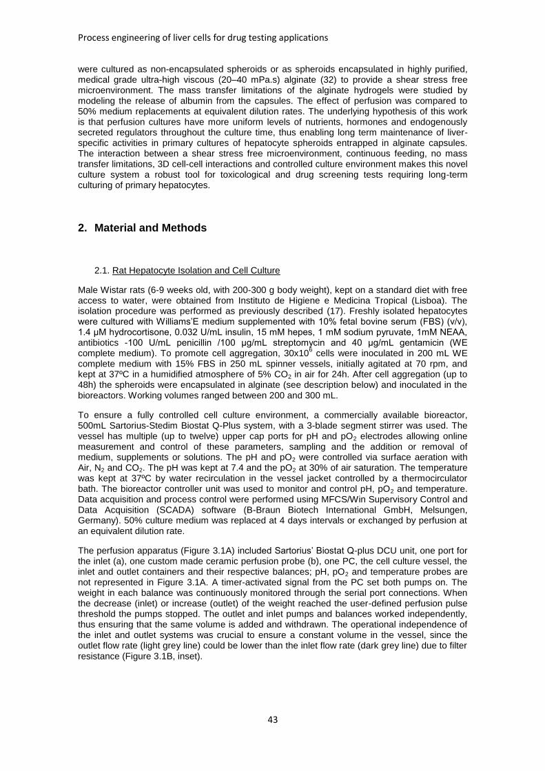
Process engineering of liver cells for drug testing applications
43
were cultured as non-encapsulated spheroids or as spheroids encapsulated in highly purified, medical grade ultra-high viscous (20–40 mPa.s) alginate (32) to provide a shear stress free microenvironment. The mass transfer limitations of the alginate hydrogels were studied by modeling the release of albumin from the capsules. The effect of perfusion was compared to 50% medium replacements at equivalent dilution rates. The underlying hypothesis of this work is that perfusion cultures have more uniform levels of nutrients, hormones and endogenously secreted regulators throughout the culture time, thus enabling long term maintenance of liver-specific activities in primary cultures of hepatocyte spheroids entrapped in alginate capsules. The interaction between a shear stress free microenvironment, continuous feeding, no mass transfer limitations, 3D cell-cell interactions and controlled culture environment makes this novel culture system a robust tool for toxicological and drug screening tests requiring long-term culturing of primary hepatocytes.
2. Material and Methods
2.1. Rat Hepatocyte Isolation and Cell Culture
Male Wistar rats (6-9 weeks old, with 200-300 g body weight), kept on a standard diet with free access to water, were obtained from Instituto de Higiene e Medicina Tropical (Lisboa). The isolation procedure was performed as previously described (17). Freshly isolated hepatocytes were cultured with Williams’E medium supplemented with 10% fetal bovine serum (FBS) (v/v), 1.4 μM hydrocortisone, 0.032 U/mL insulin, 15 mM hepes, 1 mM sodium pyruvate, 1mM NEAA, antibiotics -100 U/mL penicillin /100 μg/mL streptomycin and 40 μg/mL gentamicin (WE complete medium). To promote cell aggregation, 30x10
6 cells were inoculated in 200 mL WE
complete medium with 15% FBS in 250 mL spinner vessels, initially agitated at 70 rpm, and kept at 37ºC in a humidified atmosphere of 5% CO2 in air for 24h. After cell aggregation (up to 48h) the spheroids were encapsulated in alginate (see description below) and inoculated in the bioreactors. Working volumes ranged between 200 and 300 mL.
To ensure a fully controlled cell culture environment, a commercially available bioreactor, 500mL Sartorius-Stedim Biostat Q-Plus system, with a 3-blade segment stirrer was used. The vessel has multiple (up to twelve) upper cap ports for pH and pO2 electrodes allowing online measurement and control of these parameters, sampling and the addition or removal of medium, supplements or solutions. The pH and pO2 were controlled via surface aeration with Air, N2 and CO2. The pH was kept at 7.4 and the pO2 at 30% of air saturation. The temperature was kept at 37ºC by water recirculation in the vessel jacket controlled by a thermocirculator bath. The bioreactor controller unit was used to monitor and control pH, pO2 and temperature. Data acquisition and process control were performed using MFCS/Win Supervisory Control and Data Acquisition (SCADA) software (B-Braun Biotech International GmbH, Melsungen, Germany). 50% culture medium was replaced at 4 days intervals or exchanged by perfusion at an equivalent dilution rate.
The perfusion apparatus (Figure 3.1A) included Sartorius’ Biostat Q-plus DCU unit, one port for the inlet (a), one custom made ceramic perfusion probe (b), one PC, the cell culture vessel, the inlet and outlet containers and their respective balances; pH, pO2 and temperature probes are not represented in Figure 3.1A. A timer-activated signal from the PC set both pumps on. The weight in each balance was continuously monitored through the serial port connections. When the decrease (inlet) or increase (outlet) of the weight reached the user-defined perfusion pulse threshold the pumps stopped. The outlet and inlet pumps and balances worked independently, thus ensuring that the same volume is added and withdrawn. The operational independence of the inlet and outlet systems was crucial to ensure a constant volume in the vessel, since the outlet flow rate (light grey line) could be lower than the inlet flow rate (dark grey line) due to filter resistance (Figure 3.1B, inset).
Chapter 3 – Bioreactor perfusion of 3D encapsulated hepatocytes
44
2.2. Cell concentration determination
Alginate beads were dissolved by incubation in 1M Na2SO4, pH=7, at 37ºC for 20 minutes. Released spheroids were digested with trypsin and the resulting single cells viability was assessed by the trypan blue exclusion method. Cell counting was performed using a Neubauer counting chamber.
2.3. Determination of albumin and urea synthesis
The secretion of albumin from hepatocytes was measured by an enzyme-linked immunosorbent assay (ELISA) using NEPHRAT albumin test kit (ref.NR002, Exocell). The assay was performed according to the manufacturer’s description. The results were expressed as μg/day/10
6 cells at
the indicated time points. The urea synthesis rate was determined using a quantitative colorimetric urea kit (QuantiChromTM Urea Assay Kit, DIUR-500, BioAssay Systems), according to the manufacturer’s instructions. The albumin and urea specific synthesis rates were calculated according to the general mass balance equation for a continuous system:
( )
[ ] (1)
where q is the specific synthesis rate,
is the rate of change of the metabolite (either urea or
albumin) in the supernatant, D is the dilution rate (0.125 day-1
), Cin and CBR are the inlet and bioreactor concentrations of the metabolite and [Xv]ave is the average viable cell concentration during t. For the 50% medium exchange strategy D was zero and CBR was adjusted to account for a 50% dilution in the t where a medium exchange had occurred. The results were expressed as μg/day/10
6 cells at the indicated time points.
2.4. Glucose concentration measurement
Glucose concentration in the culture medium was measured using the YSI 7100 analyzer (Yellow Spring Instruments).
2.5. CYP450 Measurement
7-Ethoxycoumarin (ECOD) activity was measured by the method described in Castell (33) with slight modifications. Salicylamide (1.5 mM), was added to the medium to prevent conjugation of 7-hydroxy metabolites (7-HC) of 7-ethoxycoumarin. The activity is measured by the rate of formation of 7-hydroxycoumarin (Umbelliferone) in μmol/day/10
6.
2.6. Entrapment of hepatocyte spheroids in alginate
Ultra-high viscosity, clinical grade alginates were produced and purified from strains of Lessonia nigrescens and Lessonia trabeculata as described elsewhere (32) (the viscosity of a 0.1% [w/v] solution in distilled water was 20–30 mPa.s). A 1:1 mixture of purified alginates was used at 0.7% (w/v) in NaCl. For cell entrapment, hepatocyte spheroids were suspended in the mixture described above, to obtain the desired cell density per mL of alginate. Cell suspension was dropped through a syringe based apparatus into a solution of 20 mM BaCl2 (pH=7.3). The resulting spherical beads with entrapped hepatocyte spheroids were washed in 0.9% NaCl solution, suspended in complete medium and inoculated into the bioreactors.
2.7. Albumin encapsulation in alginate for diffusion experiments
To encapsulate albumin, alginate capsules were prepared similarly to spheroid capsules; however, instead of cells, Bovine Serum Albumin (BSA) was added to all the solutions in concentrations of 0.8, 1.5 and 2% (w/v), to obtain alginate capsules with these three concentrations. The capsules were then transferred to a PBS solution and the albumin release from the capsules was monitored by a fluorometrical measurement of the albumin concentration
Process engineering of liver cells for drug testing applications
45
in solution along time, at 37ºC, in a HORIBA Jobin-Yvon Fluoromax spectrofluorimeter (excitation: 280 nm, emission: 350 nm).
To determine the diffusion coefficient of albumin in the alginate capsules eq. 2 (34, 35) was applied:
∑
( )
(2)
where Mt is the albumin concentration in solution at time t, M is the albumin concentration in solution when equilibrium is reached, D is the effective diffusion of the solute inside the capsule,
R is the average radius of the alginate capsules (200 m) and n is the number of summation terms used (n=6 was enough to obtain an accurate solution); this equation was fitted against the
average fractional release (
) obtained experimentally at the three different intra-capsule
albumin concentrations. A custom algorithm was built, using MATLAB, where a range of values of D where tested to solve equation (2), the chosen value being the one which minimized the sum of squared errors between the theoretical curve and the experimental data. It was assumed that there was no resistance to mass transfer due to the high ratio of solution volume to total capsules volume (more than 20 fold).
2.8. Modified Dämkohler number
The modified Dämkohler number was calculated according to the equation:
( ) (3)
where Qalb is the albumin volumetric synthesis rate, in g.s-1
.(ml alginate)-1
, D is the diffusion coefficient of albumin in alginate, in cm
2.s
-1, R is the average radius of the alginate capsule, 0.02
cm, and Cout and Cin are the albumin concentrations in the culture medium bulk and the albumin
concentration inside the alginate capsule, respectively, in g.mL-1
.
2.9. Statistical analysis
Unless otherwise stated, all results were subject to an ANOVA single factor analysis, with
=0.05, using Microsoft Excell’s data analysis toolpack; p values are presented for statistically significant results (p<0.05, n≥3).
3. Results
3.1. Perfusion bioreactor set-up
The aim of this work was to improve the functionality of hepatocyte spheroids cultured in a fully controlled bioreactor by continuously feeding and washing-out nutrients and secondary metabolites. To ensure uniform levels of nutrients, hormones and endogenously secreted regulators throughout culture time, a gravimetrically controlled perfusion apparatus was set-up (Figure 3.1) maintaining a stable, continuous dilution rate in the perfusion bioreactors. The perfusion cultures were compared with 50% medium exchange cultures (control) at the same dilution rates, by assessing their ability for long-term preservation of liver-specific phenotype. The effects of perfusion feeding were studied in hepatocyte spheroids alone or in a shear stress free microenvironment, provided by the encapsulation of the spheroids within ultra-high viscosity clinical grade alginate.
Chapter 3 – Bioreactor perfusion of 3D encapsulated hepatocytes
46
Figure 3.1 – Perfusion bioreactor system with gravimetric control. A) Perfusion apparatus (See the “material and methods” section for further details on the system’s operation). B) Example of several perfusion pulses.
3.2. Perfusion bioreactor cultures of hepatocyte spheroids
Figure 3.2B shows that the perfusion culture of non encapsulated hepatocyte spheroids produced over 10 fold more albumin than the control cultures after three weeks. The other liver-specific activities analyzed, namely urea synthesis and ECOD biotransformation, were not significantly different between the two culture feeding strategies throughout the entire culture period (Figure 3.2A and D). The specific oxygen consumption rate was 1.5 fold higher in the control culture when compared to perfusion (Figure 3.2C, p<0.001). The improvement of
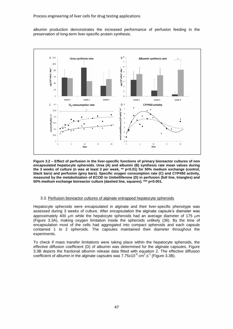
Process engineering of liver cells for drug testing applications
47
albumin production demonstrates the increased performance of perfusion feeding in the preservation of long-term liver-specific protein synthesis.
Figure 3.2 – Effect of perfusion in the liver-specific functions of primary bioreactor cultures of non encapsulated hepatocyte spheroids. Urea (A) and albumin (B) synthesis rate mean values during the 3 weeks of culture (n was at least 3 per week, ** p<0.01) for 50% medium exchange (control, black bars) and perfusion (grey bars). Specific oxygen consumption rate (C) and CYP450 activity, measured by the metabolization of ECOD to Umbelliferone (D) in perfusion (full line, triangles) and 50% medium exchange bioreactor culture (dashed line, squares). *** p<0.001.
3.3. Perfusion bioreactor cultures of alginate entrapped hepatocyte spheroids
Hepatocyte spheroids were encapsulated in alginate and their liver-specific phenotype was assessed during 3 weeks of culture. After encapsulation the alginate capsule’s diameter was
approximately 400 m while the hepatocyte spheroids had an average diameter of 175 m (Figure 3.3A), making oxygen limitation inside the spheroids unlikely (36). By the time of encapsulation most of the cells had aggregated into compact spheroids and each capsule contained 1 to 2 spheroids. The capsules maintained their diameter throughout the experiments.
To check if mass transfer limitations were taking place within the hepatocyte spheroids, the effective diffusion coefficient (D) of albumin was determined for the alginate capsules. Figure 3.3B depicts the fractional albumin release data fitted with equation 2. The effective diffusion coefficient of albumin in the alginate capsules was 7.75x10
-9 cm
2.s
-1 (Figure 3.3B).
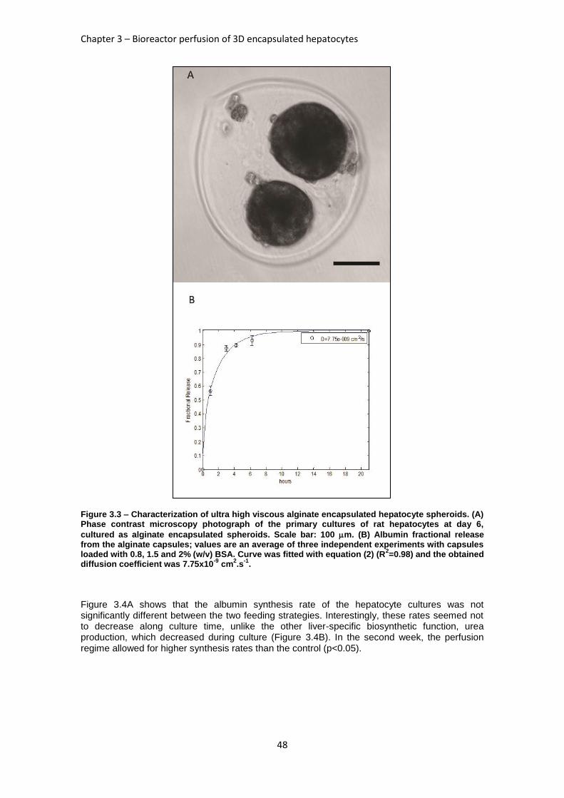
Chapter 3 – Bioreactor perfusion of 3D encapsulated hepatocytes
48
Figure 3.3 – Characterization of ultra high viscous alginate encapsulated hepatocyte spheroids. (A) Phase contrast microscopy photograph of the primary cultures of rat hepatocytes at day 6,
cultured as alginate encapsulated spheroids. Scale bar: 100 m. (B) Albumin fractional release from the alginate capsules; values are an average of three independent experiments with capsules loaded with 0.8, 1.5 and 2% (w/v) BSA. Curve was fitted with equation (2) (R
2=0.98) and the obtained
diffusion coefficient was 7.75x10⁻⁹
cm2.s
-1.
Figure 3.4A shows that the albumin synthesis rate of the hepatocyte cultures was not significantly different between the two feeding strategies. Interestingly, these rates seemed not to decrease along culture time, unlike the other liver-specific biosynthetic function, urea production, which decreased during culture (Figure 3.4B). In the second week, the perfusion regime allowed for higher synthesis rates than the control (p<0.05).
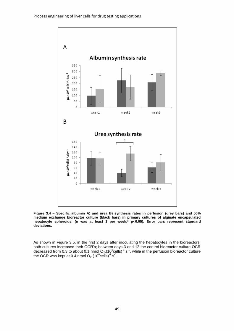
Process engineering of liver cells for drug testing applications
49
Figure 3.4 – Specific albumin A) and urea B) synthesis rates in perfusion (grey bars) and 50% medium exchange bioreactor culture (black bars) in primary cultures of alginate encapsulated hepatocyte spheroids. (n was at least 3 per week,* p<0.05). Error bars represent standard deviations.
As shown in Figure 3.5, in the first 2 days after inoculating the hepatocytes in the bioreactors, both cultures increased their OCR’s; between days 3 and 12 the control bioreactor culture OCR decreased from 0.3 to about 0.1 nmol O2.(10
6cells)
-1.s
-1, while in the perfusion bioreactor culture
the OCR was kept at 0.4 nmol O2.(106cells)
-1.s
-1.
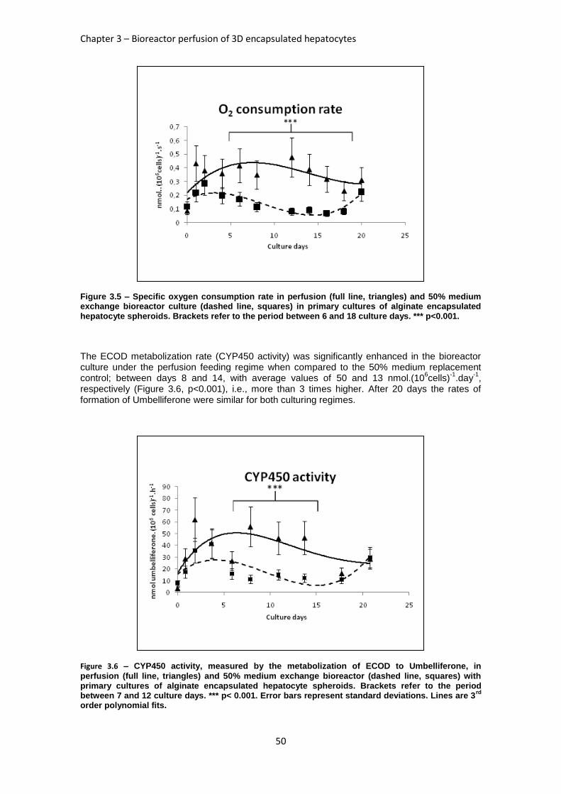
Chapter 3 – Bioreactor perfusion of 3D encapsulated hepatocytes
50
Figure 3.5 – Specific oxygen consumption rate in perfusion (full line, triangles) and 50% medium exchange bioreactor culture (dashed line, squares) in primary cultures of alginate encapsulated hepatocyte spheroids. Brackets refer to the period between 6 and 18 culture days. *** p<0.001.
The ECOD metabolization rate (CYP450 activity) was significantly enhanced in the bioreactor culture under the perfusion feeding regime when compared to the 50% medium replacement control; between days 8 and 14, with average values of 50 and 13 nmol.(10
6cells)
-1.day
-1,
respectively (Figure 3.6, p<0.001), i.e., more than 3 times higher. After 20 days the rates of formation of Umbelliferone were similar for both culturing regimes.
Figure 3.6 – CYP450 activity, measured by the metabolization of ECOD to Umbelliferone, in
perfusion (full line, triangles) and 50% medium exchange bioreactor (dashed line, squares) with primary cultures of alginate encapsulated hepatocyte spheroids. Brackets refer to the period between 7 and 12 culture days. *** p< 0.001. Error bars represent standard deviations. Lines are 3
rd
order polynomial fits.
Process engineering of liver cells for drug testing applications
51
4. Discussion
Maintaining liver-specific activities of primary cultures of hepatocytes have been attempted in many approaches (reviewed in (31, 37-39)). Most recent outcomes use controlled delivery of nutrients, namely oxygen, to the 3D cell clusters and providing a physical matrix to protect cells from shear stress. Our group has previously demonstrated the relevance of 3D cell-cell interactions and the bioreactor’s fully controlled environment (17); more recently, the beneficial effects of alginate entrapment in primary bioreactor cultures of hepatocyte spheroids has also been established (20). In this work, both these approaches have been conjugated with a continuous feeding strategy to further enhance the stability of the hepatocytes’ culture environment; the improvement resulting from the perfusion feeding, both in non-encapsulated as well as encapsulated hepatocyte spheroids, was compared to 50% medium replenishments (the control cultures) at the same dilution rate (0.125 day
-1).
The results presented herein confirm that encapsulated hepatocyte spheroids cultured under perfusion improved liver-specific phenotypes, except for the albumin synthesis rate, which was improved in non encapsulated spheroids albumin synthesis with perfusion culture (compared to the control). This was not due to mass transfer limitations of albumin within the alginate capsules; these limitations can be estimated by the calculation of a modified adimensional Dämkohler number, Da (equation 3), which represents the ratio between the time scales of albumin synthesis and diffusion, calculated for each culture time point, where. For Cin values ranging between 10 and 100 fold Cout, Da varies between 1.8 ± 1.4 and 0.17 ± 0.13, respectively, implying that the measured rates of albumin production are hardly influenced by limited diffusion of this protein inside the alginate capsule. This conclusion is further reinforced by the fact that the calculated effective diffusion coefficient is within the same magnitude of other literature values (34). The albumin synthesis rates achieved in the alginate entrapped
hepatocyte spheroids, 300 g.(106cells)
-1.day
-1 at 3 weeks culture time, are close to the
physiological values. These may be estimated from data obtained in rat studies which report albumin production rates in vivo of approximately 50 mg albumin/4.6g liver/day (40); from (41, 42) one can obtain an average yield of hepatocytes per gram of liver of 17-18 million cells.
Thus, the in vivo albumin production rate is in the order of 700 g.(106cells)
-1.day
-1, which
indicates that this culture of encapsulated hepatocyte spheroids reached albumin production rates nearing physiological levels, independently of the feeding strategy. These near physiological rates are at the top end of earlier reported values which range from 100 to 300
g.(106cells)
-1.day
-1 in a microfabricated hepatocyte-fibroblast co-culture system (38). It is also
worth to note that the levels of albumin synthesis rates were maintained throughout the encapsulated spheroid culture time; in non encapsulated cultures (both perfusion and control) the albumin production rates were lower, possibly reflecting the variability in protein synthesis between different batches of isolated rat hepatocytes; still, these values are in accordance with lower values found in the literature for rat hepatocyte spheroid culture (43).
In the encapsulated spheroid culture, the specific urea synthesis rate of the perfusion regime was 3-fold higher than the control during the second week; values ranged from 300, in the
beginning of the culture, to 50 g.(106cells)
-1.day
-1, after 3 weeks. These values are in
accordance with earlier literature reports (23, 27, 44).
The oxygen consumption rate (OCR) in the non encapsulated 50% medium replenishment spheroid culture (control) was 1.5 fold higher than in the perfusion culture (Figure 3.2C). However, in the system using encapsulated spheroids the perfusion system enabled a 3 fold increase in OCR when compared to the 50% medium exchange control. This latter result agrees with 2 experiments investigating the consumption characteristics of rat hepatocytes during the first culture day, 0.38-0.6 nmol O2.(10
6cells)
-1.s
-1 (45, 46); these authors also describe
a trend for rat hepatocyte cultures to decrease their OCR along culture time. In fact, in this work, the OCR of both cultures reached the same value of approximately 0.25 nmol O2.(10
6cells)
-1.s
-1
after 20 days of culture (Figure 3.5). However, from day 8 to day 18 the OCR of encapsulated
Chapter 3 – Bioreactor perfusion of 3D encapsulated hepatocytes
52
hepatocyte spheroids cultures was, on average, 3 times higher under the perfusion feeding regime than the control (Figure 3.5A, p<0.001). These OCR results show that the perfusion culture enabled the alginate encapsulated spheroids to maintain their initial metabolic functions for longer than the control feeding regime.
The activity of phase I CYP450 isozymes is quickly downregulated when hepatocytes are cultured ex vivo. The metabolization of ECOD by hepatocytes is mediated by more than one isoform of this group of enzymes, so it can be regarded as a general test for CYP450 enzymatic activity (47). When cultured under perfusion the encapsulated hepatocyte spheroids yielded a 3 fold improvement in CYP450 activity over the control culture, by the second culture week, emphasizing the importance of maintaining a continuous feeding regime.
The fact that perfusion feeding appears to be more critical in encapsulated cells may be explained by the retention of endogenously secreted extracellular matrix (ECM) components, which are essential for the spheroids’ cohesion within the alginate capsules. This would mask a possible improvement of perfusion against the 50% medium exchange strategy which would only become clear for the encapsulated system. To further elucidate the importance of ECM in the synergy between alginate encapsulation and perfusion, it would be necessary to evaluate the hepatocyte spheroids ECM composition (fibronectin and collagens I and IV, for instance), in both encapsulated and non-encapsulated systems along culture time and correlate it to the liver-specific phenotype obtained.
The general 3-fold improvement obtained (CYP450 activity, OCR and Urea production rate) by combining the homogeneous environment provided by controlled stirred bioreactors with perfusion and alginate encapsulation, for primary cultures of hepatocyte spheroids, suggests that the simultaneous exploitation of physical, chemical and biological tools with this novel comprehensive strategy leads to a positive synergistic effect between alginate entrapped hepatocyte spheroids and the continuous feeding of culture medium.
In comparison with the more established hollow fiber system (23, 48), it is clear that the absence of mass transfer limitations and the possibility of intensive hepatocyte sampling makes the bioreactor system described herein a better alternative to drug testing and toxicological studies than the hollow fiber reactors.
It is also noteworthy that this was not only an end-point improvement, but more notably a 1 week time extension of highly functional hepatocyte spheroid cultures. Such a system will certainly be an added value toward long-term studies in drug development.
5. References
1. Quinlan GJ, Martin GS, Evans TW. Albumin: Biochemical properties and therapeutic potential. Hepatology 2005;41:1211-1219. 2. Pilkis SJ, Granner DK. MOLECULAR PHYSIOLOGY OF THE REGULATION OF HEPATIC GLUCONEOGENESIS AND GLYCOLYSIS. Annual Review of Physiology 1992;54:885-909. 3. Watford M, Chellaraj V, Ismat A, Brown P, Raman P. Hepatic glutamine metabolism. Nutrition 2002;18:301-303. 4. Gebhardt R, Hengstler JG, Muller D, Glockner R, Buenning P, Laube B, Schmelzer E, et al. New hepatocyte in vitro systems for drug metabolism: Metabolic capacity and recommendations for application in basic research and drug development, standard operation procedures. Drug Metabolism Reviews 2003;35:145-213. 5. Gomez-Lechon MJ, Donato MT, Castell JV, Jover R. Human hepatocytes as a tool for studying toxicity and drug metabolism. Current Drug Metabolism 2003;4:292-312. 6. LeCluyse EL. Human hepatocyte culture systems for the in vitro evaluation of cytochrome P450 expression and regulation. European Journal of Pharmaceutical Sciences 2001;13:343-368.
Process engineering of liver cells for drug testing applications
53
7. Maurel P. The use of adult human hepatocytes in primary culture and other in vitro systems to investigate drug metabolism in man. Advanced Drug Delivery Reviews 1996;22:105-132. 8. Lee WM. MEDICAL PROGRESS - ACUTE LIVER-FAILURE. New England Journal of Medicine 1993;329:1862-1872. 9. Sinz M, Wallace G, Sahi J. Current industrial practices in assessing CYP450 enzyme induction: Preclinical and clinical. Aaps Journal 2008;10:391-400. 10. Park J, Li Y, Berthiaume F, Toner M, Yarmush ML, Tilles AW. Radial flow hepatocyte bioreactor using stacked microfabricated grooved substrates. Biotechnology and Bioengineering 2008;99:455-467. 11. Coecke S, Rogiers V, Bayliss M, Castell J, Doehmer J, Fabre G, Fry J, et al. The use of long-term hepatocyte cultures for detecting induction of drug metabolising enzymes: The current status - ECVAM hepatocytes and metabolically competent systems task force report 1. Atla-Alternatives to Laboratory Animals 1999;27:579-638. 12. Nakamura T, Yoshimoto K, Nakayama Y, Tomita Y, Ichihara A. RECIPROCAL MODULATION OF GROWTH AND DIFFERENTIATED FUNCTIONS OF MATURE RAT HEPATOCYTES IN PRIMARY CULTURE BY CELL CELL CONTACT AND CELL-MEMBRANES. Proceedings of the National Academy of Sciences of the United States of America-Biological Sciences 1983;80:7229-7233. 13. Nakamura T, Tomita Y, Hirai R, Yamaoka K, Kaji K, Ichihara A. INHIBITORY EFFECT OF TRANSFORMING GROWTH-FACTOR-BETA ON DNA-SYNTHESIS OF ADULT-RAT HEPATOCYTES IN PRIMARY CULTURE. Biochemical and Biophysical Research Communications 1985;133:1042-1050. 14. Vermeir M, Annaert P, Mamidi RNVS, Roymans D, Meuldermans W, Mannens G. Cell-based models to study hepatic drug metabolism and enzyme induction in humans. Expert Opin Drug Metab Toxicol 2005;1:75-90. 15. Abu-Absi SF, Friend JR, Hansen LK, Hu WS. Structural polarity and functional bile canaliculi in rat hepatocyte spheroids. Experimental Cell Research 2002;274:56-67. 16. Wu F, Friend J, Remmel R, Cerra F, Hu W. Enhanced cytochrome P450 IA1 activity of self-assembled rat hepatocyte spheroids. Cell Transplant 1999;8:233-246. 17. Miranda JP, Leite SB, Muller-Vieira U, Rodrigues A, Carrondo MJT, Alves PM. Towards an Extended Functional Hepatocyte In Vitro Culture. Tissue Engineering Part C-Methods 2009;15:157-167. 18. Guo XL, Yang KS, Hyun JY, Kim WS, Lee DH, Min KE, Park LS, et al. Morphology and metabolism of Ba-alginate-encapsulated hepatocytes with galactosylated chitosan and poly(vinyl alcohol) as extracellular matrices. Journal of Biomaterials Science-Polymer Edition 2003;14:551-565. 19. Takabatake H, Koide N, Tsuji T. ENCAPSULATED MULTICELLULAR SPHEROIDS OF RAT HEPATOCYTES PRODUCE ALBUMIN AND UREA IN A SPOUTED BED CIRCULATING CULTURE SYSTEM. Artificial Organs 1991;15:474-480. 20. Miranda J, Rodrigues A, Tostoes R, Leite S, Zimmermann H, Carrondo M, Alves P. Extending hepatocyte functionality for drug testing applications using high viscosity alginate encapsulated 3D cultures in bioreactors. Tissue Eng Part C Methods 2010. 21. Schneider S, Feilen P, Brunnenmeier F, Minnemann T, Zimmermann H, Zimmermann U, Weber M. Long-term graft function of adult rat and human islets encapsulated in novel alginate-based microcapsules after transplantation in immunocompetent diabetic mice. Diabetes 2005;54:687-693. 22. Haussinger D, Lamers WH, Moorman AFM. HEPATOCYTE HETEROGENEITY IN THE METABOLISM OF AMINO-ACIDS AND AMMONIA. Enzyme 1992;46:72-93. 23. De Bartolo L, Salerno S, Curcio E, Piscioneri A, Rende M, Morelli S, Tasselli F, et al. Human hepatocyte functions in a crossed hollow fiber membrane bioreactor. Biomaterials 2009;30:2531-2543. 24. Feng ZQ, Chu XH, Huang NP, Wang T, Wang YC, Shi XL, Ding YT, et al. The effect of nanofibrous galactosylated chitosan scaffolds on the formation of rat primary hepatocyte aggregates and the maintenance of liver function. Biomaterials 2009;30:2753-2763. 25. Flendrig LM, laSoe JW, Jorning GGA, Steenbeek A, Karlsen OT, Bovee W, Ladiges N, et al. In vitro evaluation of a novel bioreactor based on an integral oxygenator and a spirally wound nonwoven polyester matrix for hepatocyte culture as small aggregates. Journal of Hepatology 1997;26:1379-1392.
Chapter 3 – Bioreactor perfusion of 3D encapsulated hepatocytes
54
26. Patzer JF. Oxygen consumption in a hollow fiber bioartificial liver-revisited. Artificial Organs 2004;28:83-98. 27. Carraro A, Hsu WM, Kulig KM, Cheung WS, Miller ML, Weinberg EJ, Swart EF, et al. In vitro analysis of a hepatic device with intrinsic microvascular-based channels. Biomedical Microdevices 2008;10:795-805. 28. Zhang MY, Lee PJ, Hung PJ, Johnson T, Lee LP, Mofrad MRK. Microfluidic environment for high density hepatocyte culture. Biomedical Microdevices 2008;10:117-121. 29. Korin N, Bransky A, Khoury M, Dinnar U, Levenberg S. Design of Well and Groove Microchannel Bioreactors for Cell Culture. Biotechnology and Bioengineering 2009;102:1222-1230. 30. Powers MJ, Janigian DM, Wack KE, Baker CS, Stolz DB, Griffith LG. Functional behavior of primary rat liver cells in a three-dimensional perfused microarray bioreactor. Tissue Engineering 2002;8:499-513. 31. Sivaraman A, Leach JK, Townsend S, Iida T, Hogan BJ, Stolz DB, Fry R, et al. A microscale in vitro physiological model of the liver: Predictive screens for drug metabolism and enzyme induction. Current Drug Metabolism 2005;6:569-591. 32. Zimmermann H, Wahlisch F, Baier C, Westhoff M, Reuss R, Zimmermann D, Behringer M, et al. Physical and biological properties of barium cross-linked alginate membranes. Biomaterials 2007;28:1327-1345. 33. Castell JV, Gómez-Lechón MJ. In vitro methods in pharmaceutical research. San Diego: Academic Press, 1997: ix, 467 p. 34. Koyama K, Seki M. Evaluation of mass-transfer characteristics in alginate-membrane liquid-core capsules prepared using polyethylene glycol. J Biosci Bioeng 2004;98:114-121. 35. Crank J. The mathematics of diffusion. 2d ed. Oxford, [Eng]: Clarendon Press, 1979: viii, 414 p. 36. Curcio E, Salerno S, Barbieri G, De Bartolo L, Drioli E, Bader A. Mass transfer and metabolic reactions in hepatocyte spheroids cultured in rotating wall gas-permeable membrane system. Biomaterials 2007;28:5487-5497. 37. Berthiaume F, Moghe PV, Toner M, Yarmush ML. Effect of extracellular matrix topology on cell structure, function, and physiological responsiveness: Hepatocytes cultured in a sandwich configuration. Faseb Journal 1996;10:1471-1484. 38. Bhatia SN, Balis UJ, Yarmush ML, Toner M. Effect of cell-cell interactions in preservation of cellular phenotype: cocultivation of hepatocytes and nonparenchymal cells. Faseb Journal 1999;13:1883-1900. 39. Pichard-Garcia L, Gerbal-Chaloin S, Ferrini JB, Fabre JM, Maurel P: Use of long-term cultures of human hepatocytes to study cytochrome P450 gene expression. In: Cytochrome P450, Pt C. Volume 357. San Diego: Academic Press Inc, 2002; 311-321. 40. Peavy D, Taylor J, Jefferson L. Correlation of albumin production rates and albumin mRNA levels in livers of normal, diabetic, and insulin-treated diabetic rats. Proc Natl Acad Sci U S A 1978;75:5879-5883. 41. Wang S, Renaud G, Infante J, Catala D, Infante R. Isolation of rat hepatocytes with EDTA and their metabolic functions in primary culture. In Vitro Cell Dev Biol 1985;21:526-530. 42. Carthew P, Maronpot R, Foley J, Edwards R, Nolan B. Method for determining whether the number of hepatocytes in rat liver is increased after treatment with the peroxisome proliferator gemfibrozil. J Appl Toxicol;17:47-51. 43. Ijima H, Kakeya Y, Yokonuma T, Hou Y, Takei T. Composition of culture medium is more important than co-culture with hepatic non-parenchymal cells in albumin production activity of primary rat hepatocytes, and the effect was enhanced by hepatocytes spheroid culture in collagen gel. Biochemical Engineering Journal 2009:226-231. 44. Seagle C, Christie MA, Winnike JH, McClelland RE, Ludlow JW, O'Connell TM, Gamcsik MP, et al. High-throughput nuclear magnetic resonance metabolomic footprinting for tissue engineering. Tissue Engineering Part C-Methods 2008;14:107-118. 45. Foy BD, Rotem A, Toner M, Tompkins RG, Yarmush ML. A DEVICE TO MEASURE THE OXYGEN-UPTAKE RATE OF ATTACHED CELLS - IMPORTANCE IN BIOARTIFICIAL ORGAN DESIGN. Cell Transplantation 1994;3:515-527. 46. Smith MD, Smirthwaite AD, Cairns DE, Cousins RB, Gaylor JD. Techniques for measurement of oxygen consumption rates of hepatocytes during attachment and post-attachment. International Journal of Artificial Organs 1996;19:36-44. 47. Phillips IR, Shephard EA. Cytochrome P450 protocols. 2nd ed. Totowa, N.J.: Humana Press, 2006: xiii, 363 p.

Process engineering of liver cells for drug testing applications
55
48. Dong J, Mandenius CF, Lubberstedt M, Urbaniak T, Nussler AKN, Knobeloch D, Gerlach JC, et al. Evaluation and optimization of hepatocyte culture media factors by design of experiments (DoE) methodology. Cytotechnology 2008;57:251-261.
56
Chapter 4
Human liver cell spheroids in extended perfusion bioreactor culture for repeated dose drug testing.
This Chapter was based on the manuscript: Tostoes R, Leite SB, Serra M, Jensen J, Björquist P, Carrondo M, Brito C, et al. Human liver cell spheroids in extended perfusion bioreactor culture for repeated dose drug testing. Hepatology 2011.
Process engineering of liver cells for drug testing applications
57
Abstract
Primary cultures of human hepatocyte spheroids are a promising in vitro model for long term studies of hepatic metabolism and cytotoxicity. The lack of robust methodologies to culture cell spheroids, a poor characterization of the human hepatocyte spheroids architecture and liver-specific functionality have hampered a widespread adoption of this 3D culture format. In this work, an automated perfusion bioreactor was used to obtain and maintain human hepatocyte spheroids. These spheroids were cultured for 3-4 weeks in serum-free conditions, sustaining their phase I enzyme expression and permitting repeated induction during long culture times; the rate of albumin and urea synthesis, as well as phase I and II drug metabolizing enzymes’ gene expression and activity of the spheroid hepatocyte cultures presented reproducible profiles, despite the basal inter-donor variability (n=3 donors). Immunofluorescence microscopy of human hepatocyte spheroids after 3-4 weeks of long term culture confirmed the presence of
the liver-specific markers hepatocyte nuclear factor 4, albumin, cytokeratin 18 and cytochrome P450 3A. Moreover, immunostaining of the atypical protein kinase C apical marker, as well as the excretion of a fluorescent dye, evidenced that these spheroids spontaneously assemble a functional bile canaliculi network extending from the surface to the interior of the spheroids after 3-4 weeks of culture. Conclusion: Perfusion bioreactor cultures of primary human hepatocyte spheroids maintain a liver-specific activity and architecture and are thus suitable for drug testing in a long term, repeated dose format.
Chapter 4 – Human liver cell spheroids in extended perfusion bioreactor culture
58
Table of Contents
1. Introduction ........................................................................................................ 59
2. Material and Methods ........................................................................................ 59
2.1. Primary cultures of human hepatocytes ............................................................ 60
2.2. Perfusion Bioreactor Culture ............................................................................. 60
2.3. Hepatocyte CYP450 enzyme inductions ........................................................... 60
2.4. Cell concentration determination ...................................................................... 61
2.5. Hepatocyte spheroids visualization and measurement ..................................... 61
2.6. Determination of albumin and urea synthesis ................................................... 61
2.7. CYP450 activity Measurement ......................................................................... 62
2.8. qRT-PCR .......................................................................................................... 62
2.9. Whole mount Immunofluorescence microscopy ................................................ 62
2.10. CryoSection immunofluorescence microscopy ............................................... 62
2.11. Antibodies for confocal immunofluorescence microscopy ............................... 62
2.12. Bile canaliculi functionality .............................................................................. 62
2.13. Statistical analysis .......................................................................................... 63
3. Results ............................................................................................................... 63
3.1. Hepatocyte spheroid size distribution, viability and liver-specific synthesis time-course profiles in bioreactor cultures. ...................................................................... 63
3.2. Phase I and II enzyme expression and activity in bioreactor cultures. .............. 65
3.3. Liver-specific markers and structural polarity in bioreactor cultured spheroids. . 66
4. Discussion ......................................................................................................... 69
5. References ......................................................................................................... 70
Process engineering of liver cells for drug testing applications
59
1. Introduction
The liver-specific functions of hepatocytes, such as albumin secretion or drug metabolizing activity, are rapidly downregulated during in vitro primary cultures, limiting their use for drug development and toxicity tests (1). For such assays, the current gold standard for long term human hepatocyte culture is the collagen sandwich in vitro model (2). The overlaying collagen layer increases cell-cell and cell-matrix contacts, providing a more 3D-like architecture than a monolayer culture. For rat hepatocyte spheroids, where cell-cell interactions are maximized, liver-specific functions (3, 4) and multicellular architecture (5, 6) are increased, when compared to monolayer cultures.
The use of microfluidic devices for primary cultures of hepatocytes is a promising approach to enable high throughput screening in drug development (7, 8). However, the down scaling enabled by these technologies makes the culture environment harder to be controlled and limits the application of microfluidics for long term primary cultures of hepatocytes. In fact, the most useful applications of micro technologies for such cultures couple either microfluidic perfusion or co-culture micropatterning to 12 (9) or 24 well plate culture plates (10), respectively; still, these technologies do not enable a physiologically relevant long-term culture of primary hepatocytes.
Bioartificial liver devices (BAL) using human hepatocytes are often built in hollow fiber formats; this configuration is likely among the best options for maintaining cultures of large numbers of hepatocytes for prolonged culture periods (11); recently, one of such devices has also been scaled down and adapted to drug testing, using human liver cells (12). The configuration of these bioreactors enables the formation of a liver-like hepatic mass throughout the culture time, making them long-term end point assays; nevertheless, these bioreactors were not designed for high throughput screening and do not allow sampling of the cellular mass throughout the culture period.
CYP450 expression is downregulated during in vitro primary culture of hepatocytes; these enzymes are fundamental to xenobiotic metabolization studies, namely in drug development, and the orphan receptor-mediated induction of their mRNA synthesis is one of the most important parameters to be assessed in such tests (13). After these enzymes have oxidized a given xenobiotic, the compound is further conjugated with polar groups by phase II enzymes and secreted in the bile canaliculi by phase III enzymes. Thus, a long-term hepatocyte culture system must not only maintain CYP450 basal expression but also to enable their de novo mRNA synthesis upon exposure to prototypical CYP450 inducers, while maintaining the remaining phase II and III activities. Since phase III activity depends on the transport of phase II metabolites through the apical membrane, the presence of bile canaliculi is also necessary for a thorough assessment of drug metabolizing activity in primary cultures of hepatocytes.
This work focuses on the use of perfusion stirred tank bioreactors for primary cultures of human hepatocyte as spheroids. When cultured in a bioreactor with essentially convective mass transfer and environmental control, the hepatocytes experience much smaller changes in pH, dissolved oxygen (DO) and culture medium composition than any culture system with a constant atmosphere and discrete medium exchanges. The pH and DO are controlled by CO2 and N2 injection through the reactor headspace, respectively, whereas the continuous addition of nutrients and removal of metabolic by-products is ensured by the automated perfusion system (14); the good mixing minimizes the gradients of these soluble factors in the culture bulk. The results indicate that this bioreactor system of primary culture of human hepatocyte spheroids enables the robust formation of hepatic-like micro-tissue units that can be repeatedly induced in long-term periods.
2. Material and Methods
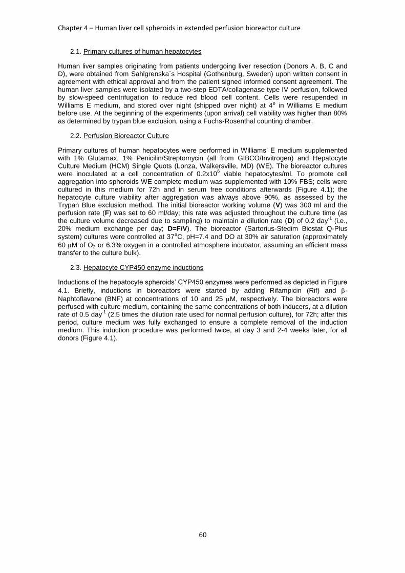
Chapter 4 – Human liver cell spheroids in extended perfusion bioreactor culture
60
2.1. Primary cultures of human hepatocytes
Human liver samples originating from patients undergoing liver resection (Donors A, B, C and D), were obtained from Sahlgrenska´s Hospital (Gothenburg, Sweden) upon written consent in agreement with ethical approval and from the patient signed informed consent agreement. The human liver samples were isolated by a two-step EDTA/collagenase type IV perfusion, followed by slow-speed centrifugation to reduce red blood cell content. Cells were resupended in
Williams E medium, and stored over night (shipped over night) at 4⁰ in Williams E medium before use. At the beginning of the experiments (upon arrival) cell viability was higher than 80% as determined by trypan blue exclusion, using a Fuchs-Rosenthal counting chamber.
2.2. Perfusion Bioreactor Culture
Primary cultures of human hepatocytes were performed in Williams’ E medium supplemented with 1% Glutamax, 1% Penicilin/Streptomycin (all from GIBCO/Invitrogen) and Hepatocyte Culture Medium (HCM) Single Quots (Lonza, Walkersville, MD) (WE). The bioreactor cultures were inoculated at a cell concentration of 0.2x10
6 viable hepatocytes/ml. To promote cell
aggregation into spheroids WE complete medium was supplemented with 10% FBS; cells were cultured in this medium for 72h and in serum free conditions afterwards (Figure 4.1); the hepatocyte culture viability after aggregation was always above 90%, as assessed by the Trypan Blue exclusion method. The initial bioreactor working volume (V) was 300 ml and the perfusion rate (F) was set to 60 ml/day; this rate was adjusted throughout the culture time (as the culture volume decreased due to sampling) to maintain a dilution rate (D) of 0.2 day
-1 (i.e.,
20% medium exchange per day; D=F/V). The bioreactor (Sartorius-Stedim Biostat Q-Plus
system) cultures were controlled at 37⁰C, pH=7.4 and DO at 30% air saturation (approximately
60 M of O2 or 6.3% oxygen in a controlled atmosphere incubator, assuming an efficient mass transfer to the culture bulk).
2.3. Hepatocyte CYP450 enzyme inductions
Inductions of the hepatocyte spheroids’ CYP450 enzymes were performed as depicted in Figure
4.1. Briefly, inductions in bioreactors were started by adding Rifampicin (Rif) and -
Naphtoflavone (BNF) at concentrations of 10 and 25 M, respectively. The bioreactors were perfused with culture medium, containing the same concentrations of both inducers, at a dilution rate of 0.5 day
-1 (2.5 times the dilution rate used for normal perfusion culture), for 72h; after this
period, culture medium was fully exchanged to ensure a complete removal of the induction medium. This induction procedure was performed twice, at day 3 and 2-4 weeks later, for all donors (Figure 4.1).
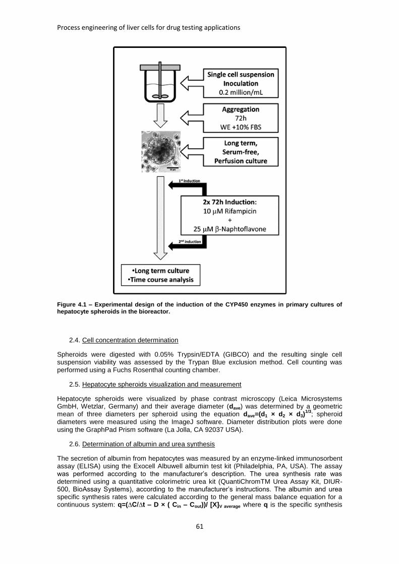
Process engineering of liver cells for drug testing applications
61
Figure 4.1 – Experimental design of the induction of the CYP450 enzymes in primary cultures of hepatocyte spheroids in the bioreactor.
2.4. Cell concentration determination
Spheroids were digested with 0.05% Trypsin/EDTA (GIBCO) and the resulting single cell suspension viability was assessed by the Trypan Blue exclusion method. Cell counting was performed using a Fuchs Rosenthal counting chamber.
2.5. Hepatocyte spheroids visualization and measurement
Hepatocyte spheroids were visualized by phase contrast microscopy (Leica Microsystems GmbH, Wetzlar, Germany) and their average diameter (dave) was determined by a geometric mean of three diameters per spheroid using the equation dave=(d1 × d2 × d3)
1/3; spheroid
diameters were measured using the ImageJ software. Diameter distribution plots were done using the GraphPad Prism software (La Jolla, CA 92037 USA).
2.6. Determination of albumin and urea synthesis
The secretion of albumin from hepatocytes was measured by an enzyme-linked immunosorbent assay (ELISA) using the Exocell Albuwell albumin test kit (Philadelphia, PA, USA). The assay was performed according to the manufacturer’s description. The urea synthesis rate was determined using a quantitative colorimetric urea kit (QuantiChromTM Urea Assay Kit, DIUR-500, BioAssay Systems), according to the manufacturer’s instructions. The albumin and urea specific synthesis rates were calculated according to the general mass balance equation for a continuous system: q=(∆C/∆t – D × ( Cin – Cout))/ [X]V average where q is the specific synthesis
Chapter 4 – Human liver cell spheroids in extended perfusion bioreactor culture
62
rate, ∆C/∆t is the rate of change of the metabolite (either urea or albumin) in the supernatant, D is the dilution rate (0.2 day
-1), Cin and Cout are the inlet and outlet concentrations of the
metabolite and [X]V average is the average viable cell concentration during . The results were
expressed as μg/106 cells/day at the indicated time points.
2.7. CYP450 activity Measurement
7-Ethoxycoumarin O-deethylation (ECOD) activity was measured using the method described in (15) with slight modifications. Briefly, Salicylamide (1.5 mM), was added to the medium to prevent conjugation of 7-hydroxy metabolites (7-HC) of 7-ethoxycoumarin. The activity is measured by the rate of formation of 7-hydroxycoumarin (Umbelliferone) in nmol/10
6 cells/day.
2.8. qRT-PCR
Hepatocyte spheroids were collected from bioreactor cultures at different time points and stored
at -20⁰C with RNAprotect Cell Reagent (Qiagen, Valencia, CA). Later total RNA was extracted using RNEasy Plus Mini Kit (, Qiagen) according to the manufacturer´s instructions. Reverse transcription was performed using 0.6ug of total RNA in a final volume of 20 ul reaction mix using High Capacity cDNA Reverse Transcriptase Kit (Applied Biosystems). Real time PCR was performed using ready-to-use TaqMan Gene Expression Assays (applied Biosystems) according to the manufacturer´s instructions. GAPDH was used as endogenous control.
2.9. Whole mount Immunofluorescence microscopy
Hepatocyte spheroids were fixed in 4% (w/v) paraformaldehyde (PFA) in PBS for 1 hour at room
temperature (RT), blocked overnight (O/N) at 4⁰C in 0.1 % Triton X-100 (Sigma-Aldrich) and 0.2% fish skin gelatin solution in PBS and subsequently incubated with primary antibodies diluted (1:100) in 0.125% fish skin gelatine in PBS for 2 days at 4°C. Cells were washed three times with PBS and the secondary antibodies (diluted 1:500 in 0.125% fish skin gelatine in PBS)
were applied to the cells overnight at 4⁰C in the dark. After three washes with PBS, the samples were mounted in Prolong gold anti-fade containing 4,6-diamidino-2-phenylindole (DAPI). Cells were visualized using point scanning (SP5, Leica Microsystems GmbH, Wetzlar, Germany) or spinning disk (Andor Technology, Belfast, Northern Ireland) confocal microscopy.
2.10. CryoSection immunofluorescence microscopy
Hepatocyte spheroids were frozen in O.C.T.TM
Tissue Tek (Sakura Finetek Europe, NL) and
sectioned in 10 m thick slices onto glass coverslips. These coverslips were blocked for 10 minutes at RT in 0.1 % Triton X-100 (Sigma-Aldrich) and 0.2% fish skin gelatin solution in PBS and subsequently incubated with primary antibodies diluted (1:100) in 0.125% fish skin gelatine in PBS for 2 hours at 4°C. Cells were washed three times with PBS and the secondary antibodies (diluted 1:500 in 0.125% fish skin gelatine in PBS) were applied to the cells for 1h at
4⁰C in the dark. After three washes in PBS, the samples were mounted in Prolong gold anti-fade containing DAPI. Cells were visualized using a Leica DMI 6000 epifluorescence microscope (Leica Microsystems GmbH, Wetzlar, Germany).
2.11. Antibodies for confocal immunofluorescence microscopy
Primary antibodies used were: goat anti-albumin, mouse anti-HNF4 (Abcam, Cambridge, UK), goat anti-CYP450 3A, mouse anti-aPKC (Santa Cruz, CA. 95060), FITC conjugated anti-Cytokeratin 18 (Sigma Aldrich, St. Louis, MO), Alexa 488 conjugated Phalloidin (Molecular Probes, Eugene, OR) . Secundary antibodies used were anti-mouse Alexa Fluor 488, anti-goat Alexa Fluor 594 (Molecular Probes, Eugene, OR) and anti-mouse Cy5 (Abcam, Cambridge, UK).
2.12. Bile canaliculi functionality
Human hepatocyte spheroids were collected from the reactor after 2 weeks of culture, washed
with PBS and incubated for 10 minutes in 2 g/ml of 5-(and-6)-carboxy-2',7'-dichlorofluorescein diacetate (CDFDA; Molecular Probes, Eugene, OR) in Williams E medium. Afterwards, spheroids were washed for 3 times with PBS and imaged in by confocal spinning disk microscopy (Andor Technology, Belfast, Northern Ireland).
Process engineering of liver cells for drug testing applications
63
2.13. Statistical analysis
Unless otherwise stated, all results were subject to an ANOVA single factor analysis, with
=0.05, using Microsoft Excell’s data analysis toolpack; p values are presented for statistically significant results (p<0.05).
3. Results
3.1. Hepatocyte spheroid size distribution, viability and liver-specific synthesis time-course profiles in bioreactor cultures.
The hepatocyte spheroid diameter is a critical variable for the maintenance of viability (16) and hepatic phenotype (17) in primary rat hepatocyte cultures. Thus, the average spheroid diameters in bioreactor cultures were measured based on phase-contrast images after 1 and 2 weeks of culture, as depicted in Figures 4.2a and 4.2b (Donor C), yielding a size distribution plot
as shown in Figure 4.2c. On average, the spheroid diameters were 65 ± 7 m after 1 week and
81 ± 4 m (value ± standard error of the mean) after 2 weeks, for the 3 different donors (Figure 4.2d), and the viability of the primary bioreactor cultures of hepatocyte spheroids was maintained above 70% of the inoculated viable hepatocytes without major cell death during culture time (Figure 4.2d).
Figure 4.2 – Hepatocyte spheroid diameter distribution and cell viability. Bright field pictures of human hepatocyte spheroids in the first (a) and second (b) week of culture show an increase in
average diameter, as quantified in c (Donor C, n= 100 spheroids; bars=200 m). The average diameter of the hepatocyte spheroids and the average cell culture viability (in % of viable inoculated hepatocytes) are depicted in d. Bars in d represent standard deviations of n= 3 donors
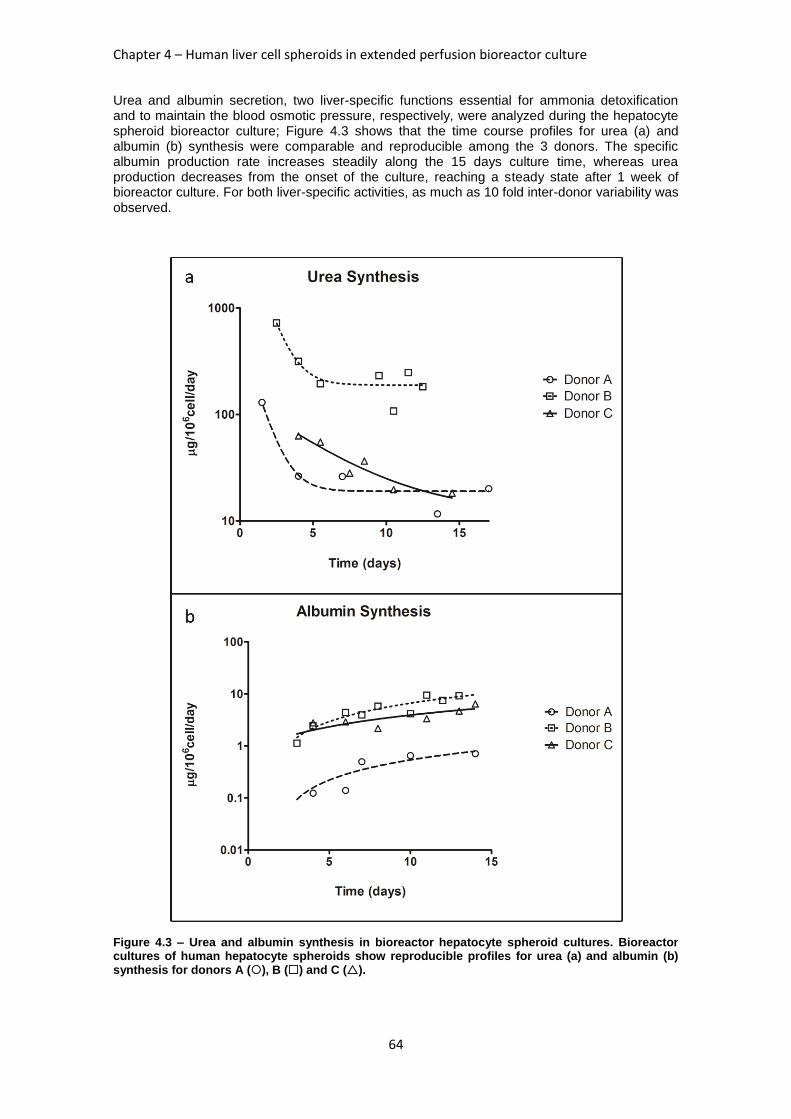
Chapter 4 – Human liver cell spheroids in extended perfusion bioreactor culture
64
Urea and albumin secretion, two liver-specific functions essential for ammonia detoxification and to maintain the blood osmotic pressure, respectively, were analyzed during the hepatocyte spheroid bioreactor culture; Figure 4.3 shows that the time course profiles for urea (a) and albumin (b) synthesis were comparable and reproducible among the 3 donors. The specific albumin production rate increases steadily along the 15 days culture time, whereas urea production decreases from the onset of the culture, reaching a steady state after 1 week of bioreactor culture. For both liver-specific activities, as much as 10 fold inter-donor variability was observed.
Figure 4.3 – Urea and albumin synthesis in bioreactor hepatocyte spheroid cultures. Bioreactor cultures of human hepatocyte spheroids show reproducible profiles for urea (a) and albumin (b) synthesis for donors A (), B () and C ().
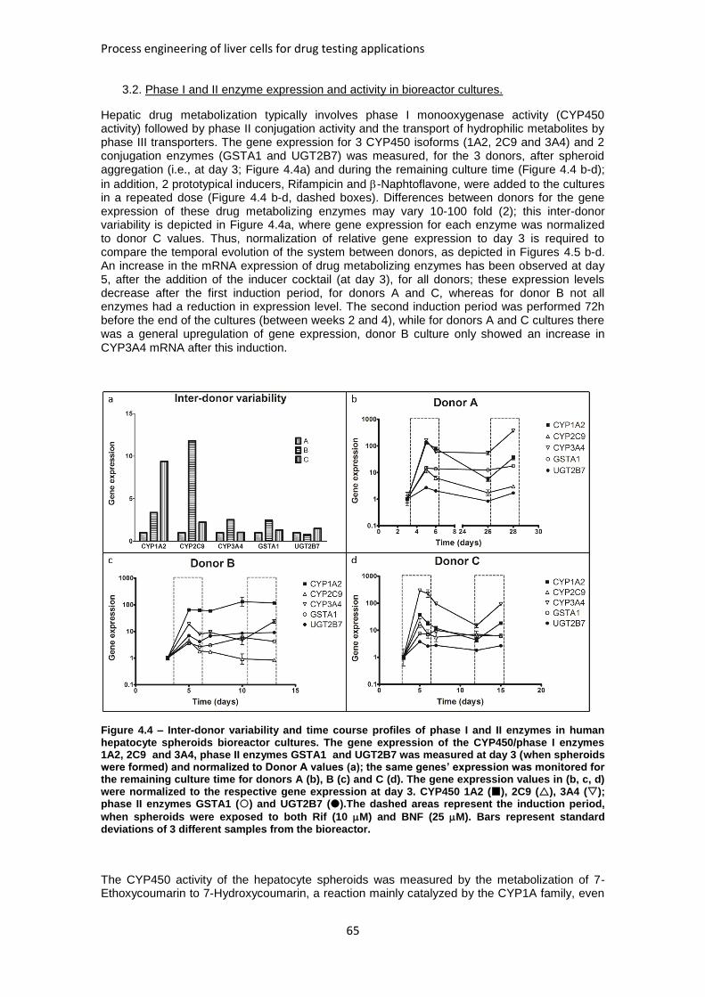
Process engineering of liver cells for drug testing applications
65
3.2. Phase I and II enzyme expression and activity in bioreactor cultures.
Hepatic drug metabolization typically involves phase I monooxygenase activity (CYP450 activity) followed by phase II conjugation activity and the transport of hydrophilic metabolites by phase III transporters. The gene expression for 3 CYP450 isoforms (1A2, 2C9 and 3A4) and 2 conjugation enzymes (GSTA1 and UGT2B7) was measured, for the 3 donors, after spheroid aggregation (i.e., at day 3; Figure 4.4a) and during the remaining culture time (Figure 4.4 b-d);
in addition, 2 prototypical inducers, Rifampicin and -Naphtoflavone, were added to the cultures in a repeated dose (Figure 4.4 b-d, dashed boxes). Differences between donors for the gene expression of these drug metabolizing enzymes may vary 10-100 fold (2); this inter-donor variability is depicted in Figure 4.4a, where gene expression for each enzyme was normalized to donor C values. Thus, normalization of relative gene expression to day 3 is required to compare the temporal evolution of the system between donors, as depicted in Figures 4.5 b-d. An increase in the mRNA expression of drug metabolizing enzymes has been observed at day 5, after the addition of the inducer cocktail (at day 3), for all donors; these expression levels decrease after the first induction period, for donors A and C, whereas for donor B not all enzymes had a reduction in expression level. The second induction period was performed 72h before the end of the cultures (between weeks 2 and 4), while for donors A and C cultures there was a general upregulation of gene expression, donor B culture only showed an increase in CYP3A4 mRNA after this induction.
Figure 4.4 – Inter-donor variability and time course profiles of phase I and II enzymes in human hepatocyte spheroids bioreactor cultures. The gene expression of the CYP450/phase I enzymes 1A2, 2C9 and 3A4, phase II enzymes GSTA1 and UGT2B7 was measured at day 3 (when spheroids were formed) and normalized to Donor A values (a); the same genes’ expression was monitored for the remaining culture time for donors A (b), B (c) and C (d). The gene expression values in (b, c, d) were normalized to the respective gene expression at day 3. CYP450 1A2 (), 2C9 (), 3A4 (); phase II enzymes GSTA1 () and UGT2B7 ().The dashed areas represent the induction period,
when spheroids were exposed to both Rif (10 M) and BNF (25 M). Bars represent standard deviations of 3 different samples from the bioreactor.
The CYP450 activity of the hepatocyte spheroids was measured by the metabolization of 7-Ethoxycoumarin to 7-Hydroxycoumarin, a reaction mainly catalyzed by the CYP1A family, even
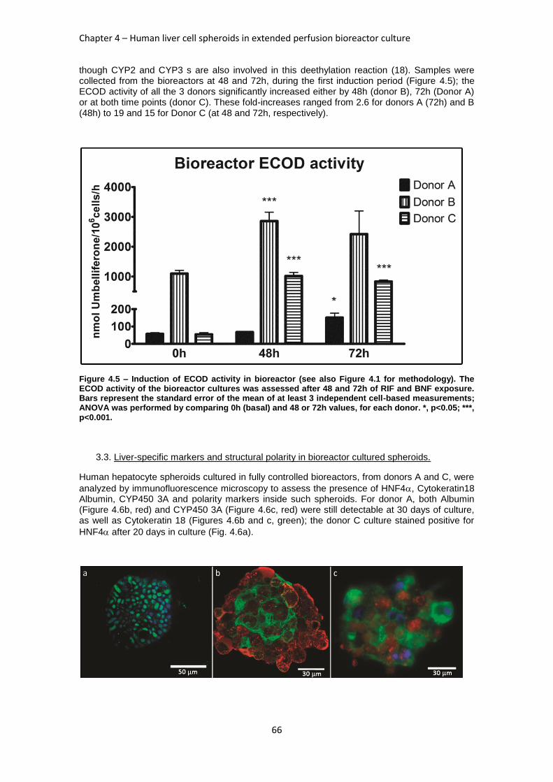
Chapter 4 – Human liver cell spheroids in extended perfusion bioreactor culture
66
though CYP2 and CYP3 s are also involved in this deethylation reaction (18). Samples were collected from the bioreactors at 48 and 72h, during the first induction period (Figure 4.5); the ECOD activity of all the 3 donors significantly increased either by 48h (donor B), 72h (Donor A) or at both time points (donor C). These fold-increases ranged from 2.6 for donors A (72h) and B (48h) to 19 and 15 for Donor C (at 48 and 72h, respectively).
Figure 4.5 – Induction of ECOD activity in bioreactor (see also Figure 4.1 for methodology). The ECOD activity of the bioreactor cultures was assessed after 48 and 72h of RIF and BNF exposure. Bars represent the standard error of the mean of at least 3 independent cell-based measurements; ANOVA was performed by comparing 0h (basal) and 48 or 72h values, for each donor. *, p<0.05; ***, p<0.001.
3.3. Liver-specific markers and structural polarity in bioreactor cultured spheroids.
Human hepatocyte spheroids cultured in fully controlled bioreactors, from donors A and C, were
analyzed by immunofluorescence microscopy to assess the presence of HNF4, Cytokeratin18 Albumin, CYP450 3A and polarity markers inside such spheroids. For donor A, both Albumin (Figure 4.6b, red) and CYP450 3A (Figure 4.6c, red) were still detectable at 30 days of culture, as well as Cytokeratin 18 (Figures 4.6b and c, green); the donor C culture stained positive for
HNF4 after 20 days in culture (Fig. 4.6a).
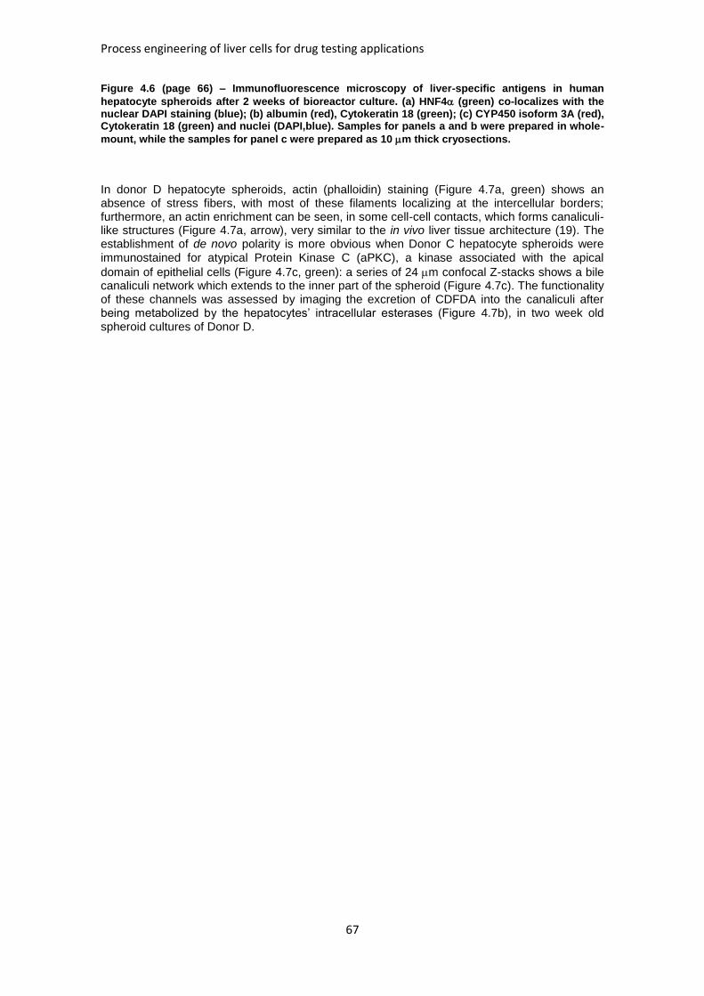
Process engineering of liver cells for drug testing applications
67
Figure 4.6 (page 66) – Immunofluorescence microscopy of liver-specific antigens in human
hepatocyte spheroids after 2 weeks of bioreactor culture. (a) HNF4 (green) co-localizes with the nuclear DAPI staining (blue); (b) albumin (red), Cytokeratin 18 (green); (c) CYP450 isoform 3A (red), Cytokeratin 18 (green) and nuclei (DAPI,blue). Samples for panels a and b were prepared in whole-
mount, while the samples for panel c were prepared as 10 m thick cryosections.
In donor D hepatocyte spheroids, actin (phalloidin) staining (Figure 4.7a, green) shows an absence of stress fibers, with most of these filaments localizing at the intercellular borders; furthermore, an actin enrichment can be seen, in some cell-cell contacts, which forms canaliculi-like structures (Figure 4.7a, arrow), very similar to the in vivo liver tissue architecture (19). The establishment of de novo polarity is more obvious when Donor C hepatocyte spheroids were immunostained for atypical Protein Kinase C (aPKC), a kinase associated with the apical
domain of epithelial cells (Figure 4.7c, green): a series of 24 m confocal Z-stacks shows a bile canaliculi network which extends to the inner part of the spheroid (Figure 4.7c). The functionality of these channels was assessed by imaging the excretion of CDFDA into the canaliculi after being metabolized by the hepatocytes’ intracellular esterases (Figure 4.7b), in two week old spheroid cultures of Donor D.
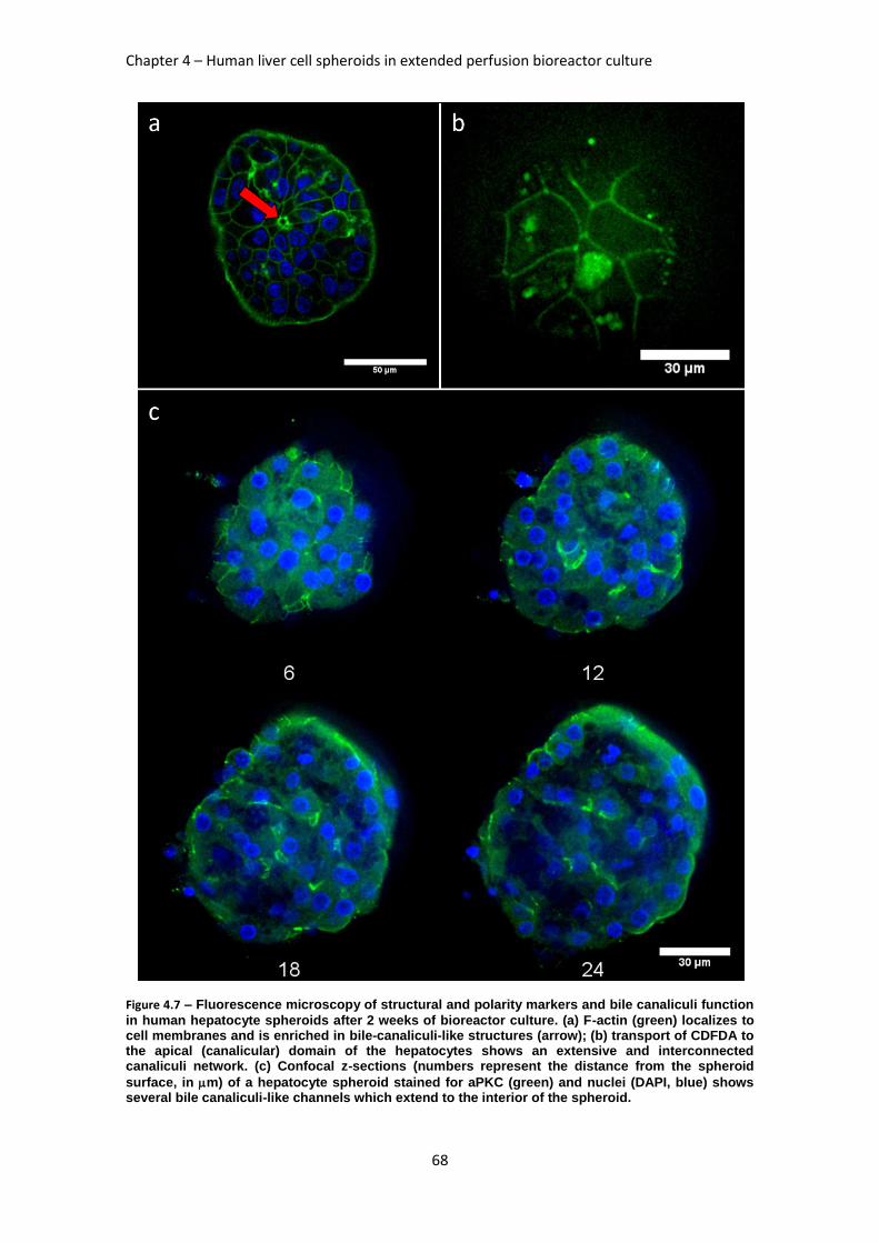
Chapter 4 – Human liver cell spheroids in extended perfusion bioreactor culture
68
Figure 4.7 – Fluorescence microscopy of structural and polarity markers and bile canaliculi function
in human hepatocyte spheroids after 2 weeks of bioreactor culture. (a) F-actin (green) localizes to cell membranes and is enriched in bile-canaliculi-like structures (arrow); (b) transport of CDFDA to the apical (canalicular) domain of the hepatocytes shows an extensive and interconnected canaliculi network. (c) Confocal z-sections (numbers represent the distance from the spheroid
surface, in m) of a hepatocyte spheroid stained for aPKC (green) and nuclei (DAPI, blue) shows several bile canaliculi-like channels which extend to the interior of the spheroid.
Process engineering of liver cells for drug testing applications
69
4. Discussion
In this work, a perfused bioreactor system for long term maintenance of primary cultures of human hepatocyte spheroids was established and tested. In this system, hepatocyte spheroids reproducibly recapitulated in vivo hepatic functions and structure, despite inter-donor variability. We hypothesize that these reproducible time-course profiles were made possible due to the tight control of critical environmental variables at physiological values, such as pH and oxygen levels. mRNA expression of CYP450 (phase I), GSTA1 and UGT2B7 (phase II) was maintained up to 4 weeks and increased when the cultures were exposed to the prototypical CYP450
inducers Rifampicin and -Naphtoflavone, in repeated dose. The phase I ECOD activity of such cultures also responded to such inducers, showing the system’s potential for more informative time course experiments. The spheroid’s inner structure resembled the liver architecture, with functional bile canaliculi-like structures and liver-specific markers. Such a system constitutes an ideal long-term culture platform for analyzing hepatic function for drug development tests.
The formation of hepatocyte spheroids was performed during the first 72 hours of culture; phase contrast microscopy data of this period (data not shown) suggests an initial 24h period of cell
clustering, when small aggregates (40-50 m) are formed; these clusters grow in size during the following 2 days (i.e., until 72h), as previously reported for primary cultures of rat hepatocyte spheroids (20).
The influence of oxygen concentration in primary cultures of hepatocytes has been the subject of several publications (16, 21-23) and the conclusions from these studies are not easily comparable. This is mainly due to the different culture systems used, i.e., static systems (24) need higher oxygen concentrations for hepatocyte culture because the mass transfer relies on diffusion; on the other hand, stirred systems, such as the bioreactor described herein, have convective mass transfer and nearly homogeneous DO concentrations in the culture bulk. Thus, the bioreactor culture oxygen concentration used in this work (30% of air saturation in culture
medium, i.e., 60 M) is in the interval between the known periportal and pericentral oxygen
concentration in the rat liver, 90 and 45 M, respectively (25).
The bulk aggregation of hepatocytes into multicellular spheroids depends on parameters such as the agitation type, vessel geometry (3, 26, 27) and cell inoculum (3). Brophy and colleagues (26) have show that rat hepatocyte spheroids could be obtained from single cells by rocking motion with a higher yield of spheroids (85%), when compared to rotational motion (54%). However, this rotational motion was based on shake flask cultures and previous work by the Hu group (20) had shown that spinner vessels could yield rat hepatocyte spheroids with an 80% efficiency in the incorporation of single cells into spheroids, after 72h. The differences between the fluid dynamics in orbitally shaken flasks and spinner vessels or the different cell inoculum used (1 and 0.3 million hepatocytes per ml, respectively) can explain the difference in both publications. In the work described herein, the previous knowledge from our group concerning the aggregation of rat hepatocytes (3, 14, 23) was adapted to the formation of multicellular spheroids in primary cultures of human hepatocytes. For these cultures the control of spheroid size becomes critical to avoid putative nutrient diffusion limitations; in the system described
herein, hepatocyte spheroids had an average size of 81 m (Figure 4.2d) and the number of
spheroids with a diameter larger than 200 m was less than 0.4% of the total population (n=3 donors). From previously published studies, it is know that rat hepatocyte spheroids with
diameters of 100 m yield a higher albumin production rate than larger ones (17), whereas
spheroids of up to 200 m diameter have been shown not to be subject to nutrient (namely oxygen) limitations (16). Since the aggregation process here described reproducibly yields
spheroids with an average 81 m diameter, it is not expected that the hepatocytes within these spheroids are subject to any significant mass transfer limitations. The data obtained for liver-specific activities (urea and albumin production, Figure 4.3; ECOD activity, Figure 4.5) and gene expression (CYP450 and phase II enzymes, Figure 4.4) confirm the inter-donor variability, which has been thoroughly described for primary human hepatocyte cultures and is a direct reflection of in vivo variability (1, 28); however, the tight control of critical variables by the perfusion bioreactor system, coupled to an easy cell sampling system, allowed reproducible liver-specific profiles to be obtained, despite inter-donor variability; the use of serum-free media
Chapter 4 – Human liver cell spheroids in extended perfusion bioreactor culture
70
after aggregation is also a likely cause for such a reproducible behavior, since serum is known to downregulate both albumin synthesis and CYP450 activity of primary cultures of human hepatocytes (22). The profiles of urea secretion rate for the 3 donors have a significant decrease from the beginning of the cultures. This reduction in urea productivity has been observed by Zeillinger and colleagues (12, 29) in a perfusion hollow fiber bioreactor and may be related to the lower oxygen concentrations inside the spheroids: retrograde liver perfusion experiments in rats have shown that lower oxygen concentrations partially inhibit the periportal urea synthesis (30).
The primary cultures of human hepatocytes in the perfusion bioreactor were inducible for the entire long-term period. The co-administration of Rif and BNF ensured increase in mRNA synthesis for CYP3A4 (Rif), CYP2C9 (Rif) and CYP1A2 (BNF) and despite the possibility of positive or negative synergies due to the use of both inducers, there is a significant induction in the 3 CYP450 isozymes; such co-administration studies constitute a unique tool to study long-term drug-drug interactions with easy access to the hepatocytes for cell-based assays. In fact, the automated perfusion, as well as the oxygen and pH control in these bioreactors, can be used to expose primary cultures of hepatocyte spheroids to repeated drug dosing, as herein described, or long term time-varying drug concentrations for chronic toxicity assessment.
The maintenance of these hepatic activities and gene expression is enabled by the cell-cell interactions which, during the bioreactor culture, evolve to a liver-like phenotype, as shown by
the presence of Albumin, Cytokeratin 18, CYP450 3A and HNF4 (Figure 4.6a-c), which are typical hepatic markers. The structural and functional polarity shown by both actin and aPKC staining and CDFDA metabolization (followed by the Multidrug resistance-associated protein 2, MRP2,-mediated transport through the apical membrane of the hepatocytes) demonstrate that these human hepatocytes cultured as spheroids resume the cuboidal geometry, without actin stress fibers, and a polarized liver-like architecture (Figure 4.7a and c). The presence of a functional bile canaliculi network in the hepatocyte spheroids (Figure 4.7b), which had not been shown to be functional in previous studies (6), ensures an efficient polarized transport of the metabolic by-products in these hepatic microtissues. The similarity to in vivo liver tissue makes this system an interesting tool for fundamental studies of the hepatic functions in physiological or bioreactor-simulated pathological conditions, namely for more complex drug transport studies. Further improvements to this system may be achieved by adding heterotypical cell-interactions, by co-culturing hepatocytes with endothelial cells (24) or fibroblasts (10, 23) or by encapsulating the hepatocyte spheroids in alginate (14).
In conclusion, the perfusion bioreactor system presented herein allows to culture defined size
human hepatocyte spheroids (80 m) which maintain hepatic liver-specific protein synthesis, CYP450, phase II and III drug metabolizing enzymes’ gene expression and activity, as well as liver-like architecture inside the spheroids for 2-4 weeks.
5. References
1. Gomez-Lechon MJ, Donato MT, Castell JV, Jover R. Human hepatocytes as a tool for studying toxicity and drug metabolism. Current Drug Metabolism 2003;4:292-312. 2. Hewitt NJ, Lechon MJG, Houston JB, Hallifax D, Brown HS, Maurel P, Kenna JG, et al. Primary hepatocytes: Current understanding of the regulation of metabolic enzymes and transporter proteins, and pharmaceutical practice for the use of hepatocytes in metabolism, enzyme induction, transporter, clearance, and hepatotoxicity studies. In: 1st Medicon Valley Hepatocyte User Form Symposium; 2006 Jan 26-27; Copenhagen, DENMARK: Taylor & Francis Inc; 2006. p. 159-234. 3. Miranda JP, Leite SB, Muller-Vieira U, Rodrigues A, Carrondo MJT, Alves PM. Towards an Extended Functional Hepatocyte In Vitro Culture. Tissue Engineering Part C-Methods 2009;15:157-167. 4. Wu F, Friend J, Remmel R, Cerra F, Hu W. Enhanced cytochrome P450 IA1 activity of self-assembled rat hepatocyte spheroids. Cell Transplant 1999;8:233-246. 5. Abu-Absi SF, Friend JR, Hansen LK, Hu WS. Structural polarity and functional bile canaliculi in rat hepatocyte spheroids. Experimental Cell Research 2002;274:56-67.
Process engineering of liver cells for drug testing applications
71
6. Dvir-Ginzberg M, Elkayam T, Aflalo ED, Agbaria R, Cohen S. Ultrastructural and functional investigations of adult hepatocyte spheroids during in vitro cultivation. Tissue Engineering 2004;10:1806-1817. 7. Goral VN, Hsieh YC, Petzold ON, Clark JS, Yuen PK, Faris RA. Perfusion-based microfluidic device for three-dimensional dynamic primary human hepatocyte cell culture in the absence of biological or synthetic matrices or coagulants. Lab on a Chip 2010;10:3380-3386. 8. Nishimura M, Hagi M, Ejiri Y, Kishimoto S, Horie T, Narimatsu S, Naito S. Secretion of Albumin and Induction of CYP1A2 and CYP3A4 in Novel Three-dimensional Culture System for Human Hepatocytes using Micro-space Plate. Drug Metabolism and Pharmacokinetics 2010;25:236-242. 9. Domansky K, Inman W, Serdy J, Dash A, Lim MHM, Griffith LG. Perfused multiwell plate for 3D liver tissue engineering. Lab on a Chip 2010;10:51-58. 10. Khetani SR, Bhatia SN. Microscale culture of human liver cells for drug development. Nat Biotechnol 2008;26:120-126. 11. Carpentier B, Gautier A, Legallais C. Artificial and bioartificial liver devices: present and future. Gut 2009;58:1690-1702. 12. Zeilinger K, Schreiter T, Darnell M, Söderdahl T, Lübberstedt M, Dillner B, Knobeloch D, et al. Scaling down of a clinical 3D perfusion multi-compartment hollow fiber liver bioreactor developed for extracorporeal liver support to an analytical scale device useful for hepatic pharmacological in vitro studies. Tissue Eng Part C Methods 2011. 13. LeCluyse EL. Human hepatocyte culture systems for the in vitro evaluation of cytochrome P450 expression and regulation. European Journal of Pharmaceutical Sciences 2001;13:343-368. 14. Tostoes R, Leite S, Miranda J, Sousa M, Wang D, Carrondo M, Alves P. Perfusion of 3D Encapsulated Hepatocytes-A Synergistic Effect Enhancing Long-Term Functionality in Bioreactors. Biotechnology and Bioengineering 2011:41-49. 15. Castell JV, Gómez-Lechón MJ. In vitro methods in pharmaceutical research. San Diego: Academic Press, 1997: ix, 467 p. 16. Curcio E, Salerno S, Barbieri G, De Bartolo L, Drioli E, Bader A. Mass transfer and metabolic reactions in hepatocyte spheroids cultured in rotating wall gas-permeable membrane system. Biomaterials 2007;28:5487-5497. 17. Glicklis R, Merchuk JC, Cohen S. Modeling mass transfer in hepatocyte spheroids via cell viability, spheroid size, and hepatocellular functions. Biotechnology and Bioengineering 2004;86:672-680. 18. Waxman DJ, Chang TK. Use of 7-ethoxycoumarin to monitor multiple enzymes in the human CYP1, CYP2, and CYP3 families. Methods Mol Biol 2006;320:153-156. 19. Dunn JCY, Tompkins RG, Yarmush ML. Long-term in vitro function of adult hepatocytes in a collagen sandwich configuration. Biotechnology Progress 1991;7:237-245. 20. Wu FJ, Friend JR, Hsiao CC, Zilliox MJ, Ko WJ, Cerra FB, Hu WS. Efficient assembly of rat hepatocyte spheroids for tissue engineering applications. Biotechnology and Bioengineering 1996;50:404-415. 21. Foy BD, Rotem A, Toner M, Tompkins RG, Yarmush ML. A DEVICE TO MEASURE THE OXYGEN-UPTAKE RATE OF ATTACHED CELLS - IMPORTANCE IN BIOARTIFICIAL ORGAN DESIGN. Cell Transplantation 1994;3:515-527. 22. Kidambi S, Yarmush RS, Novik E, Chao P, Yarmush ML, Nahmias Y. Oxygen-mediated enhancement of primary hepatocyte metabolism, functional polarization, gene expression, and drug clearance. Proceedings of the National Academy of Sciences of the United States of America 2009;106:15714-15719. 23. Leite SB, Teixeira AP, Miranda JP, Tostões RM, Clemente JJ, Sousa MF, Carrondo MJ, et al. Merging bioreactor technology with 3D hepatocyte-fibroblast culturing approaches: improved in vitro models for Toxicological applications. Toxicol In Vitro 2011. 24. Kidambi S, Yarmush RS, Novik E, Chao P, Yarmush ML, Nahmias Y. Oxygen-mediated enhancement of primary hepatocyte metabolism, functional polarization, gene expression, and drug clearance. Proc Natl Acad Sci U S A 2009;106:15714-15719. 25. Jungermann K, Kietzmann T. Role of oxygen in the zonation of carbohydrate metabolism and gene expression in liver. Kidney Int 1997;51:402-412. 26. Brophy CM, Luebke-Wheeler JL, Amiot BP, Khan H, Remmel RP, Rinaldo P, Nyberg SL. Rat hepatocyte spheroids formed by rocked technique maintain differentiated hepatocyte gene expression and function. Hepatology 2009;49:578-586.
Chapter 4 – Human liver cell spheroids in extended perfusion bioreactor culture
72
27. Khaoustov VI, Darlington GJ, Soriano HE, Krishnan B, Risin D, Pellis NR, Yoffe B. Induction of three-dimensional assembly of human liver cells by simulated microgravity. In Vitro Cellular & Developmental Biology-Animal 1999;35:501-509. 28. Ponsoda X, Pareja E, Gómez-Lechón MJ, Fabra R, Carrasco E, Trullenque R, Castell JV. Drug biotransformation by human hepatocytes. In vitro/in vivo metabolism by cells from the same donor. J Hepatol 2001;34:19-25. 29. Schmelzer E, Mutig K, Schrade P, Bachmann S, Gerlach JC, Zeilinger K. Effect of Human Patient Plasma Ex Vivo Treatment on Gene Expression and Progenitor Cell Activation of Primary Human Liver Cells in Multi-Compartment 3D Perfusion Bioreactors for Extra-Corporeal Liver Support. Biotechnology and Bioengineering 2009;103:817-827. 30. Katz NR. Metabolic heterogeneity of hepatocytes across the liver acinus. J Nutr 1992;122:843-849.
73
Chapter 5
Spheroid formation of human embryonic stem cell-derived hepatic progenitors for improved hepatic differentiation
This work was performed in collaboration with Cellartis AB, Sweden, and it will be submitted for
publication after Cellartis authorization
Chapter 5 – Spheroid formation of hESC-derived hepatic progenitors
74
Abstract
A key unmet need in pharmaceutical development is reliable, available, cost-effective and predictive models for determining the metabolic and toxicological properties of drug compounds. Human hepatocytes are the regulator’s gold standard model for evaluating drug toxicity; however, they suffer from scarce and inconsistent availability. In vivo models are prohibitively expensive, have low throughput and are often not predictive for humans (1), which ultimately results in high failure rates and risk to volunteers in Phase I trials. In addition to the potential use in the pharmaceutical industry, the generation of hepatocytes for transplantation medicine, as a means to increase the number of functional hepatocytes in the liver or as a biological component for bioartificial liver (BAL) devices, make their production extremely attractive to the clinical industry as an alternative therapeutic approach to whole organ transplantation (2). In this work, the directed hepatic differentiation of hESC, previously developed at Cellartis AB, has been adapted to mimic the in vivo developmental morphogenesis; hESC-derived definitive endoderm cells, obtained after directed differentiation in monolayer culture, were aggregated in stirred tank vessels and the resulting spheroids were encapsulated in an alginate hydrogel. This novel differentiation process resulted in higher liver-specific gene expression, phenotype and activities than the monolayer cultures.
Process engineering of liver cells for drug testing applications
75
Table of Contents
1. Introduction ........................................................................................................... 76
2. Materials and Methods ......................................................................................... 77
2.1. Human embryonic stem cell-derived hepatic progenitor culture ........................ 77
2.2. Optimization of spheroid formation ................................................................... 77
2.2.1. Medium composition .............................................................................. 77
2.2.2. Cell aggregation process ....................................................................... 77
2.3. Spheroid measurement .................................................................................... 77
2.4. Alginate ............................................................................................................ 77
2.5. Monolayer culture of hESC-derived hepatic progenitors ................................... 77
2.6. 3D differentiation of hESC-derived hepatic progenitors .................................... 78
2.7. Alginate microencapsulation and dissolution .................................................... 78
2.8. Cell concentration determination ...................................................................... 78
2.9. qRT-PCR .......................................................................................................... 78
2.10. Cell membrane integrity assay. ...................................................................... 78
2.11. Albumin and Urea measurement .................................................................... 78
3. Results ................................................................................................................... 79
3.1. Optimization of spheroid formation ................................................................... 79
3.1.1. Medium composition .............................................................................. 79
3.1.2. Cell aggregation process ....................................................................... 79
3.2. Differentiation of hESC-derived hepatic progenitors ......................................... 80
3.3. Hepatic progenitors viability in the 3D differentiation process ........................... 82
3.4. Urea and Albumin production ........................................................................... 82
4. Discussion............................................................................................................. 82
5. References ............................................................................................................ 83
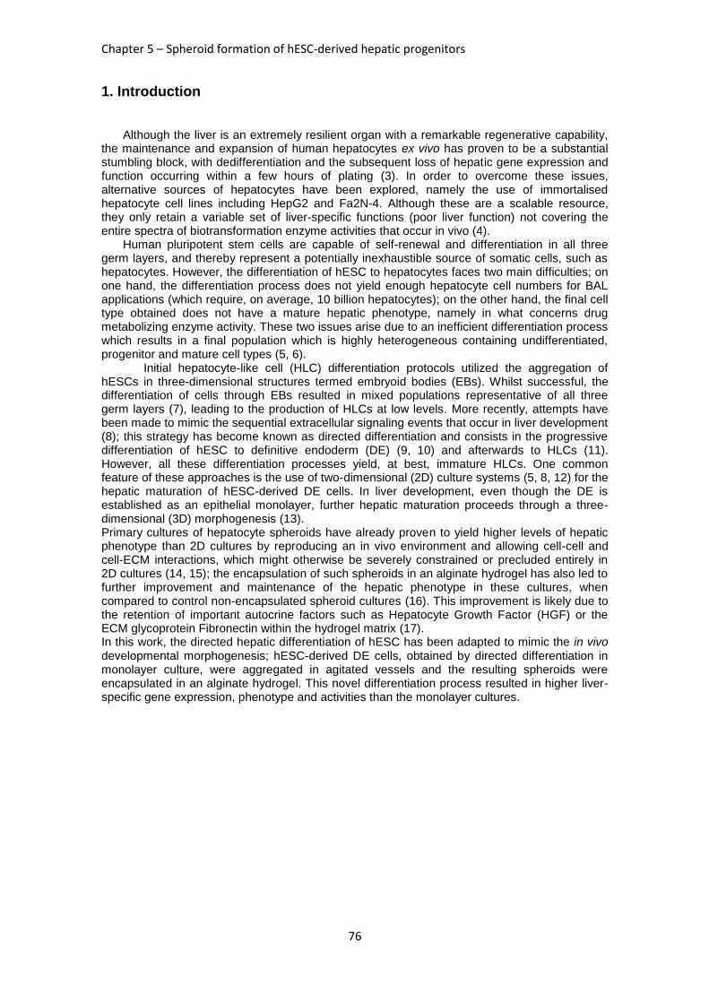
Chapter 5 – Spheroid formation of hESC-derived hepatic progenitors
76
1. Introduction
Although the liver is an extremely resilient organ with a remarkable regenerative capability, the maintenance and expansion of human hepatocytes ex vivo has proven to be a substantial stumbling block, with dedifferentiation and the subsequent loss of hepatic gene expression and function occurring within a few hours of plating (3). In order to overcome these issues, alternative sources of hepatocytes have been explored, namely the use of immortalised hepatocyte cell lines including HepG2 and Fa2N-4. Although these are a scalable resource, they only retain a variable set of liver-specific functions (poor liver function) not covering the entire spectra of biotransformation enzyme activities that occur in vivo (4).
Human pluripotent stem cells are capable of self-renewal and differentiation in all three germ layers, and thereby represent a potentially inexhaustible source of somatic cells, such as hepatocytes. However, the differentiation of hESC to hepatocytes faces two main difficulties; on one hand, the differentiation process does not yield enough hepatocyte cell numbers for BAL applications (which require, on average, 10 billion hepatocytes); on the other hand, the final cell type obtained does not have a mature hepatic phenotype, namely in what concerns drug metabolizing enzyme activity. These two issues arise due to an inefficient differentiation process which results in a final population which is highly heterogeneous containing undifferentiated, progenitor and mature cell types (5, 6).
Initial hepatocyte-like cell (HLC) differentiation protocols utilized the aggregation of hESCs in three-dimensional structures termed embryoid bodies (EBs). Whilst successful, the differentiation of cells through EBs resulted in mixed populations representative of all three germ layers (7), leading to the production of HLCs at low levels. More recently, attempts have been made to mimic the sequential extracellular signaling events that occur in liver development (8); this strategy has become known as directed differentiation and consists in the progressive differentiation of hESC to definitive endoderm (DE) (9, 10) and afterwards to HLCs (11). However, all these differentiation processes yield, at best, immature HLCs. One common feature of these approaches is the use of two-dimensional (2D) culture systems (5, 8, 12) for the hepatic maturation of hESC-derived DE cells. In liver development, even though the DE is established as an epithelial monolayer, further hepatic maturation proceeds through a three-dimensional (3D) morphogenesis (13). Primary cultures of hepatocyte spheroids have already proven to yield higher levels of hepatic phenotype than 2D cultures by reproducing an in vivo environment and allowing cell-cell and cell-ECM interactions, which might otherwise be severely constrained or precluded entirely in 2D cultures (14, 15); the encapsulation of such spheroids in an alginate hydrogel has also led to further improvement and maintenance of the hepatic phenotype in these cultures, when compared to control non-encapsulated spheroid cultures (16). This improvement is likely due to the retention of important autocrine factors such as Hepatocyte Growth Factor (HGF) or the ECM glycoprotein Fibronectin within the hydrogel matrix (17). In this work, the directed hepatic differentiation of hESC has been adapted to mimic the in vivo developmental morphogenesis; hESC-derived DE cells, obtained by directed differentiation in monolayer culture, were aggregated in agitated vessels and the resulting spheroids were encapsulated in an alginate hydrogel. This novel differentiation process resulted in higher liver-specific gene expression, phenotype and activities than the monolayer cultures.
Process engineering of liver cells for drug testing applications
77
2. Materials and Methods
2.1. Human embryonic stem cell-derived hepatic progenitor culture
Human embryonic stem cell (hESC) line SA 181-derived hepatic progenitors (hepatic progenitors, day 11 of the hepatic differentiation, were resupended in KnockOut DMEM (KO-
DMEM, GIBCO/Invitrogen) and stored overnight (shipped overnight) at 4⁰ before use. At the
beginning of the experiments (upon arrival, day 12 of the hepatic differentiation at Cellartis (confidential protocol)) cell viability was higher than 80% as determined by trypan blue exclusion, using a Fuchs-Rosenthal counting chamber. These hepatic progenitors were cultured in Progenitor Medium (PM) (a confidential medium formulation developed at Cellartis) for three days (day 15 of the hepatic differentiation); all cultures were maintained in an incubator at 37ºC and controlled atmosphere (5% CO2 in air).
2.2. Optimization of spheroid formation
2.2.1. Medium composition
The hanging drop (HD) culture method was used to determine the best medium composition for the aggregation of hepatic progenitors. This HD culture system has a high throughput 96 well plate format which allows testing several medium compositions per drop. In the optimization herein described, one HD plate has been used to assess the influence of the Rock inhibitor (Rocki) Y-27632 (Sigma) in the aggregation of the hepatic progenitors, under the droplets
microgravity conditions; the aggregation time was 72h, the drop volume was 20 L and each drop contained 1000 cells. Thus, 48 drops without Rocki were compared with the same number of drops using Rocki. The aggregation was evaluated by light microscopy (Leica Microsystems GmbH, Wetzlar, Germany).
2.2.2. Cell aggregation process
A single cell suspension of hepatic progenitors in PM supplemented with 10 M Rocki was inoculated in spinners (Wheaton, Techne, NJ, USA) with ball or paddle impeller, at 0.4 million cells/mL; both shake flasks (Corning, Corning, NY, USA) and Aggrewell 400 wells (Stemcell Technologies, Grenoble, France), were inoculated at a concentration of 1 million hepatic progenitors/mL. Aggrewell plates were centrifuged at 100g (as recommended by the
manufacturer) to force the cells into the microwells.
2.3. Spheroid measurement
hESC-derived hepatic progenitor spheroids were visualized by phase contrast microscopy (Leica Microsystems GmbH, Wetzlar, Germany) and their average diameter (dave) was determined by a geometric mean of three diameters per spheroid (n>35 spheroids) using the equation dave=(d1 × d2 × d3)
1/3; spheroid diameters were measured using the ImageJ software.
Diameter distribution plots were done using the GraphPad Prism software (La Jolla, CA 92037 USA).
2.4. Alginate
Ca2+-UP MVG alginate was dissolved by incubating the microcapsules with a chelating solution (50 mM EDTA and 10 mM HEPES in PBS) for 5 min at 37°C [31]. Spheroids were washed twice with PBS and incubated with culture medium until further analysis.
2.5. Monolayer culture of hESC-derived hepatic progenitors
For monolayer (2D) cultures, 6-well plates were coated with Matrigel (BD) at room temperature for one hour; hepatic progenitors were seeded onto these plates at 0.2 million cells/cm
2 and
cultured in PM supplemented with 10 M of Rocki for 24 hours. After this period the culture medium was changed to PM without Rocki for another 48 hours (until day 15 of the hepatic
Chapter 5 – Spheroid formation of hESC-derived hepatic progenitors
78
differentiation). At day 16 the culture medium was changed to Maturation Medium (MM), whose composition was developed at Cellartis and is also confidential. This culture medium was used until the end of the culture, with full medium exchanges every two days.
2.6. 3D differentiation of hESC-derived hepatic progenitors
Hepatic progenitors (at day 12 of differentiation) were inoculated in spinner vessels with a ball impeller; the cells were inoculated in the vessels at a concentration of 0.4 million cells/mL in PM
supplemented with 10 M of Rocki, at an agitation rate of 50 rpm, for 72h, i.e., up to day 15 of the differentiation protocol. By day 15 spheroids were separated from single cells by settling and encapsulated in calcium-crosslinked alginate (see below); the encapsulated spheroids were transferred to spinners with paddle impeller and cultured in MM until the end of the culture, with 50% medium exchanges every two days.
2.7. Alginate microencapsulation and dissolution
Ultra Pure MVG alginate (UP MVG NovaMatrix, Pronova Biomedical, Oslo, Norway) was prepared at a concentration of 1.1% (w/v) in 0.9% (w/v) NaCl solution. Microcapsules were prepared by passing the alginate-cell mixture using a 1 mL syringe through an air-jet generator at an air flow rate of 2–3.5 L/min and an air pressure of 1 bar. These encapsulation conditions yielded microcapsules with a diameter of approximately 400-600 µm. For cross-linkage of the UP MVG alginate, a 100 mM CaCl2/10 mM HEPES solution adjusted to pH 7.4 was used. Alginate microcapsules were washed twice with 0.9% (w/v) NaCl solution and once with DMEM-KO medium before being transferred to the spinner vessels (18).
Ca2+
-UP MVG alginate was dissolved by incubating the microcapsules with a chelating solution (50 mM EDTA and 10 mM HEPES in PBS) for 5 min at 37°C (18).Cells were washed twice with PBS and incubated with culture medium until further analysis.
2.8. Cell concentration determination
Hepatic progenitors monolayers were digested with TrypLE™ Select (Invitrogen, Paisley, UK), whereas spheroids were digested with 0.05% Trypsin/EDTA (GIBCO), for 6-8 minutes at 37ºC and the resulting single cell suspension viability was assessed by the trypan blue exclusion method. Cell counting was performed using a Fuchs Rosenthal counting chamber.
2.9. qRT-PCR
Hepatic progenitor spheroids and monolayer single cell suspensions were collected from the cultures at different time points and stored at -20⁰C with RNAprotect Cell Reagent (Qiagen,
Valencia, CA) for up to one month. Total RNA was extracted using RNEasy Plus Mini Kit (Qiagen, Valencia, CA) according to the manufacturer´s instructions. Reverse transcription was performed using 0.6ug of total RNA in a final volume of 20 ul reaction mix using High Capacity cDNA Reverse Transcriptase Kit (Applied Biosystems). Real time PCR was performed using ready-to-use TaqMan Gene Expression Assays (applied Biosystems) according to the manufacturer´s instructions. GAPDH was used as endogenous control.
2.10. Cell membrane integrity assay.
The qualitative assessment of the cell plasma membrane integrity during culture was performed using the enzyme substrate fluorescein diacetate (FDA; Sigma-Aldrich, Steinheim, Germany) and the DNA-dye propidium iodide (PI; Sigma-Aldrich, Steinheim, Germany) as described in the literature (18). Briefly, spheroids/microcapsules were incubated with 20 µg/mL FDA and 10 µg/mL PI in PBS for 2–5 min and then observed using fluorescence microscopy (Leica Microsystems GmbH, Wetzlar, Germany).
2.11. Albumin and Urea measurement
The concentration of albumin in the culture medium supernatant was measured by an enzyme-linked immunosorbent assay (ELISA) using the Exocell Albuwell albumin test kit (Philadelphia, PA, USA). The assay was performed according to the manufacturer’s description. The concentration of albumin in the culture medium supernatant was determined using a
Process engineering of liver cells for drug testing applications
79
quantitative colorimetric urea kit (QuantiChromTM Urea Assay Kit, DIUR-500, BioAssay Systems), according to the manufacturer’s instructions. The supernatant concentrations were normalized to RNA concentration in the corresponding cellular fraction of the samples.
3. Results
3.1. Optimization of spheroid formation
3.1.1. Medium composition
The effect of Rocki on the aggregation of hepatic progenitors was assessed in Hanging Drop cultures. Figure 5.1A and B show that without this chemical agent the microgravity conditions of the drops are not enough to promote the aggregation of hepatic progenitors (Figure5.1 A); on the other hand, the presence of Rocki led to the formation of aggregation clusters (arrows, Figure5.1 B). Such aggregates maintained both metabolic activity and membrane integrity, unlike single cells, as shown in Figure 5.1C and D.
Figure 5.1 – Effect of Rocki on the aggregation and viability of hepatic progenitor cells under HD
microgravity conditions. Each 20 L drop of PM contained 1000 cells which were cultured for 72h in the absence (A) or presence of Rocki (B). Arrows in B indicate multicellular aggregates of cells. The spheroids obtained with Rocki-containing PM retained their membrane integrity and metabolic activity, whereas single cells did not (arrowheads), as assessed by FDA/PI staining. These are representative photographs of 48 Hanging Drops per condition tested; bars measure 200 and 100
m in A-B and C-D, respectively.
3.1.2. Cell aggregation process
Several methods for increasing the efficiency of the aggregation process have been tested
using PM supplemented with 10 M Rocki, for 72h. Figure 5.2 summarizes the conditions and results of this scale-up experiment. The size distribution plots show that aggregation within a spinner vessel with a paddle impeller or shake flasks yielded the lowest average spheroid
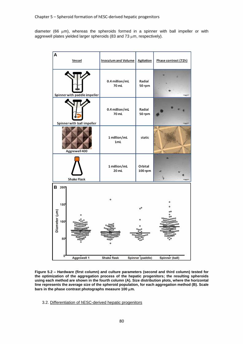
Chapter 5 – Spheroid formation of hESC-derived hepatic progenitors
80
diameter (66 m), whereas the spheroids formed in a spinner with ball impeller or with
aggrewell plates yielded larger spheroids (83 and 73 m, respectively).
Figure 5.2 – Hardware (first column) and culture parameters (second and third column) tested for the optimization of the aggregation process of the hepatic progenitors; the resulting spheroids using each method are shown in the fourth column (A). Size distribution plots, where the horizontal line represents the average size of the spheroid population, for each aggregation method (B). Scale
bars in the phase contrast photographs measure 100 m.
3.2. Differentiation of hESC-derived hepatic progenitors
A
B
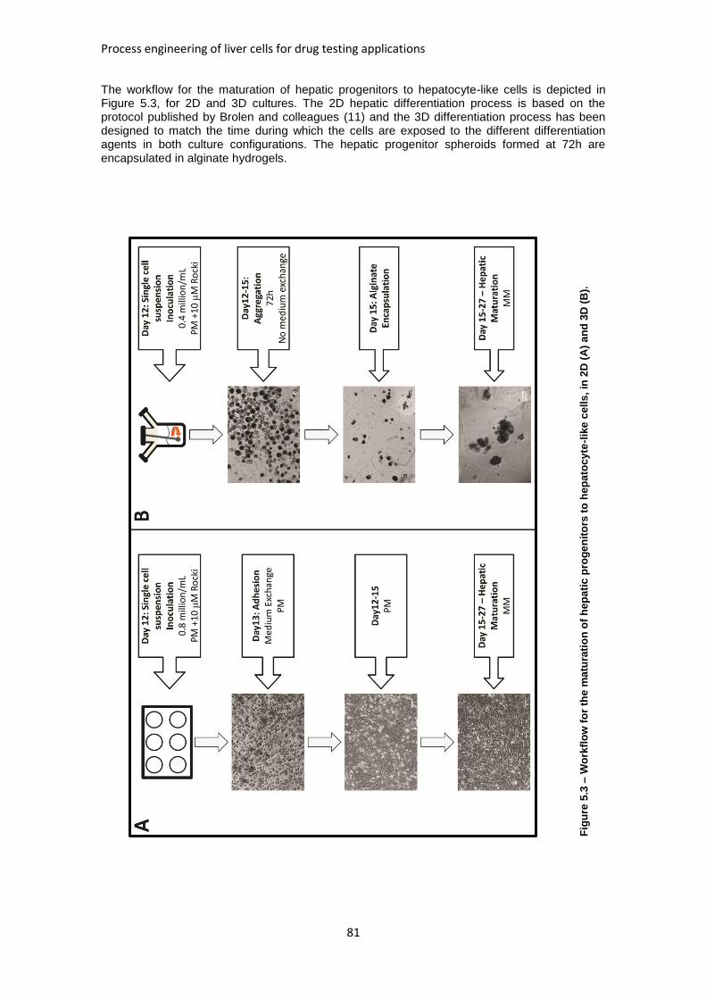
Process engineering of liver cells for drug testing applications
81
The workflow for the maturation of hepatic progenitors to hepatocyte-like cells is depicted in Figure 5.3, for 2D and 3D cultures. The 2D hepatic differentiation process is based on the protocol published by Brolen and colleagues (11) and the 3D differentiation process has been designed to match the time during which the cells are exposed to the different differentiation agents in both culture configurations. The hepatic progenitor spheroids formed at 72h are encapsulated in alginate hydrogels.
Fig
ure
5.3
– W
ork
flo
w f
or
the
ma
tura
tio
n o
f h
ep
ati
c p
rog
en
ito
rs t
o h
ep
ato
cy
te-l
ike
ce
lls
, in
2D
(A
) a
nd
3D
(B
).
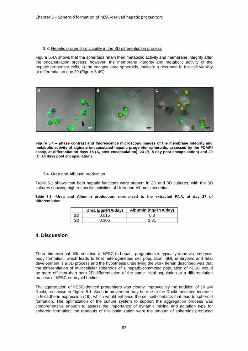
Chapter 5 – Spheroid formation of hESC-derived hepatic progenitors
82
3.3. Hepatic progenitors viability in the 3D differentiation process
Figure 5.4A shows that the spheroids retain their metabolic activity and membrane integrity after the encapsulation process; however, the membrane integrity and metabolic activity of the hepatic progenitor cells, in the encapsulated spheroids, indicate a decrease in the cell viability at differentiation day 29 (Figure 5.4C).
Figure 5.4 – phase contrast and fluorescence microscopy images of the membrane integrity and metabolic activity of alginate encapsulated hepatic progenitor spheroids, assessed by the FDA/PI assay, at differentiation days 15 (A, post encapsulation), 23 (B, 8 day post encapsulation) and 29 (C, 14 days post encapsulation).
3.4. Urea and Albumin production
Table 5.1 shows that both hepatic functions were present in 2D and 3D cultures, with the 3D cultures showing higher specific activities of Urea and Albumin secretion.
Table 5.1 –Urea and Albumin production, normalized to the extracted RNA, at day 27 of
differentiation.
Urea (g/RNA/day) Albumin (ng/RNA/day)
2D 0.015 0.8
3D 0.305 2.31
4. Discussion
Three dimensional differentiation of hESC to hepatic progenitors is typically done via embryoid body formation, which leads to final heterogeneous cell population. Still, embryonic and fetal development is a 3D process and the hypothesis underlying the work herein described was that the differentiation of multicellular spheroids of a hepatic-committed population of hESC would be more efficient than both 2D differentiation of the same initial population or a differentiation process of hESC embryoid bodies.
The aggregation of hESC-derived progenitors was clearly improved by the addition of 10 M Rocki, as shown in Figure 5.1. Such improvement may be due to the Rocki-mediated increase in E-cadherin expression (19), which would enhance the cell-cell contacts that lead to spheroid formation. The optimization of the culture system to support the aggregation process was comprehensive enough to assess the importance of dynamic mixing and agitation type for spheroid formation; the readouts of this optimization were the amount of spheroids produced
Process engineering of liver cells for drug testing applications
83
(normalized to the cost) and the diameter distribution. This latter outcome was not critical since
no spheroid exceeded 200 m.
The poor performance of the paddle spinner for the aggregation of hESC-derived hepatic progenitors suggests that this agitation regime may result in shear stress levels that prevent aggregation. Comparing the spinner with ball impeller and the Aggrewell plate at 1 million cells/mL, and assuming that the differences in average diameter obtained by the 2 methods (73
m Aggrewell vs 83 m spinner with ball impeller) are negligible, the spinner with ball impeller is the most cost-efficient option: the cost per million cell aggregated (assuming similar aggregation efficiencies) is 6-fold higher using the Aggrewell plates (calculations considering the amount of Rock inhibitor used and the cost of the Aggrewell plates).
Several published works report an improvement of the liver-specific functionality of alginate encapsulated hepatocytes (20-23); the effects of alginate encapsulation, which include the protection from shear stress and the retention of ECM molecules (17), were hypothesized to improve the maturation of hESC-derived hepatic progenitor spheroids to hepatocyte-like cells. Although some improvement in Urea and Albumin secretion of such encapsulated spheroids could be seen (Table 5.1), when compared to the 2D hepatic maturation, there was also a marked decrease in cell viability (Figure 5.4) at the end of this differentiation period. This apparent increase in cell death may be due to the retention of proteases or other large molecular weight apoptotic agents. While this hypothesis can be investigated, culturing the progenitor spheroids without alginate encapsulation (at the expense of an increased shear stress at the cells’ surface) seems to be the most straightforward step in this work.
While the optimization of spheroid formation (section 3.1) relies on statistically significant results, the hepatic differentiation sections (sections 3.2, 3.3 and 3.4) report one single experiment and must thus be viewed and discussed with caution, until the 3D differentiation hepatic differentiation experiments are repeated and statistically significant results are obtained.
Since the major bottleneck in this process is the cell viability throughout the hepatic maturation, another strategy to be tested is the co-culture of the hESC-derived hepatic progenitors with human feeder cells, such as human foreskin fibroblasts (HFF), which are regularly used in hESC culture to maintain their undifferentiated growth (24); moreover, the co-culture of hepatocytes with fibroblasts is known to improve these latter cells’ liver-specific functions.
5. References
1. Huang SM, Strong JM, Zhang L, Reynolds KS, Nallani S, Temple R, Abraham S, et al. New era in drug interaction evaluation: US Food and Drug Administration update on CYP enzymes, transporters, and the guidance process. J Clin Pharmacol 2008;48:662-670. 2. Miki T, Ring A, Gerlach J. Hepatic Differentiation of Human Embryonic Stem Cells Is Promoted by Three-Dimensional Dynamic Perfusion Culture Conditions. Tissue Eng Part C Methods 2011. 3. Miranda JP, Leite SB, Muller-Vieira U, Rodrigues A, Carrondo MJT, Alves PM. Towards an Extended Functional Hepatocyte In Vitro Culture. Tissue Engineering Part C-Methods 2009;15:157-167. 4. Sinz M, Wallace G, Sahi J. Current industrial practices in assessing CYP450 enzyme induction: Preclinical and clinical. Aaps Journal 2008;10:391-400. 5. Agarwal S, Holton KL, Lanza R. Efficient differentiation of functional hepatocytes from human embryonic stem cells. Stem Cells 2008;26:1117-1127. 6. Hay DC, Zhao D, Ross A, Mandalam R, Lebkowski J, Cui W. Direct differentiation of human embryonic stem cells to hepatocyte-like cells exhibiting functional activities. Cloning and Stem Cells 2007;9:51-62. 7. Itskovitz-Eldor J, Schuldiner M, Karsenti D, Eden A, Yanuka O, Amit M, Soreq H, et al. Differentiation of human embryonic stem cells into embryoid bodies compromising the three embryonic germ layers. Mol Med 2000;6:88-95.
Chapter 5 – Spheroid formation of hESC-derived hepatic progenitors
84
8. Cai J, Zhao Y, Liu YX, Ye F, Song ZH, Qin H, Meng S, et al. Directed differentiation of human embryonic stem cells into functional hepatic cells. Hepatology 2007;45:1229-1239. 9. D'Amour KA, Agulnick AD, Eliazer S, Kelly OG, Kroon E, Baetge EE. Efficient differentiation of human embryonic stem cells to definitive endoderm. Nature Biotechnology 2005;23:1534-1541. 10. Lock LT, Tzanakakis ES. Expansion and differentiation of human embryonic stem cells to endoderm progeny in a microcarrier stirred-suspension culture. Tissue Eng. Part A 2009;15:2051-2063. 11. Brolen G, Sivertsson L, Bjorquist P, Eriksson G, Ek M, Semb H, Johansson I, et al. Hepatocyte-like cells derived from human embryonic stem cells specifically via definitive endoderm and a progenitor stage. J Biotechnol;145:284-294. 12. Brolen G, Sivertsson L, Bjorquist P, Eriksson G, Ek M, Semb H, Johansson I, et al. Hepatocyte-like cells derived from human embryonic stem cells specifically via definitive endoderm and a progenitor stage. Journal of Biotechnology 2010;145:284-294. 13. Zaret KS. Hepatocyte differentiation: from the endoderm and beyond. Curr Opin Genet Dev 2001;11:568-574. 14. Miranda JP, Leite SB, Muller-Vieira U, Rodrigues A, Carrondo MJ, Alves PM. Towards an extended functional hepatocyte in vitro culture. Tissue Eng Part C Methods 2009;15:157-167. 15. Brophy CM, Luebke-Wheeler JL, Amiot BP, Khan H, Remmel RP, Rinaldo P, Nyberg SL. Rat hepatocyte spheroids formed by rocked technique maintain differentiated hepatocyte gene expression and function. Hepatology 2009;49:578-586. 16. Miranda J, Rodrigues A, Tostoes R, Leite S, Zimmerman H, Carrondo M, Alves P. Extending Hepatocyte Functionality for Drug-Testing Applications Using High-Viscosity Alginate-Encapsulated Three-Dimensional Cultures in Bioreactors. Tissue Engineering Part C-Methods 2010:1223-1232. 17. Williams CM, Mehta G, Peyton SR, Zeiger AS, Van Vliet KJ, Griffith LG. Autocrine-controlled formation and function of tissue-like aggregates by primary hepatocytes in micropatterned hydrogel arrays. Tissue Eng Part A 2011;17:1055-1068. 18. Serra M, Correia C, Malpique R, Brito C, Jensen J, Bjorquist P, Carrondo MJ, et al. Microencapsulation technology: a powerful tool for integrating expansion and cryopreservation of human embryonic stem cells. PLoS One 2011;6:e23212. 19. Xu Y, Zhu X, Hahm HS, Wei W, Hao E, Hayek A, Ding S. Revealing a core signaling regulatory mechanism for pluripotent stem cell survival and self-renewal by small molecules. Proc Natl Acad Sci U S A 2010;107:8129-8134. 20. Falasca L, Miccheli A, Sartori E, Tomassini A, Devirgiliis LC. Hepatocytes entrapped in alginate gel beads and cultured in bioreactor: Rapid repolarization and reconstitution of adhesion areas. Cells Tissues Organs 2001;168:126-136. 21. Lan SF, Safiejko-Mroczka B, Starly B. Long-term cultivation of HepG2 liver cells encapsulated in alginate hydrogels: A study of cell viability, morphology and drug metabolism. Toxicology in Vitro 2010;24:1314-1323. 22. Miranda J, Rodrigues A, Tostoes R, Leite S, Zimmermann H, Carrondo M, Alves P. Extending hepatocyte functionality for drug testing applications using high viscosity alginate encapsulated 3D cultures in bioreactors. Tissue Eng Part C Methods 2010. 23. Tostoes R, Leite S, Miranda J, Sousa M, Wang D, Carrondo M, Alves P. Perfusion of 3D Encapsulated Hepatocytes-A Synergistic Effect Enhancing Long-Term Functionality in Bioreactors. Biotechnology and Bioengineering 2011:41-49. 24. Serra M, Brito C, Sousa M, Jensen J, Tostões R, Clemente J, Strehl R, et al. Improving expansion of pluripotent human embryonic stem cells in perfused bioreactors through oxygen control. J Biotechnol 2010;148:208-215.

85
Chapter 6
Discussion and conclusions
Chapter 6 – Discussion and conclusions
86
Table of Contents
1. Discussion............................................................................................................. 87
1.1. Cell culture technologies: Alginate microencapsulation and perfusion .............. 88
1.1.1. Operational bottlenecks ............................................................................. 90
1.2. Process validation for drug development tests: primary cultures of human hepatocytes and repeated dose drug induction. ...................................................... 90
1.2.1. CYP450 induction in primary cultures of human hepatocytes at weeks 1-4; ............................................................................................................................ 90
1.2.2. A 3D structure which closely resembles the liver structure; ........................ 90
1.2.3. Technical inter-donor reproducibility. .......................................................... 90
1.2.4. System limitations ...................................................................................... 90
1.3. Human embryonic stem cells as potential source of functional hepatic cells. .... 91
2. Future Work ........................................................................................................... 91
2.1. Improving the hepatocyte spheroid model: co-culture ....................................... 91
2.2. New hepatic cell sources .................................................................................. 91
3. Conclusions .......................................................................................................... 93
4. References ............................................................................................................ 93
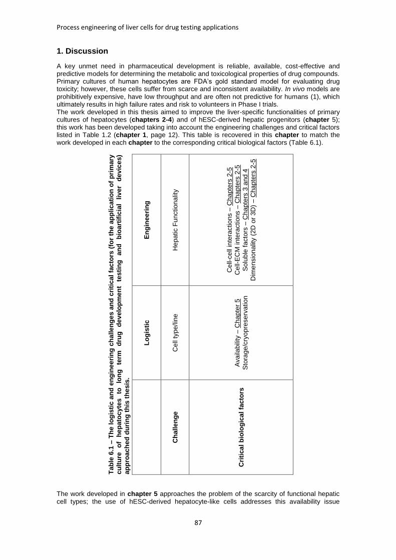
Process engineering of liver cells for drug testing applications
87
1. Discussion
A key unmet need in pharmaceutical development is reliable, available, cost-effective and predictive models for determining the metabolic and toxicological properties of drug compounds. Primary cultures of human hepatocytes are FDA’s gold standard model for evaluating drug toxicity; however, these cells suffer from scarce and inconsistent availability. In vivo models are prohibitively expensive, have low throughput and are often not predictive for humans (1), which ultimately results in high failure rates and risk to volunteers in Phase I trials. The work developed in this thesis aimed to improve the liver-specific functionalities of primary cultures of hepatocytes (chapters 2-4) and of hESC-derived hepatic progenitors (chapter 5); this work has been developed taking into account the engineering challenges and critical factors listed in Table 1.2 (chapter 1, page 12). This table is recovered in this chapter to match the work developed in each chapter to the corresponding critical biological factors (Table 6.1).
Tab
le 6
.1 –
Th
e lo
gis
tic a
nd
en
gin
ee
rin
g c
hallen
ge
s a
nd
cri
tical
facto
rs (
for
the a
pp
lic
ati
on
of
pri
mary
cu
ltu
re
of
hep
ato
cyte
s
to
lon
g
term
d
rug
d
evelo
pm
en
t te
sti
ng
an
d
bio
art
ific
ial
liv
er
dev
ice
s)
ap
pro
ach
ed
du
rin
g t
his
th
esis
.
En
gin
ee
rin
g
Hepatic F
unction
alit
y
Cell-
cell
inte
ractions –
Cha
pte
rs 2
-5
Cell-
EC
M inte
ractions –
Ch
apte
rs 2
-5
Solu
ble
facto
rs –
Ch
apte
rs 3
an
d 4
Dim
ensio
na
lity (
2D
or
3D
) – C
ha
pte
rs 2
-5
Lo
gis
tic
Cell
type/lin
e
Availa
bili
ty –
Cha
pte
r 5
Sto
rage/c
ryopre
serv
ation
Ch
allen
ge
Cri
tica
l b
iolo
gic
al fa
cto
rs
The work developed in chapter 5 approaches the problem of the scarcity of functional hepatic cell types; the use of hESC-derived hepatocyte-like cells addresses this availability issue
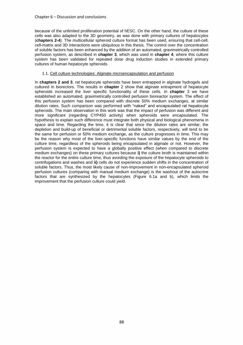
Chapter 6 – Discussion and conclusions
88
because of the unlimited proliferation potential of hESC. On the other hand, the culture of these cells was also adapted to the 3D geometry, as was done with primary cultures of hepatocytes (chapters 2-4). The multicellular spheroid culture format has been used, ensuring that cell-cell, cell-matrix and 3D interactions were ubiquitous in this thesis. The control over the concentration of soluble factors has been enhanced by the addition of an automated, gravimetrically controlled perfusion system, as described in chapter 3, which was used in chapter 4, where this culture system has been validated for repeated dose drug induction studies in extended primary cultures of human hepatocyte spheroids.
1.1. Cell culture technologies: Alginate microencapsulation and perfusion
In chapters 2 and 3, rat hepatocyte spheroids have been entrapped in alginate hydrogels and cultured in biorectors. The results in chapter 2 show that alginate entrapment of hepatocyte spheroids increased the liver specific functionality of these cells. In chapter 3 we have established an automated, gravimetrically controlled perfusion bioreactor system. The effect of this perfusion system has been compared with discrete 50% medium exchanges, at similar dilution rates. Such comparison was performed with “naked” and encapsulated rat hepatocyte spheroids. The main observation in this work was that the impact of perfusion was different and more significant (regarding CYP450 activity) when spheroids were encapsulated. The hypothesis to explain such difference must integrate both physical and biological phenomena in space and time. Regarding the time, it is clear that since the dilution rates are similar, the depletion and build-up of beneficial or detrimental soluble factors, respectively, will tend to be the same for perfusion or 50% medium exchange, as the culture progresses in time. This may be the reason why most of the liver-specific functions have similar values by the end of the culture time, regardless of the spheroids being encapsulated in alginate or not. However, the perfusion system is expected to have a globally positive effect (when compared to discrete medium exchanges) on these primary cultures because i) the culture broth is maintained within the reactor for the entire culture time, thus avoiding the exposure of the hepatocyte spheroids to centrifugations and washes and ii) cells do not experience sudden shifts in the concentration of soluble factors. Thus, the most likely cause of non-improvement in non-encapsulated spheroid perfusion cultures (comparing with manual medium exchange) is the washout of the autocrine factors that are synthesized by the hepatocytes (Figure 6.1a and b), which limits the improvement that the perfusion culture could yield.
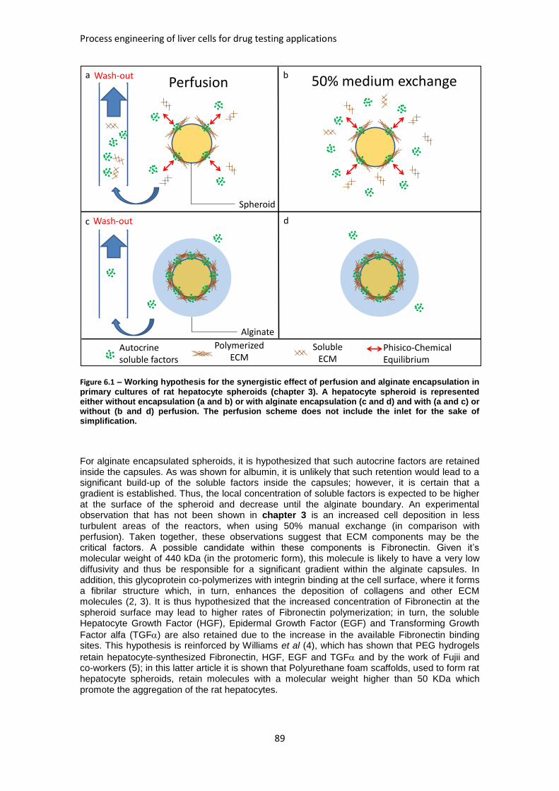
Process engineering of liver cells for drug testing applications
89
Figure 6.1 – Working hypothesis for the synergistic effect of perfusion and alginate encapsulation in
primary cultures of rat hepatocyte spheroids (chapter 3). A hepatocyte spheroid is represented either without encapsulation (a and b) or with alginate encapsulation (c and d) and with (a and c) or without (b and d) perfusion. The perfusion scheme does not include the inlet for the sake of simplification.
For alginate encapsulated spheroids, it is hypothesized that such autocrine factors are retained inside the capsules. As was shown for albumin, it is unlikely that such retention would lead to a significant build-up of the soluble factors inside the capsules; however, it is certain that a gradient is established. Thus, the local concentration of soluble factors is expected to be higher at the surface of the spheroid and decrease until the alginate boundary. An experimental observation that has not been shown in chapter 3 is an increased cell deposition in less turbulent areas of the reactors, when using 50% manual exchange (in comparison with perfusion). Taken together, these observations suggest that ECM components may be the critical factors. A possible candidate within these components is Fibronectin. Given it’s molecular weight of 440 kDa (in the protomeric form), this molecule is likely to have a very low diffusivity and thus be responsible for a significant gradient within the alginate capsules. In addition, this glycoprotein co-polymerizes with integrin binding at the cell surface, where it forms a fibrilar structure which, in turn, enhances the deposition of collagens and other ECM molecules (2, 3). It is thus hypothesized that the increased concentration of Fibronectin at the spheroid surface may lead to higher rates of Fibronectin polymerization; in turn, the soluble Hepatocyte Growth Factor (HGF), Epidermal Growth Factor (EGF) and Transforming Growth
Factor alfa (TGF) are also retained due to the increase in the available Fibronectin binding sites. This hypothesis is reinforced by Williams et al (4), which has shown that PEG hydrogels
retain hepatocyte-synthesized Fibronectin, HGF, EGF and TGF and by the work of Fujii and co-workers (5); in this latter article it is shown that Polyurethane foam scaffolds, used to form rat hepatocyte spheroids, retain molecules with a molecular weight higher than 50 KDa which promote the aggregation of the rat hepatocytes.
Phisico-ChemicalEquilibrium
PolymerizedECM
Perfusion
Autocrinesoluble factors
50% medium exchangeWash-out
Wash-out
Spheroid
Alginate
SolubleECM
a b
c d
Chapter 6 – Discussion and conclusions
90
This washout of soluble ECM components could also be minimized in non-encapsulated cultures. In the absence of the retention of ECM by alginate, the diffusion boundary layer that surrounds the spheroids is the mass transfer barrier limiting the transport of soluble ECM components from the surface of the spheroid to the culture medium bulk. One option would be to increase this boundary layer by reducing the stirring rate; however, such stirring rate must ensure that the spheroids do not deposit in the bottom of the vessel. For the pitched-bladed impellers used throughout this work, a minimum of 60 RPM would prevent such cell deposition.
1.1.1. Operational bottlenecks
Despite improving the liver-specific activities of hepatocyte spheroids, alginate encapsulation has three disadvantages: i) cell loss during encapsulation, ii) lower cell concentration and iii) the fact that encapsulation is laborious and time consuming. When combined, i) and ii) result in a very diluted culture, which in turn leads to an increase in the volume of culture medium used; on the other hand, the cell loss problem becomes significantly detrimental when accounting for the scarcity of hepatocytes. These were the main reasons not to use alginate encapsulation for the validation of the perfusion bioreactor system with primary cultures of human hepatocyte spheroids, in chapter 4. An increase in the number of hepatocytes per volume of alginate would minimize all these issues, since less encapsulations would be needed (less labor and lower losses), while the culture would be less diluted. In a first approach, to have cell concentrations similar to those used in chapter 4, the number of hepatocytes per mL of alginate should be 4-fold the current 1 million per mL of alginate, i.e., 4-5 million hepatocytes per mL of alginate. Such a ratio has not been tested in chapter 2, when the hepatocyte number per volume of alginate ratio was optimized. In fact, the values tested were 0.5, 1 and 10 million hepatocytes per mL of alginate; the lower value is 2-fold diluted compared to the optimal 1 million/mL alginate, whereas the higher value is 10-fold concentrated.
1.2. Process validation for drug development tests: primary cultures of human hepatocytes and repeated dose drug induction.
The perfusion bioreactor system has been validated for repeated dose drug induction tests in primary culture of human hepatocyte spheroids in chapter 4. In this work, the knowledge collected from rat studies has been combined with the induction of the hepatic CYP450 enzymes in the bioreactor system.
1.2.1. CYP450 induction in primary cultures of human hepatocytes at weeks 1-4;
The most notable achievement in the published literature claimed hepatocyte cultures could be maintained for 2 weeks and has shown induction of CYP450 activity (6); furthermore, long term maintenance of these enzyme’s activities has been demonstrated by Zeillinger and colleagues (7, 8) in the Charité’s hollow fiber reactors, up to 23 days, without inducing CYP450 activity. In the work herein described, a repeated induction of the CYP450 enzymes was achieved in 2-4 weeks cultures, which constitutes a clear improvement beyond the state of the art.
1.2.2. A 3D structure which closely resembles the liver structure;
The location of the apical marker aPKC, as well as MRP2-specific activity, has shown that these spheroids self-assemble to form a polarized structure, with 3D bile canaliculi. Although extensive references exist regarding the formation of such structures in 3D cultures (8, 9), this has been (to the best of our knowledge), the most clear demonstration of the three-dimensionality of functional bile canaliculi formation in vitro.
1.2.3. Technical inter-donor reproducibility.
The natural variability within human hepatocyte donors was offset by reproducible time-course profiles of Urea and Albumin which, we hypothesize, may be attributed to the control of pH, DO, temperature and most soluble factor (coupled with good mixing).
1.2.4. System limitations
This system is currently limited by (i) the lack of data on the efficiency of spheroid formation and (ii) the amount of hepatocytes needed for one reactor. Regarding the first limitation (i), visual
Process engineering of liver cells for drug testing applications
91
inspection (by light microscopy) of the hepatocyte cultures herein described suggest that the aggregation efficiency is below 50%, i.e., no more than half of the inoculated hepatocytes are incorporated in multicellular spheroids, after 72 hours; this may be due to detrimental effects of collagenase digestion of the liver sample, during the isolation of the hepatocytes (10). Still, the large majority of viable hepatocyte preparations (from different donors) aggregate after 72h with 10% FBS, in both stirred tank bioreactors with pitch bladed impellers and paddle impeller
spinners. To have more quantitative data, a study should be performed using 40 m filters to separate single cells, doublets and triplets from multicellular spheroids and thus determine the efficiency of the spheroid formation process. Alternatively, the use of automated methods for particle measurement and counting, such as a Multisizer™ 3 COULTER COUNTER®, would certainly provide an accurate measurement of the aggregation efficiency. As for the high cell numbers used (ii), it is a problem that spawns from the scarcity of freshly isolated human hepatocytes; nevertheless, considering that these cells are used for drug development tests, the question is: how to fit this perfusion bioreactor system within the drug development pre-clinical tests workflow? Clearly, the usefulness of this system lies on the long term maintenance of hepatic phenotype. One possible application, after animal tests (typically rodents) yield dose-toxicity information, could be to assess the long term effects of such dosing in the hepatic metabolism and viability in the presence (or absence) of other drugs. Moreover, the possibility of withdrawing a large sample from the bioreactor and subculturing in other systems, with higher throughput, for short term (such as shake flasks or multititer plates) or exposing other cell types (such as neurons or cardiac cells) to the products of the hepatic drug metabolism, increases the possible applications of this system.
1.3. Human embryonic stem cells as potential source of functional hepatic cells.
The differentiation of hESC to hepatocytes is an expanding field of research, mainly because an efficient differentiation process would contribute to improve the problem of human hepatocyte scarcity, either for BAL or drug development applications. However, the current processes and protocols for the hepatic differentiation of hESC yield limited numbers of non-mature hepatocytes, i.e., the final product lacks quantity and quality. While most of the differentiation protocols described in the literature (11-15) are based on the exposure of 2D cultured hESC to soluble factors such as HGF or Bone Morphogenetic Proteins (BMPs), the effect of 3D culture systems on this differentiation process has only been assessed in embryoid bodies (EBs), with limited success (16). In chapter 5 the effect of 3D culture on the hepatic differentiation of hESC
has been assessed and compared with 2D cultures, using a previously published protocol (17).
2. Future Work
Although the work described in this thesis has achieved progress beyond the state of the art, there is still room for improvement towards an (economically viable) physiological liver culture system.
2.1. Improving the hepatocyte spheroid model: co-culture
In addition to the effect of the 3D microenvironment, it has been published that heterotypic interactions, provided either by stromal cells such as fibroblasts or by endothelial cells, provide both mechanical and biochemical stimuli that enhance liver specific functions (18, 19). Also, during liver development, it is postulated that the cell contact of endothelial cells with the endoderm is crucial for the differentiation to liver progenitors (20, 21).
Thus, the recreation of the 3D microenvironment associated with the effect of the heterotypic interactions on these culture systems may, on one hand, enhance and prolong primary hepatocyte functionality and, on the other hand, provide a better system for the differentiation process of hESC to hepatocyte-like cells.
2.2. New hepatic cell sources
The hepatocyte culture bioprocessing field has been developing towards the discovery and/or generation of new hepatic cell types, i.e., trying to overcome the logistic challenge imposed by
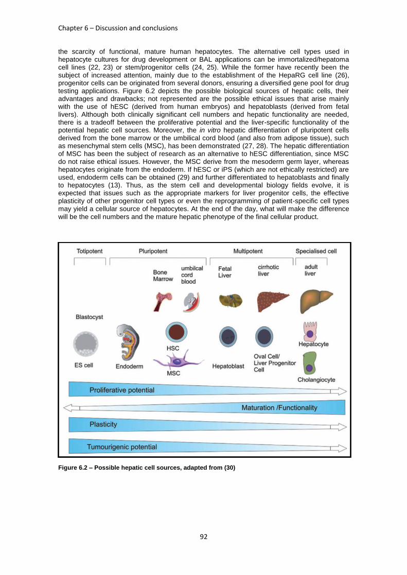
Chapter 6 – Discussion and conclusions
92
the scarcity of functional, mature human hepatocytes. The alternative cell types used in hepatocyte cultures for drug development or BAL applications can be immortalized/hepatoma cell lines (22, 23) or stem/progenitor cells (24, 25). While the former have recently been the subject of increased attention, mainly due to the establishment of the HepaRG cell line (26), progenitor cells can be originated from several donors, ensuring a diversified gene pool for drug testing applications. Figure 6.2 depicts the possible biological sources of hepatic cells, their advantages and drawbacks; not represented are the possible ethical issues that arise mainly with the use of hESC (derived from human embryos) and hepatoblasts (derived from fetal livers). Although both clinically significant cell numbers and hepatic functionality are needed, there is a tradeoff between the proliferative potential and the liver-specific functionality of the potential hepatic cell sources. Moreover, the in vitro hepatic differentiation of pluripotent cells derived from the bone marrow or the umbilical cord blood (and also from adipose tissue), such as mesenchymal stem cells (MSC), has been demonstrated (27, 28). The hepatic differentiation of MSC has been the subject of research as an alternative to hESC differentiation, since MSC do not raise ethical issues. However, the MSC derive from the mesoderm germ layer, whereas hepatocytes originate from the endoderm. If hESC or iPS (which are not ethically restricted) are used, endoderm cells can be obtained (29) and further differentiated to hepatoblasts and finally to hepatocytes (13). Thus, as the stem cell and developmental biology fields evolve, it is expected that issues such as the appropriate markers for liver progenitor cells, the effective plasticity of other progenitor cell types or even the reprogramming of patient-specific cell types may yield a cellular source of hepatocytes. At the end of the day, what will make the difference will be the cell numbers and the mature hepatic phenotype of the final cellular product.
Figure 6.2 – Possible hepatic cell sources, adapted from (30)
Process engineering of liver cells for drug testing applications
93
3. Conclusions
In this thesis a robust and technically reproducible system for long term, repeated dose drug induction tests using primary cultures of hepatocytes was developed and tested. The study of novel technical solutions, in chapters 2 and 3, allowed extending the bioreactor culture time of human hepatocyte spheroids which resume the liver-specific architecture, as described in chapter 4.
The resolution of the logistic problems which is spawned by the scarcity of freshly isolated human hepatocytes has been approached in chapter 5, where the hepatic differentiation of human embryonic stem cell-derived hepatic progenitor spheroids has been studied. Three dimensional cell culture approaches have been used throughout this thesis, proving to be a critical factor in primary and stem cell culture, as well as in tissue engineering.
4. References
1. Huang SM, Strong JM, Zhang L, Reynolds KS, Nallani S, Temple R, Abraham S, et al. New era in drug interaction evaluation: US Food and Drug Administration update on CYP enzymes, transporters, and the guidance process. J Clin Pharmacol 2008;48:662-670. 2. Magnusson MK, Mosher DF. Fibronectin: structure, assembly, and cardiovascular implications. Arterioscler Thromb Vasc Biol 1998;18:1363-1370. 3. Sottile J, Hocking DC. Fibronectin polymerization regulates the composition and stability of extracellular matrix fibrils and cell-matrix adhesions. Mol Biol Cell 2002;13:3546-3559. 4. Williams CM, Mehta G, Peyton SR, Zeiger AS, Van Vliet KJ, Griffith LG. Autocrine-controlled formation and function of tissue-like aggregates by primary hepatocytes in micropatterned hydrogel arrays. Tissue Eng Part A 2011;17:1055-1068. 5. Fujii Y, Nakazawa K, Funatsu K. Intensive promotion of spheroid formation by soluble factors in a hepatocyte-conditioned medium. Journal of Biomaterials Science-Polymer Edition 2000;11:731-745. 6. Khetani SR, Bhatia SN. Microscale culture of human liver cells for drug development. Nat Biotechnol 2008;26:120-126. 7. Mueller D, Tascher G, Müller-Vieira U, Knobeloch D, Nuessler AK, Zeilinger K, Heinzle E, et al. In-depth physiological characterization of primary human hepatocytes in a 3D hollow-fiber bioreactor. J Tissue Eng Regen Med 2011;5:e207-218. 8. Zeilinger K, Schreiter T, Darnell M, Söderdahl T, Lübberstedt M, Dillner B, Knobeloch D, et al. Scaling down of a clinical 3D perfusion multi-compartment hollow fiber liver bioreactor developed for extracorporeal liver support to an analytical scale device useful for hepatic pharmacological in vitro studies. Tissue Eng Part C Methods 2011. 9. Abu-Absi SF, Friend JR, Hansen LK, Hu WS. Structural polarity and functional bile canaliculi in rat hepatocyte spheroids. Experimental Cell Research 2002;274:56-67. 10. Lecluyse EL, Alexandre E. Isolation and culture of primary hepatocytes from resected human liver tissue. Methods Mol Biol 2010;640:57-82. 11. Agarwal S, Holton KL, Lanza R. Efficient differentiation of functional hepatocytes from human embryonic stem cells. Stem Cells 2008;26:1117-1127. 12. Baharvand H, Hashemi SM, Shahsavani M. Differentiation of human embryonic stem cells into functional hepatocyte-like cells in a serum-free adherent culture condition. Differentiation 2008;76:465-477. 13. Cai J, Zhao Y, Liu YX, Ye F, Song ZH, Qin H, Meng S, et al. Directed differentiation of human embryonic stem cells into functional hepatic cells. Hepatology 2007;45:1229-1239. 14. Duan YY, Ma XC, Zou W, Wang C, Bahbahan IS, Ahuja TP, Tolstikov V, et al. Differentiation and Characterization of Metabolically Functioning Hepatocytes from Human Embryonic Stem Cells. Stem Cells 2010;28:674-686. 15. Hay DC, Zhao D, Ross A, Mandalam R, Lebkowski J, Cui W. Direct differentiation of human embryonic stem cells to hepatocyte-like cells exhibiting functional activities. Cloning and Stem Cells 2007;9:51-62. 16. Baharvand H, Hashemi SM, Ashtian SK, Farrokhi A. Differentiation of human embryonic stem cells into hepatocytes in 2D and 3D culture systems in vitro. International Journal of Developmental Biology 2006;50:645-652.
Chapter 6 – Discussion and conclusions
94
17. Brolen G, Sivertsson L, Bjorquist P, Eriksson G, Ek M, Semb H, Johansson I, et al. Hepatocyte-like cells derived from human embryonic stem cells specifically via definitive endoderm and a progenitor stage. J Biotechnol;145:284-294. 18. Kidambi S, Yarmush RS, Novik E, Chao P, Yarmush ML, Nahmias Y. Oxygen-mediated enhancement of primary hepatocyte metabolism, functional polarization, gene expression, and drug clearance. Proc Natl Acad Sci U S A 2009;106:15714-15719. 19. Chen AA, Thomas DK, Ong LL, Schwartz RE, Golub TR, Bhatia SN. Humanized mice with ectopic artificial liver tissues. Proc Natl Acad Sci U S A 2011;108:11842-11847. 20. Lemaigre F, Zaret KS. Liver development update: new embryo models, cell lineage control, and morphogenesis. Curr Opin Genet Dev 2004;14:582-590. 21. Han S, Dziedzic N, Gadue P, Keller GM, Gouon-Evans V. An endothelial cell niche induces hepatic specification through dual repression of Wnt and Notch signaling. Stem Cells 2011;29:217-228. 22. Chang TT, Hughes-Fulford M. Monolayer and Spheroid Culture of Human Liver Hepatocellular Carcinoma Cell Line Cells Demonstrate Distinct Global Gene Expression Patterns and Functional Phenotypes. Tissue Engineering Part A 2009;15:559-567. 23. Aninat C, Piton A, Glaise D, Le Charpentier T, Langouët S, Morel F, Guguen-Guillouzo C, et al. Expression of cytochromes P450, conjugating enzymes and nuclear receptors in human hepatoma HepaRG cells. Drug Metab Dispos 2006;34:75-83. 24. Duan YY, Catana A, Meng Y, Yamamoto N, He SQ, Gupta S, Gambhir SS, et al. Differentiation and enrichment of hepatocyte-like cells from human embryonic stem cells in vitro and in vivo. Stem Cells 2007;25:3058-3068. 25. Sasaki K, Kon J, Mizuguchi T, Chen Q, Ooe H, Oshima H, Hirata K, et al. Proliferation of hepatocyte progenitor cells isolated from adult human livers in serum-free medium. Cell Transplant 2008;17:1221-1230. 26. Darnell M, Schreiter T, Zeilinger K, Urbaniak T, Söderdahl T, Rossberg I, Dillnér B, et al. Cytochrome P450-dependent metabolism in HepaRG cells cultured in a dynamic three-dimensional bioreactor. Drug Metab Dispos 2011;39:1131-1138. 27. Banas A, Teratani T, Yamamoto Y, Tokuhara M, Takeshita F, Quinn G, Okochi H, et al. Adipose tissue-derived mesenchymal stem cells as a source of human hepatocytes. Hepatology 2007;46:219-228. 28. Hong SH, Gang EJ, Jeong JA, Ahn C, Hwang SH, Yang IH, Park HK, et al. In vitro differentiation of human umbilical cord blood-derived mesenchymal stem cells into hepatocyte-like cells. Biochem Biophys Res Commun 2005;330:1153-1161. 29. D'Amour KA, Agulnick AD, Eliazer S, Kelly OG, Kroon E, Baetge EE. Efficient differentiation of human embryonic stem cells to definitive endoderm. Nature Biotechnology 2005;23:1534-1541. 30. Dan YY, Yeoh GC. Liver stem cells: A scientific and clinical perspective. Journal of Gastroenterology and Hepatology 2008;23:687-698.