Embed Size (px)
Citation preview
ChemicalScience
EDGE ARTICLE
Ope
n A
cces
s A
rtic
le. P
ublis
hed
on 0
9 Ju
ly 2
020.
Dow
nloa
ded
on 3
/14/
2022
12:
18:4
9 A
M.
Thi
s ar
ticle
is li
cens
ed u
nder
a C
reat
ive
Com
mon
s A
ttrib
utio
n 3.
0 U
npor
ted
Lic
ence
.
View Article OnlineView Journal | View Issue
Probing biotin re
Nanochemistry and Bioimaging Group, La
CNRS UMR 7021, Universite de Strasbour
France. E-mail: andrey.klymchenko@unistra
† Electronic supplementary informa10.1039/d0sc01973a
‡ These authors contributed equally.
Cite this: Chem. Sci., 2020, 11, 8240
All publication charges for this articlehave been paid for by the Royal Societyof Chemistry
Received 6th April 2020Accepted 8th July 2020
DOI: 10.1039/d0sc01973a
rsc.li/chemical-science
8240 | Chem. Sci., 2020, 11, 8240–8
ceptors in cancer cells withrationally designed fluorogenic squaraine dimers†
Kyong T. Fam, ‡ Mayeul Collot ‡* and Andrey S. Klymchenko *
Fluorogenic probes enable imaging biomolecular targets with high sensitivity and maximal signal-to-
background ratio under non-wash conditions. Here, we focus on the molecular design of biotinylated
dimeric squaraines that undergo aggregation-caused quenching in aqueous media through
intramolecular H-type dimerization, but turn on their fluorescence in apolar environment due to target-
mediated disaggregation. Our structure–property study revealed that depending on the linkers used to
connect the squaraine dyes, different aggregation patterns could be obtained (intramolecular
dimerization versus intermolecular aggregation) leading to different probing efficiencies. Using
a relatively short L-lysine linker we developed a bright fluorogenic probe, Sq2B, displaying only
intramolecular dimerization-caused quenching properties in aqueous media. The latter was successfully
applied for imaging biotin receptors, in particular sodium-dependent multivitamin transporter (SMVT),
which are overexpressed at the surface of cancer cells. Competitive displacement with SMVT-targets,
such as biotin, lipoic acid or sodium pantothenate, showed Sq2B targeting ability to SMVT. This
fluorogenic probe for biotin receptors could distinguish cancer cells (HeLa and KB) from model non-
cancer cell lines (NIH/3T3 and HEK293T). The obtained results provide guidelines for development of
new dimerization-based fluorogenic probes and propose bright tools for imaging biotin receptors, which
is particularly important for specific detection of cancer cells.
Introduction
Biotin is an essential vitamin playing its role in cellular carbo-hydrate, amino acid and lipid metabolism.1 Unlike bacteria,mammalian cell machinery does not produce biotin, thereforebiotin is supplemented exogenously.1 There is evidence thatexpression of biotin receptors (BRs) is correlated with cancer.2
Among BRs, sodium-dependent multivitamin transporter(SMVT) is essential to deliver vitamins, like biotin, to cancercells.3 Therefore, SMVT is a potentially useful cancer biomarkerfor tumor diagnostics. While BRs have already been exploitedfor specic targeting of cancer cells,3,4 there is lack of robustimaging probes for BRs (in particular SMVT) in cancerous cellsand evaluation of new targeted therapeutics.
Fluorogenic probes are particularly adapted for decipheringbiological processes with background-free imaging.5–11
Although environment-sensitive biotin probes have beendeveloped for SMVT imaging operating in the visibleregion,3,4,12–14 detection of SMVT at low concentration requiressuperior brightness. Alternatively, recent efforts resulted in
boratoire de Bioimagerie et Pathologies,
g, Faculte de Pharmacie, 67401 Illkirch,
.fr; [email protected]
tion (ESI) available. See DOI:
248
development of hybrid nanoprobes to distinguish SMVT incancer cells and 3D spheroids.15–18 There is a particular demandin development of probes operating in far-red and near-infrared(NIR) region to image deeper in the tissues and potentially invivo.19–22 A number of concepts to generate uorogenic responseto target-binding was developed in the last decade.6 First, oneshould mention push–pull dyes, which are poorly uorescent inwater, but light up in apolar environments of cellular recep-tors.23,24 Second, molecular rotors,25 which are poorly uores-cent in aqueous media, but light up upon binding to the targetswith highly viscous environment.26,27 One should also mentionaggregation-induced emission approach, where the non-emissive dyes light up because the target triggers dye aggrega-tion.28 Of particular interest are probes operating byaggregation-caused quenching (ACQ) mechanism.29 ACQ iscaused by strong hydrophobic effects and p–p stacking of dyesat high concentrations.30 ACQ is common for uorophores inaqueous media since their planar hydrophobic aromaticsystems provoke formation of non-radiative excimers/exci-plexes.30 Aggregation is thermodynamically favorable in water,but it could be disrupted in an appropriate environment.30 Overthe years, a variety of uorogenic probes has been developed,where the initially quenched aggregated dye species arespatially separated upon interaction with the biological target(Fig. 1A).29 Both intermolecular and intramolecular aggregationprocesses have been used to design ACQ-driven probes.
This journal is © The Royal Society of Chemistry 2020
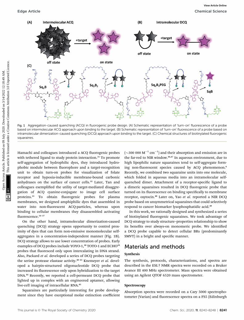
Fig. 1 Aggregation-caused quenching (ACQ) in fluorogenic probe design. (A) Schematic representation of ‘turn-on’ fluorescence of a probebased on intermolecular ACQ approach upon binding to the target. (B) Schematic representation of ‘turn-on’ fluorescence of a probe based onintramolecular dimerization-caused quenching (DCQ) approach upon binding to the target. (C) Chemical structures of biotinylated fluorogenicsquaraines.
Edge Article Chemical Science
Ope
n A
cces
s A
rtic
le. P
ublis
hed
on 0
9 Ju
ly 2
020.
Dow
nloa
ded
on 3
/14/
2022
12:
18:4
9 A
M.
Thi
s ar
ticle
is li
cens
ed u
nder
a C
reat
ive
Com
mon
s A
ttrib
utio
n 3.
0 U
npor
ted
Lic
ence
.View Article Online
Hamachi and colleagues introduced a ACQ uorogenic probeswith tethered ligand to study protein interaction.31 To promoteself-aggregation of hydrophilic dyes, they introduced hydro-phobic module between uorophore and a target-recognitionunit to obtain turn-on probes for visualization of folatereceptor and hypoxia-inducible membrane-bound carbonicanhydrases on the surface of cancer cells.32 Later, Tan andcolleagues exemplied the utility of target-mediated disaggre-gation of ACQ cyanine-conjugate to image cell surfaceproteins.33 To obtain uorogenic probes for plasmamembranes, we designed amphiphilic dyes that assembled inwater into non-uorescent ACQ-particles, whereas uponbinding to cellular membranes they disassembled activatinguorescence.34–37
On the other hand, intramolecular dimerization-causedquenching (DCQ) strategy opens opportunity to control prox-imity of dyes that can form non-emissive monomolecular self-aggregates in a concentration-independent manner (Fig. 1B).DCQ strategy allows to use lower concentration of probes. Earlyexamples of DCQ probes include YOYO-1,38 TOTO-1 and ECHO39
probes that uoresced only upon intercalating in DNA strand.Also, Packard et al. developed a series of DCQ probes targetingthe serine protease elastase activity.40–43 Knemeyer et al. devel-oped a hairpin-structured oligonucleotide DCQ probe thatincreased its uorescence only upon hybridization to the targetDNA.44 Recently, we reported a cell-permeant DCQ probe thatlighted up in complex with an engineered aptamer, allowinglive-cell imaging of intracellular RNA.45
Squaraines are particularly interesting for probe develop-ment since they have exceptional molar extinction coefficient
This journal is © The Royal Society of Chemistry 2020
(�300 000 M�1 cm�1) and their absorption and emission are inthe far-red to NIR window.46,47 In aqueous environment, due tohigh lipophilic nature squaraines tend to self-aggregate form-ing non-uorescent species caused by ACQ phenomenon.6
Recently, we combined two squaraine units into one molecule,which folded in aqueous media into an intramolecular self-quenched dimer. Attachment of a receptor-specic ligand toa dimeric squaraines resulted in DCQ uorogenic probe thatturned on its uorescence on binding specically to membranereceptor, oxytocin.48 Later on, Yao et al. reported a NIR DCQprobe based on unsymmetrical squaraines that could selectivelyrespond to cancer biomarker lysophosphatidic acid.49
In this work, we rationally designed and synthesized a seriesof biotinylated uorogenic squaraines. We took advantage ofDCQ strategy to study structure–properties relationship to showits benets over always-on monomeric probe. We identieda DCQ probe capable to detect cellular BRs (predominantlySMVT) in a bright and specic manner.
Materials and methodsSynthesis
The synthesis, protocols, characterizations, and spectra aredescribed in the ESI.† NMR spectra were recorded on a BrukerAvance III 400 MHz spectrometer. Mass spectra were obtainedusing an Agilent QTOF 6520 mass spectrometer.
Spectroscopy
Absorption spectra were recorded on a Cary 5000 spectropho-tometer (Varian) and uorescence spectra on a FS5 (Edinburgh
Chem. Sci., 2020, 11, 8240–8248 | 8241
Chemical Science Edge Article
Ope
n A
cces
s A
rtic
le. P
ublis
hed
on 0
9 Ju
ly 2
020.
Dow
nloa
ded
on 3
/14/
2022
12:
18:4
9 A
M.
Thi
s ar
ticle
is li
cens
ed u
nder
a C
reat
ive
Com
mon
s A
ttrib
utio
n 3.
0 U
npor
ted
Lic
ence
.View Article Online
Instruments) spectrouorometer. The uorescence signal wascorrected for the lamp intensity uctuations and forwavelength-dependent sensitivity of the detector. Relative uo-rescence quantum yields were measured using DiD in meth-anol50 (QY ¼ 0.33) as standard.
Microscopy imaging
Cells were grown at 37 �C in humidied atmosphere containing5% CO2: KB cells (ATCC® CCL-17) in Dulbecco's Modied EagleMedium without phenol red (DMEM, Gibco-Invitrogen) with10% fetal bovine serum (FBS, Lonza), 1% non-essential aminoacids (Gibco-Invitrogen), 1% MEM vitamin solution (Gibco-Invitrogen), 1% L-glutamine (Sigma Aldrich) and 0.1% antibi-otic solution (gentamicin, Sigma-Aldrich); HEK293T (ATCC®CRL-3216™) and HeLa (ATCC® CCL-2™) in DMEM withoutphenol red supplemented with 10% FBS (Lonza), 1% L-gluta-mine (Sigma Aldrich) and 1% antibiotic solution (Penicillin–Streptomycin, Sigma-Aldrich); NIH/3T3 (ATCC® CRL-1658™) inDMEM without phenol red supplemented with 10% bovine calfserum, iron fortied (Sigma Aldrich), 1% L-glutamine (SigmaAldrich) and 1% antibiotic solution (Penicillin–Streptomycin,Sigma-Aldrich). Cells were seeded onto a chambered coverglass(IBiDi®) 24 h before the microscopy measurement. Forimaging, the culture medium was removed, the attached cellswere washed with Hank's Balanced Salt Solution (HBSS, Gibco-Invitrogen) and incubated with solution of Sq2B (0.2 mM). Incompetition experiment, KB cells were pretreated withcompetitor (100 mM) for 30 min prior to incubation with Sq2Bprobe. Images were taken with Nikon Ti-E inverted microscope,equipped with CFI Plan Apo � 60 oil (NA ¼ 1.4) objective, X-light spinning disk module (CresOptics) and a HamamatsuOrca Flash 4 sCMOS camera, was used. The microscopy settingswere: Hoechst (ex. 405 nm, em. 510 � 42 nm), squaraine (ex.638 nm, em. 705� 36 nm). The images were recorded using NISelements and then processed with Icy open source imagingsoware.
Flow cytometry
Cells were grown at 37 �C in humidied atmosphere containing5% CO2 in 25 cm2 (Nunc™ EasYFlask™, ThermoFisher). On theday of the analysis, the cells were washed and harvested. Thecell suspension (3 � 105 cells per mL) was incubated with cor-responding Sq2B probe (0.2 mM) for 5 min at room temperatureand analyzed immediately using ow cytometry (MACSQuantVYB, Miltenyi Biotec).
Cytotoxicity assay
Cytotoxicity of the dyes was quantied by the MTT assay (3-(4,5-dimethylthiazol-2-yl)-2,5-diphenyltetrazolium bromide). A totalof 1 � 104 KB cells per well were seeded in a 96-well plate 24 hprior to the cytotoxicity assay in growth medium and wereincubated in a 5% CO2 incubator at 37 �C. Aer mediumremoval, an amount of 100 mL DMEM containing 5 mM, 1 mM or0.2 mM of a probe was added to the KB cell and incubated for24 h at 37 �C (5% CO2). As control, for each 96-well plate, thecells were incubated with DMEM containing the same
8242 | Chem. Sci., 2020, 11, 8240–8248
percentage of DMSO (0.5% v/v) as the solution with the testeddyes. Aer 24 h of dye incubation, the medium was replaced by100 mL of a mix containing DMEM + MTT solution (diluted inPBS beforehand) and the cells were incubated for 4 h at 37 �C.Then, 75 mL of the mix was replaced by 50 mL of DMSO (100%)and gently shaken for 15 min at room temperature in order todissolve the insoluble purple formazan reduced in living cells.The absorbance at 540 nm was measured (absorbance of thedyes at 540 nm were taken into account). Data were shown asthe mean value (n ¼ 6) plus a standard deviation (�SD). Foreach concentration, we calculated the percentage of cell viabilityin reference of the control DMEM+0.5% DMSO.
Results and discussionSynthesis of biotinylated squaraine dimers
In order to nd the optimal design of uorogenic dimer, weconsidered two types of dimeric probes. The rst one is Y-structure, where two squaraine (Sq.) unites are connected tolysine linker, which was further coupled to a biotin ligand(Sq2B, Fig. 1C). In contrast to our originally proposed system forGPCR receptor,48 the present design is simpler with shorterdistance between the Sq dyes and it bears small biotin ligandinstead of cyclic peptide. A monomeric dye analogue of thisprobe (SqB, Fig. 1C) has also been obtained as a control. In thesecond design strategy, we developed symmetrical U-shapedimers of squaraines connected with a exible PEG8-bridgeand bearing two biotin moieties (Sq2B2, Sq2B2PEG, Fig. 1C).Activated carbonate 5 (Schemes S1 and S2 ESI†) was coupled toPEG8-diamine resulting in 6 with two propargyl handles(Scheme S2, ESI†), which were further clicked to N3-biotin or N3-PEG3-biotin, respectively (Scheme S3, ESI†). We assumed thatintroduction of additional short PEG-linker between biotin andSq unit in Sq2B2PEG would increase the solubility of the probein aqueous solutions as well as add the degree of exibility todynamically fold and prevent nonspecic binding tobiomolecules.
Fluorogenic properties of squaraine dimers
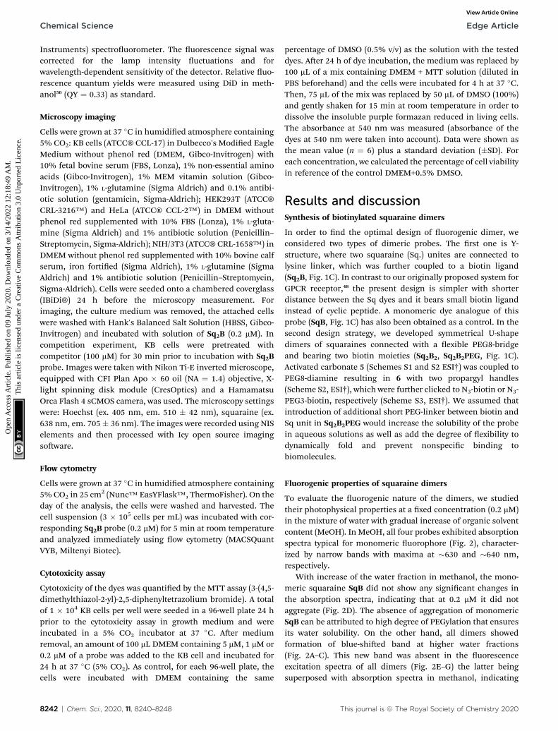
To evaluate the uorogenic nature of the dimers, we studiedtheir photophysical properties at a xed concentration (0.2 mM)in the mixture of water with gradual increase of organic solventcontent (MeOH). In MeOH, all four probes exhibited absorptionspectra typical for monomeric uorophore (Fig. 2), character-ized by narrow bands with maxima at �630 and �640 nm,respectively.
With increase of the water fraction in methanol, the mono-meric squaraine SqB did not show any signicant changes inthe absorption spectra, indicating that at 0.2 mM it did notaggregate (Fig. 2D). The absence of aggregation of monomericSqB can be attributed to high degree of PEGylation that ensuresits water solubility. On the other hand, all dimers showedformation of blue-shied band at higher water fractions(Fig. 2A–C). This new band was absent in the uorescenceexcitation spectra of all dimers (Fig. 2E–G) the latter beingsuperposed with absorption spectra in methanol, indicating
This journal is © The Royal Society of Chemistry 2020
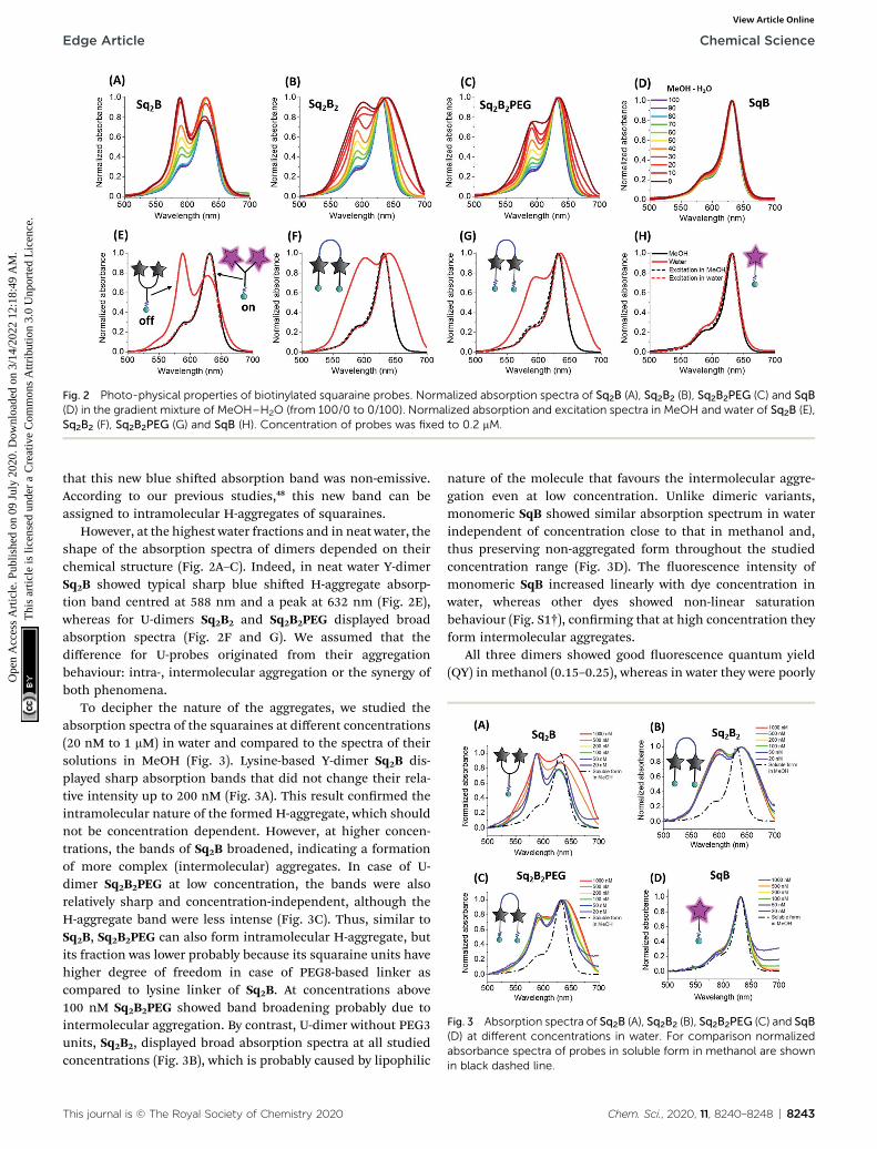
Fig. 2 Photo-physical properties of biotinylated squaraine probes. Normalized absorption spectra of Sq2B (A), Sq2B2 (B), Sq2B2PEG (C) and SqB(D) in the gradient mixture of MeOH–H2O (from 100/0 to 0/100). Normalized absorption and excitation spectra in MeOH and water of Sq2B (E),Sq2B2 (F), Sq2B2PEG (G) and SqB (H). Concentration of probes was fixed to 0.2 mM.
Fig. 3 Absorption spectra of Sq2B (A), Sq2B2 (B), Sq2B2PEG (C) and SqB(D) at different concentrations in water. For comparison normalizedabsorbance spectra of probes in soluble form in methanol are shownin black dashed line.
Edge Article Chemical Science
Ope
n A
cces
s A
rtic
le. P
ublis
hed
on 0
9 Ju
ly 2
020.
Dow
nloa
ded
on 3
/14/
2022
12:
18:4
9 A
M.
Thi
s ar
ticle
is li
cens
ed u
nder
a C
reat
ive
Com
mon
s A
ttrib
utio
n 3.
0 U
npor
ted
Lic
ence
.View Article Online
that this new blue shied absorption band was non-emissive.According to our previous studies,48 this new band can beassigned to intramolecular H-aggregates of squaraines.
However, at the highest water fractions and in neat water, theshape of the absorption spectra of dimers depended on theirchemical structure (Fig. 2A–C). Indeed, in neat water Y-dimerSq2B showed typical sharp blue shied H-aggregate absorp-tion band centred at 588 nm and a peak at 632 nm (Fig. 2E),whereas for U-dimers Sq2B2 and Sq2B2PEG displayed broadabsorption spectra (Fig. 2F and G). We assumed that thedifference for U-probes originated from their aggregationbehaviour: intra-, intermolecular aggregation or the synergy ofboth phenomena.
To decipher the nature of the aggregates, we studied theabsorption spectra of the squaraines at different concentrations(20 nM to 1 mM) in water and compared to the spectra of theirsolutions in MeOH (Fig. 3). Lysine-based Y-dimer Sq2B dis-played sharp absorption bands that did not change their rela-tive intensity up to 200 nM (Fig. 3A). This result conrmed theintramolecular nature of the formed H-aggregate, which shouldnot be concentration dependent. However, at higher concen-trations, the bands of Sq2B broadened, indicating a formationof more complex (intermolecular) aggregates. In case of U-dimer Sq2B2PEG at low concentration, the bands were alsorelatively sharp and concentration-independent, although theH-aggregate band were less intense (Fig. 3C). Thus, similar toSq2B, Sq2B2PEG can also form intramolecular H-aggregate, butits fraction was lower probably because its squaraine units havehigher degree of freedom in case of PEG8-based linker ascompared to lysine linker of Sq2B. At concentrations above100 nM Sq2B2PEG showed band broadening probably due tointermolecular aggregation. By contrast, U-dimer without PEG3units, Sq2B2, displayed broad absorption spectra at all studiedconcentrations (Fig. 3B), which is probably caused by lipophilic
This journal is © The Royal Society of Chemistry 2020
nature of the molecule that favours the intermolecular aggre-gation even at low concentration. Unlike dimeric variants,monomeric SqB showed similar absorption spectrum in waterindependent of concentration close to that in methanol and,thus preserving non-aggregated form throughout the studiedconcentration range (Fig. 3D). The uorescence intensity ofmonomeric SqB increased linearly with dye concentration inwater, whereas other dyes showed non-linear saturationbehaviour (Fig. S1†), conrming that at high concentration theyform intermolecular aggregates.
All three dimers showed good uorescence quantum yield(QY) in methanol (0.15–0.25), whereas in water they were poorly
Chem. Sci., 2020, 11, 8240–8248 | 8243
Chemical Science Edge Article
Ope
n A
cces
s A
rtic
le. P
ublis
hed
on 0
9 Ju
ly 2
020.
Dow
nloa
ded
on 3
/14/
2022
12:
18:4
9 A
M.
Thi
s ar
ticle
is li
cens
ed u
nder
a C
reat
ive
Com
mon
s A
ttrib
utio
n 3.
0 U
npor
ted
Lic
ence
.View Article Online
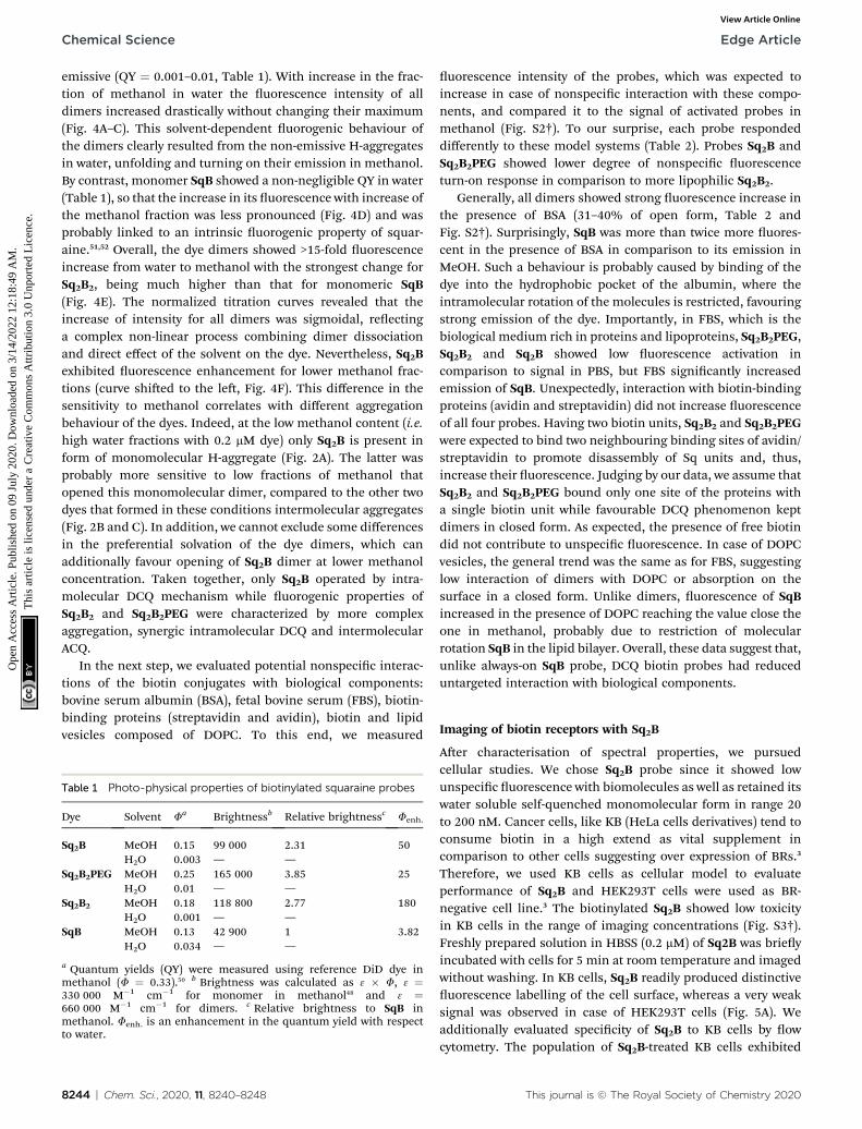
emissive (QY ¼ 0.001–0.01, Table 1). With increase in the frac-tion of methanol in water the uorescence intensity of alldimers increased drastically without changing their maximum(Fig. 4A–C). This solvent-dependent uorogenic behaviour ofthe dimers clearly resulted from the non-emissive H-aggregatesin water, unfolding and turning on their emission in methanol.By contrast, monomer SqB showed a non-negligible QY in water(Table 1), so that the increase in its uorescence with increase ofthe methanol fraction was less pronounced (Fig. 4D) and wasprobably linked to an intrinsic uorogenic property of squar-aine.51,52 Overall, the dye dimers showed >15-fold uorescenceincrease from water to methanol with the strongest change forSq2B2, being much higher than that for monomeric SqB(Fig. 4E). The normalized titration curves revealed that theincrease of intensity for all dimers was sigmoidal, reectinga complex non-linear process combining dimer dissociationand direct effect of the solvent on the dye. Nevertheless, Sq2Bexhibited uorescence enhancement for lower methanol frac-tions (curve shied to the le, Fig. 4F). This difference in thesensitivity to methanol correlates with different aggregationbehaviour of the dyes. Indeed, at the low methanol content (i.e.high water fractions with 0.2 mM dye) only Sq2B is present inform of monomolecular H-aggregate (Fig. 2A). The latter wasprobably more sensitive to low fractions of methanol thatopened this monomolecular dimer, compared to the other twodyes that formed in these conditions intermolecular aggregates(Fig. 2B and C). In addition, we cannot exclude some differencesin the preferential solvation of the dye dimers, which canadditionally favour opening of Sq2B dimer at lower methanolconcentration. Taken together, only Sq2B operated by intra-molecular DCQ mechanism while uorogenic properties ofSq2B2 and Sq2B2PEG were characterized by more complexaggregation, synergic intramolecular DCQ and intermolecularACQ.
In the next step, we evaluated potential nonspecic interac-tions of the biotin conjugates with biological components:bovine serum albumin (BSA), fetal bovine serum (FBS), biotin-binding proteins (streptavidin and avidin), biotin and lipidvesicles composed of DOPC. To this end, we measured
Table 1 Photo-physical properties of biotinylated squaraine probes
Dye Solvent Fa Brightnessb Relative brightnessc Fenh.
Sq2B MeOH 0.15 99 000 2.31 50H2O 0.003 — —
Sq2B2PEG MeOH 0.25 165 000 3.85 25H2O 0.01 — —
Sq2B2 MeOH 0.18 118 800 2.77 180H2O 0.001 — —
SqB MeOH 0.13 42 900 1 3.82H2O 0.034 — —
a Quantum yields (QY) were measured using reference DiD dye inmethanol (F ¼ 0.33).50 b Brightness was calculated as 3 � F, 3 ¼330 000 M�1 cm�1 for monomer in methanol48 and 3 ¼660 000 M�1 cm�1 for dimers. c Relative brightness to SqB inmethanol. Fenh. is an enhancement in the quantum yield with respectto water.
8244 | Chem. Sci., 2020, 11, 8240–8248
uorescence intensity of the probes, which was expected toincrease in case of nonspecic interaction with these compo-nents, and compared it to the signal of activated probes inmethanol (Fig. S2†). To our surprise, each probe respondeddifferently to these model systems (Table 2). Probes Sq2B andSq2B2PEG showed lower degree of nonspecic uorescenceturn-on response in comparison to more lipophilic Sq2B2.
Generally, all dimers showed strong uorescence increase inthe presence of BSA (31–40% of open form, Table 2 andFig. S2†). Surprisingly, SqB was more than twice more uores-cent in the presence of BSA in comparison to its emission inMeOH. Such a behaviour is probably caused by binding of thedye into the hydrophobic pocket of the albumin, where theintramolecular rotation of the molecules is restricted, favouringstrong emission of the dye. Importantly, in FBS, which is thebiological medium rich in proteins and lipoproteins, Sq2B2PEG,Sq2B2 and Sq2B showed low uorescence activation incomparison to signal in PBS, but FBS signicantly increasedemission of SqB. Unexpectedly, interaction with biotin-bindingproteins (avidin and streptavidin) did not increase uorescenceof all four probes. Having two biotin units, Sq2B2 and Sq2B2PEGwere expected to bind two neighbouring binding sites of avidin/streptavidin to promote disassembly of Sq units and, thus,increase their uorescence. Judging by our data, we assume thatSq2B2 and Sq2B2PEG bound only one site of the proteins witha single biotin unit while favourable DCQ phenomenon keptdimers in closed form. As expected, the presence of free biotindid not contribute to unspecic uorescence. In case of DOPCvesicles, the general trend was the same as for FBS, suggestinglow interaction of dimers with DOPC or absorption on thesurface in a closed form. Unlike dimers, uorescence of SqBincreased in the presence of DOPC reaching the value close theone in methanol, probably due to restriction of molecularrotation SqB in the lipid bilayer. Overall, these data suggest that,unlike always-on SqB probe, DCQ biotin probes had reduceduntargeted interaction with biological components.
Imaging of biotin receptors with Sq2B
Aer characterisation of spectral properties, we pursuedcellular studies. We chose Sq2B probe since it showed lowunspecic uorescence with biomolecules as well as retained itswater soluble self-quenched monomolecular form in range 20to 200 nM. Cancer cells, like KB (HeLa cells derivatives) tend toconsume biotin in a high extend as vital supplement incomparison to other cells suggesting over expression of BRs.3
Therefore, we used KB cells as cellular model to evaluateperformance of Sq2B and HEK293T cells were used as BR-negative cell line.3 The biotinylated Sq2B showed low toxicityin KB cells in the range of imaging concentrations (Fig. S3†).Freshly prepared solution in HBSS (0.2 mM) of Sq2B was brieyincubated with cells for 5 min at room temperature and imagedwithout washing. In KB cells, Sq2B readily produced distinctiveuorescence labelling of the cell surface, whereas a very weaksignal was observed in case of HEK293T cells (Fig. 5A). Weadditionally evaluated specicity of Sq2B to KB cells by owcytometry. The population of Sq2B-treated KB cells exhibited
This journal is © The Royal Society of Chemistry 2020
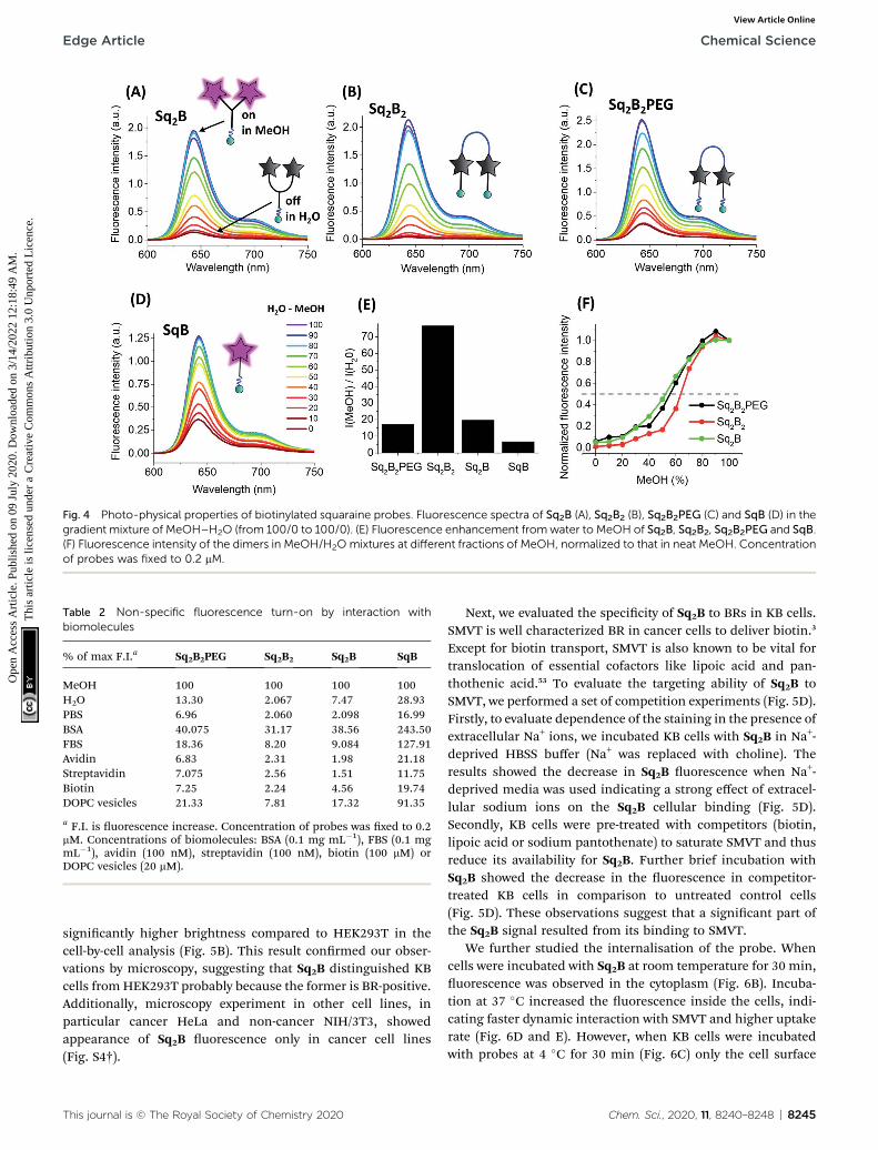
Fig. 4 Photo-physical properties of biotinylated squaraine probes. Fluorescence spectra of Sq2B (A), Sq2B2 (B), Sq2B2PEG (C) and SqB (D) in thegradient mixture of MeOH–H2O (from 100/0 to 100/0). (E) Fluorescence enhancement fromwater to MeOH of Sq2B, Sq2B2, Sq2B2PEG and SqB.(F) Fluorescence intensity of the dimers in MeOH/H2O mixtures at different fractions of MeOH, normalized to that in neat MeOH. Concentrationof probes was fixed to 0.2 mM.
Table 2 Non-specific fluorescence turn-on by interaction withbiomolecules
% of max F.I.a Sq2B2PEG Sq2B2 Sq2B SqB
MeOH 100 100 100 100H2O 13.30 2.067 7.47 28.93PBS 6.96 2.060 2.098 16.99BSA 40.075 31.17 38.56 243.50FBS 18.36 8.20 9.084 127.91Avidin 6.83 2.31 1.98 21.18Streptavidin 7.075 2.56 1.51 11.75Biotin 7.25 2.24 4.56 19.74DOPC vesicles 21.33 7.81 17.32 91.35
a F.I. is uorescence increase. Concentration of probes was xed to 0.2mM. Concentrations of biomolecules: BSA (0.1 mg mL�1), FBS (0.1 mgmL�1), avidin (100 nM), streptavidin (100 nM), biotin (100 mM) orDOPC vesicles (20 mM).
Edge Article Chemical Science
Ope
n A
cces
s A
rtic
le. P
ublis
hed
on 0
9 Ju
ly 2
020.
Dow
nloa
ded
on 3
/14/
2022
12:
18:4
9 A
M.
Thi
s ar
ticle
is li
cens
ed u
nder
a C
reat
ive
Com
mon
s A
ttrib
utio
n 3.
0 U
npor
ted
Lic
ence
.View Article Online
signicantly higher brightness compared to HEK293T in thecell-by-cell analysis (Fig. 5B). This result conrmed our obser-vations by microscopy, suggesting that Sq2B distinguished KBcells fromHEK293T probably because the former is BR-positive.Additionally, microscopy experiment in other cell lines, inparticular cancer HeLa and non-cancer NIH/3T3, showedappearance of Sq2B uorescence only in cancer cell lines(Fig. S4†).
This journal is © The Royal Society of Chemistry 2020
Next, we evaluated the specicity of Sq2B to BRs in KB cells.SMVT is well characterized BR in cancer cells to deliver biotin.3
Except for biotin transport, SMVT is also known to be vital fortranslocation of essential cofactors like lipoic acid and pan-thothenic acid.53 To evaluate the targeting ability of Sq2B toSMVT, we performed a set of competition experiments (Fig. 5D).Firstly, to evaluate dependence of the staining in the presence ofextracellular Na+ ions, we incubated KB cells with Sq2B in Na+-deprived HBSS buffer (Na+ was replaced with choline). Theresults showed the decrease in Sq2B uorescence when Na+-deprived media was used indicating a strong effect of extracel-lular sodium ions on the Sq2B cellular binding (Fig. 5D).Secondly, KB cells were pre-treated with competitors (biotin,lipoic acid or sodium pantothenate) to saturate SMVT and thusreduce its availability for Sq2B. Further brief incubation withSq2B showed the decrease in the uorescence in competitor-treated KB cells in comparison to untreated control cells(Fig. 5D). These observations suggest that a signicant part ofthe Sq2B signal resulted from its binding to SMVT.
We further studied the internalisation of the probe. Whencells were incubated with Sq2B at room temperature for 30 min,uorescence was observed in the cytoplasm (Fig. 6B). Incuba-tion at 37 �C increased the uorescence inside the cells, indi-cating faster dynamic interaction with SMVT and higher uptakerate (Fig. 6D and E). However, when KB cells were incubatedwith probes at 4 �C for 30 min (Fig. 6C) only the cell surface
Chem. Sci., 2020, 11, 8240–8248 | 8245
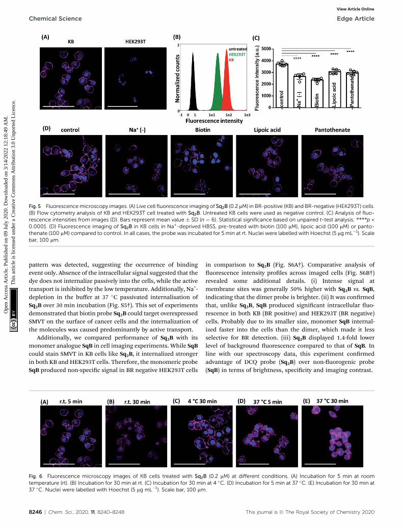
Fig. 5 Fluorescencemicroscopy images. (A) Live cell fluorescence imaging of Sq2B (0.2 mM) in BR-positive (KB) and BR-negative (HEK293T) cells.(B) Flow cytometry analysis of KB and HEK293T cell treated with Sq2B. Untreated KB cells were used as negative control. (C) Analysis of fluo-rescence intensities from images (D). Bars represent mean value � SD (n ¼ 6). Statistical significance based on unpaired t-test analysis: ****p <0.0001. (D) Fluorescence imaging of Sq2B in KB cells in Na+-deprived HBSS, pre-treated with biotin (100 mM), lipoic acid (100 mM) or panto-thenate (100 mM) compared to control. In all cases, the probe was incubated for 5 min at rt. Nuclei were labelled with Hoechst (5 mg mL�1). Scalebar, 100 mm.
Chemical Science Edge Article
Ope
n A
cces
s A
rtic
le. P
ublis
hed
on 0
9 Ju
ly 2
020.
Dow
nloa
ded
on 3
/14/
2022
12:
18:4
9 A
M.
Thi
s ar
ticle
is li
cens
ed u
nder
a C
reat
ive
Com
mon
s A
ttrib
utio
n 3.
0 U
npor
ted
Lic
ence
.View Article Online
pattern was detected, suggesting the occurrence of bindingevent only. Absence of the intracellular signal suggested that thedye does not internalize passively into the cells, while the activetransport is inhibited by the low temperature. Additionally, Na+-depletion in the buffer at 37 �C passivated internalisation ofSq2B over 30 min incubation (Fig. S5†). This set of experimentsdemonstrated that biotin probe Sq2B could target overexpressedSMVT on the surface of cancer cells and the internalization ofthe molecules was caused predominantly by active transport.
Additionally, we compared performance of Sq2B with itsmonomer analogue SqB in cell imaging experiments. While SqBcould stain SMVT in KB cells like Sq2B, it internalized strongerin both KB and HEK293T cells. Therefore, the monomeric probeSqB produced non-specic signal in BR negative HEK293T cells
Fig. 6 Fluorescence microscopy images of KB cells treated with Sq2Btemperature (rt). (B) Incubation for 30 min at rt. (C) Incubation for 30 min37 �C. Nuclei were labelled with Hoechst (5 mg mL�1). Scale bar, 100 mm
8246 | Chem. Sci., 2020, 11, 8240–8248
in comparison to Sq2B (Fig. S6A†). Comparative analysis ofuorescence intensity proles across imaged cells (Fig. S6B†)revealed some additional details. (i) Intense signal atmembrane sites was generally 50% higher with Sq2B vs. SqB,indicating that the dimer probe is brighter. (ii) It was conrmedthat, unlike Sq2B, SqB produced signicant intracellular uo-rescence in both KB (BR positive) and HEK293T (BR negative)cells. Probably due to its smaller size, monomer SqB internal-ized faster into the cells than the dimer, which made it lessselective for BR detection. (iii) Sq2B displayed 1.4-fold lowerlevel of background uorescence compared to that of SqB. Inline with our spectroscopy data, this experiment conrmedadvantage of DCQ probe (Sq2B) over non-uorogenic probe(SqB) in terms of brightness, specicity and imaging contrast.
(0.2 mM) at different conditions. (A) Incubation for 5 min at roomat 4 �C. (D) Incubation for 5 min at 37 �C. (E) Incubation for 30 min at.
This journal is © The Royal Society of Chemistry 2020
Edge Article Chemical Science
Ope
n A
cces
s A
rtic
le. P
ublis
hed
on 0
9 Ju
ly 2
020.
Dow
nloa
ded
on 3
/14/
2022
12:
18:4
9 A
M.
Thi
s ar
ticle
is li
cens
ed u
nder
a C
reat
ive
Com
mon
s A
ttrib
utio
n 3.
0 U
npor
ted
Lic
ence
.View Article Online
Conclusions
In conclusion, we designed and synthesized a series of bio-tinylated uorogenic probes for biotin receptors overexpressedin cancer cells. We focused on the molecular design of dimericsquaraines operating by ACQ mechanism that enabled tuningthe uorogenic properties while minimizing nonspecic inter-action with biomolecules and reducing the backgrounduorescence.
We made a systematic analysis of structure effect on uo-rogenic properties of dimeric squaraines. When designingdimeric uorogenic dyes following factors should be taken intoconsideration: (i) close proximity of uorophores to each otherto form self-aggregated monomolecular species in aqueousenvironment so that the designed probe operates by DCQmechanism; (ii) hydrophilic linkers to prevent nonspecicbinding; (iii) effect of the ligand on inter-/intramolecularinteractions. Our structure–properties study revealed thata relatively short L-lysine linker served as a better connector inDCQ-probe design in comparison to PEG8-linker. Additionalsmall PEG-linkers increased water solubility, promoted reten-tion of the monomolecular form while preventing nonspecicinteractions in cellular environment.
This strategy enabled us to develop a monomolecular DCQprobe, Sq2B, as a promising candidate to report biotin receptorin microscopy and ow cytometry analysis. We showed thatSq2B underwent active translocation into the cancer cells atleast partially via SMVT. In particular, Sq2B transport wassodium-dependent and inhibited by native vitamins like biotin,lipoic acid and pantothenic acid.
We believe that this study will provide useful guidelines indesigning uorogenic DCQ probes for bioimaging. The devel-oped probes can be of potential interest for specic detection ofcancer cells overexpressing BRs. Additionally, specic Sq2Bprobe will contribute in deciphering the biological role of BRs,in particular SMVT, and built new strategies for evaluation ofnew therapeutics.
Conflicts of interest
There are no conicts of interest to declare.
Acknowledgements
This work received nancial support from Agence Nationale dela Recherche (BrightRiboProbes, ANR-16-CE11-0010-01/02) andERC Consolidator grant (BrightSens, 648528). Authors thankDmytro Danylchuck for providing DOPC vesicles.
Notes and references
1 H. M. Said, in Water Soluble Vitamins: Clinical Research andFuture Application, ed. O. Stanger, Springer Netherlands,Dordrecht, 2012, pp. 1–19.
2 G. Russell-Jones, K. McTavish, J. McEwan, J. Rice andD. Nowotnik, J. Inorg. Biochem., 2004, 98, 1625–1633.
This journal is © The Royal Society of Chemistry 2020
3 W. X. Ren, J. Han, S. Uhm, Y. J. Jang, C. Kang, J.-H. Kim andJ. S. Kim, Chem. Commun., 2015, 51, 10403–10418.
4 M. Gao, F. Yu, C. Lv, J. Choo and L. Chen, Chem. Soc. Rev.,2017, 46, 2237–2271.
5 H. Zhu, J. Fan, J. Du and X. Peng, Acc. Chem. Res., 2016, 49,2115–2126.
6 A. S. Klymchenko, Acc. Chem. Res., 2017, 50, 366–375.7 R. Godin, H.-W. Liu and G. Cosa, Chem. Sci., 2014, 5, 2525–2529.
8 J. Bucevicius, J. Keller-Findeisen, T. Gilat, S. W. Hell andG. Lukinavicius, Chem. Sci., 2019, 10, 1962–1970.
9 H.-R. Jia, Y.-X. Zhu, K.-F. Xu, G.-Y. Pan, X. Liu, Y. Qiao andF.-G. Wu, Chem. Sci., 2019, 10, 4062–4068.
10 R. Subiros-Funosas, V. C. L. Ho, N. D. Barth, L. Mendive-Tapia, M. Pappalardo, X. Barril, R. Ma, C.-B. Zhang,B.-Z. Qian, M. Sintes, O. Ghashghaei, R. Lavilla andM. Vendrell, Chem. Sci., 2020, 11, 1368–1374.
11 H. Zong, J. Peng, X.-R. Li, M. Liu, Y. Hu, J. Li, Y. Zang, X. Liand T. D. James, Chem. Commun., 2020, 56, 515–518.
12 S. Chen, X. Zhao, J. Chen, J. Chen, L. Kuznetsova, S. S. Wongand I. Ojima, Bioconjugate Chem., 2010, 21, 979–987.
13 D. Jung, S. Maiti, J. H. Lee, J. H. Lee and J. S. Kim, Chem.Commun., 2014, 50, 3044–3047.
14 K. Pal, A. Sharma and A. L. Koner, Org. Lett., 2018, 20, 6425–6429.
15 H. Jin, Q. Jin, Z. Liang, Y. Liu, X. Qu and Q. Sun, Anal. Chem.,2019, 91, 8958–8965.
16 Y. Gao, R. Li, W. Zheng, X. Shang, J. Wei, M. Zhang, J. Xu,W. You, Z. Chen and X. Chen, Chem. Sci., 2019, 10, 5452–5460.
17 J. Karges, O. Blacque, H. Chao and G. Gasser, Inorg. Chem.,2019, 58, 12422–12432.
18 A. Doeringer, N. N. Quang, E. Gravel, G. Pinna,M. Vandamme, F. Duconge and E. Doris, Chem. Commun.,2018, 54, 3613–3616.
19 L. Yuan, W. Lin, K. Zheng, L. He and W. Huang, Chem. Soc.Rev., 2013, 42, 622–661.
20 J. Jiang, Z. Zhao, Z. Hai, H. Wang and G. Liang, Anal. Chem.,2017, 89, 9625–9628.
21 K. Li, W. Dong, Q. Liu, G. Lv, M. Xie, X. Sun, L. Qiu and J. Lin,J. Photochem. Photobiol., B, 2019, 190, 1–7.
22 P. Chen, W. Kuang, Z. Zheng, S. Yang, Y. Liu, L. Su, K. Zhaoand G. Liang, Theranostics, 2019, 9, 7359–7369.
23 I. A. Karpenko, R. Kreder, C. Valencia, P. Villa, C. Mendre,B. Mouillac, Y. Mely, M. Hibert, D. Bonnet andA. S. Klymchenko, ChemBioChem, 2014, 15, 359–363.
24 E. Prii, L. Reymond, M. Umebayashi, R. Hovius,H. Riezman and K. Johnsson, ACS Chem. Biol., 2014, 9,606–612.
25 M. K. Kuimova, Phys. Chem. Chem. Phys., 2012, 14, 12671–12686.
26 M. Kubankova, I. Lopez-Duarte, J. A. Bull, D. M. Vadukul,L. C. Serpell, M. de Saint Victor, E. Stride andM. K. Kuimova, Biomaterials, 2017, 139, 195–201.
27 P. Ashokkumar, A. H. Ashoka, M. Collot, A. Das andA. S. Klymchenko, Chem. Commun., 2019, 55, 6902–6905.
Chem. Sci., 2020, 11, 8240–8248 | 8247
Chemical Science Edge Article
Ope
n A
cces
s A
rtic
le. P
ublis
hed
on 0
9 Ju
ly 2
020.
Dow
nloa
ded
on 3
/14/
2022
12:
18:4
9 A
M.
Thi
s ar
ticle
is li
cens
ed u
nder
a C
reat
ive
Com
mon
s A
ttrib
utio
n 3.
0 U
npor
ted
Lic
ence
.View Article Online
28 R. T. K. Kwok, C. W. T. Leung, J. W. Y. Lam and B. Z. Tang,Chem. Soc. Rev., 2015, 44, 4228–4238.
29 D. Zhai, W. Xu, L. Zhang and Y.-T. Chang, Chem. Soc. Rev.,2014, 43, 2402–2411.
30 J. R. Lakowicz, Principles of Fluorescence Spectroscopy,Springer US, 3rd edn, 2006.
31 K. Mizusawa, Y. Ishida, Y. Takaoka, M. Miyagawa, S. Tsukijiand I. Hamachi, J. Am. Chem. Soc., 2010, 132, 7291–7293.
32 K. Mizusawa, Y. Takaoka and I. Hamachi, J. Am. Chem. Soc.,2012, 134, 13386–13395.
33 T.-C. Hou, Y.-Y. Wu, P.-Y. Chiang and K.-T. Tan, Chem. Sci.,2015, 6, 4643–4649.
34 O. A. Kucherak, S. Oncul, Z. Darwich, D. A. Yushchenko,Y. Arntz, P. Didier, Y. Mely and A. S. Klymchenko, J. Am.Chem. Soc., 2010, 132, 4907–4916.
35 M. Collot, R. Kreder, A. L. Tatarets, L. D. Patsenker, Y. Melyand A. S. Klymchenko, Chem. Commun., 2015, 51, 17136–17139.
36 M. Collot, P. Ashokkumar, H. Anton, E. Boutant, O. Faklaris,T. Galli, Y. Mely, L. Danglot and A. S. Klymchenko, CellChem. Biol., 2019, 26, 600–614.
37 M. Collot, E. Boutant, M. Lehmann and A. S. Klymchenko,Bioconjugate Chem., 2019, 30, 192–199.
38 H. S. Rye, S. Yue, D. E. Wemmer, M. A. Quesada,R. P. Haugland, R. A. Mathies and A. N. Glazer, NucleicAcids Res., 1992, 20, 2803–2812.
39 A. Okamoto, Chem. Soc. Rev., 2011, 40, 5815–5828.40 B. Z. Packard, D. D. Toptygin, A. Komoriya and L. Brand,
Proc. Natl. Acad. Sci. U. S. A., 1996, 93, 11640.41 B. Z. Packard, A. Komoriya, D. D. Toptygin and L. Brand, J.
Phys. Chem. B, 1997, 101, 5070–5074.
8248 | Chem. Sci., 2020, 11, 8240–8248
42 B. Z. Packard, D. D. Toptygin, A. Komoriya and L. Brand,Biophys. Chem., 1997, 67, 167–176.
43 B. Z. Packard, A. Komoriya, V. Nanda and L. Brand, J. Phys.Chem. B, 1998, 102, 1820–1827.
44 J.-P. Knemeyer, N. Marme, B. Hafner, G. Habl, G. Schafer,M. Muller, O. Nolte, M. Sauer and J. Wolfrum, Int. J.Environ. Anal. Chem., 2005, 85, 625–637.
45 F. Bouhedda, K. T. Fam, M. Collot, A. Autour, S. Marzi,A. Klymchenko and M. Ryckelynck, Nat. Chem. Biol., 2020,16, 69–76.
46 J. O. Escobedo, O. Rusin, S. Lim and R. M. Strongin, Mol.Imaging, 2010, 14, 64–70.
47 K. Ilina, W. M. MacCuaig, M. Laramie, J. N. Jeouty,L. R. McNally and M. Henary, Bioconjugate Chem., 2020, 31,194–213.
48 I. A. Karpenko, M. Collot, L. Richert, C. Valencia, P. Villa,Y. Mely, M. Hibert, D. Bonnet and A. S. Klymchenko, J. Am.Chem. Soc., 2015, 137, 405–412.
49 D. Yao, Z. Lin and J. Wu, ACS Appl. Mater. Interfaces, 2016, 8,5847–5856.
50 I. Texier, M. Goutayer, A. Da Silva, L. Guyon, N. Djaker,V. Josserand, E. Neumann, J. Bibette and F. Vinet, J.Biomed. Opt., 2009, 14, 054005.
51 G. E. Dobretsov, T. I. Syrejschikova and N. V. Smolina,Biophysics, 2014, 59, 183–188.
52 I. A. Karpenko, A. S. Klymchenko, S. Gioria, R. Kreder,I. Shulov, P. Villa, Y. Mely, M. Hibert and D. Bonnet, Chem.Commun., 2015, 51, 2960–2963.
53 A. Dutt Vadlapudi, R. Krishna Vadlapatla and A. K. Mitra,Curr. Drug Targets, 2012, 13, 994–1003.
This journal is © The Royal Society of Chemistry 2020