Embed Size (px)
Citation preview

Pro-apoptotic Activity of Novel Isatin-Schiff Base Copper(II)Complexes Depends on Oxidative Stress Induction andOrganelle-selective Damage*□S
Received for publication, November 27, 2006, and in revised form, February 22, 2007 Published, JBC Papers in Press, February 27, 2007, DOI 10.1074/jbc.M610927200
Giuseppe Filomeni‡1, Giselle Cerchiaro§2, Ana Maria Da Costa Ferreira§, Angelo De Martino‡, Jens Z. Pedersen‡,Giuseppe Rotilio‡, and Maria R. Ciriolo‡3
From the ‡Department of Biology, University of Rome “ Tor Vergata,” Via della Ricerca Scientifica, 00133 Rome, Italy and the§Departamento de Quımica Fundamental, Instituto de Quımica, Universidade de Sao Paulo, P. O. Box 26077,CEP 05513-970 Sao Paulo, Sao Paulo, Brazil
We characterized the pro-apoptotic activity of two new syn-thesized isatin-Schiff base copper(II) complexes, obtained fromisatin and 1,3-diaminopropane or 2-(2-aminoethyl)pyridine:(Cu(isapn)) and (Cu(isaepy)2), respectively. We demonstratedthat these compounds trigger apoptosis via the mitochondrialpathway. The early induction of the p53/p21 system indicates arole for p53 in cell death, however, experiments carried out withsmall interfering RNA against p53, or with cells lacking p53,support that a p53-independentmechanism can also occur. Theextent of apoptosis mirrors the kinetics of intracellular copperuptake. Particularly, Cu(isaepy)2 enters the cells more effi-ciently and specifically damages nuclei and mitochondria, asevidenced by atomic absorption analysis of copper content andby the extent of nuclear and mitochondrial integrity. Con-versely, Cu(isapn), although less permeable, induces a wide-spread oxidative stress, as demonstrated by analyses of reactiveoxygen species concentration, and oxidation of proteins and lip-ids. The increase of the antioxidant defense, through the over-expression of Cu,Zn-SOD, partially counteracts cell death;whereas retinoic acid-mediated differentiation completely res-cues cells from apoptosis induced by both compounds. The acti-vation of JNK- andAkt-mediated phosphorylative pathways hasbeen found to be not functional for apoptosis induction. On thecontrary, apoptosis significantly decreased when the analogouszinc complexwas used orwhenCu(isaepy)2was incubated in thepresence of a copper chelator. Altogether, our data provide evi-dence for a dual role of these copper(II) complexes: they are ableto vehicle copper into the cell, thus producing reactive oxygenspecies, and could behave as delocalized lipophilic cation-likemolecules, thus specifically targeting organelles.
In the last years synthesis and characterization of novelanti-tumor compounds have represented a field of researchthat has aroused expectations for more specific and less toxictherapies. Besides DNA and cellular replication, which so farrepresented the principal targets of cancer treatment, otherintracellular compartments and other cell functions, as wellas the microenvironment of cancer cells, have become thetargets of new and more specific therapies (1–6). Forinstance, tumor cells are known to show different redox sen-sitivity or to have low levels of antioxidants, which may leadto an increase of radical species. This phenomenon repre-sents a double-edged sword: on one hand it allows the acti-vation of several redox-sensitive transcription factors induc-ing tumor proliferation and cell cycle progression (7–10); onthe other it can represent an important tool in selectivelyinducing apoptosis (11–14). Many chemical agents still usedin chemotherapy are exploiting this feature; in fact they eas-ily undergo one-electron redox cycling with oxygen, givingrise to superoxide production and oxidative insult. Doxoru-bicin, daunorubicin, and bleomycin, are among themost usedand well known examples of such chemotherapeutics (15–17),but recently particular attention has been addressed also totransition metals, such as copper (18, 19).Copper is amicronutrient essential for cell survival because it
functions as cofactor of several metalloenzymes (e.g. Cu,Zn-SOD4 and cytochrome c oxidase), but it is also toxic when pres-ent at high concentrations (20). In fact, existing in two redoxstates, copper(I) and copper(II), it represents an excellent cata-lyst of redox cycles in the presence of oxygen (21), generatingpartially reduced and highly reactive O2 derivatives, theso-called “reactive oxygen species” (ROS). Besides their wellknown detrimental effects, ROS can also act as secondmessen-gers, which, depending on their concentration and the protein
* This work was supported in part by grants from Ministero della Salute, Min-istero dell�Universita e della Ricerca (to M. R. C.), Fondo per gli Investimentidella Ricerca di Base (FIRB) (to G. R.), and Brazilian agency Fundacao deAmparo a Pesquisa do Estado de Sao Paulo (to A. M. D.). The costs of pub-lication of this article were defrayed in part by the payment of pagecharges. This article must therefore be hereby marked “advertisement” inaccordance with 18 U.S.C. Section 1734 solely to indicate this fact.
□S The on-line version of this article (available at http://www.jbc.org) containssupplemental Figs. S1–S2.
1 Recipient of fellowship “Santina Grillini” from Italian Association for CancerResearch (AIRC-FIRC).
2 Recipient of a fellowship from Coordenacao de Aperfeicoamento de Pessoalde Nıvel Superior (CAPES) while at the University of Rome “Tor Vergata.”
3 To whom correspondence should be addressed. Tel.: 39-06-7259-4369; Fax:39-06-7259-4311; E-mail: [email protected].
4 The abbreviations used are: SOD, superoxide dismutase; AIF, apoptosisinducing factor; Cu(isaepy)2, bis-[(2-oxindol-3-yl-imino)-2-(2-aminoeth-yl)pyridine-N,N�]copper(II); Cu(isapn), [bis-(2-oxindol-3-yl-imino)-1,3-di-aminopropane-N,N�,O,O�]copper(II); EPR, electron paramagnetic reso-nance; JNK, c-Jun N-terminal kinase; PARP, poly(ADP-ribose) polymerase;RA, retinoic acid; ROS, reactive oxygen species; TRIEN, triethylenetetra-mine; Zn(isaepy), [(2-oxindol-3-yl-imino)-2-(2-aminoethyl)pyridine-N,N�]zinc(II); zVAD-fmk, benzyloxycarbonyl-Val-Ala-Asp-fluoromethyl ketone;TES,2-{[2-hydroxy-1,1-bis(hydroxymethyl)ethyl]amino}ethanesulfonicacid; PBS, phosphate-buffered saline; siRNA, small interfering RNA.
THE JOURNAL OF BIOLOGICAL CHEMISTRY VOL. 282, NO. 16, pp. 12010 –12021, April 20, 2007© 2007 by The American Society for Biochemistry and Molecular Biology, Inc. Printed in the U.S.A.
12010 JOURNAL OF BIOLOGICAL CHEMISTRY VOLUME 282 • NUMBER 16 • APRIL 20, 2007
by guest on January 14, 2019http://w
ww
.jbc.org/D
ownloaded from

target involved, can trigger different signal transduction path-ways ultimately leading to cell survival or death response (22).Because the clinical success of cisplatin for the treatment of
several tumor cell types has been amply demonstrated, othermetal-based compounds have been tested for their anti-tumoractivity, to discover more effective and less toxic drugs thancisplatin (23, 24). In this context copper has a long history ofmedical application, however, its potential anti-tumor proper-ties have been explored only in the last few decades (25). Anti-tumor activity of copper thiosemicarbazone complexes wasreported as early as the 1960s (26), however, although manycopper-based anti-tumor agents induced cell death in vitro,their further usewas limited, due to the lowwater solubility andrelatively high toxicity in vivo (26–28).
Wepreviously synthesized novel isatin-Schiff base copper(II)complexes and characterized their chemical, physical, andbiological properties, suggesting a potential role of some ofthem in the activation of the apoptotic program in differenttumor cell lines (29, 30). In this study, we have investigatedthe molecular mechanisms underlying the activation of apo-ptosis upon treatment with two specific isatin-diimine cop-per(II) complexes: Cu(isapn), and Cu(isaepy)2, in SH-SY5Yneuroblastoma cells, demonstrating that their pro-apoptoticactivity involves the mitochondrial pathway. Because of thedifferent copper coordinations characterizing the two com-plexes, the molecular events upstream of the execution ofapoptosis are peculiar of each compound and seem to betightly associated with the relative kinetics of copper uptakeinside the cells.
EXPERIMENTAL PROCEDURES
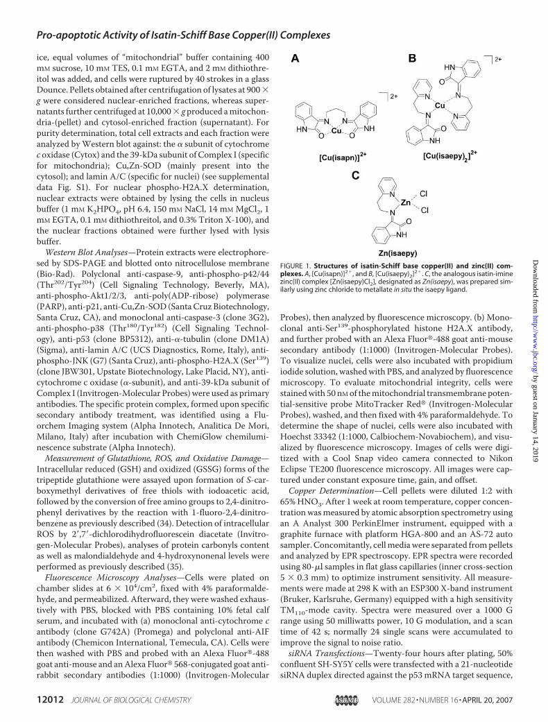
Materials—Isatin-diimine copper(II) complexes bis-[(2-ox-indol-3-yl-imino)-1,3-diaminopropane-N,N�,O,O�]copper(II)perchlorate ([Cu(isapn)](ClO4)2) and [bis-(2-oxindol-3-yl-imino)-2-(2-aminoethyl)pyridine-N,N�]copper(II) perchlorate([Cu(isaepy)2](ClO4)2), named hereCu(isapn) andCu(isaepy)2,respectively (Fig. 1, A and B), were synthesized as previouslydescribed (30). The analogous isatin-imine zinc(II) complex([Zn(isaepy)Cl2]), designated as Zn(isaepy), was prepared sim-ilarly using zinc chloride to metallate in situ the isaepy ligand(Fig. 1C). Dimethyl sulfoxide (Me2SO), dithiothreitol, EDTA,EGTA, paraformaldehyde, propidium iodide, tert-butylhydroperoxide, sodium orthovanadate, triethylenetetramine(TRIEN), and Triton X-100 were from Sigma. Goat anti-mouse and anti-rabbit IgG (H�L)-horseradish peroxidase con-jugate was from Bio-Rad. TES was from U.S. Biological Corp.(Cleveland, OH). All other chemicals were obtained fromMerck (Darmstadt, Germany).Cell Culture—Human neuroblastoma cells SH-SY5Y were
purchased from the European Collection of Cell Culture andgrown in Dulbecco’s modified Eagle’s medium, F-12 medium.Promonocytoma U937 were from the American Type CultureCollection; melanomaM14 were kindly provided by Dr. Gabri-ella Zupi from the Experimental Chemotherapy Laboratory,Regina Elena Cancer Institute of Rome, and grown in RPMI1640 medium; cervical carcinoma HeLa cells stably transfectedwith pSUPER vector containing p53 siRNAs (pSUPER-p53), orwith empty vector (pSUPER) were a gift of Dr. Anna Maria
Biroccio from the Experimental Chemotherapy Laboratory,Regina Elena Cancer Institute of Rome, and grown in Dulbec-co’s modified Eagle’s medium. All cell media were supple-mented with 10% fetal calf serum, and the cells were grown at37 °C in an atmosphere of 5%CO2 in air. Monoclonal SH-SY5Ycell lines transfected with human wild type Cu,Zn-superoxidedismutase (named hSOD) were obtained as previouslydescribed (31). During the experiments cells were plated at adensity of 4 � 104/cm2 (for SH-SY5Y, HeLa and M14) or 2 �105/ml (for U937), unless otherwise indicated.Treatments—A 5 mM solution of Cu(isapn), Cu(isaepy)2, or
Zn(isaepy)was prepared just before the experiments by dissolv-ing the lyophilized compounds in Me2SO. Treatments wereperformed with a concentration of 50 �M at 37 °C in mediumsupplemented with serum. This concentration was chosen forall the experiments because it gave a substantial degree of apo-ptosis at the times selected (29). As control, equal volumes ofMe2SO (1%) were added to untreated cells. The pancaspaseinhibitor zVAD-fmk (Alexis Biochemicals) was used at a finalconcentration of 100 �M, preincubated for 1 h before the addi-tion of Cu(isapn) and Cu(isaepy)2, and maintained throughoutthe experimental time. TRIEN, at a final concentration of 150�M, was added 3 h before the addition of Cu(isaepy)2 andmain-tained throughout the experiment. A 5 mM solution of coppersulfate in water was prepared just before the experiments andadded to culturemedia at a concentration of 50�M.Treatmentswith the specific c-Jun-N-terminal kinase (JNK) inhibitor,SP600125 (Calbiochem-Novabiochem, La Jolla, CA), and thephosphoinositide 3-kinase/Akt pathway inhibitor wortmannin(Calbiochem-Novabiochem) were performed at concentra-tions of 10 and 5 �M, respectively, because under our experi-mental conditions they did not result to be toxic. They wereadded 30 min before the addition of Cu(isapn) or Cu(isaepy)2andmaintained throughout the experiment. Retinoic acid (RA)was added to culture media at a concentration of 20 �M andmaintained for 7 days of culture to allow differentiation of cells,which was monitored by analyzing the increased expressionof the differentiation marker, growth-associated protein-43(GAP-43).Analysis of Cell Viability and Apoptosis—Adherent (after
trypsinization) and detached cells were combined, washedwithPBS, and stained with 50�g/ml propidium iodide prior to anal-ysis by a FACScalibur instrument (BD Biosciences). The per-centages of apoptotic cells were evaluated according to Nico-letti et al. (32) by calculating peak area of hypodiploid nuclei(sub-G1). Alternatively, cells were collected and counted aftertrypan blue staining by optical microscopy using a Thomachamber.Cell Fractionation and Protein Extraction—Total protein
extracts were obtained by rupturing cells with a 30-min incu-bation on ice in lysis buffer (50 mM Tris-HCl, pH 7.4, 1 mMEDTA, 1 mM EGTA, 1% Triton X-100, 10 mM NaF, 1 mMsodium orthovanadate) and protease inhibitor mixture (RocheApplied Science,Monza, Italy) and centrifuged at 22,300� g for20 min at 4 °C. Cell fractions were obtained as previouslyreported (33). Briefly, cells were incubated in hypotonicmedium (10 mM Tris-HCl, pH 7.5, 15 mM MgCl2, 10 mM KCl,and protease inhibitor mixture). After 10 min of incubation on
Pro-apoptotic Activity of Isatin-Schiff Base Copper(II) Complexes
APRIL 20, 2007 • VOLUME 282 • NUMBER 16 JOURNAL OF BIOLOGICAL CHEMISTRY 12011
by guest on January 14, 2019http://w
ww
.jbc.org/D
ownloaded from
ice, equal volumes of “mitochondrial” buffer containing 400mM sucrose, 10 mM TES, 0.1 mM EGTA, and 2 mM dithiothre-itol was added, and cells were ruptured by 40 strokes in a glassDounce. Pellets obtained after centrifugation of lysates at 900�g were considered nuclear-enriched fractions, whereas super-natants further centrifuged at 10,000� gproduced amitochon-dria-(pellet) and cytosol-enriched fraction (supernatant). Forpurity determination, total cell extracts and each fraction wereanalyzed byWestern blot against: the � subunit of cytochromec oxidase (Cytox) and the 39-kDa subunit of Complex I (specificfor mitochondria); Cu,Zn-SOD (mainly present into thecytosol); and lamin A/C (specific for nuclei) (see supplementaldata Fig. S1). For nuclear phospho-H2A.X determination,nuclear extracts were obtained by lysing the cells in nucleusbuffer (1 mM K2HPO4, pH 6.4, 150 mM NaCl, 14 mM MgCl2, 1mM EGTA, 0.1 mM dithiothreitol, and 0.3% Triton X-100), andthe nuclear fractions obtained were further lysed with lysisbuffer.Western Blot Analyses—Protein extracts were electrophore-
sed by SDS-PAGE and blotted onto nitrocellulose membrane(Bio-Rad). Polyclonal anti-caspase-9, anti-phospho-p42/44(Thr202/Tyr204) (Cell Signaling Technology, Beverly, MA),anti-phospho-Akt1/2/3, anti-poly(ADP-ribose) polymerase(PARP), anti-p21, anti-Cu,Zn-SOD (SantaCruz Biotechnology,Santa Cruz, CA), and monoclonal anti-caspase-3 (clone 3G2),anti-phospho-p38 (Thr180/Tyr182) (Cell Signaling Technol-ogy), anti-p53 (clone BP5312), anti-�-tubulin (clone DM1A)(Sigma), anti-lamin A/C (UCS Diagnostics, Rome, Italy), anti-phospho-JNK (G7) (Santa Cruz), anti-phospho-H2A.X (Ser139)(clone JBW301, Upstate Biotechnology, Lake Placid, NY), anti-cytochrome c oxidase (�-subunit), and anti-39-kDa subunit ofComplex I (Invitrogen-Molecular Probes) were used as primaryantibodies. The specific protein complex, formed upon specificsecondary antibody treatment, was identified using a Flu-orchem Imaging system (Alpha Innotech, Analitica De Mori,Milano, Italy) after incubation with ChemiGlow chemilumi-nescence substrate (Alpha Innotech).Measurement of Glutathione, ROS, and Oxidative Damage—
Intracellular reduced (GSH) and oxidized (GSSG) forms of thetripeptide glutathione were assayed upon formation of S-car-boxymethyl derivatives of free thiols with iodoacetic acid,followed by the conversion of free amino groups to 2,4-dinitro-phenyl derivatives by the reaction with 1-fluoro-2,4-dinitro-benzene as previously described (34). Detection of intracellularROS by 2�,7�-dichlorodihydrofluorescein diacetate (Invitro-gen-Molecular Probes), analyses of protein carbonyls contentas well as malondialdehyde and 4-hydroxynonenal levels wereperformed as previously described (35).Fluorescence Microscopy Analyses—Cells were plated on
chamber slides at 6 � 104/cm2, fixed with 4% paraformalde-hyde, and permeabilized. Afterward, they were washed exhaus-tively with PBS, blocked with PBS containing 10% fetal calfserum, and incubated with (a) monoclonal anti-cytochrome cantibody (clone G742A) (Promega) and polyclonal anti-AIFantibody (Chemicon International, Temecula, CA). Cells werethen washed with PBS and probed with an Alexa Fluor�-488goat anti-mouse and anAlexa Fluor� 568-conjugated goat anti-rabbit secondary antibodies (1:1000) (Invitrogen-Molecular
Probes), then analyzed by fluorescence microscopy. (b) Mono-clonal anti-Ser139-phosphorylated histone H2A.X antibody,and further probed with an Alexa Fluor�-488 goat anti-mousesecondary antibody (1:1000) (Invitrogen-Molecular Probes).To visualize nuclei, cells were also incubated with propidiumiodide solution, washedwith PBS, and analyzed by fluorescencemicroscopy. To evaluate mitochondrial integrity, cells werestainedwith 50nMof themitochondrial transmembrane poten-tial-sensitive probe MitoTracker Red� (Invitrogen-MolecularProbes), washed, and then fixed with 4% paraformaldehyde. Todetermine the shape of nuclei, cells were also incubated withHoechst 33342 (1:1000, Calbiochem-Novabiochem), and visu-alized by fluorescence microscopy. Images of cells were digi-tized with a Cool Snap video camera connected to NikonEclipse TE200 fluorescence microscopy. All images were cap-tured under constant exposure time, gain, and offset.Copper Determination—Cell pellets were diluted 1:2 with
65%HNO3. After 1 week at room temperature, copper concen-tration wasmeasured by atomic absorption spectrometry usingan A Analyst 300 PerkinElmer instrument, equipped with agraphite furnace with platform HGA-800 and an AS-72 autosampler. Concomitantly, cellmediawere separated frompelletsand analyzed by EPR spectroscopy. EPR spectra were recordedusing 80-�l samples in flat glass capillaries (inner cross-section5 � 0.3 mm) to optimize instrument sensitivity. All measure-ments were made at 298 K with an ESP300 X-band instrument(Bruker, Karlsruhe, Germany) equipped with a high sensitivityTM110-mode cavity. Spectra were measured over a 1000 Grange using 50 milliwatts power, 10 G modulation, and a scantime of 42 s; normally 24 single scans were accumulated toimprove the signal to noise ratio.siRNA Transfections—Twenty-four hours after plating, 50%
confluent SH-SY5Y cells were transfected with a 21-nucleotidesiRNA duplex directed against the p53mRNA target sequence,
FIGURE 1. Structures of isatin-Schiff base copper(II) and zinc(II) com-plexes. A, [Cu(isapn)]2�, and B, [Cu(isaepy)2]2�. C, the analogous isatin-iminezinc(II) complex [Zn(isaepy)Cl2], designated as Zn(isaepy), was prepared sim-ilarly using zinc chloride to metallate in situ the isaepy ligand.
Pro-apoptotic Activity of Isatin-Schiff Base Copper(II) Complexes
12012 JOURNAL OF BIOLOGICAL CHEMISTRY VOLUME 282 • NUMBER 16 • APRIL 20, 2007
by guest on January 14, 2019http://w
ww
.jbc.org/D
ownloaded from

FIGURE 2. Cu(isapn) and Cu(isaepy)2 induce a p53/p21-associated apoptosis in SH-SY5Y cells via the mitochondrial pathway. A, SH-SY5Y cellswere treated with 50 �M Cu(isapn) or Cu(isaepy)2 for 24 and 48 h, washed, and stained with propidium iodide. Analysis of sub-G1 (apoptotic) cells wasperformed by a FACScalibur instrument and percentages of staining positive cells were calculated using WinMDI version 2.8 software. Data areexpressed as mean � S.D., n � 12; *, p � 0.001. B, SH-SY5Y cells were treated with 50 �M Cu(isapn) or Cu(isaepy)2 for 6, 12, and 24 h. 25 �g of total proteinextract was loaded onto each lane for detection of p53 and p21. �-Tubulin was used as loading control. Western blots are from one experimentrepresentative of three that gave similar results. C, SH-SY5Y cells were grown on chamber slides, treated for 24 h with 50 �M Cu(isapn) or Cu(isaepy)2, andconcomitantly incubated with antibodies anti-AIF (red), to visualize mitochondria, and anti-cytochrome c (green). Images were digitized with a CoolSnap video camera connected to a Nikon Eclipse TE200 fluorescence microscopy. White arrows indicate cells where cytochrome c was not localized intomitochondria; therefore no superimposition of the two fluorescence was evidenced. D, alternatively, SH-SY5Y cells were treated with 50 �M Cu(isapn)or Cu(isaepy)2 for 24 and 48 h. 40 �g of total protein extract was loaded onto each lane for detection of pro- and active caspase-9, pro-caspase 3 andPARP. Western blots are from one experiment representative of three that gave similar results. E, SH-SY5Y cells were incubated for 1 h with or without100 �M pancaspase inhibitor zVAD-fmk, treated with 50 �M Cu(isapn) or Cu(isaepy)2 for 24, washed, and stained with propidium iodide. Analysis ofsub-G1 (apoptotic) cells was performed by a FACScalibur instrument and percentages of staining positive cells were calculated using WinMDI version 2.8software. Data are expressed as mean � S.D., n � 5; *, p � 0.001.
Pro-apoptotic Activity of Isatin-Schiff Base Copper(II) Complexes
APRIL 20, 2007 • VOLUME 282 • NUMBER 16 JOURNAL OF BIOLOGICAL CHEMISTRY 12013
by guest on January 14, 2019http://w
ww
.jbc.org/D
ownloaded from

5�-GACUCCAGUGGUAAUCUACTT-3� (sip53) (MWG Bio-tech, Ebersberg, Germany). Control cells were transfected witha scramble siRNA duplex, which does not present homologywith any other humanmRNAs (siScr). Cells were transfected byelectroporation using a Gene Pulser Xcell system (Bio-Rad)according to the manufacturer’s instructions and immediatelyseeded into fresh medium. Transfection efficiency of siRNAinto SH-SY5Y cells was estimated by co-transfecting p53siRNA with nonspecific rhodamine-conjugated oligonucleo-tides and found to be �80%.Protein Determination—Proteins were determined by the
method of Lowry et al. (36).Data Presentation—All experiments were done at least five
different times unless otherwise indicated. Datawere expressedasmean� S.D. and significance was assessed by Student’s t test
corrected by Bonferroni’s method.Differences with p values �0.05were considered significant.
RESULTS AND DISCUSSION
Cu(isapn) andCu(isaepy)2 InduceCell Cycle Arrest and Caspase-dependent Apoptosis—To dissectthe mechanisms by which isatin-Shiff base copper(II) complexesinduce cell death in tumor cell lines,on the basis of the results previouslyobtained (29), we selected the mosteffective molecules, Cu(isapn) andCu(isaepy)2 (Fig. 1, A and B), andused them at a concentration of 50�M. Fig. 2A shows representativehistograms from cytofluorimetricanalyses of SH-SY5Y cells treatedwith Cu(isapn) and Cu(isaepy)2 for24 and 48 h, where a more effectiveincrease in the apoptotic cells withCu(isaepy)2 treatment was evi-denced (Fig. 2A).Western blot anal-yses of p53 and p21 showed a strongactivation of these two proteinsalthough with different kinetics:rapid for Cu(isaepy)2 and moregradual for Cu(isapn) with a peak ofinduction at 12 and 24 h, respec-tively (Fig. 2B), indicating that thep53/p21 pathway is activated asresponse to cell damage. The typicalmitochondrial localization of theapoptosis inducing factor (AIF) andendonuclease G showed that no“caspase-independent” mechanismwas operative under our experi-mental conditions (data not shown);searching for the mechanismunderlying apoptotic induction, wetherefore used the anti-AIF anti-body to probemitochondria. As evi-
denced by immunofluorescence analyses of SH-SY5Y cellstreated for 24 h with copper complexes, AIF fluorescence didnot superimpose cytochrome c staining (Fig. 2C), indicatingthat cytochrome c was efficiently released from mitochondriainto the cytosol, and that the intrinsic mitochondrial pathwaywas operative under our experimental conditions. Concomi-tantly, Western blot analyses of pro- and active caspase-9, aswell as pro-caspase-3 and PARP indicated that each step of theapoptotic program was executed upon treatment with eitherCu(isapn) or Cu(isaepy)2 (Fig. 2D). In fact, immunoreactivebands of proteolyzed caspase-9 and PARP, together with a sig-nificant decrease of pro-caspase-3 were already detected after24 h of treatment. As final evidence that a caspase-dependentapoptotic response occurred after treatment with both coppercomplexes, we preincubated the cells with 100 �M of the pan-
FIGURE 3. p53 RNA interference partially counteracts apoptosis induced by Cu(isapn) and Cu(isaepy)2.A, SH-SY5Y cells were transiently transfected with siRNA duplex directed against the p53 mRNA targetsequence (sip53) or with a scramble siRNA duplex, which does not present homology with any other humanmRNAs (siScr). Cell adhesion has been allowed for 9 h, then the cells were treated with 50 �M Cu(isapn) orCu(isaepy)2 for the next 24, washed, and stained with propidium iodide. Analysis of sub-G1 (apoptotic) cells wasperformed by a FACScalibur instrument and percentages of staining positive cells were calculated using Win-MDI version 2.8 software. Data are expressed as mean � S.D., n � 6; *, p � 0.05. B, to evaluate the degree of p53decrease, at the time points indicated, sip53 or siScr cells were harvested and lysed. 25 �g of total proteinextract was loaded onto each lane for detection of p53. �-Tubulin was used as loading control. After 9 h fromtransfection with siScr or sip53, SH-SY5Y cells were then treated with 50 �M Cu(isapn) or Cu(isaepy)2 for 24 h. 40�g of total protein extract was loaded onto each lane for detection of p53 and PARP. �-Tubulin was used asloading control. Western blots are from one experiment representative of three that gave similar results.C, HeLa cells, stably transfected with a pSUPER vector containing p53 siRNA (pSUPER-p53) or with empty vector(pSUPER) were treated with 50 �M Cu(isapn) or Cu(isaepy)2 for 24 h, washed, and stained with propidium iodide.Analysis of sub-G1 (apoptotic) cells was performed by a FACScalibur instrument and percentages of stainingpositive cells were calculated using WinMDI version 2.8 software. Data are expressed as mean � S.D., n � 6;*, p � 0.05. D, pSUPER and pSUPER-p53 cells were treated with 50 �M Cu(isapn) or Cu(isaepy)2 for 24 h. 40 �gof total protein extract was loaded onto each lane for detection of p53 and PARP. �-Tubulin was used as loadingcontrol. Western blots are from one experiment representative of three that gave similar results.
Pro-apoptotic Activity of Isatin-Schiff Base Copper(II) Complexes
12014 JOURNAL OF BIOLOGICAL CHEMISTRY VOLUME 282 • NUMBER 16 • APRIL 20, 2007
by guest on January 14, 2019http://w
ww
.jbc.org/D
ownloaded from

caspase inhibitor zVAD-fmk for 1 h and then added Cu(isapn)or Cu(isaepy)2. Fig. 2E shows that a recovery of cell viabilityresulted from the inhibition of caspases activation upon treat-ment with both compounds, confirming that caspase-mediatedapoptosis is the principalmechanism for cell death induction inour experimental system.To evaluate the role of p53 in apoptosis induction, we trans-
fected SH-SY5Y cells with siRNA against p53 (sip53) or with ascramble sequence that does not present homology with anyother humanmRNAs (siScr). Western blot analyses of p53 lev-els indicated that the concentration of the protein rapidlydecreased after transfection (see Fig. 3B, upper panel). There-fore, we decided to treat the cells with Cu(isapn) or Cu(isaepy)2after 9 h from transfection, time that allowed the cells adheringto the flask and p53 interference being still in progress. Cyt-ofluorimetric analyses show that sip53 cells were significantlyresistant to apoptosis if compared with siScr (Fig. 3A). This wasalso confirmed byWestern blot of PARP that indicated a moreefficient cleavage of the protein occurring in siScr than in sip53cells (Fig. 3B, bottom panel). We also performed experimentsonHeLa cells stably transfected with a pSUPER vector contain-ing siRNAs for p53 (pSUPER-p53) or with an empty vector(pSUPER). Fig. 3C shows cytofluorimetric analyses of apoptosisupon 24 h treatment with Cu(isapn) or Cu(isaepy)2. As previ-ously observed for SH-SY5Y, also for HeLa, the percentage ofsub-G1 cells was significantly reduced upon p53 interference.This result was further confirmed by Western blot analyses ofPARP (Fig. 3D), suggesting that p53 activation contributes tocopper complex-mediated apoptosis. Nevertheless, a consider-able amount of p53-independent apoptosis was also observed.Cu(isapn) and Cu(isaepy)2 Cross the Cell Membrane and
Selectively Induce Oxidative Stress—To characterize the capa-bility of Cu(isapn) and Cu(isaepy)2 to enter the cells and thekinetics of their accumulation, we followed copper uptake byatomic absorption analyses. Fig. 4A shows that treatments withboth compounds resulted in a rapid increase of intracellularcopper content that reached a plateau after 12 h. This result wasparticularly significant, especially when compared with thatobtained with copper sulfate, used as control of cellular incor-poration of the metal ion. Cu(isaepy)2 seems to be more effi-ciently incorporated within the cells with respect to Cu(isapn).These results demonstrated a direct relationship between cop-per uptake and the extent of apoptosis, with Cu(isaepy)2 beingmore permeating andmore efficient in inducing cell death thanCu(isapn). Because the molecules we have synthesized havecharacteristic EPR spectra (23), we measured the extracellularconcentration of Cu(isaepy)2 by EPR spectroscopy. Fig. 4Breports the spectra of Cu(isaepy)2 in cell culture media and itsrelative concentration up to 12 h of treatment. The data suggestthat a decrease in the content of copper complex, most proba-bly due to its uptake by cells, was operative under our experi-mental conditions. Moreover, these results also suggested thatthe complex was chemically stable during the experimentaltimes selected, as no changes of the EPR parameters were evi-denced. To further confirm both hypotheses, we performed thesame experiments at 4 °C, a temperature at which metabolicprocesses are strongly reduced. Fig. 4C show that, under theseexperimental conditions, no significant difference in the extra-
cellular concentration of Cu(isaepy)2 was evidenced. The sameresults were achieved with Cu(isapn), for which, however, aslower uptake was observed (data not shown), confirming thatthe decrease of EPR signals measured in cell media after treat-mentwas due to cell uptake of both complexes rather than theirstability (see Fig. S2 to compare the different time courses ofCu(isaepy)2 uptake at 37 and 4 °C).Copper could behave as pro-oxidant by catalyzing intracel-
lular redox cycles with oxygen thus generating free radicals. Byspin trapping EPR experiments, using 5,5-dimethyl-1-pyrrolineN-oxide as the spin scavenger, we previously reported that bothCu(isapn) and Cu(isaepy)2 are able to produce ROS in the pres-ence of hydrogen peroxide, with the formermore efficient thanthe latter in generating 5,5-dimethyl-1-pyrroline N-oxide-OH�
adducts (29). To determine whether they were still able to gen-erate ROS in a cell system, we measured ROS content by stain-ing SH-SY5Y cells with 2�,7�-dichlorodihydrofluorescein diac-etate. Cytofluorimetric analyses shown in Fig. 5A demonstratethat Cu(isapn) and Cu(isaepy)2 were pro-oxidants, as ROS pro-duction increased with respect to untreated cells. In particular,Cu(isapn) was more effective as an upstream ROS inducer,because the increase of fluorescence was detectable as early as3 h after treatment. On the other hand, Cu(isaepy)2 did notaffect ROS production up to 24 h, the time corresponding to anincreased rate of apoptotic cells, allowing us to suggest that thisphenomenon could be a downstream event of the death proc-ess. Moreover, Cu(isapn)-induced transient damages to bothproteins (Fig. 5B) and lipids (Fig. 5C), as determined by West-
FIGURE 4. Cu(isapn) and Cu(isaepy)2 induce intracellular copper uptake.A, SH-SY5Y cells were treated with 50 �M Cu(isapn) or Cu(isaepy)2 for 6, 12,and 24 h, exhaustively washed with PBS containing 1 mM EDTA to avoid con-tamination of extracellularly membrane-bound copper, diluted 1:2 with 65%HNO3, and analyzed for copper content by atomic absorption. 50 �M coppersulfate (CuSO4) was used as control of copper uptake. Data are expressed asnanomole of copper/mg of total protein and represent the mean � S.D., n �5; *, p � 0.001. B, SH-SY5Y cells were treated with 50 �M Cu(isaepy)2 for 12 h at4 and 37 °C. Cell media were harvested, centrifuged to remove detached cells,and analyzed by EPR technique to measure the copper complex content.
Pro-apoptotic Activity of Isatin-Schiff Base Copper(II) Complexes
APRIL 20, 2007 • VOLUME 282 • NUMBER 16 JOURNAL OF BIOLOGICAL CHEMISTRY 12015
by guest on January 14, 2019http://w
ww
.jbc.org/D
ownloaded from

ern blot of carbonyls and colorimetric analyses of lipid peroxi-dation by products, respectively. Interestingly, the major capa-bility of this compound in generating ROS was in accordancewith the occurrence of an earlier appearance of protein carbon-yls as well as malondialdehyde and 4-hydroxynonenal (3–6 hafter treatment). In contrast, Cu(isaepy)2 treatment resulted ina later oxidative protein damage (12 h) without compromisinglipid structures.We measured the concentration and the redox state of glu-
tathione, as this molecule represents the most important lowmolecular weight antioxidant, particularly involved in ROSscavenging and xenobiotic detoxification. Fig. 5D shows that,after 6 h of treatment with Cu(isapn) and Cu(isaepy)2, bothGSH andGSSG increased with an extentmirroring the kineticsof copper uptake, suggesting that the levels of glutathione, dueto its ability to bind copper and detoxify xenobiotics, might becoupled with the intracellular content of copper compounds.
Cu(isapn) and Cu(isaepy)2-medi-ated Cytotoxicity Does Not Dependon Phosphorylative Pathways—Todetermine whether apoptosis wasmediated by phosphorylative path-ways downstreamof oxidative stressproduced by Cu(isapn) and Cu(isa-epy)2, we performed Western blotanalysis of phospho-active levels ofthe different members of mitogen-activated protein kinases and pro-tein kinase B, also known as Akt. Nosignificant changes in the immuno-reactive bands of active p38MAPK
and ERK1/2 (extracellular regulatedkinase 1 and 2) were detected upontreatment with the copper com-plexes (data not shown), but thephospho-isoforms of JNK and Aktincreased significantly, particularlyafter Cu(isaepy)2 treatment (Fig.6A), suggesting a role for these pro-teins in the induction of the celldeath program. However, becausethis increase occurred very earlyafter treatment (3–6 h), no correla-tion between Akt or JNK activationand ROS production can be stated,as far as Cu(isaepy)2 treatment isconcerned. To determine the role ofAkt in cell response to Cu(isapn)and Cu(isaepy)2, we preincubatedSH-SY5Y cells with wortmannin,a specific inhibitor of the Aktupstream kinase phosphoinositide3-kinase, and then treated the cellswith the copper complexes. Fig. 6Bshows that inhibition of Akt beforetreatment with Cu(isapn) and Cu-(isaepy)2 resulted in a significantincrease of sub-G1 cell population.
Western blot analyses of cell lysates confirmed that wortman-nin incubation produced blockage of the Akt-mediated path-way (Fig. 6C) and concomitantly showed no effect on the phos-pho-active form of JNK after Cu(isapn) and Cu(isaepy)2treatments, suggesting that Akt and JNK activation are inde-pendent responses. Preincubation with the specific JNK path-way inhibitor SP600125 did not result in modified cell survivalto treatments with either Cu(isapn) or Cu(isaepy)2 (Fig. 6D),indicating that JNK activation could represent just an epiphe-nomenon of the copper complex-induced cytotoxicity, whichdoes not directly relate to the apoptotic process.Increase of Antioxidant Defense or Differentiation by Retinoic
Acid Rescues SH-SY5Y Cells from Cu(isapn) and Cu(isaepy)2-induced Cytotoxicity—The results so far presented indicate adifferent time dependence and occurrence of the oxidativemarkers upon treatments with Cu(isapn) or Cu(isaepy)2,which could reflect the different structures of the copper
FIGURE 5. Cu(isapn) and Cu(isaepy)2 induce oxidative stress. A, SH-SY5Y cells were treated with 50 �M
Cu(isapn) or Cu(isaepy)2 for 3, 6, 12, and 24 h, and incubated with 50 �M DCF-DA at 37 °C. At the indicated timepoints, cells were washed with PBS and ROS production was analyzed by a FACScalibur instrument. Histogramsshown are representative of three experiments that gave similar results. B, at the same time points proteincarbonyls were identified upon derivatization with dinitrophenylhydrazine followed by immunoblot usinganti-dinitrophenylhydrazine antibody. 20 �g of derivatized proteins were loaded onto each lane. A represent-ative Western blot of three that gave similar results is shown. C, alternatively lipid peroxidation was evaluatedby measuring the levels of malondialdehyde and 4-hydroxynonenal using a colorimetric method. Data areexpressed as % of control and represent the mean � S.D., n � 5; *, p � 0.001. D, SH-SY5Y cells were treated with50 �M Cu(isapn) or Cu(isaepy)2 for 6, 12, and 24 h, exhaustively washed with PBS containing 1 mM EDTA, toavoid glutathione oxidation, and used for high performance liquid chromatography determination of intracel-lular GSH and GSSG. Data are expressed as % of control and represent the mean � S.D., n � 5; *, p � 0.05; **,p � 0.001.
Pro-apoptotic Activity of Isatin-Schiff Base Copper(II) Complexes
12016 JOURNAL OF BIOLOGICAL CHEMISTRY VOLUME 282 • NUMBER 16 • APRIL 20, 2007
by guest on January 14, 2019http://w
ww
.jbc.org/D
ownloaded from

complexes used. Particularly, the former could represent amore efficient ROS producer and the latter a more toxic andpermeating compound. To evaluate the contribution of oxi-dative stress in the execution of apoptosis, we used SH-SY5Ycells transfected with an additional copy of the human super-oxide dismutase 1 gene (hSOD cells) or differentiated bymeans of 7 days of incubation with 20 �M RA. Fig. 7A showsthe results obtained by direct counts of dead cells aftertrypan blue staining, and demonstrates that hSOD cells weremore resistant to both compounds than parental SH-SY5Y,even though to different extents. The increased antioxidantdefense let hSOD cells be highly resistant to Cu(isapn)-in-duced cytotoxicity, whereas a moderate decrease in dead cellsuponCu(isaepy)2 treatment was observed at 48 h. These resultsgive support to a different mode of action of the two coppercomplexes, in which ROS production represents a crucialupstream event in apoptosis induced by Cu(isapn), but onlyplays a downstream additional role in Cu(isaepy)2 cytotoxicity.The p53/p21 pathway was also activated upon both treatments,although with different kinetics. In fact, p53 and p21 seemedhighly expressed after 6 and 12 h of treatmentswithCu(isaepy)2and Cu(isapn), respectively; but no activation was observedafter 24 h (Fig. 7B). These results suggest that Cu(isapn) and
Cu(isaepy)2 exert similar inhibitoryeffects on cell growth of parentalSH-SY5Y and hSOD cells, a resultconfirmed by cell direct counts ofattached viable cells (data notshown). To test whether such com-pounds functioned as nonspecifictoxic agents, or were less efficienttoward untransformed cells, weinduced differentiation with pro-longed RA incubations. The resultsobtained would have providedsignificant information about apotential approach for a selectiveuse of copper complexes in cancertreatment. Seven days incubationwith 20 �M RA conferred efficientprotection against both coppercomplexes. A significant decrease ofsub-G1 cells to values close tountreated cells (about 4.25% after48 h as shown in Fig. 7C) and noactivation of the p53/p21 systemwas observed under these experi-mental conditions (Fig. 7D). Theaverage 1-fold increase of SODand catalase activity (data notshown), togetherwith the inhibitionof cell proliferation, features foundin normal (untransformed) cells,are some of the modificationsinduced by RA incubation andmay contribute to cell resistanceagainst Cu(isapn)- and Cu(isaepy)2-mediated toxicity.
Cu(isapn) and Cu(isaepy)2-mediated Toxicity Is a Copper-mediated Event That Induces Nuclear and MitochondrialDysfunction—Cu(isapn) and Cu(isaepy)2 are lipophilic com-pounds that may vehicle copper and allow the catalysis of redoxreactions and ROS production to take place also within specificintracellular organelles.We evaluated the copper content in cellu-lar fractions of SH-SY5Y cells (the purity of which is shown insupplemental dataFig. S2) treated fordifferent timeswith the cop-per complexes. Fig. 8A shows atomic absorption analyses, whichreveal a significant increase of copper in nuclear and mitochon-drial fractions upon treatmentwith both compounds,with a trendmirroring total copper uptake and suggesting a likely copper-me-diated site-directed injury. To evaluate the degree of the insultinduced, we investigated if mitochondria and nuclei were dam-aged upon treatment with the two copper complexes. Fig. 8Bshows SH-SY5Ynuclei stainedwith an antibody against the phos-pho-active histone H2A.X, which is phosphorylated on Ser139after DNAdouble strand break. After 6 and 12 h of treatment, theappearance of discrete foci, indicating the recruiting sites of theDNA repair machinery, revealed DNA-specific damagemediatedby Cu(isaepy)2 and, to a lesser extent by Cu(isapn) treatment, aphenomenon thatwas further confirmedbyWesternblot analysesof nuclear fractions (Fig. 8C). Concomitantly, we incubated
FIGURE 6. JNK and Akt are not involved in Cu(isapn) and Cu(isaepy)2-induced apoptosis. A, SH-SY5Y cellswere treated with 50 �M Cu(isapn) or Cu(isaepy)2 up to 24 h. 20 �g of total protein extract was loaded onto eachlane for detection of phosphorylated forms of JNK (P-JNK) and Akt (P-Akt). �-Tubulin was used as loadingcontrol. Western blots are from one experiment representative experiment of three that gave similar results.B, SH-SY5Y cells were treated with 50 �M Cu(isapn) or Cu(isaepy)2 for 24 h previous to a 1-h incubation with thephosphoinositide 3-kinase/Akt pathway inhibitor, wortmannin. Cells were washed and stained with pro-pidium iodide. Analysis of sub-G1 (apoptotic) cells was performed by a FACScalibur instrument and percent-ages of staining positive cells were calculated using WinMDI version 2.8 software. Data are expressed asmean � S.D., n � 5; *, p � 0.05; **, p � 0.001. C, alternatively 20 �g of total protein extract was loaded onto eachlane and used for the detection of phosphorylated forms of JNK (P-JNK) and Akt (P-Akt). Western blots are fromone experiment representative of three that gave similar results. D, SH-SY5Y cells were treated with 50 �M
Cu(isapn) or Cu(isaepy)2 for 24 h previous to a 1-h incubation with the specific JNK inhibitor, SP600125. Cellswere washed and stained with propidium iodide. Analyses of sub-G1 (apoptotic) cells was performed by aFACScalibur instrument and percentages of staining positive cells were calculated using WinMDI version 2.8software. Data are expressed as mean � S.D., n � 5.
Pro-apoptotic Activity of Isatin-Schiff Base Copper(II) Complexes
APRIL 20, 2007 • VOLUME 282 • NUMBER 16 JOURNAL OF BIOLOGICAL CHEMISTRY 12017
by guest on January 14, 2019http://w
ww
.jbc.org/D
ownloaded from

SH-SY5Y cells with MitoTracker Red, a red fluorescent dye thatstains mitochondria in viable cells and that accumulates depend-ing upon transmembrane potential (�mit). Fig. 8D shows thatespecially for Cu(isaepy)2 treatment, a huge number ofmitochon-dria resulted completely depolarized 12 h after treatment;�mit-dependent fluorescence was localized only in single spots anddid not look like a continuous network as in untreated cells.These results give strength to the hypothesis that althoughCu(isaepy)2 does produce less ROS, it is more efficient thanCu(isapn) in inducing apoptosis, because of its ability to spe-cifically accumulate and damage fundamental organelles,such as the nucleus and mitochondria.To evaluate further the contribution of copper in Cu(isa-
epy)2-induced apoptosis, we synthesized an analogous complexmolecule in which zinc, a non-redox active metal ion, replacedcopper in the complex. Alternatively, before Cu(isaepy)2administration, we incubated the cells with 150 �M of the non-permeating copper chelator TRIEN, which is able to removecopper from the complex and prevent its uptake by the cells.Cytofluorimetric analyses shown in Fig. 8E demonstrate thateither upon treatment with Zn(isaepy) (Fig. 1C), or by preincu-bating the cells with TRIEN, the extent of sub-G1 cell popula-tion strongly decreased with respect to Cu(isaepy)2-treatedcells, although a significant percentage of apoptosis was still
detectable (n � 6, p � 0.05 withrespect to untreated cells). Theseresults suggest that copper plays afundamental role in Cu(isaepy)2-mediated apoptosis, presumablydue to its action as catalyst of redoxreactions; however, the isatin-Schiffbase could also contribute to thetoxicity observed.Cu(isapn) andCu(isaepy)2 Induce
Apoptosis via the MitochondrialPathway in Other Tumor CellTypes—Finally, to verify a generalpro-apoptotic activity of the moreefficient copper complex Cu(isa-epy)2, we selected two other humantumor cells, the promonocytomaU937 and the melanoma M14. Aspreviously done with SH-SY5Ycells, we treatedU937 andM14 cellswith 50 �M Cu(isaepy)2. Fig. 9Ashows representative cytofluori-metric panels that demonstrate thattreatment with this copper complexinduced apoptosis after 24 h in boththe tumor cell lines selected, withU937 cells being more susceptible.Western blot analyses of caspase-9,caspase-3, and PARP, performed at24 and 48 h, further confirmed thatthe intrinsicmitochondrial pathwayrepresents the preferential route forthe induction of apoptosis (Fig. 9B).These results demonstrate the abil-
ity of Cu(isaepy)2 in inducing cell death in different tumor cells,and suggest a general application of the molecule.
CONCLUSIONS
In this paper we report that two recent synthesized isatin-Schiff base copper(II) complexes Cu(isapn) and Cu(isaepy)2 areable to induce apoptosis via the mitochondrial pathway in neu-roblastoma SH-SY5Y cells and in other tumor histotypes,mainly by copper-dependent oxidative stress and nuclear/mi-tochondrial site-directed damage. Although both compoundsare capable of producing such effects, the difference in the timeof appearance of pro-apoptotic and oxidative markers and theextent of cell death depends on their efficiency to permeate thecell. Cu(isapn), although less permeating, seems more prone toproduce oxyradicals and induce oxidative stress, whereas Cu-(isaepy)2 easily crosses cell membranes, accumulates, and dam-ages nuclear and mitochondrial compartments at very earlytimes. In line with these features, we speculate that the absenceof a detectable “cytosolic” oxidative stress upon treatment withCu(isaepy)2 explain the absence of a critical redox activation ofthe JNK-mediated signaling cascade in the events leading to celldeath.Moreover, our data are equally supportive of parallel p53-de-
pendent and -independent pathways, as demonstrated by experi-
FIGURE 7. The increase of antioxidant defense or differentiation by retinoic acid protects SH-SY5Y cellsagainst Cu(isapn) and Cu(isaepy)2-induced cytotoxic effects. A, SH-SY5Y and hSOD cells were treated with50 �M Cu(isapn) or Cu(isaepy)2 for 24 and 48 h, adherent and floating cells were collected, washed with PBS,and counted upon trypan blue staining. Data are expressed as % of control and represent the mean � S.D., n �6; **, p � 0.001, with respect to parental cell line. B, hSOD cells were treated with 50 �M Cu(isapn) or Cu(isaepy)2for 6, 12, and 24 h. 25 �g of total protein extract was loaded onto each lane for detection of p53 and p21.Western blots are from one experiment representative of three that gave similar results. C, SH-SY5Y cells wereincubated with 20 �M retinoic acid, to induce differentiation, then treated with 50 �M Cu(isapn) or Cu(isaepy)2for 24 h, washed, and stained with propidium iodide. Analysis of sub-G1 (apoptotic) cells was performed by aFACScalibur instrument and percentages of staining positive cells were calculated using WinMDI version 2.8software. Data are expressed as mean � S.D., n � 6. D, SH-SY5Y cells were incubated with 20 �M retinoic acid,then treated with 50 �M Cu(isapn) or Cu(isaepy)2 for 6, 12, and 24 h. 25 �g of total protein extract was loadedonto each lane for detection of p53 and p21. Western blots are from one experiment representative of threethat gave similar results.
Pro-apoptotic Activity of Isatin-Schiff Base Copper(II) Complexes
12018 JOURNAL OF BIOLOGICAL CHEMISTRY VOLUME 282 • NUMBER 16 • APRIL 20, 2007
by guest on January 14, 2019http://w
ww
.jbc.org/D
ownloaded from

ments carried out in p53 knocked-down cells by siRNA or in celllines lacking p53. These results are of great interest because func-tional p53 is frequently lost in human tumorigenesis.
The results obtained indicate that the role of copper in Cu-(isapn) and Cu(isaepy)2 cytotoxicity is fundamental, because ofits capability to catalyze one-electron redox cycle reactions
FIGURE 8. Copper is fundamental in Cu(isapn) and Cu(isaepy)2-induced apoptosis and is associated with the specific damage to nuclear and mito-chondrial compartments. A, SH-SY5Y cells were treated with 50 �M Cu(isapn) or Cu(isaepy)2 for 3, 6, and 12 h, and exhaustively washed with PBS containing1 mM EDTA to avoid contamination of extracellularly membrane-bound copper. Cells were then subjected to separation into nuclear, mitochondrial, andcytosolic enriched fractions, subsequently diluted 1:2 with 65% HNO3, maintained at room temperature for 1 week, and analyzed by atomic absorption forcopper content. Data are expressed as nanomole of copper/mg of total protein and represent the mean � S.D., n � 5; *, p � 0.05; **, p � 0.001, with respectto cytosolic copper concentrations. B, SH-SY5Y cells were grown on chamber slides, treated for 6 and 12 h with 50 �M Cu(isapn) or Cu(isaepy)2, and incubatedwith an anti-phospho-histone H2A.X antibody (green), to detect nuclear damage, and propidium iodide (red), to visualize nuclei. Images were digitized with aCool Snap video camera connected to Nikon Eclipse TE200 fluorescence microscopy. C, alternatively, after 3, 6, and 12 h of treatment, nuclei of SH-SY5Y cellswere isolated and lysed. 50 �g of total nuclear extract was loaded onto each lane and used for the detection of the phosphorylated form of histone H2A.X.Lamin A/C was used as nuclear loading control. D, SH-SY5Y cells were grown on chamber slides and treated for 12 h with 50 �M Cu(isapn) or Cu(isaepy)2. Beforefixation, cells were incubated for 30 min with 50 nM MitoTracker Red and subsequently stained with the specific nuclear vital dye Hoechst 33342. White arrowsindicate the formation of nuclear fragment characteristic of cells undergoing apoptosis. Images were digitized with a Cool Snap video camera connected toNikon Eclipse TE200 fluorescence microscopy. All of images were captured under constant exposure time, gain and offset. E, SH-SY5Y cells were treated with50 �M Cu(isapn), Zn(isaepy), or 50 �M Cu(isaepy)2 with or without the non-permeating copper chelator TRIEN (150 �M) for 24 h. Cells were then washed andstained with propidium iodide. Analysis of sub-G1 (apoptotic) cells was performed by a FACScalibur instrument and percentages of staining positive cells werecalculated using WinMDI version 2.8 software. Data are expressed as mean � S.D., n � 6; **, p � 0.001, with respect to Cu(isaepy)2-treated cells.
Pro-apoptotic Activity of Isatin-Schiff Base Copper(II) Complexes
APRIL 20, 2007 • VOLUME 282 • NUMBER 16 JOURNAL OF BIOLOGICAL CHEMISTRY 12019
by guest on January 14, 2019http://w
ww
.jbc.org/D
ownloaded from

with oxygen thus producingROS; the combination of this prop-erty with the chemical structure of the organic ligand bindingthemetal ion seems to give the specificity of the cellular damageinduced. We suggest that the presence of copper is mainlyrequired for nuclear damage, similar to what iron does in thepresence of bleomycin (37, 38), whereas the isatin-imine ligandis important to carry the redox-active metal ion across cellularmembranes, and could be more active at the mitochondriallevel. This hypothesis is strengthened by the similarity of the
chemical structure of Cu(isapn) and Cu(isaepy)2 with the delo-calized lipophilic cations, a class of molecules able to permeatethe cell in response to negative transmembrane potentials andincrease their concentration, particularly into mitochondria(39). The higher plasma and mitochondrial membrane poten-tials of tumor cells compared with normal cells account for thepreferential accumulation of delocalized lipophilic cations incarcinoma mitochondria (40, 41). Because most delocalizedlipophilic cations are toxic to mitochondria at high concentra-tions, their selective accumulation in the mitochondria oftumor cells, and consequent mitochondrial toxicity, providethe basis for selective tumor cell killing. The capability of thiscompound to preferentially induce detrimental effects in trans-formed cells can also be suggested on the basis of the prelimi-nary results obtained in the presence of RA, which is able toprotect the cells by making them similar to differentiated lines.Overall, the results obtained allow us to suggest a dual role
for Cu(isapn) and Cu(isaepy)2 in the induction of apoptosis: onone hand they are able to vehicle copper into the cell, thusproducing ROS; on the other they could behave as delocalizedlipophilic cations, thus specifically targeting mitochondria.Therefore the chemical structure of the isatin-Shiff base rep-resents the “switch” between these properties. This suggeststhat by specifically changing the chemical characteristics ofthis ligand type, we may modulate the cytotoxic effectsinduced, thus exploiting the plasticity of this new class ofcompounds to improve the therapeutic selectivity to differ-ent tumor histotypes.
REFERENCES1. Blagosklonny, M. V., and Pardee, A. B. (2001) Cancer Res. 61, 4301–43052. Shapiro, G. I. (2006) J. Clin. Oncol. 24, 1770–17833. Townsend, D. M., Findlay, V. L., and Tew, K. D. (2005)Methods Enzymol.
401, 287–3074. Kalinowski, D. S., and Richardson, D. R. (2005) Pharmacol. Rev. 57,
547–5835. Lyko, F., and Brown, R. (2005) J. Natl. Cancer Inst. 97, 1498–15066. Wilson, L., and Jordan, M. A. (2004) J. Chemother. 16, Suppl. 4, 83–857. Gius, D., and Spitz, D. R. (2006) Antioxid. Redox. Signal. 8, 1249–12528. Griguer, C. E., Oliva, C. R., Kelley, E. E., Giles, G. I., Lancaster, J. R., Jr., and
Gillespie, G. Y. (2006) Cancer Res. 66, 2257–22639. Martin, V., Herrera, F., Carrera-Gonzalez, P., Garcia-Santos, G., Antolin, I.,
Rodriguez-Blanco, J., and Rodriguez, C. (2006)Cancer Res. 66, 1081–108810. Prusty, B. K., and Das, B. C. (2005) Int. J. Cancer 113, 951–96011. Na, H. K., and Surh, Y. J. (2006)Mol. Carcinog. 45, 368–38012. Biaglow, J. E., and Miller, R. A. (2005) Cancer Biol. Ther. 4, 6–1313. Engel, R. H., and Evens, A. M. (2006) Front. Biosci. 11, 300–31214. Cleveland, J. L., and Kastan, M. B. (2000) Nature 407, 390–39515. Miller, R. A.,Woodburn, K.W., Fan, Q., Lee, I., Miles, D., Duran, G., Sikic,
B., and Magda, D. (2001) Clin. Cancer Res. 7, 3215–322116. Gewirtz, D. A. (1999) Biochem. Pharmacol. 57, 727–74117. Kotamraju, S., Kalivendi, S. V., Konorev, E., Chitambar, C. R., Joseph, J.,
and Kalyanaraman, B. (2004)Methods Enzymol. 378, 362–38218. Daniel, K. G., Gupta, P., Harbach, R. H., Guida, W. C., and Dou, Q. P.
(2004) Biochem. Pharmacol. 67, 1139–115119. Chen, D., Peng, F., Cui, Q. C., Daniel, K. G., Orlu, S., Liu, J., and Dou, Q. P.
(2005) Front. Biosci. 10, 2932–293920. Gaetke, L. M., and Chow, C. K. (2003) Toxicology 189, 147–16321. Rossi, L., Aquilano, K., Filomeni, G., Lombardo, M. F., Rotilio, G., and
Ciriolo, M. R. (2004) in Frontiers in Neurodegenerative Disorders andAging: Fundamental Aspects, Clinical Perspectives and New Insights(Ozben, K., and Chevion, M., eds) pp. 207–250, IOS Press, Amsterdam
22. Filomeni, G., Rotilio, G., and Ciriolo, M. R. (2005) Cell Death Differ. 12,
FIGURE 9. Cu(isapn) and Cu(isaepy)2 induce mitochondria-dependentapoptosis in U937 and M14 cells. A, human melanoma M14 and promono-cytoma U937 were treated with 50 �M Cu(isaepy)2 for 24 h, washed, andstained with propidium iodide. Analysis of cell cycle and apoptosis was per-formed by a FACScalibur instrument and percentages of staining positivecells were calculated using WinMDI version 2.8 software. The cell cycle plotsreported are from a typical experiment done in triplicate of five that gavesimilar results. The percentages of apoptotic nuclei are shown above eachhistogram. B, alternatively, M14 and U937 cells were treated with 50 �M Cu-(isaepy)2 for 24 and 48 h. 40 �g of total protein extract was loaded onto eachlane for detection of pro- and active caspase-9, pro-caspase 3, and PARP.Western blots are from one experiment representative of three that gavesimilar results.
Pro-apoptotic Activity of Isatin-Schiff Base Copper(II) Complexes
12020 JOURNAL OF BIOLOGICAL CHEMISTRY VOLUME 282 • NUMBER 16 • APRIL 20, 2007
by guest on January 14, 2019http://w
ww
.jbc.org/D
ownloaded from

1555–156323. Radulovic, S., Tesic, Z., and Manic, S. (2002) Curr. Med. Chem. 9,
1611–161824. Katsaros, N., and Anagnostopoulou, A. (2002) Crit. Rev. Oncol. Hematol.
42, 297–30825. Rosenzweig, A. C. (2001) Acc. Chem. Res. 34, 119–12826. Wang, T., and Guo, Z. (2006) Curr. Med. Chem. 13, 525–53727. Saryan, L. A., Ankel, E., Krishnamurti, C., Petering, D. H., and Elford, H.
(1979) J. Med. Chem. 22, 1218–122128. Antholine, W. E., Knight, J. M., and Petering, D. H. (1976) J. Med. Chem.
19, 339–34129. Cerchiaro, G., Aquilano, K., Filomeni, G., Rotilio, G., Ciriolo, M. R., and
Ferriera, A. M. D. C. (2005) J. Inorg. Biochem. 99, 1433–144030. Cerchiaro, G., Micke, G. A., Tavares, M. F. M., and Ferriera, A. M. D. C.
(2004) J. Mol. Catalysis A Chem. 221, 29–3931. Ciriolo, M. R., De Martino, A., Lafavia, E., Rossi, L., Carri, M. T., and
Rotilio, G. (2000) J. Biol. Chem. 275, 5065–507232. Nicoletti, I., Migliorati, G., Pagliacci, M. C., Grignani, F., and Riccardi, C.
(1991) J. Immunol. Methods 139, 271–27933. Filomeni, G., Aquilano, K., Rotilio, G., and Ciriolo, M. R. (2005) Antioxid.
Redox. Signal. 7, 446–45534. Filomeni, G., Rotilio, G., and Ciriolo, M. R. (2003) FASEB J. 17, 64–6635. Filomeni, G., Aquilano, K., Rotilio, G., and Ciriolo, M. R. (2003) Cancer
Res. 63, 5940–594936. Lowry, O. H., Rosebrough, N. J., Farr, A. L., and Randall, R. J. (1951) J. Biol.
Chem. 193, 265–27537. Ciriolo, M. R., Peisach, J., and Magliozzo, R. S. (1989) J. Biol. Chem. 264,
1443–144938. Harsch, A., Marzilli, L. A., Bunt, R. C., Stubbe, J., and Vouros, P. (2000)
Nucleic Acids Res. 28, 1978–198539. Modica-Napolitano, J. S., and Aprille, J. R. (2001)Adv. Drug Deliv. Rev. 49,
63–7040. Davis, S., Weiss, M. J., Wong, J. R., Lampidis, T. J., and Chen, L. B. (1985)
J. Biol. Chem. 260, 13844–1385041. Modica-Napolitano, J. S., and Aprille, J. R. (1987) Cancer Res. 47,
4361–4365
Pro-apoptotic Activity of Isatin-Schiff Base Copper(II) Complexes
APRIL 20, 2007 • VOLUME 282 • NUMBER 16 JOURNAL OF BIOLOGICAL CHEMISTRY 12021
by guest on January 14, 2019http://w
ww
.jbc.org/D
ownloaded from

Martino, Jens Z. Pedersen, Giuseppe Rotilio and Maria R. CirioloGiuseppe Filomeni, Giselle Cerchiaro, Ana Maria Da Costa Ferreira, Angelo De
on Oxidative Stress Induction and Organelle-selective DamagePro-apoptotic Activity of Novel Isatin-Schiff Base Copper(II) Complexes Depends
doi: 10.1074/jbc.M610927200 originally published online February 27, 20072007, 282:12010-12021.J. Biol. Chem.
10.1074/jbc.M610927200Access the most updated version of this article at doi:
Alerts:
When a correction for this article is posted•
When this article is cited•
to choose from all of JBC's e-mail alertsClick here
Supplemental material:
http://www.jbc.org/content/suppl/2007/03/01/M610927200.DC1
http://www.jbc.org/content/282/16/12010.full.html#ref-list-1
This article cites 40 references, 12 of which can be accessed free at
by guest on January 14, 2019http://w
ww
.jbc.org/D
ownloaded from

![Isatin as a Strategic Motif for Asymmetric Catalysisdigital.csic.es/bitstream/10261/111234/4/Isatin as a Strategic.pdf · it is a current open area of research in asymmetric catalysis.[12]](https://img.pdfslide.us/doc/110x75/5f0881af7e708231d4225981/isatin-as-a-strategic-motif-for-asymmetric-as-a-strategicpdf-it-is-a-current.jpg)