Embed Size (px)
Citation preview
Prion diseases or transmissible spongiform encephalopathies (TSEs)
• rare progressive neurodegenerative disorders that affect both humans and animals.
• They are distinguished by long incubation periods, characteristic spongiform changes associated with neuronal loss, and a failure to induce inflammatory response.

Prion diseases
• Normal prion protein PrPc – encoded by the prion gene (PRNP) on human chromosome 20
• The function of PrPc
role in anti-oxidant systems
cellular coper metabolism

Prion diseases
• Prion disease – normal gene produces normal PrPc, post-translational confirmational change to a disease related form – PrPsc
• PrPsc - insoluble and protease resistant protein accumulates in tissues forming amyloid structures
Prion diseases
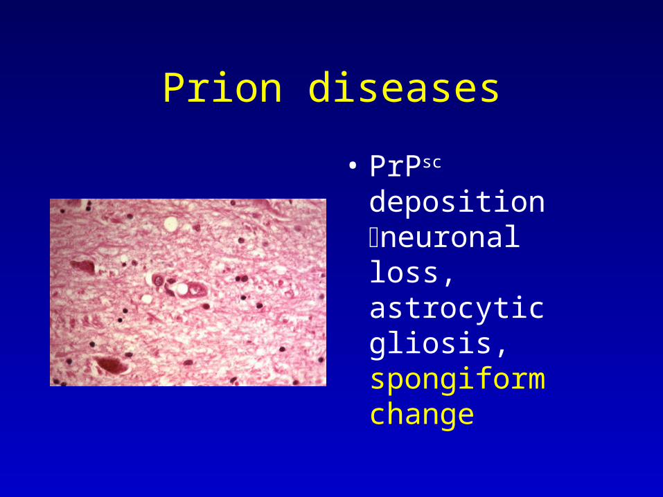
• PrPsc deposition neuronal loss, astrocytic gliosis, spongiform change

Prion diseases
• In human prion diseases – common polymorphism at codon 129 important effects on susceptibility to disease
• At codon 129 of PRNP an individual may encode for methionin or valin
• 80% of UK sporadic JCD – MM

Prion diseases
• Creutzfeldt-Jakob Disease (CJD)
• Variant Creutzfeldt-Jakob Disease (vCJD)
• Gerstmann-Straussler-Scheinker Syndrome
• Fatal Familial Insomnia

Creutzfeldt – Jakob sporadic form
• 90%
• Annual frequency – 1/milion/ per year
• Middle age (55-70 years)

Creutzfeldt – Jakob sporadic form
• Mental deterioration• Speech disorders• Memory loss • Cerebellar signs• Visual –• Pyramidal , extrapyramidal signs• Involuntary movements (myoklonus)• Mutism, global dementia – death (6M-2R)• Lost ability to walk

The typical periodic EEG seen in many cases of sporadic CJD.

(A) sCJD: axial FLAIR image at the level of the basal ganglia showing symmetrical high signal in the caudate head and anterior putamen (arrows).
(B) vCJD: axial FLAIR image at the level of the basal ganglia showing symmetrical high signal in the pulvinar and dorsomedial nuclei of the thalamus (arrows).

sCJch – MRI - diagnostika
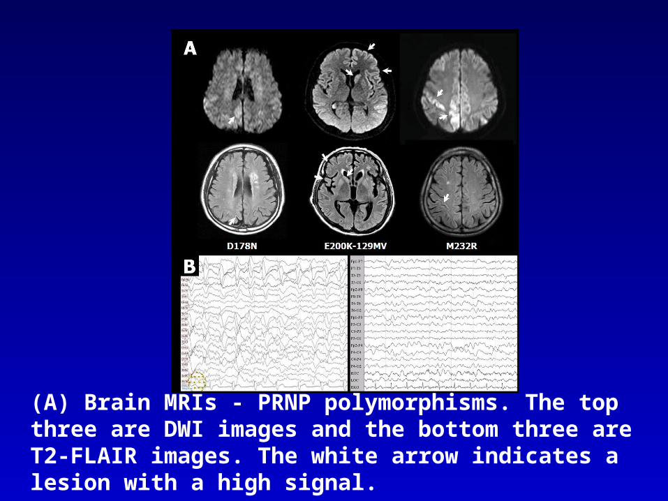
(A) Brain MRIs - PRNP polymorphisms. The top three are DWI images and the bottom three are T2-FLAIR images. The white arrow indicates a lesion with a high signal.

sCJch – diagnostika

Creutzfeldt – Jakob
• CSF – protein 14-3-3
• Normal protein being released to CSF following neuronal damage
• Not specific for JCD
• Sensitivity – 94%
• Genetic testing – most common mutation – E200K

sCJch – diagnostika
Spongiform changes Patological PrP

Creutzfeldt – Jakobiatrogenic –accidentally trasmitted
• Accidentally introduced into the body
• Length of incubation – 2 years in cases when infection introduced directly into the brain , 15 years – after s.c. inoculation
• Now - rare
• Corneal graft, stereotactic EEG
Creutzfeldt – Jakobnew variant (vCJD)
• Due to consumption of beef contamined by the agent of bovine spongiform encephalopathy (BSE)
• Young age at onset of ilness (27-50)
• Psychiatric or sensory disturbance
• Long duration of illness (14 months)
• Clinical feature – like sporadic form (dementia, myoclonus, multisystem neurological deficits)
nvCJch
• Etiological agent – in peripheral lymphatic nodes, increased riskof transmision
• Patological PrP we can confirm by biopsy from retikuloendotelial system - tonsilar biopsy, or appendix

MRI – pulvinar sign

Creutzfeldt – Jacob variant (vCJD)
• There are no changes on EEG• There is no protein 14-3-3 in CSF• MRI – abnormally high symmetrical
signal in pulvinar talami – strong diagnostic clue
• Neuropathological examination – difuse spongiform changes, especially in BG, posterior thalamus and cerebellum

Bovine spongiform encephalopathy

Gerstmann-Sträussler-Scheinker sy (GSS)
• Begins between the ages of 45 and 50
• Slowly evolving ataxia
• Mental deterioration
• Dementia, myoclonus, duration 5-10 years• Point mutation at codon 102, 105 (spastic paraparesis),
117 (pseudobulbar signs), 145, 198, 217 (GSS + AD)

Fatal familial insomnia (FFI)
• Autonomic and endocrine dysfunction
• Insomnia (during day - somnolence)
• Unexplained disorders of temperature, cardiovascular and respiratory regulation
• Later – pyramidal, extrapyramidal signs, , cerebellar ataxia, myoclonus
• duration 1 –2 years• Mutation at codon 178

Acquired immunodeficiency syndrom (AIDS) Human immunodeficiency virus (HIV)
• Neurological complications
• Aseptic meningitis
• Cognitive disturbances – adults
• Progressive encephalopathy – children
• Myelopathy • Neuropathy (inflammatory demyelinizating
polyneuropathy, brachial plexopathy, mononeuritis)
• Myopathies – myopathy, myositis

AIDS
• tumors
• Primary lymfoma of CNS (PCNSL)
most frequent, children, adult – 5%
clinical feature – headache, confusion, impaired memory , seizures, cran. nn. )
Dg.: MRI
• MTS non-Hodgkin lymfoma into CNS
• Kaposi sarcoma

AIDS
• Oportune infections • Bacterial – (Mycobacterium tuberculosis,
Treponema pallidum, Nocardia, ...)
• Viral – (Cytomegalovirus, Herpes simplex, Varicella zoster, JC, ...)
• Fungal – (Cryptococcus neoformans, candida, ...)
• Protozoa – (Toxoplazma gondii, ...)

AIDS dementia complex (ADC)brain atrophy, wide ventricles and subarachnoid space

AIDS dementia complex (ADC)
• T2- MRI:
• Enlargement of ventricles,
hyperintenzity in subcortical white matter of both frontal lobes