Embed Size (px)
Citation preview

Retinoscopy
Michael Davidson, D.V.M. Diplomate, American College of
Veterinary Ophthalmologists Professor, Ophthalmology
College of Veterinary MedicineNorth Carolina State University
Raleigh, North Carolina, USA

Retinoscopy: Research Applications
Normal refractive state and prevalence of refractive error in dogs, cats, horses, rats, raptors, gorilla, ostrich, elephant. Numerous studies.
Effect of environment on refractive state in cats. Belkin et al. Doc Ophthal 42:433-7.
Myopia and ammetropia in dog breeds, guide dogs. Murphy CL et al. Invest Ophthalmol Vis Sci 1992; 33: 2459-63.
Refractive state of aphakic/pseudophkic dogs, modal IOL strength. Davidson MG et al. Am J Vet Res 1993; 54; 174-7.
Modal IOL strength in cats. Gilger BC et al. Am J Vet Res 1998; 59: 1339-43.

Retinoscopy: Research Applications
Naturally occurring canine models of myopia. Kubai MA et al. Invest Ophthal Vis Sci 2013; 54: 7324-8; Williams LA et al. Optom Vis Sci2011; 88: 269-74; Black J. et al. Invest Ophthalmol Vis Sci 2008; 49: 4784-9; Mutti DO et al. Invest Ophthalmol Vis Sci 1999; 40: 1577-84.
Refractive state with foldable IOLs in dogs. Gaiddon JA et al. J Am Vet Med Assoc 2000; 216: 864-9.
Astigmatism in infant monkeys. Kee CS et al. Vis Res 2003; 43: 2721-39
Refractive state in cats with aqueous humor misdirection syndrome. Czederpiltz JM et al. J Am Vet Med Assoc 2005; 227: 1434-41.
No refractive error in Appaloosa with CSNB. Sandmeyer LS et al. Vet Ophthalmol 2007; 10: 368-75

Retinoscopy: Research Applications
Breed-related trends in ammetropia. Kubai MA et al. Am J Vet Res 2008; 69: 946-51.
Refractive state with different IOL designs in dogs. Gift BW et al. Vet Ophthalmol 2009; 12: 13-21.
“Modal” IOL dioptric power in horses. McMullen RJ et al. Am J Vet Res 2010; 71: 809-16.
Refractive state after vitreoretinal surgery in dogs. Hoffman A et al. Am J Vet Res 2012; 73: 1299-304.
Comparison of autorefractor vs. streak retinoscopy in dogs. Groth AD. Vet Ophthalmol 2013; 16: 319-323.
Effect of tropicamide on refractive state and aberrant retinoscopic reflexs in horses. McMullen RJ et al. Vet Ophthalmol 2014; 7: 120-5.

Refractive Error Is Relevant in our Patients
Following lens removal, vitreoretinal surgery, corneal surgery
Performance dogs Ofri R. et al. AVJR 2012, 73; 546-50 Assistance dogs Murphy CL et al. IOVS 1992; 33: 2459-63 Performance horses While naturally occurring, clinically significant refractive error
is relatively uncommon in our patients, retinoscopy allows clinician to rule out ammetropia as cause of visual problem

Lens Systems of the Mammalian Eye
cornea: 70-80% of refractive power 40-45 diopters in dog
crystalline lens: 20-30% of refractive power 13-15 diopters in dog
in emmetropic eye, brings incident light rays from optical infinity to point source on retina

Basic Definitions of Refraction and Refractive Properties
Vergence - the character of light rays, defined by the curvature of its wave front. The rays may have a negative (divergent), positive (convergent) or plano (parallel) vergence
Refraction - bending of light rays,as with a glass lens or the lens systems of the eye. Plus lenses (convex) converge parallel light rays while minus lens (concave) diverge light rays
Diopter (D) - a measure of lens power, defined by its focal point in meters (e.g., 5 diopter lens has a focal point of 0.2 meters or 1meter/5D)
Optical Infinity - an distance greater than 6 meters
www.cybersight.org

Basic Definitions of Refraction and Refractive Properties
Meridian - an imaginary line on the surface of a spherical body. A corneal meridian is this line marking the intersection with the corneal surface and an anterior-posterior plane passing through the apex of the cornea
www.helio.com
www.cybersight.org

Basic Definitions of Refraction and Refractive Properties
Emmetropia - an eye without refractive error where the plus lens of the the cornea and crystalline lenses refract light to a pint source on the retina
Ammetropia - an eye with a refractive error, generally from variations in the axial length of the eye, astigmatisms, or a shift in position or absence of the lens
Hyperopia - an eye with a refractive error caused by relatively too little refractive power, generally caused by a shorter than normal axial length
Myopia - an eye with a refractive error caused by relatively too great a refractive power, generally caused by a longer than normal axial length
Anisimetropia - difference in refractive state of the two
www.patient.info

Basic Definitions of Refraction and Refractive Properties
Astigmatism - an aspherical ammetropia, caused when the refractive surfaces of the eye have different radii of curvature in different meridians, generally caused by difference in corneal curvatures. Such an eye has two or more principle focal points, or two or more points of focus on incident light rays.
www.eyeglassguide.com
www.simplyoptometry.com

Principles of Retinoscopy or “Putting Yourself at the Far Point
of the Patient’s Eye”
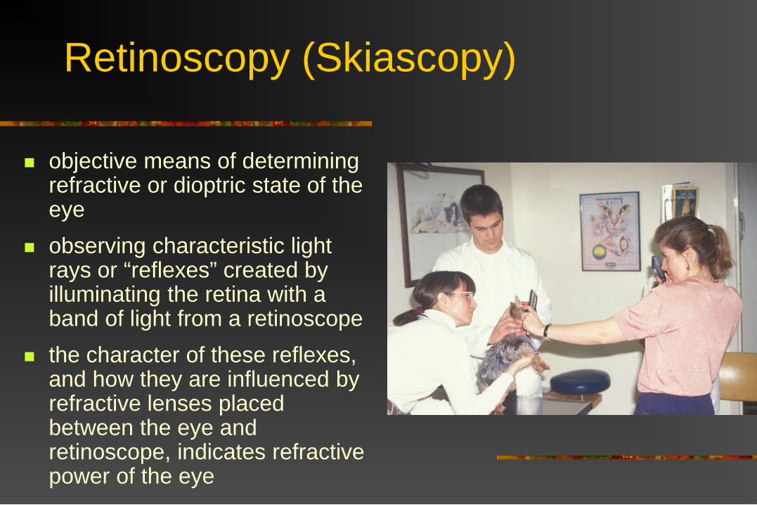
Retinoscopy (Skiascopy)
objective means of determining refractive or dioptric state of the eye
observing characteristic light rays or “reflexes” created by illuminating the retina with a band of light from a retinoscope
the character of these reflexes, and how they are influenced by refractive lenses placed between the eye and retinoscope, indicates refractive power of the eye

Design of Retinoscope
light projection system: tungsten bulb filament
emits a streak of light condensing lens which
changes vergence of light sleeve which controls
vergence by changing orientation of mirror, and controls (horizontal or vertical) direction of light streak

Design of Retinoscope
light projection system: tungsten bulb filament
emits a streak of light condensing lens which
changes vergence of light
sleeve which controls vergence by changing orientation of mirror, and controls (horizontal or vertical) direction of light streak

Design of Retinoscope

Design of Retinoscope
examiner observation system: peephole aperture allows
examiner to view emergent light rays from the eye

Retinoscopes
Welch Allyn Hiene

Refracting Lens
trial lens set: plus and minus spherical lenses
in 0.25D increments plus cylinder lenses for
spherocylindrical refraction technique
lens (skiascopy) bar or rack: series of spherical plus and
minus lenses in increments of 0.5D to 1.0D
in U.S., black bar contains plus lenses, red bar minus lenses, European designs may be the opposite
Luneau Lens Bars

Incident Light Rays and Refractive State
incident light rays acted on by lens systems of the eye
emmetropic eye: focal point on retina
hyperopic eye: focussed beyond retina
myopic eye: focussed in front of retina (in
vitreous)
www.patient.info
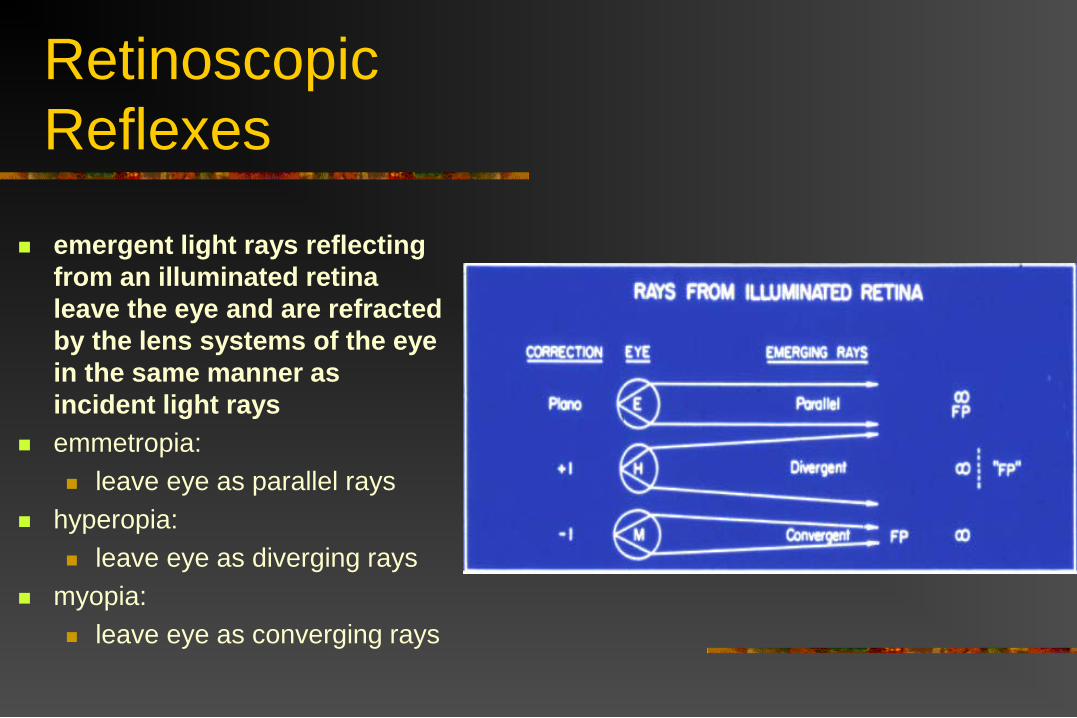
Retinoscopic Reflexes
emergent light rays reflecting from an illuminated retina leave the eye and are refracted by the lens systems of the eye in the same manner as incident light rays
emmetropia: leave eye as parallel rays
hyperopia: leave eye as diverging rays
myopia: leave eye as converging rays

Far Point of the Eye point in space, conjugate with, or
corresponding to, the retina emmetropic eye:
emergent light as parallel rays; far point AT infinity
hyperopic eye: emergent light as divergent
rays; far point BEYOND infinity
myopic eye: emergent light as convergent
rays; far point IN FRONT OF infinity
with emergent light rays, the further the far point is from infinity, the greater the refractive error

Emergent Light Rays from a Retinoscope appear as band of light, with
adjacent shadow as streak is passed across patient’s pupil
diverging or parallel light rays: “with” motion (moves in same
direction as sweep) light rays have come to a
focal point and crossed: “against” motion (moves in
opposite direction to sweep) light rays at the far point (in
the process of crossing): pupil fills with light, no motion
seen…“neutralization”

Emergent Light Rays from a Retinoscope
appear as band of light, with adjacent shadow as streak is passed across patient’s pupil
diverging or parallel light rays: “with” motion (moves in same direction
as sweep) light rays have come to a focal
point and crossed: “against” motion (moves in opposite
direction to sweep) light rays at the far point (in the
process of crossing): pupil fills with light, no motion
seen…“neutralization”

Emergent Light Rays from a Retinoscope
appear as band of light, with adjacent shadow as streak is passed across patient’s pupil
diverging or parallel light rays: “with” motion (moves in
same direction as sweep) light rays have come to a
focal point and crossed: “against” motion (moves in
opposite direction to sweep)
light rays at the far point (in the process of crossing): pupil fills with light, no
motion seen…“neutralization”

Emergent Light Rays from a Retinoscope
appear as band of light, with adjacent shadow as streak is passed across patient’s pupil
diverging or parallel light rays: “with” motion (moves in same
direction as sweep) light rays have come to a focal
point and crossed: “against” motion (moves in
opposite direction to sweep) light rays at the far point (in the
process of crossing): pupil fills with light, no motion
seen…“neutralization”

Emergent Light Rays from a Retinoscope
appear as band of light, with adjacent shadow as streak is passed across patient’s pupil
diverging or parallel light rays: “with” motion (moves in same
direction as sweep) light rays have come to a focal
point and crossed: “against” motion (moves in
opposite direction to sweep) light rays at the far point (in the
process of crossing): pupil fills with light, no motion
seen…“neutralization”

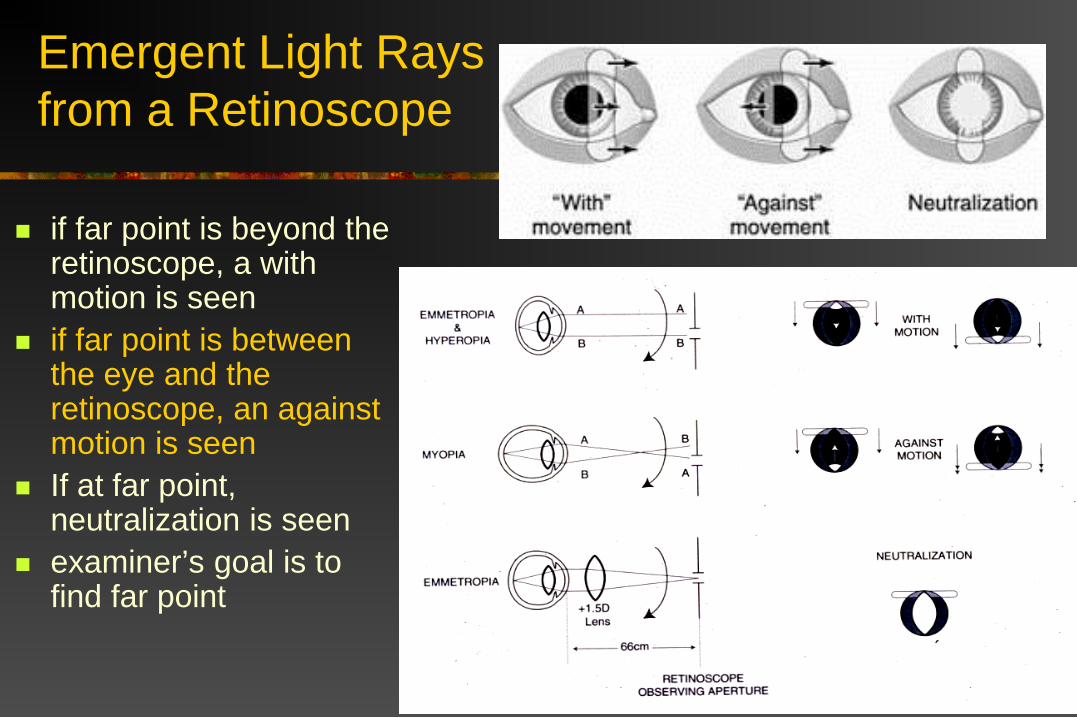
Emergent Light Rays from a Retinoscope
if far point is beyond the retinoscope, a with motion is seen
if far point is between the eye and the retinoscope, an against motion is seen
If at far point, neutralization is seen
examiner’s goal is to find far point
Emergent Light Rays from a Retinoscope
if far point is beyond the retinoscope, a with motion is seen
if far point is between the eye and the retinoscope, an against motion is seen
If at far point, neutralization is seen
examiner’s goal is to find far point

Emergent Light Rays from a Retinscope
if far point is beyond the retinoscope, a with motion is seen
if far point is between the eye and the retinoscope, an against motion is seen
If at far point, neutralization is seen
examiner’s goal is to find far point

Retinoscopy Simulator
VIDEO

Retinoscopic Reflexes Viewed at Infinity
emergent light rays from emmetropic and hyperopic eyes have not yet converged to a focal point: “with” motion
emergent light rays from myopic eye have converged, crossed, and begun to diverge: “against” motion

Retinoscopy Working Distance optical infinity (>6meters)
too distant from eye to perform retinoscopy
infinity recreated by placing retinoscope at a known distance from eye, the “working distance” and placing a “working lens” in the path of reflected light rays

Retinoscopy at 1 Meter emmetropia and
hyperopia: “with” motion
myopia >1 diopter: against motion
add 1 D “working lens” in front of eye: emmetropic eye at far point =
“neutralization” to reach far point for other
refractive states: add more plus lens to 1 D for
hyperopic eye add more minus lenses to 1
D for myopic eye

Retinoscopy at 1 Meter emmetropia and hyperopia:
“with” motion myopia >1diopter:
against motion add 1 D “working lens” in
front of eye: emmetropic eye at far point =
“neutralization” to reach far point for other
refractive states: add more plus lens to 1 D for
hyperopic eye add more minus lenses to 1
D for myopic eye

Retinoscopy at 1 Meter
emmetropia and hyperopia: “with” motion
myopia >1 diopter: against motion
add 1 D “working lens” in front of eye: emmetropic eye at far point =
“neutralization” to reach far point for other
refractive states: add more plus lens to 1 D for
hyperopic eye add more minus lenses to 1
D for myopic eye

Finding the Far Point
“with” motion wants PLUS lenses
“against” motion wants MINUS lenses

Retinoscopy at 66 cm
with no working lens: emmetropia, hyperopia, &
myopia <1.5 D show “with” motion
myopia 1.5 D shows neutralization
myopia >1.5 D shows “against” motion
use 1.5 D “working” lens: emmetropia shows
neutralization hyperopia shows “with”
motion (add plus lenses) myopia shows “against”
motion (add minus lenses)

Retinoscopy at 66 cm
with no working lens: emmetropia, hyperopia, &
myopia <1.5 D show “with” motion
myopia 1.5 D shows neutralization
myopia >1.5 D shows “against” motion
use 1.5 D “working” lens: emmetropia shows
neutralization hyperopia shows “with” motion
(add plus lenses) myopia shows “against” motion
(add minus lenses)

Retinoscopy Working Distance
a single lens is used for both the “working lens” and additional “correcting” lenses
when neutralization is reached, subtract the working lens strength from gross (total) refraction to yield net refraction
66cm = working lens of +1.5D
50cm = working lens of +2.0D
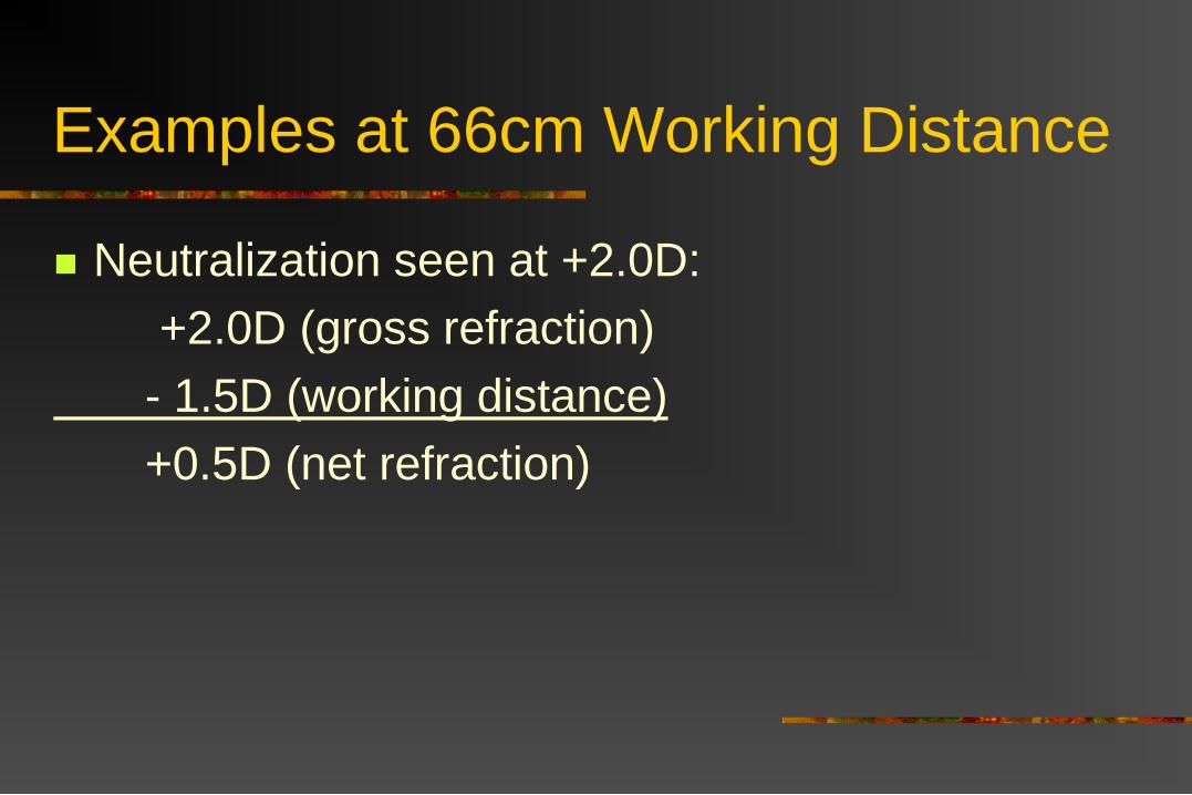
Examples at 66cm Working Distance
Neutralization seen at +2.0D:+2.0D (gross refraction)
- 1.5D (working distance)+0.5D (net refraction)

Examples at 66cm Working Distance
Neutralization seen at +0.5D:+0.5D (gross refraction)
- 1.5D (working distance)-1.0D (net refraction)

Examples at 66cm Working Distance
Neutralization seen at -1.5D:-1.5D (gross refraction)
- 1.5D (working distance)-3.0D (net refraction)

Examples at 50cm Working Distance
Neutralization seen at +3.0D:+3.0D (gross refraction)
- 2.0D (working distance)+1.0D (net refraction)

Examples at 50cm Working Distance
Neutralization seen at -1.0D:-1.0D (gross refraction)
- 2.0D (working distance)-3.0D (net refraction)

Technique of Retinoscopy
semidarkened room, assistant holds animal, directs gaze
retinoscope held in palm, thumb on sleeve, lens bar in other hand, distance 66 or 50 cm from patient
vergence set by moving sleeve down, direction set so vertical streak projected on eye
optical alignment…align Purkinje images on anterior cornea and lens
streaks brought into pupil with slow, deliberate movement (shake head back and forth), find neutral point
direction of beam is then rotated to produce horizontal streak and this meridian is assessed…ALWAYS ASSESS BOTH MERIDIANS

Identifying Neutrality
with no lenses, determine if “with” motion, “against” motion, or neutrality: note that all emmetropes and almost all ammetropes will show a
“with” motion at 66 cm with no refractive lenses with motion = add progressively stronger plus lenses against motion = add minus lenses because against motion more difficult to see and
confusing: to confirm, reverse vergence, “against” becomes a “with”!! approach neutrality from “with” side….go past neutrality until
with motion seen, bracket back to neutrality
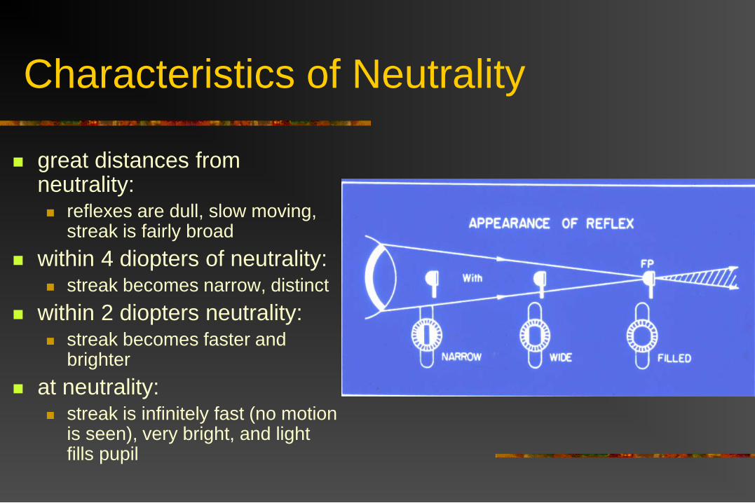
Characteristics of Neutrality
great distances from neutrality: reflexes are dull, slow moving,
streak is fairly broad within 4 diopters of neutrality:
streak becomes narrow, distinct within 2 diopters neutrality:
streak becomes faster and brighter
at neutrality: streak is infinitely fast (no motion
is seen), very bright, and light fills pupil

Characteristics of Neutrality
great distances from neutrality: reflexes are dull, slow moving,
streak is fairly broad within 4 diopters of neutrality:
streak becomes narrow, distinct within 2 diopters neutrality:
streak becomes faster and brighter
at neutrality: streak is infinitely fast (no
motion is seen), very bright, and light fills pupil
Retinoscopy Simulator
VIDEO
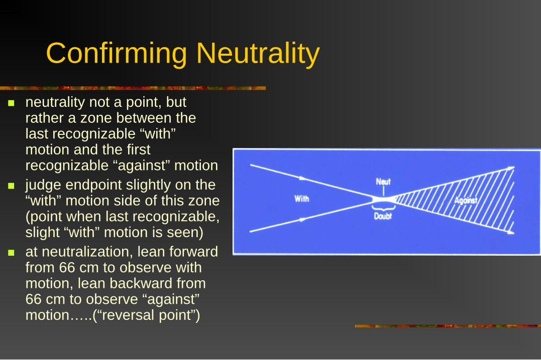
Confirming Neutrality neutrality not a point, but
rather a zone between the last recognizable “with” motion and the first recognizable “against” motion
judge endpoint slightly on the “with” motion side of this zone (point when last recognizable, slight “with” motion is seen)
at neutralization, lean forward from 66 cm to observe with motion, lean backward from 66 cm to observe “against” motion…..(“reversal point”)

Estimating Hyperopia
“enhancement” estimates gross hyperopia
at working distance, compare thickness of beam in pupil (retinal band) vs. outside the pupil (face band)
slowly raise vergence until the beam of light is the thinnest possible

Estimating Hyperopia <1.0 D gross hyperopia:
beam will not enhance 1-3 D gross hyperopia:
retinal band thinner (1/2 to 3/4) than face band
4-5 D gross hyperopia: retinal band may be
enhanced to thin streak, and it is only slightly more narrow than face band
emmetrope has +1.5 D of gross hyperopia at 66 cm: retinal band 3/4 width
of face band, which is broad

Estimating Myopia
“far point determination” estimates net myopia
if against motion observed at 66 cm, >1.5 D myopia present
change vergence by moving sleeve up to confirm
move sleeve back down, slowly move progressively slower to eye, streaking beam until neutralization reached
estimate your distance from the eye at neutrality:
neutralization at 33cm = -3.0D refractive stateneutralization at 50cm = -5.0D refractive state

Astigmatism Astigmatism - an aspherical ammetropia, caused when the refractive
surfaces of the eye have different radii of curvature in different meridians, generally caused by difference in corneal curvatures. Such an eye has two or more principle focal points, or two or more points of focus on incident light rays.

Astigmatic Refractive Errors
neutralization seen with different lenses in two different meridians
or…when neutralization reached in one meridian, streak is rotated, either a “with” or “against” motion is seen
major or principle meridians: least and most refractive
meridians generally oriented with axes
at or near 90 degrees and 180 degrees

Astigmatic Refractive Errors
simple astigmatism emmetropia/ammetropia
compound astigmatism hyperopia/hyperopia or myopia/myopia
mixed astigmatism hyperopia/myopia

Astigmatic Refractive Errors
regular astigmatism principle meridians 90
degrees apart irregular astigmatism
principle meridians not 90 degrees apart
Astigmatic Refractive Errors
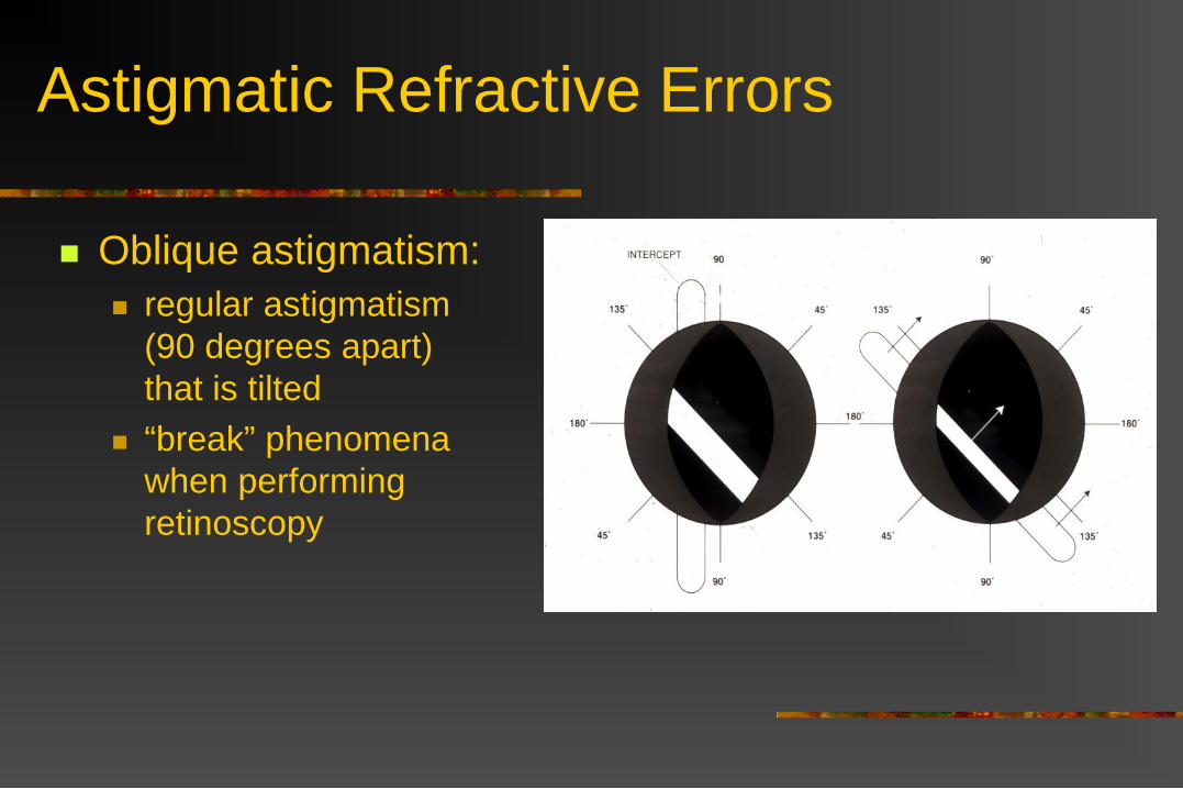
Oblique astigmatism: regular astigmatism
(90 degrees apart) that is tilted
“break” phenomena when performing retinoscopy

Astigmatic Refractive Errors
”with the rule” astigmatism most refractive corneal
meridian vertical “against the rule”
astigmatism most refractive corneal
meridian horizontal
*Vertical retinoscopic streak measures power in horizontal corneal meridian

Designating Refractive Error
determine net refraction in both vertical and horizontal meridians
if refraction is same, eye is “spherical”, if two meridians are different eye is “astigmatic”
average the two meridians to get “average” refractive state
or…designate two meridians with “lens cross”

Designating Refractive Error
determine net refraction in both vertical and horizontal meridians
if refraction is same, eye is “spherical”, if two meridians are different eye is “astigmatic”
average the two meridians to get “average” refractive state
or…designate two meridians with “lens cross”
+1.5D
+0.5D

Practical Aspects of Veterinary Retinoscopy
use retinoscopy bar vs. trial lens set good assistant invaluable estimating techniques useful to perform first
and reduce refraction time optical alignment (Purkinje images) critical,
must constantly realign

Practical Aspects of Veterinary Retinoscopy
retinoscopy should generally be performed without mydriasis
cycloplegia/mydriasis used in humans to eliminate accommodation
while retinoscopy results not significantly different with and without mydriasis in dogs and horses, mydriasis reduces accuracy in identifying neutrality due to spherical abberation:
full mydriasis often causes swirling or “scissors” motion
if mydriasis present, concentrate on center of pupil
Practical Aspects of Veterinary Retinoscopy
brightness of tapetum is useful in identifying neutrality
refracting aphakes or pseudophakes challenging: opaque ocular media surgically-induced astigmatism on pseudophakes, reflex different in pupil covered by IOL
optic and that area outside of IOL optic


Retinoscopy Model Eyes

Check Your Working Distance

Retinoscopy Simulator
http://eyeontechs.com/wp-content/uploads/2009/04/retinoscopysimulator.swf