Embed Size (px)
Citation preview
Principles of Human Anatomy and Physiology, 11e 1
Chapter 10
Muscle Tissue
Lecture Outline
Principles of Human Anatomy and Physiology, 11e 2
INTRODUCTION
• Motion results from alternating contraction (shortening) and relaxation of muscles; the skeletal system provides leverage and a supportive framework for this movement.
• The scientific study of muscles is known as myology.
Principles of Human Anatomy and Physiology, 11e 3
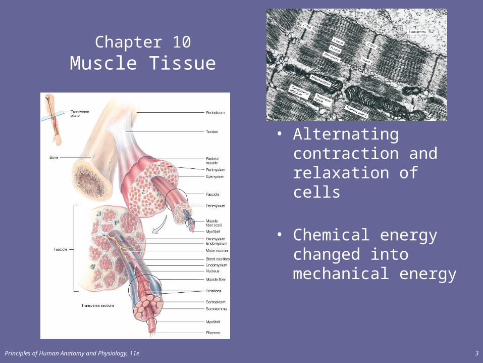
Chapter 10Muscle Tissue
• Alternating contraction and relaxation of cells
• Chemical energy changed into mechanical energy
Principles of Human Anatomy and Physiology, 11e 4
OVERVIEW OF MUSCLE TISSUE
• Types of Muscle Tissue• Skeletal muscle tissue is primarily attached to bones. It is
striated and voluntary.• Cardiac muscle tissue forms the wall of the heart. It is
striated and involuntary.• Smooth (visceral) muscle tissue is located in viscera. It is
nonstraited (smooth) and involuntary. • Table 4.4 compares the different types of muscle.
Principles of Human Anatomy and Physiology, 11e 5
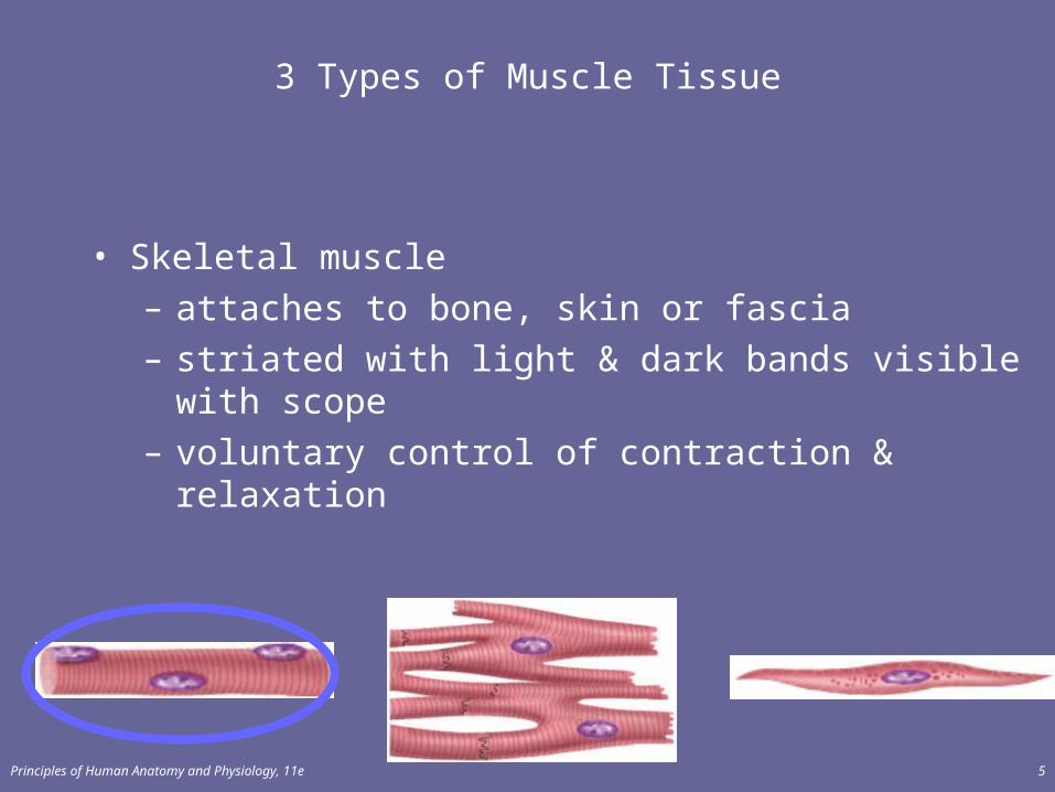
3 Types of Muscle Tissue
• Skeletal muscle– attaches to bone, skin or fascia– striated with light & dark bands visible with scope – voluntary control of contraction & relaxation
Principles of Human Anatomy and Physiology, 11e 6
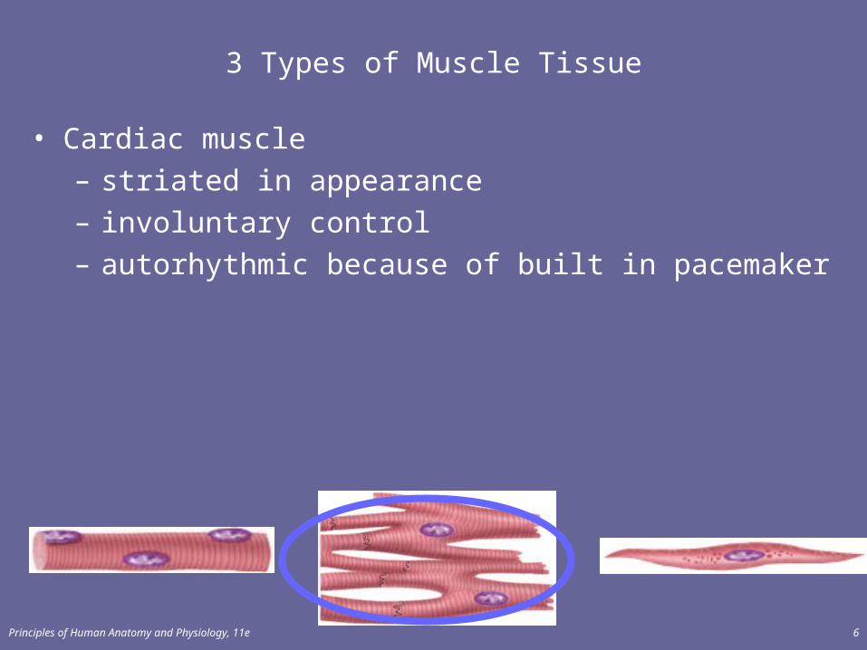
3 Types of Muscle Tissue
• Cardiac muscle– striated in appearance– involuntary control– autorhythmic because of built in pacemaker
Principles of Human Anatomy and Physiology, 11e 7
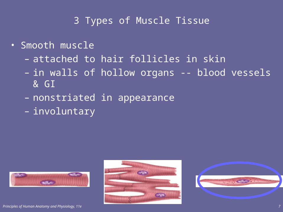
3 Types of Muscle Tissue
• Smooth muscle– attached to hair follicles in skin– in walls of hollow organs -- blood vessels & GI– nonstriated in appearance– involuntary
Principles of Human Anatomy and Physiology, 11e 8
Functions of Muscle Tissue
• Producing body movements• Stabilizing body positions• Regulating organ volumes
– bands of smooth muscle called sphincters• Movement of substances within the body
– blood, lymph, urine, air, food and fluids, sperm• Producing heat
– involuntary contractions of skeletal muscle (shivering)
Principles of Human Anatomy and Physiology, 11e 9
Properties of Muscle Tissue
• Excitability– respond to chemicals released from nerve cells
• Conductivity– ability to propagate electrical signals over membrane
• Contractility– ability to shorten and generate force
• Extensibility– ability to be stretched without damaging the tissue
• Elasticity– ability to return to original shape after being stretched
Principles of Human Anatomy and Physiology, 11e 10
SKELETAL MUSCLE TISSUE
• Each skeletal muscle is a separate organ composed of cells called fibers.
Principles of Human Anatomy and Physiology, 11e 11
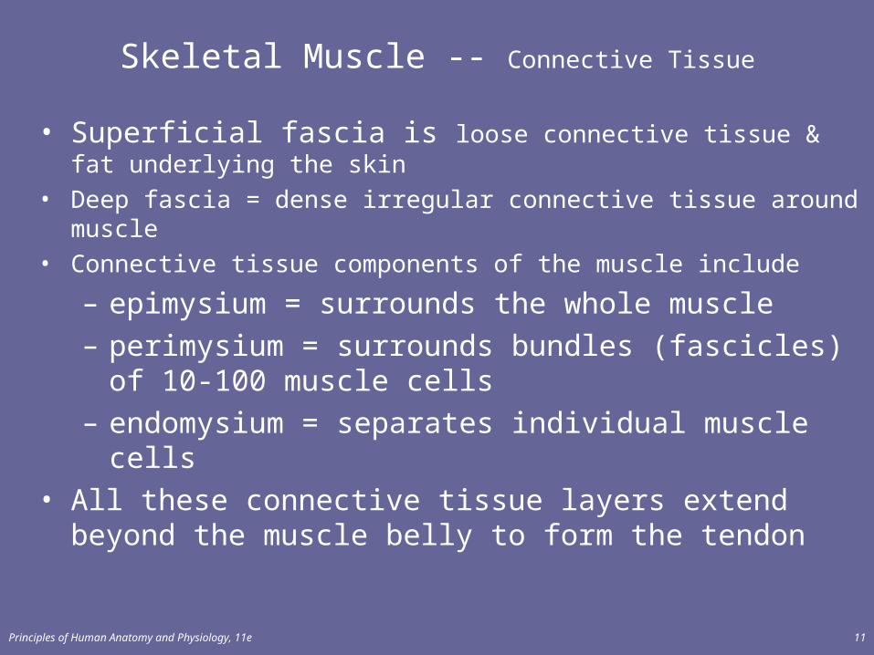
Skeletal Muscle -- Connective Tissue
• Superficial fascia is loose connective tissue & fat underlying the skin
• Deep fascia = dense irregular connective tissue around muscle• Connective tissue components of the muscle include
– epimysium = surrounds the whole muscle – perimysium = surrounds bundles (fascicles) of 10-100
muscle cells– endomysium = separates individual muscle cells
• All these connective tissue layers extend beyond the muscle belly to form the tendon
Principles of Human Anatomy and Physiology, 11e 12
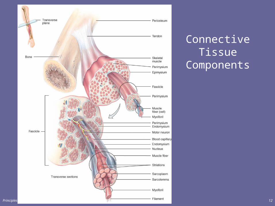
Connective Tissue Components
Principles of Human Anatomy and Physiology, 11e 13
Connective Tissue Components
• Tendons and aponeuroses are extensions of connective tissue beyond muscle cells that attach muscle to bone or other muscle.
• A tendon is a cord of dense connective tissue that attaches a muscle to the periosteum of a bone (Figure 11.22).
• An aponeurosis is a tendon that extends as a broad, flat layer (Figure 11.4c).
Principles of Human Anatomy and Physiology, 11e 14
Nerve and Blood Supply
• Each skeletal muscle is supplied by a nerve, artery and two veins.
• Each motor neuron supplies multiple muscle cells (neuromuscular junction)
• Each muscle cell is supplied by one motor neuron terminal branch and is in contact with one or two capillaries.– nerve fibers & capillaries are found in the endomysium
between individual cells
Principles of Human Anatomy and Physiology, 11e 15
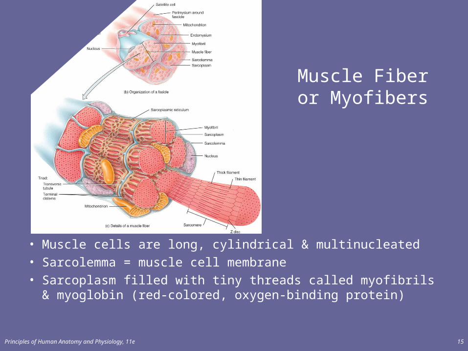
Muscle Fiber or Myofibers
• Muscle cells are long, cylindrical & multinucleated
• Sarcolemma = muscle cell membrane
• Sarcoplasm filled with tiny threads called myofibrils & myoglobin (red-colored, oxygen-binding protein)
Principles of Human Anatomy and Physiology, 11e 16
Sarcolemma, T Tubules, and Sarcoplasm
• Skeletal muscle consists of fibers (cells) covered by a sarcolemma (Figure 10.3b).– The fibers contain T tubules and sarcoplasm– T tubules are tiny invaginations of the sarcolemma that
quickly spread the muscle action potential to all parts of the muscle fiber.
• Sarcoplasm is the muscle cell cytoplasm and contains a large amount of glycogen for energy production and myoglobin for oxygen storage.
Principles of Human Anatomy and Physiology, 11e 17
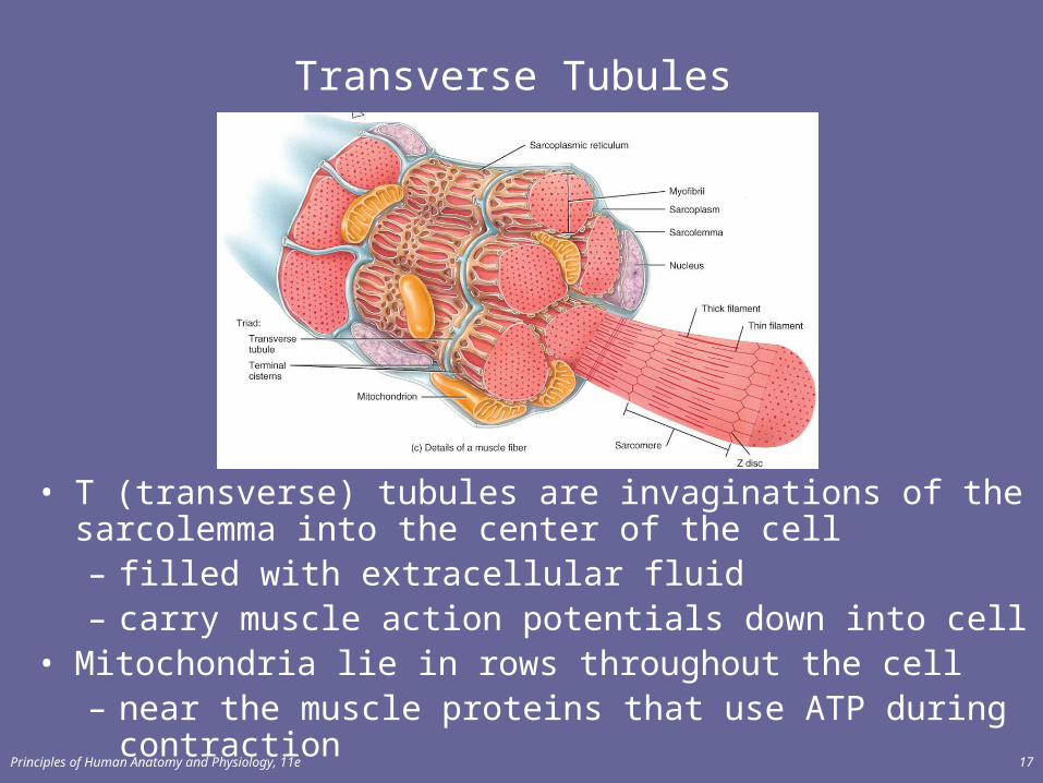
Transverse Tubules
• T (transverse) tubules are invaginations of the sarcolemma into the center of the cell– filled with extracellular fluid– carry muscle action potentials down into cell
• Mitochondria lie in rows throughout the cell– near the muscle proteins that use ATP during contraction
Principles of Human Anatomy and Physiology, 11e 18
Myofibrils and Sarcoplasmic Reticulum
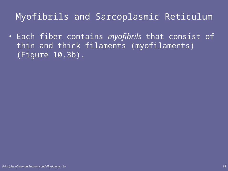
• Each fiber contains myofibrils that consist of thin and thick filaments (myofilaments) (Figure 10.3b).
Principles of Human Anatomy and Physiology, 11e 19
Myofibrils & Myofilaments
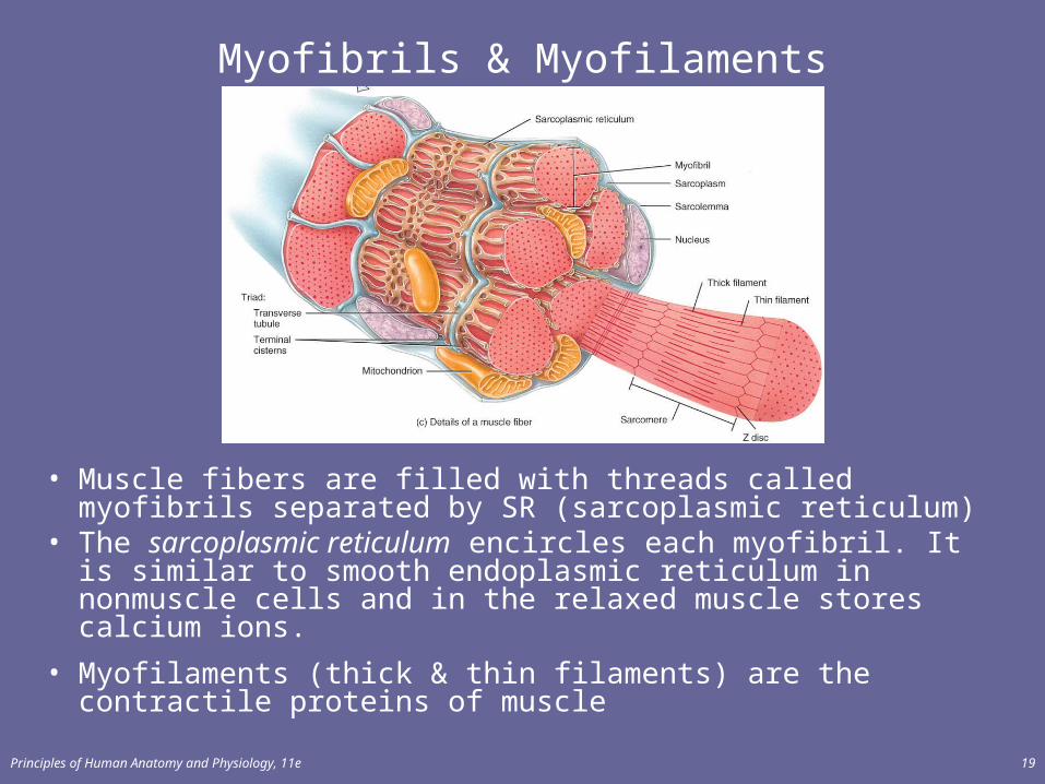
• Muscle fibers are filled with threads called myofibrils separated by SR (sarcoplasmic reticulum)
• The sarcoplasmic reticulum encircles each myofibril. It is similar to smooth endoplasmic reticulum in nonmuscle cells and in the relaxed muscle stores calcium ions.
• Myofilaments (thick & thin filaments) are the contractile proteins of muscle
Principles of Human Anatomy and Physiology, 11e 20
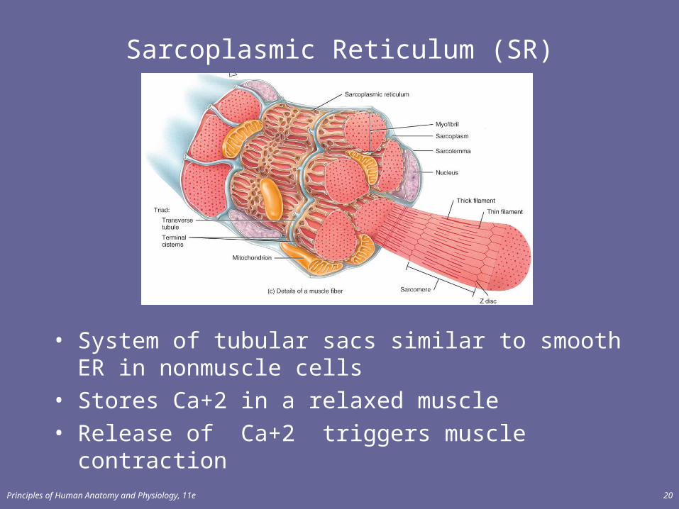
Sarcoplasmic Reticulum (SR)
• System of tubular sacs similar to smooth ER in nonmuscle cells
• Stores Ca+2 in a relaxed muscle• Release of Ca+2 triggers muscle contraction
Principles of Human Anatomy and Physiology, 11e 21
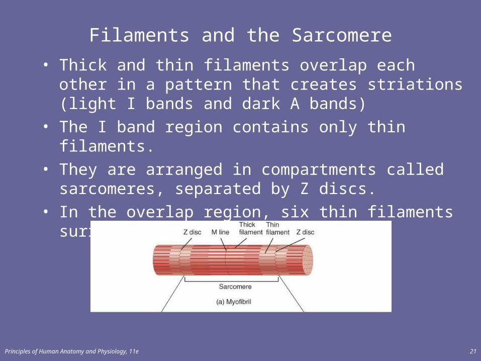
Filaments and the Sarcomere
• Thick and thin filaments overlap each other in a pattern that creates striations (light I bands and dark A bands)
• The I band region contains only thin filaments.• They are arranged in compartments called sarcomeres,
separated by Z discs.• In the overlap region, six thin filaments surround each
thick filament
Principles of Human Anatomy and Physiology, 11e 22
Sarcomere
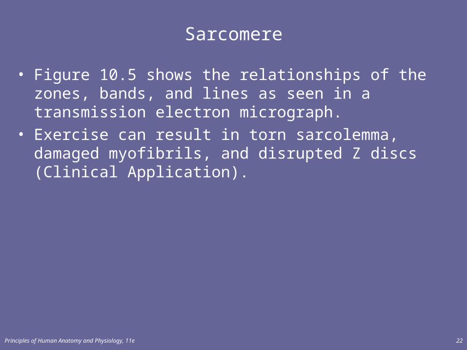
• Figure 10.5 shows the relationships of the zones, bands, and lines as seen in a transmission electron micrograph.
• Exercise can result in torn sarcolemma, damaged myofibrils, and disrupted Z discs (Clinical Application).
Principles of Human Anatomy and Physiology, 11e 23
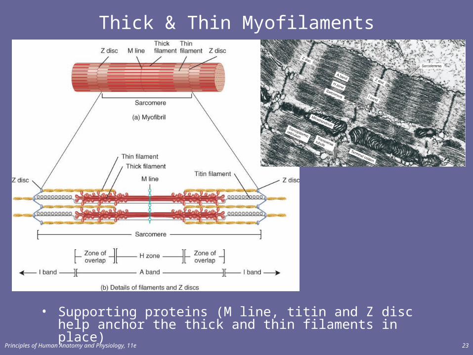
Thick & Thin Myofilaments
• Supporting proteins (M line, titin and Z disc help anchor the thick and thin filaments in place)
Principles of Human Anatomy and Physiology, 11e 24
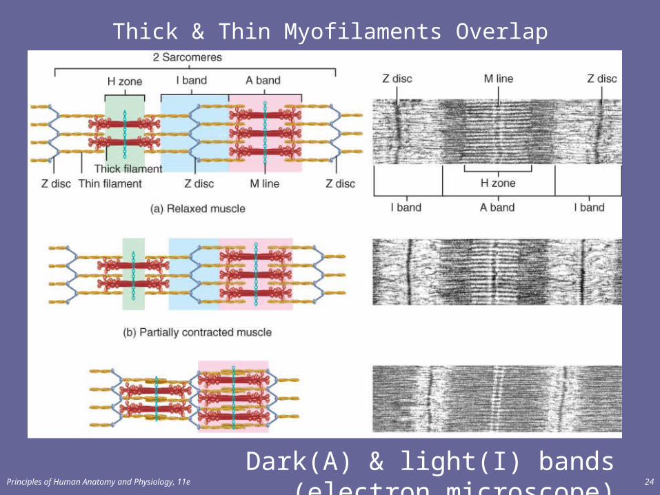
Thick & Thin Myofilaments Overlap
Dark(A) & light(I) bands (electron microscope)
Principles of Human Anatomy and Physiology, 11e 25
Muscle Proteins
Principles of Human Anatomy and Physiology, 11e 26
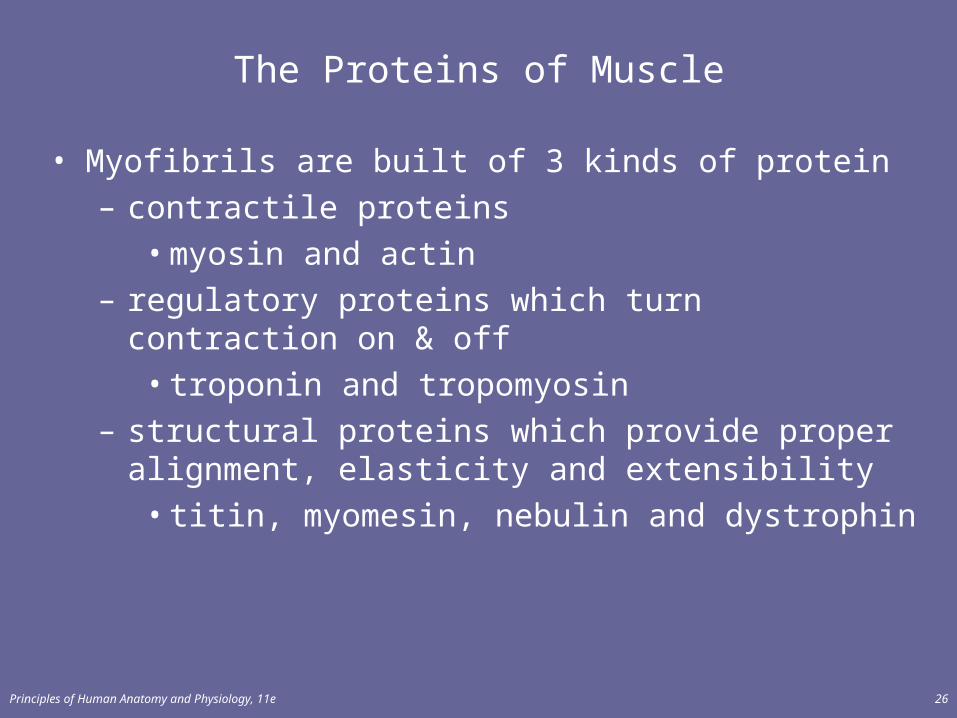
The Proteins of Muscle
• Myofibrils are built of 3 kinds of protein– contractile proteins
• myosin and actin– regulatory proteins which turn contraction on & off
• troponin and tropomyosin– structural proteins which provide proper alignment,
elasticity and extensibility• titin, myomesin, nebulin and dystrophin
Principles of Human Anatomy and Physiology, 11e 27
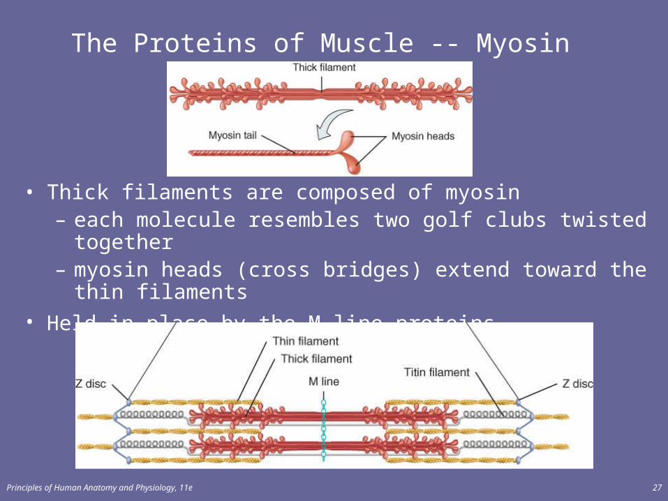
The Proteins of Muscle -- Myosin
• Thick filaments are composed of myosin – each molecule resembles two golf clubs twisted together– myosin heads (cross bridges) extend toward the thin
filaments
• Held in place by the M line proteins.
Principles of Human Anatomy and Physiology, 11e 28
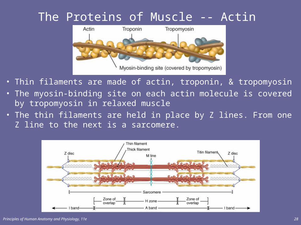
The Proteins of Muscle -- Actin
• Thin filaments are made of actin, troponin, & tropomyosin
• The myosin-binding site on each actin molecule is covered by tropomyosin in relaxed muscle
• The thin filaments are held in place by Z lines. From one Z line to the next is a sarcomere.
Principles of Human Anatomy and Physiology, 11e 29
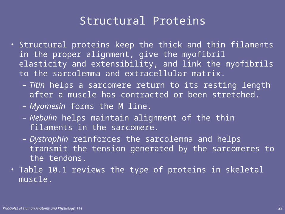
Structural Proteins
• Structural proteins keep the thick and thin filaments in the proper alignment, give the myofibril elasticity and extensibility, and link the myofibrils to the sarcolemma and extracellular matrix.– Titin helps a sarcomere return to its resting length after a
muscle has contracted or been stretched.– Myomesin forms the M line.– Nebulin helps maintain alignment of the thin filaments in
the sarcomere.– Dystrophin reinforces the sarcolemma and helps transmit
the tension generated by the sarcomeres to the tendons.• Table 10.1 reviews the type of proteins in skeletal muscle.
Principles of Human Anatomy and Physiology, 11e 30
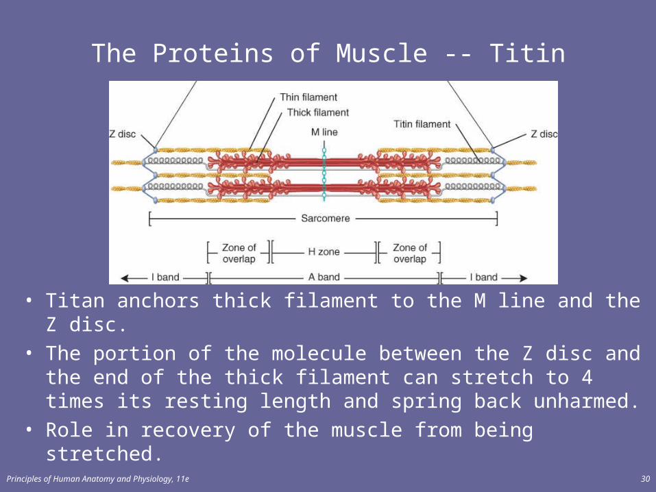
The Proteins of Muscle -- Titin
• Titan anchors thick filament to the M line and the Z disc.• The portion of the molecule between the Z disc and the end of
the thick filament can stretch to 4 times its resting length and spring back unharmed.
• Role in recovery of the muscle from being stretched.
Principles of Human Anatomy and Physiology, 11e 31
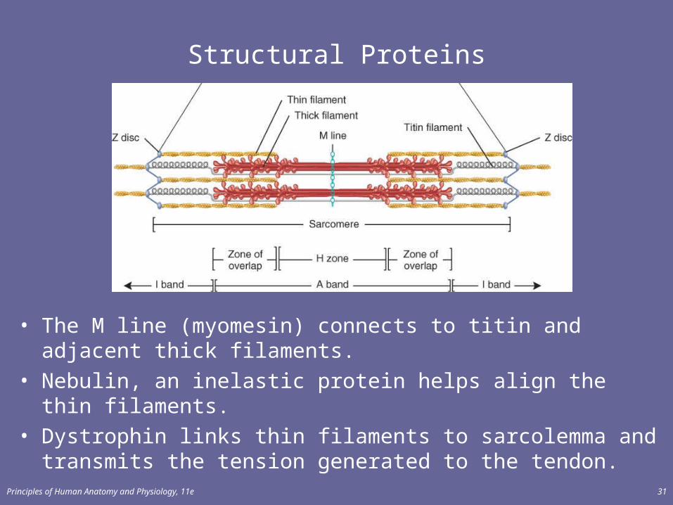
Structural Proteins
• The M line (myomesin) connects to titin and adjacent thick filaments.
• Nebulin, an inelastic protein helps align the thin filaments.• Dystrophin links thin filaments to sarcolemma and transmits
the tension generated to the tendon.
Principles of Human Anatomy and Physiology, 11e 32
Sliding Filament Mechanism Of Contraction
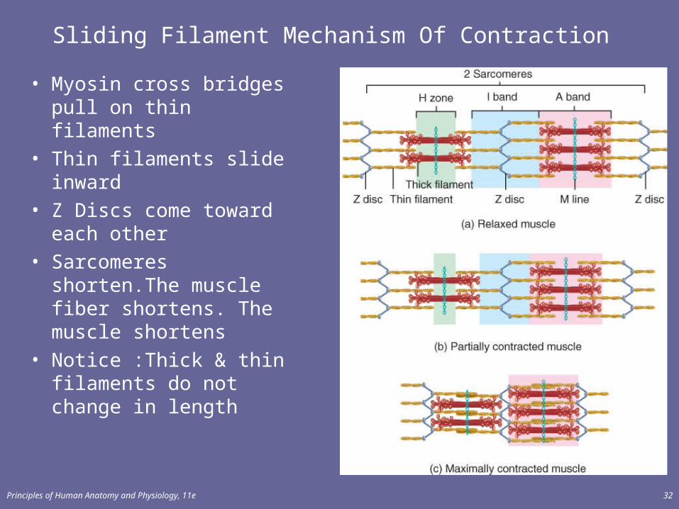
• Myosin cross bridgespull on thin filaments
• Thin filaments slide inward
• Z Discs come toward each other
• Sarcomeres shorten.The muscle fiber shortens. The muscle shortens
• Notice :Thick & thin filaments do not change in length

Principles of Human Anatomy and Physiology, 11e 33
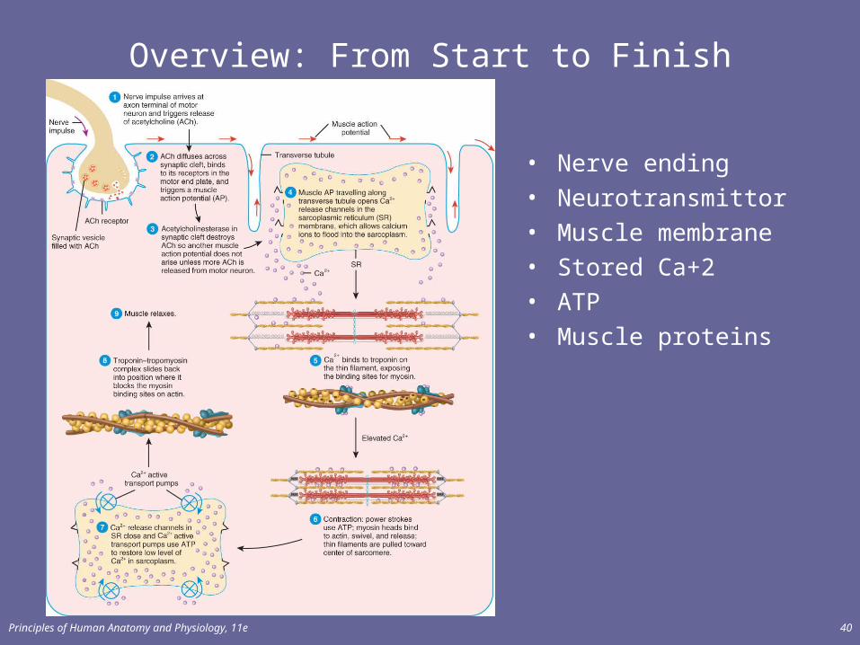
Overview: From Start to Finish
Basic Structures• Nerve ending• Neurotransmitter• Muscle membrane• Stored Ca+2• ATP• Muscle proteins
Principles of Human Anatomy and Physiology, 11e 34
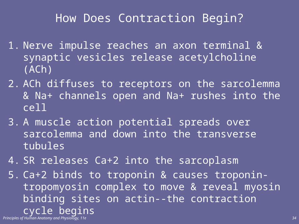
How Does Contraction Begin?
1. Nerve impulse reaches an axon terminal & synaptic vesicles release acetylcholine (ACh)
2. ACh diffuses to receptors on the sarcolemma & Na+ channels open and Na+ rushes into the cell
3. A muscle action potential spreads over sarcolemma and down into the transverse tubules
4. SR releases Ca+2 into the sarcoplasm
5. Ca+2 binds to troponin & causes troponin-tropomyosin complex to move & reveal myosin binding sites on actin--the contraction cycle begins

Principles of Human Anatomy and Physiology, 11e 35
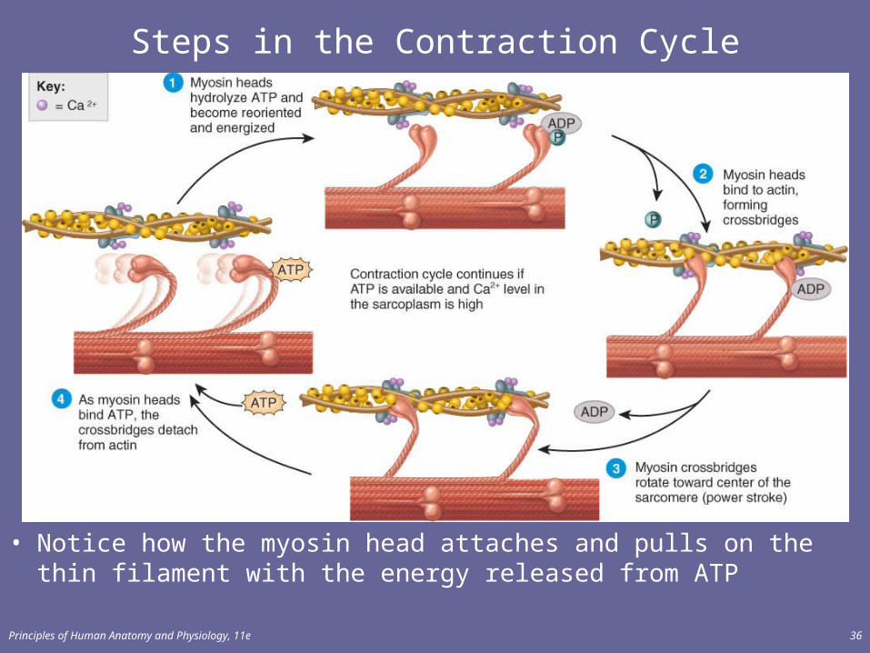
Contraction Cycle
• Repeating sequence of events that cause the thick & thin filaments to move past each other.
• 4 steps to contraction cycle– ATP hydrolysis– attachment of myosin to actin to form crossbridges– power stroke– detachment of myosin from actin
• Cycle keeps repeating as long as there is ATP available & there is a high Ca+2 level near the filaments.
Principles of Human Anatomy and Physiology, 11e 36
Steps in the Contraction Cycle
• Notice how the myosin head attaches and pulls on the thin filament with the energy released from ATP
Principles of Human Anatomy and Physiology, 11e 37
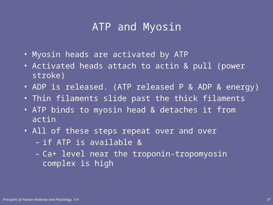
ATP and Myosin
• Myosin heads are activated by ATP• Activated heads attach to actin & pull (power stroke)• ADP is released. (ATP released P & ADP & energy)• Thin filaments slide past the thick filaments• ATP binds to myosin head & detaches it from actin• All of these steps repeat over and over
– if ATP is available &– Ca+ level near the troponin-tropomyosin complex is
high
Principles of Human Anatomy and Physiology, 11e 38
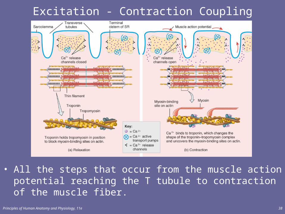
Excitation - Contraction Coupling
• All the steps that occur from the muscle action potential reaching the T tubule to contraction of the muscle fiber.
Principles of Human Anatomy and Physiology, 11e 39
Relaxation• Acetylcholinesterase (AChE) breaks down ACh within the
synaptic cleft• Muscle action potential ceases• Ca+2 release channels close• Active transport pumps Ca2+ back into storage in the
sarcoplasmic reticulum• Calcium-binding protein (calsequestrin) helps hold Ca+2 in
SR (Ca+2 concentration 10,000 times higher than in cytosol)• Tropomyosin-troponin complex recovers binding site on the
actin
Principles of Human Anatomy and Physiology, 11e 40
Overview: From Start to Finish
• Nerve ending• Neurotransmittor• Muscle membrane• Stored Ca+2• ATP• Muscle proteins
Principles of Human Anatomy and Physiology, 11e 41
Length-Tension Relationship
• The forcefulness of muscle contraction depends on the length of the sarcomeres within a muscle before contraction begins.
• Figure 10.10 plots the length-tension relationships for skeletal muscle.
Principles of Human Anatomy and Physiology, 11e 42
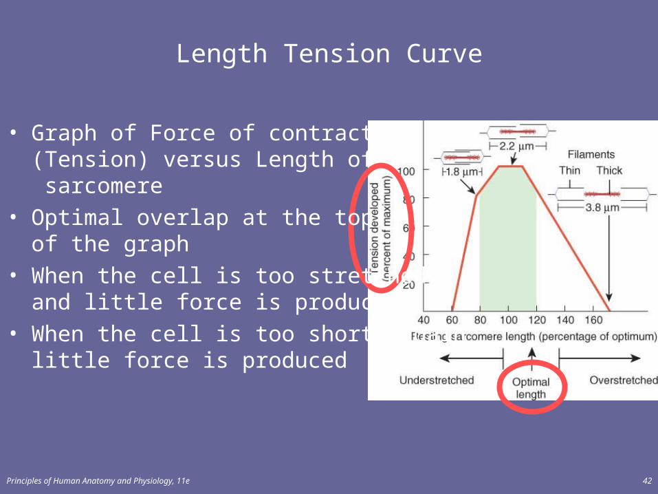
Length Tension Curve
• Graph of Force of contraction(Tension) versus Length of sarcomere
• Optimal overlap at the topof the graph
• When the cell is too stretchedand little force is produced
• When the cell is too short, againlittle force is produced
Principles of Human Anatomy and Physiology, 11e 43
• Optimal overlap of thick & thin filaments– produces greatest number of crossbridges and the
greatest amount of tension• As stretch muscle (past optimal length)
– fewer cross bridges exist & less force is produced • If muscle is overly shortened (less than optimal)
– fewer cross bridges exist & less force is produced– thick filaments crumpled by Z discs
• Normally– resting muscle length remains between 70 to 130% of the
optimum
Length of Muscle Fibers
Principles of Human Anatomy and Physiology, 11e 44
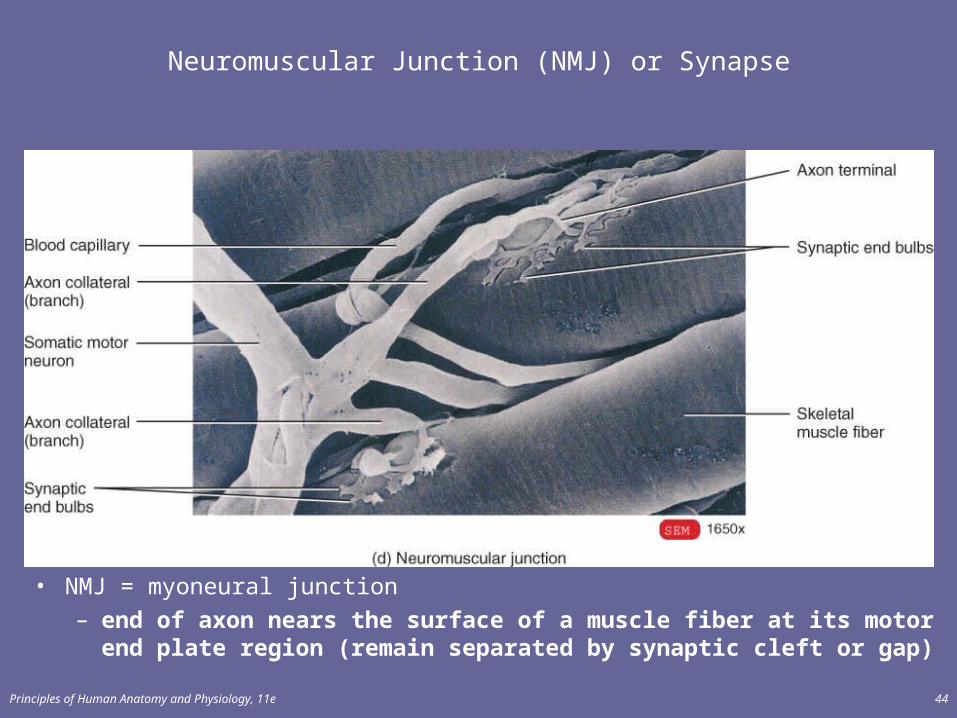
Neuromuscular Junction (NMJ) or Synapse
• NMJ = myoneural junction– end of axon nears the surface of a muscle fiber at its motor end
plate region (remain separated by synaptic cleft or gap)
Principles of Human Anatomy and Physiology, 11e 45
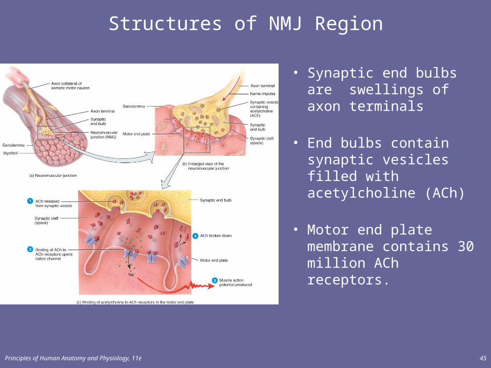
Structures of NMJ Region
• Synaptic end bulbs are swellings of axon terminals
• End bulbs contain synaptic vesicles filled with acetylcholine (ACh)
• Motor end plate membrane contains 30 million ACh receptors.
Principles of Human Anatomy and Physiology, 11e 46
Events Occurring After a Nerve Signal
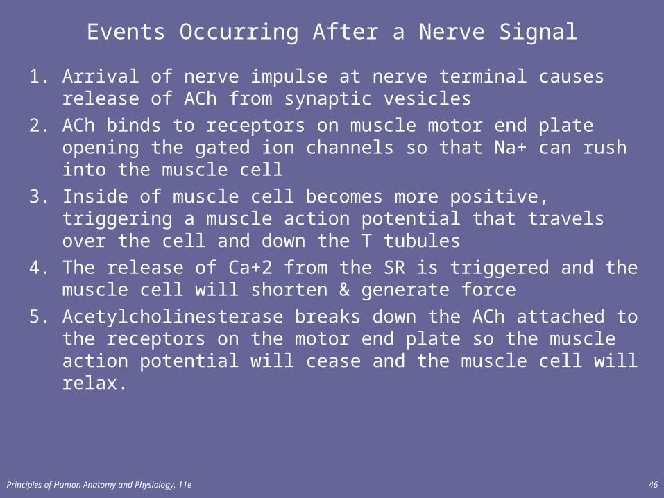
1. Arrival of nerve impulse at nerve terminal causes release of ACh from synaptic vesicles
2. ACh binds to receptors on muscle motor end plate opening the gated ion channels so that Na+ can rush into the muscle cell
3. Inside of muscle cell becomes more positive, triggering a muscle action potential that travels over the cell and down the T tubules
4. The release of Ca+2 from the SR is triggered and the muscle cell will shorten & generate force
5. Acetylcholinesterase breaks down the ACh attached to the receptors on the motor end plate so the muscle action potential will cease and the muscle cell will relax.
Principles of Human Anatomy and Physiology, 11e 47
Pharmacology of the NMJ
• Botulinum toxin blocks release of neurotransmitter at the NMJ so muscle contraction can not occur– bacteria found in improperly canned food– death occurs from paralysis of the diaphragm
• Curare (plant poison from poison arrows)– causes muscle paralysis by blocking the ACh receptors – used to relax muscle during surgery
• Neostigmine (anticholinesterase agent)– blocks removal of ACh from receptors so strengthens weak
muscle contractions of myasthenia gravis– also an antidote for curare after surgery is finished
Principles of Human Anatomy and Physiology, 11e 48
Muscle MetabolismProduction of ATP in Muscle Fibers
• Muscle uses ATP at a great rate when active• Sarcoplasmic ATP only lasts for few seconds• 3 sources of ATP production within muscle
– creatine phosphate– anaerobic cellular respiration– aerobic cellular respiration
Principles of Human Anatomy and Physiology, 11e 49
MUSCLE METABOLISM
• Creatine phosphate and ATP can power maximal muscle contraction for about 15 seconds and is used for maximal short bursts of energy (e.g., 100-meter dash) (Figure 10.13a).– Creatine phosphate is unique to muscle fibers.
Principles of Human Anatomy and Physiology, 11e 50
MUSCLE METABOLISM
• The partial catabolism of glucose to generate ATP occurs in anaerobic cellular respiration (Figure 10.13b). This system can provide enough energy for about 30-40 seconds of maximal muscle activity (e.g., 300-meter race).
• Muscular activity lasting more than 30 seconds depends increasingly on aerobic cellular respiration (reactions requiring oxygen). This system of ATP production involves the complete oxidation of glucose via cellular respiration (biological oxidation) (Figure 10.13c).
Principles of Human Anatomy and Physiology, 11e 51
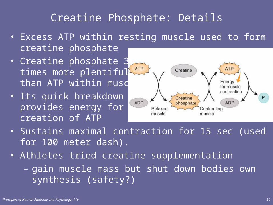
Creatine Phosphate: Details
• Excess ATP within resting muscle used to form creatine phosphate
• Creatine phosphate 3-6 times more plentiful than ATP within muscle
• Its quick breakdownprovides energy for creation of ATP
• Sustains maximal contraction for 15 sec (used for 100 meter dash).
• Athletes tried creatine supplementation – gain muscle mass but shut down bodies own synthesis
(safety?)
Principles of Human Anatomy and Physiology, 11e 52
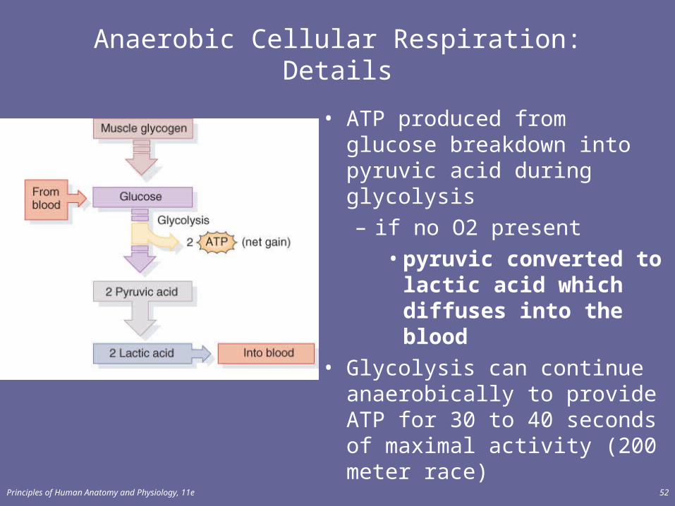
Anaerobic Cellular Respiration: Details
• ATP produced from glucose breakdown into pyruvic acid during glycolysis – if no O2 present
• pyruvic converted to lactic acid which diffuses into the blood
• Glycolysis can continue anaerobically to provide ATP for 30 to 40 seconds of maximal activity (200 meter race)
Principles of Human Anatomy and Physiology, 11e 53
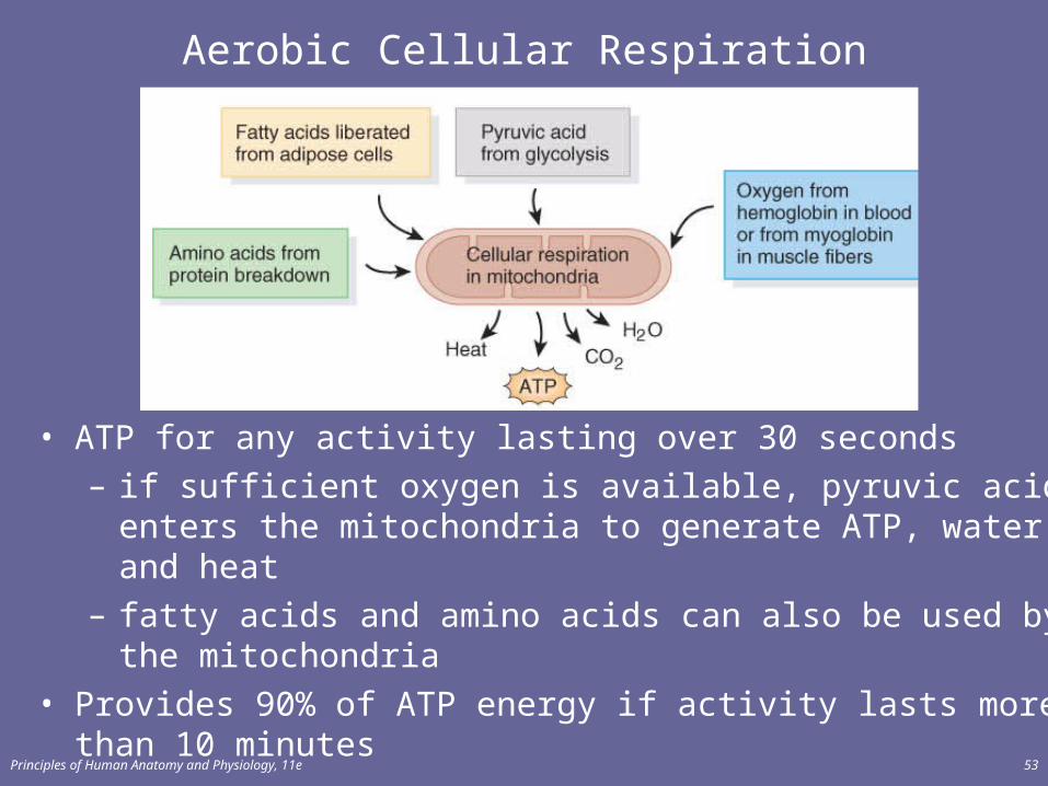
Aerobic Cellular Respiration
• ATP for any activity lasting over 30 seconds – if sufficient oxygen is available, pyruvic acid enters the
mitochondria to generate ATP, water and heat– fatty acids and amino acids can also be used by the
mitochondria• Provides 90% of ATP energy if activity lasts more than 10
minutes
Principles of Human Anatomy and Physiology, 11e 54
Muscle Fatigue
• Inability to contract after prolonged activity• Factors that contribute to fatigue
– central fatigue is feeling of tiredness and a desire to stop (protective mechanism)
– insufficient release of acetylcholine from motor neurons
– depletion of creatine phosphate– decline of Ca+2 within the sarcoplasm– insufficient oxygen or glycogen– buildup of lactic acid and ADP
Principles of Human Anatomy and Physiology, 11e 55
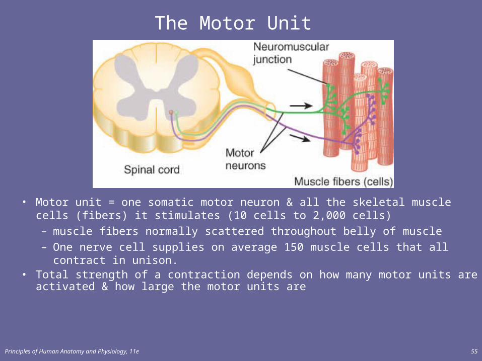
The Motor Unit
• Motor unit = one somatic motor neuron & all the skeletal muscle cells (fibers) it stimulates (10 cells to 2,000 cells)– muscle fibers normally scattered throughout belly of muscle– One nerve cell supplies on average 150 muscle cells that all contract in unison.
• Total strength of a contraction depends on how many motor units are activated & how large the motor units are
Principles of Human Anatomy and Physiology, 11e 56
CONTROL OF MUSCLE TENSION
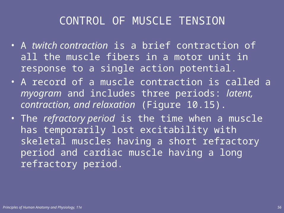
• A twitch contraction is a brief contraction of all the muscle fibers in a motor unit in response to a single action potential.
• A record of a muscle contraction is called a myogram and includes three periods: latent, contraction, and relaxation (Figure 10.15).
• The refractory period is the time when a muscle has temporarily lost excitability with skeletal muscles having a short refractory period and cardiac muscle having a long refractory period.
Principles of Human Anatomy and Physiology, 11e 57
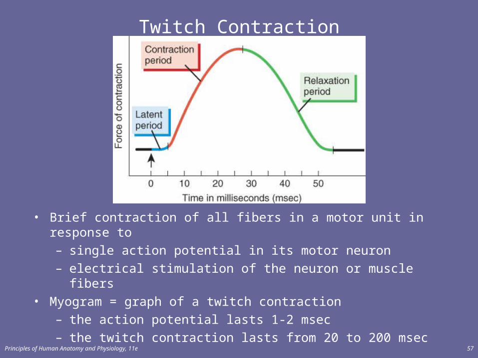
Twitch Contraction
• Brief contraction of all fibers in a motor unit in response to – single action potential in its motor neuron– electrical stimulation of the neuron or muscle fibers
• Myogram = graph of a twitch contraction– the action potential lasts 1-2 msec– the twitch contraction lasts from 20 to 200 msec
Principles of Human Anatomy and Physiology, 11e 58
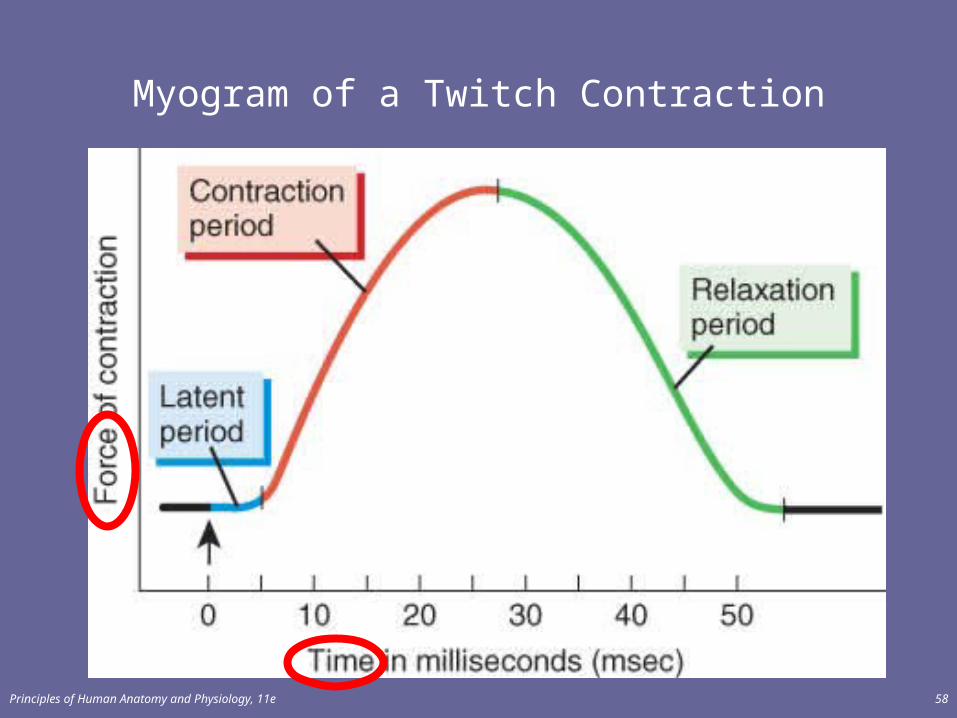
Myogram of a Twitch Contraction
Principles of Human Anatomy and Physiology, 11e 59
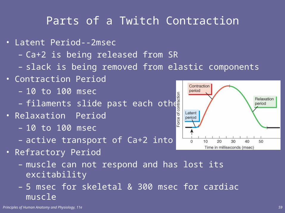
Parts of a Twitch Contraction
• Latent Period--2msec– Ca+2 is being released from SR– slack is being removed from elastic components
• Contraction Period– 10 to 100 msec– filaments slide past each other
• Relaxation Period– 10 to 100 msec – active transport of Ca+2 into SR
• Refractory Period– muscle can not respond and has lost its excitability– 5 msec for skeletal & 300 msec for cardiac muscle
Principles of Human Anatomy and Physiology, 11e 60
Frequency of Stimulation
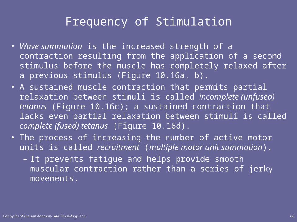
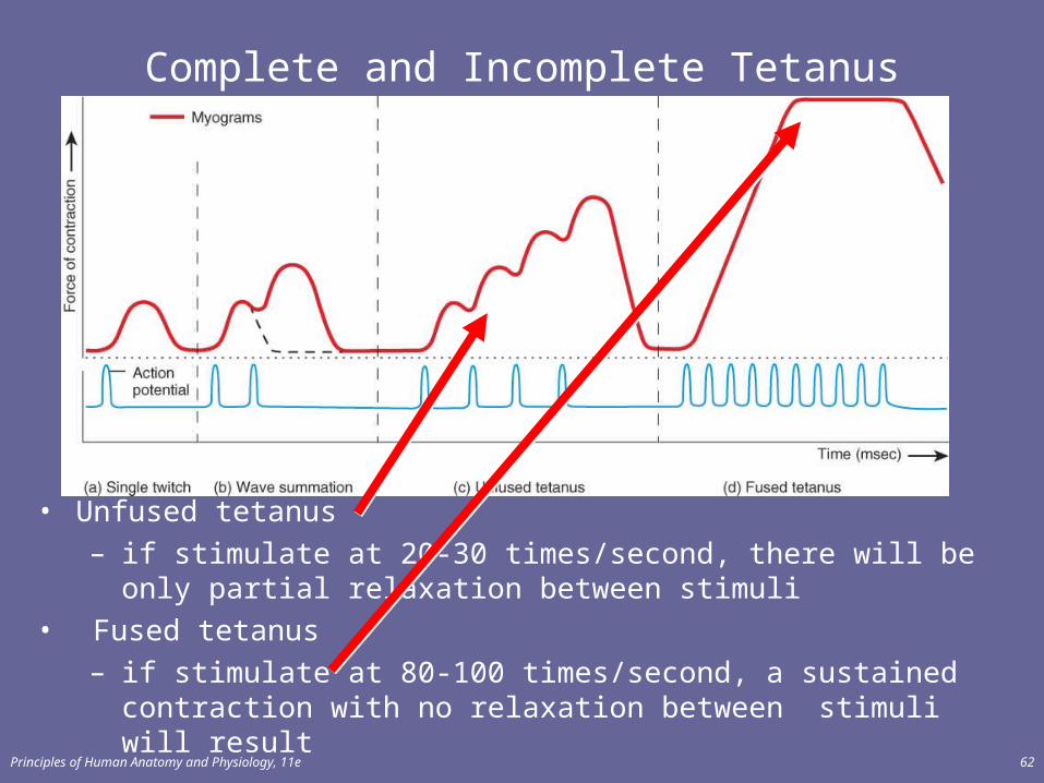
• Wave summation is the increased strength of a contraction resulting from the application of a second stimulus before the muscle has completely relaxed after a previous stimulus (Figure 10.16a, b).
• A sustained muscle contraction that permits partial relaxation between stimuli is called incomplete (unfused) tetanus (Figure 10.16c); a sustained contraction that lacks even partial relaxation between stimuli is called complete (fused) tetanus (Figure 10.16d).
• The process of increasing the number of active motor units is called recruitment (multiple motor unit summation).– It prevents fatigue and helps provide smooth muscular
contraction rather than a series of jerky movements.

Principles of Human Anatomy and Physiology, 11e 61
Wave Summation
• If second stimulation applied after the refractory period but before complete muscle relaxation---second contraction is stronger than first
Principles of Human Anatomy and Physiology, 11e 62
Complete and Incomplete Tetanus
• Unfused tetanus– if stimulate at 20-30 times/second, there will be only partial
relaxation between stimuli• Fused tetanus
– if stimulate at 80-100 times/second, a sustained contraction with no relaxation between stimuli will result
Principles of Human Anatomy and Physiology, 11e 63
Explanation of Summation & Tetanus
• Wave summation & both types of tetanus result from Ca+2 remaining in the sarcoplasm
• Force of 2nd contraction is easily added to the first, because the elastic elements remain partially contracted and do not delay the beginning of the next contraction
Principles of Human Anatomy and Physiology, 11e 64
Motor Unit Recruitment
• Motor units in a whole muscle fire asynchronously– some fibers are active others are relaxed – delays muscle fatigue so contraction can be sustained
• Produces smooth muscular contraction– not series of jerky movements
• Precise movements require smaller contractions– motor units must be smaller (less fibers/nerve)
• Large motor units are active when large tension is needed
Principles of Human Anatomy and Physiology, 11e 65
Muscle Tone
• Involuntary contraction of a small number of motor units (alternately active and inactive in a constantly shifting pattern)– keeps muscles firm even though relaxed– does not produce movement
• Essential for maintaining posture (head upright)• Important in maintaining blood pressure
– tone of smooth muscles in walls of blood vessels
Principles of Human Anatomy and Physiology, 11e 66
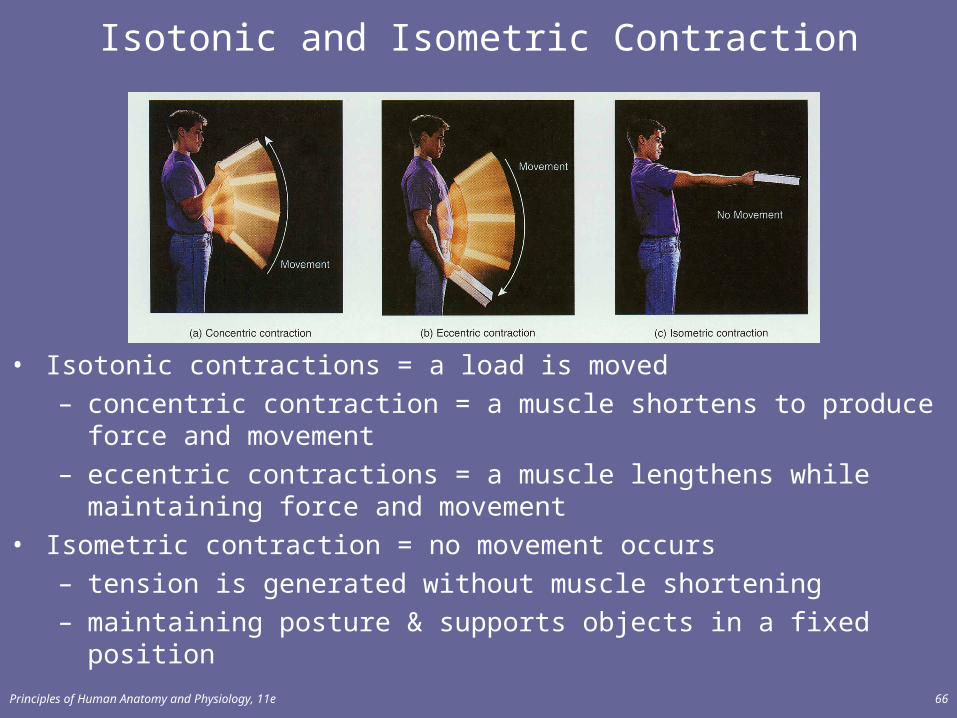
Isotonic and Isometric Contraction
• Isotonic contractions = a load is moved – concentric contraction = a muscle shortens to produce force and
movement– eccentric contractions = a muscle lengthens while maintaining force and
movement• Isometric contraction = no movement occurs
– tension is generated without muscle shortening– maintaining posture & supports objects in a fixed position

Principles of Human Anatomy and Physiology, 11e 67
TYPES OF SKELETAL MSUCLE FIBERS
• On the basis of structure and function, skeletal muscle fibers are classified as – slow oxidative, – oxidative-glycolytic, or – fast glycolytic fibers.
Principles of Human Anatomy and Physiology, 11e 68
Variations in Skeletal Muscle Fibers
• Myoglobin, mitochondria and capillaries– red muscle fibers
• more myoglobin, an oxygen-storing reddish pigment • more capillaries and mitochondria
– white muscle fibers• less myoglobin and less capillaries give fibers their
pale color• Contraction and relaxation speeds vary
– how fast myosin ATPase hydrolyzes ATP• Resistance to fatigue
– different metabolic reactions used to generate ATP
Principles of Human Anatomy and Physiology, 11e 69
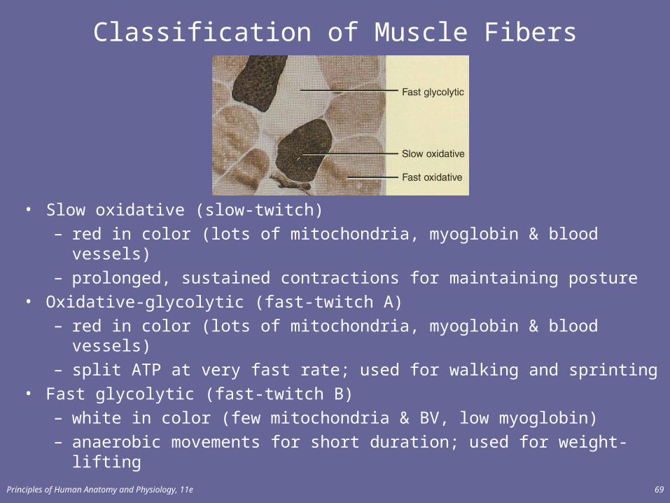
Classification of Muscle Fibers
• Slow oxidative (slow-twitch)– red in color (lots of mitochondria, myoglobin & blood vessels)– prolonged, sustained contractions for maintaining posture
• Oxidative-glycolytic (fast-twitch A)– red in color (lots of mitochondria, myoglobin & blood vessels)– split ATP at very fast rate; used for walking and sprinting
• Fast glycolytic (fast-twitch B)– white in color (few mitochondria & BV, low myoglobin)– anaerobic movements for short duration; used for weight-lifting
Principles of Human Anatomy and Physiology, 11e 70
Fiber Types within a Whole Muscle
• Most muscles contain a mixture of all three fiber types• Proportions vary with the usual action of the muscle
– neck, back and leg muscles have a higher proportion of postural, slow oxidative fibers
– shoulder and arm muscles have a higher proportion of fast glycolytic fibers
• All fibers of any one motor unit are same.• Different fibers are recruited as needed.
Principles of Human Anatomy and Physiology, 11e 71
Distribution and Recruitment of Different Types of Fibers
• Although the number of different skeletal muscle fibers does not change, the characteristics of those present can be altered by various types of exercise.
• The use of anabolic steroids by athletes to increase muscle size, strength, and endurance has been shown to have very serious side effects, some of which are life-threatening.
Principles of Human Anatomy and Physiology, 11e 72
Anabolic Steroids
• Similar to testosterone• Increases muscle size, strength, and endurance• side effects:
– liver cancer– kidney damage– heart disease– mood swings– facial hair & voice deepening in females– atrophy of testicles & baldness in males
Principles of Human Anatomy and Physiology, 11e 73
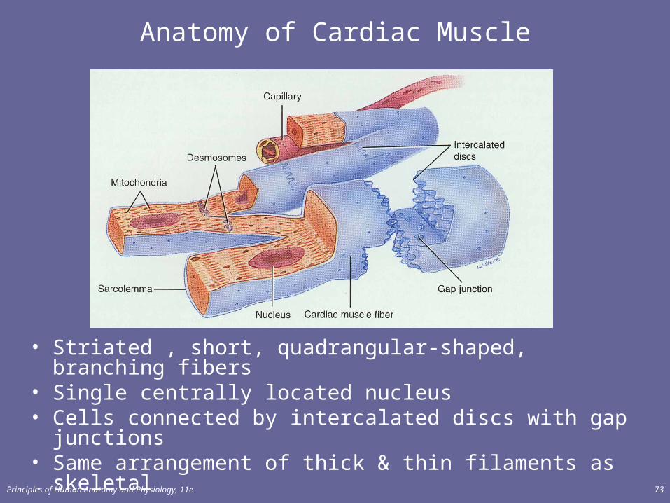
Anatomy of Cardiac Muscle
• Striated , short, quadrangular-shaped, branching fibers • Single centrally located nucleus• Cells connected by intercalated discs with gap junctions• Same arrangement of thick & thin filaments as skeletal
Principles of Human Anatomy and Physiology, 11e 74
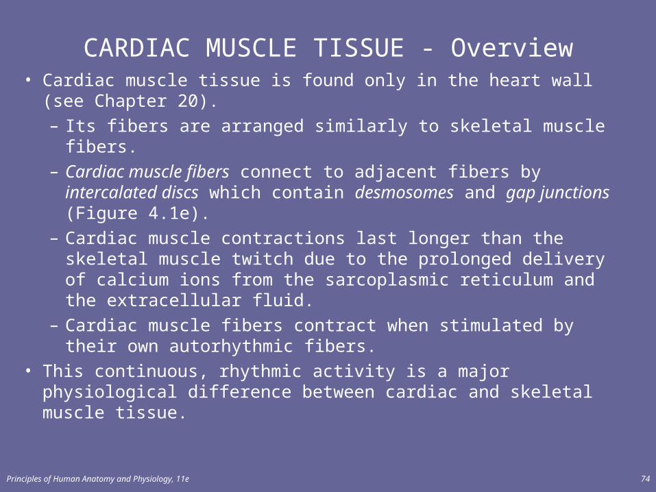
CARDIAC MUSCLE TISSUE - Overview• Cardiac muscle tissue is found only in the heart wall (see
Chapter 20).– Its fibers are arranged similarly to skeletal muscle fibers.– Cardiac muscle fibers connect to adjacent fibers by
intercalated discs which contain desmosomes and gap junctions (Figure 4.1e).
– Cardiac muscle contractions last longer than the skeletal muscle twitch due to the prolonged delivery of calcium ions from the sarcoplasmic reticulum and the extracellular fluid.
– Cardiac muscle fibers contract when stimulated by their own autorhythmic fibers.
• This continuous, rhythmic activity is a major physiological difference between cardiac and skeletal muscle tissue.
Principles of Human Anatomy and Physiology, 11e 75
Cardiac versus Skeletal Muscle
• More sarcoplasm and mitochondria• Larger transverse tubules located at Z discs, rather
than at A-l band junctions• Less well-developed SR• Limited intracellular Ca+2 reserves
– more Ca+2 enters cell from extracellular fluid during contraction
• Prolonged delivery of Ca+2 to sarcoplasm, produces a contraction that last 10 -15 times longer than in skeletal muscle
Principles of Human Anatomy and Physiology, 11e 76
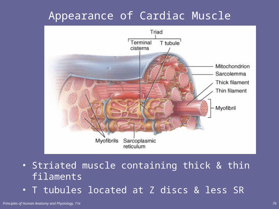
Appearance of Cardiac Muscle
• Striated muscle containing thick & thin filaments• T tubules located at Z discs & less SR

Principles of Human Anatomy and Physiology, 11e 77
Physiology of Cardiac Muscle
• Autorhythmic cells– contract without stimulation
• Contracts 75 times per min & needs lots of O2
• Larger mitochondria generate ATP aerobically • Extended contraction is possible due to slow Ca+2 delivery
– Ca+2 channels to the extracellular fluid stay open
Principles of Human Anatomy and Physiology, 11e 78
SMOOTH MUSCLE
• Smooth muscle tissue is nonstriated and involuntary and is classified into two types: visceral (single unit) smooth muscle (Figure 10.18a) and multiunit smooth muscle (Figure 10.18b).
– Visceral (single unit) smooth muscle is found in the walls of hollow viscera and small blood vessels; the fibers are arranged in a network and function as a “single unit.”
– Multiunit smooth muscle is found in large blood vessels, large airways, arrector pili muscles, and the iris of the eye. The fibers operate singly rather than as a unit.
Principles of Human Anatomy and Physiology, 11e 79
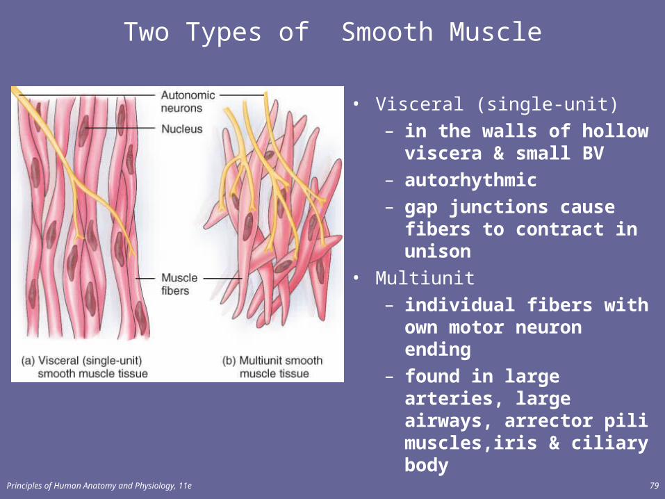
Two Types of Smooth Muscle
• Visceral (single-unit)– in the walls of hollow
viscera & small BV– autorhythmic– gap junctions cause fibers
to contract in unison• Multiunit
– individual fibers with own motor neuron ending
– found in large arteries, large airways, arrector pili muscles,iris & ciliary body
Principles of Human Anatomy and Physiology, 11e 80
Microscopic Anatomy of the Smooth Muscle
• Sarcoplasm of smooth muscle fibers contains both thick and thin filaments which are not organized into sarcomeres.
• Smooth muscle fibers contain intermediate filaments which are attached to dense bodies. (Figure 10.19)
Principles of Human Anatomy and Physiology, 11e 81
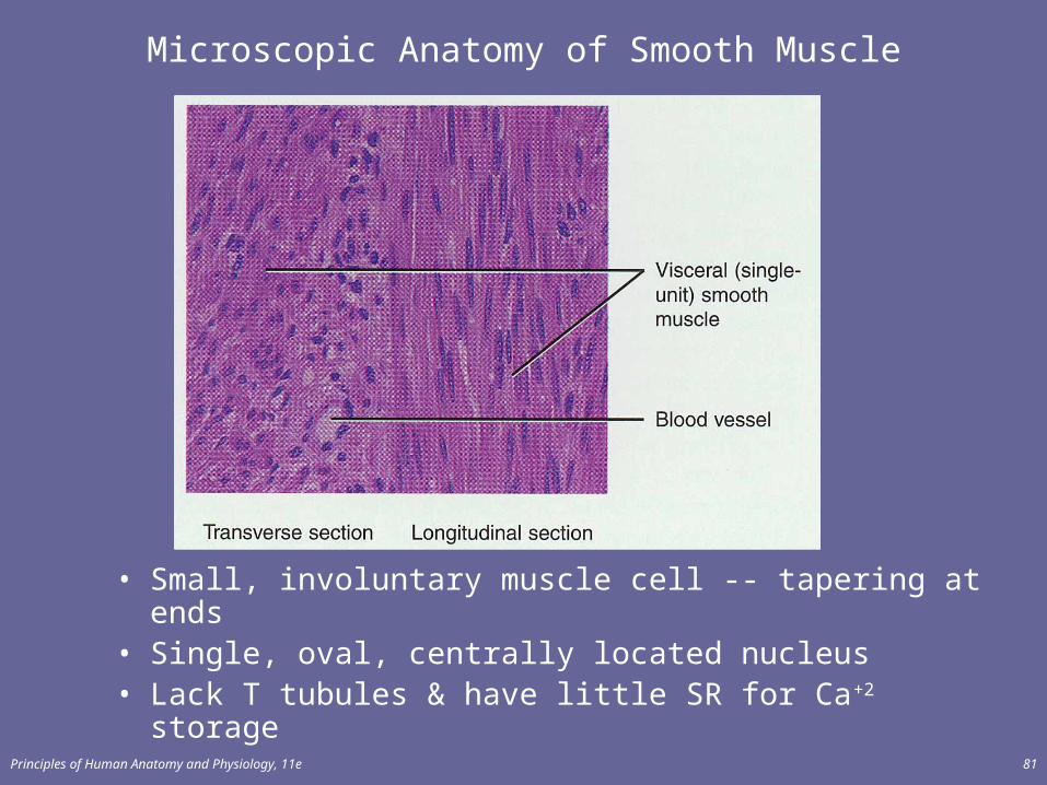
Microscopic Anatomy of Smooth Muscle
• Small, involuntary muscle cell -- tapering at ends• Single, oval, centrally located nucleus• Lack T tubules & have little SR for Ca+2 storage
Principles of Human Anatomy and Physiology, 11e 82
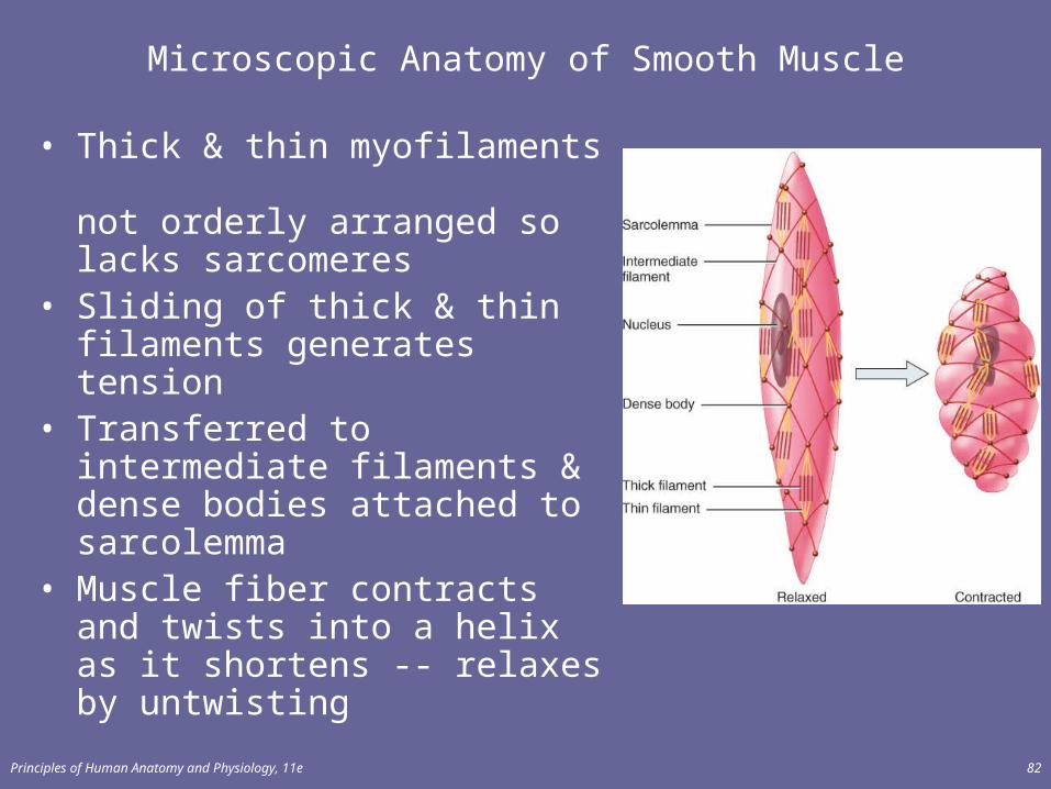
Microscopic Anatomy of Smooth Muscle
• Thick & thin myofilaments not orderly arranged so lacks sarcomeres
• Sliding of thick & thin filaments generates tension
• Transferred to intermediate filaments & dense bodies attached to sarcolemma
• Muscle fiber contracts and twists into a helix as it shortens -- relaxes by untwisting
Principles of Human Anatomy and Physiology, 11e 83
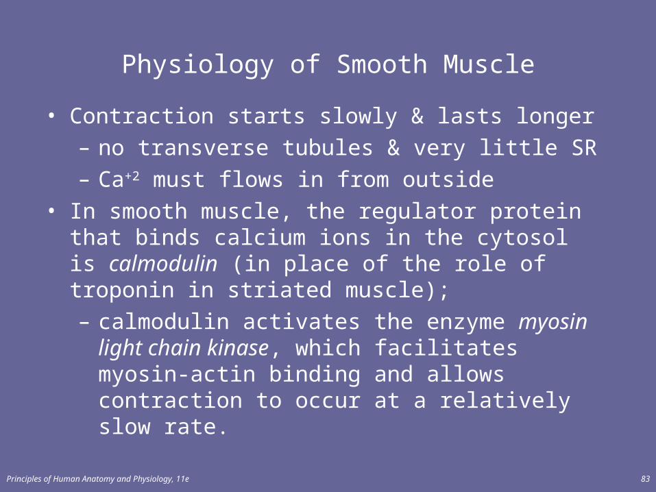
Physiology of Smooth Muscle
• Contraction starts slowly & lasts longer– no transverse tubules & very little SR– Ca+2 must flows in from outside
• In smooth muscle, the regulator protein that binds calcium ions in the cytosol is calmodulin (in place of the role of troponin in striated muscle); – calmodulin activates the enzyme myosin light chain
kinase, which facilitates myosin-actin binding and allows contraction to occur at a relatively slow rate.
Principles of Human Anatomy and Physiology, 11e 84
Smooth Muscle Tone
• The prolonged presence of calcium ions in the cytosol of smooth muscle fibers provides for smooth muscle tone, a state of continued partial contraction.
• Smooth muscle fibers can stretch considerably without developing tension; this phenomenon is termed the stress-relaxation response.
• Useful for maintaining blood pressure or a steady pressure on the contents of GI tract
Principles of Human Anatomy and Physiology, 11e 85
DEVELOPMENT OF MUSCLE
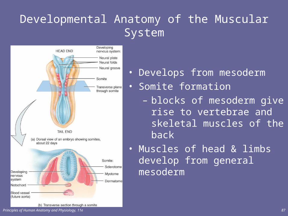
• With few exceptions, muscles develop from mesoderm (Figure 6.13a)
• Skeletal muscles of the head and extremities develop from general mesoderm; the remainder of the skeletal muscles develop from mesoderm of somites (Figure 10.20a).
Principles of Human Anatomy and Physiology, 11e 86
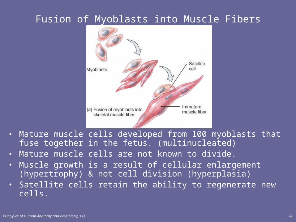
Fusion of Myoblasts into Muscle Fibers
• Mature muscle cells developed from 100 myoblasts that fuse together in the fetus. (multinucleated)
• Mature muscle cells are not known to divide.• Muscle growth is a result of cellular enlargement (hypertrophy) & not cell
division (hyperplasia)• Satellite cells retain the ability to regenerate new cells.
Principles of Human Anatomy and Physiology, 11e 87
Developmental Anatomy of the Muscular System
• Develops from mesoderm• Somite formation
– blocks of mesoderm give rise to vertebrae and skeletal muscles of the back
• Muscles of head & limbs develop from general mesoderm
Principles of Human Anatomy and Physiology, 11e 88
Regeneration of Muscle
• Skeletal muscle fibers cannot divide after 1st year– growth is enlargement of existing cells– repair
• satellite cells & bone marrow produce some new cells
• if not enough numbers---fibrosis occurs most often• Cardiac muscle fibers cannot divide or regenerate
– all healing is done by fibrosis (scar formation)• Smooth muscle fibers (regeneration is possible)
– cells can grow in size (hypertrophy)– some cells (uterus) can divide (hyperplasia)– new fibers can form from stem cells in BV walls
Principles of Human Anatomy and Physiology, 11e 89
Aging and Muscle Tissue
• Skeletal muscle starts to be replaced by fat beginning at 30 – “use it or lose it”
• Slowing of reflexes & decrease in maximal strength• Change in fiber type to slow oxidative fibers may be due to
lack of use or may be result of aging
Principles of Human Anatomy and Physiology, 11e 90
Myasthenia Gravis
• Progressive autoimmune disorder that blocks the ACh receptors at the neuromuscular junction
• The more receptors are damaged the weaker the muscle. • More common in women 20 to 40 with possible line to thymus
gland tumors• Begins with double vision & swallowing difficulties & progresses
to paralysis of respiratory muscles • Treatment includes steroids that reduce antibodies that bind to
ACh receptors and inhibitors of acetylcholinesterase
Principles of Human Anatomy and Physiology, 11e 91
Muscular Dystrophies
• Inherited, muscle-destroying diseases • Sarcolemma tears during muscle contraction• Mutated gene is on X chromosome so problem is with
males almost exclusively• Appears by age 5 in males and by 12 may be unable to
walk• Degeneration of individual muscle fibers produces
atrophy of the skeletal muscle• Gene therapy is hoped for with the most common form =
Duchenne muscular dystrophy
Principles of Human Anatomy and Physiology, 11e 92
Abnormal Contractions
• Spasm = involuntary contraction of single muscle• Cramp = a painful spasm• Tic = involuntary twitching of muscles normally under
voluntary control--eyelid or facial muscles• Tremor = rhythmic, involuntary contraction of
opposing muscle groups• Fasciculation = involuntary, brief twitch of a motor
unit visible under the skin
Principles of Human Anatomy and Physiology, 11e 93
Atrophy and Hypertrophy
• Atrophy– wasting away of muscles– caused by disuse (disuse atrophy) or severing of the nerve
supply (denervation atrophy)– the transition to connective tissue can not be reversed
• Hypertrophy– increase in the diameter of muscle fibers – resulting from very forceful, repetitive muscular activity and
an increase in myofibrils, SR & mitochondria
Principles of Human Anatomy and Physiology, 11e 94
Exercise-Induced Muscle Damage
• Intense exercise can cause muscle damage– electron micrographs reveal torn sarcolemmas,
damaged myofibrils an disrupted Z discs– increased blood levels of myoglobin & creatine
phosphate found only inside muscle cells• Delayed onset muscle soreness
– 12 to 48 Hours after strenuous exercise– stiffness, tenderness and swelling due to
microscopic cell damage
Principles of Human Anatomy and Physiology, 11e 95
Rigor Mortis
• Rigor mortis is a state of muscular rigidity that begins 3-4 hours after death and lasts about 24 hours
• After death, Ca+2 ions leak out of the SR and allow myosin heads to bind to actin
• Since ATP synthesis has ceased, crossbridges cannot detach from actin until proteolytic enzymes begin to digest the decomposing cells.

Principles of Human Anatomy and Physiology, 11e 96
end