Embed Size (px)
Citation preview

Principles and Technical Aspectsof PCR Amplifi cation
Elizabeth van Pelt-VerkuilAlex van BelkumJohn P. Hays
Principles and Technical Aspects of PCR Amplifi cation
Elizabeth van Pelt-VerkuilHogeschool Leiden, LeidenThe Netherlands
Alex van BelkumErasmus MC, RotterdamThe Netherlands
John P. HaysErasmus MC, RotterdamThe Netherlands
ISBN 978-1-4020-6240-7 e-ISBN 978-1-4020-6241-4DOI 10.1007/978-1-4020-6241-4
Library of Congress Control Number: 2007942548
© 2008 Springer Science + Business Media B.V.No part of this work may be reproduced, stored in a retrieval system, or transmitted in any form or by any means, electronic, mechanical, photocopying, microfilming, recording or otherwise, without written permission from the Publisher, with the exception of any material supplied specifically for the purpose of being entered and executed on a computer system, for exclusive use by the purchaser of the work.
Printed on acid-free paper
9 8 7 6 5 4 3 2 1
springer.com
Foreword
This book aims to provide an introduction to: (i) the concept of PCR, (ii) PCR technologies, (iii) PCR applications and (iv) PCR quality, with particular emphasis being placed upon PCR applications and techniques relevant to the clinical laboratory. This book will without doubt be useful as a reference work pertaining to technical aspects of PCR for all bio-medical students, research technicians, medics and scientists interested in the PCR technique and it’s applications per se.
Wherever possible, the authors have tried to provide figures, scientific publica-tions, or references to commercially available products, in order to illustrate any particularly important concepts or comments. Indeed, all commercial PCR bio-technology companies offer information about their products on internet sites and in online technical manuals. These online resources will be invaluable for any readers requiring more detailed PCR protocols.
The authors have provided references for many PCR concepts and applications that are directly useful to the clinical laboratory audience. These references provide a starting point for a more detailed investigation into the PCR techniques and con-cepts mentioned. In particular, further detailed information may be acquired by (i) referring to the reference section of cited publications, and (ii) by referring to other (more recent) publications published by the cited author(s).
Great efforts have been made to include descriptions of the vast majority of PCR applications and technologies that currently exist, though the dynamic nature of the PCR field means that no book can ever be regarded as totally inclusive of all the PCR refinements that have been (and are currently being) developed.
The writing of this book has been facilitated by the Hogeschool Leiden (The Netherlands), the Department of Medical Microbiology and Infectious Diseases at Erasmus MC (Rotterdam, The Netherlands) and an unrestricted financial grant donated by Roche Molecular Diagnostics (Almere, The Netherlands).
Finally, the authors would like to wish everyone success in their PCR-related studies!
April 2007 Elizabeth van Pelt-VerkuilAlex van Belkum
John P. Hays
v
Contents
Foreword . . . . . . . . . . . . . . . . . . . . . . . . . . . . . . . . . . . . . . . . . . . . . . . . . . . . . . . . v
Chapter 1 The Polymerase Chain Reaction . . . . . . . . . . . . . . . . . . . . . . . . . . 11.1 An Overview of the PCR Process . . . . . . . . . . . . . . . . . . . . . . . 11.2 Before PCR and Beyond . . . . . . . . . . . . . . . . . . . . . . . . . . . . . . 2
Chapter 2 A Brief Comparison Between In Vivo DNA Replication and In Vitro PCR Amplifi cation . . . . . . . . . . . . . . . . . . . . . . . . . . . 92.1 Nucleic Acid Targets . . . . . . . . . . . . . . . . . . . . . . . . . . . . . . . . . 9
2.1.1 DNA . . . . . . . . . . . . . . . . . . . . . . . . . . . . . . . . . . . . . . . 92.1.2 RNA . . . . . . . . . . . . . . . . . . . . . . . . . . . . . . . . . . . . . . 11
2.2 Target DNA Strand Separation and Primer Annealing . . . . . . 122.3 DNA Dependent DNA Polymerase and Oligonucleotide
Primers . . . . . . . . . . . . . . . . . . . . . . . . . . . . . . . . . . . . . . . . . . 132.4 Deoxyribonucleotides and Additional Factors . . . . . . . . . . . . 14
Chapter 3 The PCR in Practice . . . . . . . . . . . . . . . . . . . . . . . . . . . . . . . . . . . 173.1 Brief Overview of PCR Requirements . . . . . . . . . . . . . . . . . . 17
3.1.1 The PCR Reaction Mix . . . . . . . . . . . . . . . . . . . . . . . . 173.1.2 The PCR Thermocycling Regime . . . . . . . . . . . . . . . . 193.1.3 Analysis of PCR Amplifi cation Products . . . . . . . . . . 223.1.4 Miscellaneous Considerations . . . . . . . . . . . . . . . . . . 22
Chapter 4 The Different Types and Varieties of Nucleic Acid Target Molecules . . . . . . . . . . . . . . . . . . . . . . . . . . . . . . . . . . . . . . 254.1 General Features . . . . . . . . . . . . . . . . . . . . . . . . . . . . . . . . . . . 254.2 A Brief Description of In Vivo DNA and RNA Targets . . . . . 264.3 DNA Samples . . . . . . . . . . . . . . . . . . . . . . . . . . . . . . . . . . . . . 33
4.3.1 DNA Isolation Procedures . . . . . . . . . . . . . . . . . . . . . 344.3.2 Comments on Nucleic Acids in Specifi c
Sample Types . . . . . . . . . . . . . . . . . . . . . . . . . . . . . . . 394.4 RNA Samples . . . . . . . . . . . . . . . . . . . . . . . . . . . . . . . . . . . . . 44
4.4.1 Working Free of RNase Contamination . . . . . . . . . . . 45
vii
viii Contents
4.4.2 RNA Isolation for RT-PCR . . . . . . . . . . . . . . . . . . . . . 474.5 Reverse Transcription and RT-PCR . . . . . . . . . . . . . . . . . . . . 50
4.5.1 cDNA Synthesis . . . . . . . . . . . . . . . . . . . . . . . . . . . . . 534.5.2 cDNA Synthesis Using RACE . . . . . . . . . . . . . . . . . . 554.5.3 RNA Extraction and cDNA
Synthesis Controls . . . . . . . . . . . . . . . . . . . . . . . . . . . 57
Chapter 5 PCR Primers . . . . . . . . . . . . . . . . . . . . . . . . . . . . . . . . . . . . . . . . . 635.1 PCR Primer Design and Quality Requirements . . . . . . . . . . . 64
5.1.1 Different Primer Species . . . . . . . . . . . . . . . . . . . . . . 685.2 Primer Hybridisation (Annealing) . . . . . . . . . . . . . . . . . . . . . 765.3 Thermodynamic Approach of T
m Calculations . . . . . . . . . . . 78
5.4 Primer Synthesis . . . . . . . . . . . . . . . . . . . . . . . . . . . . . . . . . . 815.5 Non-radioactive Primer Labelling . . . . . . . . . . . . . . . . . . . . . 835.6 The Effect of Mismatches Between PCR
Primer and Target . . . . . . . . . . . . . . . . . . . . . . . . . . . . . . . . . . 865.7 Primer Concentration . . . . . . . . . . . . . . . . . . . . . . . . . . . . . . . 87
Chapter 6 Deoxynucleotide Triphosphates and Buffer Components . . . . . 916.1 Factors Affecting the Choice of dNTP Concentration . . . . . . 926.2 Modifi ed dNTPs and Their Applications . . . . . . . . . . . . . . . . 936.3 The PCR Buffer . . . . . . . . . . . . . . . . . . . . . . . . . . . . . . . . . . . 94
6.3.1 Monovalent Ions . . . . . . . . . . . . . . . . . . . . . . . . . . . . 986.3.2 Magnesium Ions . . . . . . . . . . . . . . . . . . . . . . . . . . . . . 99
Chapter 7 Taq and Other Thermostable DNA Polymerases . . . . . . . . . . . 1037.1 The Advantages and Disadvantages of Taq over
Klenow Fragment DNA Polymerase . . . . . . . . . . . . . . . . . . 1047.2 Misincorporation of Nucleotides and Fidelity
of DNA Synthesis by Taq Polymerase . . . . . . . . . . . . . . . . . 1077.3 Taq DNA Polymerase and Its Modifi cations . . . . . . . . . . . . 1107.4 Taq Polymerase Unit Defi nition and Working
Concentrations . . . . . . . . . . . . . . . . . . . . . . . . . . . . . . . . . . . 1137.5 Other Thermostable Polymerases
and Their Applications . . . . . . . . . . . . . . . . . . . . . . . . . . . . . 1137.6 Mixtures of Thermostable Polymerases . . . . . . . . . . . . . . . . 117
Chapter 8 Important Considerations for Typical, Quantitative and Real-Time PCR Protocols . . . . . . . . . . . . . . . . . . . . . . . . . . 119 8.1 The Typical PCR Amplifi cation Protocol . . . . . . . . . . . . . . . 119
8.1.1 Denaturation (Melting) of the Template DNA . . . . . 1218.1.2 Annealing (Hybridisation) of PCR Primers . . . . . . . 1228.1.3 Calculating the Primer Annealing
Temperature (Tm) . . . . . . . . . . . . . . . . . . . . . . . . . . . 124
8.1.4 DNA Chain Extension/Elongation . . . . . . . . . . . . . . 125
8.1.5 PCR Cycle Number . . . . . . . . . . . . . . . . . . . . . . . 1268.1.6 The “Plateau Phase” and Final Stages of PCR
Thermocycling . . . . . . . . . . . . . . . . . . . . . . . . . . . 1278.1.7 PCR Sensitivity . . . . . . . . . . . . . . . . . . . . . . . . . . 128
8.2 Quantitative PCR Protocols . . . . . . . . . . . . . . . . . . . . . . . 1288.2.1 Quantitative PCR Controls . . . . . . . . . . . . . . . . . 129
8.3 Real-Time PCR Protocols . . . . . . . . . . . . . . . . . . . . . . . . 1338.4 RNA Extraction and Treatment . . . . . . . . . . . . . . . . . . . . 137
Chapter 9 Analysis of PCR Amplifi cation Products . . . . . . . . . . . . . . . . . 1419.1 Visualizing PCR Amplifi cation Products . . . . . . . . . . . . . 141
9.1.1 Intercalating Chemical Dyes and Silver Ions . . . 1419.1.2 Fluorescent or Hapten Labelled Amplimers . . . . 144
9.2 Post-PCR Electrophoretic Analysis of Amplimers . . . . . 1469.2.1 Gel Electrophoresis Methodologies . . . . . . . . . . 1479.2.2 Probe Hybridisation Methodologies . . . . . . . . . . 156
9.3 Real-Time Analysis of PCR Amplimers . . . . . . . . . . . . . 1689.3.1 In vitro Analysis Using Intercalating
Chemical Dyes . . . . . . . . . . . . . . . . . . . . . . . . . . . 1699.3.2 FRET Quenching Assays . . . . . . . . . . . . . . . . . . 1709.3.3 TaqMan Probes . . . . . . . . . . . . . . . . . . . . . . . . . . 1729.3.4 FRET Enhancement Reactions . . . . . . . . . . . . . . 173
9.4 Nucleic Acid Sequencing . . . . . . . . . . . . . . . . . . . . . . . . 1739.4.1 DNA Sequencing Using Non-thermostable
DNA Polymerases . . . . . . . . . . . . . . . . . . . . . . . . 1779.4.2 PCR Sequencing Using Thermostable
DNA Polymerases . . . . . . . . . . . . . . . . . . . . . . . . 1789.4.3 The Fidelity of PCR Sequencing Reactions . . . . 178
Chapter 10 Ensuring PCR Quality – Laboratory Organisation, PCR Optimization and Controls . . . . . . . . . . . . . . . . . . . . . . . . . . . . 18310.1 The Primary Level of Quality Control – Laboratory
Organization and the Prevention of PCR Contamination . . . . . . . . . . . . . . . . . . . . . . . . . . . . . . . . . 18310.1.1 Sources and Routes of Contamination . . . . . . . 18610.1.2 PCR Contamination Issues Within
Individual PCR Laboratories . . . . . . . . . . . . . . . 18710.1.3 Detecting and Preventing PCR
Contamination . . . . . . . . . . . . . . . . . . . . . . . . . . 18910.2 The Secondary Level of PCR Quality Control – PCR
Design and Optimization . . . . . . . . . . . . . . . . . . . . . . . . . 19210.2.1 Extrinsic and Intrinsic Factors . . . . . . . . . . . . . . 19210.2.2 The Developmental Steps Needed
to Achieve High Quality PCR Results . . . . . . . 194
Contents ix
10.2.3 The Use of Positive and Negative Controls in PCR Quality . . . . . . . . . . . . . . . . . . . . . . . . . 199
10.2.4 Causes and Solutions for False Positive and False Negative PCR Results . . . . . . . . . . . . 201
10.3 Quality Considerations Specifi c for RT-PCRMethodologies . . . . . . . . . . . . . . . . . . . . . . . . . . . . . . . . . 20710.3.1 Problems Likely to Cause False Positive
Results in RT-PCR Assays . . . . . . . . . . . . . . . . 20810.3.2 Problems Likely to Cause False Negative
Results in RT-PCR Assays . . . . . . . . . . . . . . . . 209
Chapter 11 Ensuring PCR Quality – Quality Criteria and Quality Assurance . . . . . . . . . . . . . . . . . . . . . . . . . . . . . . . . . . . . . . . . . . 21311.1 Quality Control Criteria and PCR . . . . . . . . . . . . . . . . . . 214
11.1.1 Sensitivity and Diagnostic Sensitivity . . . . . . . . 21411.1.2 Specifi city and Diagnostic Specifi city . . . . . . . . 21411.1.3 Reference and Threshold Values . . . . . . . . . . . . 21711.1.4 The Predictive Value . . . . . . . . . . . . . . . . . . . . . 21811.1.5 Effi ciency . . . . . . . . . . . . . . . . . . . . . . . . . . . . . . 21911.1.6 Error and Accuracy . . . . . . . . . . . . . . . . . . . . . . 21911.1.7 Precision and Correctness . . . . . . . . . . . . . . . . . 22011.1.8 Defi ning the Analytical or Quantifi cation
Range and Sensitivity . . . . . . . . . . . . . . . . . . . . 22211.1.9 Recovery, Reproducibility and Quality
Assurance . . . . . . . . . . . . . . . . . . . . . . . . . . . . . 22411.2 Quality Assurance and Multicenter Studies . . . . . . . . . . 226
Chapter 12 Variants and Adaptations of the Standard PCR Protocol . . . 23112.1 Generating Labelled PCR Amplimers for PCR
Product Visualization, DNA Probes and Cloning . . . . . . 23112.2 Two-Step PCR Protocol . . . . . . . . . . . . . . . . . . . . . . . . . . 23412.3 Booster PCR . . . . . . . . . . . . . . . . . . . . . . . . . . . . . . . . . . 23512.4 Hot-Start and Time-Release PCR Protocols . . . . . . . . . . 23612.5 Inverse PCR . . . . . . . . . . . . . . . . . . . . . . . . . . . . . . . . . . . 24012.6 Asymmetric PCR . . . . . . . . . . . . . . . . . . . . . . . . . . . . . . . 24112.7 PCR Mediated DNA Sequencing Strategies . . . . . . . . . . . 243
12.7.1 Generating Single-Stranded DNA for Sanger Sequencing Reactions . . . . . . . . . . . . 244
12.7.2 Classical Sanger Sequencing of Single-Stranded PCR Products . . . . . . . . . . . 244
12.7.3 Direct PCR Sequencing . . . . . . . . . . . . . . . . . . . 24512.7.4 Four-Tube Cycle Sequencing . . . . . . . . . . . . . . 24512.7.5 One-Tube Cycle Sequencing . . . . . . . . . . . . . . . 24812.7.6 Diffi cult to Sequence Templates . . . . . . . . . . . . 250
x Contents

12.8 Touchdown and Touch-Up PCR . . . . . . . . . . . . . . . . . . 250 12.9 Multiplex PCR . . . . . . . . . . . . . . . . . . . . . . . . . . . . . . . . 25212.10 PCR Using Degenerate Primers . . . . . . . . . . . . . . . . . . . 25312.11 Repeat and Inter-repeat PCR. . . . . . . . . . . . . . . . . . . . . . 254
12.11.1 Repeat PCR . . . . . . . . . . . . . . . . . . . . . . . . . . . 25512.11.2 Inter-repeat PCR and Random Amplifi cation
of Polymorphic DNA (RAPD) . . . . . . . . . . . . 25512.12 AFLP Fingerprinting . . . . . . . . . . . . . . . . . . . . . . . . . . . . 25812.13 Base Excision Sequence Scanning (BESS-T-Scan)
for Mutation Detection . . . . . . . . . . . . . . . . . . . . . . . . . . 25912.14 Differential Display RT-PCR (DD-PCR) . . . . . . . . . . . . 26112.15 The Protein Truncation Test (PTT) . . . . . . . . . . . . . . . . 26312.16 Methylation Specifi c PCR and PCR in the Detection
of Mutagens . . . . . . . . . . . . . . . . . . . . . . . . . . . . . . . . . . 26512.17 Breakpoint PCR . . . . . . . . . . . . . . . . . . . . . . . . . . . . . . . 26612.18 Site Directed Mutagenesis by PCR . . . . . . . . . . . . . . . . 26712.19 PCR Amplimers for Cloning and Expression . . . . . . . . 26812.20 SAGE . . . . . . . . . . . . . . . . . . . . . . . . . . . . . . . . . . . . . . . 27112.21 PCR Inhibition by DNA Specifi c Antibiotics
and Mutagens . . . . . . . . . . . . . . . . . . . . . . . . . . . . . . . . . 273
Chapter 13 In Situ PCR Amplifi cation (ISA) – Major Considerations, Sample Processing and Applications . . . . . . . . . . . . . . . . . . . 277 13.1 Tissue Processing – Nucleic Acid Fixation/Extraction . . 278
13.1.1 Fixation . . . . . . . . . . . . . . . . . . . . . . . . . . . . . . . 27813.1.2 Type of Nucleic Acid . . . . . . . . . . . . . . . . . . . . 28313.1.3 Detrimental Effects of Various Fixatives
on Nucleic Acids . . . . . . . . . . . . . . . . . . . . . . . . 28413.1.4 Effects of Tissue Processing Steps
(Decalcifi cation, Dehydration, Intermedium Application, Embedding) and Storage of Paraffi n Blocks . . . . . . . . . . . . . . . . . . . . . . . 286
13.1.5 Effects of Histological and Histochemical Staining . . . . . . . . . . . . . . . . . . . . . . . . . . . . . . . 287
13.2 Differences in Approach for ISH, ISA and Standard PCR . . . . . . . . . . . . . . . . . . . . . . . . . . . . . . . . . 28813.2.1 Different Types of Tissue Preparations . . . . . . . 28813.2.2 DNA and RT-PCR on Paraffi n-Embedded
Tissue Sections . . . . . . . . . . . . . . . . . . . . . . . . . 28913.2.3 Improvement of PCR Effi ciency Using
Fixed Tissue Sections . . . . . . . . . . . . . . . . . . . . 290 13.3 An Introduction to In Situ Amplifi cation (ISA) . . . . . . . 292 13.4 Considerations in the Development of ISA Protocols . . 294
13.4.1 IS-PCR or PCR-ISH . . . . . . . . . . . . . . . . . . . . . 29413.4.2 Diffusion of Nucleic Acids . . . . . . . . . . . . . . . . 295
Contents xi
13.4.3 The Correct Fixative . . . . . . . . . . . . . . . . . . . . 295 13.4.4 Damage Caused by Paraffi n Embedding . . . . . 296 13.4.5 Detachment of Cells and Tissue Sections . . . . 296 13.4.6 Specimen Proteolysis . . . . . . . . . . . . . . . . . . . . 297 13.4.7 Acetylation and Other Forms of Tissue
Section Pre-treatment . . . . . . . . . . . . . . . . . . . 299 13.4.8 Pre-treatment of Preparations for IS-PCR . . . . 299 13.4.9 Testing for Loss of Amplimers Due
to Leakage from Their Site of Production . . . . 30113.4.10 Miscellaneous IS-PCR Considerations . . . . . . 30113.4.11 Mispriming . . . . . . . . . . . . . . . . . . . . . . . . . . . 30413.4.12 Primer Independent Non-specifi c
DNA Synthesis . . . . . . . . . . . . . . . . . . . . . . . . 30413.4.13 Evaporation of Reactants During IS-PCR/
Wet Hot Start Procedure . . . . . . . . . . . . . . . . . 30713.4.14 Cell Thickness and ISA . . . . . . . . . . . . . . . . . . 307 13.4.15 Choosing a Hybridisation Control
for Testing Amplimer Specifi city . . . . . . . . . . 30813.4.16 Choice of the PCR Processor . . . . . . . . . . . . . 30913.4.17 Choice of the Final Detection Method . . . . . . 309
13.5 ISA Optimisation . . . . . . . . . . . . . . . . . . . . . . . . . . . . . . 309 13.6 ISA Controls . . . . . . . . . . . . . . . . . . . . . . . . . . . . . . . . . . 311
Index . . . . . . . . . . . . . . . . . . . . . . . . . . . . . . . . . . . . . . . . . . . . . . . . . . . . . . . . . 319
Color Plates . . . . . . . . . . . . . . . . . . . . . . . . . . . . . . . . . . . . . . . . . . . . . . . . . . . 325
xii Contents
Chapter 1
The Polymerase Chain Reaction
1.1 An Overview of the PCR Process
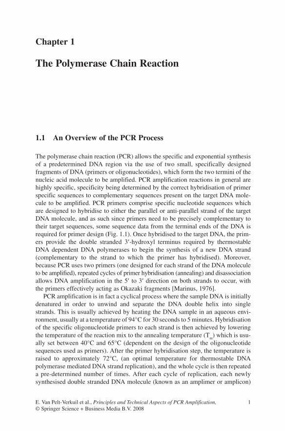
The polymerase chain reaction (PCR) allows the specific and exponential synthesis of a predetermined DNA region via the use of two small, specifically designed fragments of DNA (primers or oligonucleotides), which form the two termini of the nucleic acid molecule to be amplified. PCR amplification reactions in general are highly specific, specificity being determined by the correct hybridisation of primer specific sequences to complementary sequences present on the target DNA mole-cule to be amplified. PCR primers comprise specific nucleotide sequences which are designed to hybridise to either the parallel or anti-parallel strand of the target DNA molecule, and as such since primers need to be precisely complementary to their target sequences, some sequence data from the terminal ends of the DNA is required for primer design (Fig. 1.1). Once hybridised to the target DNA, the prim-ers provide the double stranded 3¢-hydroxyl terminus required by thermostable DNA dependent DNA polymerases to begin the synthesis of a new DNA strand (complementary to the strand to which the primer has hybridised). Moreover, because PCR uses two primers (one designed for each strand of the DNA molecule to be amplified), repeated cycles of primer hybridisation (annealing) and disassociation allows DNA amplification in the 5¢ to 3¢ direction on both strands to occur, with the primers effectively acting as Okazaki fragments [Marinus, 1976].
PCR amplification is in fact a cyclical process where the sample DNA is initially denatured in order to unwind and separate the DNA double helix into single strands. This is usually achieved by heating the DNA sample in an aqueous envi-ronment, usually at a temperature of 94°C for 30 seconds to 5 minutes. Hybridisation of the specific oligonucleotide primers to each strand is then achieved by lowering the temperature of the reaction mix to the annealing temperature (T
m) which is usu-
ally set between 40°C and 65°C (dependent on the design of the oligonucleotide sequences used as primers). After the primer hybridisation step, the temperature is raised to approximately 72°C, (an optimal temperature for thermostable DNA polymerase mediated DNA strand replication), and the whole cycle is then repeated a pre-determined number of times. After each cycle of replication, each newly synthesised double stranded DNA molecule (known as an amplimer or amplicon)
E. Van Pelt-Verkuil et al., Principles and Technical Aspects of PCR Amplification, 1© Springer Science + Business Media B.V. 2008
2 1 The Polymerase Chain Reaction
contains terminal sequences, which are complementary to the primer sequences used (Fig. 1.2). This process allows each amplimer to serve as a template for repli-cation in subsequent rounds of PCR cycling, resulting in a theoretical doubling (exponential amplification) of the number of target molecules during each cycle. Some of the fundamental principles introduced above are detailed in a large body of international scientific literature (e.g. [Jain, 2002; Lubeck and Hoorfar, 2003; Klein, 2002; Wolk et al., 2001; Foy and Parkes, 2001; Erlich, 1999; Kiechle, 1999; Lisby, 1999]).
1.2 Before PCR and Beyond
Before PCR, molecular biologists utilised nucleic acid sequence data (sequence motifs) to design “hybridisation” probes for use in assays for the detection and identification of specific RNA and DNA fragments (moieties). These “hybridisa-tion” assays were used to determine the presence/absence of specific RNA or DNA sequences within complex mixtures of nucleic acids, via the use of specifically designed (semi-synthetic) complementary DNA or RNA molecules (nucleic acid probes) which had been equipped with radioactive labels for detection purposes. The target nucleic acid population was initially attached to a solid carrier phase and
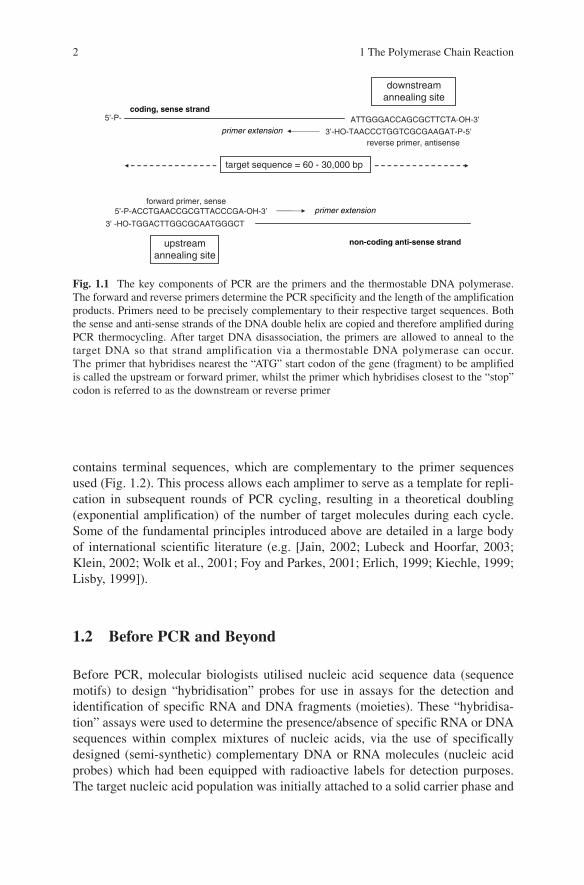
Fig. 1.1 The key components of PCR are the primers and the thermostable DNA polymerase. The forward and reverse primers determine the PCR specificity and the length of the amplification products. Primers need to be precisely complementary to their respective target sequences. Both the sense and anti-sense strands of the DNA double helix are copied and therefore amplified during PCR thermocycling. After target DNA disassociation, the primers are allowed to anneal to the target DNA so that strand amplification via a thermostable DNA polymerase can occur. The primer that hybridises nearest the “ATG” start codon of the gene (fragment) to be amplified is called the upstream or forward primer, whilst the primer which hybridises closest to the “stop” codon is referred to as the downstream or reverse primer
ATTGGGACCAGCGCTTCTA-OH-3’
3’-HO-TAACCCTGGTCGCGAAGAT-P-5’ reverse primer, antisense
downstreamannealing site
forward primer, sense 5’-P-ACCTGAACCGCGTTACCCGA-OH-3’
3’ -HO-TGGACTTGGCGCAATGGGCT
upstreamannealing site
coding, sense strand
non-coding anti-sense strand
primer extension
target sequence = 60 - 30,000 bp
5’-P-
primer extension
1.2 Before PCR and Beyond 3
then hybridised with specific labelled probe. After stringent hybridisation and extensive washing procedures, the presence of the target DNA fragment could be determined by the presence/absence of the radioactively labelled probe on the solid carrier phase. Alternatively, direct visualisation of the probe and target molecule was achieved via electron microscopy, with hybridisation being quantified on the basis of the different widths of double stranded (hybridised) versus single stranded (non-hybridised) nucleic acids. The most convenient of these hybridisation test systems utilised filter hybridisation (where the target DNA extract was first immo-bilised on nitrocellulose or nylon filters), in combination with post-hybridisation
Fig. 1.2 Schematic representation indicating the principle of PCR and its key components. The duplication of a region within a DNA target molecule is facilitated by the specific hybridisation of two different oligonucleotide primers (primer 1 and primer 2). A thermostable DNA-dependent DNA polymerase recognises these primers and extends the DNA strand in the 5¢ to 3¢ direction while consuming dNTPs. Repeated cycles of strand separation by heat denaturation (melting), primer hybridisation (annealing) and new DNA strand synthesis (elongation) results in the expo-nential amplification of a specific DNA region, as defined by the user designed primers. In general, the minimum number of cycles of PCR performed is 20 cycles with 50 cycles usually being regarded as an upper limit
cycle 1
cycle 2
cycle 3
cycle 4
primer 1
primer 2
amplimers
Mg2+, Taq, dNT P
{
4 1 The Polymerase Chain Reaction
autoradiography or scintillography. This format greatly increased test sensitivity and vastly improved the technical reliability and speed of the hybridisation proce-dure, allowing the detection of picogram quantities of target material. However, one major disadvantage of these hybridisation systems was the requirement for radioactively labelled probes, not least because working with radioactive materials is hazardous for your health, requires legal permits, correct disposal systems, and is relatively expensive to use. For these reasons, radioactive labels have been largely replaced by various (non-radioactive) chemical labels, facilitating the development of colorimetric, chemo-luminescent and chemo-fluorescent hybridi-sation detection methods. However, these “second generation” chemical-labelling and detection systems do not generally yield as high a degree of sensitivity as the original radio-labelling and detection systems, though both systems are amenable to automation and high throughput applications. To date, a variety of elegant tech-niques based upon the basic hybridisation principle have been developed (e.g. sandwich hybridisation, Southern- and Northern-blot hybridisations, etc.) and these are frequently applied in both fundamental research and clinical diagnostics.
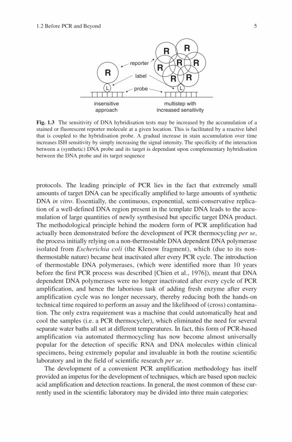
Newer advances and adaptations in nucleic acid hybridisation have allowed the detection of nucleic acids within target tissues or cells per se, without the need to extract the nucleic acids from their “natural environment”. Many elegant applica-tions for this “in situ hybridisation” (ISH) technology have been developed and refined [Brigati et al., 1983]. Moreover, the fact that ISH assays can be coupled to microscopic analysis, means that single copies of target regions present within individual cells may be visualised [Unger et al., 1991]. The sensitivity of such ISH tests may be up to 1,000x greater than traditional filter hybridisation techniques. Currently, most ISH tests are coupled to amplification techniques which include a visualisation step involving the gradual accumulation of a reporter stain (such as biotin-tyramine) in the presence of probe hybridisation (Fig. 1.3). The introduction of a few additional steps to the detection protocol facilitates the use of (multiplex) ISH testing protocols using multiple probes and specimens. However, the disadvantage of ISH/stain-accumulation coupled protocols is a reduction in the ability to accurately localise the focal point of probe hybridisation within a cell due to diffusion of amplimers and stain(s) within the cellular matrix. Loss of target or target inaccessi-bility are well-known complications of ISH protocols and separate optimisation reactions are required for each cell type and ISH probe used in order to achieve the best results.
The need to detect very small numbers of clinically relevant molecules has significantly increased over the past few years. For example, the detection of low-titre viral infections, minimal residual disease in leukaemia patients, point mutations in genes or genetic aberrations in tumours etc., all require highly sensitive methodolo-gies. This has led to the development of novel approaches specifically aimed at the amplification of target (gene) sequences prior to detection, such that sensitivity issues related to hybridisation/probe detection protocols are no longer the limiting step of DNA and RNA detection protocols.
The PCR technique provided the first practical solution to overcoming the limitations of sensitivity which were inherent in early nucleic acid hybridisation and detection
protocols. The leading principle of PCR lies in the fact that extremely small amounts of target DNA can be specifically amplified to large amounts of synthetic DNA in vitro. Essentially, the continuous, exponential, semi-conservative replica-tion of a well-defined DNA region present in the template DNA leads to the accu-mulation of large quantities of newly synthesised but specific target DNA product. The methodological principle behind the modern form of PCR amplification had actually been demonstrated before the development of PCR thermocycling per se, the process initially relying on a non-thermostable DNA dependent DNA polymerase isolated from Escherichia coli (the Klenow fragment), which (due to its non-thermostable nature) became heat inactivated after every PCR cycle. The introduction of thermostable DNA polymerases, (which were identified more than 10 years before the first PCR process was described [Chien et al., 1976]), meant that DNA dependent DNA polymerases were no longer inactivated after every cycle of PCR amplification, and hence the laborious task of adding fresh enzyme after every amplification cycle was no longer necessary, thereby reducing both the hands-on technical time required to perform an assay and the likelihood of (cross) contamina-tion. The only extra requirement was a machine that could automatically heat and cool the samples (i.e. a PCR thermocycler), which eliminated the need for several separate water baths all set at different temperatures. In fact, this form of PCR-based amplification via automated thermocycling has now become almost universally popular for the detection of specific RNA and DNA molecules within clinical specimens, being extremely popular and invaluable in both the routine scientific laboratory and in the field of scientific research per se.
The development of a convenient PCR amplification methodology has itself provided an impetus for the development of techniques, which are based upon nucleic acid amplification and detection reactions. In general, the most common of these cur-rently used in the scientific laboratory may be divided into three main categories:
LL
R
R
R R
RR
R
R
insensitive approach
multistep with increased sensitivity
reporter
label
probe
Fig. 1.3 The sensitivity of DNA hybridisation tests may be increased by the accumulation of a stained or fluorescent reporter molecule at a given location. This is facilitated by a reactive label that is coupled to the hybridisation probe. A gradual increase in stain accumulation over time increases ISH sensitivity by simply increasing the signal intensity. The specificity of the interaction between a (synthetic) DNA probe and its target is dependant upon complementary hybridisation between the DNA probe and its target sequence
1.2 Before PCR and Beyond 5
6 1 The Polymerase Chain Reaction
1. Target amplification: The initial amount of target nucleic acid (or a specific region within this target nucleic acid) is exponentially amplified using an opti-mised reaction mix and a thermostable DNA dependent polymerase (e.g. PCR). Other variants (related to PCR but using different reaction mixes and enzymes) include – the ligase chain reaction (LCR) [Laffler et al., 1993], transcription amplification systems (TAS) [Kwoh et al., 1989] and the nucleic acid sequence based amplification (NASBA) technique [Malek et al., 1994; Cook, 2003]. The latter two techniques may be especially useful in amplifying RNA instead of DNA, and involve the generation of DNA/RNA intermediates.
2. Probe amplification: This technology depends on an initial hybridisation between a probe molecule and the target nucleic acid, where the probe contains additional sequence elements that enable detection of the probe via a specific interaction with these additional sequence elements, e.g. the Qb-replicase system which exponentially amplifies RNA using the bacteriophage Qb RNA dependent RNA polymerase. Other systems involving “replicable probes” and “branched DNA assays” (which enable the Christmas tree-like “networking” of multiple probes), have been produced and developed into commercially available diag-nostic products. Though these are currently much less widely available or used than the “target amplification” systems mentioned above [Qian and Lloyd, 2003; Blok and Kramer, 1997].
3. Signal amplification: This is the common term for all of the techniques that amplify a signal previously generated by either the target amplification systems or probe amplification systems described above. Signal amplification systems fre-quently utilise chemical reactions that result in an increase in the accumulation of coloured, fluorescent or luminescent product (i.e. signal) after target amplification, thereby resulting in an increase in test sensitivity [e.g. Ness et al., 2003].
This book is specifically dedicated to the polymerase chain reaction (developed in 1985 by Saiki et al. [1985]), and to the large multitude of additional applications and formats which have been described for PCR over the past 2 decades. The goal of this book is to provide the reader with an overview of the technique, development and application of PCR technology today, rather than providing an all-encompassing and intimately detailed account of all aspects of PCR (as if this were possible!). To this extent, the PCR process is first compared to DNA replication, and different PCR test formats. Further, the identification of amplified PCR products is discussed in detail, and the design and execution of PCR tests (including quality control and trouble shooting) are also discussed.
References
Blok HJ, Kramer FR. 1997. Amplifiable hybridization probes containing a molecular switch. Mol Cell Probes 11:187–194.
Brigati DJ, Myerson D, Leary JJ, Spalholz B, Travis SZ, Fong CK, Hsiung GD, Ward DC. 1983. Detection of viral genomes in cultured cells and paraffin-embedded tissue sections using biotin-labeled hybridization probes. Virol 126:32–50.
Chien A, Edgar DB, Trela JM. 1976. Deoxyribonucleic acid polymerase from the extreme thermophile Thermus aquaticus. J Bacteriol 127:1550–1557.
Cook N. 2003. The use of NASBA for the detection of microbial pathogens in food and environmental samples. J Microbiol Methods 53:165–174.
Erlich HA. 1999. Principles and applications of the polymerase chain reaction. Rev Immunogenet 1:127–134.
Foy CA, Parkes HC. 2001. Emerging homogeneous DNA-based technologies in the clinical laboratory. Clin Chem 47:990–1000.
Jain KK. 2002. Current trends in molecular diagnostics. Med Device Technol 13:14–18.Kiechle FL. 1999. DNA technology in the clinical laboratory. Arch Pathol Lab Med
123:1151–1153.Klein D. 2002. Quantification using real-time PCR technology: applications and limitations.
Trens Mol Med 8:257–260.Kwoh DY, Davis GR, Whitfield KM, Chappelle HL, DiMichele LJ, Gingeras TR. 1989.
Transcription based amplification system and detection of amplified human immunodefi-ciency virus type I with a bead-based sandwich hybridization format. Proc Natl Acad Sci USA 86:1173–1177.
Laffler TG, Carrino JJ, Marshall RL. 1993. The ligase chain reaction in DNA-based diagnosis. Ann Biol Clin 51:821–826.
Lisby G. 1999. Application of nucleic acid amplification in clinical microbiology. Mol Biotechnol 12:75–99.
Lubeck PS, Hoorfar J. 2003. PCR technology and applications to zoonotic food-borne bacterial pathogens. Methods Mol Biol 216:65–84.
Malek L, Sooknanan R, Compton J. 1994. Nucleic acid sequence based amplification (NASBA). Methods Mol Biol 28:253–260.
Marinus MG. 1976. Adenine methylation of Okazaki fragments in Escherichia coli. J Bacteriol 128:853–854.
Ness JM, Akhtar RS, Latham CB, Roth KA. 2003. Combined tyramide signal amplification and quantum dots for sensitive and photostable immunofluorescence detection. J Histochem Cytochem 51:981–987.
Qian X, Lloyd RV. 2003. Recent developments in signal amplification methods for in situ hybridi-sation. Diagn Mol Pathol 12:1–13.
Saiki RK, Scharf S, Faloona F, Mullis KB, Horn GT, Erlich HA, Arnheim N. 1985. Enzymatic amplification of beta-globin genomic sequences and restriction site analysis for diagnosis of sickle cell anemia. Science 230:1350–1354.
Unger ER, Hammer ML, Chenggis ML. 1991. Comparison of 35S and biotin as labels for in situ hybridization: usa of an HPV model system. J Histochem Cytochem 39:145–150.
Wolk D, Mitchel S, Patel R. 2001. Principles of molecular microbiology testing methods. Infect Dis Clin North Am 15:1157–1204.
References 7
Chapter 2
A Brief Comparison Between In Vivo DNA Replication and In Vitro PCR Amplification
In principle, PCR generates large quantities of DNA from a minute amount of nucleic acid starting material using a methodology similar to (but much simpler than) that seen in living cells. For living cells, in vivo DNA synthesis is dependent upon a well defined but complex set of enzymes and co-factors, which have evolved to act in a concerted fashion during the synthetic phase (S-phase) of the cell cycle. In comparison, PCR facilitates in vitro DNA synthesis in a much simpler fashion, making use of a smaller set of defined ingredients and reaction conditions involving relatively high temperatures. The range of factors contributing to successful PCR amplification is reviewed below.
2.1 Nucleic Acid Targets
2.1.1 DNA
In vivo DNA duplication, which is essentially a form of limited DNA amplification, is performed under the direction of a select and diverse set of structural proteins, enzymes and additional co-factors (a detailed description of which is beyond the scope of this book and interested readers are referred to the international literature for a more in-depth look into this particular topic, e.g. Shcherbakova et al., 2003; Goren and Cedar, 2003; Kelman, 2000; Nasheuer et al., 2002; Nasmyth, 2002). In eukaryotes, the DNA molecule is intimately associated with positively charged proteins, which are strongly electrostatically bound to the phosphate moieties on the DNA chain. These “histone” proteins associate into octameric complexes, bind to approximately 400 base pairs (bp) of genomic DNA, and constitute approxi-mately half of the mass of the eukaryotic chromosome. These histones do not com-pletely disassociate from the DNA during replication. An additional level of complexity also exists in eukaryotic DNA, in that further folding processes induce secondary (300 nm) and tertiary (700 nm) order folding on the template DNA, thereby generating tightly coiled DNA with a high molecular density [Stewart, 1997] (Fig. 2.1). For prokaryotes, the situation regarding the in vivo state of DNA
E. Van Pelt-Verkuil et al., Principles and Technical Aspects of PCR Amplification, 9© Springer Science + Business Media B.V. 2008
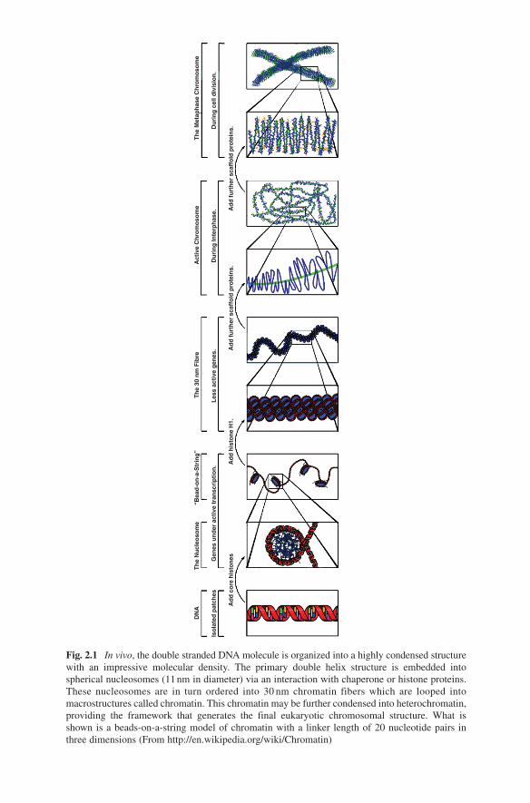
Fig. 2.1 In vivo, the double stranded DNA molecule is organized into a highly condensed structure with an impressive molecular density. The primary double helix structure is embedded into spherical nucleosomes (11 nm in diameter) via an interaction with chaperone or histone proteins. These nucleosomes are in turn ordered into 30 nm chromatin fibers which are looped into macrostructures called chromatin. This chromatin may be further condensed into heterochromatin, providing the framework that generates the final eukaryotic chromosomal structure. What is shown is a beads-on-a-string model of chromatin with a linker length of 20 nucleotide pairs in three dimensions (From http://en.wikipedia.org/wiki/Chromatin)
DN
A
Iso
late
d p
atch
es
Ad
d c
ore
his
ton
esA
dd
his
ton
e H
1.A
dd
fu
rth
er s
caff
old
pro
tein
s.A
dd
fu
rth
er s
caff
old
pro
tein
s.
Les
s ac
tive
gen
es.
Du
rin
g In
terp
has
e.D
uri
ng
cel
l div
isio
n.
Gen
es u
nd
er a
ctiv
e tr
ansc
rip
tio
n.
Th
e N
ucl
eoso
me
“Bea
d-o
n-a
-Str
ing
”T
he
30 n
m F
ibre
Act
ive
Ch
rom
oso
me
Th
e M
etap
has
e C
hro
mo
som
e
2.1 Nucleic Acid Targets 11
is generally considered to be somewhat simpler, with microorganisms lacking a visible nucleus and containing only one (circular) chromosomal copy per cell. However, it is known that prokaryotic DNA is also associated with a variety of histone-like proteins, which intimately interact with the cellular DNA, protecting it and conferring complex structure (though this structure is generally less ordered or regular than that associated with eukaryotic chromosomes). Therefore, one of the major requirements for DNA replication in in vivo systems is the systematic uncoiling of specific regions of genomic DNA in order to expose single strands of DNA ready for replication. In both eukaryotic and prokaryotic kingdoms this process is achieved via specific DNA topoisomerases, helicases and gyrases, which act together to help uncoil the double stranded DNA.
In contrast, the uncoiling of DNA during PCR amplification is not enzymati-cally controlled but is in fact achieved using a far less complicated procedure, i.e. the DNA to be amplified is usually chemically extracted from its host chaperone proteins, and the residual tertiary or secondary structure removed by heating the naked DNA. This heating step also provides the mechanism by which the two DNA strands are separated (melted) into single strands ready for PCR amplification. As a further point, whereas in vivo DNA chromosomal replication involves the replica-tion of many millions of nucleotides, PCR amplification products are generally designed to be shorter than 1,000 bp in length (a parameter largely imposed on the PCR amplification process by the type of heat-stable DNA dependent DNA polymerases used). However in extreme cases, the successful PCR amplification of regions of DNA over 35 kilobases in length have been reported using special heat-stable DNA dependent DNA polymerases combinations [Hu et al., 2002; Davies and Gray, 2002]. In principle, DNA from all types of viruses, cells (plant, animal and bacterial), or tissues (lung, brain, etc.) may be amplified using nucleic acid extraction techniques coupled to PCR amplification.
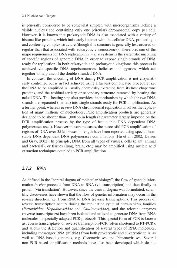
2.1.2 RNA
As defined in the “central dogma of molecular biology”, the flow of genetic infor-mation in vivo proceeds from DNA to RNA (via transcription) and then finally to protein (via translation). However, since the central dogma was formulated, scien-tific discoveries have shown that the flow of genetic information may occur in the reverse direction, i.e. from RNA to DNA (reverse transcription). This process of reverse transcription occurs during the replication cycle of certain virus families (Retroviridae, Hepadnaviridae and Caulimoviridae), and the relevant enzymes (reverse transcriptases) have been isolated and utilized to generate DNA from RNA molecules in specially adapted PCR protocols. This special form of PCR is known as reverse transcriptase- or reverse transcription-PCR (often shortened to RT-PCR), and allows the detection and quantification of several types of RNA molecules, including messenger RNA (mRNA) from both prokaryotic and eukaryotic cells, as well as RNA-based genomes, e.g. Coronaviruses and Picornaviruses. Several non-PCR-based amplification methods have also been developed which do not
utilise reverse transcriptase enzymes and hence do not make use of an intermediate DNA template. These techniques amplify RNA (and not double stranded DNA as is the case with PCR) using an initial RNA template.
2.2 Target DNA Strand Separation and Primer Annealing
During in vivo DNA replication, cell associated proteins (including DNAa, DNAb, DNAc, single stranded binding proteins, helicases and gyrases, ligase and a variety of polymerase subunit proteins), all act in concert to uncoil the stable a-helical DNA structure, break the hydrogen bonds between the purine and pyrimidine bases, and expose a DNA replication origin [Nasheuer et al., 2002]. In contrast, DNA required for PCR amplification is separated from its chaperone proteins using chemical and/or enzymatic extraction methods, with the DNA then being separated into single strands via thermal disassociation, i.e. via incubation at approximately 94°C for 30 seconds to 5 minutes, which causes breaking of the hydrogen bonds between the complementary base pairs present on opposite strands. This method is not available to living cells as proteins are rapidly denatured at 94°C and cellular integrity irretrievably breaks down. In fact, the melting temperature (T
m) of a DNA
molecule is defined as the temperature at which half of it’s constitutive bases are no longer paired to their complementary partner on the opposite strand. For most natu-ral species of DNA, the melting temperature lies somewhere between 70°C and 100°C, with the actual T
m being dependent on both the length of the DNA molecule
to be melted and on the base composition of the strands. Moreover, there exists a linear relationship between the percentage of guanosine and cytosine bases present in the DNA strands (referred to as the “GC content”) and the melting temperature of that particular DNA helix, with GC-rich DNA melting at a higher temperature than adenosine and thymidine rich (AT-rich) DNA. Because of the fact that DNA melting curves are relatively steep, temperatures of T
m+5°C and T
m−5°C above or
below the calculated Tm value will result in the double helix being completely dena-
tured or completely intact, respectively.Several other factors may also facilitate DNA helix destabilization, all of them
operating by destabilizing the interaction between the various complementary DNA base moieties. Extreme pH values, denaturants such as formamide or urea and the overall salt concentration are important parameters in this respect (Rauch et al., 2000). Essentially, the addition of these reagents shifts the DNA melting curve to the left, with the result that the absolute T
m for the double stranded DNA
value decreases. This decrease in Tm may be tens of degrees centigrade dependent
on the factor included in the reaction mix. Conversely, several compounds actu-ally stabilize the DNA double helix, including magnesium ions and elevated salt concentrations, which help neutralize the triple-negative phosphate charges on the opposing DNA strands in the duplex (therefore limiting electrostatic repul-sion), as well as (partially) neutralizing the effect that the water dipole has on DNA hydrogen bonding.
12 2 A Brief Comparison Between In Vivo DNA and In Vitro PCR
2.3 DNA Dependent DNA Polymerase and Oligonucleotide Primers 13
The heating procedure used to melt DNA during the PCR process is a simple and reliable method for ensuring that DNA strand separation occurs and that DNA binding sites for specifically designed PCR primers are exposed. However, at DNA melting temperatures, PCR primers also remain disassociated from the target DNA, and effective binding of the PCR primers to the target DNA can only take place at a reduced temperature when the thermal energy is low enough to allow complemen-tary base pairing. In most applications (random amplification of polymorphic DNA or RAPD excepted), PCR primers are designed to specifically bind to known sequences of target DNA and to anneal to the melted target DNA at a temperature of between 45°C and 65°C (the “annealing” temperature). Of course, at this tem-perature the target DNA also re-anneals to its complementary strand. However, the excess concentration of primers added to PCR mixes ensures that binding of at least some PCR primers will occur during each thermocycle (dependent of course on the presence of primer-complementary sequences in the target DNA).
2.3 DNA Dependent DNA Polymerase and Oligonucleotide Primers
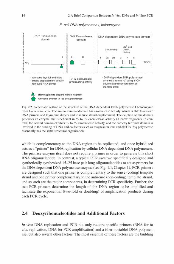
DNA dependent DNA polymerase is an essential component of both in vivo DNA replication and the PCR process (Fig. 2.2), though the DNA dependent DNA polymerases found in the vast majority of organisms are heat sensitive (thermolabile), one of the major stumbling blocks hindering the initial success of PCR. However, the discovery of thermophilic organisms and the subsequent isolation of thermostable DNA dependent DNA polymerases from these organisms heralded a new chapter in PCR amplification technology [Saiki et al., 1988], such that successive cycles of heat-ing and cooling (necessary for target DNA melting and primer annealing during PCR thermocycling) no longer resulted in the concomitant denaturation of the thermolabile DNA dependent DNA polymerase enzyme. The discovery of these thermostable enzymes allowed the whole PCR process to become far more convenient, less time consuming and more user-friendly for laboratory personnel. The first thermostable DNA dependent DNA polymerase enzyme to be widely used in PCR (Taq polymerase) was derived from the hot spring-dwelling bacterium Thermus aquaticus, though many other commercially available non-Taq thermostable DNA dependent DNA polymerase enzymes are now available on the market, e.g. Pfu from Pyrococcus furiosus, Vent from Thermococcus litoralis, etc. The advantages and disadvantages of some of these different thermostable polymerases with respect to PCR thermocycling are described more fully in Chapter 7.
DNA dependent DNA polymerases per se are actually incapable of performing DNA synthesis from a purely single stranded piece of DNA and require an addi-tional shorter “priming” oligonucleotide to initiate DNA replication. In in vivo DNA replication, an RNA oligonucleotide (generated by a DNA dependent RNA polymerase called primase) acts as the primer for DNA replication. This primase enzyme synthesizes a short RNA molecule (of approximately ten nucleotides),
14 2 A Brief Comparison Between In Vivo DNA and In Vitro PCR
which is complementary to the DNA region to be replicated, and once hybridized acts as a “primer” for DNA replication by cellular DNA dependent DNA polymerase. The primase enzyme itself does not require a primer in order to generate this short RNA oligonucleotide. In contrast, a typical PCR uses two specifically designed and synthetically synthesized 15–25 base pair long oligonucleotides to act as primers for the DNA dependent DNA polymerase enzyme (see Fig. 1.1, Chapter 1). PCR primers are designed such that one primer is complementary to the sense (coding) template strand and one primer complementary to the antisense (non-coding) template strand, and as such are the major components, in determining PCR specificity. Further, the two PCR primers determine the length of the DNA region to be amplified and facilitate the exponential (two-fold or doubling) of amplification products during each PCR cycle.
2.4 Deoxyribonucleotides and Additional Factors
In vivo DNA replication and PCR not only require specific primers (RNA for in vivo replication, DNA for PCR amplification) and a (thermostable) DNA polymer-ase, but also several other factors. The most essential of these factors are the building
Fig. 2.2 Schematic outline of the structure of the DNA dependent DNA polymerase I holoenzyme from Escherichia coli. The amino terminal domain has exonuclease activity, which is able to remove RNA primers and thymidine dimers and to induce strand displacement. The deletion of this domain generates an enzyme that is deficient in 5¢- to 3¢- exonuclease activity (Klenow fragment). In con-trast, the central domain exhibits 3¢- to 5¢- exonuclease activity, and the carboxy terminal domain is involved in the binding of DNA and co-factors such as magnesium ions and dNTPs. Taq polymerase essentially has the same structural organisation
E. coli DNA-polymerase l, holoenzyme
5’-3’ Exonucleasedomain
- removes thymidine-dimers- strand displacement activity- removes RNA-primer
cleaving point to prepare Klenow fragment
functional deletion in Taq DNA polymerase
- DNA-dependent DNA polymerase synthesis from 5’ -3’ using 3’-OH double strand configuration as startting point
3’ -5’ exonucleaseproofreading activity
NH2
I II III 1 2a
DNA-bindingMg2+ andDNTPbinding
2b 3 4 5
COOH
3’-5’ Exonucleasedomain
DNA-dependent DNA polymerase domain
blocks or free deoxynucleotide triphosphate molecules that are incorporated into the growing DNA chain by the DNA dependent DNA polymerase. These compounds can be acquired in vivo as nutrients, but are generally synthesized via complex pathways involving the reduction of ribonucleotide diphosphates and addition of a further phosphate group by a kinase enzyme. The four major deoxynucleotides required for DNA synthesis (adenosine-, guanosine-, thymidine- and cytosine-triphosphate) contain an energy-rich triphosphate moiety, which is utilized by the DNA polymerase to catalyze a phosphodiester link between the 3′-hydroxy termi-nus of the primer (or previously added deoxynucleotide triphosphate) and the newly acquired deoxynucleotide triphosphate on the growing DNA strand. Hydrogen bonding between complementary nucleotides on adjacent strands then completes the double stranded primary structure of the DNA molecule. The mecha-nism by which deoxyribonucleotide triphosphates (dNTPs) are added to the grow-ing DNA chain is identical for both in vivo replication and PCR amplification processes, though the concentration of dNTPs in in vitro amplification reactions may be easily manipulated to artificially high levels. As well as deoxyribonucle-otides, successful in vivo DNA synthesis and amplification requires a large number of chemical components such as (1) the bivalent metal ions magnesium (a cofactor for the DNA polymerase enzyme) and manganese and (2) simple chemicals such as sodium chloride, etc. which help to maintain the correct pH of the reaction or allow for the synthesis of new DNA with a complex secondary structure (see Chapters 4–7 and Wilson et al., [2002] for a more detailed discussion).
References
Davies PA, Gray G. 2002. Long-range PCR. Methods Mol Biol 187:51–55.Goren A, Cedar H. 2003. Replicating by the clock. Nat Rev Mol Cell Biol 4:25–32.Hu M, Chilton NB, Gasser RB. 2002. Long PCR based amplification of the entire mitochondrial
genome from single parasitic nematodes. Mol Cell Probes 16:261–267.Kelman Z. 2000. DNA replication in the third domain of life. Curr Protein Pept Sci 1:139–154.Nasheuer HP, Smith R, Bauerschmidt C, Grosse F, Weisshart K. 2002. Initiation of eukaryotic
DNA replication: regulation and mechanisms. Prog Nucleic Acid Res Mol Biol 72:41–94.Nasmyth K. 2002. Segregating sister genomes: the molecular biology of chromosome separation.
Science 297:559–565.Rauch J, Wolf D, Hausmann M, Cremer C. 2000. The influence of formamide on thermal dena-
turation profiles of DNA and metaphase chromosomes in suspension. Z Naturforsch 55:737–746.
Saiki RK, Gelfand DH, Stoffel S, Scharf SJ, Higuchi R, Horn GT, Mullis KB, Erlich HA. 1988. Primer directed enzymatic amplification of DNA with a thermostable DNA polymerase. Science 239:487–491.
Shcherbakova PV, Bebenel K, Kunkel TA. 2003. Functions of eukaryotic DNA polymerases. Sci Aging Knowl Environ 26:RE3.
Stewart RD. 1997. A theoretical investigation of cell cycle effects and interspecies radiosensitivi-ties. Ph.D. thesis.
Wilson T, Carson J, Bowman J. 2002. Optimisation of one-tube PCR-ELISA to detect femtogram amounts of genomic DNA. J Microbiol Methods 51:163–170.
References 15
Chapter 3
The PCR in Practice
3.1 Brief Overview of PCR Requirements
3.1.1 The PCR Reaction Mix
A typical PCR reaction mix includes target DNA (Chapter 4), specifically designed oligonucleotide primers (Chapter 5), deoxyribonucleotide triphosphates/magnesium ions/a buffer component (Chapter 6), a thermostable DNA polymerase (Chapter 7) and water. The range of quantities for each ingredient of a typical PCR mix is indicated below:
– 10 fg–10 µg of chemically extracted DNA or reverse transcribed RNA in 1–10 µl of rehydration buffer or water
– 0.1–1.0 µM each of oligonucleotide primers, one complementary to the positive (sense) strand and the other complementary to the downstream (antisense) strand
– Approximately 200 µM of each of the four deoxyribonucleotide triphosphate DNA building blocks, i.e. adenosine-, cytosine-, guanosine- and thymidine triphosphate
– 0.2–2.0 units of Taq polymerase (other commercially available thermostable DNA polymerases may also be used but bear in mind differences in proof-reading ability and processivity)
– Tris-buffer (pH 8.3), 50 mM KCl (specific buffers are usually supplied free by the manufacturer of the thermostable enzyme used)
– 500–1,000 µg gelatin or Bovine Serum Albumin (BSA) per ml (optional).– 0.5–5.0 mM of magnesium chloride salt solution (MgCl
2 is a DNA polymerase
cofactor)
All prepared in a total typical PCR reaction volume of 50 µl (5–100 µl = the usual range).
Note that the composition of the PCR reaction mix may vary dependant on the nature (and quality) of the heat-resistant thermostable DNA dependent DNA polymerase used. Low concentrations of detergents, such as Triton X-100, Tween-20, betain or dimethylsulphoxide (DMSO) may also be included in the PCR mix to help increase the specificity of primer binding. The same compounds may assist in overcoming problems caused by secondary structure or possibly even inhibitory
E. Van Pelt-Verkuil et al., Principles and Technical Aspects of PCR Amplification, 17© Springer Science + Business Media B.V. 2008
18 3 The PCR in Practice
compounds. The composition of the PCR mix should ideally be optimized for every new PCR protocol developed, as any change in the PCR methodology (including changes in primer design, deoxyribonucleotide composition, template nucleic acid and type of thermostable DNA polymerase used) may influence the specificity of amplification. All PCR reaction ingredients should be stored in a freezer in a dedi-cated “clean” room where strict guidelines are enforced to help prevent possible contamination of reaction mixes and ingredients (Chapter 10).
In order to decrease the likelihood of non-specific primer binding occurring during the initial (i.e. pre-first cycle) stage of the PCR thermocycling program (when the temperature is low enough to allow partial binding of the primer), one of the reaction components (usually the DNA polymerase or magnesium salt) may be omitted from the reaction mix and added once the initial target DNA melting tem-perature of 90–95°C is reached (PCR amplification will not proceed in the absence of magnesium ions). Unfortunately however, the simple act of opening and closing a PCR reaction tube greatly increases the chance of contamination of that tube. One elegant alternative to this problem is to physically separate the reaction components into two separate compartments, e.g. via a thin layer of wax. Magnesium chloride solution may then be added to the upper surface of the wax bead. The wax bead then melts at a temperature that is too high for non-specific binding of the PCR primers to the DNA template to occur. Some suppliers provide wax beads already impregnated with magnesium salt. Other elegant alternatives devised to decrease the likelihood of non-specific primer binding during the initial stage of the PCR thermocycling program, include heat-activatable thermostable DNA polymerases, which require approximately 10 minutes of heat activation at around 90°C before becoming activate. Other methods utilise polymerases coated with antibodies, which inhibit the action of the polymerase until the temperature is high enough to denature and remove the antibodies. All of these approaches are collectively named “hot-start” procedures (Chapter 7).
When performing multiple PCRs in a single “batch” (i.e. at the same time), it is advisable to prepare a “master mix” of reaction ingredients (comprising a multiple number of individual reaction mixes prepared in a single tube), and then pipette out the individual volumes into individual reaction tubes, rather than pipetting each separate component into every individual reaction tube one after the other. This process significantly reduces the number of pipetting actions that need to be per-formed and as such greatly contributes to the reproducibility and reliability of the PCR test protocol. The volume of a particular reagent required in a master mix is dependent on the number of PCRs to be performed, and may be calculated by multiplying the volume of the ingredient required for a single reaction, by the number of tests to be performed, including duplicates and positive and negative control samples. Additionally, for every master mix reaction prepared, an extra aliquot of each ingredient should be added (in order to account for volume errors inherent in any repeat pipetting action). Preferably, once prepared, the master mix should be immediately dispensed or stored on ice for short periods of time. Mastermixes and/or individual PCR reaction mix aliquots may be stored frozen for a prolonged period of time and immediately thawed prior to use. In this case, it is preferable to
3.1 Brief Overview of PCR Requirements 19
prepare master mixes without thermostable DNA polymerase and then add the required volume of enzyme after thawing (immediately prior to PCR thermocycling). The time period during which frozen mastermix aliquots may be stored without loss of PCR amplification sensitivity should ideally be determined for each particular mix to be prepared and be routinely quality controlled [Kofler and Klausegger, 1999]. From the authors’ own experience, PCR master mix aliquots without thermostable DNA polymerase may be stored for 2–3 months at −20°C without loss of PCR sensitivity.
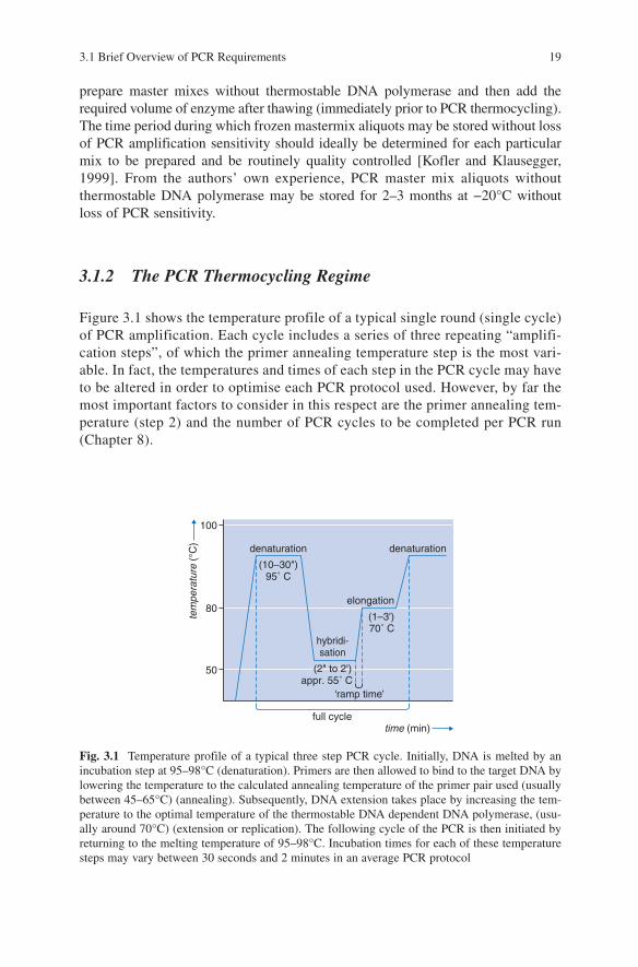
3.1.2 The PCR Thermocycling Regime
Figure 3.1 shows the temperature profile of a typical single round (single cycle) of PCR amplification. Each cycle includes a series of three repeating “amplifi-cation steps”, of which the primer annealing temperature step is the most vari-able. In fact, the temperatures and times of each step in the PCR cycle may have to be altered in order to optimise each PCR protocol used. However, by far the most important factors to consider in this respect are the primer annealing tem-perature (step 2) and the number of PCR cycles to be completed per PCR run (Chapter 8).
Fig. 3.1 Temperature profile of a typical three step PCR cycle. Initially, DNA is melted by an incubation step at 95–98°C (denaturation). Primers are then allowed to bind to the target DNA by lowering the temperature to the calculated annealing temperature of the primer pair used (usually between 45–65°C) (annealing). Subsequently, DNA extension takes place by increasing the tem-perature to the optimal temperature of the thermostable DNA dependent DNA polymerase, (usu-ally around 70°C) (extension or replication). The following cycle of the PCR is then initiated by returning to the melting temperature of 95–98°C. Incubation times for each of these temperature steps may vary between 30 seconds and 2 minutes in an average PCR protocol
denaturation
(10–30")95˚ C
elongation
(1–3')70˚ C
‘ramp time’
hybridi-sation
(2" to 2')appr. 55˚ C
denaturation
tem
pera
ture
(°C
)
time (min)
100
80
50
full cycle
20 3 The PCR in Practice
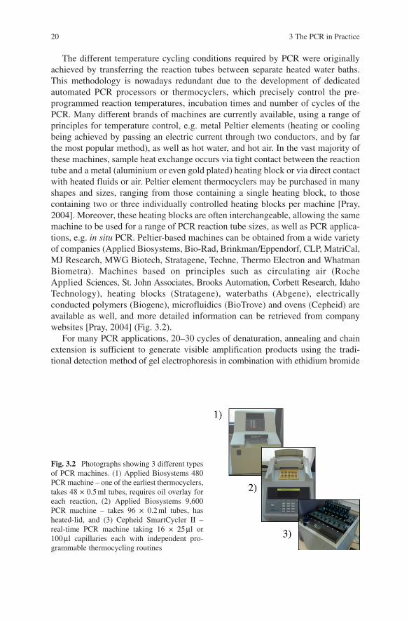
The different temperature cycling conditions required by PCR were originally achieved by transferring the reaction tubes between separate heated water baths. This methodology is nowadays redundant due to the development of dedicated automated PCR processors or thermocyclers, which precisely control the pre-programmed reaction temperatures, incubation times and number of cycles of the PCR. Many different brands of machines are currently available, using a range of principles for temperature control, e.g. metal Peltier elements (heating or cooling being achieved by passing an electric current through two conductors, and by far the most popular method), as well as hot water, and hot air. In the vast majority of these machines, sample heat exchange occurs via tight contact between the reaction tube and a metal (aluminium or even gold plated) heating block or via direct contact with heated fluids or air. Peltier element thermocyclers may be purchased in many shapes and sizes, ranging from those containing a single heating block, to those containing two or three individually controlled heating blocks per machine [Pray, 2004]. Moreover, these heating blocks are often interchangeable, allowing the same machine to be used for a range of PCR reaction tube sizes, as well as PCR applica-tions, e.g. in situ PCR. Peltier-based machines can be obtained from a wide variety of companies (Applied Biosystems, Bio-Rad, Brinkman/Eppendorf, CLP, MatriCal, MJ Research, MWG Biotech, Stratagene, Techne, Thermo Electron and Whatman Biometra). Machines based on principles such as circulating air (Roche Applied Sciences, St. John Associates, Brooks Automation, Corbett Research, Idaho Technology), heating blocks (Stratagene), waterbaths (Abgene), electrically conducted polymers (Biogene), microfluidics (BioTrove) and ovens (Cepheid) are available as well, and more detailed information can be retrieved from company websites [Pray, 2004] (Fig. 3.2).
For many PCR applications, 20–30 cycles of denaturation, annealing and chain extension is sufficient to generate visible amplification products using the tradi-tional detection method of gel electrophoresis in combination with ethidium bromide
Fig. 3.2 Photographs showing 3 different types of PCR machines. (1) Applied Biosystems 480 PCR machine – one of the earliest thermocyclers, takes 48 × 0.5 ml tubes, requires oil overlay for each reaction, (2) Applied Biosystems 9,600 PCR machine – takes 96 × 0.2 ml tubes, has heated-lid, and (3) Cepheid SmartCycler II – real-time PCR machine taking 16 × 25 µl or 100 µl capillaries each with independent pro-grammable thermocycling routines
staining (Chapter 9). If a higher sensitivity is required, for instance due to an apparent limit on the quantity of target DNA, then the number of programmed cycles may be increased from 30 cycles to a generally accepted maximum of 50 cycles. The amplification factor required for the determination of visible amplification product from biologically available quantities of starting DNA material using ethidium bromide staining lies in the order of 105–106.
The vast majority of PCR protocols currently use a ramping temperature/time of approximately 1°C/second, which allows for 20–30 cycles of amplification in approxi-mately 2–3 hours. Less frequently used PCR protocols may considerably increase the average time for 20–30 cycles of amplification, e.g. >16 hours for PCRs intended to amplify multiple-kilobasepair sized fragments. Further, more recent technological developments have also facilitated the implementation of high-speed PCR tests. By using thin glass capillaries, the volume of the PCR reaction mix may be reduced and heat transfer rates (ramp rates) vastly increased from 1°C per second to 20°C per second. PCR processing times are also therefore greatly reduced from several hours to less than 30 minutes, though the number of samples that can be processed per PCR “run” are somewhat reduced in capillary PCR machines.
The duration of a particular PCR program is not only determined by the incubation time required for the individual steps and the total number of cycles programmed for that PCR protocol, but also includes; (a) the time required for the initial melting of the target DNA prior to PCR thermocycling per se (usually a “soak” time of between 2 and 10 minutes at 90–95°C used to ensure absolute melting of the target
3.1 Brief Overview of PCR Requirements 21
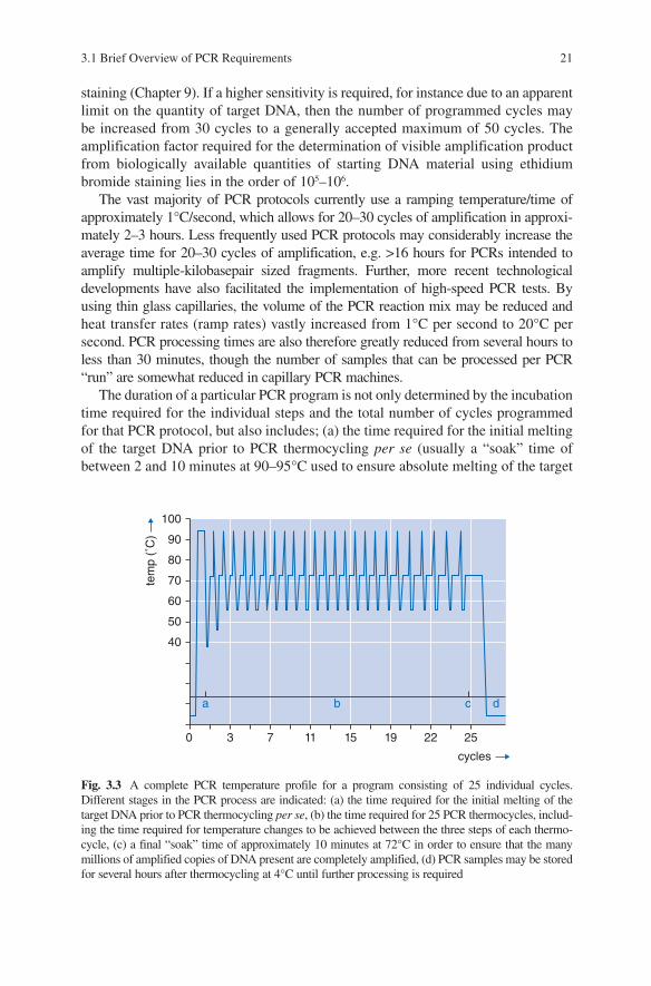
Fig. 3.3 A complete PCR temperature profile for a program consisting of 25 individual cycles. Different stages in the PCR process are indicated: (a) the time required for the initial melting of the target DNA prior to PCR thermocycling per se, (b) the time required for 25 PCR thermocycles, includ-ing the time required for temperature changes to be achieved between the three steps of each thermo-cycle, (c) a final “soak” time of approximately 10 minutes at 72°C in order to ensure that the many millions of amplified copies of DNA present are completely amplified, (d) PCR samples may be stored for several hours after thermocycling at 4°C until further processing is required
100
0 3 7 11 15 19 22 25
70
60
50
40
a b c d
cycles
tem
p (˚
C)
80
90
22 3 The PCR in Practice
DNA and/or for activation of “hot-start” DNA polymerase enzymes), (b) the time required to change the temperature between the three steps of each PCR cycle, and (c) a final “soak” time of approximately 10 minutes at 72°C (added after the final PCR thermocycle is complete) in order to ensure that the many millions of ampli-fied copies of DNA present at this stage of the PCR process are completely amplified. When the total PCR run is completed, the samples are usually cooled down to 4°C for an indefinite period, a task which may be performed by most modern PCR machines (some are even capable of freezing the samples), thereby providing stable very short-term storage conditions (Fig. 3.3).
3.1.3 Analysis of PCR Amplification Products
The visualisation of PCR amplification products is traditionally facilitated by gel electrophoresis and ethidium bromide staining, a chemical which inserts or “intercalates” between the two DNA strands and is detected by fluorescence during exposure to ultraviolet (UV) light (Chapter 9). Though this is still the method most widely used to detect PCR products, other fluorescent dyes are now available which are extremely useful in PCR applications, e.g. in the PCR sequencing of DNA. Recent develop-ments have also facilitated the direct visualisation of amplified PCR products during actual PCR thermocycling per se (e.g. SYBR Green). This technology has allowed the development of “real-time” PCR and “semi-quantitative” PCR testing (where an estimate of the initial quantity of template DNA may be made based on calculations derived from the final quantity of specific post-PCR amplification after a pre-programmed number of PCR cycles).
3.1.4 Miscellaneous Considerations
Several additional considerations may determine the success or failure of a given PCR protocol. These include the nature and use of the PCR reaction tube, which needs to be adequately sealed during the PCR run (to prevent evaporation of the reaction mix ingredients), and which ideally should be thin-walled and have excel-lent heat conduction capacities (in order to facilitate a rapid rate of heat transfer between the external heating/cooling system and internal PCR mix environment). The most widely used PCR reaction tube formats include 0.2 ml Eppendorf tubes (during the early years of PCR, 0.5 ml sized Eppendorf tubes were used), sealable glass capillaries or microtitre plates. The current generation of reaction vials are designed for use in PCR machines with a heated lid, which ensures rapid thermal ramping rates and negates the need to add an evaporation-preventing layer of oil to the surface of the PCR mix. Further progress in PCR reaction vial design has led to the 96- or 384-well ELISA plate format, which allows for high-throughput (auto-mated) PCR applications with a requirement for only limited hands-on time.