Embed Size (px)
Citation preview
Principle, management & monitoring of
patients on ECMO
Role of ECMO & ECCO2 R in ARDS
Kodati Rakesh

HISTORY
ECLS (Extracorporeal life support)
• Encompasses all extracorporeal technologies and life
support components including oxygenation, carbon
dioxide removal, and haemodynamic support; renal and
liver support may also be incorporated
• ECMO ( VA & VV ECMO)
• ECPR
• ECCO2R
History
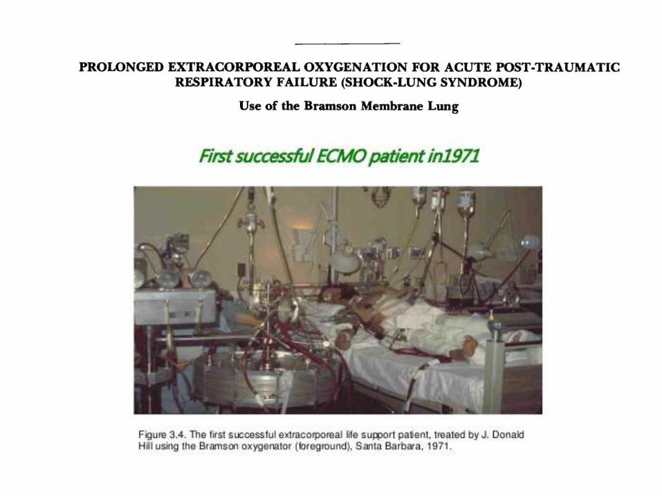
• 1971 – first successfully treated case of ARDS
• 1975 – first neonatal case
• 1978 – first description of ECCO2R
• 1989 – foundation of ESLO (Extracorporeal life
support organisation)
• 2009 – renewed interest in ARDS (CESAR trial)
• 2009 – H1N1 pandemic, lead to widespread use
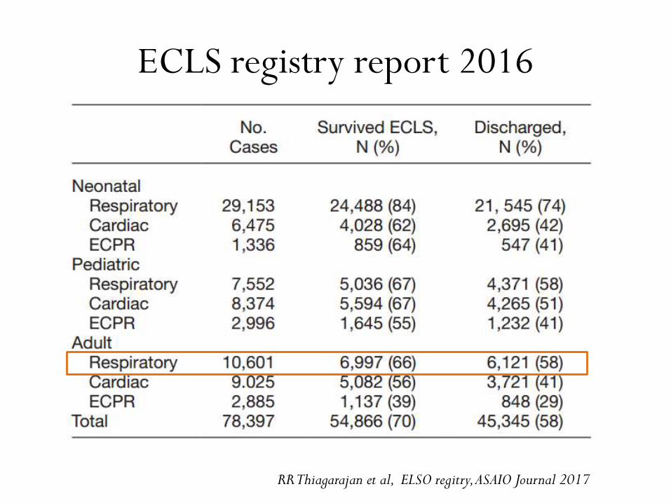
ECLS registry report 2016
RR Thiagarajan et al, ELSO regitry, ASAIO Journal 2017
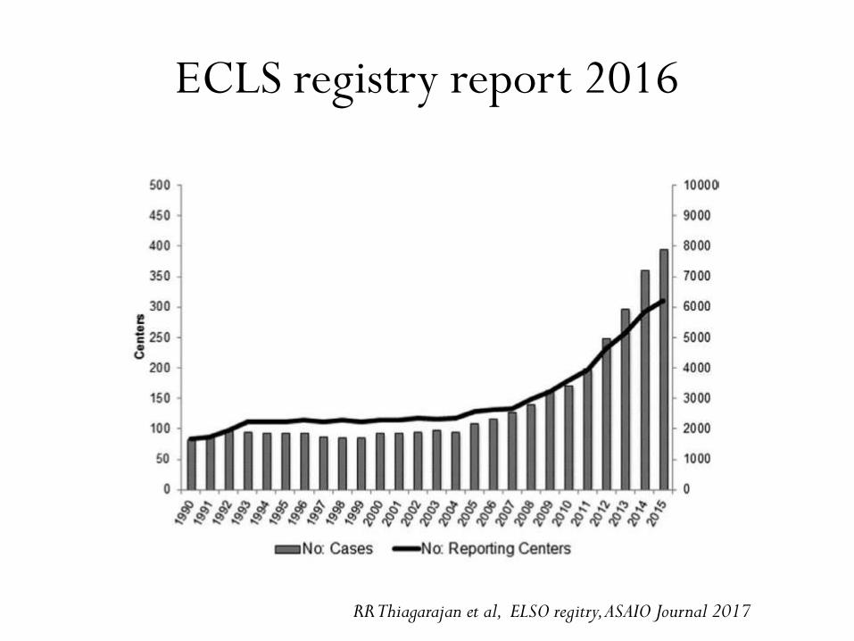
ECLS registry report 2016
RR Thiagarajan et al, ELSO regitry, ASAIO Journal 2017
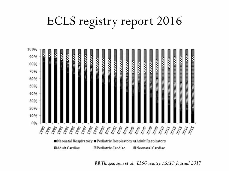
ECLS registry report 2016
RR Thiagarajan et al, ELSO regitry, ASAIO Journal 2017

ECLS in respiratory failure
RR Thiagarajan et al, ELSO regitry, ASAIO Journal 2017

BASIC PRINCIPLES
Principles
• To provide cardiopulmonary support in cases refractory
to conventional management
• To correct gas exchange abnormalities not maintained
by conventional support
• Used to prevent ventilator associated lung injury
– Reducing the delivered volumes and airway
pressures
– Low FiO2 levels
Principles
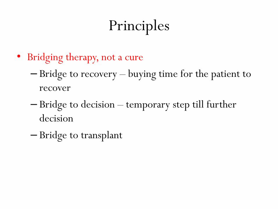
• Bridging therapy, not a cure
– Bridge to recovery – buying time for the patient to
recover
– Bridge to decision – temporary step till further
decision
– Bridge to transplant
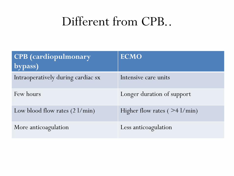
Different from CPB..
CPB (cardiopulmonary
bypass)
ECMO
Intraoperatively during cardiac sx Intensive care units
Few hours Longer duration of support
Low blood flow rates (2 l/min) Higher flow rates ( >4 l/min)
More anticoagulation Less anticoagulation
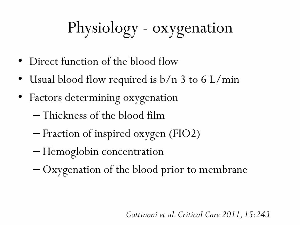
Physiology - oxygenation
• Direct function of the blood flow
• Usual blood flow required is b/n 3 to 6 L/min
• Factors determining oxygenation
– Thickness of the blood film
– Fraction of inspired oxygen (FIO2)
– Hemoglobin concentration
– Oxygenation of the blood prior to membrane
Gattinoni et al. Critical Care 2011, 15:243
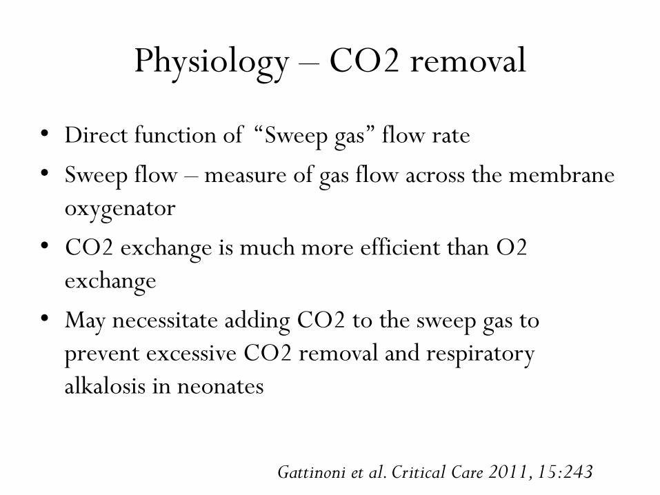
Physiology – CO2 removal
• Direct function of ‘‘Sweep gas’’ flow rate
• Sweep flow – measure of gas flow across the membrane
oxygenator
• CO2 exchange is much more efficient than O2
exchange
• May necessitate adding CO2 to the sweep gas to
prevent excessive CO2 removal and respiratory
alkalosis in neonates
Gattinoni et al. Critical Care 2011, 15:243

Physiology - Artificial lung
Oxygenation
High extracorporeal
blood flow
Low sweep flow
CO2 removal
High sweep flow
Low blood flow
Gattinoni et al. Critical Care 2011, 15:243
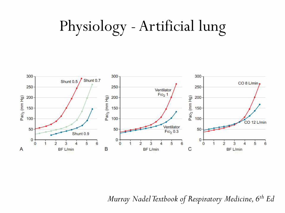
Physiology - Artificial lung
Murray NadelTextbook of Respiratory Medicine, 6th Ed
Modes of access
• Veno - venous (V-V)
– Isolated respiratory failure
• Veno - arterial (V-A)
– Isolated cardiac failure
– Cardiorespiratory failure
Venovenous ECMO
• Blood is extracted from the vena cava or right atrium
and returned to the right atrium
• Provides respiratory support, but the patient is
dependent upon his or her own hemodynamics
• Systemic blood flow and pressure are the result of the
native cardiac function unrelated to the extracorporeal
flow
Venovenous ECMO
• The PaO2 is determined by the mixing effect of
oxygenated blood returning from the ECMO circuit to
the right heart and deoxygenated blood returning from
the bronchial admixture, coronary sinus, and vena cava
• Connected in series with lungs and heart
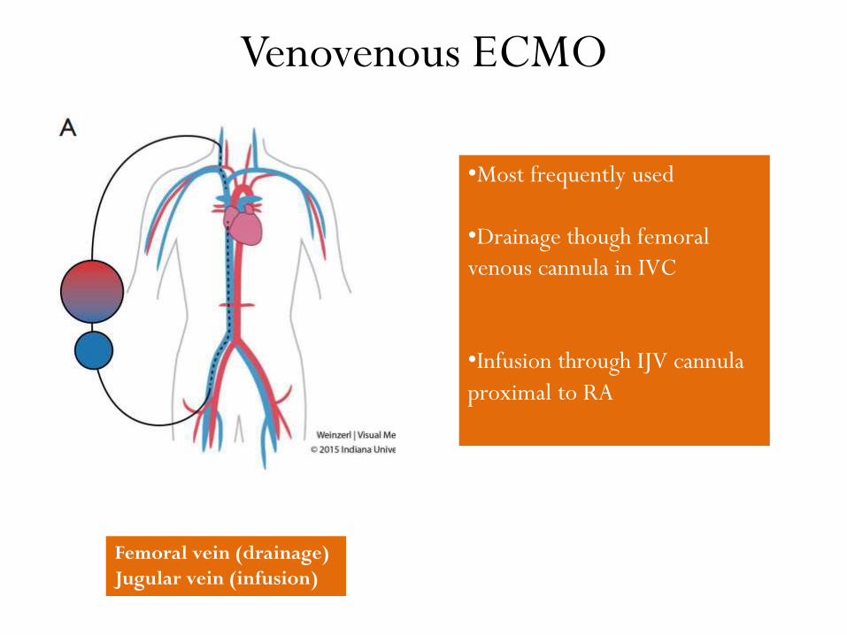
Femoral vein (drainage)
Jugular vein (infusion)
Venovenous ECMO
•Most frequently used
•Drainage though femoral
venous cannula in IVC
•Infusion through IJV cannula
proximal to RA
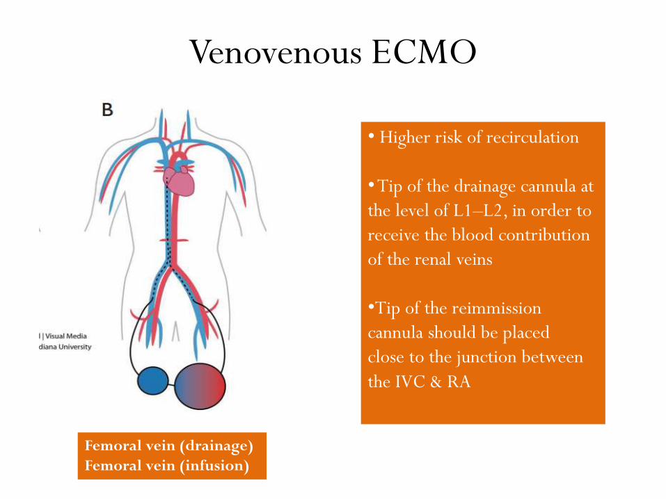
Venovenous ECMO
Femoral vein (drainage)
Femoral vein (infusion)
• Higher risk of recirculation
•Tip of the drainage cannula at
the level of L1–L2, in order to
receive the blood contribution
of the renal veins
•Tip of the reimmission
cannula should be placed
close to the junction between
the IVC & RA
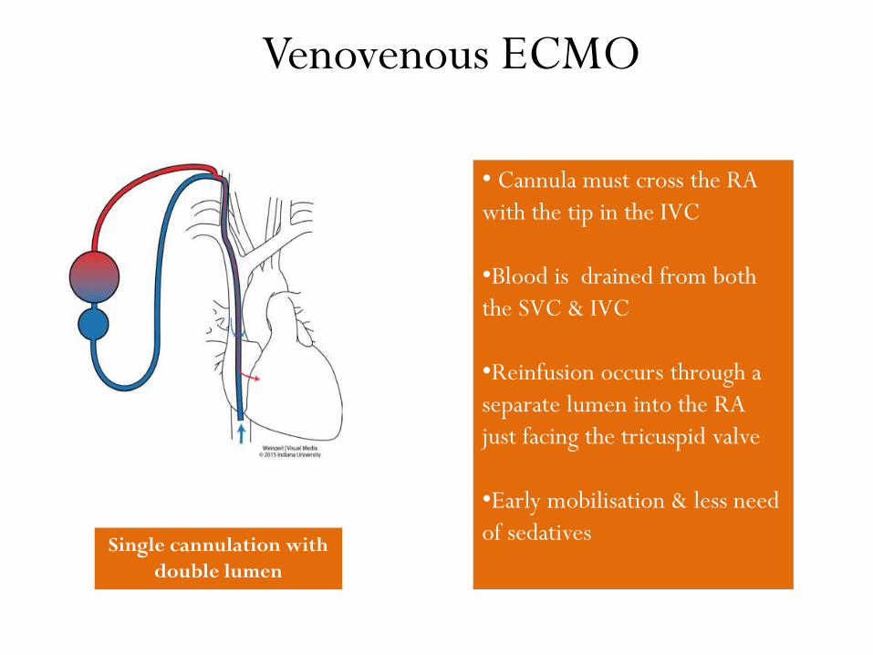
Venovenous ECMO
Single cannulation with
double lumen
• Cannula must cross the RA
with the tip in the IVC
•Blood is drained from both
the SVC & IVC
•Reinfusion occurs through a
separate lumen into the RA
just facing the tricuspid valve
•Early mobilisation & less need
of sedatives
Venoarterial ECMO
• Blood is extracted from the right atrium or vena cava
(for drainage), and returned to the arterial system
either through peripheral cannulations via femoral,
axillary or carotid arteries (for infusion)
• Connected in parallel with heart and lungs
Venoarterial ECMO
• Systemic flow, PO2 and CO2 levels are determined by
combination of the blood added from the
extracorporeal circuit plus the amount of blood passing
through the native heart and lungs
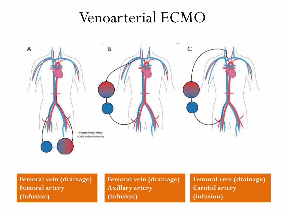
Venoarterial ECMO
Femoral vein (drainage)
Femoral artery
(infusion)
Femoral vein (drainage)
Axillary artery
(infusion)
Femoral vein (drainage)
Carotid artery
(infusion)
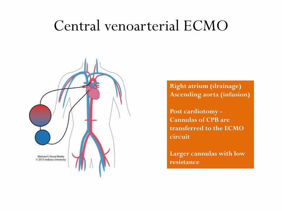
Central venoarterial ECMO
Right atrium (drainage)
Ascending aorta (infusion)
Post cardiotomy -
Cannulas of CPB are
transferred to the ECMO
circuit
Larger cannulas with low
resistance
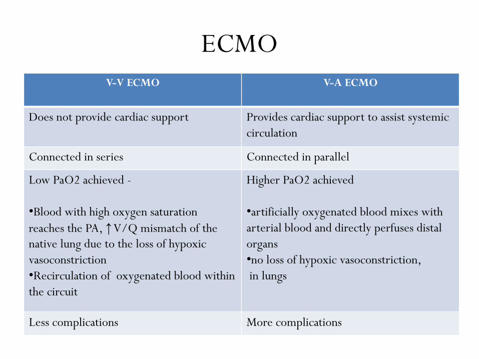
ECMO
V-V ECMO V-A ECMO
Does not provide cardiac support Provides cardiac support to assist systemic
circulation
Connected in series Connected in parallel
Low PaO2 achieved -
•Blood with high oxygen saturation
reaches the PA, ↑V/Q mismatch of the
native lung due to the loss of hypoxic
vasoconstriction
•Recirculation of oxygenated blood within
the circuit
Higher PaO2 achieved
•artificially oxygenated blood mixes with
arterial blood and directly perfuses distal
organs
•no loss of hypoxic vasoconstriction,
in lungs
Less complications More complications
INDICATIONS &
CONTRAINDICATIONS
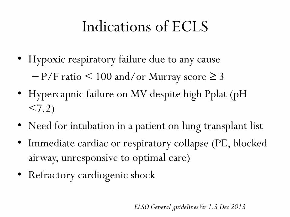
Indications of ECLS
• Hypoxic respiratory failure due to any cause
– P/F ratio < 100 and/or Murray score ≥ 3
• Hypercapnic failure on MV despite high Pplat (pH
<7.2)
• Need for intubation in a patient on lung transplant list
• Immediate cardiac or respiratory collapse (PE, blocked
airway, unresponsive to optimal care)
• Refractory cardiogenic shock
ELSO General guidelines Ver 1.3 Dec 2013
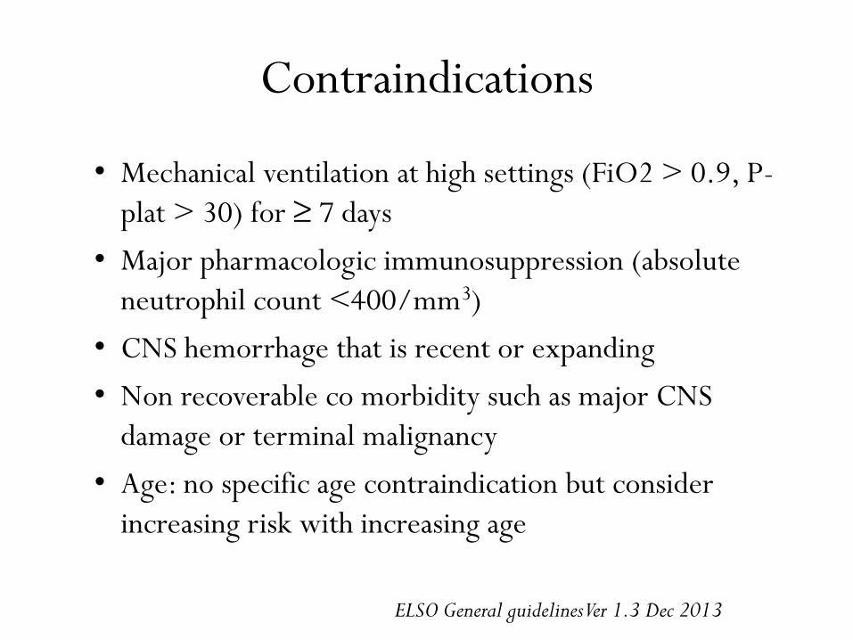
Contraindications
• Mechanical ventilation at high settings (FiO2 > 0.9, P-
plat > 30) for ≥ 7 days
• Major pharmacologic immunosuppression (absolute
neutrophil count <400/mm3)
• CNS hemorrhage that is recent or expanding
• Non recoverable co morbidity such as major CNS
damage or terminal malignancy
• Age: no specific age contraindication but consider
increasing risk with increasing age
ELSO General guidelines Ver 1.3 Dec 2013
ECMO CIRCUITRY &
COMPONENTS
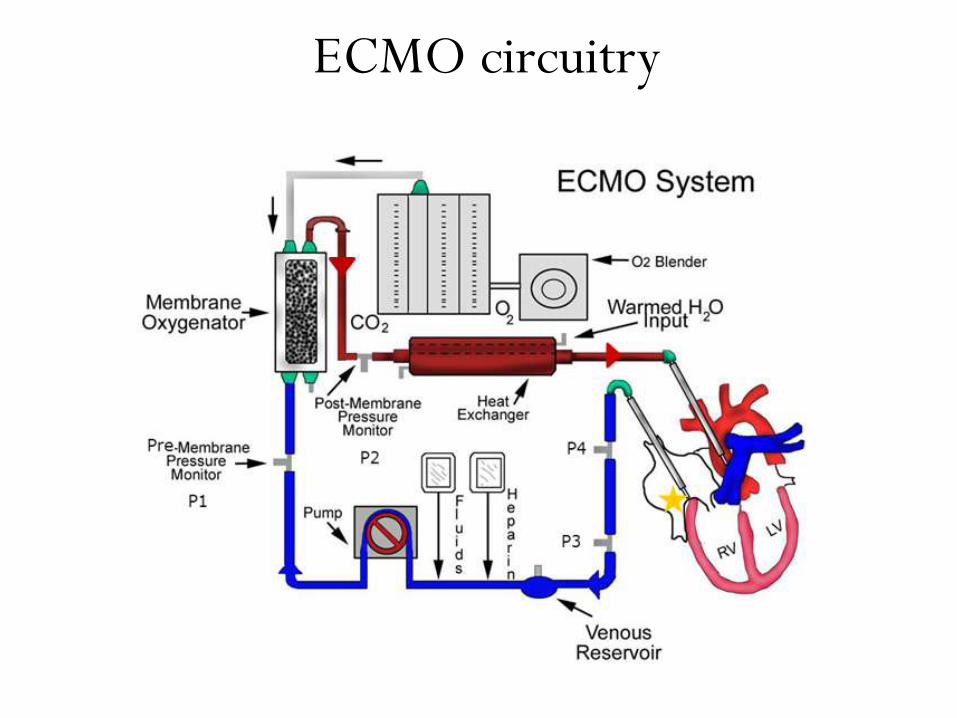
ECMO circuitry
Components
• Cannulas
• Pumps
• Oxygenator /Membrane lung
• Heat exchanger
• Tubings
Components - Cannulas
• Best cannulation technique should be chosen on the basis of patients and clinical settings
• Intrathoracic or extrathoracic
• Percutaneous or surgical
• Percutaneous approach is standard of care in VV ECMO
– Less risk of bleeding
– Short operative time
– Easier mobilization & nursing
Laurance Lequier et al; Pediatr Crit Care Med. 2013
Components - Cannulas
• Cannula size
– Big enough to ensure adequate flow with relatively low
suction pressure
– Shouldn’t exceed 2/3 rd of vessel diameter
• Positioning
– Important to minimise recirculation
– Tip should be in a high flow vessel
Laurance Lequier et al; Pediatr Crit Care Med. 2013
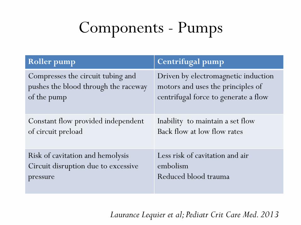
Components - Pumps
Roller pump Centrifugal pump
Compresses the circuit tubing and
pushes the blood through the raceway
of the pump
Driven by electromagnetic induction
motors and uses the principles of
centrifugal force to generate a flow
Constant flow provided independent
of circuit preload
Inability to maintain a set flow
Back flow at low flow rates
Risk of cavitation and hemolysis
Circuit disruption due to excessive
pressure
Less risk of cavitation and air
embolism
Reduced blood trauma
Laurance Lequier et al; Pediatr Crit Care Med. 2013
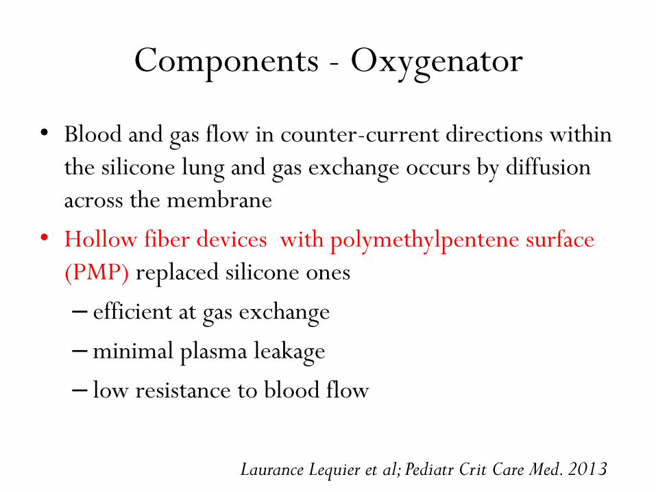
Components - Oxygenator
• Blood and gas flow in counter-current directions within
the silicone lung and gas exchange occurs by diffusion
across the membrane
• Hollow fiber devices with polymethylpentene surface
(PMP) replaced silicone ones
– efficient at gas exchange
– minimal plasma leakage
– low resistance to blood flow
Laurance Lequier et al; Pediatr Crit Care Med. 2013
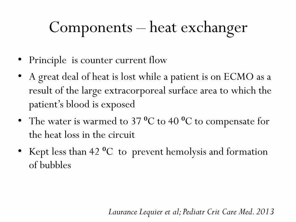
Components – heat exchanger
• Principle is counter current flow
• A great deal of heat is lost while a patient is on ECMO as a
result of the large extracorporeal surface area to which the
patient’s blood is exposed
• The water is warmed to 37 ⁰C to 40 ⁰C to compensate for
the heat loss in the circuit
• Kept less than 42 ⁰C to prevent hemolysis and formation
of bubbles
Laurance Lequier et al; Pediatr Crit Care Med. 2013
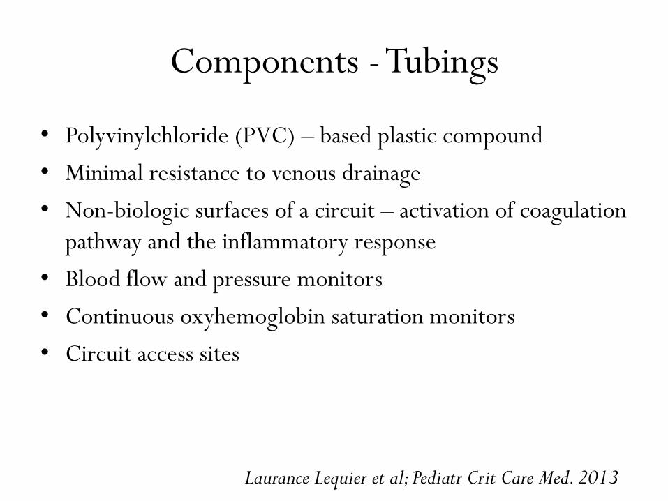
Components -Tubings
• Polyvinylchloride (PVC) – based plastic compound
• Minimal resistance to venous drainage
• Non-biologic surfaces of a circuit – activation of coagulation
pathway and the inflammatory response
• Blood flow and pressure monitors
• Continuous oxyhemoglobin saturation monitors
• Circuit access sites
Laurance Lequier et al; Pediatr Crit Care Med. 2013
Components -Tubings
• Biocompatible lining to reduce the systemic
inflammatory response and risk of thrombosis and
bleeding
• Fewer the connectors and stopcocks – less is the flow
turbulence and blood stasis
Laurance Lequier et al; Pediatr Crit Care Med. 2013
Components - Bridge
• Connection between the venous (drain) and
arterial (return) components of the circuit
• Bypass to allow the isolation of the patient from the
circuit
• If adequate gas exchange and hemodynamics can be
maintained while flow continues through the bridge
after its opening
INITIATION & MAINTENANCE
Technique of ECMO
• Should only be performed by clinicians with training
and experience in its initiation, maintenance, and
discontinuation
• The patient is anticoagulated with IV heparin
• Cannulae are inserted into the vessels
• Cannulae are connected to the limbs of circuit
• ECMO support is initiated after that
Gattinoni et al. Critical Care 2011, 15:243
S Allen et al; Journal of Intensive Care Medicine 2011
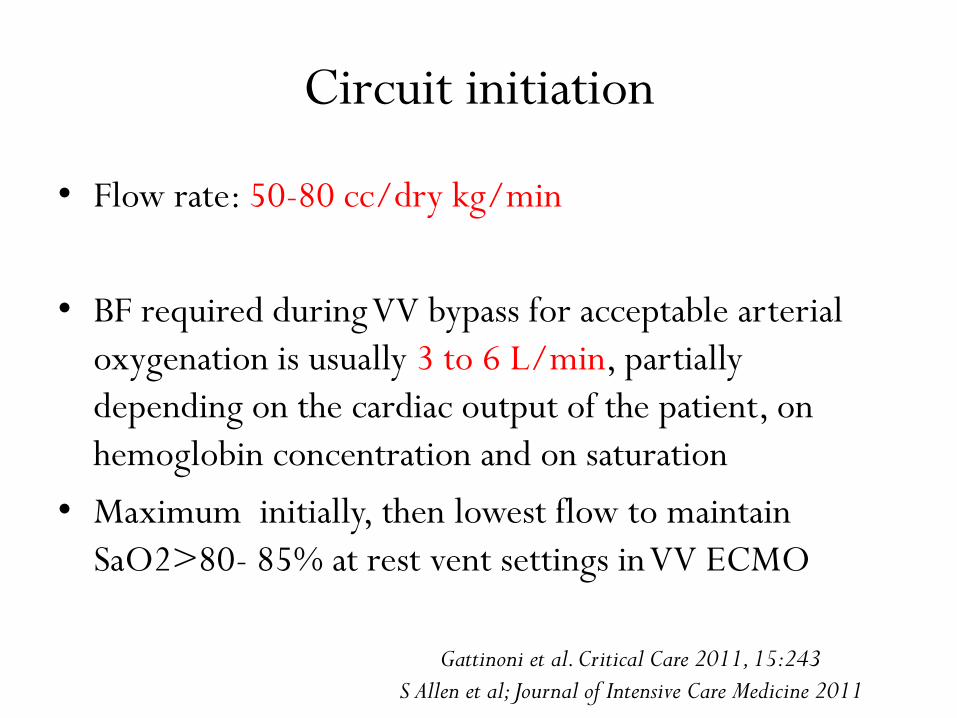
Circuit initiation
• Flow rate: 50-80 cc/dry kg/min
• BF required during VV bypass for acceptable arterial
oxygenation is usually 3 to 6 L/min, partially
depending on the cardiac output of the patient, on
hemoglobin concentration and on saturation
• Maximum initially, then lowest flow to maintain
SaO2>80- 85% at rest vent settings in VV ECMO
Gattinoni et al. Critical Care 2011, 15:243
S Allen et al; Journal of Intensive Care Medicine 2011
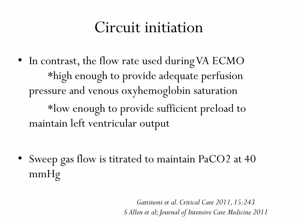
Circuit initiation
• In contrast, the flow rate used during VA ECMO
*high enough to provide adequate perfusion
pressure and venous oxyhemoglobin saturation
*low enough to provide sufficient preload to
maintain left ventricular output
• Sweep gas flow is titrated to maintain PaCO2 at 40
mmHg
Gattinoni et al. Critical Care 2011, 15:243
S Allen et al; Journal of Intensive Care Medicine 2011
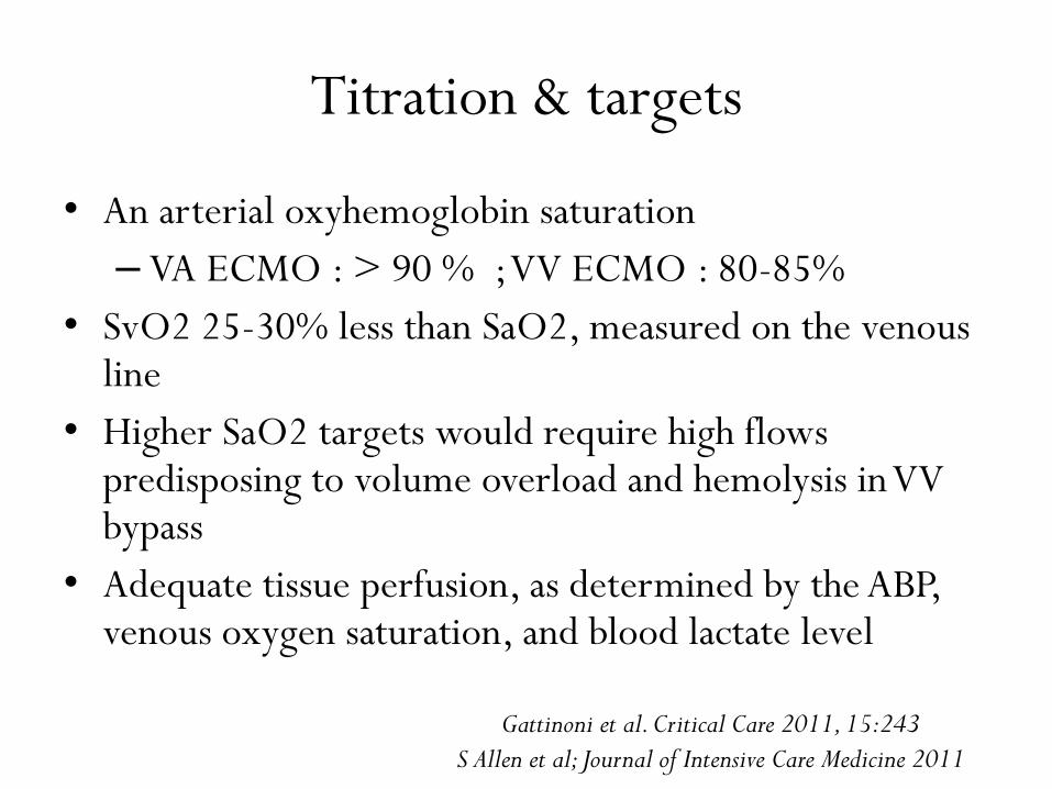
Titration & targets
• An arterial oxyhemoglobin saturation
– VA ECMO : > 90 % ; VV ECMO : 80-85%
• SvO2 25-30% less than SaO2, measured on the venous line
• Higher SaO2 targets would require high flows predisposing to volume overload and hemolysis in VV bypass
• Adequate tissue perfusion, as determined by the ABP, venous oxygen saturation, and blood lactate level
Gattinoni et al. Critical Care 2011, 15:243
S Allen et al; Journal of Intensive Care Medicine 2011
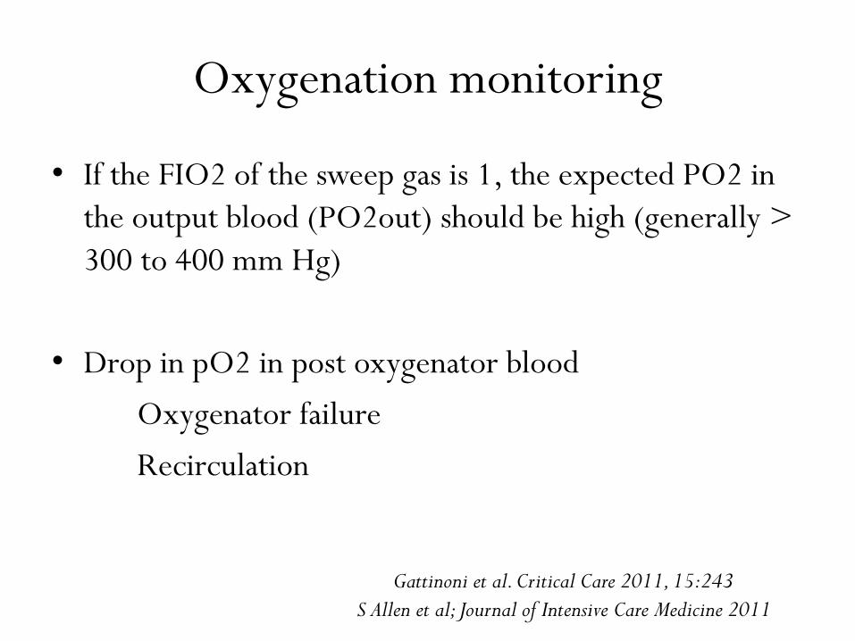
Oxygenation monitoring
• If the FIO2 of the sweep gas is 1, the expected PO2 in
the output blood (PO2out) should be high (generally >
300 to 400 mm Hg)
• Drop in pO2 in post oxygenator blood
Oxygenator failure
Recirculation
Gattinoni et al. Critical Care 2011, 15:243
S Allen et al; Journal of Intensive Care Medicine 2011
Oxygenation monitoring
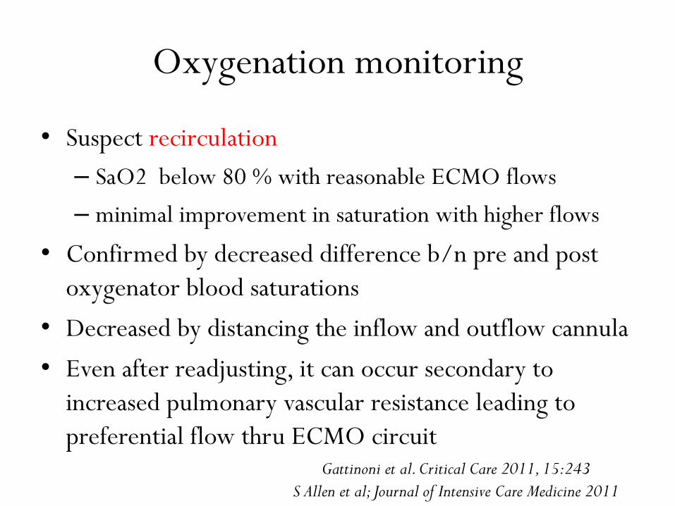
• Suspect recirculation
– SaO2 below 80 % with reasonable ECMO flows
– minimal improvement in saturation with higher flows
• Confirmed by decreased difference b/n pre and post
oxygenator blood saturations
• Decreased by distancing the inflow and outflow cannula
• Even after readjusting, it can occur secondary to
increased pulmonary vascular resistance leading to
preferential flow thru ECMO circuitGattinoni et al. Critical Care 2011, 15:243
S Allen et al; Journal of Intensive Care Medicine 2011
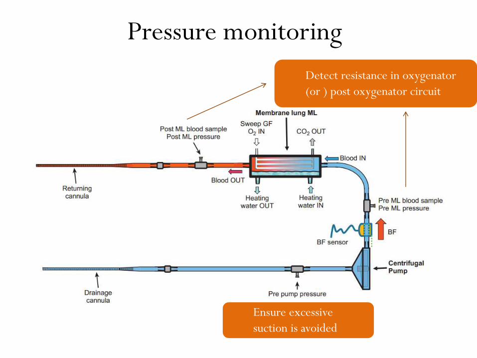
Pressure monitoring
Ensure excessive
suction is avoided
Detect resistance in oxygenator
(or ) post oxygenator circuit
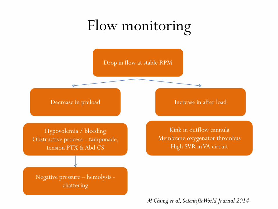
Flow monitoring
Drop in flow at stable RPM
Increase in after loadDecrease in preload
Hypovolemia / bleeding
Obstructive process – tamponade,
tension PTX & Abd CS
Negative pressure – hemolysis -
chattering
Kink in outflow cannula
Membrane oxygenator thrombus
High SVR in VA circuit
M Chung et al, Scientific World Journal 2014
MAP monitoring
• Essential in case of V A ECMO bypass - MAP > 65 mm Hg
• MAP should not exceed 90 mm Hg in order to limit the
afterload
• MAP can be increased by administering the volume or by
increasing the RPM
• Correction of volume status and vasopressor support as
indicated to maintain MAP
M Chung et al, Scientific World Journal 2014
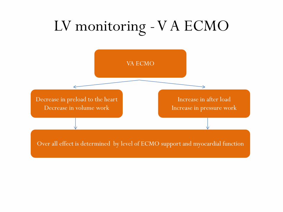
LV monitoring -V A ECMO
VA ECMO
Increase in after load
Increase in pressure work
Decrease in preload to the heart
Decrease in volume work
Over all effect is determined by level of ECMO support and myocardial function
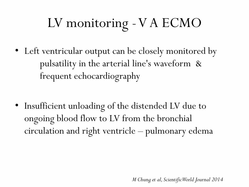
LV monitoring -V A ECMO
• Left ventricular output can be closely monitored by
pulsatility in the arterial line's waveform &
frequent echocardiography
• Insufficient unloading of the distended LV due to
ongoing blood flow to LV from the bronchial
circulation and right ventricle – pulmonary edema
M Chung et al, Scientific World Journal 2014
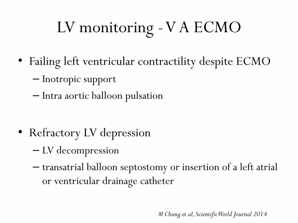
LV monitoring -V A ECMO
• Failing left ventricular contractility despite ECMO
– Inotropic support
– Intra aortic balloon pulsation
• Refractory LV depression
– LV decompression
– transatrial balloon septostomy or insertion of a left atrial
or ventricular drainage catheter
M Chung et al, Scientific World Journal 2014
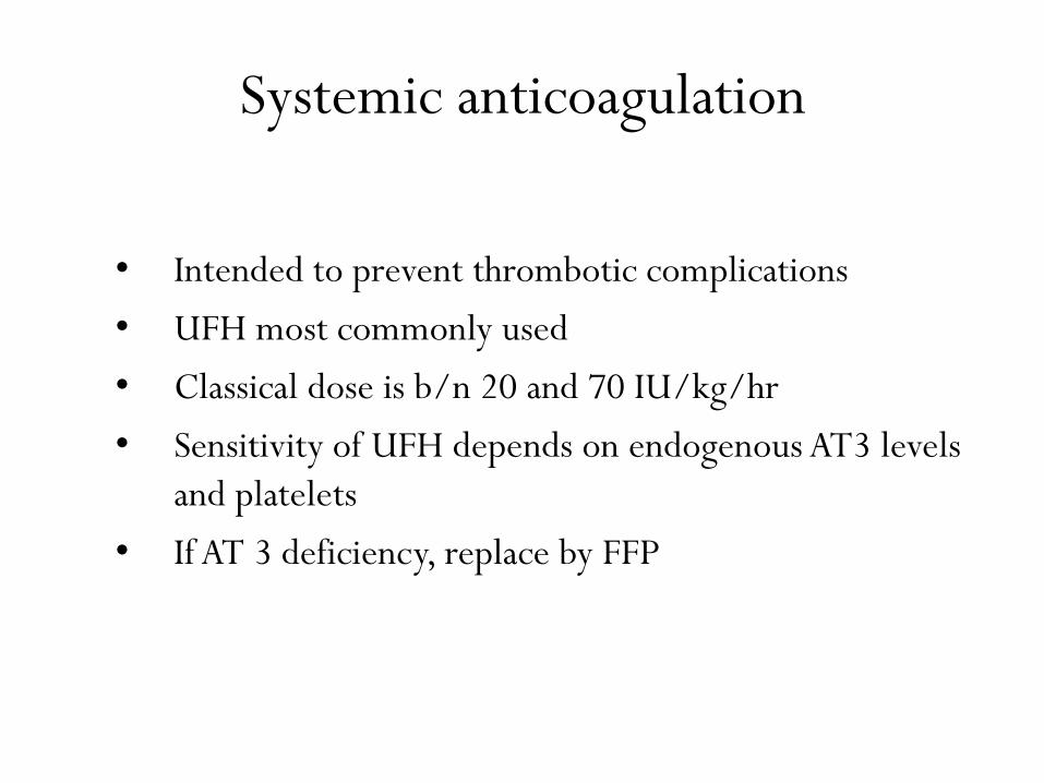
Systemic anticoagulation
• Intended to prevent thrombotic complications
• UFH most commonly used
• Classical dose is b/n 20 and 70 IU/kg/hr
• Sensitivity of UFH depends on endogenous AT3 levels
and platelets
• If AT 3 deficiency, replace by FFP
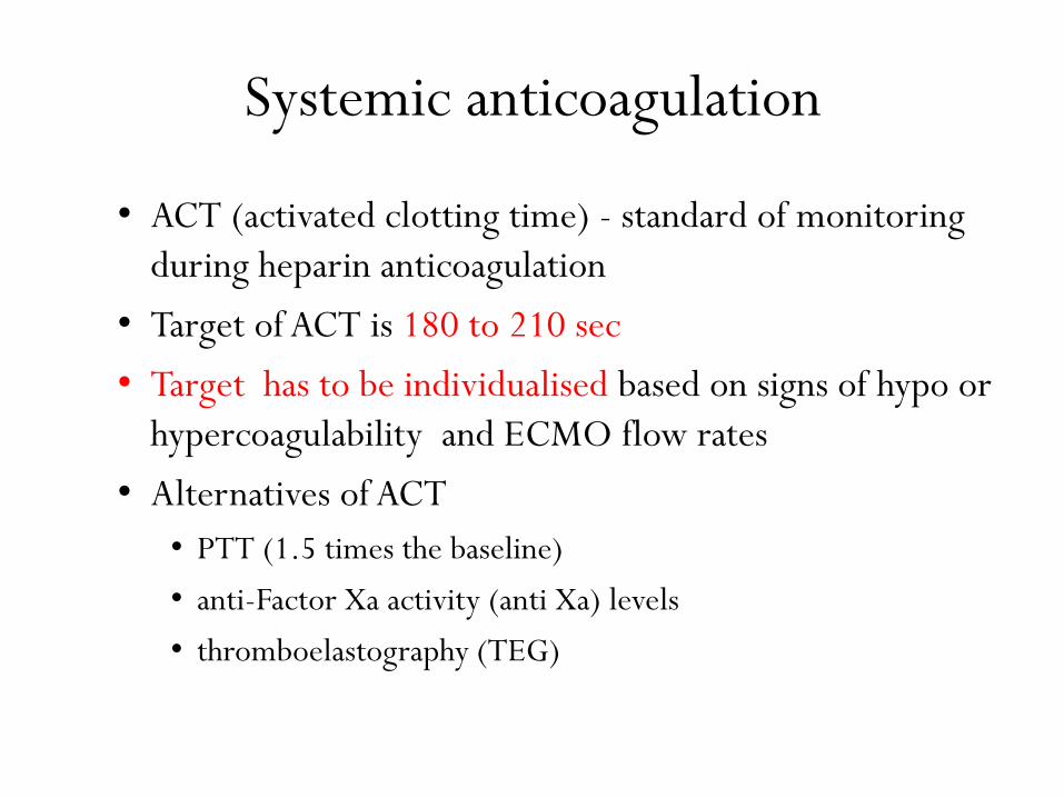
Systemic anticoagulation
• ACT (activated clotting time) - standard of monitoring
during heparin anticoagulation
• Target of ACT is 180 to 210 sec
• Target has to be individualised based on signs of hypo or
hypercoagulability and ECMO flow rates
• Alternatives of ACT
• PTT (1.5 times the baseline)
• anti-Factor Xa activity (anti Xa) levels
• thromboelastography (TEG)
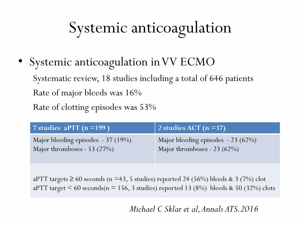
Systemic anticoagulation
• Systemic anticoagulation in VV ECMO
Systematic review, 18 studies including a total of 646 patients
Rate of major bleeds was 16%
Rate of clotting episodes was 53%
7 studies aPTT (n =199 ) 2 studies ACT (n =37)
Major bleeding episodes - 37 (19%)
Major thromboses - 53 (27%)
Major bleeding episodes - 23 (62%)
Major thromboses - 23 (62%)
aPTT targets ≥ 60 seconds (n =43, 5 studies) reported 24 (56%) bleeds & 3 (7%) clot
aPTT target < 60 seconds(n = 156, 3 studies) reported 13 (8%) bleeds & 50 (32%) clots
Michael C Sklar et al, Annals ATS.2016
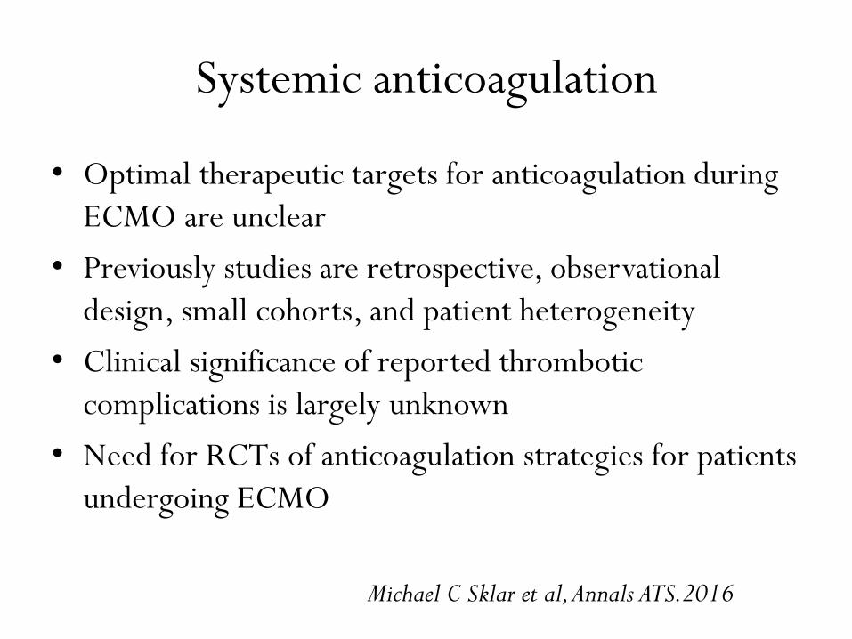
Systemic anticoagulation
• Optimal therapeutic targets for anticoagulation during
ECMO are unclear
• Previously studies are retrospective, observational
design, small cohorts, and patient heterogeneity
• Clinical significance of reported thrombotic
complications is largely unknown
• Need for RCTs of anticoagulation strategies for patients
undergoing ECMO
Michael C Sklar et al, Annals ATS.2016

MV - ECMO
• Significant knowledge gap in understanding the benefits
and risks of MV during ECMO
• Risk of VILI
– Limitation of the alveolar strain by decrease in Vt
– High PEEP with low Vt to prevent atelectrauma
– Avoid oxygen toxicity to the lung from a high FiO2
& reabsorption atelectasis
Schmidt et al. Critical Care 2014, 18:203
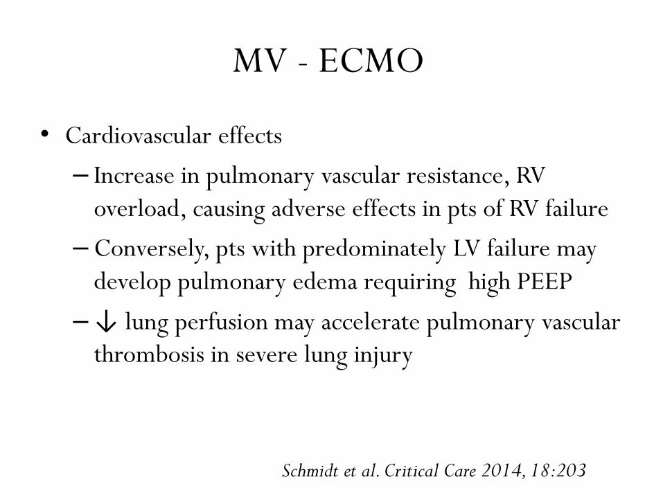
MV - ECMO
• Cardiovascular effects
– Increase in pulmonary vascular resistance, RV
overload, causing adverse effects in pts of RV failure
– Conversely, pts with predominately LV failure may
develop pulmonary edema requiring high PEEP
–↓ lung perfusion may accelerate pulmonary vascular
thrombosis in severe lung injury
Schmidt et al. Critical Care 2014, 18:203
MV - ECMO
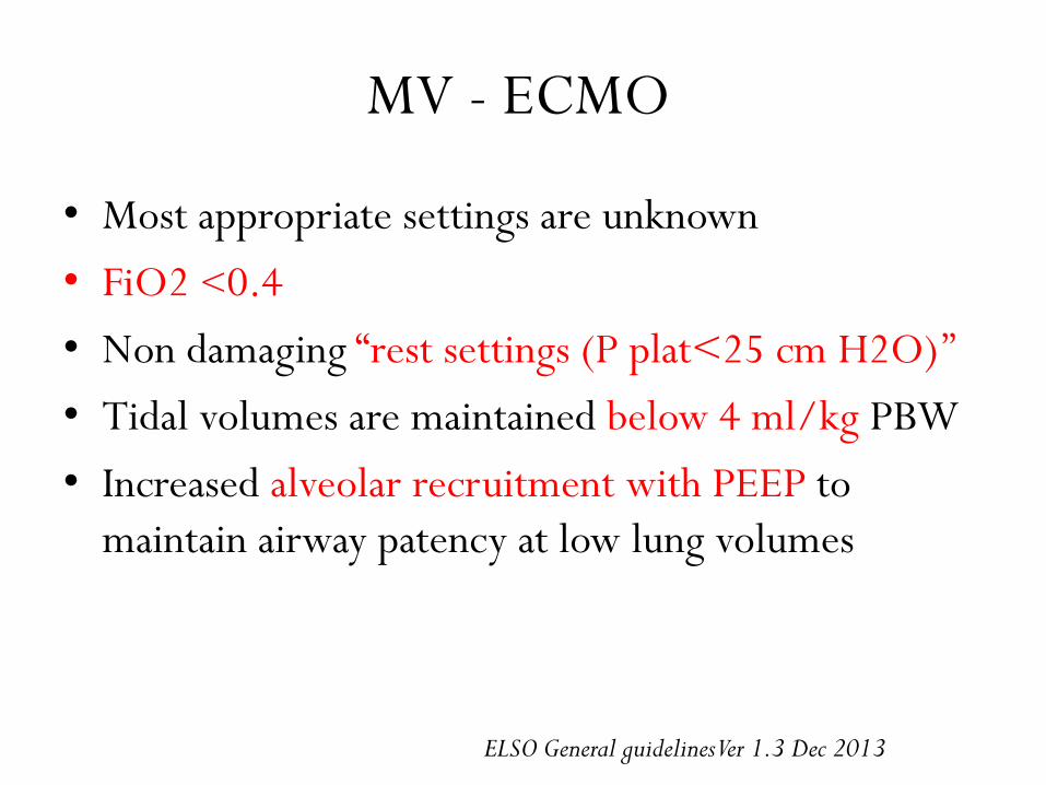
• Most appropriate settings are unknown
• FiO2 <0.4
• Non damaging “rest settings (P plat<25 cm H2O)’’
• Tidal volumes are maintained below 4 ml/kg PBW
• Increased alveolar recruitment with PEEP to
maintain airway patency at low lung volumes
ELSO General guidelines Ver 1.3 Dec 2013
MV - ECMO
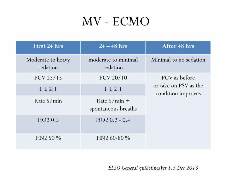
First 24 hrs 24 – 48 hrs After 48 hrs
Moderate to heavy
sedation
moderate to minimal
sedation
Minimal to no sedation
PCV 25/15 PCV 20/10 PCV as before
or take on PSV as the
condition improvesI: E 2:1 I: E 2:1
Rate 5/min Rate 5/min +
spontaneous breaths
FiO2 0.5 FiO2 0.2 - 0.4
FiN2 50 % FiN2 60-80 %
ELSO General guidelines Ver 1.3 Dec 2013
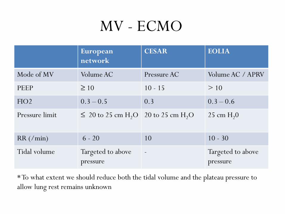
MV - ECMO
European
network
CESAR EOLIA
Mode of MV Volume AC Pressure AC Volume AC / APRV
PEEP ≥ 10 10 - 15 > 10
FIO2 0.3 – 0.5 0.3 0.3 – 0.6
Pressure limit ≤ 20 to 25 cm H2O 20 to 25 cm H2O 25 cm H20
RR (/min) 6 - 20 10 10 - 30
Tidal volume Targeted to above
pressure
- Targeted to above
pressure
* To what extent we should reduce both the tidal volume and the plateau pressure to
allow lung rest remains unknown
Transfusion support
• The benefit of enhanced oxygen delivery must be
weighed against the potential harm of transfusion
• Many centers recommend transfusion who are
receiving ECMO until their hematocrit levels are in
the normal range
• Lesser blood flows in the circuit are required if
hematocrit is maintained
Daniel Brodie NEJM 2011;365:1905-14.
Transfusion support
• Platelets are continuously consumed during ECMO
because they are activated by exposure to the
foreign surface area
• Platelet counts should be maintained greater than
50,000/microL, which may require platelet
transfusion
Daniel Brodie NEJM 2011;365:1905-14.
Weaning from ECMO
• Progressive reduction of the ECMO contribution to
oxygenation and CO2 removal as the gas exchange
capability of the native lung improves and the
patient’s clinical conditions stabilize
• Requires regular monitoring the pts respiratory
function (gas exchange function, respiratory
mechanics) and hemodynamics
Steve Allen et al, Journal of Intensive Care Medicine 26(1) 13-26
Weaning from ECMO
• Respiratory failure
– when 50% to 80% of total gas exchange is by the native lungs
– when the patient’s lung compliance improves
– improving chest x-ray
• Cardiac failure
– Enhanced aortic pulsatility correlates with improved left
ventricular output
– Decrease in mixed-venous oxygenation saturation
– MAP> 60 mmHg in the absence of “high-dose” inopressors
Steve Allen et al, Journal of Intensive Care Medicine 26(1) 13-26
Weaning from ECMO
• VV ECMO trials
– Sweep low rate is slowly decreased
– Ventilator is placed on full support
– Successful weaning is confirmed if the patient remains
stable at a FGF of 0 L/min for a period of 4 to 24 hours
Steve Allen et al, Journal of Intensive Care Medicine 26(1) 13-26
Weaning from ECMO
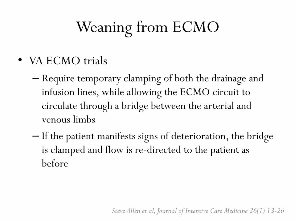
• VA ECMO trials
– Require temporary clamping of both the drainage and
infusion lines, while allowing the ECMO circuit to
circulate through a bridge between the arterial and
venous limbs
– If the patient manifests signs of deterioration, the bridge
is clamped and flow is re-directed to the patient as
before
Steve Allen et al, Journal of Intensive Care Medicine 26(1) 13-26

COMPLICATIONS
Complications ECLS
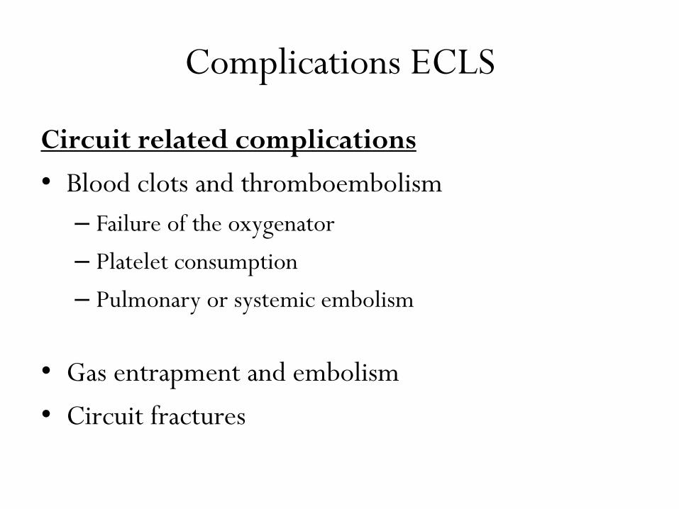
Circuit related complications
• Blood clots and thromboembolism
– Failure of the oxygenator
– Platelet consumption
– Pulmonary or systemic embolism
• Gas entrapment and embolism
• Circuit fractures
Complications ECLS
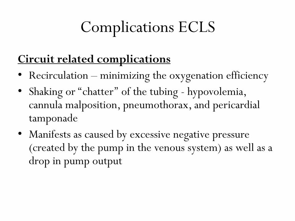
Circuit related complications
• Recirculation – minimizing the oxygenation efficiency
• Shaking or ‘‘chatter’’ of the tubing - hypovolemia, cannula malposition, pneumothorax, and pericardial tamponade
• Manifests as caused by excessive negative pressure (created by the pump in the venous system) as well as a drop in pump output
Complications ECLS
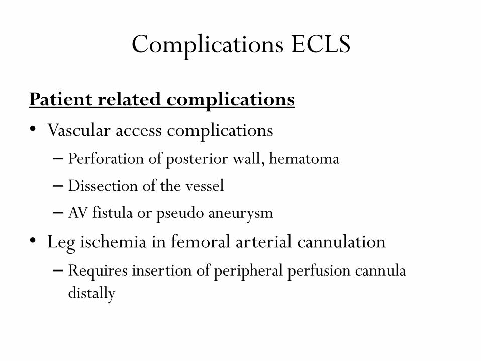
Patient related complications
• Vascular access complications
– Perforation of posterior wall, hematoma
– Dissection of the vessel
– AV fistula or pseudo aneurysm
• Leg ischemia in femoral arterial cannulation
– Requires insertion of peripheral perfusion cannula
distally
Complications ECLS
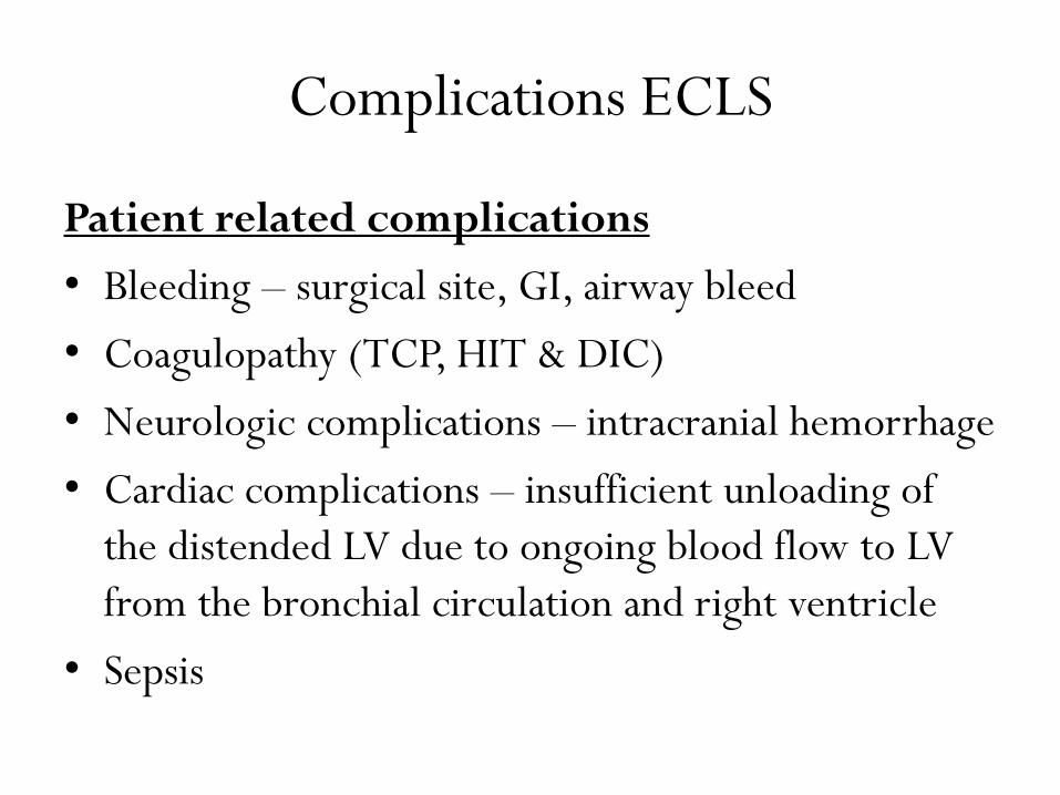
Patient related complications
• Bleeding – surgical site, GI, airway bleed
• Coagulopathy (TCP, HIT & DIC)
• Neurologic complications – intracranial hemorrhage
• Cardiac complications – insufficient unloading of
the distended LV due to ongoing blood flow to LV
from the bronchial circulation and right ventricle
• Sepsis
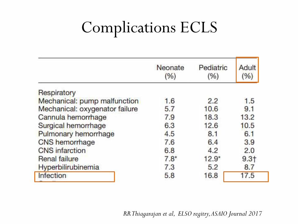
Complications ECLS
RR Thiagarajan et al, ELSO regitry, ASAIO Journal 2017

ECMO - ARDS
ECMO in ARDS
• Currently a salvage therapy for the most severe
cases of ARDS
• Benefit of ECMO as compared to conventional,
standard of care management for ARDS has yet to
be demonstrated
• Increasing potential for ECMO to enhance the way
ARDS is managed
M Parekh et al; Ann Transl Med 2017
Benefit in ARDS
• Complete lung rest – Lung protective ventilation
• Complete avoidance of VILI
• Adequate gas exchange extracorporeally
• Decreases Oxygen toxicity to lung
M Parekh et al; Ann Transl Med 2017
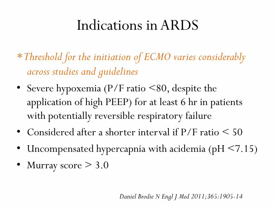
Indications in ARDS
* Threshold for the initiation of ECMO varies considerably
across studies and guidelines
• Severe hypoxemia (P/F ratio <80, despite the
application of high PEEP) for at least 6 hr in patients
with potentially reversible respiratory failure
• Considered after a shorter interval if P/F ratio < 50
• Uncompensated hypercapnia with acidemia (pH <7.15)
• Murray score > 3.0
Daniel Brodie N Engl J Med 2011;365:1905-14
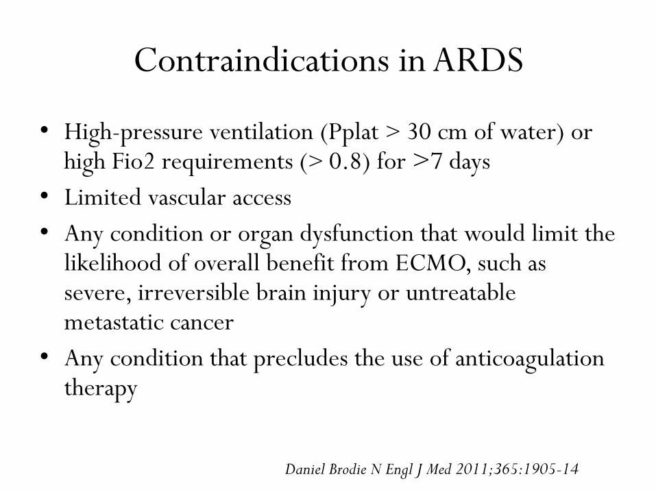
Contraindications in ARDS
• High-pressure ventilation (Pplat > 30 cm of water) or high Fio2 requirements (> 0.8) for >7 days
• Limited vascular access
• Any condition or organ dysfunction that would limit the likelihood of overall benefit from ECMO, such as severe, irreversible brain injury or untreatable metastatic cancer
• Any condition that precludes the use of anticoagulation therapy
Daniel Brodie N Engl J Med 2011;365:1905-14
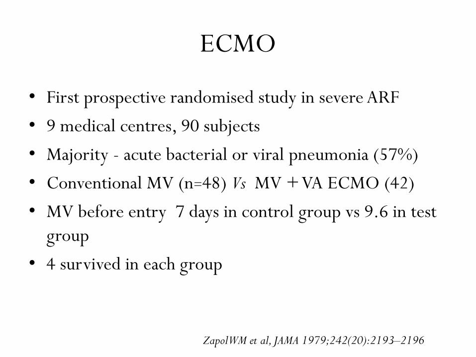
ECMO
• First prospective randomised study in severe ARF
• 9 medical centres, 90 subjects
• Majority - acute bacterial or viral pneumonia (57%)
• Conventional MV (n=48) Vs MV + VA ECMO (42)
• MV before entry 7 days in control group vs 9.6 in test
group
• 4 survived in each group
Zapol WM et al, JAMA 1979;242(20):2193–2196
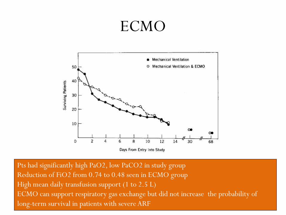
ECMO
Pts had significantly high PaO2, low PaCO2 in study group
Reduction of FiO2 from 0.74 to 0.48 seen in ECMO group
High mean daily transfusion support (1 to 2.5 L)
ECMO can support respiratory gas exchange but did not increase the probability of
long-term survival in patients with severe ARF
ECMO
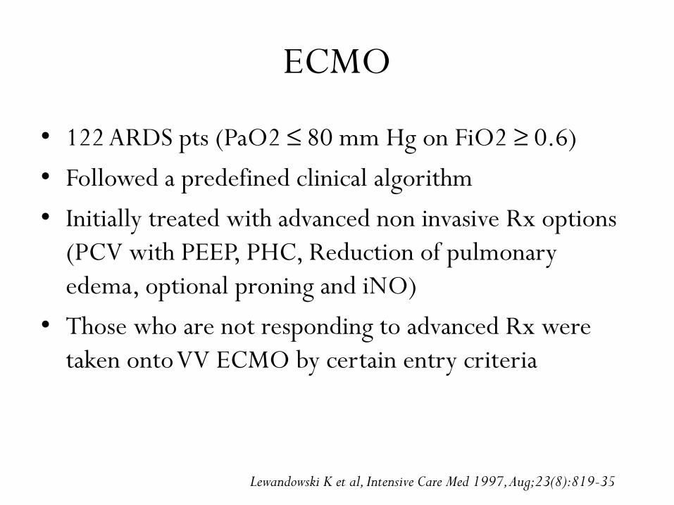
• 122 ARDS pts (PaO2 ≤ 80 mm Hg on FiO2 ≥ 0.6)
• Followed a predefined clinical algorithm
• Initially treated with advanced non invasive Rx options
(PCV with PEEP, PHC, Reduction of pulmonary
edema, optional proning and iNO)
• Those who are not responding to advanced Rx were
taken onto VV ECMO by certain entry criteria
Lewandowski K et al, Intensive Care Med 1997, Aug;23(8):819-35
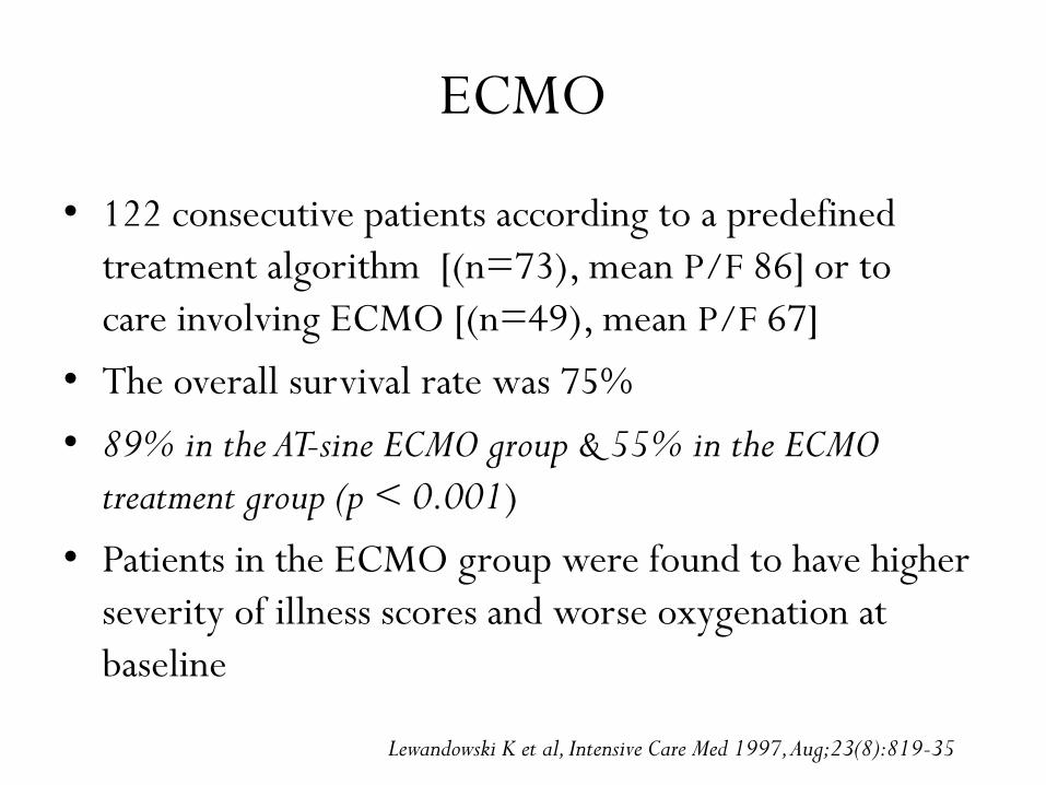
ECMO
• 122 consecutive patients according to a predefined
treatment algorithm [(n=73), mean P/F 86] or to
care involving ECMO [(n=49), mean P/F 67]
• The overall survival rate was 75%
• 89% in the AT-sine ECMO group & 55% in the ECMO
treatment group (p < 0.001)
• Patients in the ECMO group were found to have higher
severity of illness scores and worse oxygenation at
baseline
Lewandowski K et al, Intensive Care Med 1997, Aug;23(8):819-35
ECMO
• Evidence from these studies suggested no definite
benefit of ECMO over conventional mechanical
ventilation
• Its usage was restricted to clinical trials and not got
widely implemented
• However these studies have little relevance now due to
changed ventilatory strategies, ECMO circuits, disease
management and increased experience with it
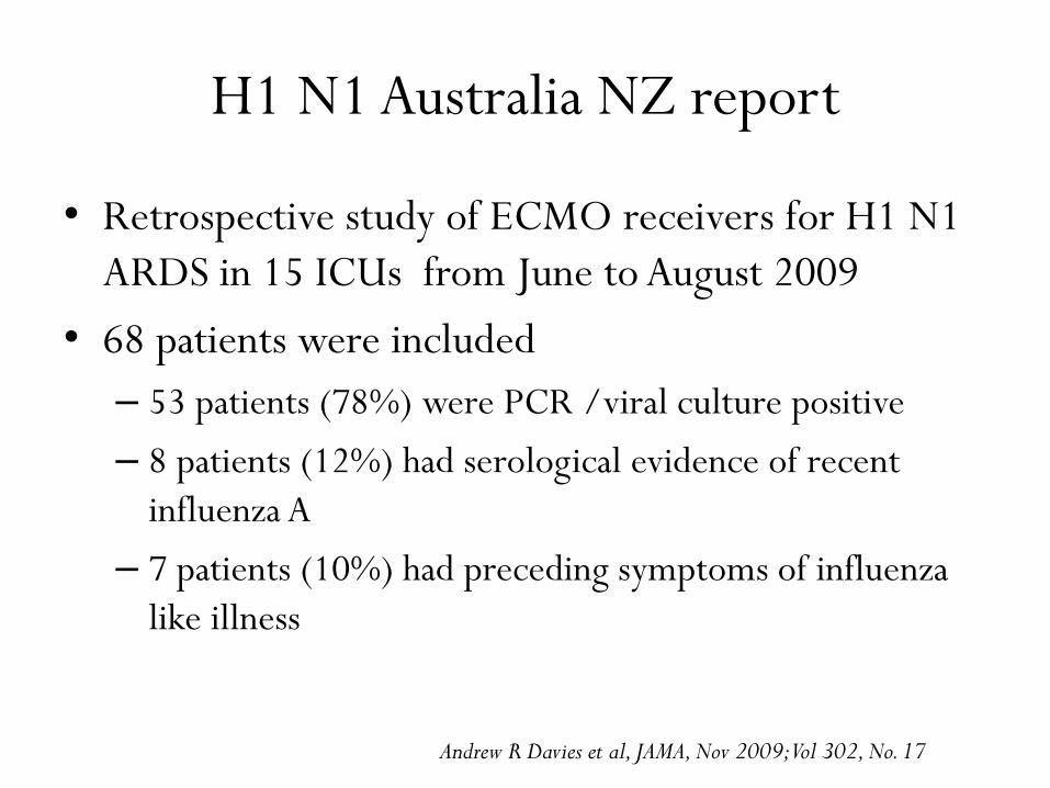
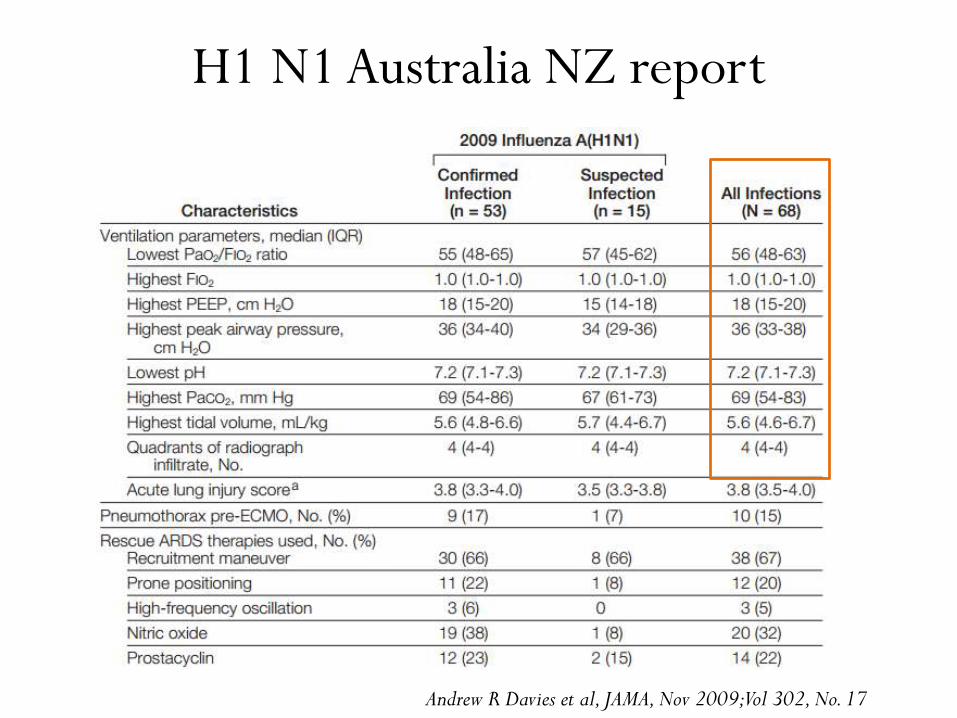
H1 N1 Australia NZ report
• Retrospective study of ECMO receivers for H1 N1
ARDS in 15 ICUs from June to August 2009
• 68 patients were included
– 53 patients (78%) were PCR /viral culture positive
– 8 patients (12%) had serological evidence of recent
influenza A
– 7 patients (10%) had preceding symptoms of influenza
like illness
Andrew R Davies et al, JAMA, Nov 2009;Vol 302, No. 17
H1 N1 Australia NZ report
Andrew R Davies et al, JAMA, Nov 2009; Vol 302, No. 17
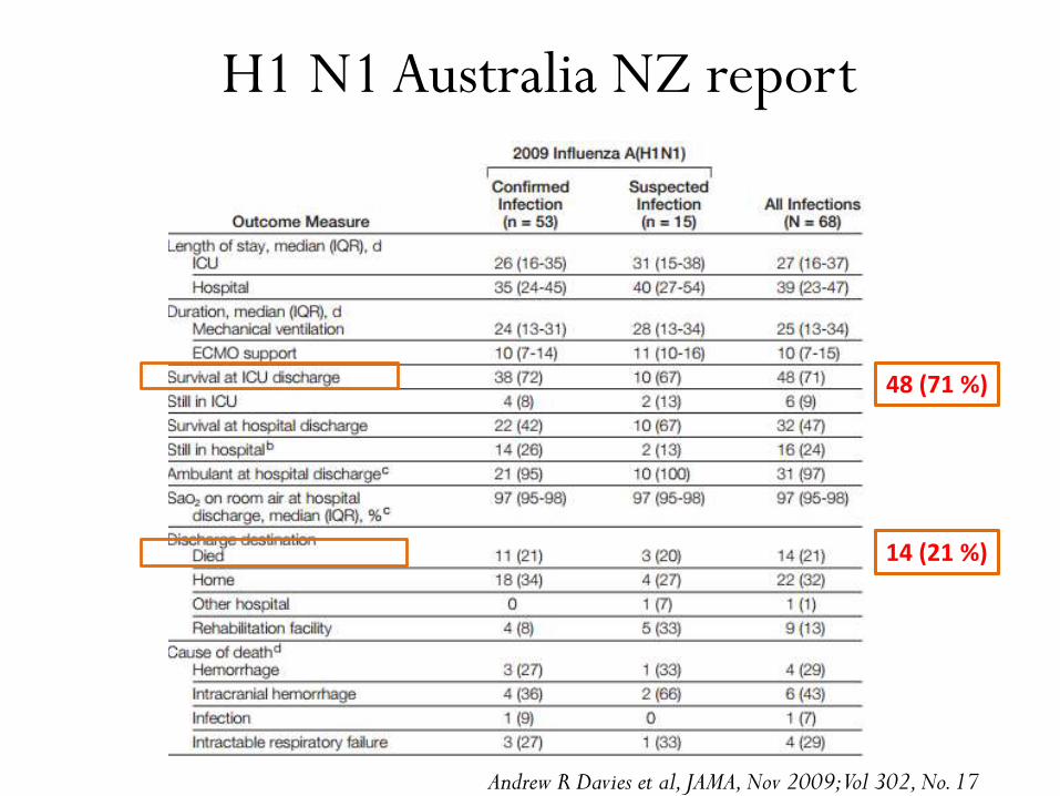
H1 N1 Australia NZ report
14 (21 %)
48 (71 %)
Andrew R Davies et al, JAMA, Nov 2009;Vol 302, No. 17
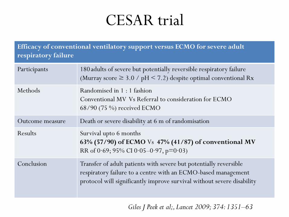
CESAR trial
Efficacy of conventional ventilatory support versus ECMO for severe adult
respiratory failure
Participants 180 adults of severe but potentially reversible respiratory failure
(Murray score ≥ 3.0 / pH < 7.2) despite optimal conventional Rx
Methods Randomised in 1 : 1 fashion
Conventional MV Vs Referral to consideration for ECMO
68/90 (75 %) received ECMO
Outcome measure Death or severe disability at 6 m of randomisation
Results Survival upto 6 months
63% (57/90) of ECMO Vs 47% (41/87) of conventional MV
RR of 0·69; 95% CI 0·05–0·97, p=0·03)
Conclusion Transfer of adult patients with severe but potentially reversible
respiratory failure to a centre with an ECMO-based management
protocol will significantly improve survival without severe disability
Giles J Peek et al;, Lancet 2009; 374: 1351–63
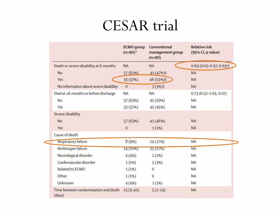
CESAR trial
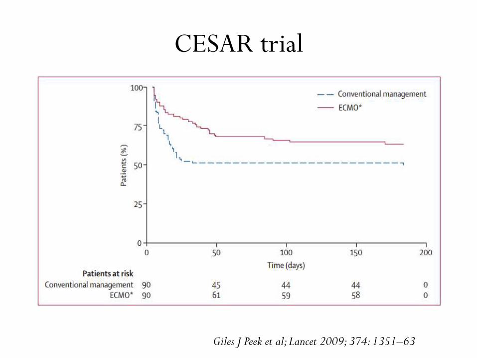
CESAR trial
Giles J Peek et al; Lancet 2009; 374: 1351–63
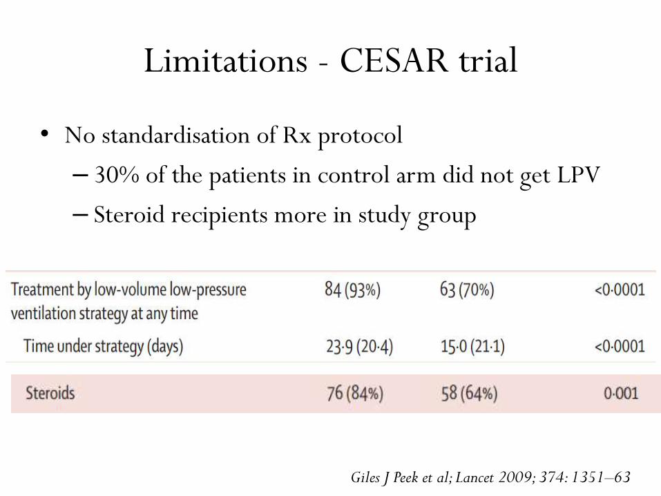
Limitations - CESAR trial
• No standardisation of Rx protocol
– 30% of the patients in control arm did not get LPV
– Steroid recipients more in study group
Giles J Peek et al; Lancet 2009; 374: 1351–63
Limitations - CESAR trial
• Intervention in CESAR was referral to an ECMO center
not treatment with ECMO
▪ 25 % of patients in ECMO referral group didnot receive
ECMO
• Two serious adverse events noted
– Mechanical failure of O2 supply during transport
– Vessel perforation during cannulation
Giles J Peek et al; Lancet 2009; 374: 1351–63
Limitations - CESAR trial
• Three patients died before they could be transferred
and two died in transit
• Risk of death during transfer of such patients
• Exclusion of pts ventilated with high pressure or
high FiO₂ for more than 7 days
• Did improved care at the single ECMO hospital lead
to the relative risk observed??
Giles J Peek et al; CESAR trial, Lancet 2009; 374: 1351–63
ECMO
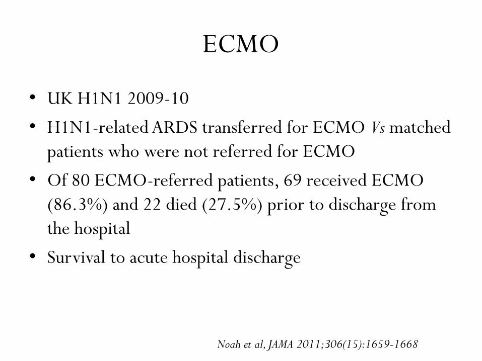
• UK H1N1 2009-10
• H1N1-related ARDS transferred for ECMO Vs matched
patients who were not referred for ECMO
• Of 80 ECMO-referred patients, 69 received ECMO
(86.3%) and 22 died (27.5%) prior to discharge from
the hospital
• Survival to acute hospital discharge
Noah et al, JAMA 2011;306(15):1659-1668
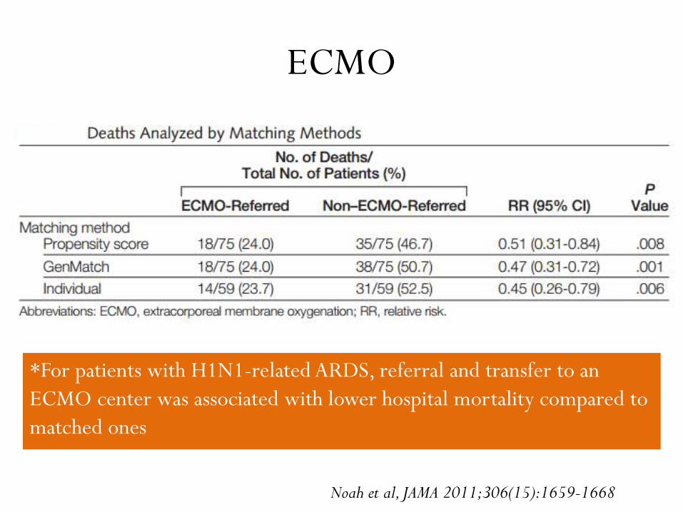
ECMO
*For patients with H1N1-related ARDS, referral and transfer to an
ECMO center was associated with lower hospital mortality compared to
matched ones
Noah et al, JAMA 2011;306(15):1659-1668
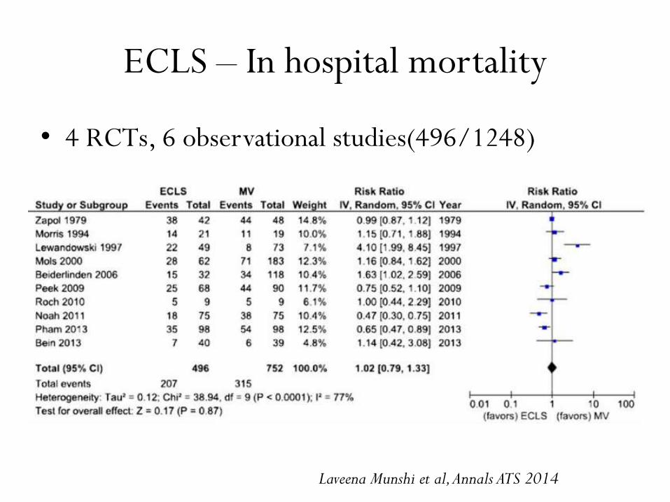
ECLS – In hospital mortality
• 4 RCTs, 6 observational studies(496/1248)
Laveena Munshi et al, Annals ATS 2014
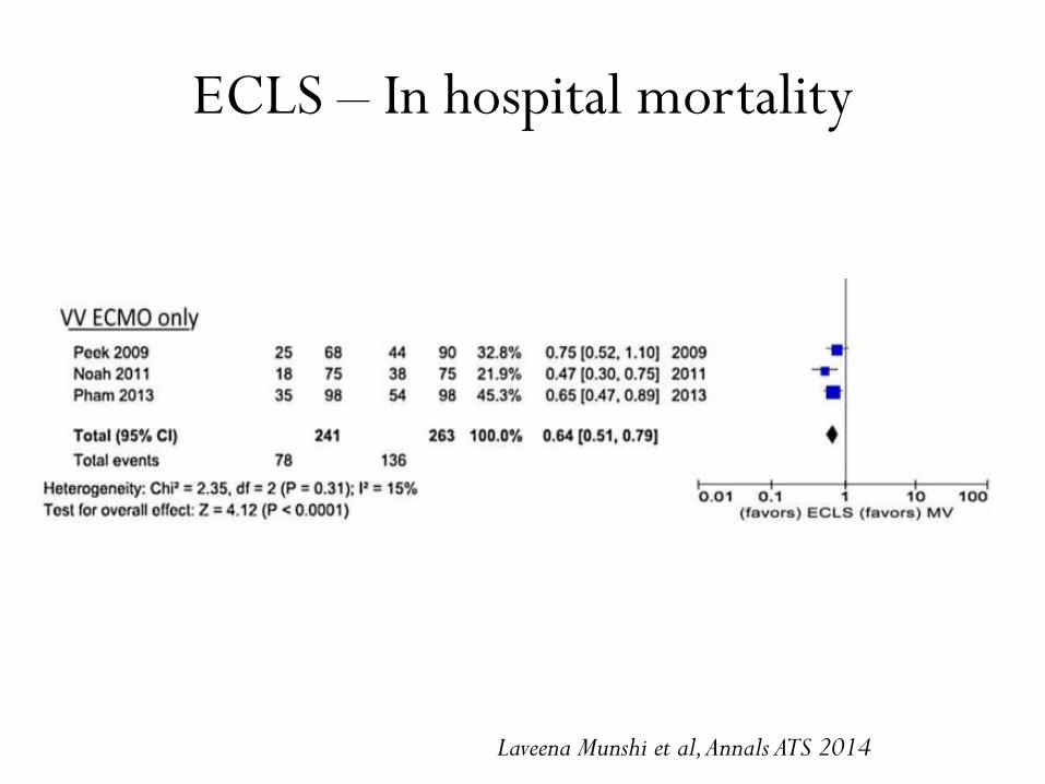
ECLS – In hospital mortality
Laveena Munshi et al, Annals ATS 2014
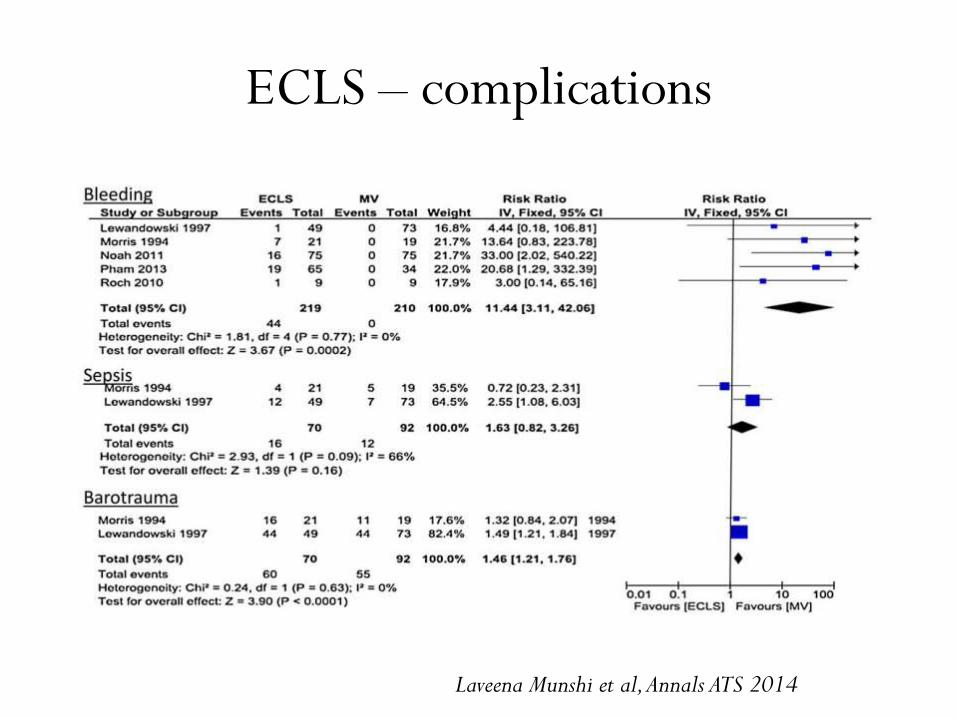
ECLS – complications
Laveena Munshi et al, Annals ATS 2014
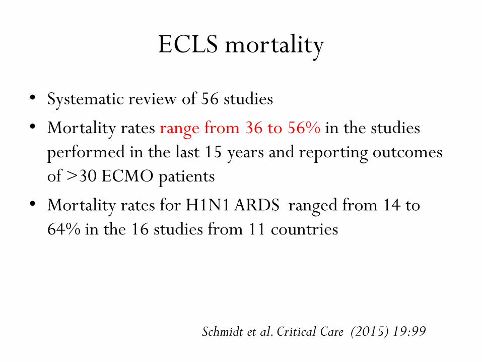
ECLS mortality
• Systematic review of 56 studies
• Mortality rates range from 36 to 56% in the studies
performed in the last 15 years and reporting outcomes
of >30 ECMO patients
• Mortality rates for H1N1 ARDS ranged from 14 to
64% in the 16 studies from 11 countries
Schmidt et al. Critical Care (2015) 19:99
ECLS mortality
• Factors associated with poor outcomes after ECMO for
acute respiratory failure
– Older age
– More days of mechanical ventilation before ECMO
– More number of organ failures
– Low pre ECMO respiratory system compliance
– Immunosuppression
Schmidt et al. Critical Care (2015) 19:99
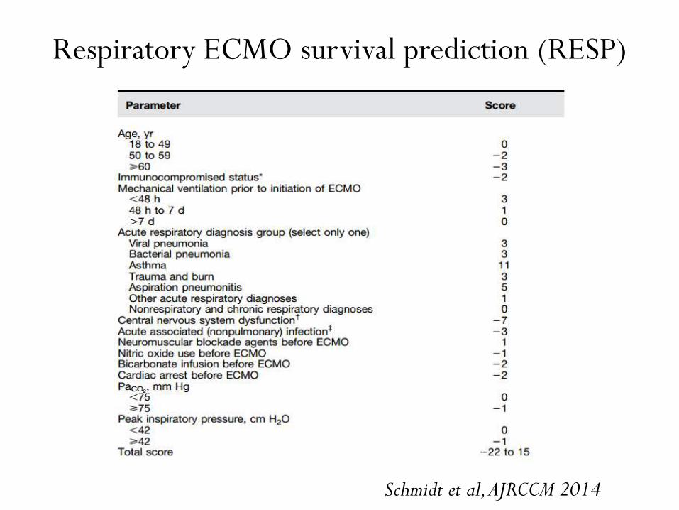
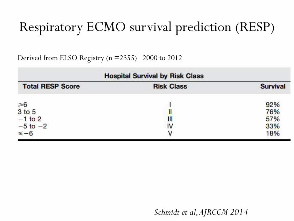
Respiratory ECMO survival prediction (RESP)
Schmidt et al, AJRCCM 2014
Respiratory ECMO survival prediction (RESP)
Schmidt et al, AJRCCM 2014
Derived from ELSO Registry (n =2355) 2000 to 2012
ECCO2 REMOVAL /
RESPIRATORY DIALYSIS
ECCO2 removal
• Technique providing artificial respiratory support by
removal of CO2 from blood through an extracorporeal
gas exchanger
• Feature of other ECLS
• Low flow VV or AV devices – provide CO2 removal without
oxygenation
History
• 1976 - Kolobow and Gattinoni explored the possibility of
treating severe respiratory failure using low frequency
PPV alongside ECCO2 removal in sheep
• 1986 – first clinical study on V-V ECCO2 removal by
Gattinoni et al
• 1997 – first clinical study on A-V ECCO2 removal
Hypercapnia detrimental?
• Inhibition of cell membrane repair
• Suppression of innate immunity & host defence
• Uncoupling of RV & pulmonary circulation – RV failure
• Increase in intracranial pressure
• Depression of myocardial contractility
Morimont et al, Critical Care 2015, 19 : 117
Benefits of ECCO2R
• Decreases the detrimental effects of hypercapnia
• Better oxygenation
– Increases the alveolar O2 concentration in accordance
with the alveolar gas equation
– By removing CO2, ECCO2R allows ventilation
strategies that are focused on oxygenation rather than
CO2 elimination
• “Rest lung” concept
Morimont et al, Critical Care 2015, 19 : 117
Benefits of ECCO2R
• COPD
Obviates the need of intubation & IMV
Facilitates withdrawal of IMV & extubation
• Weaning from MV
• Bridge to lung transplantation
Morelli et al, Intensive Care Med 2017, 43 : 519-30
In ARDS...
• Decreases the ventilator induced lung injury (VILI) by
allowing to ventilate the lung at low volumes and
pressures
• Allows to continue low tidal volume ventilation (< 6
ml/kg IBW)
• Upto 50 % reduction in MV can be obtained while
maintaining normocarbia
Morimont et al, Critical Care 2015, 19 : 117
Morelli et al, Intensive Care Med 2017, 43 : 519-30
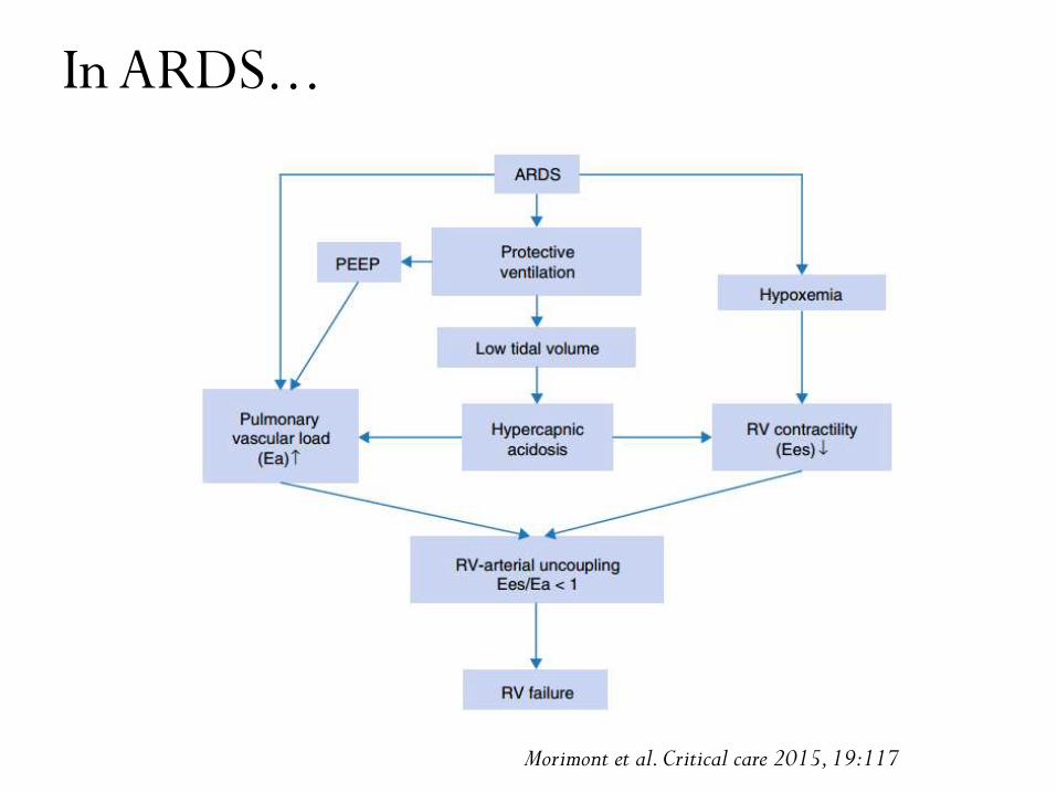
In ARDS...
Morimont et al. Critical care 2015, 19:117
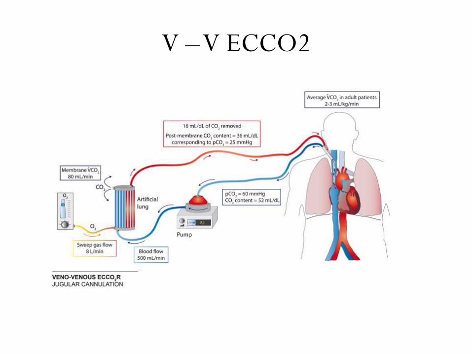
V –V ECCO2
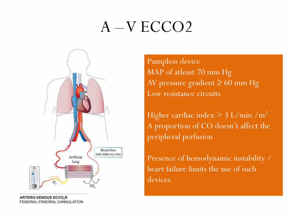
A –V ECCO2
Pumpless device
MAP of atleast 70 mm Hg
AV pressure gradient ≥ 60 mm Hg
Low resistance circuits
Higher cardiac index > 3 L/min /m2
A proportion of CO doesn’t affect the
peripheral perfusion
Presence of hemodynamic instability /
heart failure limits the use of such
devices
Various devices
• The Pump-Assisted Lung Protection (PALP)
(Maquet, Rastatt, Germany)
• The iLA Activve® (Novalung, Germany)
• The Hemolung® system (Alung Technologies,
Pittsburgh, USA)
• The Decap® system (Hemodec, Salerno, Italy)
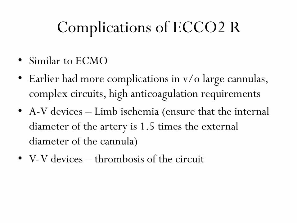
Complications of ECCO2 R
• Similar to ECMO
• Earlier had more complications in v/o large cannulas,
complex circuits, high anticoagulation requirements
• A-V devices – Limb ischemia (ensure that the internal
diameter of the artery is 1.5 times the external
diameter of the cannula)
• V-V devices – thrombosis of the circuit
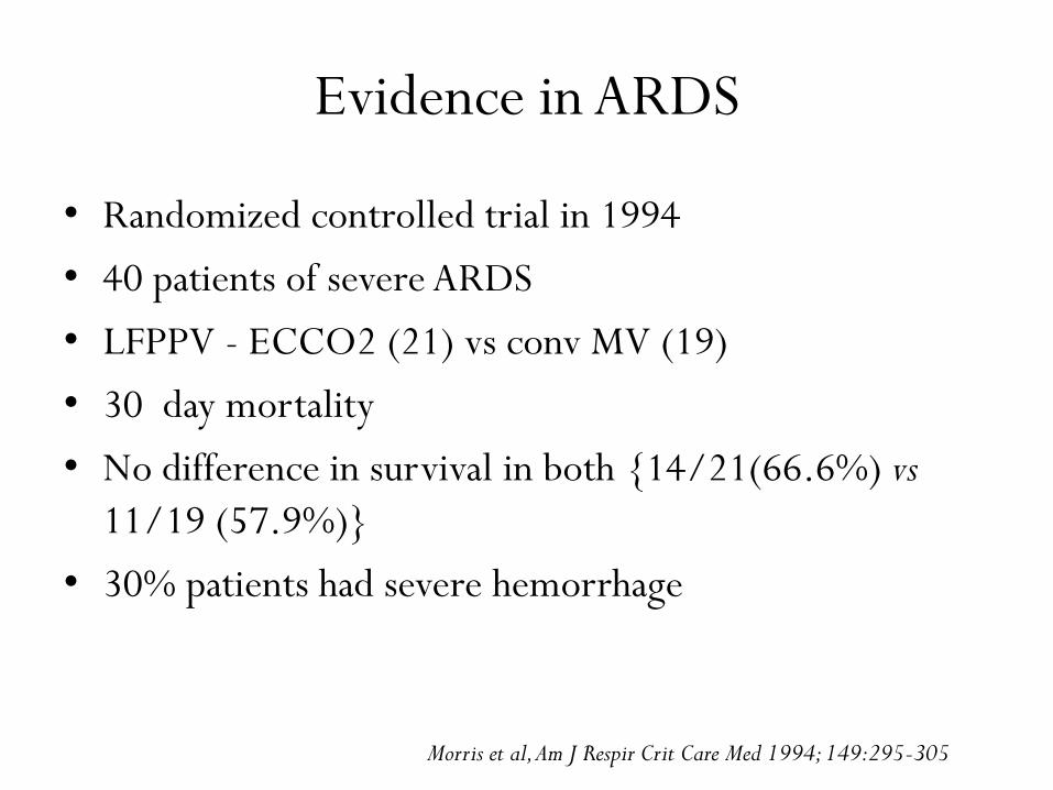
Evidence in ARDS
• Randomized controlled trial in 1994
• 40 patients of severe ARDS
• LFPPV - ECCO2 (21) vs conv MV (19)
• 30 day mortality
• No difference in survival in both {14/21(66.6%) vs
11/19 (57.9%)}
• 30% patients had severe hemorrhage
Morris et al, Am J Respir Crit Care Med 1994; 149:295-305
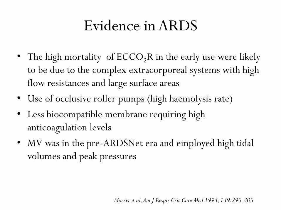
Evidence in ARDS
• The high mortality of ECCO2R in the early use were likely
to be due to the complex extracorporeal systems with high
flow resistances and large surface areas
• Use of occlusive roller pumps (high haemolysis rate)
• Less biocompatible membrane requiring high
anticoagulation levels
• MV was in the pre-ARDSNet era and employed high tidal
volumes and peak pressures
Morris et al, Am J Respir Crit Care Med 1994; 149:295-305
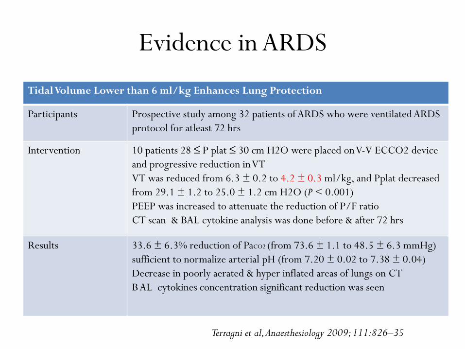
Evidence in ARDS
Tidal Volume Lower than 6 ml/kg Enhances Lung Protection
Participants Prospective study among 32 patients of ARDS who were ventilated ARDS
protocol for atleast 72 hrs
Intervention 10 patients 28 ≤ P plat ≤ 30 cm H2O were placed on V-V ECCO2 device
and progressive reduction in VT
VT was reduced from 6.3 ± 0.2 to 4.2 ± 0.3 ml/kg, and Pplat decreased
from 29.1 ± 1.2 to 25.0 ± 1.2 cm H2O (P < 0.001)
PEEP was increased to attenuate the reduction of P/F ratio
CT scan & BAL cytokine analysis was done before & after 72 hrs
Results 33.6 ± 6.3% reduction of PaCO2 (from 73.6 ± 1.1 to 48.5 ± 6.3 mmHg)
sufficient to normalize arterial pH (from 7.20 ± 0.02 to 7.38 ± 0.04)
Decrease in poorly aerated & hyper inflated areas of lungs on CT
B AL cytokines concentration significant reduction was seen
Terragni et al, Anaesthesiology 2009; 111:826–35
Evidence in ARDS
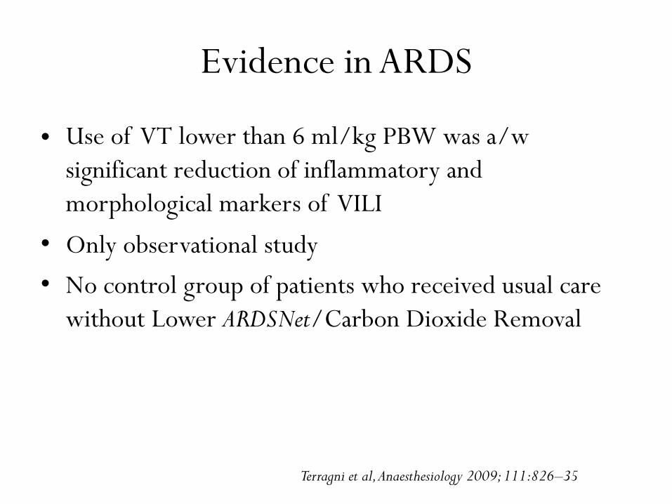
• Use of VT lower than 6 ml/kg PBW was a/w
significant reduction of inflammatory and
morphological markers of VILI
• Only observational study
• No control group of patients who received usual care
without Lower ARDSNet/Carbon Dioxide Removal
Terragni et al, Anaesthesiology 2009; 111:826–35
Evidence in ARDS
• Fanelli et al, Critical Care (2016) 20:36
• Participants
• Prospective study among 15 patients of moderate ARDS
• Intervention
• VT was gradually reduced from 6 to a min value of 4 mL/kg by 0.5 mL/kg every 30 min & PEEP was increased to target a Pplat between 23 and 25 cmH2O
• If arterial pH was <7.25 with PaCO2 >60 mmHg, despite an increase in RR up to 35/min, ECCO2R device was switched on
• Results
• Initial reduction in VT, without ECCO2R
• resulted in significant respiratory acidosis (pH <7.25) in all
• significant reduction in Pplat from 27.7 ± 1.6 to 23.9 ± 1 cmH2OMortality at 28 days was 47 %, which was expected in moderate ARDS1/3 rd required either proning or ECMO for refractory hypoxia
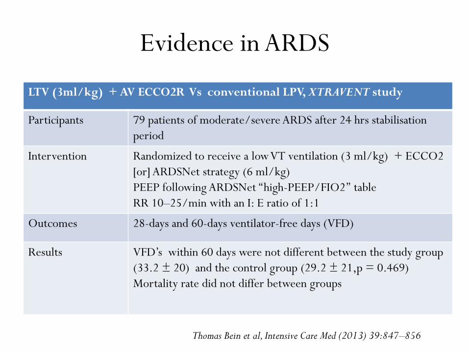
LTV (3ml/kg) + AV ECCO2R Vs conventional LPV, XTRAVENT study
Participants 79 patients of moderate/severe ARDS after 24 hrs stabilisation
period
Intervention Randomized to receive a low VT ventilation (3 ml/kg) + ECCO2
[or] ARDSNet strategy (6 ml/kg)
PEEP following ARDSNet ‘‘high-PEEP/FIO2’’ table
RR 10–25/min with an I: E ratio of 1:1
Outcomes 28-days and 60-days ventilator-free days (VFD)
Results VFD’s within 60 days were not different between the study group
(33.2 ± 20) and the control group (29.2 ± 21,p = 0.469)
Mortality rate did not differ between groups
Thomas Bein et al, Intensive Care Med (2013) 39:847–856
Evidence in ARDS
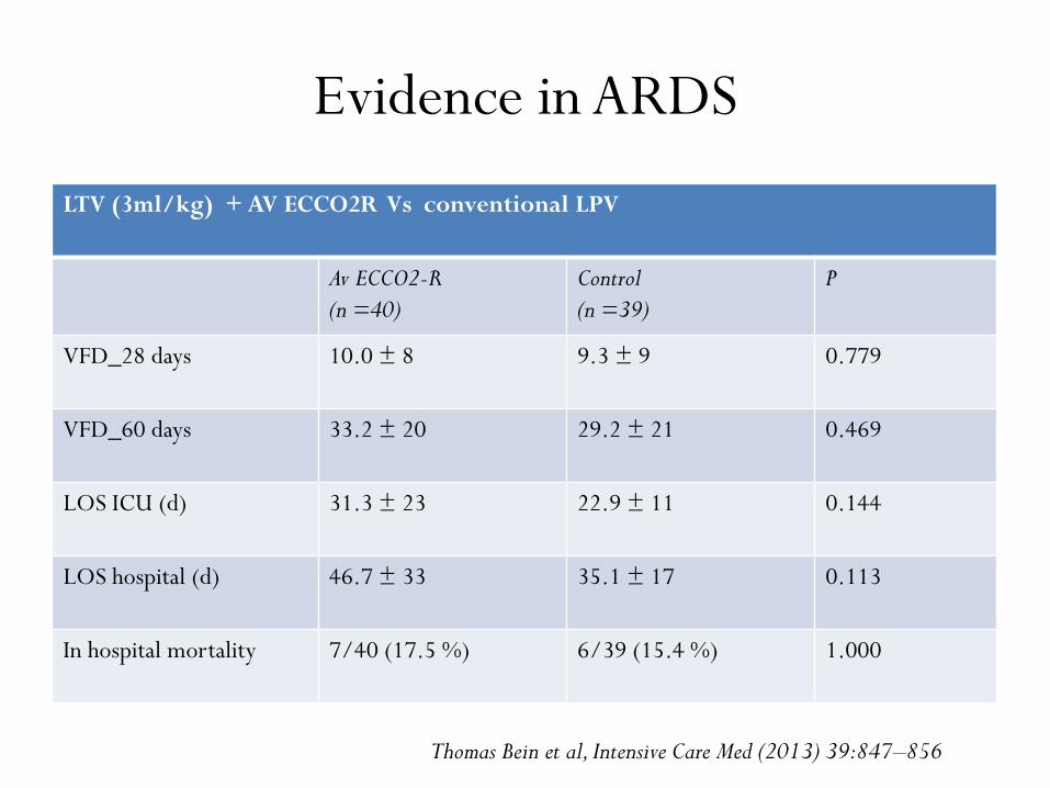
LTV (3ml/kg) + AV ECCO2R Vs conventional LPV
Av ECCO2-R
(n =40)
Control
(n =39)
P
VFD_28 days 10.0 ± 8 9.3 ± 9 0.779
VFD_60 days 33.2 ± 20 29.2 ± 21 0.469
LOS ICU (d) 31.3 ± 23 22.9 ± 11 0.144
LOS hospital (d) 46.7 ± 33 35.1 ± 17 0.113
In hospital mortality 7/40 (17.5 %) 6/39 (15.4 %) 1.000
Thomas Bein et al, Intensive Care Med (2013) 39:847–856
Evidence in ARDS
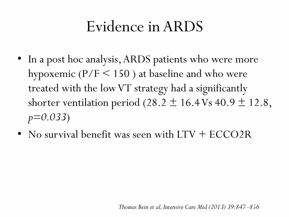
• In a post hoc analysis, ARDS patients who were more
hypoxemic (P/F < 150 ) at baseline and who were
treated with the low VT strategy had a significantly
shorter ventilation period (28.2 ± 16.4 Vs 40.9 ± 12.8,
p=0.033)
• No survival benefit was seen with LTV + ECCO2R
Thomas Bein et al, Intensive Care Med (2013) 39:847–856
Evidence in ARDS
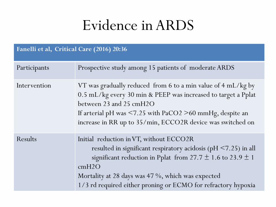
Fanelli et al, Critical Care (2016) 20:36
Participants Prospective study among 15 patients of moderate ARDS
Intervention VT was gradually reduced from 6 to a min value of 4 mL/kg by
0.5 mL/kg every 30 min & PEEP was increased to target a Pplat
between 23 and 25 cmH2O
If arterial pH was <7.25 with PaCO2 >60 mmHg, despite an
increase in RR up to 35/min, ECCO2R device was switched on
Results Initial reduction in VT, without ECCO2R
resulted in significant respiratory acidosis (pH <7.25) in all
significant reduction in Pplat from 27.7 ± 1.6 to 23.9 ± 1
cmH2O
Mortality at 28 days was 47 %, which was expected
1/3 rd required either proning or ECMO for refractory hypoxia
Evidence in ARDS
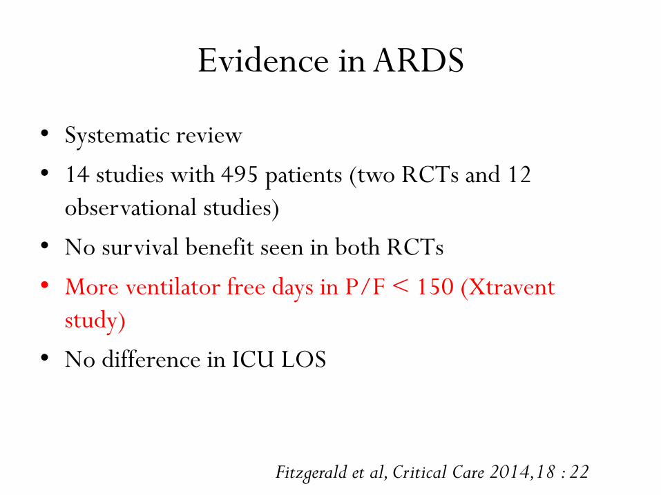
• Systematic review
• 14 studies with 495 patients (two RCTs and 12
observational studies)
• No survival benefit seen in both RCTs
• More ventilator free days in P/F < 150 (Xtravent
study)
• No difference in ICU LOS
Fitzgerald et al, Critical Care 2014,18 : 22
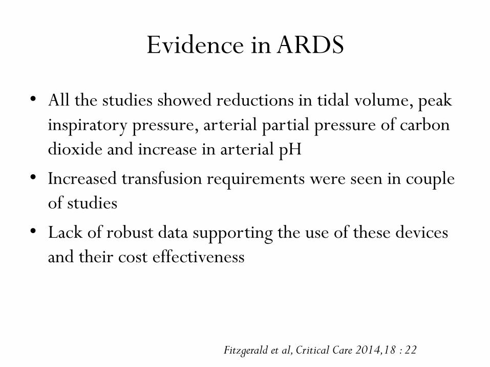
Evidence in ARDS
• All the studies showed reductions in tidal volume, peak
inspiratory pressure, arterial partial pressure of carbon
dioxide and increase in arterial pH
• Increased transfusion requirements were seen in couple
of studies
• Lack of robust data supporting the use of these devices
and their cost effectiveness
Fitzgerald et al, Critical Care 2014,18 : 22
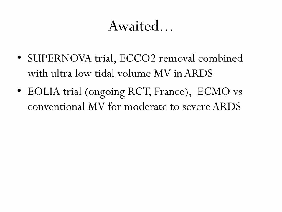
Awaited...
• SUPERNOVA trial, ECCO2 removal combined
with ultra low tidal volume MV in ARDS
• EOLIA trial (ongoing RCT, France), ECMO vs
conventional MV for moderate to severe ARDS
Take home message
• Requires early & careful selection of patients with
reversible disease and without significant comorbidities
• Rescue therapy for patients with severe ARDS
• Evidence of benefit in H1 N1 related ARDS
• VV ECMO – therapy of choice in ARDS
• ECCO2 R therapeutic adjunct in moderate to severe
ARDS

![Intrafirm Monitoring of Executive Compensation · 2019. 3. 28. · 2016] INTRAFIRM MONITORING 697 of executive pay while maintaining fidelity to the core principle that the board](https://img.pdfslide.us/doc/110x75/604018d9fb2e4f47c82f12b0/intrafirm-monitoring-of-executive-compensation-2019-3-28-2016-intrafirm-monitoring.jpg)