Embed Size (px)
Citation preview

AD_________________
Award Number: W81XWH-04-1-0126 TITLE: Radiation-Induced Immune Modulation in Prostate Cancer PRINCIPAL INVESTIGATOR: William H. McBride, D.Sc. CONTRACTING ORGANIZATION: University of California Los Angeles, CA 90024 REPORT DATE: January 2006 TYPE OF REPORT: Annual PREPARED FOR: U.S. Army Medical Research and Materiel Command Fort Detrick, Maryland 21702-5012 DISTRIBUTION STATEMENT: Approved for Public Release; Distribution Unlimited The views, opinions and/or findings contained in this report are those of the author(s) and should not be construed as an official Department of the Army position, policy or decision unless so designated by other documentation.

REPORT DOCUMENTATION PAGE Form Approved
OMB No. 0704-0188 Public reporting burden for this collection of information is estimated to average 1 hour per response, including the time for reviewing instructions, searching existing data sources, gathering and maintaining the data needed, and completing and reviewing this collection of information. Send comments regarding this burden estimate or any other aspect of this collection of information, including suggestions for reducing this burden to Department of Defense, Washington Headquarters Services, Directorate for Information Operations and Reports (0704-0188), 1215 Jefferson Davis Highway, Suite 1204, Arlington, VA 22202-4302. Respondents should be aware that notwithstanding any other provision of law, no person shall be subject to any penalty for failing to comply with a collection of information if it does not display a currently valid OMB control number. PLEASE DO NOT RETURN YOUR FORM TO THE ABOVE ADDRESS. 1. REPORT DATE 01-01-2006
2. REPORT TYPEAnnual
3. DATES COVERED 1 Jan 2005 – 31 Dec 2005
4. TITLE AND SUBTITLE
5a. CONTRACT NUMBER
Radiation-Induced Immune Modulation in Prostate Cancer
5b. GRANT NUMBER W81XWH-04-1-0126
5c. PROGRAM ELEMENT NUMBER
6. AUTHOR(S)
5d. PROJECT NUMBER
William H. McBride, D.Sc. 5e. TASK NUMBER
5f. WORK UNIT NUMBER
7. PERFORMING ORGANIZATION NAME(S) AND ADDRESS(ES)
8. PERFORMING ORGANIZATION REPORT NUMBER
University of California Los Angeles, CA 90024
9. SPONSORING / MONITORING AGENCY NAME(S) AND ADDRESS(ES) 10. SPONSOR/MONITOR’S ACRONYM(S)U.S. Army Medical Research and Materiel Command
Fort Detrick, Maryland 21702-5012 11. SPONSOR/MONITOR’S REPORT NUMBER(S) 12. DISTRIBUTION / AVAILABILITY STATEMENT Approved for Public Release; Distribution Unlimited
13. SUPPLEMENTARY NOTES Original contains colored plates ALL DTIC reproductions will be in black and white
14. ABSTRACT This proposal is to determine if radiation affects presentation of prostate specific antigen (PSA) through endogenous and exogenous pathways by dendritic cells (DCs) and to devise strategies to enhance the manner in which radiation-induced cell death is translated into the generation of tumor-specific immunity so as to achieve the best therapeutic outcome from radiation therapy. From the conventional point of view, radiotherapy is usually related to cell killing. However, our hypothesis is that radiation is more than just depleting cells. It also influences functional antigen presentation by DCs without killing them. We chose PSA as antigen for this study, but because of the high risk nature of the experiments and the high PSA expression levels in prostate cancer patients, we have also explored the possibility of monitoring the immune responses to the antigen survivin in prostate cancer. Our approach to translate radiation-induced immunosuppression into beneficial tumor immunogenicity focuses on IL-3 and/or GM-CSF treatment. Our studies on combined treatment of radiotherapy and IL-3, and on the effects of radiation on PSA presentation by DCs are underway and are presented in this Progress Report, along with other milestone that have been attained.
15. SUBJECT TERMSRadiation, Dendritic Cells, Cytokines
16. SECURITY CLASSIFICATION OF:
17. LIMITATION OF ABSTRACT
18. NUMBER OF PAGES
19a. NAME OF RESPONSIBLE PERSONUSAMRMC
a. REPORT U
b. ABSTRACT U
c. THIS PAGE U
UU
16
19b. TELEPHONE NUMBER (include area code)
Standard Form 298 (Rev. 8-98)Prescribed by ANSI Std. Z39.18

3
Table of Contents
Cover…………………………………………………………………………………… 1
SF 298……………………………………………………………………………..…… 2
Introduction…………………………………………………………….………….... 4
Body……………………………………………………………………………………. 4
Key Research Accomplishments………………………………………….……… 9
Reportable Outcomes………………………………………………………………. 9
Conclusions………………………………………………………………………….. 10
References…………………………………………………………………………… n/a
Appendices…………………………………………………………………………… 10
McBride, William H.W81XWH-04-1-0126
4
Introduction
The immunosuppressive effects of ionizing radiation are generally considered tobe due to killing of radiosensitive lymphocytes. However, we discover another and novelmechanism of radiation-induced immunosuppression, which is the inability of dendriticcells (DCs) to process antigens. We believe that radiation does this by inhibitingproteasome function and/or the expression of cytokines and related molecules by DCs.To overcome this radiation-induced immunosuppression, we plan to investigate thecombined effects of radiation and cytokines that we have shown to increase tumorimmunogenicity, specifically IL-3 and/or GM-CGF. The overall goal in this study is totranslate radiation-induced tumor cell killing into generation of tumor immunity in thehope of optimizing therapy for localized and disseminated prostate cancer. The aims areunaltered from the original submission.
BodyThe statement of work for this year covers experiments aimed at studying the
effects of radiation on the immune responses to DCs with adenovirally delivered PSA(AdV-PSA), on radiation-induced cytokines, cytokine receptors, and proteasomestructures, and the development of a treatment regimen with AdV-IL-3 combined withradiation.
We have spent this year establishing a prostate cancer mouse model, preparingvectors, cell lines and reagents for future experiments and refining our experimentapproach. Our AdV-PSA has been constructed, as has our murine prostate cancer cell lineexpressing PSA and we have initiated our murine studies using these reagents. Whilethese were being developed, we explored a number of the specific hypotheses in theproposal.
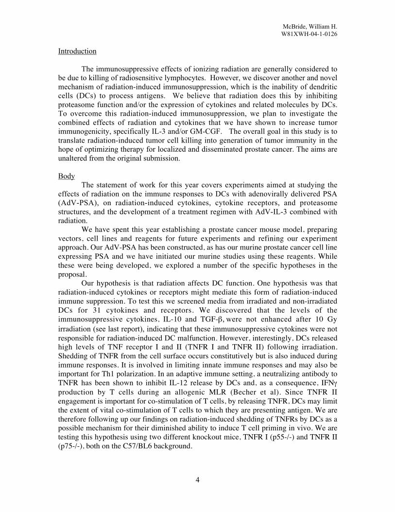
Our hypothesis is that radiation affects DC function. One hypothesis was thatradiation-induced cytokines or receptors might mediate this form of radiation-inducedimmune suppression. To test this we screened media from irradiated and non-irradiatedDCs for 31 cytokines and receptors. We discovered that the levels of theimmunosuppressive cytokines, IL-10 and TGF-β, were not enhanced after 10 Gyirradiation (see last report), indicating that these immunosuppressive cytokines were notresponsible for radiation-induced DC malfunction. However, interestingly, DCs releasedhigh levels of TNF receptor I and II (TNFR I and TNFR II) following irradiation.Shedding of TNFR from the cell surface occurs constitutively but is also induced duringimmune responses. It is involved in limiting innate immune responses and may also beimportant for Th1 polarization. In an adaptive immune setting, a neutralizing antibody toTNFR has been shown to inhibit IL-12 release by DCs and, as a consequence, IFNγproduction by T cells during an allogenic MLR (Becher et al). Since TNFR IIengagement is important for co-stimulation of T cells, by releasing TNFR, DCs may limitthe extent of vital co-stimulation of T cells to which they are presenting antigen. We aretherefore following up our findings on radiation-induced shedding of TNFRs by DCs as apossible mechanism for their diminished ability to induce T cell priming in vivo. We aretesting this hypothesis using two different knockout mice, TNFR I (p55-/-) and TNFR II(p75-/-), both on the C57/BL6 background.

McBride, William H.W81XWH-04-1-0126
5
0
20
40
60
80
100
WT p55 p75
MHC IICD86double positivedouble positive high
% c
ells
stai
ning
pos
itive
k/o k/o
Fig. 1. Maturation status of BMDCs. DCs were grownin the presence of IL-4 and GM-CSF and harvested onday 9. Cells were double stained with antibodies forMHC II and CD86 and analyzed by flow cytometry
Initial experiments aimed todefine the importance of TNFR Iand II (TNFR I = p55 and TNFR II= p75) to the DC phenotype andthen to characterize whatconsequences this has for theirfunctional capacity to presenttumor antigen during the responseto ionizing radiation. Remarkably,even the morphology of bonemarrow-derived DCs from theTNFR ko mice was different.Those from TNFR I ko mice didnot show the irregular appearance
typically associated with DCs. However, according to maturation markers on the cellsurface, bone marrow-derived DCs (BMDCs) from these TNFR I ko mice not only had ahigher proportion of cells staining positive for CD86, MHC II, and both molecules, butalso had higher levels of these surface markers than WT BM-DCs (Fig. 1). BMDCs fromTNFR II ko appeared to have somewhat similar expression of markers to the WTBMDCs and similar morphology. We concluded that loss of TNFR I alters themorphology of DCs and enhances their expression of the co-accessory markers that areassociated with superior antigen presentation.
We then examined whether these phenotypic differences between DCs from WTmice and TNFR ko mice were reflected differences in their functional status. Initially, wechose the AdV-MART antigen to perform these studies because this was our original“test” system for the radiation effects on DCs and because we had more experience withthis than with PSA, although similar experiments with the PSA system are planned.Overall, BM-DCs from TNFR ko were superior to WT BM-DCs in terms of antigenpresentation, as confirmed by ELISPOT (Fig. 2), indicating that these receptors generallyact as a ‘brake’ on immunity, as has been suggested previously. With time it emergedthat this effect was robust and consistent, in particular in the case of the TNFR I ko DCs(not shown). The major ‘brake’ on the development of immunity by DCs seems therefore
0 100 200 300 400 500 600 700
non-immunized
C57/Bl6
C57/Bl6 -10Gy
TNFR I
TNFR I -10Gy
TNFR II
TNFR II -10Gy
no ex vivo stimulationEL4EL4-MART
IFNγ dots / 100 000 cells
strainFig. 2. Antigen presentation byBMDCs. DCs were grown frombone marrow cultures of WT, p55-/-or p75-/- in the presence of IL-4 andGM-CSF. On the day of harvest,DCs were irradiated with 10Gy ornot and transduced with AdMARTprior to injection into WT C57/Bl6mice (5x10⁵ DCs/mouse). 7 dayslater splenocytes were harvested, re-stimulated ex vivo with either EL4 orEL4-MART system or left non-stimulated. T cell cultures wereanalyzed by ELISPOT to measureIFNγ production by activated T cells.
McBride, William H.W81XWH-04-1-0126
6
to be TNFR I. It is possible that it inhibits their phenotypic maturation. This wassomewhat surprising since it has been reported in the literature that DCs respond toautocrine TNF during maturation, even more so through TNFR I than TNFR II.
Regarding the response to radiation, it appears that DCs from TNFR II ko micerespond to 10 Gy in a similar fashion to those from WT mice, in that they have adecreased ability to generate T cell responses to antigen following injection. In contrast,10 Gy irradiation did not suppress this ability in TNFR I ko DCs. These experimentssuggest that TNFR I shedding following irradiation may be responsible for radiation-induced inhibition of DC function. Furthermore, they suggest a means of circumventingthis effect through the use of drugs that inhibit shedding or antibodies to TNFR II thatmight stimulate DC maturation and bias the response towards radiation resistance.Further analysis will involve these approaches and the use of double knockouts, whichwill allow discrimination between the positive and negative effects of these molecules.
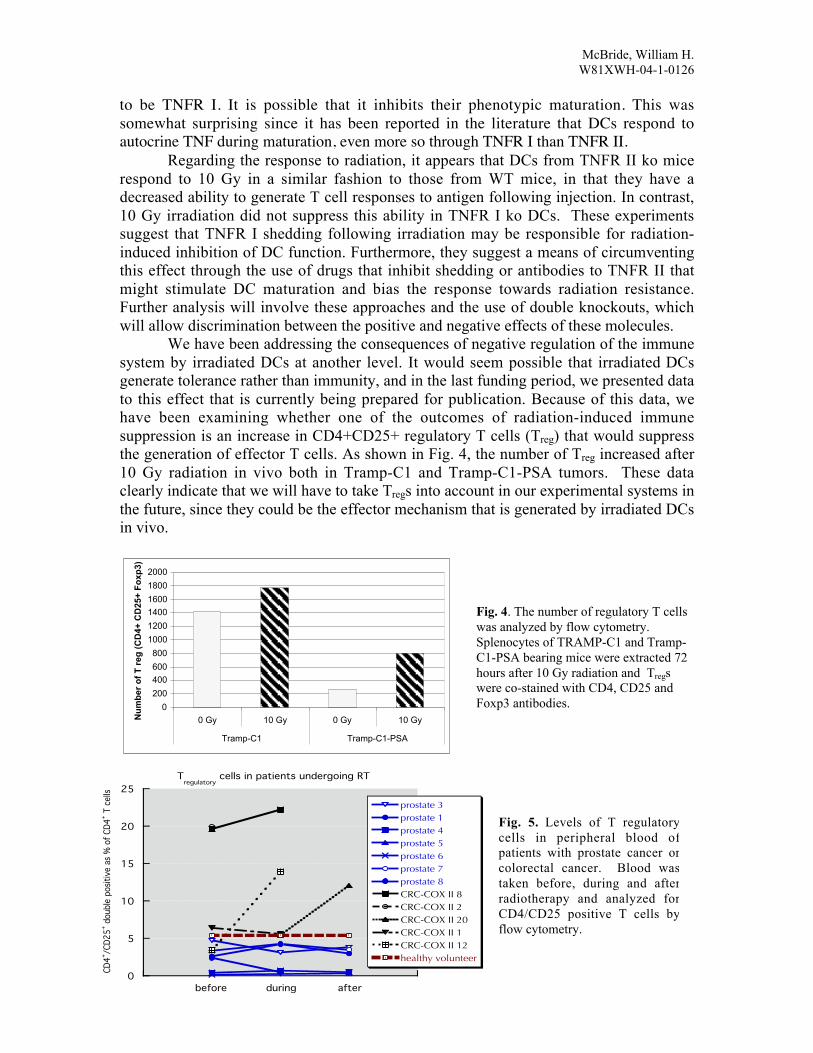
We have been addressing the consequences of negative regulation of the immunesystem by irradiated DCs at another level. It would seem possible that irradiated DCsgenerate tolerance rather than immunity, and in the last funding period, we presented datato this effect that is currently being prepared for publication. Because of this data, wehave been examining whether one of the outcomes of radiation-induced immunesuppression is an increase in CD4+CD25+ regulatory T cells (Treg) that would suppressthe generation of effector T cells. As shown in Fig. 4, the number of Treg increased after10 Gy radiation in vivo both in Tramp-C1 and Tramp-C1-PSA tumors. These dataclearly indicate that we will have to take Tregs into account in our experimental systems inthe future, since they could be the effector mechanism that is generated by irradiated DCsin vivo.
0
200
400
600
800
1000
1200
1400
1600
1800
2000
0 Gy 10 Gy 0 Gy 10 Gy
Tramp-C1 Tramp-C1-PSA
Nu
mb
er o
f T
reg
(C
D4+
CD
25+
Fo
xp3)
Fig. 4. The number of regulatory T cellswas analyzed by flow cytometry.Splenocytes of TRAMP-C1 and Tramp-C1-PSA bearing mice were extracted 72hours after 10 Gy radiation and Tregswere co-stained with CD4, CD25 andFoxp3 antibodies.
0
5
10
15
20
25
before during after
prostate 3prostate 1prostate 4prostate 5prostate 6prostate 7prostate 8CRC-COX II 8CRC-COX II 2CRC-COX II 20CRC-COX II 1CRC-COX II 12healthy volunteer
CD4+ /C
D25+ d
oubl
e po
sitive
as
% o
f CD4
+ T c
ells
Radiotherapy
Tregulatory
cells in patients undergoing RT
Fig. 5. Levels of T regulatorycells in peripheral blood ofpatients with prostate cancer orcolorectal cancer. Blood wastaken before, during and afterradiotherapy and analyzed forCD4/CD25 positive T cells byflow cytometry.

McBride, William H.W81XWH-04-1-0126
7
Although not part of this proposal, because of our concerns about Tregs followingirradiation, we undertook to study whether or not there were significant changes in Treg
levels in prostate cancer patients receiving radiation treatment. Tregs have been found athigh levels in many cancers, >5% of CD4+ T cells, but they seemed low in patients withprostate cancer and did not increase following radiation therapy (Fig. 5). The low levelsmay be due to the relatively localized disease in this patient cohort, but it did reassure usthat our attempts at modulating immunity in prostate cancer would not have to overcomethe effects of high levels of these cells.
Utilizing IL-3 is one of our proposed approaches to overcome radiation-inducedimmunosuppression. We started with examining the effects of AdV-IL-3 combined withradiation on co-stimulatory molecules in DCs. As can be seen in Fig. 6 (left), theexpression of MHC class II and CD86 was upregulated 4 days after transduction of DCswith AdV-IL-3. In fact, these data may underestimate the effect of IL-3 since there was asubpopulation that had uniquely high CD86 levels after AdV-IL-3 transduction. IL-3expression was confirmed by ELISA assay (Fig. 6 (right)). The data suggest that IL-3-transduced DCs might have better ability to present antigen. There was no suggestion thatthese cells would be affected more by irradiation. The experiments to test their functionalability with and without irradiation are underway.
0
20
40
60
80
100
120
Control 10 Gy AdV-b-gal AdV-b-gal 10Gy AdV-IL-3 AdV-IL-3 10Gy
Dendritic Cells
% S
tain
ing
MHC II
CD86
Double Stain
High Staining
IL-3 release by BM-DC
0
100
200
300
400
500
DC DC10Gy
AdV-bgal
AdV-bgal10Gy
AdVIL-3
AdVIL-3 10Gy
IL-3
[n
g/
1x1
0exp
6 c
ell
s]
Fig. 6. Co-stimulatory molecules of AdV-IL-3 transduced DC by flow cytometric analysis 24 hr afterradiation. DCs were transduced with AdV-IL-3 on day 5 and irradiated with 10 Gy on day 8 (left). Thelevel of IL-3 expressed in AdV-IL3 transduced DCs was measured by ELISA (right).
While constructing AdV-PSA, we initiated a pilot study to explore the possibilitythat in vivo irradiation may block local DCs ability to present antigens and ways toprevent this by injection of antigen-laden DCs, with an emphasis on the timing of DCadministration. As can be seen in Fig. 7, splenic responses could be detected in controlmice with B16 tumors (group 2). Tumor irradiation alone decreased the responses (group3). Intratumoral AdVMART1/DC injection enhanced splenic T cell responses (group 4)and this was not affected and may have been slightly enhanced by irradiation given oneday later (group 5). In contrast, local tumor irradiation prior to AdVMART1/DC injectioninhibited the generation of splenic T cell responses (group 6). This suggests thatirradiation of tumor decreases immunity and blocks its generation, possibly throughinduction of immune tolerance. This has important implications for the proposed study inthis grant, which will explore the use of AdV-PSA and AdV-IL-3 as means forimmunizing mice and enhancing the effects of radiation therapy that will be performed inthe next grant period.

McBride, William H.W81XWH-04-1-0126
8
As mentioned in the last report, because of the high risk of the proposedexperiment and high background of natural PSA level in prostate cancer patient, wechose to investigate survivin, which is over expressed in many prostate cancers, as apotential back-up antigen. We have incorporated a pilot prostate study into a larger effortthat involves collaboration with other labs in Europe. As can be seen in Fig, 8,lymphocytes from patients with prostate and colon carcinoma, taken before, during andafter presurgical chemoradiotherapy were tested for the level of CD8+ T cells whichrecognize the tumor-associated antigen survivin by tetramer assay. We were able todetect survivin-specific CD8+ T cells in some patients. These tumor-specific T cellsappeared to accumulate after completion of radiation therapy in some patients (abstractattached). These experiments will help guide those in this proposal while not being partof the specific aims of the grant.
Fig. 8. Analysis of tumor-specific CD8+ T cells in peripheral blood of 11 patients with different cancersundergoing radiotherapy. Patients were confirmed to be HLA-A2 positive prior to staining with tetramerspresenting the survivin peptide LMLGEFLKL. Cells were co-stained for CD8+ and non-viable cells wereexcluded on grounds of high 7-AAD up-take. One healthy volunteer served as control throughout. Datashown as % CD8+ T cells positive for tetramer a. of individual patients and b. as overall time course.Colorectal cancer patients (CRC-COX II) were part of a clinical study using radiation treatment inconjunction with a COX II inhibitor. Patients that received placebo are indicated with *.
0
20
40
60
80
100
120
Unim
mun
ized
B16 t
umor
10 G
y
AdVM
ART1
/DC
AdVM
ART1
/DC
--10 G
y
10 G
y--
AdVM
ART1
/DC
IFN-γ
spot
s/105 ce
lls
control
EL4
EL4(MART-1)
in vitro
Fig. 7. IFN-γ expressionmeasured by ELISPOT assay.AdVMART1/DCs wereinjected intratumorally intoB16 tumors and the tumorswere irradiated with 10 Gyone day later. Another groupof tumors was irradiated (10Gy) prior to immunizationwith 5 × 10⁵ intratumoralAdVMART1/DC.
1 2 3 4 5 6
0
0.5
1
1.5
before during after
CRC-COX II 14
CRC-COX II 15
CRC-COX II 16
CRC-COX II 19
CRC-COX II 7
CRC-COX II 21
CRC-COX II 5
prostate 5
rectum 5
rectum 8
rectum 10
healthy volunteer
% o
f CD
8+ lym
phoc
ytes
pos
itive
for t
etra
mer
Radiotherapy
0
0.5
1
1.5
before during after
CRC-COX II 14
CRC-COX II 15
CRC-COX II 16
CRC-COX II 19
CRC-COX II 7
CRC-COX II 21
CRC-COX II 5
prostate 5
rectum 5
rectum 8
rectum 10
healthy volunteertetra
mer
pos
itive
s as %
of C
D8+ ly
mph
ocyt
es
*
*
0
0.2
0.4
0.6
0.8
1
1.2
1.4
before during after healthy
% C
D8+ ly
mph
ocyt
es p
ositi
ve fo
r tet
ram
er
Radiotherapy volunteer

McBride, William H.W81XWH-04-1-0126
9
Key Research Accomplishments
1. Study of the effects of radiation on DC co-accessory molecules--completed.2. Study of the effects of radiation on proteasome structure and function--completed.3. Study of the effects of radiation on different DC subpopulations--completed.4. Study of the effects of IL-3 combined with radiation on DC function--50%
completed.5. Development of an AdV-PSA vector--completed.6. Development of a survivin system--80% completed.
Reportable Outcomes – Manuscripts and Abstracts
Pajonk, F., A. van Ophoven, C. Weissenberger and W.H. McBride: The Proteasomeinhibitor MG-132 sensitizes PC-3 prostate cancer cells to ionizing radiation by a DNA-PK-independent mechanism. Biomedcentral Cancer 5:76, 2005.
Pervan, M., K.S. Iwamoto and W.H. McBride: Proteasome structures affected by ionizingradiation. Molecular Cancer Research 3:381-390, 2005
Demaria, S., N. Bhardwaj, W.H. McBride and S.C. Formenti: Combing radiotherapy andimmunotherapy: a revived partnership. Int. J. Radiat. Oncol. Bio. Phys. 63:655-666,2005.
Tsai, C-H., J-H. Hong, K-F. Hsieh, H-W. Hsiao, W-L. Chuang, C-C. Lee, W.H.McBride, and C-S. Chiang: Tetracycline-regulated intratumoral expression of interleukin-3 enhances the efficacy of radiation therapy for murine prostate cancer. Submitted, 2006.
Schaue, D., Y. Liao, B. Comin-Anduix, A. Ribas, D.C. Altieri, A. Debucquoy, K.Haustermans and W.H. McBride: The Effect of Radiation Therapy on Tumor-SpecificImmune Responses. In: Abstracts of papers for the 52nd Annual Meeting of the RadiationResearch Society, Denver, Colorado, 2004.
Reportable Outcomes – Presentations
4/27/05Department of Radiation OncologyNew York University School of Medicine, New York, NYInvited Lecture: “Sense of Danger from Radiation”
10/6-10/8/0519th Meeting of European Macrophages and Dendritic Cell Society, Amsterdam, theNetherlandsAbstract presentation: “Radiation Affects Antigen Processing by Dendritic Cells – ANovel Form of Immune Suppression”

McBride, William H.W81XWH-04-1-0126
10
Reportable Outcomes – Presentations (cont.)
11/16/05Massey Cancer Center, Virginia Commonwealth University, Richmond, VAInvited speaker: “The Proteasome in Cancer biology and Therapy”
Conclusions
This has been a year of refinement of approach and development of morereagents. We have made important advances in the elucidation of the mechanismunderlying the effects of irradiation on DC function through examining the roles ofTNFR I and II in DC maturation in vitro. We have also examined in some detail theeffects of IL-3 expression on DC maturation and function and are greatly encouraged thatthis approach will reverse radiation-induced immune suppression and allow tumorirradiation to overcome immune suppression, We have started investigating thepossibility that local DC irradiation might generate Treg cells that would inhibit immunity.This is a concern in animal models, but may not be clinically as prostate cancer patientsdo not seem to have high levels of these cells, at least in early stage disease, but this stillremains a clinical concern for the future. We have had no changes in the directions of theproposed work in the grant, although we have set up a new system using survivin as atumor antigen in case the high natural level of PSA in prostate cancer patients proves tobe a problem. We feel that although we have made progress on many fronts, we are onlynow in a position to finally bring these efforts together in a truly meaningful way. We areencouraged however that nationally we have been instrumental in enthusing clinicians inthe concept of combining radiation therapy with immunotherapy and there is a real desireto move this approach into the clinic.
Appendices
One published manuscript and one abstract are in the appendix.
Schaue, D., Y. Liao, B. Comin-Anduix, A. Ribas, D.C. Altieri, A. Debucquoy, K.Haustermans and W.H. McBride: The Effect of Radiation Therapy on Tumor-SpecificImmune Responses. In: Abstracts of papers for the 52nd Annual Meeting of the RadiationResearch Society, Denver, Colorado, 2004.
Becher, B., M. Blain, P.S. Giacomini, and J.P. Antel: Inhibition of Th1 Polarization bySoluble TNF Receptors is Dependent on Antigen-Presenting Cell-Derived IL-12. J.Immunol. 162:684-688, 1999.

11
The Effect of Radiation Therapy on Tumor-Specific Immune Responses
In: Abstracts of Papers for the 52nd Annual Meeting of the Radiation Research Society,Denver, CO, 2004.
Dörthe Schaue, Yu-Pei Liao, Begonya Comin-Anduix, Antoni Ribas, AnneliesDebucquoy, Karin Haustermans, and William H. McBride
We have shown in a mouse B16 melanoma tumor model that local tumor irradiationdepresses tumor-specific systemic immunity. We have also shown that in vitro irradiationof dendritic cells affects their function, effectively blocking tumor antigen processing andtheir ability to generate protective CD8+ T cell anti-tumor responses. Indeed, irradiateddendritic cells actually suppress the generation of responses. The overall aim of thisstudy is to determine if irradiation of human cancer also results in a loss of tumor-specific T cell reactivity.
Lymphocytes from patients with prostate and colon carcinoma taken before, during andafter presurgical chemoradiotherapy were tested for the level of CD8+ T cells thatrecognize the tumor-associated antigen survivin. Survivin has has been shown to beover-expressed in many cancers samples. We have attempted to enumerate the numberof CD8+ cells that recognize that specific immunodominant survivin-associated antigenicpeptide in the context of HLA-A2.1 using tetramer analysis by flow cytometry. In addition,all patient samples were examined for the number of CD4+, CD25+, Foxp3+ Tregulatory cells in the circulation since we hypothesized that these would also changefollowing treatment and might be a cause of radiation-induced immune suppression.Some of the patients with colon carcinoma were part of a clinical trial where theyreceived COX-2 inhibitor as a radiosensitizer to determine if this would enhance tumor-specific immunity and influence the generation of T regulatory cells.The long term basis for this study is the notion that if radiation-induced tumor cell killwere to be better translated into the generation of tumor-specific immunity, local curerates might be increased and micrometastases inhibited. This study will lay the basis forsuch future investigations aimed at converting this state of radiation-induced immunesuppression into a more positive anti-tumor immune response.
Key words: tumor-specific T cells, T regulatory cells, survivin, tetramer

Inhibition of Th1 Polarization by Soluble TNF Receptor IsDependent on Antigen-Presenting Cell-Derived IL-121
Burkhard Becher,2 Manon Blain, Paul S. Giacomini, and Jack P. Antel
Th1-polarized CD41 T cells are considered central to the development of a number of target-directed autoimmune disordersincluding multiple sclerosis. The APC-derived cytokine IL-12 is a potent inducer of Th1 polarization in T cells. Inhibition of IL-12in vivo blocks the development of experimental allergic encephalomyelitis, the animal model for multiple sclerosis. Based onprevious work that suggests that the production of IL-12 by activated human central nervous system-derived microglia is regu-lated by autocrine TNF-a, we wanted to determine whether inhibition of TNF could induce a reduction of Th1 responses by itsimpact on systemic APCs. We found that soluble TNFR p75-IgG fusion protein (TNFR:Fc) inhibited production of IFN-g byallo-Ag-activated blood-derived human CD4 T cells. We documented reduced IL-12 p70 production by APCs in the MLR. Byadding back recombinant IL-12, we could rescue IFN-g production, indicating that TNFR:Fc acts on APC-derived IL-12. Con-sistent with an inhibition of the Th1 polarization, we found a decreased expression of IL-12R-b2 subunit on the T cells. Further-more, the capacity of T cells to secrete IFN-g upon restimulation when previously treated with TNFR:Fc is impaired, whereas IL-2secretion was not altered. Our results define a TNF-dependent cytokine network that favors development of Th1 immuneresponses. The Journal of Immunology,1999, 162: 684–688.
CD41 Th cells can be divided into Th1 and Th2 cells bytheir polarized expression of cytokines (1). Th1 cells pre-dominantly produce IFN-g whereas Th2 cells produce
IL-4 and IL-10. Proinflammatory Th1 T cells are considered cen-tral to the development of target-directed autoimmune inflamma-tory disorders including multiple sclerosis (MS)3 and rheumatoidarthritis (RA) (2, 3). In the animal model of MS, experimentalautoimmune encephalomyelitis (EAE), T cells recovered from theinflamed central nervous system (CNS) are predominantly Th1-polarized cells (4). In passive transfer experiments, EAE-inducingencephalitogenic T cell lines or clones are Th1 polarized (5, 6).Th1 cells have an advantage in crossing an endothelial barrier bybinding to P- and E-selectin (7). Simultaneous administration ofTh2 cytokines (IL-10, IL-4) inhibits disease development (8–10).
The APC-derived cytokine IL-12 is a potent activator of the Th1phenotype in T cells. In contrast to the Th1 cytokines IFN-g andTNF, the APC-derived cytokine IL-12 is crucial for the inductionof EAE (11–13). Th1 cells express higher levels of IL-12Rb2
subunit, resulting in higher responsiveness to IL-12 compared withTh2 cells (14). IL-12 production by APC is itself tightly regulated(15, 16). IL-12 production in dendritic cells can be induced by cellto cell contact with activated (CD1541) T cells (17), and IFN-gcan further enhance IL-12 secretion. Our previous in vitro studies
using human adult CNS derived microglial cells as a source ofIL-12 indicated that TNF-a regulates IL-12 production via an au-tocrine network (18). We found that LPS-stimulated human mi-croglial cells produce TNF-a before IL-12 and that administrationof TNFR:Fc or anti-TNF Abs significantly inhibits IL-12 produc-tion. These observations indicate that TNF should be consideredamong cytokines that can drive Th1 responses.
EAE can be induced by active systemic immunization or passiveT cell transfer, but not by direct intrathecal administration of my-elin reactive cells, indicating a role of systemic immune regulationin the disease process and a rationale for systemic therapeutic in-tervention. TNFR:Fc and anti-TNF Abs, given systemically, havebeen used to ablate the development of EAE and have been usedin clinical trials for RA and MS (7, 19, 20). Therapeutic effects inEAE were attributed to blocking the effector functions of TNF andlymphotoxin (LT); the effect on immunoregulatory functions wasnot considered (19). Monthly administration of a TNFR-Ig fusionprotein containing the 50-kDa TNFR has been shown to be clin-ically efficacious in the treatment of RA (21). In contrast, a clinicaltrial for MS, in which TNFR:Fc was also used in the form of monthlysystemic injections, was prematurely halted due to concerns regardingadverse impacts on the disease (22). The mechanism of action (or lackthere of) in these clinical conditions was not established.
We wished to establish whether TNFR:Fc would inhibit IL-12production by systemic APCs and whether there was an associatedreduction in Th1 (IFN-g) cytokine production by T cells respondingto these APCs. For our studies, we determined the effects of TNFR:Fcin an MLR assay, as this system serves as an in vitro model forcell-mediated immunity. Establishing the in vitro effects on immunereactivity of inhibiting TNF/LT should help to predict what effectsmight be expected by systemic TNF/LT-directed therapies involvingmolecules that may have complicated in vivo pharmacokinetics.
Materials and MethodsReagents
IFN-g and TNF-a were obtained from Genzyme (Cambridge, MA), IL-12and anti-IL-12 mAbs from R&D Systems (Minneapolis, MN), shu-TNFRwas a generous gift from A. Troutt (Immunex, Seattle, WA).
Neuroimmunology Unit, Montreal Neurological Institute, Department of Neurologyand Neurosurgery, McGill University, Montreal, Quebec, Canada
Received for publication March 25, 1998. Accepted for publication September 29, 1998.
The costs of publication of this article were defrayed in part by the payment of pagecharges. This article must therefore be hereby markedadvertisementin accordancewith 18 U.S.C. Section 1734 solely to indicate this fact.1 B.B. has a fellowship award from the German Academic Exchange Service (DAAD/HSPIII). This work has been supported by the Canadian Multiple Sclerosis Societyand the London Life Award.2 Address correspondence and reprint requests to Dr. Burkhard Becher, MontrealNeurological Institute, 3801 University, Montreal, QC, H3A 2B4, Canada. E-mailaddress: [email protected] Abbreviations used in this paper: MS, multiple sclerosis; CNS, central nervoussystem; EAE, experimental allergic encephalomyelitis; RA, rheumatoid arthritis;TNFR:Fc, soluble human p75 TNF receptor-IgG:Fc fusion protein; LT, lymphotoxin;LDH, lactate dehydrogenase.
Copyright © 1999 by The American Association of Immunologists 0022-1767/99/$02.00

Isolation of peripheral blood-derived cells
PBMC were isolated from healthy adult volunteer donors by density gra-dient centrifugation using Ficoll-Hypaque (Pharmacia, Baie D’Urfe, Can-ada). For the isolation of enriched APCs, the PBMC were washed twicewith PBS and cultured for 1 h in RPMI 1640 medium (Life Technologies,Burlington, ON, Canada) supplemented with 10% FCS, 2.5 mg/ml peni-cillin, 2.5 mg/ml streptomycin, and 2 mM glutamine (all from Life Tech-nologies) in 75-cm2 tissue culture flasks (Falcon, VWR Scientific, Mon-treal, Canada). The nonadherent cells were removed by gentle shaking. Theadherent cells consisted of 95% HLA-DR/B7-2 positive monocytes.
CD41 T cells were isolated from PBMC using anti-CD4 mAbs conju-gated to magnetic beads (Dynal, Great Neck, NY). The beads were de-tached from the cells after isolation following the supplied protocol. Thecells were then washed with PBS and their purity was of$96% as assessedby flow cytometry (23).
Semiquantitative PCR analysis
Total RNA was isolated using TRIZOL Reagent (Life Technologies). Totranscribe into cDNA, 3mg RNA, 3.3 mM random hexamer primers(Boehringer Mannheim, Manheim, Germany), reverse transcriptase buffer,3 mM dNTPs, 400 U Maloney murine leukemia virus reverse transcriptase(all from Life Technologies), 0.6ml RNA guard, and 3 mM DTT (bothfrom Pharmacia) were added to a total volume of 32ml. The reactionmixture was incubated for 1 h at42°C followed by a 10-min incubation at75°C. Primers used for PCR were obtained from Life Technologies and hadthe following sequences: IL-12Rb2 forward, 59-ACAGGACACACCTCCTGGAC-39; reverse, 59-AGAGGGACCTGTGTGTCACC-39; andb-ac-tin forward, 59-ATGCCATCCTGCGTCTGGACCTGGC-39; reverse, 59-AGCATTTGCGGTGCACGATGGAGGG-39. The primers for IL-12Rb2
andb-actin were constructed to generate fragments of 281 bp and 378 bp,respectively. cDNA 200 ng was added to the reaction mixture containingPCR buffer, 0.2 mM dNTPs (Life Technologies), 50 pMol of either primerset for IL-12Rb2 or b-actin, and 0.5ml Taq polymerase (Life Technolo-gies). The reaction mixture was completed with H2O to a total volume of50 ml. Samples were placed in a Gene Amp PCR system 9600 (Cetus,Perkin-Elmer, Norwalk, CT) for 25 cycles of 94°C for 30 s, 60°C for 45 s,and 72°C for 1 min followed by a 10-min extension at 72°C. After am-plification, 15ml of each sample was electrophoresed on a 1.5% agarosegel (Life Technologies). The bands were visualized with ethidium bromide.For quantification purposes, 2.5mCi of [32P]dCTP (DuPont/NEN, Missis-sauga, ON, Canada) was added to the reaction mixture before PCR. Thegels were dried and the bands were analyzed using a phosphorimaging andImage Quant software (Molecular Dynamics, Sunny Valley, CA).
MLR
A total of 105 T cells was cocultured with either 23 104 or 5 3 104
allogeneic APC. For proliferation and cytokine assays, primary MLRswere conducted in 96-well plates. After 5 days, unless indicated otherwise,1 mCi [3H]thymidine was added to the wells for 5 h. The cells were har-vested and thymidine uptake was determined using a beta-scintillationcounter. Culture medium was recovered from sister cultures to determinethe cytokine concentrations. For secondary MLRs, T cells were recoveredfrom the primary MLRs and cocultured with freshly isolated allogeneicAPC from the same donor as in the primary MLR. Secondary MLRs wereconducted for 3 more days at which time cytokine release was measured.
L929 cytotoxicity assay
L929 cells (104) were cultured in RPMI 1680 medium with 10% FCS.We added either 50 or 100 U/ml (1.7 or 0.85 ng/ml) of TNF-a in thepresence of different concentrations of TNFR:Fc (Genzyme, Cam-bridge, MA) or carrier buffer. The cells were incubated for 24 h andsupernatants were analyzed for lactate dehydrogenase (LDH) content aspreviously described (24).
Cytokine ELISA
IL-12 ELISA kits were obtained from R&D Systems. IL-2, IL-10, andIFN-g ELISA kits were obtained from BioSource International (Camarillo,CA). Tissue culture supernatants were stored at280°C until analysis.ELISA assays were performed following manufacturers instructions.
ResultsTNFR:Fc inhibits TNF-a-mediated cytotoxicity
In initial studies, we established that TNFR:Fc could specificallyinhibit the cytotoxic effect of TNF-a by exposing TNF-sensitive
L929 cells to TNF-a and TNFR:Fc or carrier control. Fig. 1Ashows the toxicity of different concentrations of rTNF-a on L929cells as assessed by LDH release assay. In a subsequent experi-ment, we have used 50 or 100 U (0.85 or 1.7 ng/ml) of TNF-a thatresults in maximum LDH release. We can block cytolysis medi-ated by 100 U of recombinant TNF-a using 140 pg/ml of TNFR:Fc, indicating that engagement of membrane TNFR on L929 cellsis completely ablated by addition of TNFR:Fc (Fig. 1B).
TNFR:Fc inhibits IFN-g production in an allogenic MLR
To analyze the effect of TNFR:Fc on immunoregulatory functions,we have performed MLRs in the presence of TNFR:Fc. CD41 Tcells were isolated from healthy donors and mixed with allogeneicAPC at different ratios (5:1–2:1) in 96-well plates. TNFR:Fc orirrelevant IgG1 mAb was added to the reaction. The MLR wasconducted for 5 days, and T cell proliferation was assessed by[3H]thymidine uptake (23). Supernatants from sister cultures wereharvested and analyzed for IFN-g by ELISA.
As shown in Fig. 2A, addition of TNFR:Fc to the primary MLRsinhibited IFN-g production in a dose-dependent fashion. Maxi-mum inhibition was first observed with 140 pg/ml of TNFR:Fc.For subsequent experiments we used 2.8mg/ml of TNFR:Fc and 4mg/ml of irrelevant IgG1 in vehicle buffer (ctrl). After 5 days,TNFR:Fc inhibited IFN-g production by 766 7% SEM, on av-erage (Table I). As shown in Fig. 2B, levels of IL-2 and IL-10 werenot altered by TNFR:Fc. We did not detect IL-4 or IL-5 under anyculture condition used. In time course studies, we observed thatTNFR:Fc inhibits IFN-g production from the earliest time pointthis cytokine could be detected (Fig. 2C). TNFR:Fc does not alterproliferation at the concentrations used over the 5-day time periodof the primary MLR.
FIGURE 1. Inhibition of TNF-a mediated toxicity by TNF-R:Fc.A,L929 cells 104 were cultured for 24 h in the presence of increasing con-centrations of TNF-a. Cytolysis was assessed by measuring LDH-release.B, TNF-a (50 and 100 U/ml) were used to determine effectiveness of in-creasing concentrations of TNFR:Fc. ctrl5 4 mg/ml carrier buffer withIgG1 isotype control.
685The Journal of Immunology

APC-dependent TNFR:Fc-mediated inhibition of IFN-gproduction
Because TNF can be produced by either APCs or T cells, and bothcell types are responsive to TNF, subsequent studies were designed
to determine whether TNFR:Fc-mediated inhibition of IFN-g pro-duction involved either an APC-dependent network and/or a directeffect on the T cells. Regarding the former, we have previouslyshown that TNFR:Fc can inhibit IL-12 production by activated-human adult microglial cells (18). When microglial cells are acti-vated with LPS, they produce TNF-a before IL-12. When we thenblock the action of autocrine TNF-a by using TNFR:Fc, we couldsignificantly inhibit IL-12 production. In the current study, the lev-els of IL-12 p70 production in the MLR were at the lower levelsof detectability (7.8 pg/ml). To overcome this limitation and todirectly determine whether IL-12 p70 production is dependent onTNF, we added anti-CD3 mAbs (0.1mg/ml) to the MLR to acti-vate nonalloresponsive T cells, which in turn results in more robustIL-12 levels. Table II shows the decrease in IL-12 p70 productionby APC in MLRs treated with TNFR:Fc measured by ELISA forIL-12 p70.
We then went on to establish whether the decrease in IL-12production by TNFR:Fc-treated APCs is responsible for the de-crease of IFN-g in an allogenic MLR. We added human rIL-12(R&D Systems) to the cells, to determine whether IFN-g produc-tion can be recovered. When 100 pg of IL-12 was added to thecultures, we were able to completely recover IFN-g productionby the T cells (Fig. 3). Addition of IL-1b did not rescue IFN-gsecretion (data not shown). We could also mimic the effect ofTNFR:Fc and decrease IFN-g production by the addition of 0.5mg/ml anti-IL-12 mAbs (Fig. 3).
To establish whether the depletion of TNF during the primarystimulation of alloreactive T cells influences their ability to secreteIFN-g during a secondary MLR in the absence of the antagonist,we isolated and extensively washed the T cells after the initial5-day primary MLR. These cells were then incubated with freshallogeneic APC for 3 more days, at which time the culture mediumwas collected and analyzed for IFN-g. Fig. 4 shows that T cells
FIGURE 2. TNFR: Fc selectively inhibits IFN-g secretion by T cells.A,five-day MLR study in which CD41 T cells were mixed with allogeneicAPC and increasing concentrations of TNFR:Fc or 4mg/ml irrelevant IgG1mAb in carrier buffer (ctrl). Culture media were harvested from triplicatewells and individually analyzed for IFN-g by ELISA. Data are expressedas IFN-g (pg/ml) 6 SEM. B, MLR was conducted for 5 days in the pres-ence of 2.8mg/ml TNFR:Fc (f) or 4mg/ml irrelevant IgG1 isotype controlin carrier buffer (M). Supernatants were analyzed for IFN-g, IL-2, andIL-10 by ELISA. The mean value6 SEM was determined for two indi-vidual experiments.C, Representative time course experiment showingproliferation and IFN-g secretion determined at different time points duringthe 5 day primary MLR. There was no difference between the treated andnontreated cultures with regards to cell recovery.
FIGURE 3. Addition of rIL-12 recovers IFN-g production in TNFR:Fc-treated cultures. MLR was conducted for 5 days. The culture medium fromtriplicate wells was pooled and analyzed for IFN-g by ELISA. Each datapoint indicates individual MLRs treated with either 4mg/ml IgG1 isotypecontrol in carrier buffer (f) or 2.8 mg/ml TNFR:Fc (Œ) or TNFR:Fc1IL-12 (100 pg/ml) (�) or a-IL-12 mAbs (0.5mg/ml) (l).
Table I. Inhibition of IFN-g production by TNFR:Fca
IFN-g Production (pg/ml)
Expt.1 Expt. 2 Expt. 3 Expt. 4 Expt. 5
MLR 1 ctrl 171 399 284 .1000 563MLR 1 TNFR:Fc 23 134 72 296 93% decrease 87 66 75 70 83
a Data indicate results of five individual experiments in which an MLR was car-ried out for 5 days in the presence of 2.8mg/ml TNF-R:Fc or 4mg/ml irrelevant IgG1isotype control in carrier buffer (ctrl). For each data point, culture medium fromtriplicate wells was pooled and analyzed for IFN-g by ELISA. Supernatants fromAPCs alone were used as background control. (% decrease5 100 3 (1 2 [IFN-g]TNFR:Fc/[IFN-g]ctrl).
Table II. Inhibition of APC-derived IL-12 in allogeneic MLR by TNFR:Fca
Expt. 1 Expt. 2 Expt. 3
Ctrl 12 14 81TNFR:Fc 1 4 26% inhibition 93 71 68
a Data indicate the results of three individual experiments in which an MLR wascarried out in the presence of 1mg/ml anti-CD3 for 24 h. A total of 2.8 ofmg/mlTNF-R:Fc or 4mg/ml isotype control in carrier buffer (ctrl) were added. For each datapoint, culture media from triplicate wells were pooled and analyzed for IL-12 p70 byELISA. The results are expressed in pg/ml IL-12 p70 (inhibition5 100 2 ([IL-12]TNFR:Fc/[IL-12]ctrl 3 100)).
686 TNF RECEPTOR INHIBITS Th1 POLARIZATION

isolated from TNFR:Fc-treated MLR cultures produce signifi-cantly less IFN-g than control cultures when restimulated by freshAPCs under regular culture conditions. As expected, the levels ofIFN-g were generally higher in the secondary response. The levelsof IL-2 in secondary MLRs were also increased compared withprimary MLR cultures but not altered due to the depletion of TNFin the primary MLR (data not shown).
Direct TNFR:Fc effects on polyclonally activated T cells
To assess the direct effects of TNFR:Fc on T cells in the absenceof APC, we stimulated CD4 T cells for 24 h by polyclonal acti-vation with anti-CD3 mAbs in the presence of TNFR:Fc. Fig. 5Ashows inhibition of IFN-g production by activated T cells. Theinhibitory effect is less pronounced than in MLRs. Addition ofTNF-a to these cultures restores normal IFN-g levels, indicatingspecificity of TNFR:Fc. In contrast to the MLR studies, we couldnot mimic the effect of TNFR:Fc by the use of anti-IL-12 mAbs(not shown). However, when recombinant IL-12 was added, wecould again increase IFN-g levels (Fig. 5B). These results demon-strate that exogenous IL-12 can override the inhibitory effect ofTNFR:Fc on IFN-g production.
As mentioned above, in addition to polarized cytokine profiles,Th1 cells can also be distinguished from Th2 cells by other phe-notypic markers. During the course of Th2 polarization induced byanti-IL-12 and IL-4 treatment in vitro, the Th0 and Th1 cellsdown-regulate IL-12Rb2 subunit and display a decreased respon-siveness to IL-12. Treatment with IFN-g or IL-12 can restorehigher levels of this receptor (14). After stimulation for 24 h withanti-CD3, we performed a radiolabeled semiquantitative PCR forIL-12R b2. The gel was analyzed by phosphorimaging. Fig. 6shows that anti-CD3-activated T cells in TNFR:Fc-treated culturesexpress significantly less IL-12Rb2 message then control cultures,indicating that TNF-a can mimic some of the functions ascribedto IFN-g.
DiscussionOur study delineates a cytokine network involving TNF/LT andIL-12 that regulates the polarization of Th1 T cells. We show thatby inhibiting the action of APC- and T cell- derived TNF/LT in anMLR, one also decreases the production of IFN-g by CD4 T cells.TNFR:Fc does not alter IL-2 or IL-10 levels. This finding supportsour conclusion that treatment with TNFR:Fc selectively inhibitsIFN-g secretion without impacting on general lymphocyte func-tion and survival or driving Th2 polarization. We have previously
shown that TNF-a and IFN-g are important costimuli for the in-duction of IL-12 expression in APC (18). IL-12 is the most potentsoluble factor driving the development of a Th1 profile by T cells(16, 25). Here, we demonstrate a significant reduction of IL-12 p70production by APCs in allogeneic MLRs supplemented withTNFR:Fc. Addition of rIL-12 to the TNFR:Fc-treated cultures re-stores normal IFN-g levels, indicating that the dominant effect ofTNFR:Fc on this system is the inhibition of IL-12 production byAPCs.
FIGURE 4. MLR was conducted for 5 days in the presence of 2.8mg/mlTNFR:Fc (Œ) or 4 mg/ml irrelevant IgG1 isotype control in carrier buffer(f). The nonadherent cells (T cells) were then harvested, extensivelywashed, and cocultured with freshly isolated APC under regular cultureconditions. After 3 days of secondary stimulation, the culture media fromtriplicate wells were pooled and analyzed for IFN-g by ELISA. Each datapoint represents an individual MLR.
FIGURE 5. Dose-dependent inhibition of IFN-g production in ploy-clonaly activated pure CD4 T cells.A, CD4 T cells were activated with 1mg/ml anti-CD3 mAbs in the presence of increasing concentrations ofTNFR:Fc or 4mg/ml irrelevant IgG1 isotype control in carrier buffer (ctrl)for 24 h. Culture media were pooled from triplicate wells and analyzed forIFN-g production by ELISA. Results are expressed as the mean of twoexperiments6 SEM. B, Activated-CD4 T cells were treated with 2.8mg/ml TNFR:Fc in the presence or absence of 100 pg/ml IL-12 p70 or 4mg/ml ml irrelevant IgG1 isotype control in carrier buffer (ctrl) for 24 h.Culture media were pooled from triplicate wells and analyzed for IFN-gproduction by ELISA. Results are expressed as the mean of two experi-ments6 SEM.
FIGURE 6. TNFR:Fc decreases IL-12Rb2 subunit expression in acti-vated-CD41 T cells. CD4 T cells were activated with 1mg/ml anti-CD3mAbs in the presence of either TNFR:Fc (2.8mg/ml) or IgG1 isotypecontrol (4 mg/ml) for 24 h. Background control is derived from nonacti-vated T cells. A representative autoradiograph from a 25-cycle PCR forIL-12R b2 (281 bp) andb-actin (378 bp) is shown.
687The Journal of Immunology

The APC-dependent mechanism may not be the only modulatorof IFN-g production by T cells. We show that in the absence ofAPCs, polyclonally activated pure CD41 T cells decrease IFN-gproduction when treated with TNFR:Fc. The effect is less pro-nounced than in APC-stimulated T cells. It is feasible that TNFacts directly on T cells to maintain a Th1 phenotype by enhancingIL-12 responsiveness (via IL-12-Rb2) and up-regulating IFN-gproduction. The direct inhibitory effect of TNFR:Fc on T cells canbe overridden by exogenous IL-12. The latter finding supports thehypothesis, that the inhibition of IFN-g production by TNFR:Fc ispredominantly achieved via its effect on APC, in particular, IL-12production. Ultimately, IFN-g itself can also contribute to this cy-tokine network in a feedback fashion by further stimulating IL-12production in activated APC.
TNFR:Fc has now been used therapeutically both in experimen-tal and human autoimmune inflammatory disorders. TNF levelsproduced by monocytes in MS are increased and correlate withdisease severity (26). Animals treated with TNFR:Fc after immu-nization with myelin basic protein or proteolipid protein but beforeonset of clinical symptoms do not develop disease but have con-tinued inflammation as assessed by the number of CNS infiltratingleukocytes (19). The authors concluded that the therapeutic effectof TNFR:Fc reflected blocking the effector functions of TNF.TNF2/2 mice also display a delayed onset of clinical symptoms(29), an effect that could be explained by either regulatory or ef-fector functions. Our data indicate that it is also possible that treat-ment with TNFR:Fc may alter the cytokine polarization of theinfiltrates by lowering the levels of IL-12 and subsequently IFN-g.Reduced IFN-g production would result in reduced activation ofbystander cells such as macrophages and microglia. Patients withMS also are reported to have elevated cerebrospinal fluid and se-rum IL-12 levels (27, 28).
Our in vitro study provides insight as to how TNFR:Fc caninfluence cytokine networks that regulate the polarization of cyto-kine patterns. Immune therapy in RA with TNFR:Fc has beenshown to be efficacious for clinical symptoms. However, whenpatients with MS were treated with TNFR p50-IgG fusion protein,there was a reported increase in the relapse rate lesion formation asassessed by magnetic resonance imaging (22). Neither study pro-vided data on whether there was skewing of the T cell cytokineresponse, in a manner demonstrated in our study. To understandthe in vivo effects of TNF/LT-directed therapy, one needs to con-sider the complicated pharmacokinetics and the pleiotrophic natureof the cytokines TNF and LT. Our current study may provide anapproach to determine that desired in vivo effects are occurring andminimize the risk of clinical toxicity.
AcknowledgmentsWe thank Dr. A. Troutt for the generous supply of TNFR:Fc.
References1. Constant, S. L., and K. Bottomly. 1997. Induction of Th1 and Th2 CD41 T cell
responses: the alternative approaches.Annu. Rev. Immunol. 15:297.2. Olsson, T. 1995. Cytokine-producing cells in experimental autoimmune enceph-
alomyelitis and multiple sclerosis.Neurology 45 (Suppl. 6):S11.3. Schulze-Koops, H., P. E. Lipsky, A. F. Kavanaugh, and L. S. Davis. 1995. El-
evated Th1- or Th0-like cytokine mRNA in peripheral circulation of patients withrheumatoid arthritis: modulation by treatment with anti-ICAM-1 correlates withclinical benefit.J. Immunol. 155:5029.
4. Renno, T., M. Krakowski, C. Piccirillo, J. Y. Lin, and T. Owens. 1995. TNF-aexpression by resident microglia and infiltrating leukocytes in the central nervous
system of mice with experimental allergic encephalomyelitis: regulation by Th1cytokines.J. Immunol. 154:944.
5. Khoruts, A., S. D. Miller, and M. K. Jenkins. 1995. Neuroantigen-specific Th2cells are inefficient suppressors of experimental autoimmune encephalomyelitisinduced by effector Th1 cells.J. Immunol. 155:5011.
6. Conboy, I. M., R. H. DeKruyff, K. M. Tate, Z. A. Cao, T. A. Moore,D. T. Umetsu, and P. P. Jones. 1997. Novel genetic regulation of T helper 1(Th1)/Th2 cytokine production and encephalitogenicity in inbred mouse strains.J. Exp. Med. 185:439.
7. Austrup, F., D. Vestweber, E. Borges, M. Lohning, R. Brauer, U. Herz, H. Renz,R. Hallmann, A. Scheffold, A. Radbruch, and A. Hamann. 1997. P- and E-selectinmediate recruitment of T-helper-1 but not T- helper-2 cells into inflammed tis-sues.Nature 385:81.
8. Cua, D. J., D. R. Hinton, and S. A. Stohlman. 1995. Self-antigen-induced Th2responses in experimental allergic encephalomyelitis (EAE)-resistant mice: Th2-mediated suppression of autoimmune disease.J. Immunol. 155:4052.
9. Racke, M. K., A. Bonomo, D. E. Scott, B. Cannella, A. Levine, C. S. Raine,E. M. Shevach, and M. Rocken. 1994. Cytokine-induced immune deviation as atherapy for inflammatory autoimmune disease.J. Exp. Med. 180:1961.
10. Rott, O., B. Fleischer, and E. Cash. 1994. Interleukin-10 prevents experimentalallergic encephalomyelitis in rats.Eur. J. Immunol. 24:1434.
11. Korner, H., D. S. Riminton, D. H. Strickland, F. A. Lemckert, J. D. Pollard, andJ. D. Sedgwick. 1997. Critical points of tumor necrosis factor action in CNSautoimmune inflammation defined by gene targeting.J. Exp. Med. 186:1585.
12. Krakowski, M., and T. Owens. 1997. The central nervous system environmentcontrols effector CD41 T cell cytokine profile in experimental allergic enceph-alomyelitis.Eur. J. Immunol. 27:2840.
13. Segal, B. M., B. K. Dwyer, and E. M. Shevach. 1998. An Interleukin (IL)-10/IL-12 immunoregulatory circuit controls susceptibility to autoimmune disease.J. Exp. Med. 187:537.
14. Szabo, S. J., A. S. Dighe, U. Gubler, and K. M. Murphy. 1997. Regulation of theinterleukin (IL)-12Rb2 subunit expression in developing T helper 1 (Th1) andTh2 cells.J. Exp. Med. 185:817.
15. Ma, X., M. Aste-Amezaga, and G. Trinchieri. 1996. Regulation of interleukin-12production.Ann. NY Acad. Sci. 795:13.
16. Trembleau, S., T. Germann, M. K. Gately, and L. Adorini. 1995. The role ofIL-12 in the induction of organ-specific autoimmune diseases.Immunol. Today16:383.
17. Koch, F., U. Stanzl, P. Jennewein, K. Janke, C. Heufler, E. Kampgen, N. Romani,and G. Schuler. 1996. High level IL-12 production by murine dendritic cells:upregulation via MHC class II and CD40 molecules and downregulation by IL-4and IL-10.J. Exp. Med. 184:741.
18. Becher, B., V. Dodelet, V. Fedorowicz, and J. P. Antel. 1996. Soluble tumornecrosis factor receptor inhibits interleukin 12 production by stimulated humanadult microglial cells in vitro.J. Clin. Invest. 98:1539.
19. Korner, H., F. A. Lemckert, G. Chaudhri, S. Etteldorf, and J. D. Sedgwick. 1997.Tumor necrosis factor blockade in actively induced experimental autoimmuneencephalomyelitis prevents clinical disease despite activated T cell infiltration tothe central nervous system.Eur. J. Immunol. 27:1973.
20. Klinkert, W. E., K. Kojima, W. Lesslauer, W. Rinner, H. Lassmann, andH. Wekerle. 1997. TNF-a receptor fusion protein prevents experimental auto-immune encephalomyelitis and demyelination in Lewis rats: an overview.J. Neu-roimmunol. 72:163.
21. Moreland, L. W., S. W. Baumgartner, M. H. Schiff, E. A. Tindall,R. M. Fleischmann, A. L. Weaver, R. E. Ettlinger, S. Cohen, W. J. Koopman,K. Mohler, M. B. Widmer, and C. M. Blosch. 1997. Treatment of rheumatoidarthritis with a recombinant human tumor necrosis factor receptor (p75)-Fc fusionprotein.N. Engl. J. Med. 337:141.
22. Andersson, P. B., E. Waubant, and D. E. Goodkin. 1997. How should we procedewith disease-modifying treatments for multiple sclerosis.Lancet 349:586.
23. Becher, B., and J. P. Antel. 1996. Comparison of phenotypic and functionalproperties of immediately ex vivo and cultured human adult microglia.Glia 18:1.
24. D’Souza, S., K. Alinauskas, E. McCrea, C. Goodyer, and J. P. Antel. 1995.Differential susceptibility of human CNS-derived cell populations to TNF-depen-dent and independent immune-mediated injury.J. Neurosci. 15:7293.
25. Trinchieri, G. 1995. Interleukin-12: a proinflammatory cytokine with immuno-regulatory functions that bridge innate resistance and antigen-specific adaptiveimmunity. Annu. Rev. Immunol. 13:251.
26. Rieckmann, P., M. Albrecht, B. Kitze, T. Weber, H. Tumani, A. Broocks,W. Luer, A. Helwig, and S. Poser. 1995. Tumor necrosis factor-a messengerRNA expression in patients with relapsing-remitting multiple sclerosis is asso-ciated with disease activity.Ann. Neurol. 37:82.
27. Drulovic, J., M. Mostarica-Stojkovic, Z. Levic, N. Stojsavljevic, V. Pravica, andS. Mesaros. 1997. Interleukin-12 and tumor necrosis factor-a levels in cerebro-spinal fluid of multiple sclerosis patients.J. Neurol. Sci. 147:145.
28. Nicoletti, F., F. Patti, C. Cocuzza, P. Zaccone, A. Nicoletti, R. Di Marco, andA. Reggio. 1996. Elevated serum levels of interleukin-12 in chronic progressivemultiple sclerosis.J. Neuroimmunol. 70:87.
29. Korner, H., D. S. Riminton, D. H. Strickland, F. A. Lemckert, J. D. Pollard, andJ. D. Sedgwick. 1997. Critical points of tumor necrosis factor action in CNSautoimmune inflammation defined by gene targeting.J. Exp. Med. 186:1585.
688 TNF RECEPTOR INHIBITS Th1 POLARIZATION