Embed Size (px)
Citation preview

Claudia U. Richter, M.D. Ophthalmic Consultants of Boston, Inc.
Ophthalmology in Primary Care 2015

No financial disclosures

Objectives
Red eye Nonvision threatening Vision threatening
Cataracts Diabetes ARMD Red Flag Signs and Symptoms

Nonvision Threatening Red Eye
Subconjunctival hemorrhage Stye/chalazion Blepharitis Conjunctivitis Dry eye

Vision Threatening Red Eye
Corneal infections Iritis Scleritis/Episcleritis Angle-closure glaucoma
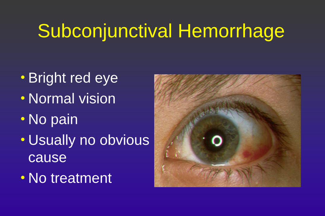
Subconjunctival Hemorrhage
Bright red eye Normal vision No pain Usually no obvious
cause No treatment

Stye/Chalazion
Stye (hordeolum): obstruction of the perifollicular glands Chalazion:
obstruction of the Meibomian glands

Stye/Chalazion

Stye/Chalazion
Treatment Warm compresses +/- topical antibiotics Systemic antibiotics for associated
preseptal cellulitis Incision and curettage for drainage

Blepharitis Chronic inflammation affecting the lash line Dysfunction of the meibomian glands Secondary infection Associated with acne rosacea

Blepharitis Symptoms
Foreign body sensation Burning Mattering of the lashes Eyelids sticking
together upon waking

Blepharitis Treatment
Warm compresses Lubricant eye drops Mechanical cleansing of lids Omega-3 fatty acid supplements (flaxseed oil,
fish oil) Counseling that this may be a chronic or
recurring problem

Blepharitis Treatment
Topical antibiotics Azasite (azithromycin in DuraSite) Topical steroids for inflammatory component
(only for short duration) Restasis Systemic doxycycline for refractory problems

Diagnosis of Conjunctivitis
Stringy white mucus: allergic Purulent discharge: bacterial Watery: viral

Allergic Conjunctivitis
Symptoms: ITCHING Clinical findings Normal exam Lid or conjunctival
edema Stringy white
discharge

Allergic Conjunctivitis Treatment Cold compresses Topical antihistamines (over the
counter) Topical mast cell stabilizers Combination topical antihistamines
and mast cell stabilizers

Topical Antihistamines
Over the counter (use QID) Vasocon-A Naphcon-A Opcon-A Visine-A

Allergic Conjunctivitis Treatment
Mast cell stabilizers with anitihistamine action BID use
• Azelastine (Optivar) • Epinastine (Elestat) • Ketotifen (Alaway) • Ketotifen (Zaditor --over the counter) • Nedocromil (Alocril) • Olopatadine (Patanol) • Pemirolast (Alamast)
Once daily use • Olopatadine (Pataday) • Alcaftadine (Lastacaft)

Viral Conjunctivitis
Adenovirus Highly contagious

Viral Conjunctivitis Symptoms Burning discomfort Associated systemic symptoms: URI, sore
throat, fever, malaise
Clinical findings Redness Watery discharge Palpable preauricular lymph node

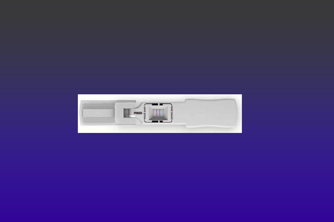
Viral Conjunctivitis Diagnosis
• AdenoPlus is immunoassay to detect adenoviral antigens
• Compared to cell culture 90% sensitivity, 96% specificity
• Cost $15-$25 per test • Reimbursable • Accurate diagnosis reduces treatment with
unnecessary and ineffective antibiotics

Viral Conjunctivitis

Viral Conjunctivitis Treatment: symptomatic Cold compresses Iced artificial tears Acetaminophen Topical betadine

Viral Conjunctivitis
Duration is 1-3 weeks Contagious period is for 1 week after
onset of symptoms Postconjunctivitis dry eye syndrome
may persist for several months

Bacterial Conjunctivitis
Caused by all common bacteria Symptoms: purulent discharge Clinical findings Conjunctival injection Purulent discharge
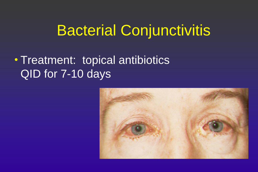
Bacterial Conjunctivitis
Treatment: topical antibiotics QID for 7-10 days

Ophthalmic Antibiotic Ointments Erythromycin Bacitracin Sulfacetamide sodium Gentamicin Tobramycin Ciprofloxacin Polymyxin B/Bacitracin Polymyxin B/Neomycin/Bacitracin Polymyxin B/Oxytetracyclin
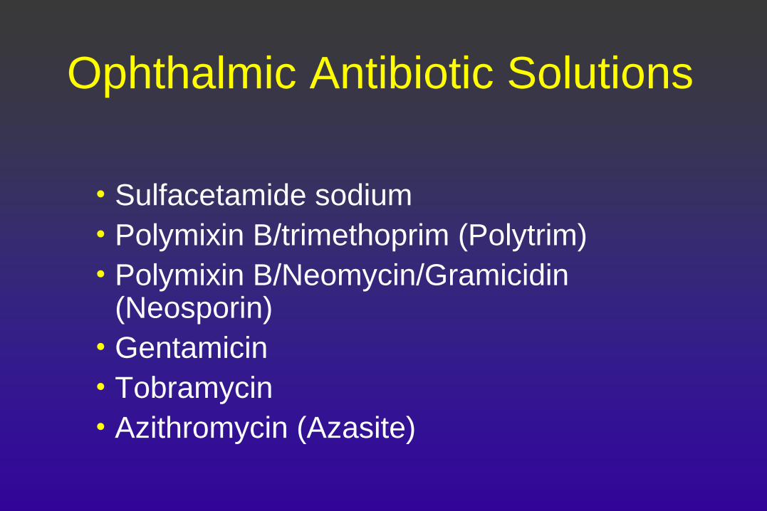
Ophthalmic Antibiotic Solutions
Sulfacetamide sodium Polymixin B/trimethoprim (Polytrim) Polymixin B/Neomycin/Gramicidin
(Neosporin) Gentamicin Tobramycin Azithromycin (Azasite)

Ophthalmic Antibiotic Solutions
Ofloxacin (Ocuflox) Ciprofloxacin (Ciloxan) Levofloxacin (Quixin) Gatifloxacin (Zymar, Zymaxid) Moxifloxacin (Vigamox) Besifloxacin (Besivance)

Hyperpurulent Bacterial Conjunctivitis
Copious discharge may indicate infection with
pseudomonas or gonorrhea and requires urgent referral

Dry Eyes
Symptoms Burning Foreign body
sensation Grittiness Tearing

Dry Eyes Associated conditions Aging Sjogren’s syndrome Rheumatoid arthritis Stevens-Johnson syndrome Systemic medications: antihistamines,
diuretics, antidepressants

Dry Eyes Treatment Lubricant eye drops (artificial tears) Lubricating ointment at bedtime Protective glasses and hat outdoors Flaxseed oil 1000 mg daily Restasis (topical cyclosporine) Punctal plugs or occlusion

Punctal Plugs

Vision Threatening Red Eye
Corneal infections Scleritis/episcleritis Iritis/uveitis Acute angle-closure glaucoma

Vision Threatening Red Eye Indications for Referral
Decreased vision Severe eye pain Light sensitivity Opacity on cornea

Corneal Infections
Viral keratitis Herpes simplex most common
Bacterial keratitis Frequently related to soft contact lens
wear Fungal keratitis

Herpes Simplex Keratitis Primary HSV Conjunctivitis with watery discharge Skin vesicles on lids Enlarged preauricular lymph nodes +/- corneal involvement with single or
multiple dendrites Recurrent HSV

Primary HSV Recurrent HSV

Bacterial Keratitis
Most common in soft contact lens wearers Red painful eye Opacity on the cornea Requires ophthalmologic referral

Bacterial Keratitis
Iritis/Uveitis Inflammation in the anterior chamber
(iritis) or involving the entire eye (uveitis) Symptoms Pain Photophobia Decreased vision

Iritis/Uveitis Clinical findings: Circumcorneal redness Pupil is smaller than normal Cell and flare in the anterior chamber
Iritis/Uveitis Etiology Nongranulomatous:
Idiopathic Traumatic Ankylosing spondylitis Behcet’s disease Inflammatory bowel disease Herpes Lyme disease Postoperative Psoriatic arthritis Reiter’s syndrome Lupus Wegener’s granulomatosis JRA
Granulomatous: Sarcoidosis Tuberculosis Syphilis Toxoplasmosis Brucellosis

Episcleritis • Benign, self-limited
inflammation of episclera (Tenon’s capsule)
• Rarely associated with systemic disease
• Mild eye pain and tenderness
• Treatment:Oral NSAIDs

Scleritis Inflammation of the wall of the eye. Severe destructive disease, underlying systemic disease Severe boring eye pain Treatment with systemic steroids, NSAIDs, and/or immunosuppressives

Scleritis

Angle Closure Glaucoma
Obstruction of aqueous outflow due to occlusion of the trabecular meshwork by the iris. Occurs in patients anatomically predisposed with shallow anterior chambers.

Angle Closure Glaucoma
Screening for susceptible patients: penlight held temporal and parallel to the iris reveals a shadow on the nasal iris in at risk patients.

Angle Closure Glaucoma Symptoms Severe ocular pain Blurred vision Halos around lights Headache Nausea and
vomiting
Clinical findings High intraocular
pressure Mid-dilated sluggish
pupil Corneal epithelial
edema Conjunctival injection Shallow AC

Angle Closure Glaucoma Medical treatment to
lower IOP Pilocarpine Topical aqueous humor
suppressants: timolol, brimonidine, carbonic anhydrase inhibitors
Definitive therapy: Iridectomy

Cataract
Clouding of the lens which may result in decreased vision
Leading cause of treatable blindness worldwide In US accounts for 50% of low vision cases in adults
> 40 Cataract surgery with IOL implantation one of most
common surgeries performed under Medicare

Risk Factors Smoking Lifetime exposure to UV-B radiation Diabetes Inhaled, topical, and oral corticosteroid use Hypertension Myopia Obesity No significant delaying effect on cataract
development with vitamin supplementation

Cataract

Treatment of Cataracts
Cataract extraction with intraocular lens implant
Indicated when patients are having difficulty with their vision due to cataracts
Typically small incision phacoemulsification

Intraocular Lenses
Posterior chamber intraocular lens inserted into the lens capsule
Anterior chamber or sutured IOL used in certain cases

Intraocular lenses
Multifocal IOL Accommodative IOL

Preoperative Preparation Decide based on patient’s needs if cataracts
are visually significant Measurements for IOL calculation Preoperative physical examination EKG in patients older than 65 Laboratory studies as medically necessary Preoperative topical antibiotics and NSAIDS Anticoagulation—frequently may be
continued, but consult with ophthalmologist

Tamsulosin Precautions Alpha blocker therapy may cause intraoperative
floppy iris syndrome and increase the risk of surgical complications
Discontinuation of alpha blockers for weeks to years does not reverse the effect
If a patient has a cataract and requires tamsulosin or other therapy, consider cataract surgery before initiating therapy
Tell ophthalmologist of any history of alpha blocker use

Benefits of Cataract Surgery Better optically corrected vision Better uncorrected vision with reduced
spectacle dependence Increased ability to read or do near work Reduced glare Improved ability to function in dim light Improved depth perception and binocular
vision (reduced risk of motor vehicle accidents and falls with broken hips and shoulders)

Diabetes
Leading cause of blindness under age 65 Prevalence increases with duration of disease Intensive control of blood glucose reduces the
risk of retinopathy in both type I and II diabetics

Nonproliferative Diabetic Retinopathy: Characteristics
Microaneurysms Leakage of intravascular fluid Intraretinal hemorrhage Retinal ischemia due to
capillary dropout

NPDR
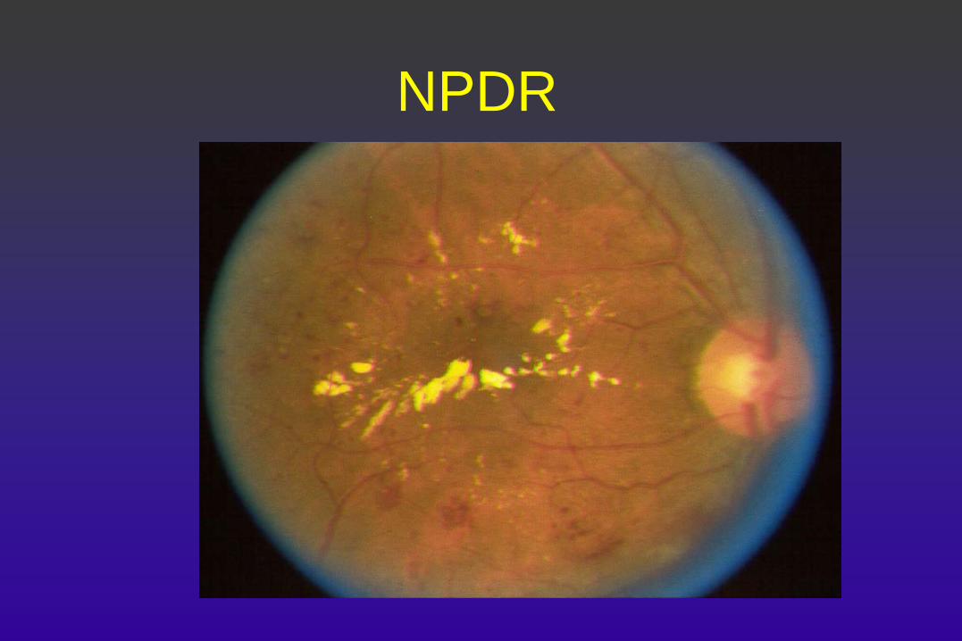
NPDR
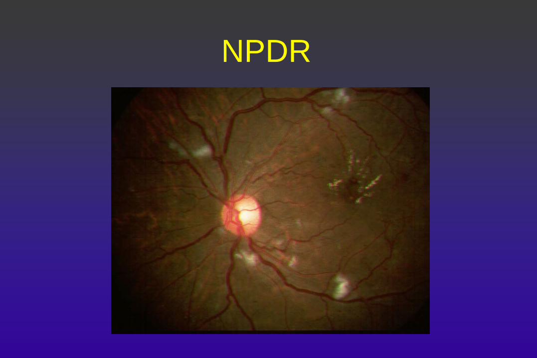
NPDR

NPDR: Treatment Intravitreal anti-VEGF (Diabetic Retinopathy
Clinical Research Net) Bevcizumab (Avastin) Ranibizumab (Lucentis) Aflibercept (Eyelea) Focal laser photocoagulation for clinically significant
macular edema (CSME) reduces rate of vision loss by more than 50% (EDTRS)
Intravitreal steroids

Proliferative Diabetic Retinopathy: Characteristics
All of the findings of NPDR PLUS formation of neovascular tissue from
the optic nerve or the retinal surface

PDR

PDR

PDR: Treatment Intravitreal anti-VEGF Panretinal photocoagulation Involute neovascularization Reduce risk of vitreous hemorrhage and/or
reduce traction retinal detachment Vitrectomy Remove nonclearing vitreous hemorrhage Repair traction retinal detachment

Pitfalls in Diagnosis
More difficult if pupils undilated CSME not visible without stereoscopic view IRMA difficult to distinguish from neovascularization Neovascularization unapparent or outside field of
direct ophthalmoscope

Screening Recommendations
Type I diabetics 5 years after onset, then yearly
Type II diabetics at time of diagnosis, then yearly
Diabetics who are pregnant early in the pregnancy and every trimester

Age-Related Macular Degeneration
A leading cause of severe, irreversible vision impairment Characterized by Drusen RPE abnormalities Geographic atrophy Neovascular maculopathy

Age-Related Macular Degeneration
Risk factors Smoking doubles risk of AMD +/-hypertension, cardiovascular disease,
inflammation Low levels of anti-oxidants

Dry ARMD

Wet ARMD

ARMD Treatment Dry ARMD
• Diet rich in green leafy vegetables, no smoking, weight control Anti-oxidant supplements for intermediate
AMD or advanced AMD in one eye Wet ARMD Intravitreal anti-VEGF
(bevacizumab,ranibizumab, aflibercept) Anti-oxidant supplements (AREDS 2 formula)

Amsler Grid
New Onset Diplopia
Is this a neurologic emergency? Diplopia that is not improved by covering
one eye requires a neuro-ophthalmic or neurologic evaluation

Diplopia Monocular:
abnormalities in the refractive media Corneal (high
astigmatism) Lenticular—cataract or
dislocated lens Retinal (rarely)
Binocular: misalignment of the visual axis Cranial nerve palsy Giant cell arteritis Demyelinating disease Myasthenia gravis Thyroid orbitopathy Orbital myositis Other causes

Flashes and Floaters
Patients need to be examined to detect and treat retinal holes and detachments.
What is the differential diagnosis?
Posterior vitreous detachment Retinal hole/detachment Vitreous hemorrhage Posterior segment inflammation Trauma Migraine


Red Flag Signs and Symptoms Require urgent referral Decreased vision Metamorphopsia (distorted vision) Severe eye pain Red eye with light sensitivity Corneal opacity Flashes and floaters Binocular double vision