Embed Size (px)
Citation preview
Abbreviations used: ALA, alpha lipoic acid; ACC, acetyl-coenzyme A carboxylase; AMPK, AMP-activated protein kinase; AUC, area under the concentration-time curve; HFD, high-fat diet; HFD+ALA, high-fat diet supplemented with 0.5% of alpha lipoic acid; LFD, low-fat diet; LFD+ALA, low-fat diet supplemented with 0.5% of alpha lipoic acid; IMCL, intramyocellular lipid(s); ipGTT, intraperitoneal glucose tolerance test.
1
Prevention of high-fat diet-induced muscular lipid accumulation in rats
by alpha lipoic acid is not mediated by AMPK activation
Silvie Timmers1,2, Johan de Vogel-van den Bosch1,2, Mhairi C. Towler3, Gert
Schaart4, Esther Moonen-Kornips2,4, Ronald P. Mensink1,2, Matthijs K.
Hesselink4, D. Grahame Hardie3 and Patrick Schrauwen1,2
1 Top Institute Food and Nutrition (TIFN), 6700 AN, Wageningen, The
Netherlands 2 Department of Human Biology, School for Nutrition, Toxicology
and Metabolism (NUTRIM), Maastricht University, 6200 MD, Maastricht, The
Netherlands 3 Division of Molecular Physiology, College of Life Sciences,
University of Dundee, DD1 5EH, Dundee, Scotland 4 Department of Human
Movement Sciences, School for Nutrition, Toxicology and Metabolism
(NUTRIM), Maastricht University, 6200 MD, Maastricht, The Netherlands
Running head: alpha lipoic acid, muscle lipid accumulation
Address correspondence to:
Patrick Schrauwen
Department of Human Biology
Maastricht University
P.O. Box 616
6200 MD Maastricht, The Netherlands
Phone: +31 (0) 43 3881502
Fax: +31 (0) 43 3670976
E-mail: [email protected]
by guest, on June 4, 2018w
ww
.jlr.orgD
ownloaded from

2
Abstract
Skeletal muscle triglyceride accumulation is associated with insulin resistance
in obesity. Recently, it has been suggested that alpha lipoic acid (ALA)
improves insulin sensitivity by lowering triglyceride accumulation in non-
adipose tissues via activation of skeletal muscle AMP-activated protein kinase
(AMPK). We examined whether chronic ALA supplementation prevents
muscular lipid accumulation that is associated with high-fat diets via activation
of AMPK. In addition, we tested if ALA supplementation was able to improve
insulin sensitivity in rats fed low- and high-fat diets.
Supplementing male Wistar rats with 0.5% ALA for eight weeks significantly
reduced body weight, both on low- and high-fat diets (-24% LFD+ALA vs.
LFD, p<0.01, and -29% HFD+ALA vs. HFD, p<0.001). Oil red O lipid staining
revealed a three-fold higher lipid content in skeletal muscle after HFD
compared to LFD and ALA-supplemented groups (p<0.05). ALA improved
whole body glucose tolerance (∼ 20% lower total AUC in ALA supplemented
groups vs. controls, p<0.05). These effects were not mediated by increased
muscular AMPK activation or ALA-induced improvement of muscular insulin
sensitivity.
To conclude, the prevention of high-fat diet-induced muscular lipid
accumulation and the improved whole body glucose tolerance are likely
secondary effects due to the anorexic nature of ALA.
Supplementary key words: diet-induced obesity, glucose tolerance, insulin
sensitivity, ectopic lipid accumulation
by guest, on June 4, 2018w
ww
.jlr.orgD
ownloaded from
3
Introduction
Obesity has reached epidemic proportions globally, and the situation is likely
to deteriorate further. It is well known that obesity predisposes individuals to a
range of serious health complications with high morbidity rates, including
insulin resistance, type 2 diabetes mellitus and cardiovascular diseases [1].
The mechanisms underlying the progression from obesity to insulin resistance
and ultimately type 2 diabetes are not fully elucidated [2]. It is speculated that
excessive FFA mobilization from adipose tissue leads to an increase in
plasma FFA with ectopic deposition of triglycerides in muscle, the liver and
the pancreas as a result. The increased intramyocellular lipid (IMCL) content
could disturb muscle insulin signaling, which ultimately results in muscle
insulin resistance [3].
Alpha lipoic acid (ALA), also known as thioctic acid, is a naturally occurring
short-chain fatty acid with a powerful antioxidant capacity. It is synthesized in
small amounts by plants and animals, including humans [4, 5]. Endogenously
synthesized ALA is an essential cofactor for several mitochondrial enzyme
complexes that catalyze critical reactions related to energy production [6].
ALA can also be exogenously derived from various food sources, such as
tomatoes, spinach, broccoli and Brussels sprouts [7]. ALA has been
suggested of having influence on glucose homeostasis. Short-term
incubations of L6 muscle cells with a rather high concentration of 10 mM of
ALA increased glucose uptake by 40-80%, and ex vivo incubation of muscles
derived from ob/ob mice with 10 mM ALA showed a substantial increase in
glucose uptake [8]. Acute and chronic parenteral treatments with ALA in
by guest, on June 4, 2018w
ww
.jlr.orgD
ownloaded from

4
obese Zucker (fa/fa) rats, an animal model of insulin resistance, significantly
improved insulin-stimulated 2-deoxyglucose uptake in epitrochlearis muscles
by 62 and 64% [9]. In an uncontrolled study of 20 patients with type 2
diabetes, intravenous infusion of 500 mg/day of racemic ALA for ten days
improved insulin-stimulated glucose disposal measured 24-hours after the last
infusion [10]. Moreover, in a placebo-controlled, multi-centre study, 74
patients with type 2 diabetes were randomized to receive 600 mg ALA or
placebo orally once, twice or three times daily, for four weeks. It was shown
that patients who received ALA significantly improved their insulin-stimulated
glucose disposal compared with those on the placebo, although no significant
differences regarding the various doses of ALA were observed [11]. These
studies suggest that ALA has the potential to improve insulin sensitivity via an
effect on skeletal muscle glucose uptake.
In that respect, recent experimental research indicates that ALA might
improve insulin sensitivity by lowering triglyceride accumulation in non-
adipose tissues, like muscle. Feeding diabetic OLETF rats with ALA-
supplemented chow reduced triglyceride accumulation in skeletal muscle and
pancreatic islets compared with their untreated counterparts [12]. The
reduced triglyceride accumulation in skeletal muscle was attributed to the
activation of skeletal muscle AMP-activated protein kinase (AMPK) [13]; an
enzyme that is activated when the cellular energy is depleted [14]. Previous
experiments in cells and animals showed that AMPK could stimulate glucose
uptake independently of insulin, lower triglyceride accumulation in non-
adipose tissues via increased fat oxidation, and increase mitochondrial
biogenesis [15-19]. Thus, activation of AMPK by ALA may lead to a lower
by guest, on June 4, 2018w
ww
.jlr.orgD
ownloaded from
5
accumulation of IMCL via an increased oxidation of long chain fatty acids and
might improve whole body insulin sensitivity.
To test this hypothesis, we studied whether supplementation of ALA to a high-
fat diet prevented diet-induced obesity and muscular lipid accumulation. In
addition, we studied if ALA supplementation could improve insulin sensitivity
over time and if this was mediated by changed activity of skeletal muscle
AMPK.
by guest, on June 4, 2018w
ww
.jlr.orgD
ownloaded from
6
Methods
Animals and diets
Thirty-two, eleven-week-old male Wistar rats (Charles River Laboratories)
were individually caged and randomly assigned to one of the four dietary
treatments for eight weeks. The first two groups were fed a semi purified diet
containing 10% of kcal as fat with an energy density of 3.85 kcal/g (D12450B;
20% energy (% E) protein, 70% E carbohydrate, and 10% E fat consisting of
225% E soybean oil and 180% E lard; Research Diets Inc., New Brunswick,
NJ) supplemented with or without 0.5% DL-ALA (Sigma-Aldrich Chemie B.V.,
Zwijndrecht, The Netherlands). The other two groups received a semi purified
diet containing 45% of kcal as fat with an energy density of 4.73 kcal/g
(D12451; 20% E protein, 35% E carbohydrate, 45% E fat consisting of 225%
E of soybean oil and 1598% E lard; Research Diets Inc., New Brunswick, NJ)
supplemented with or without 0,5% DL-ALA. Rats were maintained at ambient
temperature (22 ±1 °C), with 12:12h light-dark cycles and allowed ad libitum
access to food and water. Food consumption and body weight were recorded
weekly. To calculate net absorption of nutrients during the diet intervention,
fecal samples were collected during the last week of the intervention period,
freeze dried and together with samples from the diet, analyzed for gross
energy content using adiabatic bomb calorimetry (Ika-calorimeter system
C4000 Heitersheim, Germany). All experiments were approved by the
Institutional Animal Care and Use Committee of the Maastricht University and
complied with the principles of laboratory animal care.
by guest, on June 4, 2018w
ww
.jlr.orgD
ownloaded from

7
Intraperitoneal glucose tolerance tests
To investigate the effect of DL-ALA on glucose tolerance, rats were subjected
to an intraperitoneal glucose tolerance test (ipGTT) after four and eight weeks
of the dietary treatment. Rats were administered 1.5 g/kg glucose (ICN
Biomedicals, Inc., Aurora, Ohio) after a six-hour fast. A baseline blood sample
was immediately taken (t = 0), and this was repeated at t = 15, 30, 60 and 120
min. Samples were collected in Na-EGTA coated Eppendorf tubes and
centrifuged immediately at 4000 rpm for 10 min. Plasma was then separated
and stored at -80°C for the determination of insulin concentrations by Insulin
(Rat) Ultrasensitive EIA (Alpco Diagnostics, Salem, NH).
Tissue collection
After eight-and-a-half weeks of ALA supplementation, rats were sacrificed
after a six-hour fast. Rats received an intraperitoneal injection of insulin
(10u/kg) and were anaesthetized after 9 minutes by a mixture of 79 % of CO2
and 21% of O2 for 1 minute, followed by cervical dislocation. For histological
analysis, the medial region of the left gastrocnemius muscle (mostly
compromised of type 2 muscle fibres) was dissected and freed from any
visible fat and blood, embedded in Tissue-Tek (Sakura Finetek, Zoeterwoude,
The Netherlands) and frozen in liquid nitrogen-cooled isopentane (2-methyl-
butane, Fluka, Zwijndrecht, The Netherlands). The right gastrocnemius
muscle was rapidly dissected and immediately frozen in liquid nitrogen-cooled
isopentane for Western blot analyses. All samples were stored at -80°C until
further analysis.
by guest, on June 4, 2018w
ww
.jlr.orgD
ownloaded from

8
Histological analysis of intramyocellular lipids ( IMCL)
Briefly, cryosections (5 µm) from the midbelly region of the gastrocnemius
were thaw mounted on uncoated pre-cleaned (96% ethanol) glass slides.
Immediately after mounting, air-dried, fresh cryosections were fixed in 3.7%
formaldehyde for 1 h. Then the sections were treated with 0.5% Triton X-100
in PBS for 5 min, and washed three times with PBS. Thereafter, sections were
incubated for 30 min with a polyclonal rabbit antibody against the basement
membrane protein laminin (Sigma-Aldrich, St. Louis, MO, USA, 1:50 dilution
in PBS) and a monoclonal antibody raised against adult human slow myosin
heavy chain (A4.840, developed by Dr. Blau; Developmental Studies
Hybridoma Bank, University of Iowa, Iowa City, IA, USA) [20] to visualize
individual cell membranes and distinguish between type 1 and 2 muscle
fibres. This was followed by 30 min incubation with the appropriate secondary
antibodies conjugated with Alexa Fluor488 and Alexa Fluor555 (Invitrogen,
Groningen, The Netherlands). After washing with PBS, glass slides were
immersed in the ORO working solution for 30 min for the detection of lipid
droplets [21] and rinsed three times with deionised water for 30 s followed by
10 min of washing with running tap water. Stained sections were embedded in
Mowiol.
All sections were examined using a Nikon E800 fluorescence microscope
(Nikon Instruments Europe B.V., Badhoevedorp, The Netherlands) coupled to
a Basler A101C progressive scan colour charge-coupled device camera. All
sections were processed and analysed using Lucia GF 4.80 software (Nikon,
Düsseldorf, Germany). Special care was taken to use the same camera
by guest, on June 4, 2018w
ww
.jlr.orgD
ownloaded from

9
settings (gain and exposure time) while grabbing all images. For IMCL > 800
cells per muscle were analysed.
All images were analysed for the lipid droplet over myocyte area fraction. To
this end, a semiautomatic macro was written that allowed: (1) autodetection of
the cellular membrane (identified in the blue channel); (2) measurement of the
area covered by the measured myocytes with distinction between type 1 and
type 2 fibres (identified in the green area); and (3) measurement of the area
covered by lipid droplets (identified in the red channel). The area fraction was
computed by dividing the area covered by the lipid droplets (in µm2) by the
cell surface of the measured myocytes (in µm2). The mean area fraction thus
reflects the percentage of total measured cell surface covered by lipid
droplets.
Western blot analyses
Muscle homogenates for Western blot analysis of p-Akt protein were
performed as described before [22]. Equal amounts of protein were loaded on
SDS-PAGE and after Western blotting, incubated with Akt and p-Akt
antibodies (#9271 and #9272 respectively, Cell Signaling Technology, Bioké,
Leiden, The Netherlands). After incubation with the appropriate secondary
antibodies, specific protein bands were visualized by chemiluminescence and
analyzed by Chemidoc XRS system (Bio-Rad, Veenendaal, The Netherlands).
For GLUT4 detection equal amounts of muscle membrane and muscle cytosol
protein fractions were loaded on SDS-PAGE and after Western blotting
incubated with a GLUT4-antibody (sc-1608, Santa Cruz, Bio-Connect,
by guest, on June 4, 2018w
ww
.jlr.orgD
ownloaded from

10
Huissen, The Netherlands). After incubation with the appropriate secondary
IRDye680-labeled antibody (Licor, Westburg, Leusden, the Netherlands), the
specific GLUT4 protein was detected and analyzed using the Odyssey near
Infrared Scanner (Licor).
AMPK activity and ACC phosphorylation
Kinase assays of total AMPK activity have been described previously [23].
Briefly, total AMPK activity was measured in the tissue lysates by
immunoprecipitating with a combination of α1- and α2-specific antibodies.
Activities are presented in units per milligram protein, where a unit is defined
as nanomoles of phosphate incorporation into the SAMS peptide per minute.
Western blot analyses of acetyl-coenzyme A carboxylase (ACC) was carried
out as follows. Muscle samples were powdered under liquid nitrogen and
homogenised at a ratio of 1:4 (w/v) in the following buffer 0.05 M Tris-HCl pH
7.4, 0.25 M Sucrose, 0.001 M EDTA, 0.001 M EGTA, 1 % TX-100, 0.001 M
NaVO4 0.05 M NaF, 0.005 M Na2PO4, 0.1 % DTT and protease inhibitor
cocktail tablet (1 tablet/10 ml), (Roche, Hertfordshire, U.K.). 15 µg of protein
was loaded on a NuPage 3-8% Tris-Acetate gel (Invitrogen, Paisley, U.K.)
and separated by SDS-PAGE, followed by transfer onto nitrocellulose
membrane. Membranes were blocked for 1 hour with Odyssey blocking
buffer (Licor, U.K.) and probed overnight with sheep polyclonal
phosphospecific ACC1/2 (P-S79/ P-S221) antibody and total ACC antibody (Cell
Signalling Technology, Beverly, MA). The phosphospecific polyclonal antibody
was raised against a 13-amino-acid peptide that comprised amino acid 221 of
by guest, on June 4, 2018w
ww
.jlr.orgD
ownloaded from

11
human ACC2 coupled to KLH, and a 12-amino-acid peptide that comprised
amino acid 79 of rat ACC1. After immunization of sheep with the peptides,
antibodies were extracted from sera by Sephadex affinity column. Columns
were washed with tris-buffered saline with 1% Tween-20, and eluted in 50 mM
glycine (pH 2.5). 75 mM tris pH 8 was added to eluted antibodies. Membranes
were washed in TBS-T and incubated for 1 hour with secondary antibodies
IRDye 800 conjugated to streptavidin (picks up total ACC), and sheep IRDye
680 (picks up P-ACC) (Rockland Inc., Lorne Laboratories, Reading, UK).
Membranes were washed again in TBS-T and analysed using the Licor
Odyssey near Infrared Scanner.
Statistical Analysis
Results are presented as the mean ± SE. Differences between groups were
analysed by one-way ANOVA with a Bonferroni’s post hoc correction. The
accepted level of statistical significance was p < 0.05 for all analyses. All
calculations were done using the Statistical Package for the Social Sciences
(SPSS 11.0 software).
by guest, on June 4, 2018w
ww
.jlr.orgD
ownloaded from

12
Results
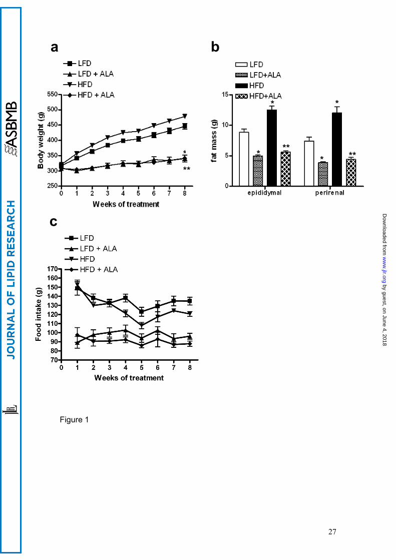
Body weight gain and composition
Body weights gradually increased over the eight weeks of diet intervention in
the LFD and the HFD rats. However, compared to animals on the LFD, rats
receiving the HFD gained more weight during the intervention period (fig 1a).
Supplementation of ALA to LFD or HFD attenuated weight gain, resulting in
significantly lower final body weights (-24% in LFD+ALA vs. LFD, p<0.01 and
-29% in HFD+ALA vs. HFD, p<0.001, fig 1a). Epididymal and perirenal fat pad
weights were significantly higher in HFD animals compared to rats on the LFD
(p<0.001). ALA treatment resulted in an approximately 50% lower epididymal
and perirenal fat mass in the ALA-treated animals compared to their controls
(p<0.01, fig 1b). Thus these data suggest that the differences in body weight
between the groups are, at least in part, due to differences in adiposity (fig
1b).
Food intake and net absorption
Analysis of food intake and faecal energy content over the last week revealed
that gross energy absorption was 30% lower in the ALA supplemented groups
compared to control groups (p<0.01) (data not shown). This reduction in gross
energy absorption was completely accounted for by a 30% lower food intake
in animals treated with ALA (p<0.001, fig 1c), with no differences in fecal
energy content between the groups (data not shown).
by guest, on June 4, 2018w
ww
.jlr.orgD
ownloaded from

13
Glucose and insulin levels following ipGTT
IpGTT were performed four and eight weeks after commencement of dietary
treatment to study time-dependent effects of the diet on whole body glucose
tolerance. Fasting blood glucose values were not different between ALA and
control animals after four and eight weeks (fig 2a and 2c). Glucose injection in
the animals after four weeks diet intervention resulted in similar peak blood
glucose values at time-point 15 minutes between groups. Glucose clearance
in the ALA treated animals was faster than in the corresponding LFD and HFD
fed rats (approximately 17% lower total AUC, p<0.05, fig 2b). After eight
weeks of treatment, glucose levels peaked again at time point 15 min after
injection of the glucose bolus and values were not different between groups.
Glucose clearance remained faster in the LFD+ALA vs. LFD group and
HFD+ALA vs. HFD group (approximately 20% lower total AUC, p<0.05, fig
2d).
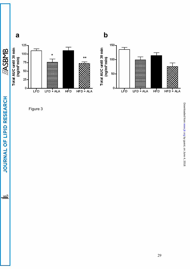
Fasting plasma insulin levels were lower in the groups receiving ALA
compared to controls after four weeks of treatment (1.08 ± 0.19 vs. 2.43 ±
0.30 ng/ml*min in LFD+ALA vs. LFD, p<0.001, and 0.66 ± 0.06 vs. 1.60 ±
0.34 ng/ml*min in HFD+ALA vs. HFD). Total area under the curve was
approximately 30% lower in the ALA supplemented animals compared to the
control LFD and HFD groups (p<0.05, fig 3a). After eight weeks of ALA
supplementation fasting plasma insulin levels remained lower compared to
the control matched animals (1.36 ± 0.31 vs. 2.52 ± 0.36 ng/ml*min in
LFD+ALA vs. LFD, p<0.05, and 0.87 ± 0.10 vs. 1.90 ± 0.21 ng/ml*min in
HFD+ALA vs. HFD, p<0.001). Total area under the curve was again ~30%
by guest, on June 4, 2018w
ww
.jlr.orgD
ownloaded from

14
lower in the ALA vs. control treated animals, however due to the high variance
this did not reach statistical significance (p=0.34, fig 3b).
Intramyocellular lipid levels
After 8 weeks of dietary intervention, IMCL content was 3-fold higher in the
animals receiving the HFD (p<0.05). There were no differences in IMCL
accumulation between type 1 and 2 muscle fibres (data not shown).
Supplementation of ALA in rats on the HFD reduced lipid accumulation
significantly, resulting in an intramyocellular lipid content that was comparable
to animals receiving the LFD (p<0.05, fig 4). ALA did not further reduce IMCL
in the LFD group. Representative images demonstrating differences in
staining between the four different diets are included as supplementary figure
1.
Insulin signalling
Muscle insulin signalling was analysed to see whether the ALA-induced
increase in whole body glucose tolerance together with the lower muscular
lipid accumulation resulted in improved insulin-stimulated glucose disposal.
Among the different dietary interventions there were no differences in insulin-
induced Akt phosphorylation (fig 5a). In parallel, ALA treatment failed to
increase insulin-stimulated GLUT4 translocation (fig 5b).
AMPK activity
No differences in total AMPK α-isoform activity were observed after 8 weeks
supplementation with 0.5% ALA to rats on both a LFD (fig 6a). Consistent with
by guest, on June 4, 2018w
ww
.jlr.orgD
ownloaded from

15
this finding, no effect of ALA on phosphorylation of ACC on either LFD or HFD
was observed (fig 6b).
by guest, on June 4, 2018w
ww
.jlr.orgD
ownloaded from

16
Discussion
Recently, it has been suggested that ALA may improve insulin sensitivity by
lowering triglyceride accumulation in non-adipose tissues [12]. The reduced
triglyceride accumulation in skeletal muscle was suggested to be attributed to
activation of skeletal muscle AMPK [13]. It is therefore possible that ALA
supplementation can prevent the lipid accumulation that is associated with
high-fat diets via activation of AMPK, and thereby improve insulin sensitivity.
Here we indeed show that ALA supplementation reduces high-fat diet-induced
accumulation of body fat and intramyocellular lipid content and improves
whole body glucose tolerance, but these effects are not associated with
increased muscular AMPK activation.
ALA supplementation prevented high fat diet-associated obesity, as
evidenced by significantly lower gain in total body weight, as well as lower fat
mass. Measuring fat pad weights at sacrifice indicated that animals receiving
the HFD had significantly more fat mass than animals on the LFD, and this
diet-induced increase in fat mass was completely prevented by
supplementation of ALA. Interestingly, the net loss of fat is independent of the
amount of fat in the diet. These effects were most likely due to pronounced
effects of ALA on food intake and body weight gain of the animals. Already
after one week of supplementation with ALA, animals had significant lower
food intake and body weight gain compared to control groups. This pattern
persisted over the entire eight-week intervention period. The reduction in food
intake was not compensated by changes in food absorption, as fecal energy
by guest, on June 4, 2018w
ww
.jlr.orgD
ownloaded from

17
loss was unaffected by ALA supplementation. These results are in agreement
with those of Kim et al. [24], who found that feeding male Sprague-Dawley
rats standard rat chow containing ALA for 2 weeks, significantly reduced food
intake and body weight in a dose-dependent manner. They suggested that the
anorexic effect of ALA is the direct result of suppression of hypothalamic
AMPK activity, since intracerebrovascular injection of small doses of ALA
reduced food intake. Indeed, several reports showed that inhibition of
hypothalamic AMPK activity has an anorexic effect [25, 26]. Furthermore, the
ALA-induced anorexic effects were not due to illness, since no adverse
pathology was seen during the study or at time of sacrifice of the animals. It
also seems unlikely that the decreased food intake is the result of taste
aversion, because intraperitoneal injection of ALA in Sprague-Dawley rats
caused a similar decrease in food consumption, and a conditioned taste
aversion test showed that there was no difference in preference ratio of
saccharine intake between ALA and saline injected animals [24].
Increasing evidence in humans and animal models indicates that
accumulation of muscle triglyceride is associated with decreased insulin
induction of glucose disposal into skeletal muscle, due to interference of fatty
acid metabolites with insulin signaling at multiple levels. Here, we found that
ALA supplementation completely prevented the high-fat diet-induced increase
in muscular lipid content. Animals receiving the HFD experienced 3-fold
higher IMCL levels, distributed both in the type 1 and type 2 fibers.
Supplementing ALA in the HFD group reduced lipid accumulation significantly,
resulting in an intramyocellular lipid content that is comparable to animals
by guest, on June 4, 2018w
ww
.jlr.orgD
ownloaded from

18
receiving the LFD. In accordance with the reduction of muscular lipid content,
whole body glucose tolerance was improved after 4 and 8 weeks of ALA
treatment. At both time points, rats receiving the LFD and HFD supplemented
with ALA had lower AUC compared to their control groups. Together with the
improved glucose tolerance, plasma insulin levels were significantly lower in
the groups receiving ALA, albeit that these results did not reach statistical
significance after 8 weeks of treatment. Together, these findings seem to
indicate that ALA supplementation is able to improve whole body insulin
sensitivity, even when high-fat diets are consumed. The mechanisms by
which ALA decreases ectopic lipid accumulation and improves whole body
glucose tolerance may be multi-factorial. First of all, it has been speculated
that ALA can activate skeletal muscle AMPK. AMPK is known to induce
GLUT4 translocation in skeletal muscle thereby improving muscular glucose
uptake independent of insulin signaling. In addition, AMPK increases fatty
acid oxidation by inhibiting ACC. Thus, ALA-induced AMPK activation in
muscle would decrease IMCL levels by stimulating fatty acid oxidation as well
as directly stimulate glucose uptake via increased GLUT4 translocation,
thereby bypassing insulin resistance induced by muscular lipid accumulation.
Indeed, 3 days of ALA supplementation to diabetic OLETF rats activated
AMPK in skeletal muscle and improved insulin sensitivity, whereas adenoviral
infection of dominant-negative AMPK prevented ALA-induced increase in
insulin-stimulated glucose uptake and failed to increase fatty acid oxidation
and ACC phosphorylation [13]. Also, numerous studies have shown that ALA
can improve glucose tolerance in both diabetic animal models as well as
individuals with type 2 diabetes [8, 10, 11, 27]. However, here we could not
by guest, on June 4, 2018w
ww
.jlr.orgD
ownloaded from

19
confirm these results. Despite the fact that ALA affected insulin sensitivity at
the whole body level, no change in muscle specific insulin-stimulated Akt
phosphorylation and GLUT4 translocation was noticed. Thus, skeletal muscle
insulin sensitivity was not affected by ALA treatment. Moreover, we could not
detect a positive effect of ALA treatment on skeletal muscle AMPK activity nor
ACC phosphorylation. It is possible that the effects of ALA treatment on whole
body glucose tolerance are due to the anorexic effects of ALA, thereby
reducing not only the triglyceride accumulation in the different fat depots, but
also targeting ectopic lipid accumulation. It is well known that moderate
reduction in caloric intake, without causing malnutrition, is effective in obese
diabetic rats to improve insulin stimulated glucose uptake and lower insulin
levels during a GTT [28, 29].
At first sight, these results are in contrast with previous findings showing that
ALA improves insulin sensitivity possibly by increasing AMPK activity [13, 30].
Even though the study of Lee et al. applied the same dosage of ALA (0.5%)
and route of administration, the duration of ALA supplementation was shorter
[13]. So, the different outcome of our study may be explained by the acute
versus chronic treatment with ALA. AMPK activity might only be influenced by
short term ALA administration. On the other hand, Gupte et al. [30] reported
that chronic ALA supplementation increased AMPK activity and improved
insulin signaling. However, a major difference in study protocol is the route of
administration of ALA. ALA was dissolved in Tris HCL (120 mM, pH 7.4) and
administered through daily injections intraperitoneally, at a dosage of 30 mg /
kg body weight. By investigating the metabolism of ALA through
administration of radioactively labeled ALA to rats, Harrison and McCormick
by guest, on June 4, 2018w
ww
.jlr.orgD
ownloaded from

20
[31] reported that the retention of the radioactive label in the body of rats was
approximately twice as high after oral administration than after intraperitoneal
administration. This was as expected given the slower rate of excretion of
radioactivity in urine and 14CO2 after oral administration [31]. Bioavailability
and excretion of ALA seems to be influenced by the route of administration,
making comparison between studies difficult. The anesthesia used and the
way of sacrifice of animals is often different in study protocols investigating
the effect of ALA on insulin sensitivity and AMPK activity. AMPK activity can
be increased if animals suffer from hypoxia [32]. Both CO2 as barbiturates
have the same working principle and with an overdose can depress the
respiratory center, which is followed by cardiac arrest [33]. So, studies that
sacrifice the animals by intraperitoneal injection of sodium pentobarbital may
influence AMPK activity. We only sedated the rats with CO2 for one minute,
after which they were sacrificed by cervical dislocation. So it is unlikely that
hypoxia occurred and affected the AMPK activity measurements.
In conclusion, we have confirmed that ALA supplementation can prevent the
lipid accumulation that is associated with high-fat diets. Moreover, we have
shown that the reduction in ectopic lipid accumulation is associated with an
improved whole body glucose tolerance. However, these effects of ALA are
not mediated via ALA-induced activation of muscle AMPK or ALA-induced
improvement in muscular insulin sensitivity. Possibly, ALA could have affected
AMPK activity in the food-regulating centre in the hypothalamus. Therefore,
we propose that the prevention of high-fat diet-induced muscular lipid
by guest, on June 4, 2018w
ww
.jlr.orgD
ownloaded from
21
accumulation and the improved whole body glucose tolerance may partly
result from the anorexic nature of ALA.
by guest, on June 4, 2018w
ww
.jlr.orgD
ownloaded from

22
Acknowledgments
The study was funded by Top Institute Food and Nutrition. TI Food and
Nutrition, formerly known as WCFS, is a unique public/private partnership that
generates vision on scientific breakthroughs in food and nutrition, resulting in
the development of innovative products and technologies that respond to
consumer demands for safe, tasty and healthy foods. Partners are major
Dutch food companies and research organizations. D. Grahame Hardie was
supported by a Programme Grant (080982) from the Wellcome Trust.
by guest, on June 4, 2018w
ww
.jlr.orgD
ownloaded from

23
References
1. Mokdad, A.H., et al., Prevalence of obesity, diabetes, and obesity-related health risk factors, 2001. Jama, 2003. 289(1): p. 76-9.
2. Hill, M.J., D. Metcalfe, and P.G. McTernan, Obesity and diabetes: lipids, 'nowhere to run to'. Clin Sci (Lond), 2009. 116(2): p. 113-23.
3. Dresner, A., et al., Effects of free fatty acids on glucose transport and IRS-1-associated phosphatidylinositol 3-kinase activity. J Clin Invest, 1999. 103(2): p. 253-9.
4. Carreau, J.P., Biosynthesis of lipoic acid via unsaturated fatty acids. Methods Enzymol, 1979. 62: p. 152-8.
5. Reed, L.J., A trail of research from lipoic acid to alpha-keto acid dehydrogenase complexes. J Biol Chem, 2001. 276(42): p. 38329-36.
6. Bustamante, J., et al., Alpha-lipoic acid in liver metabolism and disease. Free Radic Biol Med, 1998. 24(6): p. 1023-39.
7. Packer, L., E.H. Witt, and H.J. Tritschler, alpha-Lipoic acid as a biological antioxidant. Free Radic Biol Med, 1995. 19(2): p. 227-50.
8. Eason, R.C., et al., Lipoic acid increases glucose uptake by skeletal muscles of obese-diabetic ob/ob mice. Diabetes Obes Metab, 2002. 4(1): p. 29-35.
9. Jacob, S., et al., The antioxidant alpha-lipoic acid enhances insulin-stimulated glucose metabolism in insulin-resistant rat skeletal muscle. Diabetes, 1996. 45(8): p. 1024-9.
10. Jacob, S., et al., Improvement of insulin-stimulated glucose-disposal in type 2 diabetes after repeated parenteral administration of thioctic acid. Exp Clin Endocrinol Diabetes, 1996. 104(3): p. 284-8.
11. Jacob, S., et al., Oral administration of RAC-alpha-lipoic acid modulates insulin sensitivity in patients with type-2 diabetes mellitus: a placebo-controlled pilot trial. Free Radic Biol Med, 1999. 27(3-4): p. 309-14.
12. Song, K.H., et al., alpha-Lipoic acid prevents diabetes mellitus in diabetes-prone obese rats. Biochem Biophys Res Commun, 2005. 326(1): p. 197-202.
13. Lee, W.J., et al., Alpha-lipoic acid increases insulin sensitivity by activating AMPK in skeletal muscle. Biochem Biophys Res Commun, 2005. 332(3): p. 885-91.
14. Hardie, D.G. and D. Carling, The AMP-activated protein kinase--fuel gauge of the mammalian cell? Eur J Biochem, 1997. 246(2): p. 259-73.
15. Bergeron, R., et al., Chronic activation of AMP kinase results in NRF-1 activation and mitochondrial biogenesis. Am J Physiol Endocrinol Metab, 2001. 281(6): p. E1340-6.
16. Hayashi, T., et al., Metabolic stress and altered glucose transport: activation of AMP-activated protein kinase as a unifying coupling mechanism. Diabetes, 2000. 49(4): p. 527-31.
17. Ruderman, N.B., et al., Malonyl-CoA, fuel sensing, and insulin resistance. Am J Physiol, 1999. 276(1 Pt 1): p. E1-E18.
18. Winder, W.W., Energy-sensing and signaling by AMP-activated protein kinase in skeletal muscle. J Appl Physiol, 2001. 91(3): p. 1017-28.
19. Zong, H., et al., AMP kinase is required for mitochondrial biogenesis in skeletal muscle in response to chronic energy deprivation. Proc Natl Acad Sci U S A, 2002. 99(25): p. 15983-7.
by guest, on June 4, 2018w
ww
.jlr.orgD
ownloaded from

24
20. Cho, M., S.G. Webster, and H.M. Blau, Evidence for myoblast-extrinsic regulation of slow myosin heavy chain expression during muscle fiber formation in embryonic development. J Cell Biol, 1993. 121(4): p. 795-810.
21. Brand, M.D., et al., Mitochondrial superoxide and aging: uncoupling-protein activity and superoxide production. Biochem Soc Symp, 2004(71): p. 203-13.
22. Schrauwen, P., et al., Uncoupling protein 3 as a mitochondrial fatty acid anion exporter. Faseb J, 2003. 17(15): p. 2272-4.
23. Davies, S.P., D. Carling, and D.G. Hardie, Tissue distribution of the AMP-activated protein kinase, and lack of activation by cyclic-AMP-dependent protein kinase, studied using a specific and sensitive peptide assay. Eur J Biochem, 1989. 186(1-2): p. 123-8.
24. Kim, M.S., et al., Anti-obesity effects of alpha-lipoic acid mediated by suppression of hypothalamic AMP-activated protein kinase. Nat Med, 2004. 10(7): p. 727-33.
25. Andersson, U., et al., AMP-activated protein kinase plays a role in the control of food intake. J Biol Chem, 2004. 279(13): p. 12005-8.
26. Minokoshi, Y., et al., AMP-kinase regulates food intake by responding to hormonal and nutrient signals in the hypothalamus. Nature, 2004. 428(6982): p. 569-74.
27. Estrada, D.E., et al., Stimulation of glucose uptake by the natural coenzyme alpha-lipoic acid/thioctic acid: participation of elements of the insulin signaling pathway. Diabetes, 1996. 45(12): p. 1798-804.
28. Park, S.Y., et al., Calorie restriction improves whole-body glucose disposal and insulin resistance in association with the increased adipocyte-specific GLUT4 expression in Otsuka Long-Evans Tokushima fatty rats. Arch Biochem Biophys, 2005. 436(2): p. 276-84.
29. Okauchi, N., et al., Is caloric restriction effective in preventing diabetes mellitus in the Otsuka Long Evans Tokushima fatty rat, a model of spontaneous non-insulin-dependent diabetes mellitus? Diabetes Res Clin Pract, 1995. 27(2): p. 97-106.
30. Gupte, A.A., et al., Lipoic acid increases heat shock protein expression and inhibits stress kinase activation to improve insulin signaling in skeletal muscle from high-fat-fed rats. J Appl Physiol, 2009. 106(4): p. 1425-34.
31. Harrison, E.H. and D.B. McCormick, The metabolism of dl-(1,6-14C)lipoic acid in the rat. Arch Biochem Biophys, 1974. 160(2): p. 514-22.
32. Frederich, M., L. Zhang, and J.A. Balschi, Hypoxia and AMP independently regulate AMP-activated protein kinase activity in heart. Am J Physiol Heart Circ Physiol, 2005. 288(5): p. H2412-21.
33. Zupthen van, L.F.M., V. Baumans, and A.C. Beynen., Anesthesie, analgesie en euthanasie., in Handboek Proefdierkunde., E. Gezondheidszorg., Editor. 2003: Maarssen. p. 286-289.
by guest, on June 4, 2018w
ww
.jlr.orgD
ownloaded from

25
Figure legends
Figure 1
ALA prevents high-fat diet-associated obesity. (A) Body weight (n=8), (B)
epididymal and perirenal fat mass at sacrifice (n=8) (C) food intake (n=8).
Values are expressed as means ± SEM. * P<0.05 vs. LFD; ** P<0.05 vs.
HFD.
Figure 2
Effect of dietary ALA in response to a two-hour intraperitoneal glucose
tolerance test (ipGTT) (1.5g/kg). Blood glucose concentrations in time (A) and
total area under curve (B) during ipGTT after four weeks of diet intervention
(n=8). Blood glucose concentrations in time (C) and total area under curve (D)
during ipGTT after eight weeks of diet intervention (n=8). (D) Values are
expressed as means ± SEM. * P<0.05 vs. LFD; ** P<0.05 vs. HFD.
Figure 3
Effect of dietary ALA on insulin concentrations during a 2-hour intraperitoneal
glucose tolerance test (ipGTT). Total area under the insulin curve after four
(n=5) (A) and eight (n=6) (B) weeks of diet intervention (n=5). Values are
expressed as means ± SEM. * P<0.05 vs. LFD; ** P<0.05 vs. HFD.
Figure 4
Intramyocellular lipid levels in left gastrocnemius muscle after eight weeks of
diet intervention (n=8). Values are expressed as means ± SEM. * p<0.05 vs.
LFD; ** P<0.05 vs. HFD.
by guest, on June 4, 2018w
ww
.jlr.orgD
ownloaded from

26
Figure 5
Effect of eight week treatment with dietary ALA on (A) Akt phosphorylation
and (B) GLUT4 translocation in right gastrocnemius muscle (n=8). Values are
expressed as means ± SEM.
Figure 6
Effect of eight week treatment with dietary ALA on (A) AMPK activity and (B)
S79/S221 ACC phosphorylation in right gastrocnemius muscle (n=8). Values
are expressed as means ± SEM.
by guest, on June 4, 2018w
ww
.jlr.orgD
ownloaded from
27
Figure 1
b a
* **
* * **
**
* *
c
by guest, on June 4, 2018w
ww
.jlr.orgD
ownloaded from

28
Figure 2
a b
c d
*
*
*
* *
* * *
** **
**
**
**
by guest, on June 4, 2018w
ww
.jlr.orgD
ownloaded from

32
Figure 6
a pACC
totalACC
b
by guest, on June 4, 2018w
ww
.jlr.orgD
ownloaded from