Embed Size (px)
Citation preview
Prevention of clinical lymphoedema after cancer treatment:Early detection and risk reduction
A guide for health professionals
Contents
1. Introduction 1
1.1 Aim of this booklet 1
1.2 What is lymphoedema? 2
1.3 Anatomy of the lymphatic system 3
1.4 Epidemiology 5
2. Awareness and Prevention 7
2.1 Risk factors for clinical lymphoedema 7
2.2 Prevention and early detection at the sub-clinical level 9
2.3 Lifestyle and prevention 10
2.3.1 Skin and nail care tips 10
2.3.2 Diet and healthy weight 11
2.3.3 Physical activity 11
2.3.4 Travel, sun and heat 12
2.3.5 Positioning of the limb to prevent constriction 13
3. Presentation and Stages of Lymphoedema 14
3.1 Presentation of lymphoedema 14
3.2 Stages of lymphoedema 15
3.3 Problems arising from lymphoedema 16
4. Cellulitis 18
4.1 Definition 18
4.2 Clinical features of cellulitis 19
4.3 Management of acute cellulitis 20
4.3.1 Hospital admission 20
4.3.2 Management at home 22
4.3.3 Antibiotic regimes 23
4.4 Antibiotics “in case” - Travel and holidays 24
4.5 Antibiotics during lymphoedema therapy 24
4.6 Recurrent cellulitis 24
4.6.1 Antibiotic prophylaxis 25
5. Self-care 26
5.1 Simple self care exercises 26
5.2 Simple lymphatic drainage 29
6. References 35
7. Links to Additional Information 39
1
1. Introduction
1.1 Aim of this Booklet
This booklet provides an opportunity for healthcare professionals, working with cancer patients, to refresh their knowledge in preventing cancer related clinical lymphoedema and in providing self-help advice for patients. The recognition of lymphoedema and intervention at its earliest stage are essential to prevent progression.
This booklet also reviews the anatomy of the lymphatic system and the pathophysiology of cancer related lymphoedema, focusing on the prevention, early detection and treatment of cellulitis.
The impact of lymphoedema on patient health is often underestimated. It is an independent predictor of reduced quality of life even when other factors such as socioeconomic status, decreased range of limb motion, age, and obesity are taken into account.1 Lymphoedema can also have a negative impact on social wellbeing, resulting in additional burdens for cancer survivors.2 Therefore the prevention of clinical lymphoedema is a priority for health professionals.
This initiative is part of the NCCP Survivorship Programme.

2
1.2 What is Lymphoedema?
Lymphoedema is the build up of excess protein-rich lymph fluid in body tissues due to lymphatic insufficiency or obstruction of lymphatic drainage back into the bloodstream.3 The affected area can become swollen and distorted in shape. This can result in pain, heaviness, discomfort, impairment of movement and it impacts on daily activities4 (Figures 1 and 2 show clinical lymphoedema).
Primary lymphoedema is related to congenital malformation of the lymphatic channels.
Secondary lymphoedema results from illness or treatment that obstructs lymphatic drainage. Cancer related lymphoedema can occur due to the physical location of a tumour, or as a result of investigations or treatments, e.g. lymph node excision and /or radiation.
Lymphoedema is most commonly associated with breast, gynaecological and urological cancers but it can also occur in patients who have had head and neck cancer, melanoma, sarcoma and lymphoma. Lymphoedema can occur in the trunk, in addition to the limbs. For example, radiation therapy to the chest wall can result in lymphoedema of the trunk.5, 6
Figure 1. Figure 2.
Source: Lymphoedema Causes - Diseases and Conditions - Mayo Clinic www.mayoclinic.org/diseases-conditions/lymphedema/basics/causes/con-20025603

3
1.3 Anatomy of the Lymphatic System
The lymphatic system is integral to the immune system. It drains and transports ‘waste materials’ from interstitial tissues to the blood stream, including cell products such as protein, water and fat. These materials are filtered by lymph nodes before entering the venous system. Lymph is a clear-to-white fluid made of:
• White blood cells, especially lymphocytes
• Fluid from the intestines, called chyle, which contains proteins and fats.7
The lymphatic system includes:
• Primary or superficial lymphatic vessels that form a complex network of lymphatic capillary channels in the skin
• The primary lymphatic vessels drain into larger, secondary lymphatic vessels located in the sub-dermal space. Primary and secondary lymphatic vessels run parallel to the superficial veins
• The lymphatic vessels then drain into a deeper third layer located in the subcutaneous fat
• An intramuscular system of lymphatic vessels runs parallel to deep arteries and drains muscular compartments and joints. (Figure 3)

4
Primary lymphatic vessels lack a muscular wall and do not have valves. The secondary and subcutaneous lymphatic vessels have muscular walls and valves which aid active, unidirectional lymphatic flow.
Figure 3. Anatomy of the lymphatic system
Cervical nodes
Axillary nodes
Lymph vessels
Lymph nodes
Bone marrowInguinal nodes
SpleenDiaphragm
Mediastinal nodes
Thymus gland
Lymphatic System Anatomy - Medscape Reference http://emedicine.medscape.com/article/1899053-overview

5
1.4 Epidemiology
Patients treated for cancer have a lifetime increased risk of developing lymphoedema. It can develop within days or after many years.8 There are little consistent data on incidence and prevalence. This is probably due to variations in definition, diagnosis and measurement, recording and reporting, and patient characteristics. Prevalence appears to be increasing as cancer survival improves.
Most research on incidence describes upper limb lymphoedema after breast cancer treatment:
• The overall incidence of breast cancer related lymphoedema is reported as ranging from 8%-56% after two years.9 Patients who had sentinel node biopsy instead of axillary node clearance have a greatly reduced risk, even with radiation therapy, at 4-17%10
• In 80% of patients with lymphoedema, onset occurred within 3 years of surgery. The remainder developed lymphoedema at a rate of 1% per year.11
Lower limb lymphoedema most often occurs after treatment for gynaecological and prostate cancers, lymphoma and melanoma:12, 13
• The greatest prevalence of gynaecological related lower limb lymphoedema is after treatment of vulval cancer (36%) and the lowest prevalence is for ovarian cancer (5%).14
It has been reported that 12-54% of patients with head and neck cancer develop secondary lymphoedema.15
6
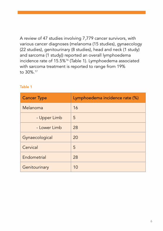
A review of 47 studies involving 7,779 cancer survivors, with various cancer diagnoses (melanoma (15 studies), gynaecology (22 studies), genitourinary (8 studies), head and neck (1 study) and sarcoma (1 study)) reported an overall lymphoedema incidence rate of 15.5%16 (Table 1). Lymphoedema associated with sarcoma treatment is reported to range from 19% to 30%.17
Table 1
Cancer Type Lymphoedema incidence rate (%)
Melanoma 16
- Upper Limb 5
- Lower Limb 28
Gynaecological 20
Cervical 5
Endometrial 28
Genitourinary 10
7
2. Awareness and Prevention
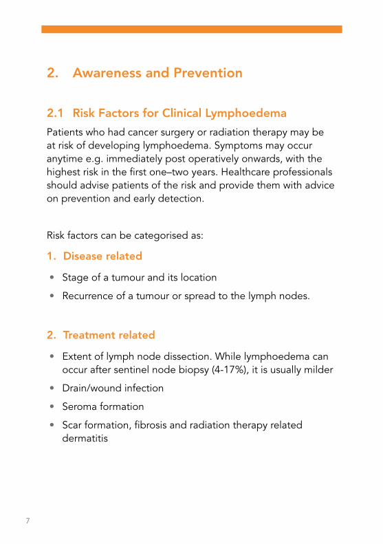
2.1 Risk Factors for Clinical Lymphoedema
Patients who had cancer surgery or radiation therapy may be at risk of developing lymphoedema. Symptoms may occur anytime e.g. immediately post operatively onwards, with the highest risk in the first one–two years. Healthcare professionals should advise patients of the risk and provide them with advice on prevention and early detection.
Risk factors can be categorised as:
1. Disease related
• Stage of a tumour and its location
• Recurrence of a tumour or spread to the lymph nodes.
2. Treatment related
• Extent of lymph node dissection. While lymphoedema can occur after sentinel node biopsy (4-17%), it is usually milder
• Drain/wound infection
• Seroma formation
• Scar formation, fibrosis and radiation therapy related dermatitis
8
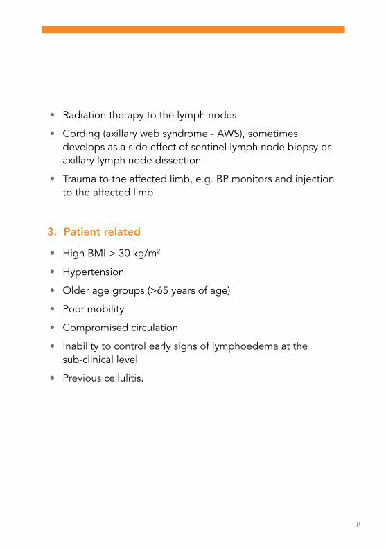
• Radiation therapy to the lymph nodes
• Cording (axillary web syndrome - AWS), sometimes develops as a side effect of sentinel lymph node biopsy or axillary lymph node dissection
• Trauma to the affected limb, e.g. BP monitors and injection to the affected limb.
3. Patient related
• High BMI > 30 kg/m2
• Hypertension
• Older age groups (>65 years of age)
• Poor mobility
• Compromised circulation
• Inability to control early signs of lymphoedema at the sub-clinical level
• Previous cellulitis.

9
2.2 Prevention and Early Detection at the Sub-Clinical Level
Prevention of clinical lymphoedema should begin before commencing cancer treatment by:
• Identifying high risk patients
• Advising the patient and family of the potential risk of lymphoedema
• Highlighting the importance of prevention, risk reduction and early recognition
• Advising on a healthy diet and referral to dietician, if required
• Demonstrating preventive exercises and encouraging these post-operatively
• Demonstrating self management techniques, for example, skin care and simple lymphatic drainage (Section 5)
• Encouraging safe resumption of physical and recreational activity.
Simple prevention tasks encourage patients to take an active role early in their recovery and to know when to seek medical advice.
The early recognition and treatment of cellulitis is essential to prevent progression to clinical lymphoedema. Information on cellulitis prevention, recognition and treatment is outlined in Section 4.

10
2.3 Lifestyle and Prevention
A lower incidence of lymphoedema has been found in patients who exercise regularly, receive lymphoedema education before treatment, and perform preventive self-care activities.18
2.3.1 Skin and Nail Care Tips
Meticulous skin hygiene and nail care are essential to reduce the risk of bacterial and fungal infection and the risk of cellulitis:
• Cut toenails straight across; see a chiropodist, as needed, to prevent ingrown nails and infections
• Only use electric razors or depilatory creams for hair removal
• Avoid dry, cracked or flaky skin. Use good quality bland unperfumed moisturiser, pH neutral, to keep the skin supple and moist
• Cracked areas of skin should be washed, with soap and water and dried carefully
• Damaged skin should be cleaned and covered with a dry dressing until it heals. Clean and change the bandage regularly
• Topical antibiotic solutions can be used to treat small breaks in the skin, for example, paper cuts
• Wear cotton socks; keep feet clean and dry
• Wear gardening and oven gloves
• Use a thimble for sewing
11
• Avoid going barefoot outdoors
• Avoid having blood tests (including finger sticks), vaccinations, intravenous lines or blood pressure monitoring in the affected limb
• Avoid exposure to extreme heat or cold - use the unaffected extremity to test temperatures (e.g. for bath water or cooking)
• Use insect repellent
• Be alert for the signs of infection (fever, swelling, redness, pain, and heat) and see your GP if you suspect infection.
2.3.2 Diet and Healthy Weight
The risk of lymphoedema increases in those who are overweight or obese. Maintaining a normal weight and eating a healthy diet are an important part of healthy survivorship.
2.3.3 Physical Activity
Gentle limb exercises should begin as soon as possible after surgery and continue during other treatments e.g. radiation therapy or chemotherapy. The exercises should be slow and methodical (Section 5). If there is pain the exercises can be reduced but not stopped. These exercises will stimulate lymph flow from the limb and reduce the risk of lymphoedema. They will also improve the range of movement and limb strength and will facilitate proper positioning of the limb in patients receiving radiation therapy.

12
Gentle and moderate physical activity does not increase the risk of lymphoedema. In the past, patients at risk of lymphoedema were advised to avoid using the affected limb as it was thought that removal of lymph nodes altered the response of the affected area to inflammation, infection, injury, and trauma. However, low level exercise has a different effect on the body than higher intensity exercise e.g. swimming is beneficial. Extreme exercise will promote inflammation and injury and should be avoided in patients at risk for lymphoedema. By contrast, slowly progressive, carefully controlled exercises are beneficial.19, 20
2.3.4 Travel, Sun and Heat
Additional precautions are recommended during long journeys and in hot weather:
• The affected area should be gently exercised and elevated, where possible
• Never stay sitting for long periods when travelling. Move about, where possible
• Drink plenty of water during hot weather
• Wear long sleeved cotton garments to protect the limbs from bites or burns
• Use of sun screen (minimum of SPF 15 – 30)
• Do not use a sauna or a hot tub or put the affected limb into a very hot bath
• Never use a sunbed.
13
2.3.5 Positioning of the Limb to Prevent Constriction
Keep the arm or leg elevated above the level of the heart when possible.
Avoid constricting the affected limb.21 People with leg lymphoedema should avoid conditions which cause stasis. Stasis refers to sitting or standing for a long period of time without moving, changing position, or elevating the legs. Excessive constriction refers to tightening or squeezing in a manner that restricts lymph flow through that area or causes tissue trauma, therefore:
• Avoid crossing legs while sitting
• Do not sit in one position for longer than 30 minutes
• Wear well fitting comfortable shoes
• Carry a handbag on the opposite arm
• Wear loose jewellery and clothes with no constricting bands
• Do not use elasticated bandages or tight clothing e.g. socks/stockings with constrictive tops
• Avoid local application of heat to the limb, as this may increase blood flow
• Avoid blood pressure monitoring on the affected arm
• Wear a professionally fitted comfortable bra.
14
3. Presentation and Stages of Lymphoedema
3.1 Presentation of Lymphoedema
Lymphoedema can begin in a subtle way and sub-clinical lymphoedema is not visible. Even when sub-clinical, patients may report a variety of symptoms, including limb heaviness, skin tightness, an itching or burning sensation or decreased joint movement. They may notice difficulty fitting clothing, jewellery or shoes.
The clinical presentation of lymphoedema may be triggered by local inflammation, for example, infection, burns or limb injury. Other causes of limb swelling should always be ruled out (e.g. deep venous thrombosis). Patients should be carefully evaluated for any evidence of cellulitis (Section 4).
Lymphoedema progresses through four stages. Intervention in the early stages (Stage 0 and Stage I) can prevent the onset of severe clinical features. However, patients with mild lymphoedema are three times more likely to develop a severe form of lymphoedema.22 Therefore, it is important to diagnose and treat at the sub-clinical stage.
15
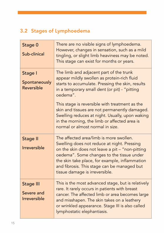
3.2 Stages of Lymphoedema
Stage 0
Sub-clinical
There are no visible signs of lymphoedema. However, changes in sensation, such as a mild tingling, or slight limb heaviness may be noted. This stage can exist for months or years.
Stage I
Spontaneously Reversible
The limb and adjacent part of the trunk appear mildly swollen as protein-rich fluid starts to accumulate. Pressing the skin, results in a temporary small dent (or pit) - “pitting oedema”.
This stage is reversible with treatment as the skin and tissues are not permanently damaged. Swelling reduces at night. Usually, upon waking in the morning, the limb or affected area is normal or almost normal in size.
Stage II
Irreversible
The affected area/limb is more swollen. Swelling does not reduce at night. Pressing on the skin does not leave a pit – “non-pitting oedema”. Some changes to the tissue under the skin take place, for example, inflammation and fibrosis. This stage can be managed but tissue damage is irreversible.
Stage III
Severe and Irreversible
This is the most advanced stage, but is relatively rare. It rarely occurs in patients with breast cancer. The affected limb or area becomes large and misshapen. The skin takes on a leathery or wrinkled appearance. Stage III is also called lymphostatic elephantiasis.

16
3.3 Problems Arising from Lymphoedema
Swelling
Initially lymphoedema may only cause cosmetic concerns or heaviness and difficulty with some movements. This is because the affected limb is swollen. The skin may become slightly shiny. It also contains extra fluid so it feels heavier. This may be all that some patients notice.
Cellulitis
The presence of extra tissue fluid causing swelling in the limb can also make the patient more likely to develop infection in the tissues (cellulitis). The lymph fluid itself is very rich in protein and is an ideal fluid for bacterial growth. Cellulitis can occur even after a minor injury. When this occurs the limb becomes red, tender, swollen and painful. The patient will probably feel generally unwell. This problem can usually be treated effectively with antibiotics, bed rest and elevation of the limb.
Fibrosis
The mere presence of significant amounts of lymph fluid in the tissues over many years can lead to scarring and fibrosis of the tissues. Once infection and inflammation resolve there will inevitably be some residual scarring of the tissues and the limb
17
may be slightly more swollen than before. This puts the limb at a slightly greater risk of infection. A vicious cycle can then develop with further infection leading to more swelling and so on. It is important to try to halt this process at an early stage when most of the changes in the limb are at a reversible stage. As well as swelling due to lymphoedema, the skin can become thickened (hyperkeratosis).
Tumours
Very rarely tumours (lymphangiosarcoma) can develop in the chronically inflamed tissues. This is sometimes called Stewart-Treves syndrome.23
18
4. Cellulitis 4.1 Definition
Cellulitis is an acute spreading inflammation of the skin and subcutaneous tissues. It usually presents with pain, warmth, swelling and erythema. The most frequent causative organisms are Group A Streptococci (S.pyogenes) or Staphylococcus aureus.
Lymphoedema is an important risk factor for cellulitis but the clinical presentation may be atypical.
The swollen skin of patients with lymphoedema is at increased risk of breakdown and subsequent infection. When infection occurs, bacterial clearance is impaired. This results in prolonged cellulitis with longer and more intense systemic inflammatory response. Persistent inflammation, in turn, worsens swelling and adds further damage to the lymphatic system placing the patient in a vicious cycle.24
Left untreated, the infection may quickly become life-threatening.25 In addition, prompt treatment is essential to avoid further damage to the lymphoedematous area which, in turn, may predispose to repeated episodes of cellulitis.26
Patients should be aware of the measures they can take to prevent cellulitis (section 2.3) and the signs and symptoms of cellulitis to enable them to seek prompt medical attention.27
19
4.2 Clinical Features of Cellulitis
Cellulitis in a lymphoedema affected area may not present with classical features. In some patients, infection is the trigger for their first episode of lymphoedema. Onset of infection may be sudden, or slow over weeks. The clinical features of cellulitis can vary. Some episodes are accompanied by severe systemic upset, with high fever or rigors; others are milder, with minimal or no fever.27 Skin manifestations may be preceded by systemic symptoms (e.g. fever, vomiting, and headache) and may be accompanied by an exacerbation of existing lymphoedema. Inflammatory markers including white cell count (WCC), C-reactive protein (CRP) or erythrocyte sedimentation rate (ESR) may not always be elevated. Therefore a high level of clinical suspicion is needed. Patients with any of the following clinical features suspicious of cellulitis need to attend their GP:
• Fevers or chills
• Skin redness or red streaks on the skin
• Increased skin warmth
• Swelling
• Tenderness
• Blistering or oozing.
20
4.3 Management of Acute Cellulitis
4.3.1 Hospital Admission
A decision whether hospital review and/or admission is indicated for a patient with cellulitis should be based on the level of systemic upset as recommended below.
1. Absolute indications for hospital review/admission = Signs of sepsis/severe sepsis
Two or more of the following criteria are present:
• Tachycardia >90 bpm
• Tachypnoea >20 bpm
• Temperature <36 or >38.3 ºC
• Acutely altered mental state
• Bedside glucose >7.7mmol/L (in the absence of diabetes mellitus)
• Systolic blood pressure <90mmHg or systolic blood pressure more than 40 below the patients normal.
21
2. Other indications for hospital review and/or admission include:
• Continuing or deteriorating systemic signs, with or without deteriorating local signs, after 48 hours antibiotic treatment
• Unresolving or deteriorating local signs, with or without systemic signs, despite trials of first and second line antibiotics.
Patients with sepsis should be managed in accordance with national guidelines.28 Choice of antibiotics in hospital should be made according to local antibiotic prescribing guidelines. Advice of the consultant microbiologist should be sought on antibiotic treatment options, if required.27
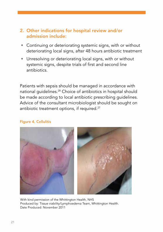
With kind permission of the Whittington Health, NHSProduced by: Tissue viability/Lymphoedema Team, Whittington Health. Date Produced: November 2011
Figure 4. Cellulitis
22
4.3.2 Management at Home
National antibiotic prescribing guidelines for primary care can be reviewed at www.antibioticprescribing.ie. However, some aspects of cellulitis treatment in patients with lymphoedema may differ from conventional cellulitis treatment (e.g. duration of treatment).
The patient should be closely monitored by the GP i.e.:
• The extent and severity of erythema should be monitored - if possible, mark and date its border
• The level of systemic upset should be monitored - pulse, temperature, respiratory rate, blood pressure
• Microbiological diagnosis should be undertaken where there are cuts or breaks in the skin before antibiotics are commenced to identify appropriate antibiotic treatment.29
23
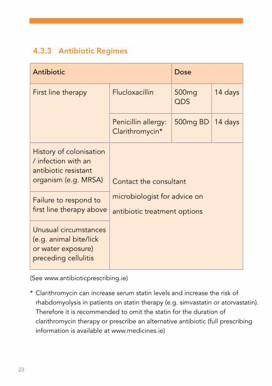
4.3.3 Antibiotic Regimes
Antibiotic Dose
First line therapy Flucloxacillin 500mg QDS
14 days
Penicillin allergy: Clarithromycin*
500mg BD 14 days
History of colonisation / infection with an antibiotic resistant organism (e.g. MRSA) Contact the consultant
microbiologist for advice on
antibiotic treatment options
Failure to respond to first line therapy above
Unusual circumstances (e.g. animal bite/lick or water exposure) preceding cellulitis
(See www.antibioticprescribing.ie)
* Clarithromycin can increase serum statin levels and increase the risk of rhabdomyolysis in patients on statin therapy (e.g. simvastatin or atorvastatin). Therefore it is recommended to omit the statin for the duration of clarithromycin therapy or prescribe an alternative antibiotic (full prescribing information is available at www.medicines.ie)

24
4.4 Antibiotics “In Case” – Travel and Holidays
The risk of further episodes of cellulitis in patients with lymphoedema is high. It is recommended that patients who have had an episode of cellulitis carry a two-week supply of antibiotics with them, particularly when away from home.25 Antibiotics should be commenced immediately when symptoms of cellulitis start and a medical opinion should be sought as soon as possible.
4.5 Antibiotics During Lymphoedema Therapy
Patients with a history of cellulitis, and who are now undergoing intensive Manual Lymphatic Drainage (MLD), may benefit from antibiotic cover for the duration of treatment. Patients with no previous episodes of cellulitis do not require antibiotics during intensive treatments. Advice from a consultant microbiologist should be sought on antibiotic therapy.
4.6 Recurrent Cellulitis
The management of patients with lymphoedema with recurrent cellulitis can be complicated. Advice should be sought from a consultant microbiologist and consultant dermatologist. Other predisposing risk factors for recurrent cellulitis should be actively managed in addition to consideration of antibiotic therapy. These include:
• Cracked, macerated inter-digital skin
• Dermatitis
• Open wounds including leg ulcers and weeping lymphangiectasia (leaking lymph blisters on the skin surface)

25
• Tinea pedis – this affects a large number of patients with severe lymphoedema as maceration between the toes creates an opportunistic environment
• Treatment of inter-digital/nail skin fungal infection is summarised at www.antibioticprescribing.ie.
4.6.1 Antibiotic Prophalaxis
Antibiotic prophylaxis should be considered in lymphoedema patients who had two or more episodes of cellulitis in a 12 month period as repeated episodes of infection exacerbate lymphoedema and predispose to further infection.30 There is ongoing debate in relation to the most appropriate drug, dose and duration.31 The advice of a consultant microbiologist should be sought.
However, there is evidence that low dose penicillin for 12 months is effective in reducing the recurrence of lower limb cellulitis i.e. Penicillin V 250mg BD or 500mg BD where the BMI ≥ 33kg/m2 or Clarithromycin 250mg BD if there is penicillin allergy.
The effect, however, may diminish when the antibiotic is stopped.25, 31 The duration of prophylaxis is unclear. However life-long therapy may be recommended if relapse occurs when antibiotics are discontinued after a two year period of successful prophylaxis.25 The requirement for antibiotic therapy needs to be balanced against the risk of Clostridium difficile infection and antimicrobial resistant organism colonisation.

26
5. Self-care5.1. Simple self care exercises32
To assist patients to regain their full range of movements following breast cancer surgery.
Exercise 1: Wall Climbing. This is done in two positions: 1. Facing a wall. 2. Sideways to a wall.
This exercise should be carried out five times a day, using the affected arm. Remember to breathe deeply while doing the exercises.
Exercises may vary slightly depending on the specialist centre attended.
Facing wall:• Keep shoulder down and place hand on wall• Walk fingers up the walls slowly and stop when it becomes painful• Hold in painful place for 6 seconds. Relax and start again• Repeat this exercise 7 times.
Sideways to wall:• Pace hand beside you on wall and keep shoulders down• Walk fingers slowly up the wall stop when it becomes painful• Hold, relax and start again for 7 times.
Facing wall
Sideways to wall
Wall Climbing
To be done in two positions - facing and sideways to wall

27
Exercise 2: The purpose of these exercises is to increase the blood flow through the arm.
Open – Close Hands: Open and close fists at various heights.
Attach the necklace: This exercise is simple and very easy to explain.
These exercises should be carried out five times a day, using the affected arm, remember to breathe deeply while doing the exercises.
Open and close fists at various heights
Purpose: To pump blood throughout the arms bringing healing nutrients and removing toxins
Frequency: 7 Pumps
Imagine you are putting on a necklace
Keep elbows relaxed
Slowly open elbows as wide as possible and hold for a count of six
Repeat 7 times
Attaching the necklaceOpen – Close Hands

28
Exercise 3: Attaching the Bra. Encourage patients to do this exercise as it is extremely important for arm rehabilitation. It can be painful but this will ease as healing progresses.
This exercise should be carried out five times a day, using the affected arm. Remember to breathe deeply while doing the exercises.
1. With healthy hand gently take hand of injured arm behind your back.2. Slowly begin pulling injured hand towards the height of your bra.3. Stop at pain - hold for 6 seconds.4. As you progress use the wall to maintain your balance; keep your back straight.5. To accomplish this may take several weeks or months - do not be discouraged. Repeat this exercise 3 times.
Attaching the Bra
Pictures courtesy of The Esmonde Technique. http://breastcancerrehabilitation.com/KeyExercises.pdf
29
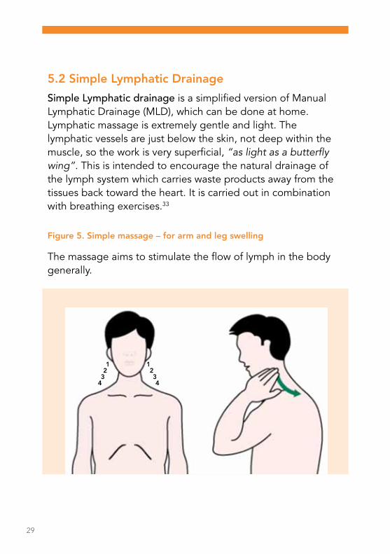
5.2 Simple Lymphatic Drainage
Simple Lymphatic drainage is a simplified version of Manual Lymphatic Drainage (MLD), which can be done at home. Lymphatic massage is extremely gentle and light. The lymphatic vessels are just below the skin, not deep within the muscle, so the work is very superficial, “as light as a butterfly wing”. This is intended to encourage the natural drainage of the lymph system which carries waste products away from the tissues back toward the heart. It is carried out in combination with breathing exercises.33
Figure 5. Simple massage – for arm and leg swelling
The massage aims to stimulate the flow of lymph in the body generally.
1223344
1
30
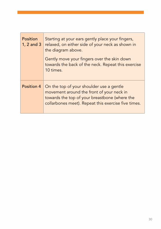
Position 1, 2 and 3
Starting at your ears gently place your fingers, relaxed, on either side of your neck as shown in the diagram above.
Gently move your fingers over the skin down towards the back of the neck. Repeat this exercise 10 times.
Position 4 On the top of your shoulder use a gentle movement around the front of your neck in towards the top of your breastbone (where the collarbones meet). Repeat this exercise five times.
31
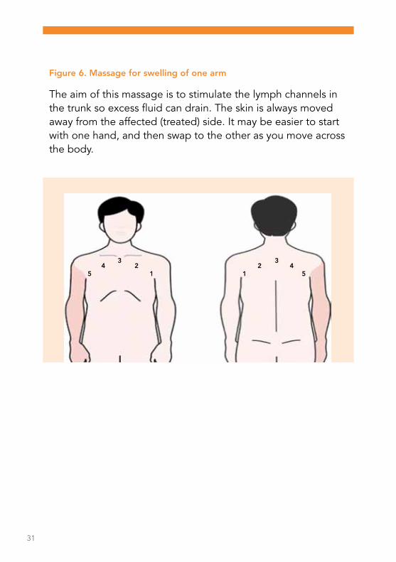
Figure 6. Massage for swelling of one arm
The aim of this massage is to stimulate the lymph channels in the trunk so excess fluid can drain. The skin is always moved away from the affected (treated) side. It may be easier to start with one hand, and then swap to the other as you move across the body.
11 5522
3344
32
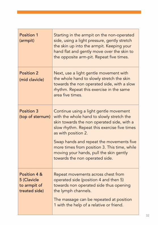
Position 1 (armpit)
Starting in the armpit on the non-operated side, using a light pressure, gently stretch the skin up into the armpit. Keeping your hand flat and gently move over the skin to the opposite arm-pit. Repeat five times.
Position 2
(mid clavicle)
Next, use a light gentle movement with the whole hand to slowly stretch the skin towards the non operated side, with a slow rhythm. Repeat this exercise in the same area five times.
Position 3 (top of sternum)
Continue using a light gentle movement with the whole hand to slowly stretch the skin towards the non operated side, with a slow rhythm. Repeat this exercise five times as with position 2.
Swap hands and repeat the movements five more times from position 3. This time, while moving your hands, pull the skin gently towards the non operated side.
Position 4 & 5 (Clavicle to armpit of treated side)
Repeat movements across chest from operated side (position 4 and then 5) towards non operated side thus opening the lymph channels.
The massage can be repeated at position 1 with the help of a relative or friend.
33
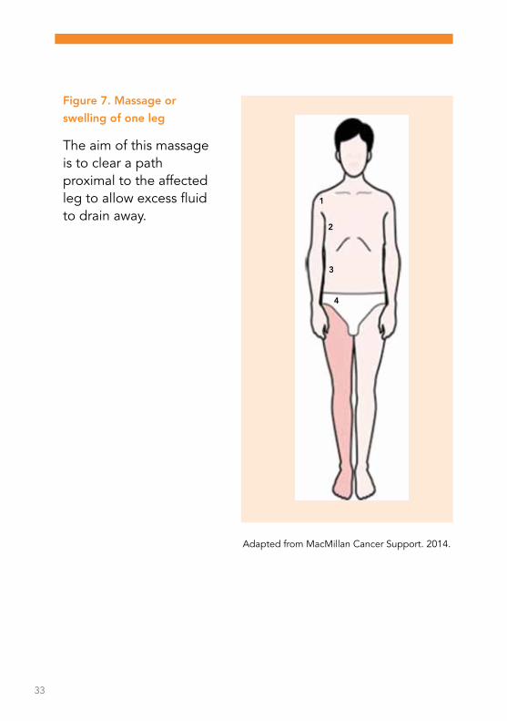
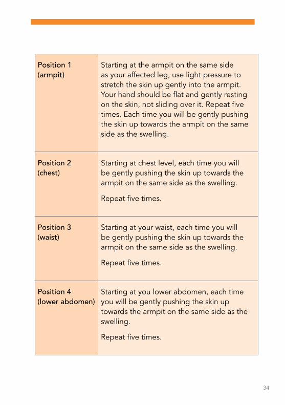
Figure 7. Massage or
swelling of one leg
The aim of this massage is to clear a path proximal to the affected leg to allow excess fluid to drain away.
1
2
3
4
Adapted from MacMillan Cancer Support. 2014.
34
Position 1(armpit)
Starting at the armpit on the same side as your affected leg, use light pressure to stretch the skin up gently into the armpit. Your hand should be flat and gently resting on the skin, not sliding over it. Repeat five times. Each time you will be gently pushing the skin up towards the armpit on the same side as the swelling.
Position 2(chest)
Starting at chest level, each time you will be gently pushing the skin up towards the armpit on the same side as the swelling.
Repeat five times.
Position 3(waist)
Starting at your waist, each time you will be gently pushing the skin up towards the armpit on the same side as the swelling.
Repeat five times.
Position 4(lower abdomen)
Starting at you lower abdomen, each time you will be gently pushing the skin up towards the armpit on the same side as the swelling.
Repeat five times.
35
6. References
1. Petrek JA. Commentary: Prospective Trial of Complete Decongestive Therapy for Upper Extremity Lymphedema After Breast Cancer Therapy. The Cancer Journal. 2004;10(1).
2. Tobin MB, Lacey HJ, Meyer L, Mortimer PS. The psychological morbidity of breast cancer-related arm swelling. Psychological morbidity of lymphoedema. Cancer. 1993;72(11):3248-3252.
3. International Society of Lymphology. The diagnosis and treatment of peripheral lymphedema: 2013 Consensus Document of the International Society of Lymphology. Lymphology. 2013;46(1):1-11.
4. Badger C, Preston N, Seers K, Mortimer P. Physical therapies for reducing and controlling lymphoedema of the limbs. Cochrane Database Syst Rev. 2004(4):Cd003141.
5. Meek AG. Breast radiotherapy and lymphedema. Cancer. 1998;83(12 Suppl American):2788-2797.
6. Roberts CC, Levick JR, Stanton AW, Mortimer PS. Assessment of truncal edema following breast cancer treatment using modified Harpenden skinfold calipers. Lymphology. 1995;28(2):78-88.
7. Armitage JO. Approach to the patient with lymphadenopathy and splenomegaly. In: Goldman L, Schafer AI, eds. Cecil Medicine: 24th edition. Philadelphia, PA: Saunders Elsevier; 2011.
8. Shaw C, Mortimer P, Judd PA. Randomized controlled trial comparing a low-fat diet with a weight-reduction diet in breast cancer-related lymphedema. Cancer. 2007;109(10):1949-1956.
9. Paskett ED, Naughton MJ, McCoy TP, Case LD, Abbott JM. The epidemiology of arm and hand swelling in premenopausal breast cancer survivors. Cancer Epidemiol Biomarkers Prev. 2007;16(4):775-782.
36
10. Francis WP, Abghari P, Du W, Rymal C, Suna M, Kosir MA. Improving surgical outcomes: standardizing the reporting of incidence and severity of acute lymphedema after sentinel lymph node biopsy and axillary lymph node dissection. Am J Surg. 2006;192(5):636-639.
11. Petrek JA, Senie RT, Peters M, Rosen PP. Lymphedema in a cohort of breast carcinoma survivors 20 years after diagnosis. Cancer. 2001;92(6):1368-1377.
12. Meneses KD, McNees MP. Upper extremity lymphedema after treatment for breast cancer: a review of the literature. Ostomy Wound Manage. 2007;53(5):16-29.
13. Paskett ED, Dean JA, Oliveri JM, Harrop JP. Cancer-related lymphedema risk factors, diagnosis, treatment, and impact: a review. J Clin Oncol. 2012;30(30):3726-3733.
14. Beesley V, Janda M, Eakin E, Obermair A, Battistutta D. Lymphedema after gynecological cancer treatment : prevalence, correlates, and supportive care needs. Cancer. 2007;109(12):2607-2614.
15. Buntzel J, Glatzel M, Mucke R, Micke O, Bruns F. Influence of amifostine on late radiation-toxicity in head and neck cancer--a follow-up study. Anticancer Res. 2007;27(4a):1953-1956.
16. Tiwari P, Coriddi M, Salani R, Povoski SP. Breast and gynecologic cancer-related extremity lymphedema: a review of diagnostic modalities and management options. World J Surg Oncol. 2013;11:237.
17. Friedmann D, Wunder JS, Ferguson P, et al. Incidence and severity of lymphoedema following limb salvage of extremity soft tissue sarcoma. Sarcoma. 2011;2011.

37
18. Cheifetz O, Haley L. Management of secondary lymphedema related to breast cancer. Can Fam Physician. 2010;56(12):1277-1284.
19. Radak Z, Chung HY, Koltai E, Taylor AW, Goto S. Exercise, oxidative stress and hormesis. Ageing Res Rev. 2008;7(1):34-42.
20. Schmitz KH. Balancing lymphedema risk: exercise versus deconditioning for breast cancer survivors. Exerc Sport Sci Rev. 2010;38(1):17-24.
21. Finegold DN, Schacht V, Kimak MA, et al. HGF and MET mutations in primary and secondary lymphedema. Lymphat Res Biol. 2008;6(2):65-68.
22. Norman SA, Localio AR, Potashnik SL, et al. Lymphedema in breast cancer survivors: incidence, degree, time course, treatment, and symptoms. J Clin Oncol. 2009;27(3):390-397.
23. www.vascular.co.nz/lymphoedema.htm. Accessed 31st July, 2014.
24. Dupuy A, Benchikhi H, Roujeau JC, et al. Risk factors for erysipelas of the leg (cellulitis): case-control study. BMJ. 1999;318(7198):1591-1594.
25. British Lymphology Society. Consensus Document on the Management of Cellulitis in Lymphoedema. 2010.
26. Australasian Lymphology Association. Management of Cellulitis in Lymphoedema. 2012.
27. Kilburn SA, Featherstone P, Higgins B, Brindle R. Interventions for cellulitis and erysipelas. Cochrane Database Syst Rev. 2010(6):Cd004299.

38
28. Health Service Executive. Sepsis Guidelines. 2014.
29. Antibiotic Expert Group. Therapeutic guidelines: antibiotic. Version 14. Melbourne: Therapeutic Guidelines Limited; 2010.
30. Vignes S, Dupuy A. Recurrence of lymphoedema-associated cellulitis (erysipelas) under prophylactic antibiotherapy: a retrospective cohort study. J Eur Acad Dermatol Venereol. 2006;20(7):818-822.
31. Nguyen L, Rowland K. Low-dose penicillin for recurrent cellulitis? J Fam Pract. 2014;63(1):E10-12.
32. The Esmonde Technique. http://breastcancerrehabilitation.com/KeyExercises.pdf.
33. Adapted from MacMillan Cancer Support. 2014.
7. Links to Additional Information
Reducing your risk of arm lymphoedema following a cancer diagnosis. Available: www.cancer.ie/sites/default/files/content attachments/reducing_your_risk_of_arm_lymphoedema_factsheet_web.pdf
Reducing your risks of leg lymphoedema following a cancer diagnosis. Available: www.cancer.ie/sites/default/files/content attachments/leg_lymphoedema_factsheet.pdf
Lymphoedema Ireland. www.lymphireland.com
MLD Ireland. www.mldireland.com
Europa Donna Ireland www.europadonnaireland.ie
39


National Cancer Control ProgrammeAn Clár Náisiúnta Rialaithe AilseKing’s Inns House200 Parnell StreetDublin 1
Tel: +353 1 828 7100 Fax: +353 1 828 7160e-mail: [email protected]
NCCP-COM-008-01 2015. © NCCP