Embed Size (px)
Citation preview
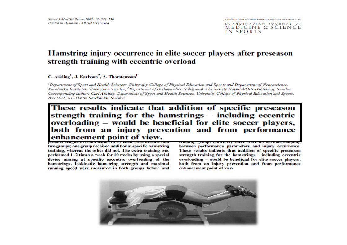
Prevention and Risk Factors
Giorgio Galanti MDProfessor of Medicine
Head Sports Medicine DepartmentDirector of Non Invasive Cardiac Laboratory
Chief of Medical Staff Fiorentina Soccer Team
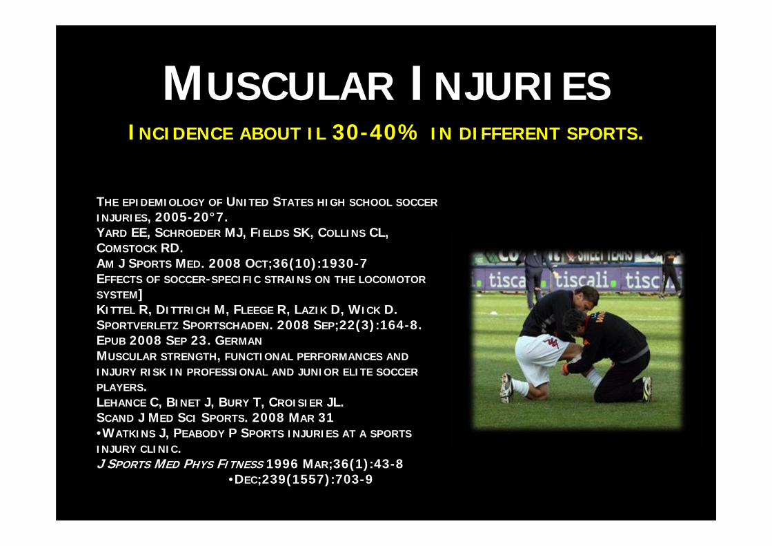
INCIDENCE ABOUT IL 30-40% IN DIFFERENT SPORTS.
THE EPIDEMIOLOGY OF UNITED STATES HIGH SCHOOL SOCCERINJURIES, 2005-20°7.YARD EE, SCHROEDER MJ, FIELDS SK, COLLINS CL, COMSTOCK RD.AM J SPORTS MED. 2008 OCT;36(10):1930-7EFFECTS OF SOCCER-SPECIFIC STRAINS ON THE LOCOMOTORSYSTEM]KITTEL R, DITTRICH M, FLEEGE R, LAZIK D, WICK D.SPORTVERLETZ SPORTSCHADEN. 2008 SEP;22(3):164-8. EPUB 2008 SEP 23. GERMANMUSCULAR STRENGTH, FUNCTIONAL PERFORMANCES ANDINJURY RISK IN PROFESSIONAL AND JUNIOR ELITE SOCCERPLAYERS.LEHANCE C, BINET J, BURY T, CROISIER JL.SCAND J MED SCI SPORTS. 2008 MAR 31•WATKINS J, PEABODY P SPORTS INJURIES AT A SPORTSINJURY CLINIC. J SPORTS MED PHYS FITNESS 1996 MAR;36(1):43-8
•DEC;239(1557):703-9
MUSCULAR INJURIES
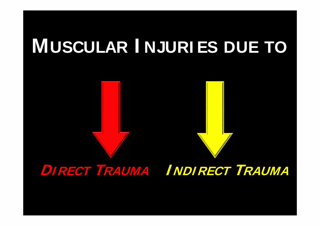
DIRECT TRAUMA INDIRECT TRAUMA
MUSCULAR INJURIES DUE TO
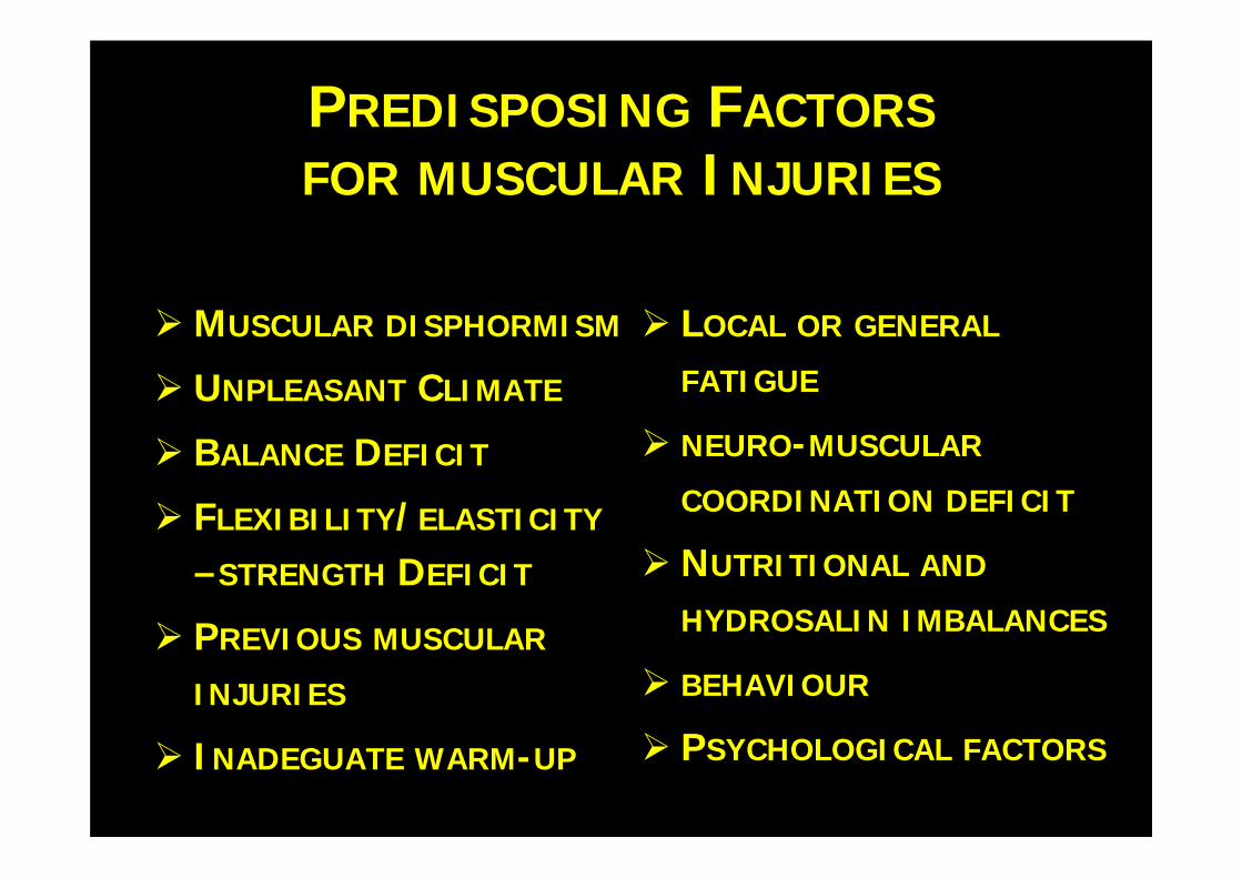
PREDISPOSING FACTORSFOR MUSCULAR INJURIES
MUSCULAR DISPHORMISM
UNPLEASANT CLIMATE
BALANCE DEFICIT
FLEXIBILITY/ELASTICITY
–STRENGTH DEFICIT
PREVIOUS MUSCULAR
INJURIES
INADEGUATE WARM-UP
LOCAL OR GENERAL
FATIGUE
NEURO-MUSCULAR
COORDINATION DEFICIT
NUTRITIONAL AND
HYDROSALIN IMBALANCES
BEHAVIOUR
PSYCHOLOGICAL FACTORS
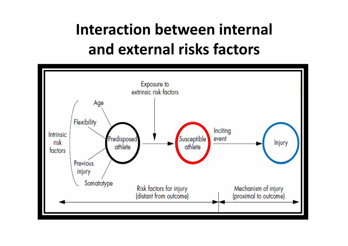
Interaction between internaland external risks factors
InjurySusceptibleathlete
Predisposedathlete
Am J Sports Med. 2012 Dec 21.
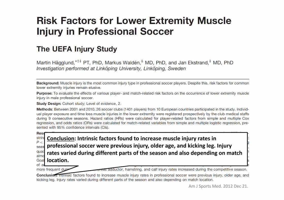
Conclusion: Intrinsic factors found to increase muscle injury rates in professional soccer were previous injury, older age, and kicking leg. Injury rates varied during different parts of the season and also depending on match location.
Am J Sports Med. 2012 Dec 21.
Am J Sports Med. 2012 Dec 21.
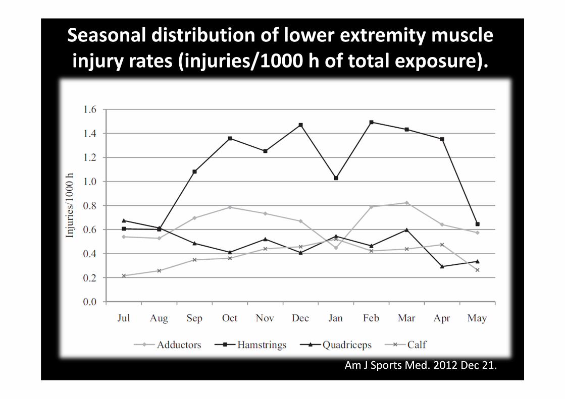
Seasonal distribution of lower extremity muscleinjury rates (injuries/1000 h of total exposure).
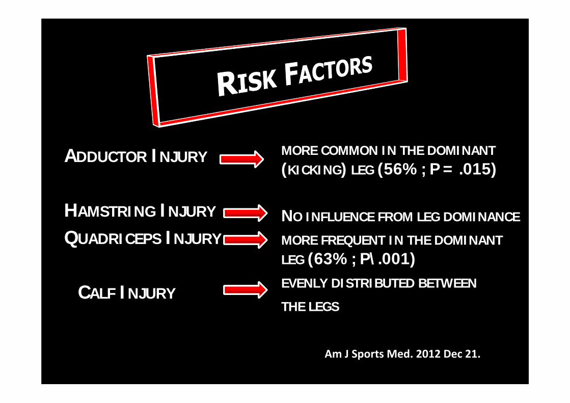
ADDUCTOR INJURY
HAMSTRING INJURY
QUADRICEPS INJURY
CALF INJURY
MORE COMMON IN THE DOMINANT(KICKING) LEG (56%; P = .015)
NO INFLUENCE FROM LEG DOMINANCE
MORE FREQUENT IN THE DOMINANTLEG (63%; P\.001)EVENLY DISTRIBUTED BETWEEN
THE LEGS
Am J Sports Med. 2012 Dec 21.
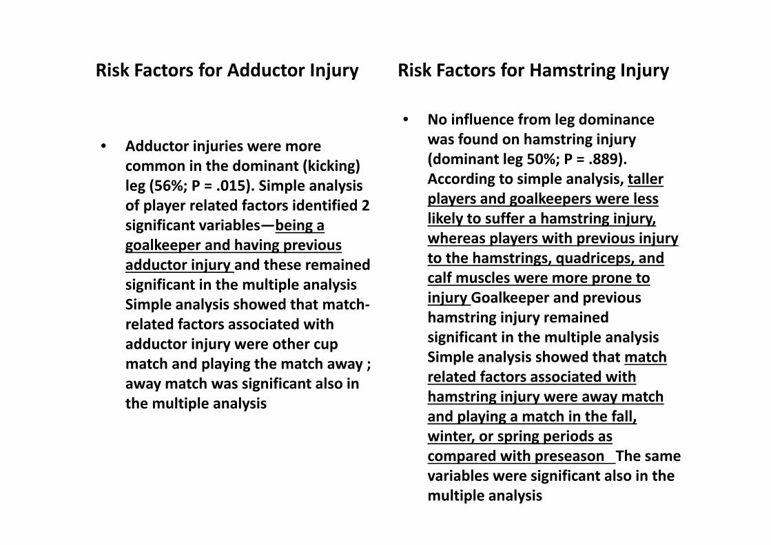
Risk Factors for Adductor Injury
• Adductor injuries were more common in the dominant (kicking) leg (56%; P = .015). Simple analysis of player related factors identified 2 significant variables—being a goalkeeper and having previous adductor injury and these remained significant in the multiple analysis Simple analysis showed that match‐related factors associated with adductor injury were other cup match and playing the match away ; away match was significant also in the multiple analysis
Risk Factors for Hamstring Injury
• No influence from leg dominance was found on hamstring injury (dominant leg 50%; P = .889). According to simple analysis, taller players and goalkeepers were less likely to suffer a hamstring injury, whereas players with previous injury to the hamstrings, quadriceps, and calf muscles were more prone to injury Goalkeeper and previous hamstring injury remained significant in the multiple analysis Simple analysis showed that match related factors associated with hamstring injury were away match and playing a match in the fall, winter, or spring periods as compared with preseason The same variables were significant also in the multiple analysis
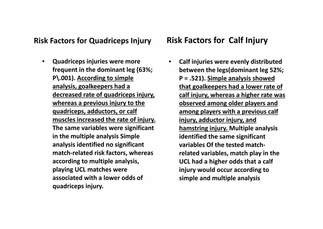
Risk Factors for Quadriceps Injury
• Quadriceps injuries were more frequent in the dominant leg (63%; P\.001). According to simple analysis, goalkeepers had a decreased rate of quadriceps injury, whereas a previous injury to the quadriceps, adductors, or calf muscles increased the rate of injury.The same variables were significant in the multiple analysis Simple analysis identified no significant match‐related risk factors, whereas according to multiple analysis, playing UCL matches were associated with a lower odds of quadriceps injury.
Risk Factors for Calf Injury
• Calf injuries were evenly distributed between the legs(dominant leg 52%; P = .521). Simple analysis showed that goalkeepers had a lower rate of calf injury, whereas a higher rate was observed among older players and among players with a previous calf injury, adductor injury, and hamstring injury. Multiple analysis identified the same significant variables Of the tested match‐related variables, match play in the UCL had a higher odds that a calf injury would occur according to simple and multiple analysis
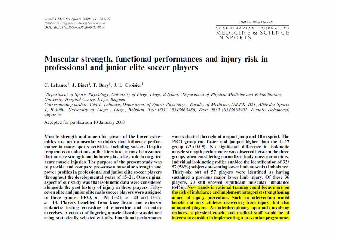
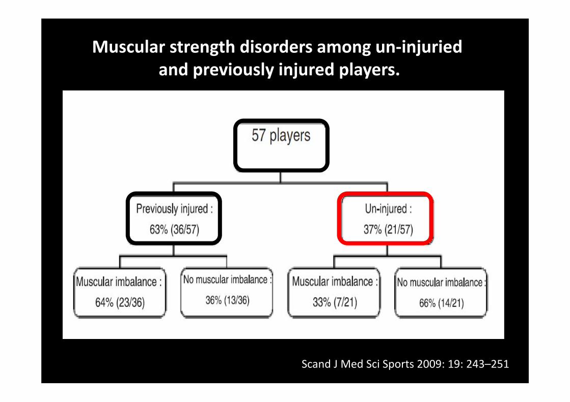
Muscular strength disorders among un‐injuriedand previously injured players.
Scand J Med Sci Sports 2009: 19: 243–251
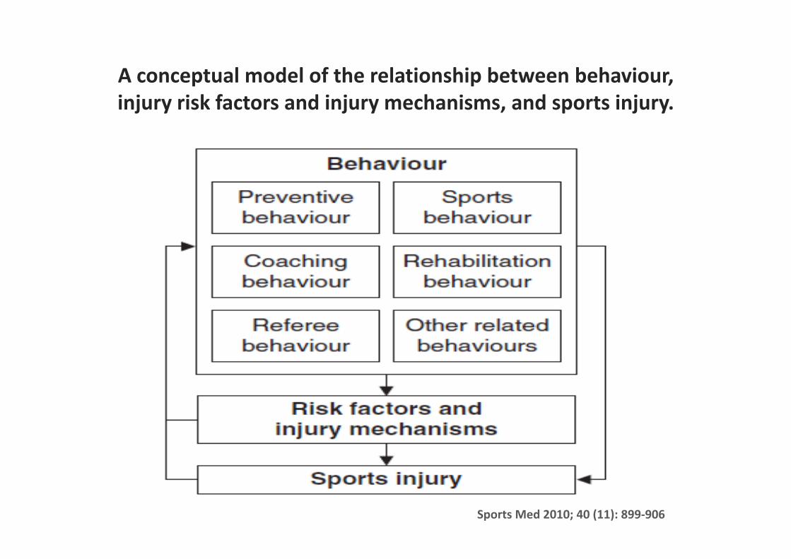
A conceptual model of the relationship between behaviour,injury risk factors and injury mechanisms, and sports injury.
Sports Med 2010; 40 (11): 899‐906
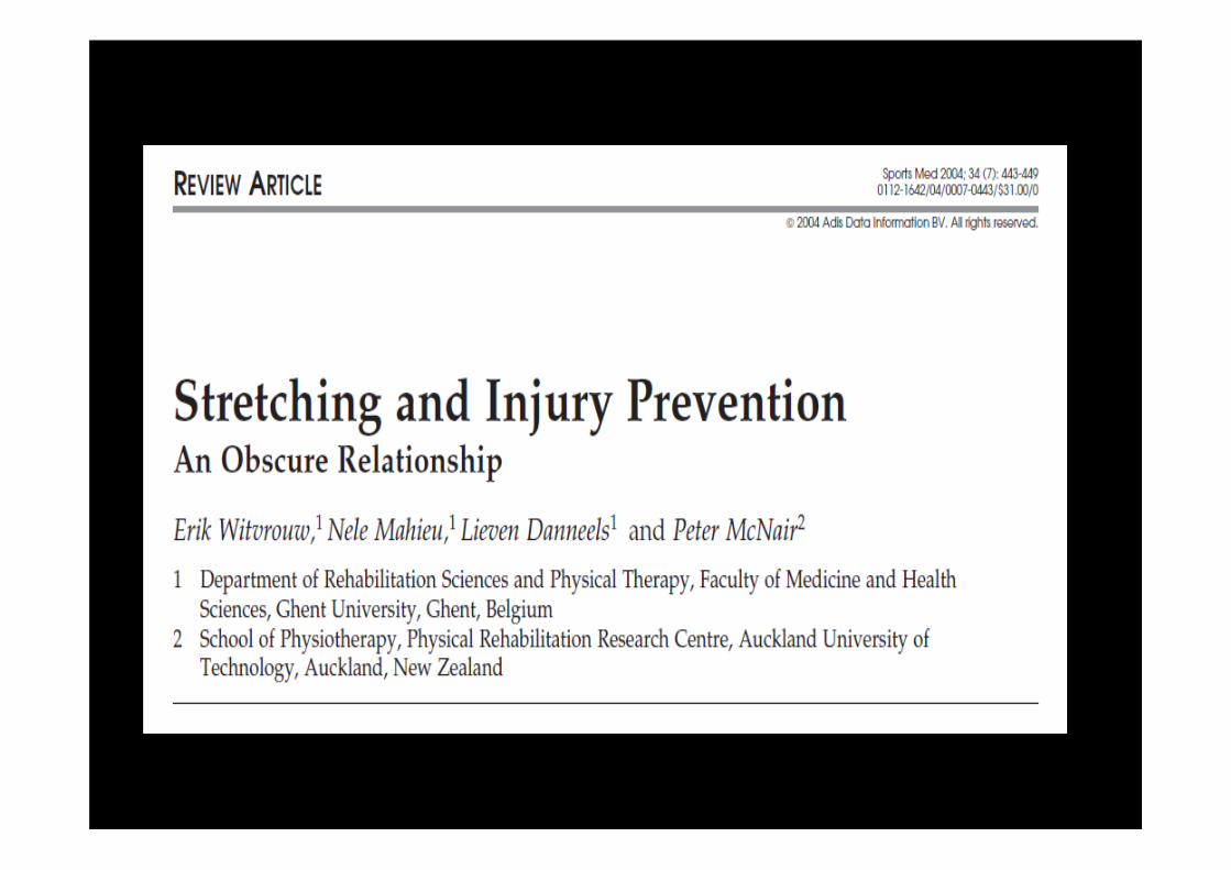
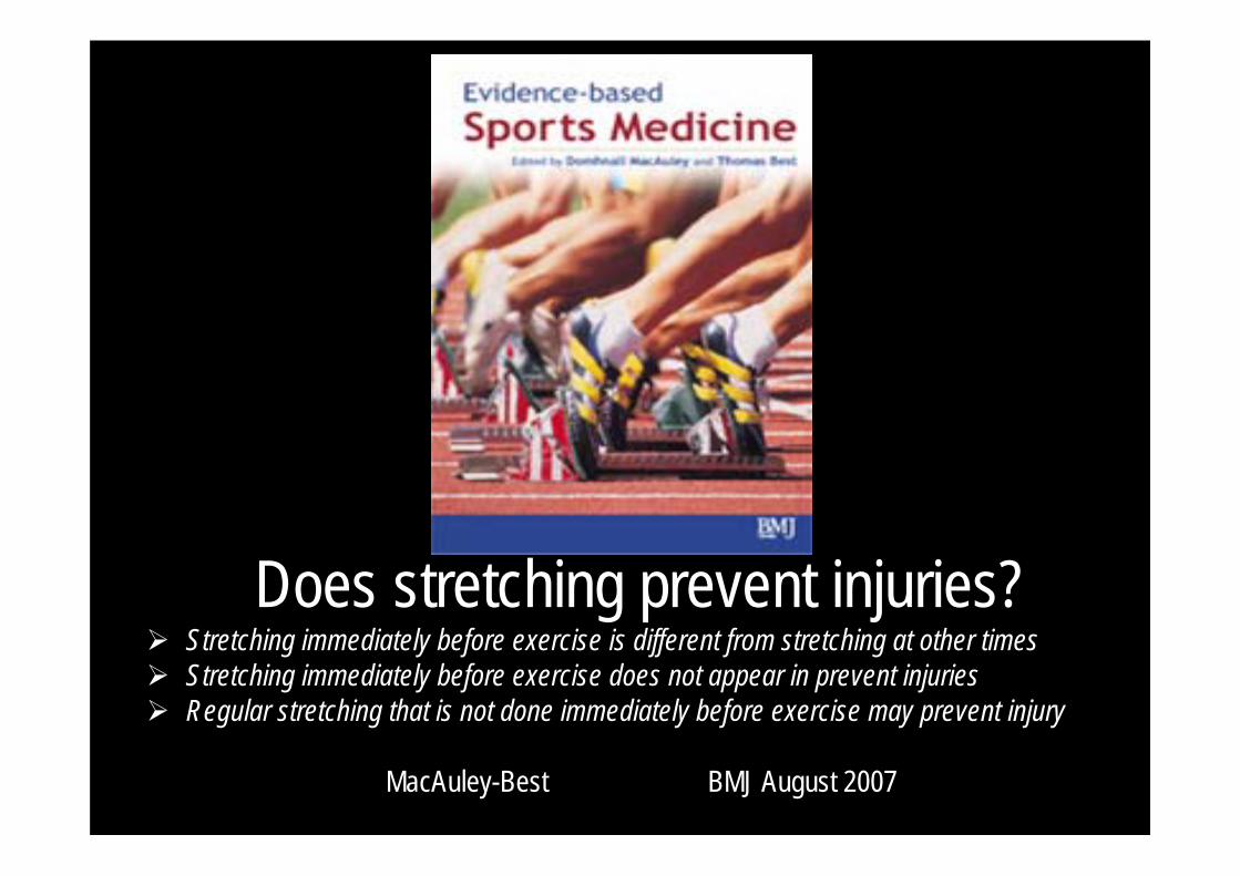
Does stretching prevent injuries?Stretching immediately before exercise is different from stretching at other timesStretching immediately before exercise does not appear in prevent injuriesRegular stretching that is not done immediately before exercise may prevent injury
MacAuley-Best BMJ August 2007
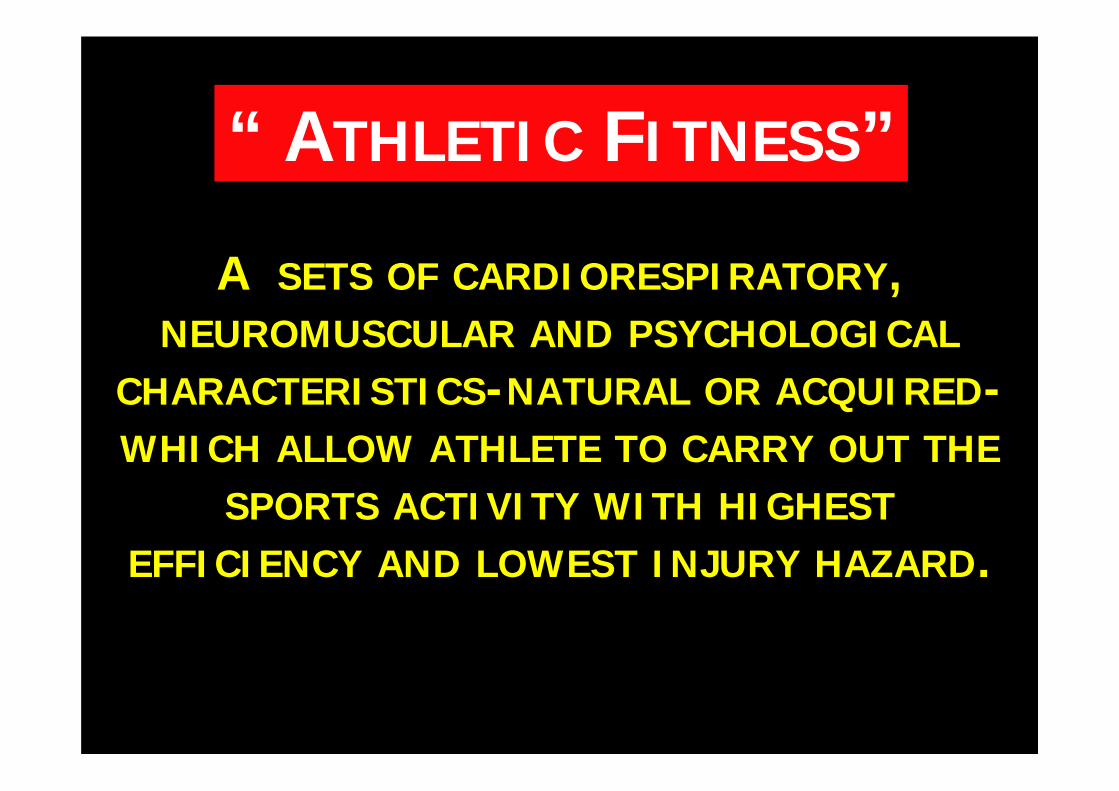
A SETS OF CARDIORESPIRATORY, NEUROMUSCULAR AND PSYCHOLOGICAL
CHARACTERISTICS-NATURAL OR ACQUIRED-WHICH ALLOW ATHLETE TO CARRY OUT THE
SPORTS ACTIVITY WITH HIGHESTEFFICIENCY AND LOWEST INJURY HAZARD.
“ ATHLETIC FITNESS”
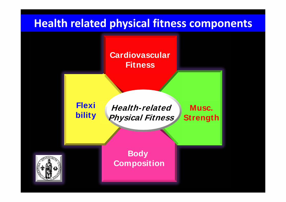
Health related physical fitness components
Body Composition
CardiovascularFitness
Flexibility
Health-relatedPhysical Fitness
Musc.Strength
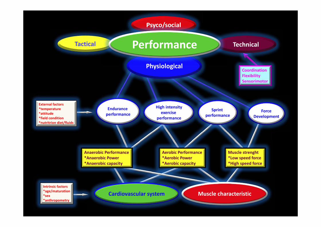
Tactical Technical
Psyco/social
Physiological
Anaerobic Performance*Anaerobic Power*Anaerobic capacity
Aerobic Performance*Aerobic Power*Aerobic capacity
Muscle strenght*Low speed force*High speed force
External factors*temperature*attitude*field condition*nutritrion diet/fluids
Intrinsic factors*age/maturation*sex*anthropometry
CoordinationFlexibilitySensorimotor
Cardiovascular system Muscle characteristic
Endurance performance
High intensityexercise
performance
Sprint performance
ForceDevelopment
Performance
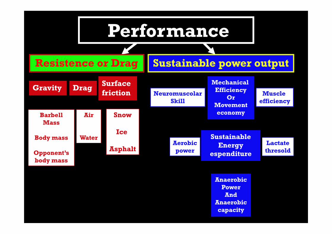
Performance
Resistence or Drag
Gravity Surfacefriction
Sustainable power output
Drag
BarbellMass
Body mass
Opponent’s body mass
Snow
Ice
Asphalt
Air
Water
NeuromuscolarSkill
Aerobicpower
AnaerobicPower
AndAnaerobiccapacity
Sustainable Energy
espenditure
Lactatethresold
Muscle efficiency
Mechanical Efficiency
OrMovementeconomy
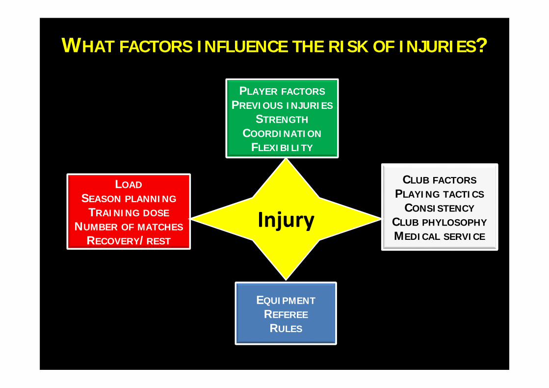
PLAYER FACTORSPREVIOUS INJURIES
STRENGTHCOORDINATION
FLEXIBILITY
LOADSEASON PLANNING
TRAINING DOSENUMBER OF MATCHES
RECOVERY/REST
EQUIPMENTREFEREERULES
CLUB FACTORSPLAYING TACTICS
CONSISTENCYCLUB PHYLOSOPHYMEDICAL SERVICE
WHAT FACTORS INFLUENCE THE RISK OF INJURIES?
Injury
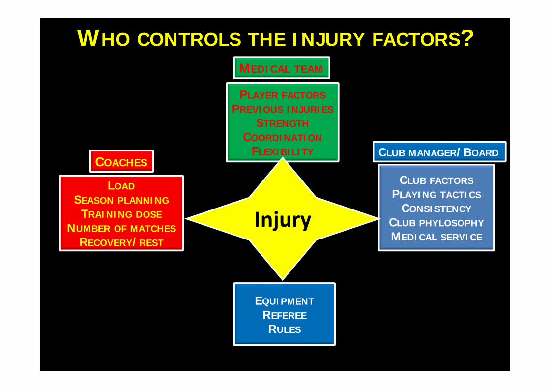
PLAYER FACTORSPREVIOUS INJURIES
STRENGTHCOORDINATION
FLEXIBILITY
LOADSEASON PLANNING
TRAINING DOSENUMBER OF MATCHES
RECOVERY/REST
EQUIPMENTREFEREERULES
CLUB FACTORSPLAYING TACTICS
CONSISTENCYCLUB PHYLOSOPHYMEDICAL SERVICE
WHO CONTROLS THE INJURY FACTORS?
COACHES
MEDICAL TEAM
CLUB MANAGER/BOARD
Injury
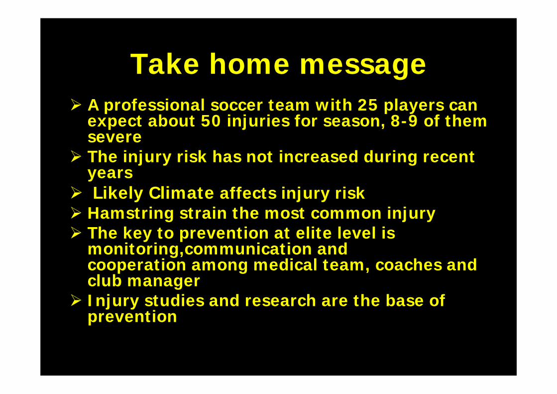
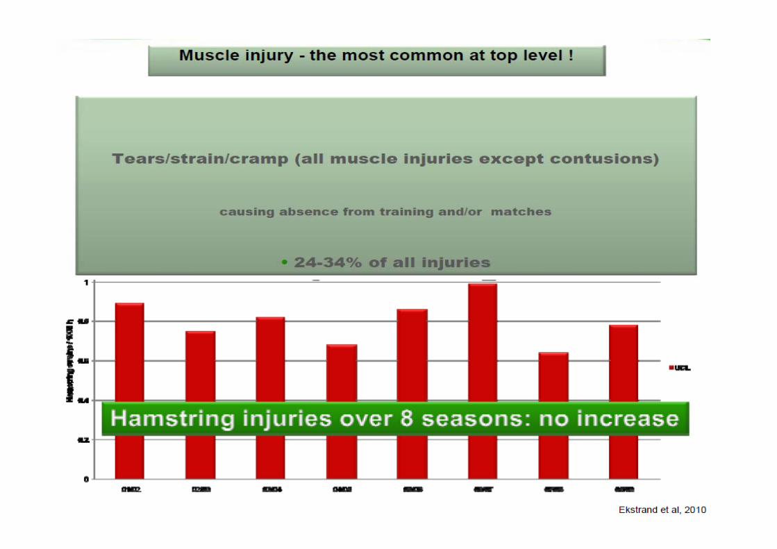
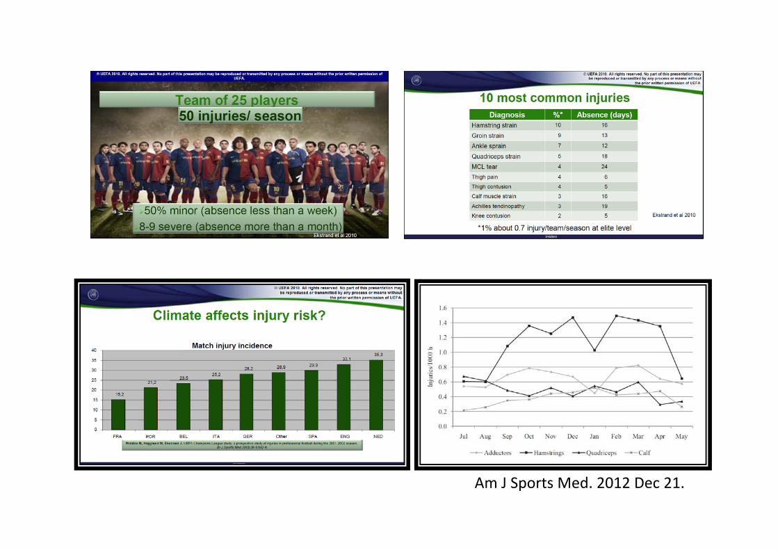
Take home messageA professional soccer team with 25 players can expect about 50 injuries for season, 8-9 of them severeThe injury risk has not increased during recent yearsLikely Climate affects injury risk
Hamstring strain the most common injuryThe key to prevention at elite level is monitoring,communication and cooperation among medical team, coaches and club managerInjury studies and research are the base of prevention
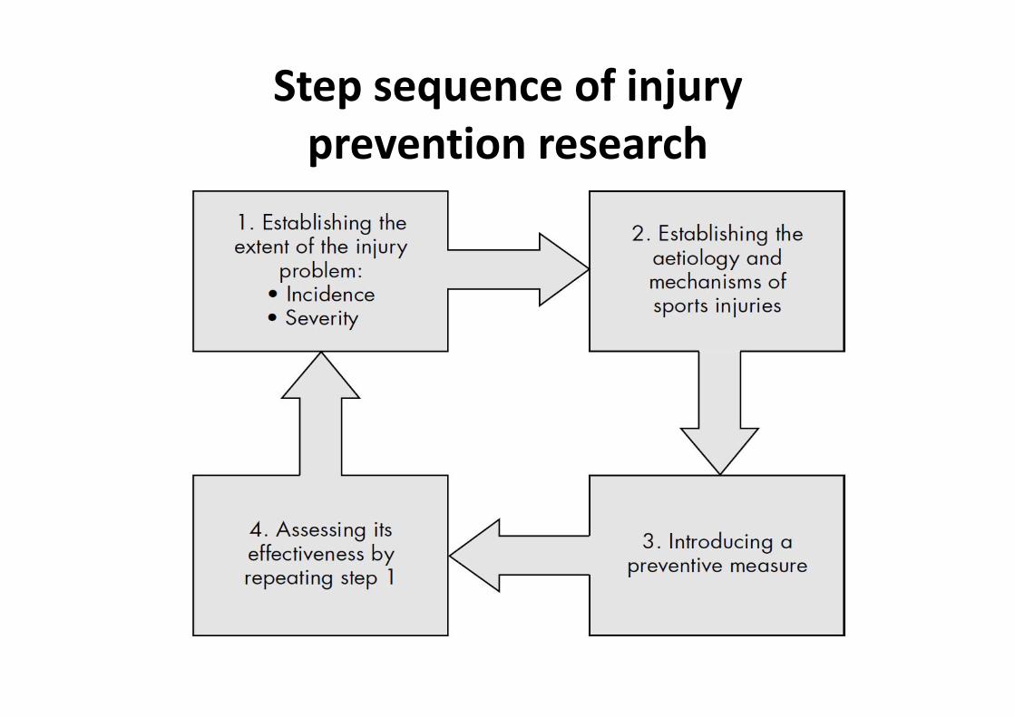
Step sequence of injuryprevention research
UNIVERSITA’ DEGLI STUDI FIRENZE
NEW TRENDS IN THERAPY
Dott. Salvatore Caruso-Dott. Andrea Moretti
Scuola di Specializzazione in Medicina dello Sport
A.O.U.C
• In traditional medicine, the drugsinjected through a syringe are absorbedaccording to the flow circulation
• This may affect the final outcome, due to the impossibility to concentrate drugs in the area of clinical interest
• Many researchers have attempted tofind alternative ways, less traumatizingthan the injection of drugs
Introduction
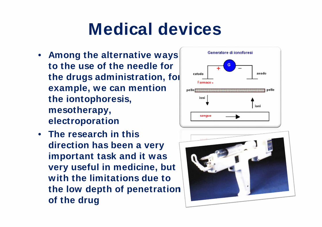
• Among the alternative waysto the use of the needle forthe drugs administration, forexample, we can mentionthe iontophoresis, mesotherapy, electroporation
• The research in thisdirection has been a veryimportant task and it wasvery useful in medicine, butwith the limitations due tothe low depth of penetrationof the drug
Medical devices
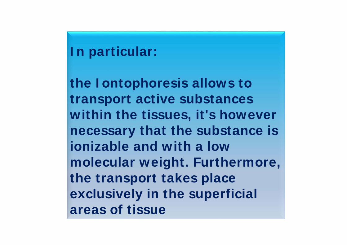
In particular:
the Iontophoresis allows totransport active substanceswithin the tissues, it's howevernecessary that the substance isionizable and with a low molecular weight. Furthermore, the transport takes placeexclusively in the superficialareas of tissue
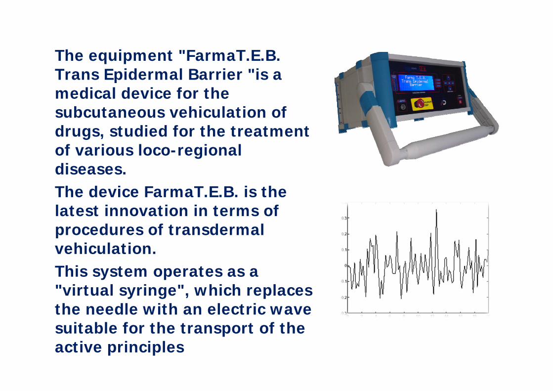
The equipment "FarmaT.E.B.Trans Epidermal Barrier "is a medical device for the subcutaneous vehiculation ofdrugs, studied for the treatment of various loco-regionaldiseases. The device FarmaT.E.B. is the latest innovation in terms ofprocedures of transdermalvehiculation.This system operates as a "virtual syringe", which replacesthe needle with an electric wavesuitable for the transport of the active principles
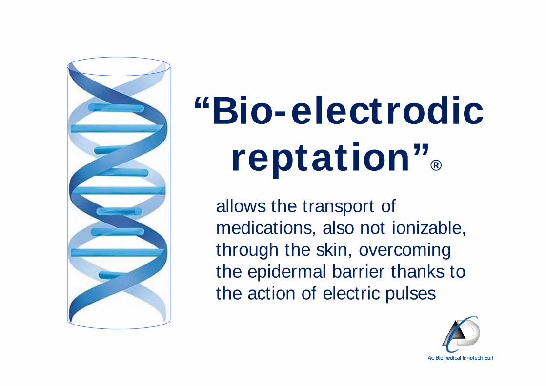
allows the transport ofmedications, also not ionizable, through the skin, overcomingthe epidermal barrier thanks tothe action of electric pulses
“Bio-electrodicreptation”®
• To enhance and optimize the vehiculation of the active principle through the epidermal barrier, the medical device Farma TEB associates the action of electromagnetic waves with the action of a mechanical massage performed with the use of an applicator tool
• The noninvasive system concentrates in a targetedmanner a high quantity of molecules, that interactswith specific receptors available in situ, realizingthe maximum of its therapeutic restorativepotential towards the injured tissues
• Vehiculating a small dose of active principle in correspondence with the lesion to be treated, itinteracts exclusively with local receptors with a therapeutic activity much more effective
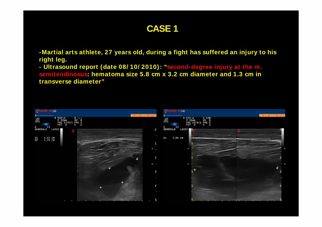
-Martial arts athlete, 27 years old, during a fight has suffered an injury to his right leg. - Ultrasound report (date 08/10/2010): “second-degree injury at the m. semitendinosus: hematoma size 5.8 cm x 3.2 cm diameter and 1.3 cm in transverse diameter”
CASE 1
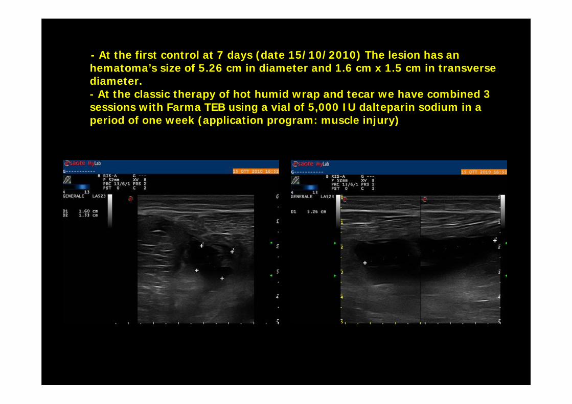
- At the first control at 7 days (date 15/10/2010) The lesion has an hematoma’s size of 5.26 cm in diameter and 1.6 cm x 1.5 cm in transverse diameter. - At the classic therapy of hot humid wrap and tecar we have combined 3 sessions with Farma TEB using a vial of 5,000 IU dalteparin sodium in a period of one week (application program: muscle injury)
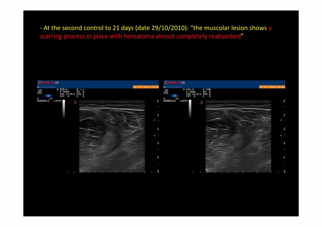
‐ At the second control to 21 days (date 29/10/2010): “the muscolar lesion shows a scarring process in place with hematoma almost completely reabsorbed”
- Sedentary patient, 76 years old. Hypertension treatedwith ACE inhibitors
- He came to our Institute for a teno-synovitis of extensors digitorum ofthe right foot of labeled grade with diffuse lymphedema. The patientreported pain and impossibility on extension of the fingers of the right foot, with consequent difficulty in walking
- At the Inspection the back of his right foot appeared swollen
- Ultrasound report of the dorsal right foot region: "The tendons of the extensors of the fingers and, to a lesser extent, of extensor of the first finger are surrounded by abundant partially corpuscular fluidcollection. During dynamic maneuvers the scrolling of the tendon is alsopreserved. In the foot and in the ankle it’s appreciated marked thickeningof the subcutaneous tissue due to diffused lymphedema
CASE 2
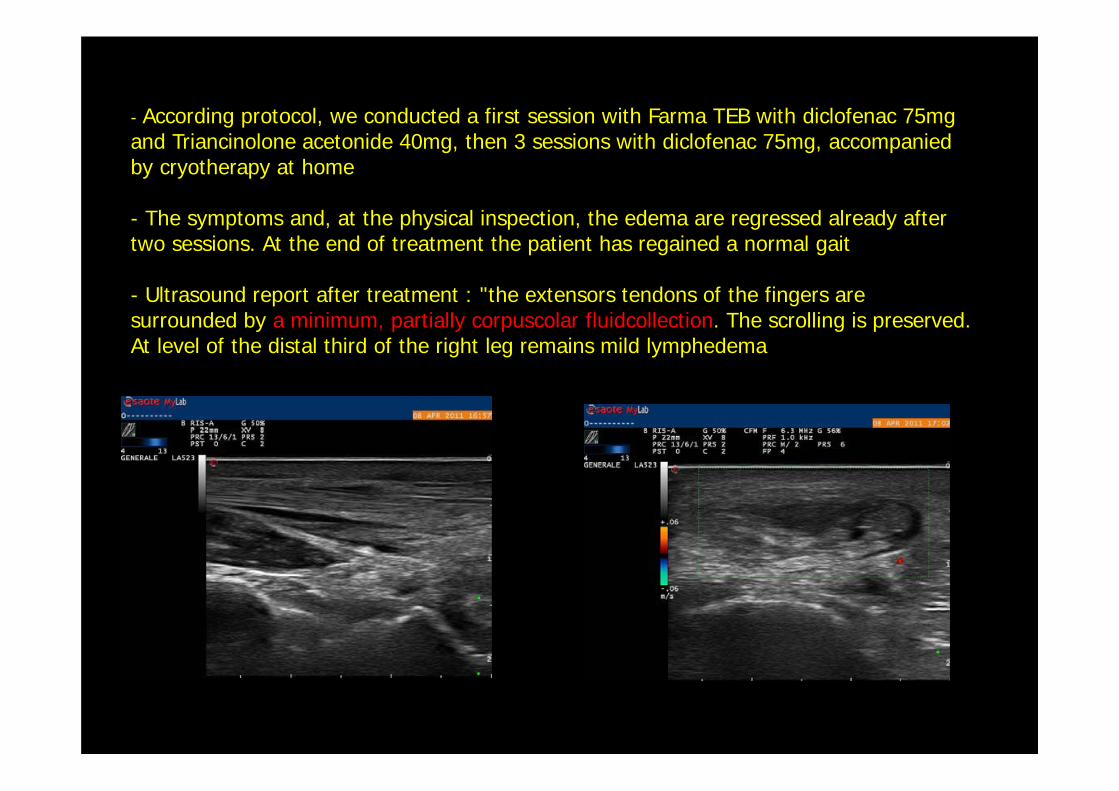
- According protocol, we conducted a first session with Farma TEB with diclofenac 75mg and Triancinolone acetonide 40mg, then 3 sessions with diclofenac 75mg, accompaniedby cryotherapy at home
- The symptoms and, at the physical inspection, the edema are regressed already aftertwo sessions. At the end of treatment the patient has regained a normal gait
- Ultrasound report after treatment : "the extensors tendons of the fingers are surrounded by a minimum, partially corpuscolar fluidcollection. The scrolling is preserved. At level of the distal third of the right leg remains mild lymphedema
- Golf player, 50 years old
- He came to our institute for a tear at level of medial gastrocnemiusmuscle of the right leg
- Ultrasound report performed on 14/07/2011 by another institute: "to the lower third of the medial gastrocnemius muscle, in subfascial intramuscularseat is currently detectable an hypoechoic oblong area that extends from the top down for about 3cm, transversely for 2 cm and in deep 0.5 cm, of finelycorpusculate liquid contents"
CASE 3
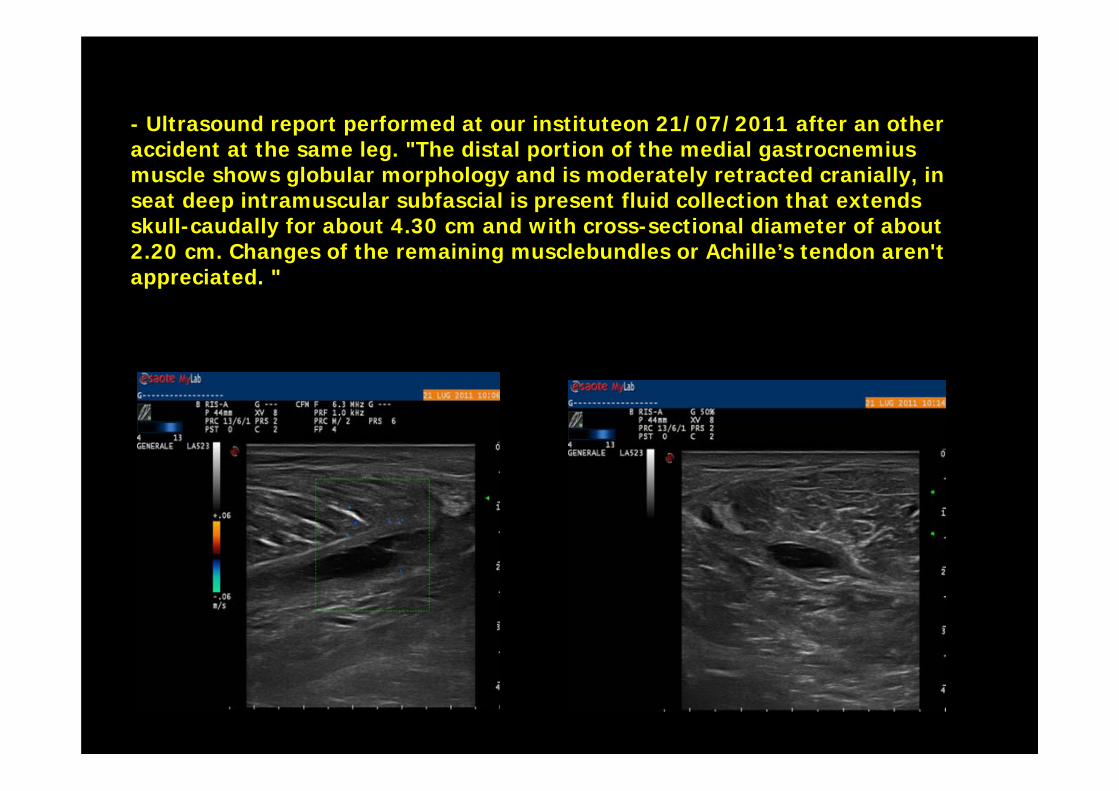
- Ultrasound report performed at our instituteon 21/07/2011 after an otheraccident at the same leg. "The distal portion of the medial gastrocnemiusmuscle shows globular morphology and is moderately retracted cranially, in seat deep intramuscular subfascial is present fluid collection that extendsskull-caudally for about 4.30 cm and with cross-sectional diameter of about2.20 cm. Changes of the remaining musclebundles or Achille’s tendon aren't appreciated. "
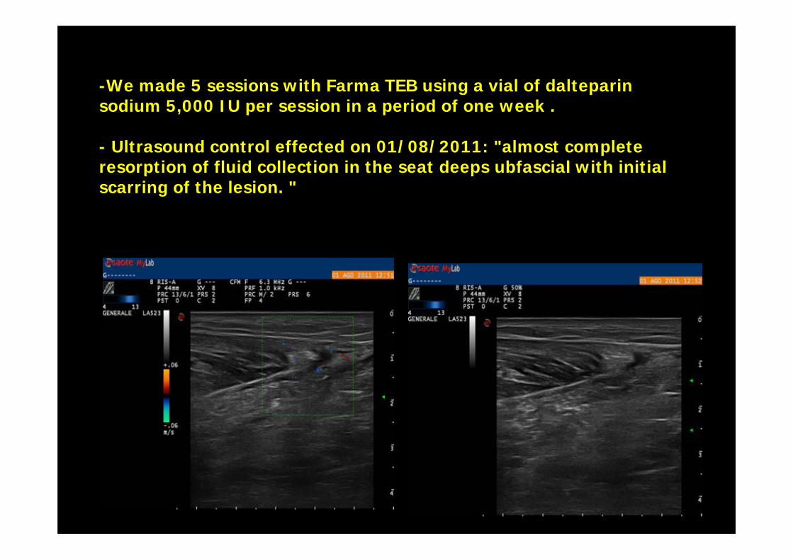
-We made 5 sessions with Farma TEB using a vial of dalteparinsodium 5,000 IU per session in a period of one week .
- Ultrasound control effected on 01/08/2011: "almost complete resorption of fluid collection in the seat deeps ubfascial with initialscarring of the lesion. "
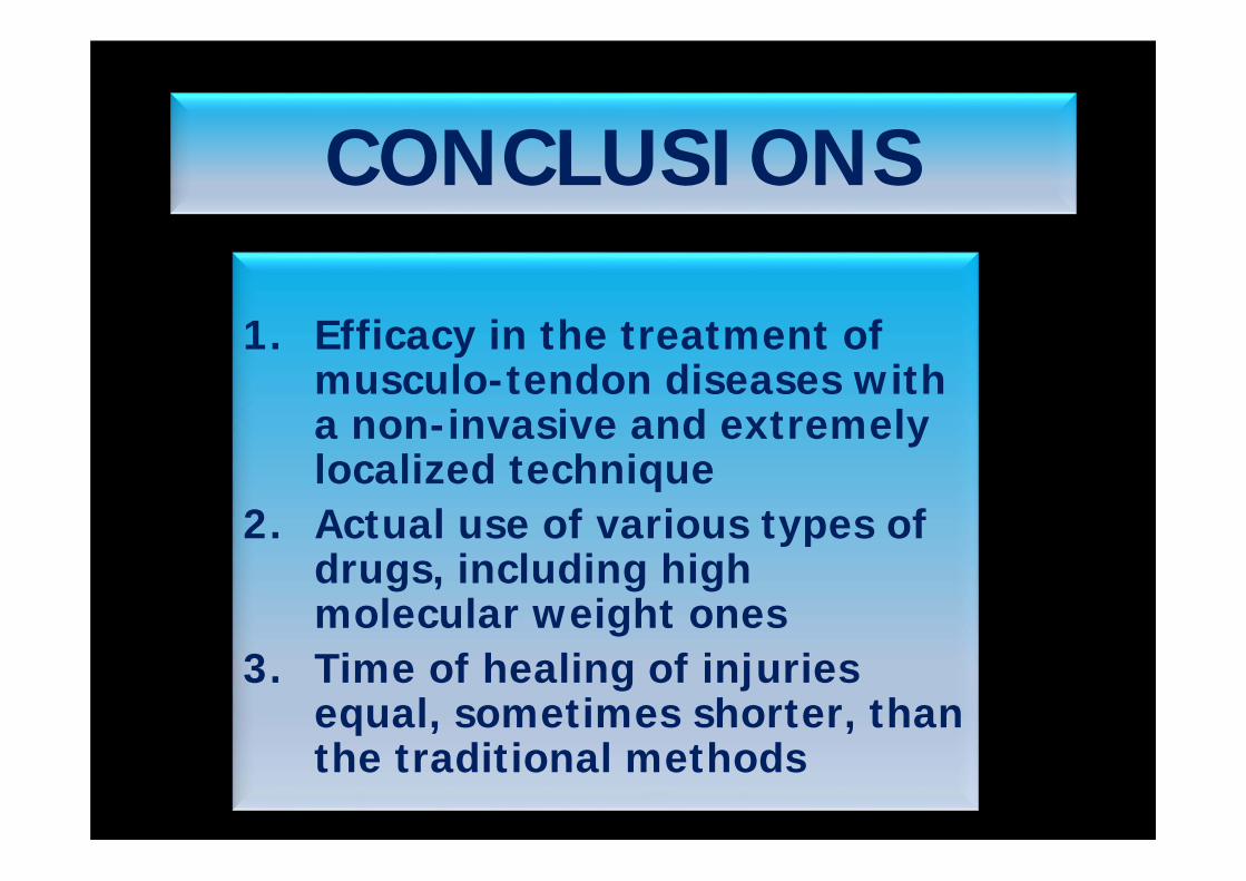
CONCLUSIONS
1. Efficacy in the treatment ofmusculo-tendon diseases witha non-invasive and extremelylocalized technique
2. Actual use of various types ofdrugs, including high molecular weight ones
3. Time of healing of injuriesequal, sometimes shorter, thanthe traditional methods