Embed Size (px)
Citation preview

Research ArticlePrevalence of Gastrointestinal Parasites in the Frugivorous andthe Insectivorous Bats in Southcentral Nepal
Roshan Babu Adhikari ,1,2,3 Mahendra Maharjan ,2 and Tirth Raj Ghimire 3
1Third Pole Conservancy, Wildlife and Eco-health, Bhaktapur, Nepal2Central Department of Zoology, Tribhuvan University, Kathmandu, Nepal3Animal Research Laboratory, Faculty of Science, Nepal Academy of Science and Technology (NAST), Lalitpur, Nepal
Correspondence should be addressed to Tirth Raj Ghimire; [email protected]
Received 27 May 2020; Revised 14 October 2020; Accepted 28 October 2020; Published 14 December 2020
Academic Editor: Emmanuel Serrano Ferron
Copyright © 2020 Roshan Babu Adhikari et al. This is an open access article distributed under the Creative Commons AttributionLicense, which permits unrestricted use, distribution, and reproduction in any medium, provided the original work isproperly cited.
Bats are the only active flying placental mammals and are traditionally classified into mega- and microbats, which are, respectively,herbivorous and insectivorous in feeding habit. Though deforestation, habitat destruction, natural calamities, illegal hunting, andclimate changes are the challenging threats for bats, the role of existing gastrointestinal (GI) parasites have not been evaluated yet inNepal. Thus, the current study aims to determine the prevalence of GI parasites in bats from the Shaktikhor area at the Chitwandistrict of Southcentral Nepal. From July 2018 to February 2019, a total of 60 fecal samples of bats (30 from frugivorous batsand 30 from the insectivorous bats) were collected. These samples were preserved at 2.5% potassium dichromate solution. Thefecal examination was carried out by the direct wet mount, concentrations, acid-fast staining, and sporulation techniques.Overall results showed the prevalence rate of 80% GI parasites. The parasites detected in the insectivorous bats were Ascaridspp., Capillarid sp., Cryptosporidium sp., Eimeria spp., Entamoeba sp., Giardia sp., Hymenolepis spp., Isospora sp., Oxyurid sp.,Strongyle, and Strongyloides sp. In contrast, Eimeria sp., Entamoeba sp., and Hymenolepis sp. were detected in the frugivorousbats. Based on a wide diversity of parasite richness and parasitic concurrency measured by the prevalence rates, we suggest thatGI parasitism might be a threatening factor in the insectivorous bats in the current study area.
1. Introduction
Bats belonging to the order Chiroptera are the only activeflying true placental mammals of the animal kingdom. Chir-optera is the second largest order of mammals (after therodents) with cosmopolitan distribution [1, 2]. Bats are tradi-tionally classified into the megabats and microbats [3–5].Megabats include flying foxes and the old-world fruit bats,which are usually herbivores and consume fruits, flowers,leaves, nectar, and pollens [3, 5, 6]. In contrast, microbatsare mostly insectivorous in feeding habits; although, few ofthese species may feed on blood, fruits, nectars, pollens, andvertebrates [3, 7–10].
It has been estimated that more than 1300 species of batsare reported in the world [1, 8]. However, many species arethreatened with extinction globally, and more than 280 spe-cies are categorized as endangered, vulnerable, or near threat-
ened by the IUCN Red List [11]. In the context of Nepal,there are a total of 54 bat species belonging to seven differentfamilies indicating this Himalayan nation to be one of therich countries in their diversities [12]. However, deforesta-tion, habitat destruction due to the operation of road con-struction projects and natural calamities, illegal hunting,and climate changes exist as challenging threats for thesemammals [13]. Among these threatening factors, diseasesmight be critical because these mammals play roles as patho-gen carriers, reservoirs, and transmitters in nature. Thedisease-causing pathogens are viruses, bacteria, fungi, andparasites, which can be life threatening in humans and ani-mals. It should be noted that several species of gastrointesti-nal (GI) protozoa, trematodes, nematodes, and cestodes havebeen predominantly reported from the bats of various geog-raphies, and they may remain as one of the major threatsfor their lives [14–21]. Moreover, infected bats act as
HindawiJournal of Parasitology ResearchVolume 2020, Article ID 8880033, 12 pageshttps://doi.org/10.1155/2020/8880033

definitive or intermediate or a paratenic host for many proto-zoan, trematode, cestode, and nematode parasites [21]. Inthese situations, feeding behavior, biological, and ecologicdiversity of bats might play a critical role in the host-parasite interactions and parasitism [21]. However, the studyof these parasitic faunae in bats has been still at virgin state inNepal. Besides, the association of GI parasitism based on thefeeding ecology of bats has not been determined and com-pared so far. Thus, in this study, we have investigated theprevalence of GI parasitic species in the frugivorous and theinsectivorous bats found in Chitwan, the Southcentral partof Nepal.
2. Materials and Methods
2.1. Study Area. The current study was conducted in ward no.9 and 10 of Kalika Municipality, the commonly called Shak-tikhor area (251m to 1003m above sea level, asl) (Figure 1).The geographic locations range from (27.69544–27.73472) Nto (84.57159–84.65498) E in Chitwan district, in the South-central part of Nepal. It is 182 kilometers (kms) away fromthe capital city and is linked to the East-West highway byroad up to the foothills. The climate is tropical to subtropical,with an average annual temperature of 29.30°C during sum-mer and 9.4°C during winter. Similarly, the yearly averagerainfall is 1993mm [22]. The vegetation of this area includeslowland Sal forest, hill Sal forests, tropical riverine forest,tropical mixed broad-leaved forest, and subtropical mixedforest [23], and a total of 13 species of birds, eight speciesof mammals, and six species of reptiles have been reportedto inhabit this area according to the Environmental ImpactAssessment done in 2019 [22].
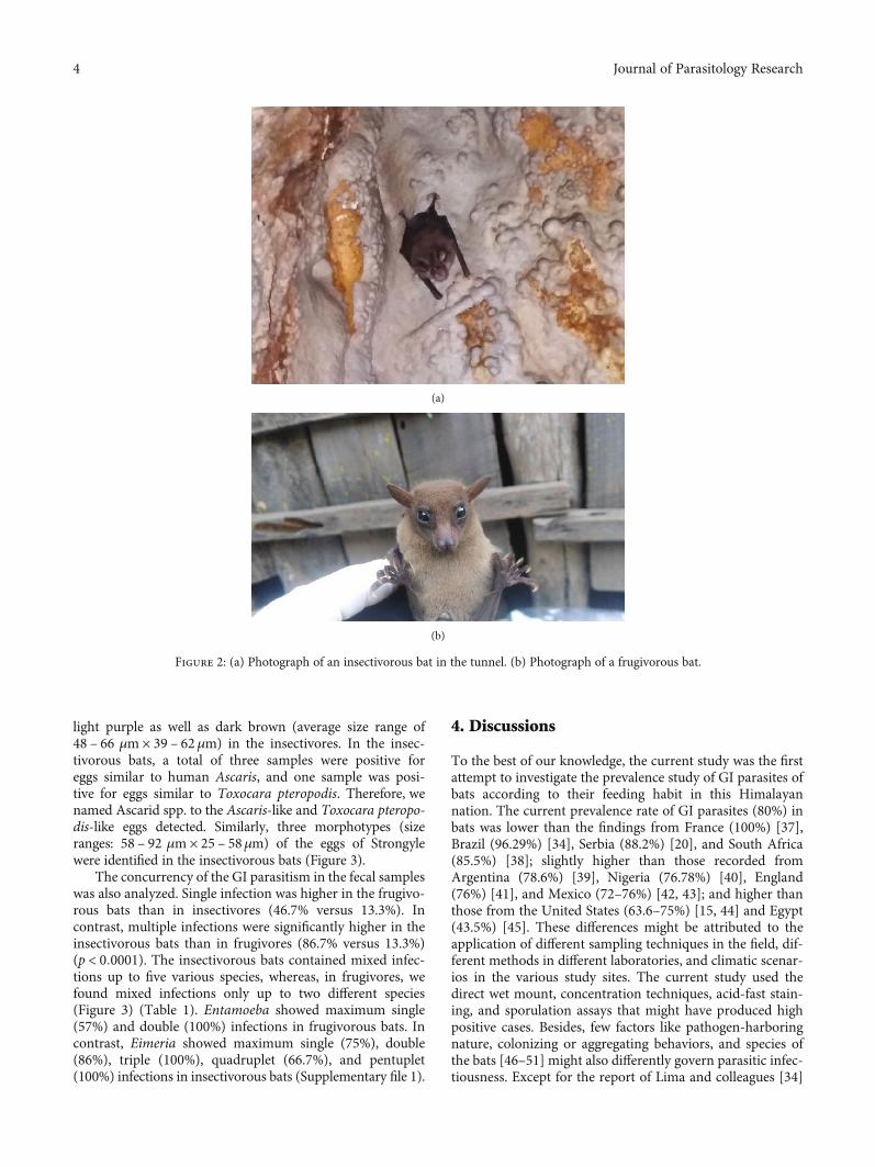
2.2. Sample Collection, Preservation, and Transportation. Atotal of 60 fecal samples (30 from the frugivorous and 30from the insectivorous bats) were collected from July 2018to February 2019 from the study area. The frugivoresincluded Rousettus leschenaulti and Eonycteris spelaea andinsectivores included Rhinolophus macrotis, Rhinolophuspusillus, and Rhinolophus pearsonii [13] (Figure 2). For fecalsample collection, 30 frugivorous bats were captured usingthe net at night time from five different spots in the buttertree (Diploknema butyracea) forest, and anal swabbing wasperformed with the help of cotton buds. In the context ofthe insectivorous bats, a total of 30 clean white plastic wereoverlaid on the floor of five different caves (six plastics percave) just below their roosts in the morning. The fecal sam-ples that fell down the plastic were collected with the helpof forceps in the evening. Quality control during sample col-lection was performed by observing the absence of othermammals inside the wet and dark caves. The collected sam-ples were immediately preserved at 2.5% potassium dichro-mate solution in 20mL sterile vials. They were transportedto Animal Research Laboratory (ARL) of the Nepal Academyof Science and Technology (NAST) and further stored at 4degrees (°) Celsius temperature.
2.3. Laboratory Processing and Examination. The fecal sam-ples were macroscopically examined for the presence of
blood, mucus, segments of cestodes, as well as whole adultnematodes and microscopically examined by the techniquesbased on the literatures, explained previously [24, 25].
2.3.1. Direct Wet Mount Technique. One to two drops ofcarefully stirred fecal samples were put in the slide with thehelp of a plastic dropper. The samples were observed directlyat 2.5% (w/v) potassium dichromate, Gram’s iodine stain,and Giemsa’s stain (1/15).
2.3.2. Saturated Salt Floatation Technique. About two grams(gms) of the fecal samples were thoroughly mixed in a 13milliliter (mL) normal saline (0.9% w/v) solution and filteredwith the help of a tea strainer. The solution was poured into a15mL conical centrifuge tube and proceeded to centrifuge(1200 revolutions per minute, rpm for 5 minutes). After dis-carding the supernatant, 12mL of salt solution (45% w/v)was added and proceeded to centrifuge (1200 rpm for 5minutes). Then, few drops of salt solution (45% w/v) wereadded in the tube to fill it, and a coverslip was placed onthe mouth of the tube. After 10 minutes, the coverslip wascarefully removed and put on the glass slide with or withoutLugol’s iodine for microscopic observation at 100x and 400xtotal magnifications.
2.3.3. Sedimentation Technique. About two gms of the fecalsamples were thoroughly mixed in 13mL normal saline(0.9% w/v), were filtered with the help of a tea strainer intoa 15mL centrifuge tube, and were proceeded to centrifuge(1200 rpm for 5 minutes). Then, the supernatant was dis-carded, and one to two drops of the sediment was put on aglass slide. Gram’s iodine and Giemsa’s stain (1/15 dilutions)were differently used in the deposits for the microscopicexaminations at 100x and 400x total magnifications.
2.3.4. Acid-Fast Staining. About one gm of the Cryptosporid-ium-positive sediments, 10% 10mL formalin, and 4mL ethylacetate were taken in a 15mL centrifuge tube and proceededto centrifuge (1200 rpm for 5 minutes). Then, the superna-tant was discarded, and the sediments were used to preparethin smears. This smear was allowed to dry at room temper-ature and then fixed in the absolute methanol for 2 minutes.The smear was stained with carbol fuchsin for 15 minutes atroom temperature and then washed with distilled waterfollowed by destaining with acid alcohol, and finally rinsedwith distilled water. The smear was further restained withmalachite green for one minute, followed by washing withdistilled water, and allowed to dry at room temperature.The dry slide was observed at 1000x total magnification usingimmersion oil.
2.3.5. Sporulation Assay. About two gms of coccidian positivesamples were incubated at equal volumes of 2.5% potassiumdichromate at 28°C ± 1 temperature in an incubator for spor-ulation assays. Then, using the floatation method, the sporu-lation states were observed at each 24 hours interval underthe microscope [26–28]. The presence of oocysts of Eimeriaspp. and Isospora sp. was confirmed by their respective sporeformulas as 0.4.2 and 0.2.4, as reviewed previously [29].
2 Journal of Parasitology Research

2.3.6. Parasite Identification. All the fecal parasites were care-fully observed under a light microscope (Optika MicroscopesItaly, B-383PLi) at a total magnification of 100x, 400x, and1000x. Photographs were taken by the camera (SXView2.2.0.172 Beta (Nov 6, 2014) Copyright (C) 2013-2014)accompanied by the microscope. The size of the parasiteswas assessed by using ImageJ 1.51k (National Institute ofHealth, USA), and identification was carried out based onvarious literature [30–36].
2.4. Data Analysis. Data were expressed as numbers of posi-tive samples as well as prevalence rates in the table usingMicrosoft Word. Prevalence rates were calculated by dividingthe number of GI positive samples (total or particular spe-cies) by the total number of samples observed [24]. We usedthe GraphPad Software (Prism 5 for Windows Version 5.00@ 1992–2007 GraphPad Software, Inc). We applied Fisher’sexact test (two-sided) to assess p values by comparing theprevalence of specific GI parasitic groups between the frugiv-orous bats and the insectivorous bats. Statistical significancewas considered at the 95% confidence interval (α = 0:05,p < 0:05).
3. Results
In the current study, out of 60 fecal samples, 80% (60% in thefrugivorous and 100% in the insectivorous bats) were positive
for at least one GI parasitic species. The sensitivity ofdifferent tests gave different results, for example, direct wetmount, sedimentation, and flotation techniques detected GIparasites in 61.7% (37/60) with seven species, 73.3% (44/60)with nine species, and 76.7% (46/60) with nine species,respectively. The overall prevalence of protozoan and hel-minth parasites was 70% and 50%, respectively. In this con-text, the prevalence of protozoa was double (93% versus46.7%) (p < 0:0001), and that of helminths was four timesgreater (80% versus 20%) (p < 0:0001) in the insectivorousbats compared to the frugivorous bats. The prevalence of spe-cific GI parasites in frugivores was Entamoeba sp. (40%),Eimeria sp. (13.3%), and Hymenolepis sp. (20%). In contrast,the insectivores possessed Eimeria spp. (83.3%), Strongyle(56.7%), Hymenolepis spp. (50%), Entamoeba sp. (30%),Isospora sp. (16.7%), Strongyloides sp. (16.7%), Ascarid spp.(16.7%), Cryptosporidium sp. (10%), Oxyurid sp. (6.7%),Giardia sp. (3.3%), and Capillarid sp. (3.3%) (Figure 3)(Table 1).
Further, we classified Eimeria spp. into six different mor-phologic forms in the insectivorous bats; however, a singlemorphotype of this coccidian was present in frugivores. Sim-ilarly, in the context of helminths, six species of parasiteswere found in the insectivorous bats, but only one Hymeno-lepis sp. was detected in the frugivorous bats. In frugivores,the eggs of Hymenolepis sp. were light purple (average sizeranges: 42 – 48 μm× 40 – 46μm). In contrast, the eggs were
Figure 1: Map of the study area showing the locations of sample collections.
3Journal of Parasitology Research
light purple as well as dark brown (average size range of48 – 66 μm× 39 – 62μm) in the insectivores. In the insec-tivorous bats, a total of three samples were positive foreggs similar to human Ascaris, and one sample was posi-tive for eggs similar to Toxocara pteropodis. Therefore, wenamed Ascarid spp. to the Ascaris-like and Toxocara pteropo-dis-like eggs detected. Similarly, three morphotypes (sizeranges: 58 – 92 μm× 25 – 58μm) of the eggs of Strongylewere identified in the insectivorous bats (Figure 3).
The concurrency of the GI parasitism in the fecal sampleswas also analyzed. Single infection was higher in the frugivo-rous bats than in insectivores (46.7% versus 13.3%). Incontrast, multiple infections were significantly higher in theinsectivorous bats than in frugivores (86.7% versus 13.3%)(p < 0:0001). The insectivorous bats contained mixed infec-tions up to five various species, whereas, in frugivores, wefound mixed infections only up to two different species(Figure 3) (Table 1). Entamoeba showed maximum single(57%) and double (100%) infections in frugivorous bats. Incontrast, Eimeria showed maximum single (75%), double(86%), triple (100%), quadruplet (66.7%), and pentuplet(100%) infections in insectivorous bats (Supplementary file 1).
4. Discussions
To the best of our knowledge, the current study was the firstattempt to investigate the prevalence study of GI parasites ofbats according to their feeding habit in this Himalayannation. The current prevalence rate of GI parasites (80%) inbats was lower than the findings from France (100%) [37],Brazil (96.29%) [34], Serbia (88.2%) [20], and South Africa(85.5%) [38]; slightly higher than those recorded fromArgentina (78.6%) [39], Nigeria (76.78%) [40], England(76%) [41], and Mexico (72–76%) [42, 43]; and higher thanthose from the United States (63.6–75%) [15, 44] and Egypt(43.5%) [45]. These differences might be attributed to theapplication of different sampling techniques in the field, dif-ferent methods in different laboratories, and climatic scenar-ios in the various study sites. The current study used thedirect wet mount, concentration techniques, acid-fast stain-ing, and sporulation assays that might have produced highpositive cases. Besides, few factors like pathogen-harboringnature, colonizing or aggregating behaviors, and species ofthe bats [46–51] might also differently govern parasitic infec-tiousness. Except for the report of Lima and colleagues [34]
(a)
(b)
Figure 2: (a) Photograph of an insectivorous bat in the tunnel. (b) Photograph of a frugivorous bat.
4 Journal of Parasitology Research

(a) (b)
(c) (d)
(e) (f)
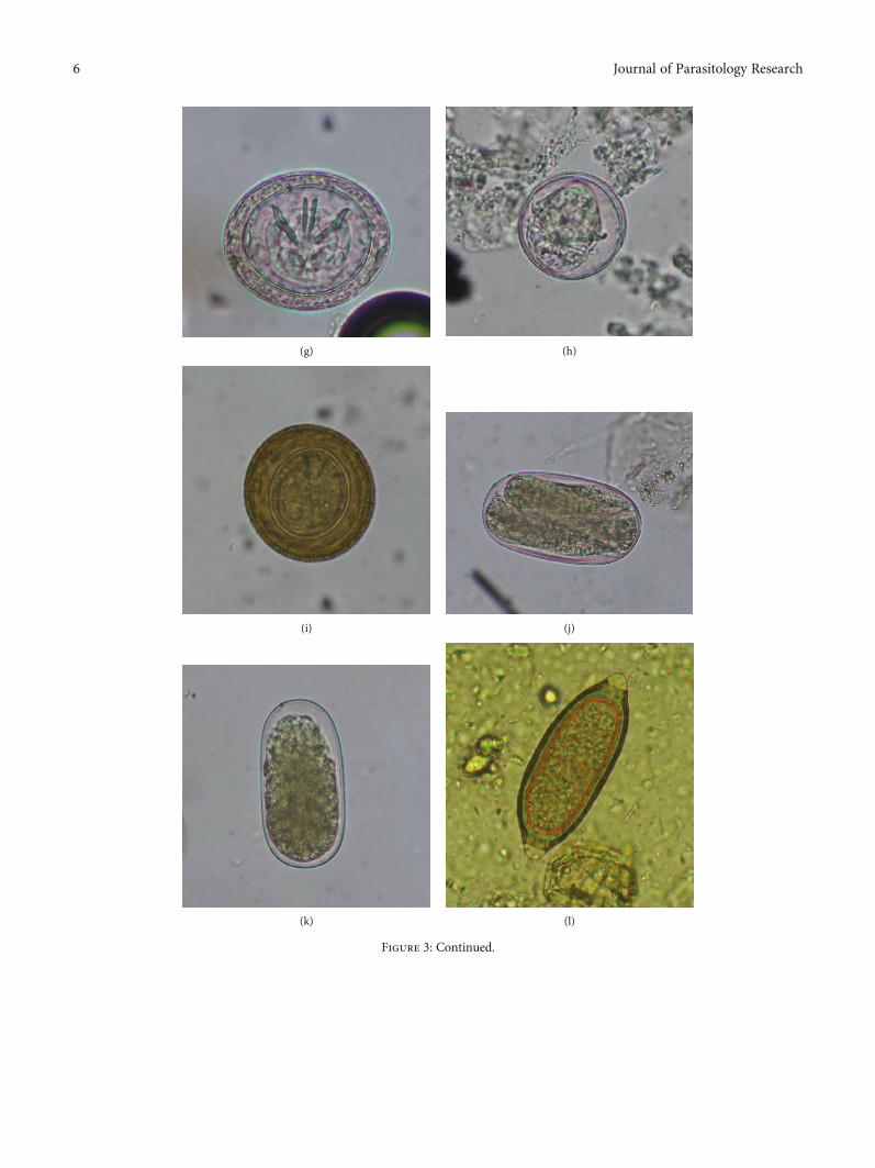
Figure 3: Continued.
5Journal of Parasitology Research
(g) (h)
(i) (j)
(k) (l)
Figure 3: Continued.
6 Journal of Parasitology Research

(m)
Figure 3: Photomicrographs of various parasitic species. (a) Oocyst of Eimeria sp. (i) (20 × 14μm), 400x, direct wet mount at Gram’s iodinestain, in insectivorous bat. (b) Oocyst of Eimeria sp. (ii) (17 × 15 μm), 400x, after flotation technique at Giemsa’s stain, in insectivorous bat. (c)Cyst of Entamoeba sp. (11 × 11 μm), 400x, direct wet mount at Lugol’s Iodine stain, in insectivorous bat. (d) Oocyst of Isospora sp. (25 × 23μm), 400x, after flotation technique, in insectivorous bat. (e) Egg of Ascarid sp. (54 × 36μm), 400x, after sedimentation technique at Giemsa’sstain, in insectivorous bat. (f) Egg of Toxocara sp. (50 × 49 μm), 400x, direct wet mount at 2.5% potassium dichromate, in insectivorous bat.(g) Light purple-colored egg of Hymenolepis sp. (52 × 43μm), 400x, after flotation technique, in insectivorous bat. (h) Egg of Hymenolepis sp.(44 × 43 μm), 400x, after flotation technique, in frugivorous bat. (i) Brown-colored egg of Hymenolepis sp. (65 × 62μm), 400x, after flotationtechnique in insectivorous bat. (j) Egg of Strongyloides sp. (87 × 46μm), 400x, after sedimentation technique at Gram’s iodine stain, ininsectivorous bat. (k) Egg of Strongyle (83 × 41μm), 400x, after flotation technique, in insectivorous bat. (l) Egg of Capillarid sp. (67 × 25μm),400x, direct wet mount at 2.5% potassium dichromate, in insectivorous bat. (m) Egg of Oxyurid sp. (93 × 36μm), 400x, after flotationtechnique, in insectivorous bat.
Table 1: Parasitic species, their concurrency, and prevalence in the frugivorous and insectivorous bats in Southcentral Nepal. Fisher’s exacttest (two-tailed) was used to calculate the p values by comparing the prevalence rates of different parasitic species or groups between thefrugivores and insectivores.
Parasitic infectionsFrugivores (N1 = 30) Insectivores (N2 = 30)
Overall (N = 60) prevalence (n × 100/N) p valuesPrevalence (n × 100/N1) Prevalence (n × 100/N2)
Entamoeba sp. 12 (40%) 9 (30%) 21 (35%)
p < 0:0001Eimeria spp. 4 (13.3%) 25 (83.3%) 29 (48.3%)
Isospora sp. 0 5 (16.7%) 5 (8.3%)
Cryptosporidium sp. 0 3 (10%) 3 (5%)
Giardia sp. 0 1 (3.3%) 1 (1.7%)
Total Protozoa 14 (46.7%) 28 (93.3%) 42 (70%)
Ascarid spp. 0 5 (16.7%) 5 (8.3%)
p < 0:0001
Hymenolepis spp. 6 (20%) 15 (50%) 21 (35%)
Strongyle 0 17 (56.7%) 17 (28.3%)
Oxyurid sp. 0 2 (6.7%) 2 (3.3%)
Strongyloides sp. 0 5 (16.7%) 5 (8.3%
Capillarid sp. 0 1 (3.3%) 1 (1.7%)
Total Helminths 6 (20%) 24 (80%) 30 (50%)
Single infection 14 (46.7%) 4 (13.3%) 18 (30%)p < 0:0001
Mixed infection 4 (13.3%) 26 (86.7%) 30 (50%)
Duplet infection 4 (13.3%) 7 (23.3%) 11 (18.3%)
Triplet infection 0 8 (26.7%) 8 (13.3%)
Quadruplet infection 0 9 (30%) 9 (15%)
Pentuplet infection 0 2 (6.7%) 2 (3.3%)
7Journal of Parasitology Research

and some experiments involving coccidian morphology [15,44], most of the studies are based on the histopathologic find-ings [20, 38, 40–43, 45], and in these contexts, it is not easy tocompare our results with their investigations.
The diversity in parasite richness and parasitic concur-rency, as measured by the parasitic prevalence, was higherin insectivores than in frugivores. This discrepancy mightbe explained based on different feeding habits and the land-scapes of the habitat used. Landscapes include available diets,roosting sites, water sources, foraging habitats, and sharedecosystems with other animals. Firstly, insectivores usuallyprefer insects like bees, beetles, caddis flies, cockroaches,crickets, flies, flying ants, grasshopper, mayflies, mosquitoes,moths, termites, and wasps [8, 40, 52]. One or more of theseinsects are also known to act as intermediate hosts or trans-port vectors for helminth and or protozoan parasites [14,53–58]. Secondly, insectivores mostly spend their lives inthe caves with high moisture contents, which are essentialfor the survival and development of the eggs, cysts, oocysts,and larva of the GI parasites [59]. Thirdly, these bats usuallyspend the full day on roosts that can result in the evaporationand extreme loss of water from their body [60]. Therefore,after coming out of the roosts, they directly visit the watersources and drink water regularly to rehydrate themselves[61]. For foraging and drinking, most insectivores are knownto utilize aquatic habitats like canals, farms, urban dams,lakes, streams, rivers, and swimming pools [62–65]. In thestudy areas, open defecation, nearby water sources, and fieldswere observed. Also, domestic animals like chicken, goats,cattle, dogs, and pigs of the study areas share the same watersources. They can contaminate them with infective cysts,eggs, oocysts, and larva of GI parasites. In these scenarios,we cannot ignore the possibility of cross-transmission ofmany parasites; although, further epidemiologic proofs areneeded to confirm this opinion. In contrast to these bats, fru-givores compensate for the requirement of water fromplant/fruit juices and occasionally use the water sources [66,67]. That is why they are less exposed to parasites.
In this research, compared with the frugivorous bats, theinsectivorous bats possessed higher concomitant infections.Similar to our study, mixed infections by protozoa (Eimeria,Entamoeba, Giardia, and Cryptosporidium) and by protozoaand helminths (Ancylostomatidae, Vampirolepis nana) inBrazilian bats have been predominantly reported [34]. Con-comitant infections are the rules rather than the exception[68], impact on the fitness of host as well as the epidemiologyof the pathogens in all biological communities [68], and helpinvestigate the role in the emergence of zoonoses [69]. Poly-parasitism is the complex interactions among various species,and the outcome of those interactions can be synergistic(positive), antagonistic (negative), or neutral [68]. In positivecase, the presence of one pathogen may enhance the infectionby other pathogens. In negative case, one pathogen inhibitsthe infection or reproduction of other pathogens, for exam-ple, cats infected by many species possessed lower Toxocaraloads [70]. In neutral case, there is no influence on infectionby other pathogens. Our results of maximum coinfection byEimeria in fecal samples suggest that further studies shouldbe conducted to link this coccidian in GI pathogenesis. Nota-
bly, the link of GI parasites in gastroenteritis has not beenfully enlightened in a polyparasitized bat host. Thus, ratherthan single species, the effects of polyparasitism by entericpathogen communities should be assessed especially in path-ologic consequences [70, 71].
It was interesting that Eimeria spp. were the predominantspecies in the insectivorous and overall bats. Their prevalencerate (83.3%) in insectivores was lower than the findings fromFrance (100%) [37] and higher than reported from Europe(80%) [72], the United States (75%) [44], Brazil (74.07%)[34], the United States (63.6%) [15], Japan and North Amer-ica (3.4%–7%) [31, 73], and Northwestern Arkansas (13%)[15]. Similarly, the prevalence of Isospora sp. was 16.7% inthe insectivorous bat, and this rate was slightly higher thanthe finding in the big brown insectivorous bats (Eptesicus fus-cus) from the United States (4.92%) [35]. Another importantcoccidian parasite detected in insectivores was Cryptosporid-ium with the prevalence of 10% which was slightly higherthan the finding from China (7.7%) [74] and the Philippines(8.8%) [43] confirmed by molecular methods and slightlylower than the results from Brazil (11.11%–16.3%) [34, 75].This coccidian parasite was also reported from the fecal sam-ples of two insectivorous bats Pipistrellus pipistrellus andMyotis ciliolabrum via the molecular methods from theUSA and Czech Republic [76] indicating these coccidia arepredominant in bats.
Regarding Sarcodina, the prevalence of Entamoeba sp.was 30% in insectivores and 40% in frugivores suggestingboth bats are critical reservoirs for this ameba. This preva-lence was lower than that reported from Molossus molossus,an insectivorous bat in Brazil (32%), and higher than thatreported from two other insectivorous species like Myotislavali (10%) and Noctilio albiventris (21.05%) [34]. More-over, amebic dysentery caused by Entamoeba histolyticawas firstly reported by techniques similar to ours and molec-ular assays in Rhinolophus rex, an insectivorous bat, fromChina indicating its pathologic consequences in bats [77].
Interestingly, only one sample (3.3%) of insectivorewas pos-itive forGiardia sp. whichwas lower than reported in the similarhosts from Brazil (11.10%) [34], indicating that this flagellatecannot be ignored during diagnosis of GI parasitism in bats.
Among the helminths, the overall prevalence ofHymeno-lepis spp. was the highest and was reported from both types ofbats. The current prevalence of this tapeworm in insectivores(50%) was slightly higher than the finding from Brazil(48.14%) [34]. Similar genera have been predominantlyreported from insectivores by other studies around the globe[21, 38–40, 78, 79]. Some of these species include secondaryhosts like insects in their life cycle. Thus, the current resultindicates that parasite transmission is related to the feedingcharacteristics of the bat hosts, and it is the reason why theinsectivorous bats were found to be positive with this cestode.Furthermore, we have reported the same genus of differentmorphotypes in the frugivorous bats, with a prevalence of20%. This rate was higher than that reported from AmazoniaBrazil (1.49%) in Artibeus planiros, a frugivorous bat [80].
In the current study, except for Hymenolepis spp., allother helminths like Ascarid spp., Strongyle, Oxyurid sp.,Strongyloides sp., and Capillarid sp. were reported only from
8 Journal of Parasitology Research

insectivores. We grouped three different morphotypes ofnematode eggs into “Strongyle-type,” because, withoutlarval cultures, it is not easy to differentiate them onlyvia the egg morphometry. Many previous histologic stud-ies of GI tracts of the insectivorous bats from variousgeographies were conducted. They reported the presenceof the adults of different Strongyles like Histostrongyluscoronatus, Macuahuitloides inexpectans, Molinostrongylusornatus, Parahistiostrongylus octacanthus, Strongylacanthaglycyrrhiza, Torrestrongylus tetradorsalis, and Bidigiticaudaserrafreire [20, 21, 38, 40, 42, 80–82]. This evidence indi-cates the predominance of a wide variety of these nematodes.
There were two morphotypes of eggs of Ascarid spp. inthe current insectivorous bats with the prevalence rates of16.7%, which was higher than the finding from Brazil [34].This roundworm species was also reported in a few researchfindings [83, 84]. Although we did not report these nema-todes from frugivores, previous studies reported the presenceof Toxocara pteropodis in frugivore bats from Australia [85],Palm Island (25%) [86], and Sri Lanka (13%) [87].
It was notable that in insectivores, we reported Strongy-loides sp. with a prevalence of 16.7%, which was slightly higherthan reported from Brazil (9.25%) [34]. Another nematodeCapillarid sp. was reported to be present in 3.3% insectivores,and this rate was similar to those reported fromBrazil (1.49%–3.7%) [34, 80, 88] and was lower than from Nigeria (18.44%)[40]. This nematode was also reported in 2% frugivorous batsfrom Amazonian Brazil [67]. Interestingly, we found eggs ofoxyurid nematodes in 6.7% of the insectivorous bats and arethe first record in published peer-reviewed journals. The pres-ence of this nematode may suggest two possible hypotheses;firstly, oxyurids are natural in bats. Secondly, bats may acquirethem via cross-transmission from animal sources; importantly,cross-transmission is known to be highly prevalent among thesehosts [89]. Cross-transmission of oxyurid in bats may occur viaoccasional feeding on rodent and avian species [7, 90].
5. Conclusions
In conclusion, the current study contributes to the under-standing of GI parasites and their roles in disease accordingto their feeding habits. The study also suggests that comparedto the frugivorous bats, the insectivorous bats have a wideand complex behavioral and ecologic landscape includingthe selection of insect diets, water bodies, and sharing of anecosystem with other vertebrates which are critical for trans-mission of the parasitic species. Based on the wide diversity ofparasite richness and parasitic concurrency measured by theprevalence rates, we suggest that GI parasitism might be athreatening factor in the insectivorous bats in the currentstudy area. However, further detailed molecular and epide-miologic studies are essential to identify the species, to assesstheir pathology, and to analyze their host specificity to clarifytheir roles in threatening the bats.
Data Availability
All data generated or analyzed during this study are includedwithin this article.
Ethical Approval
The authors declare that the study was conducted on natu-rally infected bats. No experimental infection was establishedduring this research work. The required permission for thecollection of the fecal samples was issued by Nepal HealthResearch Council (NHRC) Ethical Review Board (Permis-sion number: 463/2018), Government of Nepal, Ministry ofForests and Environment, Department of Forestry (Permis-sion number: 203/2018), District Forest Office (Permissionnumber: 65/2018), Kalika Municipality (Permission number:55/2018), and Veterinary Services, Kalika Municipality, Chit-wan (Permission number: 05/2018).
Conflicts of Interest
The authors declare that they have no conflict of interest.
Authors’ Contributions
Roshan Babu Adhikari conceived and designed the study aswell as performed the field and laboratory works. MahendraMaharjan supervised the work. Tirth Raj Ghimire investigatedparasites in the laboratory, provided laboratory facilities, andanalyzed data. All three authors wrote the manuscript andfinalized it.
Acknowledgments
The authors would like to acknowledge Prof. Dr. Tej BahadurThapa, Head of Department, Central Department of Zool-ogy, Tribhuvan University, Kathmandu, for permitting thedissertation works; Ms. Jaishree Sijapati, Chief, Faculty ofScience, Nepal Academy of Science and Technology (NAST)for permitting the laboratory works in Animal Research Lab-oratory; Mr. Ganga Ram Regmi and Mr. Purna Ale, ThirdPole Conservancy (TPC), Bhaktapur, for their supports infield works and in preparing GIS map; Nepal HealthResearch Council (NHRC), Government of Nepal for theethical approval of the research; Ministry of Forests andEnvironment, Department of Forestry; District Forest Office,Chitwan; Kalika Municipality, and Kalika Municipality Vet-erinary Services for granting the permission of the works.
Supplementary Materials
Supplementary file 1: patterns of parasitic species in frugivo-rous and insectivorous bats. (Supplementary Materials)
References
[1] American Society of Mammalogists, Taxon Summary Statis-tics. Mammal Diversity DatabaseNovember 2019, https://mammaldiversity.org/summary.
[2] S. Mickleburgh, K. Waylen, and P. Racey, “Bats as bushmeat: aglobal review,” Oryx, vol. 43, no. 2, pp. 217–234, 2009.
[3] D. R. Prothero, The Princeton field guide to prehistoric mam-mals. Princeton Field Guides. Vol. 112, Princeton UniversityPress, 2016.
9Journal of Parasitology Research

[4] K. E. Jones, N. G. Patel, M. A. Levy et al., “Global trends inemerging infectious diseases,” Nature, vol. 451, no. 7181,pp. 990–993, 2008.
[5] I. Schwab and J. Pettigrew, “A choroidal sleight of hand,” BritishJournal of Ophthalmology, vol. 89, no. 11, pp. 1398–1398, 2005.
[6] J. O. Whitaker Jr., “Chitinase in insectivorous bats,” Journal ofMammalogy, vol. 85, no. 1, pp. 15–18, 2004.
[7] M. B. Fenton and T. H. Fleming, “Ecological interactionsbetween bats and nocturnal birds,” Biotropica, vol. 8, no. 2,pp. 104–110, 1976.
[8] M. B. Fenton and N. B. Simmons, Bats: AWorld of Science andMystery, University of Chicago Press, 2015.
[9] P. W. Freeman, Form, function, and evolution in skulls andteeth of bats. Papers in Natural Resources, p. 9, 1998.
[10] L. Hammond, Bats: Biology and Behaviour, Oxford UniversityPress, 1996.
[11] IUCN, The IUCN Red List of Threatened Species. Version 2019-2. 2019 23March 2020, http://www.iucnredlist.org.
[12] P. R. Acharya and L. A. Ruedas, The Bat Fauna of Nepal: ACurrent Conspectus, 2007, BAT NET NEWSLETTER.
[13] T. R. Ghimire, R. B. Adhikari, and G. R. Regmi, The zig-zagtrail of symbiosis among Chepang, bat, and butter tree: ananalysis of conservation threat in Nepal, in Wild plants: theTreasure of Natural Healers, M. Rai, S. Bhattarai, and C. M.Feitosa, Eds., CRC Press-Taylor and Francis Group, BocaRaton, Florida, 2020.
[14] C. D. Hilton and T. L. Best, Gastrointestinal helminth parasitesof bats in Alabama. in Fourth colloquium on conservation ofmammals in the southeastern United States. Occasional Papersof the North Carolina Museum of Natural Sciences and theNorth Carolina Biological Survey, North Carolina Museum ofNatural Sciences, 2000.
[15] C. T. McAllister and S. J. Upton, “Two new species of Eimeria(Apicomplexa: Eimeriidae) from eastern red bats, Lasiurus bor-ealis (Chiroptera: Vespertilionidae), in Arkansas and North Car-olina,” Journal of Parasitology, vol. 95, no. 4, pp. 991–993, 2009.
[16] M. E. Marshall and G. C. Miller, “Some digenetic trematodesfrom Ecuadorian bats including five new species and onenew genus,” The Journal of Parasitology, vol. 65, no. 6,pp. 909–917, 1979.
[17] C. McAllister, R. S. Seville, R. Arlen, and M. B. Connior, “Anew species of Eimeria (Apicomplexa: Eimeriidae) from tri-colored bats, Perimyotis subflavus (Chiroptera: Vespertilioni-dae), from the Ouachitas of Arkansas,” Acta Parasitologica,vol. 59, no. 4, pp. 690–693, 2014.
[18] C. T. McAllister, R. S. Seville, and C. R. Bursey, “Helminth(Cestoda, Nematoda) and coccidian (Apicomplexa: Eimerii-dae) parasites of the eastern small-footed myotis, Myotis leibii(Chiroptera: Vespertilionidae) from Arkansas, with a descrip-tion of a new species of Eimeria,” Acta Parasitologica, vol. 62,no. 2, pp. 377–381, 2017.
[19] F. Schär, U. Trostdorf, F. Giardina et al., “Strongyloides stercor-alis: global distribution and risk factors,” PLoS Neglected Trop-ical Diseases, vol. 7, no. 7, article e2288, 2013.
[20] Ž. Horvat, B. Čabrilo, M. Paunović et al., “The helminth faunaof the greater horseshoe bat (Rhinolophus ferrumequinum)(-Chiroptera: Rhinolophidae) on the territory of Serbia,” Biolo-gia Serbica, vol. 37, no. 1-2, pp. 64–67, 2016.
[21] C. P. Santos and D. I. Gibson, “Checklist of the helminth par-asites of South American bats,” Zootaxa, vol. 3937, no. 3,pp. 471–499, 2015.
[22] IDML, Environmental Impact Assessment of Shaktikhor Indus-trial District, Chitwan. 2019, Industrial District ManagementLimited: Report Submitted to Government of Nepal, Ministryof Forests and Environment, Singh Durbar, Kathmandu, Nepa-lIndustrial District Management Limited.
[23] A. Rijal, “Living knowledge of the healing plants: ethno-phytotherapy in the Chepang communities from the Mid-Hills of Nepal,” Journal of Ethnobiology and Ethnomedicine,vol. 4, no. 1, p. 23, 2008.
[24] T. R. Ghimire and N. Bhattarai, “A survey of gastrointestinalparasites of goats in a goat market in Kathmandu, Nepal,”Journal of Parasitic Diseases, vol. 43, no. 4, pp. 686–695, 2019.
[25] A. M. Zajac and G. A. Conboy, Veterinary Clinical Parasitol-ogy, John Wiley & Sons, 2012.
[26] B. K. Baek, C. S. Kim, J. H. Kim, K. S. Han, and Y. G. Kim,“Studies on isosporosis in dogs. I: isolation and sporulationof Isospora ohioensis,” The Korean Journal of Parasitology,vol. 31, no. 3, pp. 201–206, 1993.
[27] S. Edgar, “Effect of temperature on the sporulation of oocystsof the protozoan, Eimeria tenella,” Transactions of the Ameri-can Microscopical Society, vol. 73, no. 3, pp. 237–242, 1954.
[28] S. Edgar, “Sporulation of oocysts at specific temperatures andnotes on the prepatent period of several species of avian cocci-dia,” The Journal of Parasitology, vol. 41, no. 2, pp. 214–216,1955.
[29] T. R. Ghimire, “Redescription of genera of family EimeriidaeMinchin, 1903,” International Journal of Life Sciences, vol. 4,pp. 26–47, 2010.
[30] D. W. Duszynski, D. W. Reduker, and B. B. Parker, “Eimeriafrom bats of the world. II. A new species in Tadarida femoro-sacca from Sonora, Mexico,” The Journal of Parasitology,vol. 74, no. 2, pp. 317–321, 1988.
[31] D. W. Duszynski, D. T. Scott, J. Aragon, A. Leach, andT. Perry, “Six new Eimeria species from vespertilionid bats ofNorth America,” The Journal of Parasitology, vol. 85, no. 3,pp. 496–503, 1999.
[32] D. W. Duszynski and S. J. Upton, “Coccidia (Apicomplexa:Eimeriidae) of the mammalian order Insectivora,” FacultyPublications from the Harold W. Manter Laboratory of Parasi-tology, vol. 4, pp. 1–67, 2000.
[33] D. W. Duszynski and P. G. Wilber, “A guideline for the prep-aration of species descriptions in the Eimeriidae,” The Journalof Parasitology, vol. 83, no. 2, pp. 333–336, 1997.
[34] V. F. S. Lima, P. A. Rocha, M. A. D. Silva et al., “Survey on hel-minths and protozoa of free-living Neotropical bats fromNortheastern Brazil,” Acta Tropica, vol. 185, pp. 267–272,2018.
[35] A. Wünschmann, J. F. X. Wellehan Jr., A. Armien et al., “Renalinfection by a new coccidian genus in big brown bats (Eptesi-cus fuscus),” Journal of Parasitology, vol. 96, no. 1, pp. 178–183, 2010.
[36] E. J. L. Soulsby, Helminths, arthropods and protozoa of domes-ticated animals seventh edition, Vol. 792, Affiliated East-WestPress Private Limited, New Delhi, 2012.
[37] E. Afonso, P. E. Baurand, P. Tournant, and N. Capelli, “Firstamplification of Eimeria hesseiDNA from the lesser horseshoebat (Rhinolophus hipposideros) and its phylogenetic relation-ships with Eimeria species from other bats and rodents,”Experimental Parasitology, vol. 139, pp. 58–62, 2014.
[38] K. Junker, O. Bain, and J. Boomker, “Helminth parasites ofNatal long-fingered bats, Miniopterus natalensis (Chiroptera:
10 Journal of Parasitology Research

Miniopteridae), South Africa,” Onderstepoort Journal of Veter-inary Research, vol. 75, no. 3, pp. 261–265, 2008.
[39] A. C. Falconaro, R. M. Vega, and G. P. Viozzi, “Helminth com-munities of two populations of Myotis chiloensis (Chiroptera:Vespertilionidae) from Argentinean Patagonia,” InternationalJournal for Parasitology: Parasites and Wildlife, vol. 7, no. 1,pp. 27–33, 2018.
[40] F. C. Okafor, I. Igbinosa, and H. Ezenwaji, “Helminth fauna ofTadarida (Chaeraphon) nigeriae (Thomas, 1913)(Microchir-optera: Molossidae),” Animal Research International, vol. 1,no. 1, pp. 64–69, 2008.
[41] J. S. Lord, S. Parker, F. Parker, and D. R. Brooks, “Gastrointesti-nal helminths of pipistrelle bats (Pipistrellus pipistrellus/Pipis-trellus pygmaeus) (Chiroptera: Vespertilionidae) of England,”Parasitology, vol. 139, no. 3, pp. 366–374, 2012.
[42] J. M. Caspeta-Mandujano, J. L. Peralta-Rodríguez, M. G.Galindo-García, and F. A. Jiménez, “A new species of Torres-trongylus (Trichostrongylidae, Anoplostrongylinae) fromMacrotus waterhousii (Chiroptera: Phyllostomidae) in CentralMexico,” Parasite, vol. 22, no. 29, pp. 28-29, 2015.
[43] F. Murakoshi, K. Koyama, T. Akasaka, N. Horiuchi, and K. Kato,“Molecular and histopathological characterization of Cryptospo-ridium and Eimeria species in bats in Japan,” Journal of Veteri-nary Medical Science, vol. 80, no. 9, pp. 1395–1399, 2018.
[44] C. T. McAllister, R. S. Seville, and Z. P. Roehrs, “A new speciesof Eimeria (Apicomplexa: Eimeriidae) from the northernmyotis, Myotis septentrionalis (Chiroptera: Vespertilionidae),in Oklahoma,” The Journal of Parasitology, vol. 98, no. 5,pp. 5–1003, 2012.
[45] M. Saoud andM. Ramadan, “Studies on the helminth parasitesof bats in Egypt and the factors influencing their occurrencewith particular reference to digenetic trematodes,” Zeitschriftfür Parasitenkunde, vol. 51, no. 1, pp. 37–47, 1976.
[46] T. N. Petney and R. H. Andrews, “Multiparasite communitiesin animals and humans: frequency, structure and pathogenicsignificance,” International Journal for Parasitology, vol. 28,no. 3, pp. 377–393, 1998.
[47] L. Drake and D. Bundy, “Multiple helminth infections in chil-dren: impact and control,” Parasitology, vol. 122, no. S1,pp. S73–S81, 2001.
[48] F. Bordes and S. Morand, “The impact of multiple infectionson wild animal hosts: a review,” Infection Ecology and Epide-miology, vol. 1, no. 1, p. 7346, 2011.
[49] F. Bordes, S. Morand, and G. Ricardo, “Bat fly species richnessin Neotropical bats: correlations with host ecology and hostbrain,” Oecologia, vol. 158, no. 1, pp. 109–116, 2008.
[50] Q. M. R. Webber and C. K. R. Willis, “Sociality, parasites, andpathogens in bats, in Sociality in Bats,” Springer, Cham, 2016.
[51] J. E. Patterson and K. E. Ruckstuhl, “Parasite infection andhost group size: a meta-analytical review,” Parasitology,vol. 140, no. 7, pp. 803–813, 2013.
[52] G. Schalk and R. M. Brigham, “Prey selection by insectivorousbats: are essential fatty acids important?,” Canadian Journal ofZoology, vol. 73, no. 10, pp. 1855–1859, 1995.
[53] M. R. Cranfield, H. Bixler, T. K. Graczyk, and R. Fayer, “Houseflies (Musca domestica) as transport hosts of Cryptosporidiumparvum,” The American Journal of Tropical Medicine andHygiene, vol. 61, no. 3, pp. 500–504, 1999.
[54] T. K. Graczyk, R. Knight, and L. Tamang, “Mechanical trans-mission of human protozoan parasites by insects,” ClinicalMicrobiology Reviews, vol. 18, no. 1, pp. 128–132, 2005.
[55] M. B. Markus, “Flies as natural transport hosts of Sarcocystisand other coccidia,” Journal of Parasitology, vol. 66, no. 2,pp. 361-362, 1980.
[56] W. Kasprzak and A. Majewska, “Transmission of Giardiacysts. I. The role of flies and cockroaches,” Wiadomosci Para-zytologiczne, vol. 27, no. 4-5, pp. 555–563, 1981.
[57] A. A. Adenusi, M. I. Akinyemi, and D. Akinsanya, “Domicili-ary cockroaches as carriers of human intestinal parasites inLagos Metropolis, Southwest Nigeria: implications for publichealth,” Journal of Arthropod-Borne Diseases, vol. 12, no. 2,pp. 141–151, 2018.
[58] R. Issa, “Musca domestica acts as transport vector hosts,” Bul-letin of the National Research Centre, vol. 43, no. 1, p. 73, 2019.
[59] A. Gajadhar, W. Scandrett, and L. Forbes, “Parásitos zoonóti-cos transmitidos por los alimentos y el agua en las granjas,”Revue Scientifique et Technique, vol. 25, no. 2, pp. 595–606,2006.
[60] P.Webb, J. Speakman, and P. Racey, “Evaporative water loss intwo sympatric species of vespertilionid bat, Plecotus auritusand Myotis daubentoni: relation to foraging mode and impli-cations for roost site selection,” Journal of Zoology, vol. 235,no. 2, pp. 269–278, 1995.
[61] D. Russo, L. Cistrone, and G. Jones, “Sensory ecology of waterdetection by bats: a field experiment,” PLoS One, vol. 7, no. 10,p. e48144, 2012.
[62] S. L. Jackrel and R. S. Matlack, “Influence of surface area, waterlevel and adjacent vegetation on bat use of artificial watersources,” The American Midland Naturalist, vol. 164, no. 1,pp. 74–79, 2010.
[63] F. Lisón and J. Calvo, “The significance of water infrastructuresfor the conservation of bats in a semiarid Mediterranean land-scape,” Animal Conservation, vol. 14, no. 5, pp. 533–541, 2011.
[64] I. Salvarina, “Bats and aquatic habitats: a review of habitat useand anthropogenic impacts,” Mammal Review, vol. 46, no. 2,pp. 131–143, 2016.
[65] C. Sirami, D. S. Jacobs, and G. S. Cumming, “Artificial wet-lands and surrounding habitats provide important foraginghabitat for bats in agricultural landscapes in theWestern Cape,South Africa,” Biological Conservation, vol. 164, pp. 30–38,2013.
[66] S. J. Ghanem, H. Ruppert, T. H. Kunz, and C. C. Voigt, “Frugiv-orous bats drink nutrient-and clay-enriched water in the Ama-zon rain forest: support for a dual function of mineral-lickvisits,” Journal of Tropical Ecology, vol. 29, no. 1, pp. 1–10, 2013.
[67] M. R. Nogueira, “Gastrointestinal helminth parasitism in fruit-eating bats (Chiroptera, Stenodermatinae) from western Ama-zonian Brazil,” Revista de Biología Tropical, vol. 52, no. 2,pp. 387–392, 2014.
[68] A. O. Hoarau, P. Mavingui, and C. Lebarbenchon, “Coinfec-tions in wildlife: focus on a neglected aspect of infectious dis-ease epidemiology,” PLoS Pathogens, vol. 16, no. 9, articlee1008790, 2020.
[69] A. J. Peel, K. Wells, J. Giles et al., “Synchronous shedding ofmultiple bat paramyxoviruses coincides with peak periods ofHendra virus spillover,” Emerging microbes & infections,vol. 8, no. 1, pp. 1314–1323, 2019.
[70] E. Serrano and J. Millán, “What is the price of neglecting par-asite groups when assessing the cost of co-infection?,” Epide-miology & Infection, vol. 142, no. 7, pp. 1533–1540, 2014.
[71] S.-X. Zhang, Y. M. Zhou, W. Xu et al., “Impact of co-infectionswith enteric pathogens on children suffering from acute
11Journal of Parasitology Research

diarrhea in southwest China,” Infectious Diseases of Poverty,vol. 5, no. 1, p. 64, 2016.
[72] E. Afonso, P. Tournant, J. C. Foltête et al., “Is the lesser horse-shoe bat (Rhinolophus hipposideros) exposed to causes thatmay have contributed to its decline? A non-invasiveapproach,” Global Ecology and Conservation, vol. 8, pp. 123–137, 2016.
[73] D. W. Duszynski, “Coccidia from bats (Chiroptera) of theworld: a new Eimeria species in Pipistrellus javanicus fromJapan,” The Journal of Parasitology, vol. 83, no. 2, pp. 280–282, 1997.
[74] W. Wang, L. Cao, B. He et al., “Molecular characterization ofCryptosporidium in bats from Yunnan province, southwesternChina,” Journal of Parasitology, vol. 99, no. 6, pp. 1148–1150,2013.
[75] J. M. N. Batista, C. de Carvalho, W. A. Pedro et al., “Identifica-tion of Cryptosporidium bat genotypes XVI–XVIII in batsfrom Brazil,” Parasitology Research, vol. 118, no. 7, pp. 2183–2191, 2019.
[76] M. Kváč, A. Hořická, B. Sak et al., “Novel Cryptosporidium batgenotypes III and IV in bats from the USA and Czech Repub-lic,” Parasitology Research, vol. 114, no. 10, pp. 3917–3921,2015.
[77] G.-Z. He, Y. X. Chen, W. Y. Tian et al., “Entamoeba histolyticainfections in a king horseshoe bat (Rhinolophus rex): a firstcase report,” Asian Journal of Animal and Veterinary, vol. 6,no. 10, pp. 1026–1030, 2011.
[78] C. Vaucher, “Revision of the genus Vampirolepis Spasskij,1954 (Cestoda: Hymenolepididae),” Memórias do InstitutoOswaldo Cruz, vol. 87, Supplement 1, pp. 299–304, 1992.
[79] A. M. F. Milano, Helmintofauna de murciélagos (Chiroptera) delnordeste argentino, in FACULTAD DE CIENCIAS NATURALESYMUSEO, UNIVERSIDADNACIONALDE LA PLATA, 2016.
[80] A. C. A. de Albuquerque, M. F. D. Moraes, A. C. Silva, I. M.Lapera, J. H. Tebaldi, and E. G. Lux Hoppe, “Helminth faunaof chiropterans in Amazonia: biological interactions betweenparasite and host,” Parasitology Research, vol. 115, no. 8,pp. 3229–3237, 2016.
[81] R. de Oliveira Simões, S. Fraga-Neto, E. M. Vilar, A. M. Júnior,and R. Val Vilela, “A new species of Bidigiticauda (Nematoda:Strongylida) from the bat Artibeus planirostris (Chiroptera:Phyllostomidae) in the Atlantic Forest and a molecular phy-logeny of the Molineid bat parasites,” Journal of Parasitology,vol. 105, no. 5, pp. 783–792, 2019.
[82] T. Genov, R. Stoykova-Hajinikolova, and F. Meszaros, “Moli-nostrongylus spp. (Nematoda: Molineidae) from bats in Bul-garia, with a review of European species,” Parasitologiahungarica, vol. 25, pp. 53–68, 1992.
[83] J. Esteban, P. Botella, R. Toledo, and J. L. Oltra-Ferrero,“Helminthfauna of bats in Spain. IV. Parasites of Rhinolophusferrumequinum (Schreber, 1774)(Chiroptera: Rhinolophi-dae),” Research and Reviews in Parasitology, vol. 59, no. 1-2,pp. 57–68, 1999.
[84] D. K. Jameson, “A survey of the parasites of five species ofbats,” The Southwestern Naturalist, vol. 4, no. 2, pp. 61–65,1959.
[85] H. Baylis, “XXXIV.—a new Ascarid from a bat,” Annals andMagazine of Natural History, vol. 17, no. 99, pp. 360–365,2009.
[86] D. E. Moorhouse, “Toxocariasis: a possible cause of the PalmIsland Mystery Disease,” Medical Journal of Australia, vol. 1,no. 4, pp. 172-173, 1982.
[87] A. C. Karawita, C. G. Himsworth, R. P. V. J. Rajapakse, T. K.Bollinger, and P. de S. Gunawardena, “Toxocara pteropodisin free-ranging Indian flying foxes (Pteropus medius) in SriLanka,” Journal of Wildlife Diseases, vol. 53, no. 2, pp. 414–416, 2017.
[88] É. M. d. Mello and R. J. D. Silva, “New records of capillariidaespecies (Nematoda, Enoplida) parasiting bats (Mammalia,Chiroptera) in Brazil,” Neotropical Helminthology, vol. 13,no. 2, pp. 353–358, 2019.
[89] A. D. Luis, D. T. S. Hayman, T. J. O'Shea et al., “A comparisonof bats and rodents as reservoirs of zoonotic viruses: are batsspecial?,” Proceedings of the Royal Society B: Biological Sciences,vol. 280, no. 1756, p. 20122753, 2013.
[90] SMCRF, Bats of Nepal: a field guide, P. R. Acharya, H. Adhi-kari, S. Dahal, A. Thapa, and S. Thapa, Eds., Small MammalsConservation and Research Foundation (SMCRF), Nepal,2010.
12 Journal of Parasitology Research


![Blood and Gastrointestinal Parasites of Chickens …poultry diseases, the knowledge of their prevalence is essential in understanding the epidemiology control measures [1]. Thus a](https://img.pdfslide.us/doc/110x75/5f7c3183e88b0858b13842c4/blood-and-gastrointestinal-parasites-of-chickens-poultry-diseases-the-knowledge.jpg)