Embed Size (px)
Citation preview
Pressure Ulcer Treatments
Contact Hours: 2.0 First Published: July 8, 2013 Course Expires: July 8, 2016
Copyright © 2013 by RN.com All Rights Reserved
Reproduction and distribution of these materials is prohibited without the
express written authorization of RN.com
Acknowledgements RN.com acknowledges the valuable contributions of… ...Shelley Lynch, MSN, RN, CCRN, co‐author of Pressure Ulcers Treatments. Shelley has over ten years of critical care nursing experience. She completed her Bachelors of Science in Nursing from Hartwick College and her Master’s of Science in Nursing with a concentration in education from Grand Canyon University. Shelley worked in a variety of intensive care units in some of the top hospitals in the United States including Johns Hopkins Medical Center, Massachusetts General Hospital, New York University Medical Center, Tulane Medical Center, and Beth Israel Deaconess Medical Center. She is the author of RN.com‘s Diabetes Overview, Thrombolytic Therapy for Acute Ischemic Stroke: t‐ PA/Activase, ICP Monitoring, Pressure Ulcer Assessment, Prevention, & Management, Care of the Ostomy Patient, Abdominal Compartment Syndrome, Chest Tube Management, Acute Coronary Syndrome: A Spectrum of Conditions and Emerging Therapies, and Understanding Intra‐Abdominal Pressures. …Kristen Lavoie, BSN, RN, CDE, CWOCN, co‐author of Pressure Ulcer Treatments. Kristen has over ten years experience as a certified Wound Ostomy and Continence nurse both in the home care setting and in the acute care arena. Recently, she has also worked towards a certification in diabetes education and has been the lead project manager on a hospital wide performance improvement imitative on caring for the hospitalized diabetic patient. She is also author of RN.com’s Pressure Ulcer Assessment, Prevention, & Management and Care of the Ostomy Patient. Conflict of Interest and Commercial Support RN.com strives to present content in a fair and unbiased manner at all times, and has a full and fair disclosure policy that requires course faculty to declare any real or apparent commercial affiliation related to the content of this presentation. Note: Conflict of interest is defined by ANCC as a situation in which an individual has an opportunity to affect educational content about products or services of a commercial interest with which he/she has a financial relationship. The author of this course does not have any conflict of interest to declare. The planners of the educational activity have no conflicts of interest to disclose. There is no commercial support being used for this course.
Purpose The purpose of Pressure Ulcer Treatments is to briefly review the skin anatomy and physiology and staging of pressure ulcers. Preventative measures for pressure ulcers, general treatment principles, and the goals for wound healing will also be reviewed. This course will educate the healthcare provider on the treatments of pressure ulcers with both a discussion of the types of dressings for pressure ulcers as well as alternative treatments for pressure ulcers. Learning Objectives After successful completion of this course, you will be able to:
1. Describe the stages of pressure ulcers and prevention strategies. 2. Understand the general treatment principles for pressure ulcers. 3. List two goals for wound healing. 4. State the different types of dressings and describe when the dressings are used. 5. List the most common type of surgery for wound closure. 6. List three treatments for each pressure ulcer by stage.
Introduction A national goal in the United States is to reduce the prevalence of pressure ulcers. When pressure ulcers happen, specialty nurses, such as Certified Wound, Ostomy & Continence Nurses (CWOCN), along with the primary RN or LPN need to understand how to care for the patient with a pressure ulcer. This course will focus on the development and prevention of pressure ulcers, dressings, and alternative treatments. This course will not focus on diabetic, arterial, or venous ulcers. It will also not focus on other skin issues and their treatments such as moles, lesions, or burns. It will focus on the treatment of the pressure‐induced wound. Although many of the same treatment modalities can be applied to both the pressure induced wound and other types of wounds, with the exclusion of burns, the only difference between pressure‐induced wounds and others are the etiology. Pressure Ulcers Assessment, Prevention, and Management A thorough understanding of skin assessment and pressure ulcers can be reviewed from the RN.com’s continuing education course titled, Pressure Ulcers Assessment, Prevention, and Management. A brief overview on pressure ulcers and prevention of pressure ulcers will be reviewed in this course, but will have a more focused concentration on the treatment of pressure ulcers.
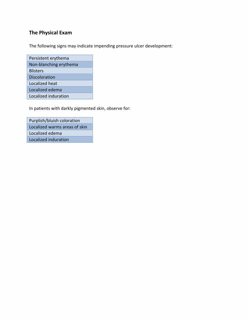
The Physical Exam The following signs may indicate impending pressure ulcer development: Persistent erythema Non‐blanching erythema Blisters Discoloration Localized heat Localized edema Localized induration In patients with darkly pigmented skin, observe for: Purplish/bluish coloration Localized warms areas of skin Localized edema Localized induration
Temperature, Moisture, Texture, and Edema Skin temperature can range from cool to warm. Warm is always normal. Note if the overall skin’s temperature is cool or warm, or if it is localized. Moisture Normally, your patient’s skin should be dry with only a slight amount of moisture. Overly moist skin may be due to environmental conditions, anxiety, obesity, hyperthyroidism, fever, or diaphoresis. Dry skin affects approximately 59% to 85% of person’s older than 64 years of age (Hess, 2008). Many factors contribute to dry skin, including a low‐humidity environment, the patient’s personal habits (smoking, alcohol intake, and poor nutrition), seasonal changes, chronic diseases, medications, and skin cleansers (Hess, 2010). Texture, Thickness, Turgor, and Mobility Inspect the skin for a normally smooth, mobile texture. You can check skin turgor by grasping the skin on the top of the hand and gently pulling up. After letting go of the skin, the skin should “snap” back into place within three seconds. Skin that remains elevated or “tented” may be due to age related changes, dehydration, or a combination of both. Edema When assessing edema it is useful to use an edema scale to guide your interpretation. This assessment is highly subjective and should be communicated at the patient’s bedside when possible so that each caregiver may interpret the degree of edema the same. Edema is often referred to as pitting or non‐pitting edema. Although clinicians commonly grade pitting edema from 1+ to 4+ (mild to severe), there is no agreed upon definition of these grades. However, this type of grading scheme may help an individual clinician record relative changes in edema in an individual patient (Rose, 2012).
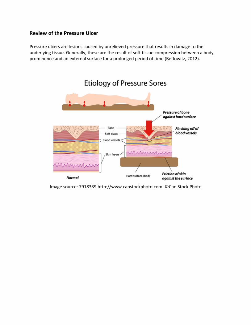
Review of the Pressure Ulcer Pressure ulcers are lesions caused by unrelieved pressure that results in damage to the underlying tissue. Generally, these are the result of soft tissue compression between a body prominence and an external surface for a prolonged period of time (Berlowitz, 2012).
Image source: 7918339 http://www.canstockphoto.com. ©Can Stock Photo
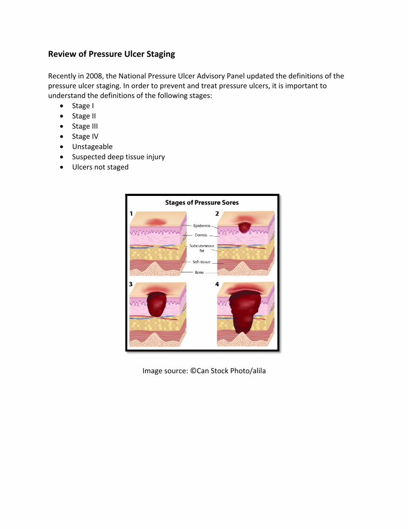
Review Recently pressureundersta
• St• St• St• St• U• S• U
of Pressu
in 2008, the ulcer staginand the defintage I tage II tage III tage IV Unstageable uspected deUlcers not sta
re Ulcer St
e National Prng. In order tnitions of the
eep tissue injaged
Ima
taging
ressure Ulceto prevent ae following s
jury
age source: ©
er Advisory Pnd treat prestages:
©Can Stock
Panel updateessure ulcers
Photo/alila
ed the defins, it is import
itions of thetant to
e
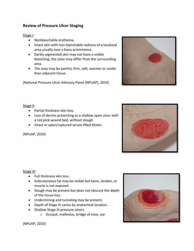
Review Stage I:
• N• In
a• D
ba
• Tth
(Nationa Stage II:
• P• Lo
a • In
(NPUAP, Stage III:
• F• S
m• S
o• U• D• S
(NPUAP,
of Pressu
Nonblanchabntact skin wirea usually oDarkly pigmelanching; threa. he area mayhan adjacent
l Pressure U
artial thicknoss of dermired pink wontact or ope
2010)
ull thicknessubcutaneoumuscle is notlough may bf the tissue
UnderminingDepth of Staghallow Stage
o Occip
2010)
re Ulcer St
ble erythemath non‐blanover a bony nted skin me color may
y be painful, t tissue.
lcer Advisor
ess skin lossis presentingound bed, wn/ruptured s
s skin loss. s fat may be exposed. be present bloss. and tunnelige III varies be III pressureut, malleolu
taging
a. chable rednprominenceay not have differ from
firm, soft, w
ry Panel [NP
s. g as a shallowithout slougserum‐filled
e visible but
ut does not
ing may be pby anatomice ulcers. s, bridge of
ess of a locae. a visible the surroun
warmer or co
UAP], 2010)
w open ulcegh. blister.
bone, tendo
obscure the
present. al location.
nose, ear
alized
nding
ooler
er with
on, or
e depth
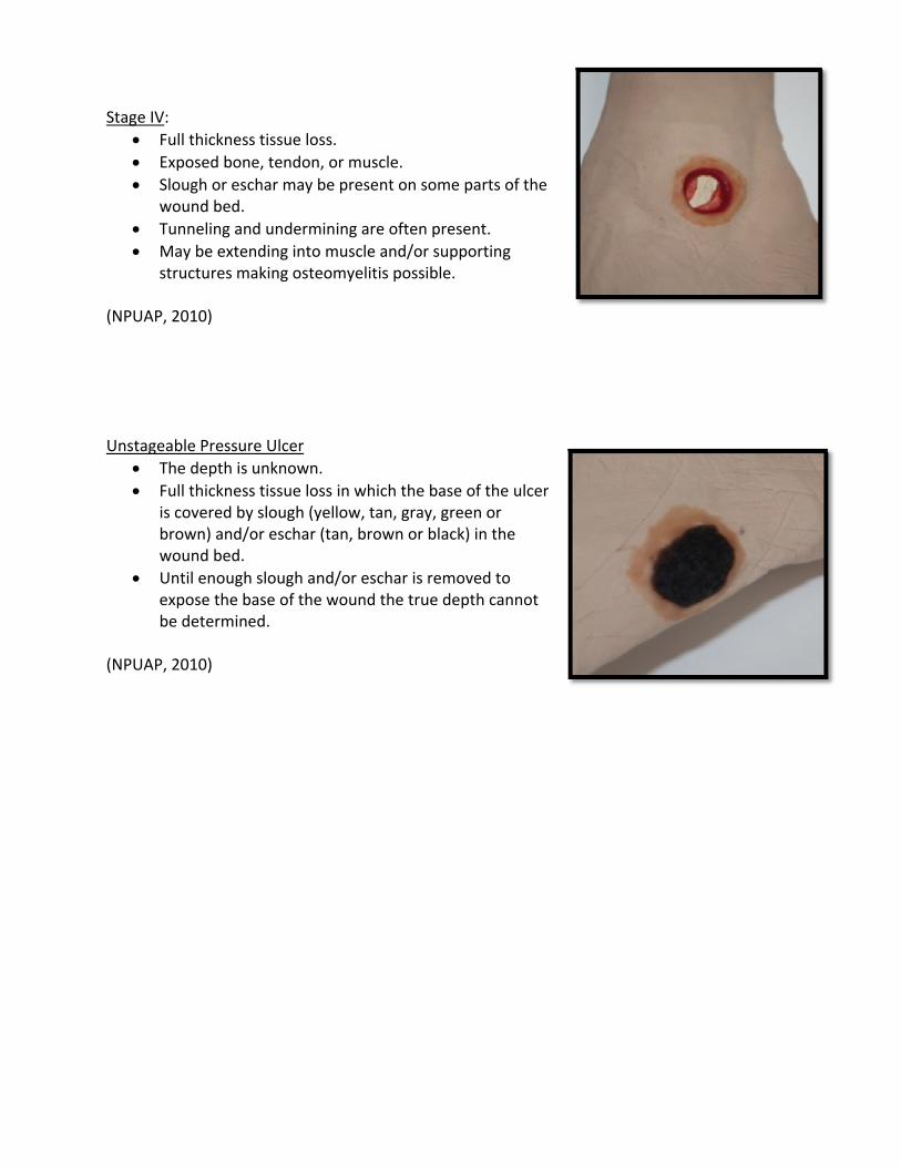
Stage IV:• F• Ex• S
w• T• M
st
(NPUAP, Unstagea
• T• F
isbw
• Uexb
(NPUAP,
ull thicknessxposed bonelough or escwound bed. unneling anMay be extentructures ma
2010)
able Pressurhe depth is uull thicknesss covered byrown) and/o
wound bed. Until enough xpose the bae determine
2010)
s tissue loss. e, tendon, ochar may be
d undermininding into making osteom
re Ulcer unknown. s tissue loss y slough (yellor eschar (ta
slough and/ase of the wed.
r muscle. present on s
ing are oftenmuscle and/omyelitis poss
in which thelow, tan, graan, brown or
/or eschar iswound the tr
some parts o
n present. or supportingsible.
e base of theay, green or r black) in th
s removed toue depth ca
of the
g
e ulcer
e
o nnot
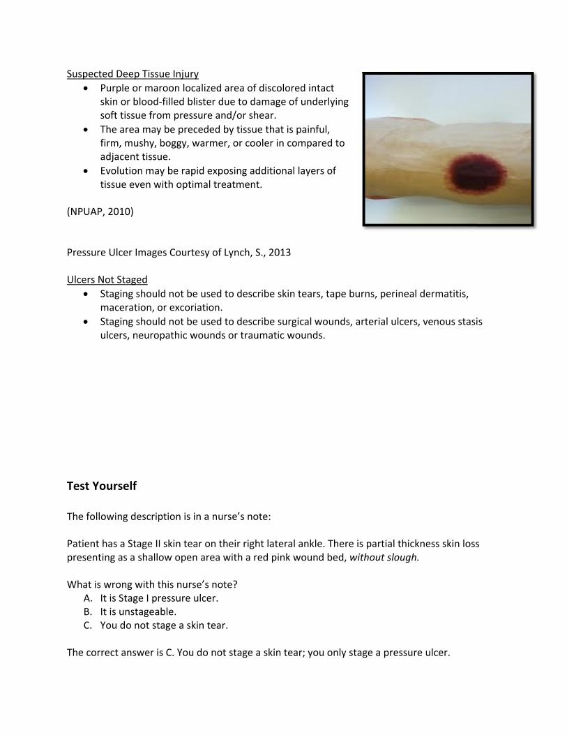
Suspecte• P
skso
• Tfia
• Evti
(NPUAP, Pressure Ulcers No
• Stm
• Stu
Test Yo The follo Patient hpresentin What is w
A. ItB. ItC. Y
The corre
ed Deep Tissurple or makin or bloodoft tissue frohe area mayrm, mushy, djacent tissuvolution massue even w
2010)
Ulcer Image
ot Staged taging shoulmaceration, otaging shoullcers, neuro
urself
wing descrip
has a Stage IIng as a shallo
wrong with tt is Stage I prt is unstageaou do not st
ect answer i
ue Injury roon localize‐filled blisteom pressurey be precedeboggy, warmue. y be rapid e
with optimal
es Courtesy
ld not be useor excoriatiold not be usepathic woun
ption is in a
I skin tear onow open are
this nurse’s ressure ulceble. tage a skin te
s C. You do n
ed area of dr due to dame and/or sheaed by tissue mer, or coole
xposing addtreatment.
of Lynch, S.,
ed to describon. ed to describnds or traum
nurse’s note
n their right ea with a red
note? r.
ear.
not stage a s
iscolored intmage of undar. that is painfer in compa
ditional layer
, 2013
be skin tears
be surgical wmatic wound
e:
lateral ankled pink woun
skin tear; yo
tact erlying
ful, red to
rs of
s, tape burns
wounds, artes.
e. There is pd bed, witho
u only stage
s, perineal d
erial ulcers, v
partial thicknout slough.
e a pressure
dermatitis,
venous stasi
ness skin loss
ulcer.
is
s
General Treatment Principles When a pressure ulcer develops, immediate actions will help prevent the worsening of the pressure ulcer. When a pressure ulcer is noted, there are six basic principles to think about to aid in healing besides the actual treatment of the wound with dressings or adjunctive therapies. The principles are:
1. Preventative 2. Monitoring 3. Healing scale 4. Pain control 5. Assessment and optimum nutrition status 6. Mattresses and tissue pressure relief
Prevention Prevention of pressure ulcers requires a multidisciplinary team approach. Together, the team is able to provide a holistic approach when caring for the patient and his/her pressure ulcer.
• All patients, regardless of whether or not they have a pressure ulcer, should have a change of position and good support to minimize tissue pressure. Pressure ulcers are most likely to occur in patients that experience sustained pressure over bony prominences.
• All patients should have a skin assessment, a risk assessment, education, and continuous evaluations in order to prevent pressure ulcers.
(Bryant & Nix, 2007) Monitoring A patient with a pressure ulcer should have the wound site monitored on a regular basis and documentation of:
1. The evaluation of the ulcer. 2. Integrity of the dressing, if present. 3. Condition of the skin surrounding the ulcer. 4. Presence of pain and adequacy of pain control. 5. Presence of possible complications such as infection.
(Berlowitz, 2013)
Genera Healing SAccordinin surface There are
• P• Se• W• P
PUSH T Accordinstage of thow the
Genera Pain ConPressure infection Treatmen
• O• O• T
Note!
l Treatmen
Scales g to Berlowie area, exte
e a few Healressure Soreessing Scale Wound Healiressure Ulce
ool
g to NPUAP the pressurepressure ulc
l Treatmen
trol ulcers can b
n, or breakdo
nt of pain inOral non‐opioOpioid analgeopical anest
• Once a Strecomme
nt Principl
itz (2013), thnt of necrot
ling Scales ue Status Too
ng Scale er Scale for H
(2010), the e ulcer, evencer is healing
nt Principl
be quite painown of the s
cludes: oids for mildesics for modthetics such
tage IV, alwaended.
es
he healing pic tissue and
sed: l (PSST)
Healing (PUS
PUSH tool isn as it heals. g.
es
nful for the purrounding
d pain derate to seas lidocaine
ays a Stage IV
rocess is desd exudates, a
SH)
s the most rYou can use
patient. The skin (Berlow
evere pain
V. Reverse s
scribed by scand the pres
eadily appliee tools like th
pain can bewitz, 2013).
taging as an
cales that casence of gran
ed. You nevehe PUSH too
e caused by i
ulcer heals
apture changnulation tiss
er change thol, to indicat
ischemia,
is not
ges sues.
he e
Patients Tolerance to Wound Care Pain:
Assess for pressure ulcer‐related pain in adults using a validated scale. Assessment of pain should include an assessment of body language and nonverbal cues. Wound pain can indicate deterioration, infection, or even inappropriate wound treatments.
Optimize pressure ulcer care to ensure that it is coordinated with pain medication administration.
Pain should be measured/rated prior to each dressing change and post‐dressing change. This is to determine if pain intervention given before the dressing change was sufficient.
(NPUAP, 2010) General Treatment Principles: Nutritional Status It is important to optimize protein and total caloric intake (especially with Stage III and Stage IV ulcers) since patients with pressure ulcers are in a chronic catabolic state. A nutrition consult should be done on these patients and all nutritional deficiencies should be corrected. Corrections could be enteral or parenteral nutrition, increase protein intake, vitamin C supplementation, and zinc supplementation (Little, 2012). Studies have shown that diets with higher protein and calories for patients with pressure ulcers has improved healing over a 12 week period versus those receiving lower limits of the recommended amounts (Van Anholt, et al., 2010). You should only correct nutritional deficiencies on patients who are deficient. Previous practice was to give routine supplements. Routine supplementation with vitamins A, E, or C, zinc, copper, or iron is no longer recommended (Little, 2012).
Test Yourself If your patient has a pressure ulcer but no nutritional deficiency, it is still a good practice to give the patient extra vitamins, zinc, and iron.
A. True B. False
The correct answer is false. Routine supplementation with vitamins A, E, or C, zinc, copper, or iron is not recommended (Little, 2012).
Genera The final supportiv Mattress
• S“alo
• Exre
• C• A
o• Li• Id
hu
• P• U
a• Li
(NPUAP,
Note!
l Treatmen
section of gve surfaces a
ses: upportive sua specializedoads, microcxamples of seplacement hoose a sup
All individualsn a regular bimit the amodeally heels eel which mse of a pressrolonged sit
Use a pressurre at risk forimit time an
2010)
• Donut‐typarea caus
nt Principl
general treatand tissue p
urfaces are dd device for pclimate, and/support surfor overlay, o
pport surfaces at risk for pbasis when aount of linenshould be “f
makes it a chasure‐redistritting results re‐redistribur pressure ul individual s
pe devices osing more da
es: Mattre
tment princiressure relie
defined by Npressure red/or other thefaces includeor seat cushe compatiblepressure ulca support sun between thfloated” off tallenge to tribution devicin a higher rution seat culcer developspends in a c
or rings haveamage than g
esses and T
ples is the def by redistri
National Presdistribution derapeutic fune: mattress, ion or seat ce with the pacers should crface is in plhe individuathe bed surfy to redistribce (Lyman, 2risk of pressuushion for inpment. chair withou
e been showgood.
Tissue Pre
discussion ofibution and
ssure Ulcer Adesigned for nctions.” integrated bcushion overatient’s needcontinue to blace. l and the suface due thebute the loa2009). ure ulcer devdividuals wh
t pressure re
n to cause is
ssure Relie
f proper matrepositionin
Advisory Panr manageme
bed system, rlay. ds. be turned an
pport surface small surfaad from the h
velopment. hose mobilit
elief.
schemia ove
ef
ttresses or ng.
nel (NPUAP) ent of tissue
mattress
nd reposition
ce. ce area of thheel through
y is reduced
r the pressu
as
ned
he h the
d and
ure
General Treatment Principles: Mattresses and Tissue Pressure Relief Pressure Redistribution/Repositioning: Immobility is the most significant risk factor for pressure ulcer development. Patients with any degree of immobility should be closely monitored for pressure ulcer development. Repositioning involves a change in position of the lying or seated person in order to redistribute pressure, therefore, enhancing comfort. This should be done at regular intervals.
• Failure to reposition will result in tissue ischemia and probable tissue damage. • Frequency of repositioning will depend on patients activity/mobility level, patients
tissue tolerance to pressure, and patients overall skin and medical condition. • Avoid positioning directly onto medical devices such as tubes or drains. • Avoid positioning on bony prominences with existing pressure ulcers. • Repositioning should be at a 30° tilted side‐lying position. Avoid positions that increase
pressure such as 90° side‐lying position or semi‐recumbent position. • Use transfer aides to reduce friction and shear.
o LIFT, don’t drag while repositioning. (NPUAP, 2010) Goals for Wound Healing Once a pressure ulcer is noted, there are goals for proper wound healing of the pressure ulcer. They include:
Prevent infection Proper cleansing Remove nonviable tissue Proper moisture levels Eliminate dead space Odor control Minimize pain Protection of wound and periwound
skin (Byrant & Nix, 2007)
Goals for Wound Healing: Prevent Infection The first goal for wound healing is the prevention and management of infection:
• Infection is a common cause of wound chronicity: o Wound infection requires prompt intervention and aggressive treatment. o While initiation of systemic antibiotics is indicated for infections involving the
bone or soft tissue it is not necessary to decrease the bacteria bioburden at the tissue level. Keeping this in mind will prevent unnecessary bacterial resistance.
• Steps to take to avoid wound infections: 1. Cover the wound to protect from outside contaminants. 2. Infection control precautions. 3. Antimicrobials when indicated.
• If infection is suspected, wound culture should be obtained. • Caveats of performing a wound culture:
1. ALWAYS obtain all cultures before administering any antibiotics. 2. Obtain the culture from healthy tissue. 3. Collect the specimen using sterile technique. 4. Be careful not to contaminate the specimen when placing it in the sterile
container. 5. Appropriately cleanse the wound prior to obtaining the culture.
(Bryant & Nix, 2007) Did You Know?
• Wound contamination is the presence of bacteria on wound surfaces with no multiplication of bacteria.
• Wound colonization refers to the presence of replicating bacteria without clinical signs and symptoms of infection.
• Infection is the presence of microorganisms that invade the tissue and there is a systemic response to the invasion.
(Bryant & Nix, 2007)
Goals for Wound Healing: Proper Cleansing The second goal of wound healing is to properly clean the pressure ulcer to encourage wound healing:
• The goal of wound cleansing is to remove bacteria and debris from wound bed while at the same time preserving and protecting healthy granulation tissue.
• Always clean a wound prior to the application of any dressing. • Cleanse wound with non‐cytotoxic cleanser. The most commonly used wound cleanser
is normal saline. Normal saline provides a moist wound environment, promotes granulation tissue formation, and causes minimal fluid shifts in healthy cells.
• Research indicates that the optimum pressure for wound cleaning is between 5 and 15 psi (Baranoski & Ayello, 2012).
• Skin cleansers (used for bathing or incontinence care) should never be used as a wound cleanser due to their toxicity. Skin cleansers are formulated to breakdown the bond between fecal matter and the skin which are stronger and more toxic than wound cleansers.
• Acetic acid, hydrogen peroxide, and sodium hypochlorite (i.e. Dakins Solution) should be avoided. These solutions may damage tissue and delay healing.
(Hess, 2005) Goals for Wound Healing: Remove Nonviable Tissue The third goal of wound healing is to remove nonviable tissue.
• It is important to examine the patients’ individual needs to determine the most appropriate debridement intervention.
• The methods of debridement should be consistent with the patients overall goals. • There are different types of wound debridement.
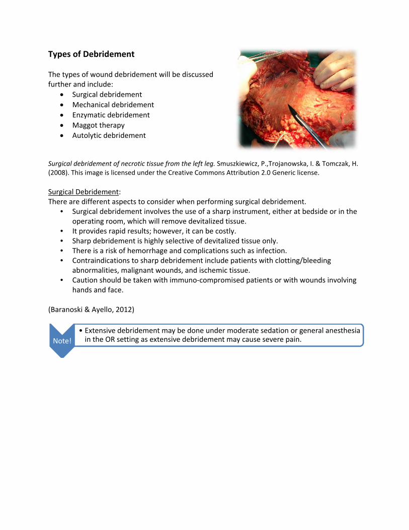
Types o The typefurther a
• S• M• E• M• A
Surgical d(2008). Th
Surgical DThere are
• So
• It• S• T• C
a• C
h (Baranos
Note!
of Debridem
s of wound nd include: urgical debrMechanical dnzymatic deMaggot theraAutolytic deb
debridement ohis image is lic
Debridemene different aurgical debrperating root provides raharp debridehere is a riskontraindicatbnormalitiesaution shouands and fac
ski & Ayello,
• Extensivein the OR
ment
debridemen idement debridementebridement apy bridement
of necrotic tiscensed under
nt: aspects to coidement invom, which wpid results; ement is higk of hemorrhtions to shars, malignantld be taken ce.
2012)
e debrideme setting as e
nt will be dis
t
ssue from the r the Creative
onsider whenvolves the uswill remove dhowever, it ghly selectivehage and corp debridemt wounds, anwith immun
nt may be dextensive deb
cussed
left leg. Smue Commons A
n performinse of a sharpdevitalized tican be costle of devitalizmplications
ment includend ischemic tno‐comprom
one under mbridement m
szkiewicz, P.,Attribution 2.0
g surgical dep instrumentssue. ly. zed tissue onsuch as infepatients wittissue.
mised patient
moderate semay cause se
,Trojanowska0 Generic lice
ebridement.t, either at b
nly. ection. th clotting/b
ts or with wo
dation or geevere pain.
a, I. & Tomczaense.
edside or in
bleeding
ounds involv
eneral anesth
ak, H.
the
ving
hesia
Types of Debridement Mechanical Debridement: Mechanical debridement involves the use of physical forces to remove necrotic tissue.
• A wet‐to‐dry dressing involves moist dressings applied to wound and allowed to dry. When the dressing is removed with force, tissue is removed with the dressing. It can be used with non‐surgical candidates; however, it is not cost‐effective if frequent dressing changes are required. These dressings may macerate the periwound skin and may cause bleeding. Most importantly, these dressing changes are often PAINFUL. Please note these dressing changes are not selective and the exposed healthy tissue will be debrided and slow the healing process.
• Wound irrigation allows removal of debris mechanically through pressurized fluids. This can be done using high‐pressured irrigation or pulsatile lavage. It is important to remember that personal protective equipment is necessary.
o High‐pressure irrigation can be obtained using a 35 mL syringe and a 19 gauge angio‐catheter. This provides enough pressure to remove wound debris without damaging the healthy tissue.
o Pulsatile lavage is obtained using machinery that provides intermittent high‐pressure irrigation combined with suction to remove the irrigant and debris. The apparatus allows adjustment of pressure to higher levels to remove debris.
• Whirlpool bath is a process by which movement of water dislodges loose debris. This may macerate the periwound skin, cause trauma to wound bed, and may lead to bacterial contamination of wound bed. This process can be labor intensive and can be time consuming. This form of debridement, however, will increase circulation to the wound bed.
(Bryant & Nix, 2007; Baranoski & Ayello, 2012) Enzymatic Debridement: The next type of debridement is the enzymatic debridement.
• This is a form of debridement where enzymes applied to the wound bed degrade and remove necrotic tissue. The enzymes will digest and dissolve the necrotic tissue in the wound bed by breaking down the wound matrix.
• This is an ideal option for patients who are non‐surgical candidates. • It is important to remember that the wound bed needs to be crosshatched or scored in
order to facilitate the effectiveness of the enzyme and facilitate local penetration of the enzymatic agent.
(Baranoski & Ayello, 2012)
Types o Maggot T
• Sed
• Tim
• Inaso
(Sherman
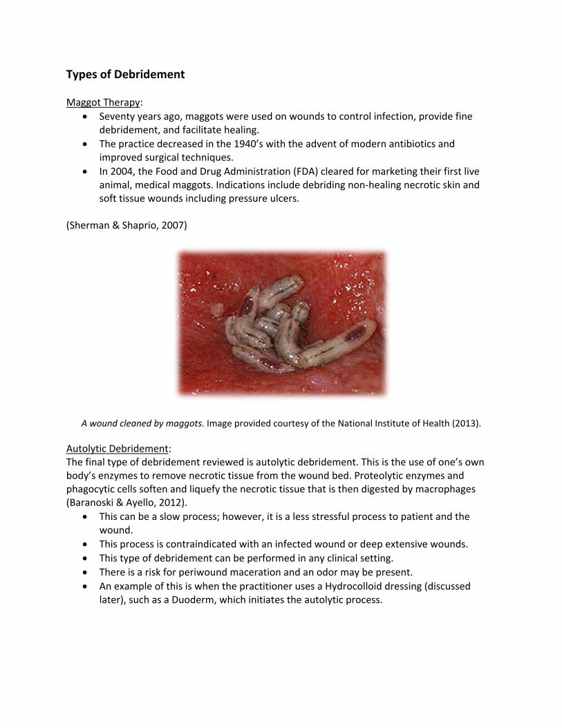
A wou
AutolyticThe final body’s enphagocyt(Baranos
• Tw
• T• T• T• A
la
of Debridem
Therapy: eventy yearsebridementhe practice dmproved surn 2004, the Fnimal, medioft tissue wo
n & Shaprio,
und cleaned b
c Debridemetype of debnzymes to retic cells softeski & Ayello, his can be a wound. his process ihis type of dhere is a riskAn example oater), such as
ment
s ago, maggo, and facilitadecreased inrgical techniqFood and Drcal maggotsounds includ
, 2007)
by maggots. Im
ent: bridement reemove necroen and lique2012). slow proces
is contraindidebridementk for periwoof this is whes a Duoderm
ots were useate healing. n the 1940’sques. ug Administ. Indicationsding pressure
mage provide
eviewed is auotic tissue frefy the necro
ss; however,
icated with at can be perfund maceraen the practm, which init
ed on wound
with the ad
tration (FDAs include debe ulcers.
ed courtesy o
utolytic debrrom the wouotic tissue th
, it is a less s
an infected wformed in antion and an itioner uses iates the au
ds to contro
dvent of mod
) cleared forbriding non‐
of the Nationa
ridement. Thund bed. Prohat is then di
stressful proc
wound or deny clinical seodor may ba Hydrocolltolytic proce
ol infection, p
dern antibio
r marketing t‐healing necr
al Institute of
his is the useoteolytic enzigested by m
cess to patie
eep extensivetting. e present. oid dressingess.
provide fine
tics and
their first livrotic skin an
Health (2013
e of one’s owzymes and macrophages
ent and the
ve wounds.
g (discussed
ve nd
3).
wn
s
Test Yo Which of
A. Ud
B. EC. UD. U
The correnecrotic Goals fo The fourt
• Fomba
• Tas
• Mah
(Baranos
Note!
urself
f the followiUse of a sharevitalized tisnzymes appUse of physicUse of one’s o
ect answer itissue.
or Wound
th goal for wrom researcf moist wou
migrate acrosurrow downnd heal the he selectionssist in prevModerate to bsorptive prealing.
ski & Ayello,
• Desiccat• Macerat
ng is the prop instrumenssue. lied to the wcal force to rown bodies
s C. Mechan
Healing: M
wound healinch in the 196nd healing (ss the wounn underneatwound. n of an approenting tissuelarge amounroperties. Th
2012)
ion = Extremion = Excess
oper definitiont, either at b
wound bed demove necrenzymes to
nical debride
Moisture
ng is the ma60’s, healthcBaranoski &d surface, mh the wound
opriate dresse desiccationnts of drainahis moisture
me drynesssive moisture
on of mechabedside or in
degrade androtic tissue.remove nec
ement is the
intenance oare provider
& Arello, 201moisture is ned bed to find
sing that man or maceraage from thebalance is im
e
anical debridn the operat
remove nec
crotic tissue
use of phys
f appropriatrs began to u2). In order eeded. Withd a moist are
aintains a motion. e wound wilmperative to
dement? ting room th
crotic tissue
from the wo
ical force to
te level of munderstand for epitheliahout moisturea for them t
oist wound e
l require a do facilitate ti
hat will remo
.
ound bed.
remove
oisture. the importaal cells to re, the cells mto move acr
environment
ressing withimely wound
ove
ance
must ross
t will
h d
Goals fo The fifth gauze dre
• Ao
• Tsp
• Itp
(Bryant & Goals fo The sixthneed to c
• Dw
• T• It• D
Note!
or Wound
goal of wouessings, andAny wound wf the woundo prevent thpace of the wt is importanromote form
& Nix, 2007)
or Wound
h goal for procleanse the wDevitalized/nwhen this scehe utilizatiot is importanDressings tha
!
• Pseudcausesand th
Healing: E
und healing i filler dressi
with depth md will enablehe open spacwound. Thisnt to ensure mation of gra
Healing: C
oper wound wound withnecrotic tissuent is presenn of charcoant to ensure at are satura
omonas aers wound infehe color of th
Eliminate D
s the eliminngs can all b
must be filled the formatice, fluff the s will facilitatthe dressinganulation tis
Control Od
healing is th each dressiue will produnt. al dressings wfrequent dreted with dra
ruginosa is oection. This he drainage
Dead Spac
ation of the be utilized tod with the drion of an absdressing pacte the healing edges comssue.
dor
he controllinng change. uce a malodo
will neutralizessing changainage will a
ne of the mobacteria wilwill be a blu
e
dead spaceo fill this dearessing. Opscess. cking and cong process.e in contact
ng of odor. T
orous scent.
ze the odor.ge to controlso produce
ost commonl produce a ue/green sha
. Absorptivead (or emptyen spaces w
ompletely fill
t with the wo
The healthca
. It is import
ol odor. an odor.
n gram negatfruity odor tade.
e dressings, y) space. within the de
l the entire
ound edges
re provider
ant to debri
tive bacteriato the woun
epth
to
will
de
a that d drainage
Goals for Wound Healing: Minimize Pain The seventh goal of proper wound healing is to address and minimize pain. This was addressed briefly in the general treatment principles section.
• Assess the patient for pain before, during, and after dressing changes. • Provide analgesia 30‐60 minutes before dressing change. • If patient’s dressing has dried out, thoroughly soak the dressing prior to removal. • Attempt to use non adherent dressings. • Avoid wet to dry dressings. • It is helpful to use dressings that require fewer frequency changes. • Pain assessment/medication prior to dressing changes is imperative. Be aware of
patient’s current status of pain. • Know your patients pain triggers and avoid whenever possible. • Avoid unnecessary manipulation of the wound during the dressing change. • Consider temperature of wound product before applying it to the wound (i.e. wound
gels can feel cold in the wound bed which is uncomfortable to some patients). • Be mindful of positioning the patient in a comfortable position during the dressing
change. • After procedure assess for patient comfort and adjust treatment regimen and dressing
change appropriately. Goals for Wound Healing: Protect the Wound and Periwound Skin The eighth (and final) goal for proper wound healing is the protection of the wound and periwound skin.
• Skin sealants, ointments, or moisture barriers should be used to protect the periwound skin from moisture and adhesives.
• Appropriate intervals of dressing changes should be followed to prevent pooling of wound drainage on periwound skin.
• With each dressing change, the periwound skin should be evaluated to assess the effectiveness of the current dressing to ensure that it is protecting the periwound skin.
(Bryant & Nix, 2007)
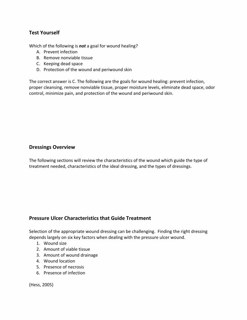
Test Yourself Which of the following is not a goal for wound healing?
A. Prevent infection B. Remove nonviable tissue C. Keeping dead space D. Protection of the wound and periwound skin
The correct answer is C. The following are the goals for wound healing: prevent infection, proper cleansing, remove nonviable tissue, proper moisture levels, eliminate dead space, odor control, minimize pain, and protection of the wound and periwound skin. Dressings Overview The following sections will review the characteristics of the wound which guide the type of treatment needed, characteristics of the ideal dressing, and the types of dressings. Pressure Ulcer Characteristics that Guide Treatment Selection of the appropriate wound dressing can be challenging. Finding the right dressing depends largely on six key factors when dealing with the pressure ulcer wound.
1. Wound size 2. Amount of viable tissue 3. Amount of wound drainage 4. Wound location 5. Presence of necrosis 6. Presence of infection
(Hess, 2005)
Characteristics of an Ideal Dressing There are important aspects to the ideal dressing that the healthcare provider should consider when assessing, placing, or removing a dressing. Most importantly, the dressing should maintain a moist wound environment to facilitate healing. Some other characteristics include:
• The dressing should be conformable for the range of use needed (i.e. to fill sinus tracts or tunnels).
• It should come in numerous size and shapes. • It should be absorbent. • The dressing needs to act as a barrier to wound contaminants. • Ideally it should be accessible. • It is important that the dressing assists with reducing pain/discomfort at the site of the
wound. • The dressing should be easy to remove from the wound bed. • The healthcare provider should assess the dressing characteristics and make sure it
aligns with the wound characteristics. • The amount of drainage that the wound produces should match the amount of
absorbency level of the dressing. • The appearance of the dressing should be assessed with each dressing change. If the
dressing is dry, then the dressing can be left in place longer to prevent drying out of the wound bed. If the dressing is saturated, the dressing should be changed at more frequent intervals.
• If the wound has depth, the dressing must be able to fill the entire depth of the wound. • If the wound is shallow, the dressing must not be too bulky and add extra pressure to
the wound bed. • If the periwound skin is fragile, the dressing adhesive must not be too powerful and
cause damage to the periwound skin. • If the wound bed is odorous, the dressing should contain a charcoal component or be
occlusive to the wound bed.
(Baranoski & Ayello, 2012)
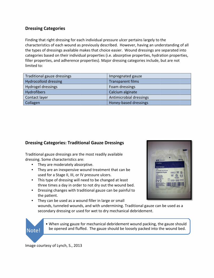
Dressin Finding tcharactethe typescategoriefiller prolimited to TraditionHydrocolHydrogelHydrofibContact lCollagen Dressin Traditiondressing.
• T• T
u• T
th• D
th• T
wse
Image co
Note!
g Categori
hat right dreristics of eacs of dressinges based on perties, ando:
nal gauze drelloid dressinl dressings ers layer
g Categori
nal gauze dre. Some charahey are modhey are an insed for a Stahis type of dhree times aDressing chanhe patient. hey can be uwounds, tunnecondary dr
ourtesy of Ly
• When be ope
ies
essing for each wound asgs available mtheir individ adherence
essings g
ies: Traditi
essings are tacteristics arderately absnexpensive wage II, III, or dressing will a day in ordenges with tra
used as a woneled woundessing or us
ynch, S., 201
using gauze ened and fluf
ach individuas previously makes that cdual propertproperties).
ional Gauz
the most reare: orptive. wound treatIV pressure need to be
er to not dry aditional gau
ound filler inds, and with ed for wet t
3
for mechanffed. The ga
al pressure udescribed. Hchoice easieies (i.e. abso Major dress
ImpregTranspFoam dCalciumAntimiHoney
ze Dressing
adily availabl
tment that culcers. changed at lout the wouuze can be p
n large or smunderminino dry mecha
ical debrideauze should
ulcer pertainHowever, har. Wound dorptive propsing categor
gnated gauzeparent filmsdressings m alginate crobial dres‐based dress
gs
le
can be
least und bed. painful to
mall ng. Traditionanical debrid
ment woundbe loosely p
ns largely to aving an undressings areerties, hydraries include,
e
ssings sings
al gauze candement.
d packing, thacked into t
the derstanding e separated iation properbut are not
n be used as
he gauze shohe wound b
of all nto rties,
a
ould bed.
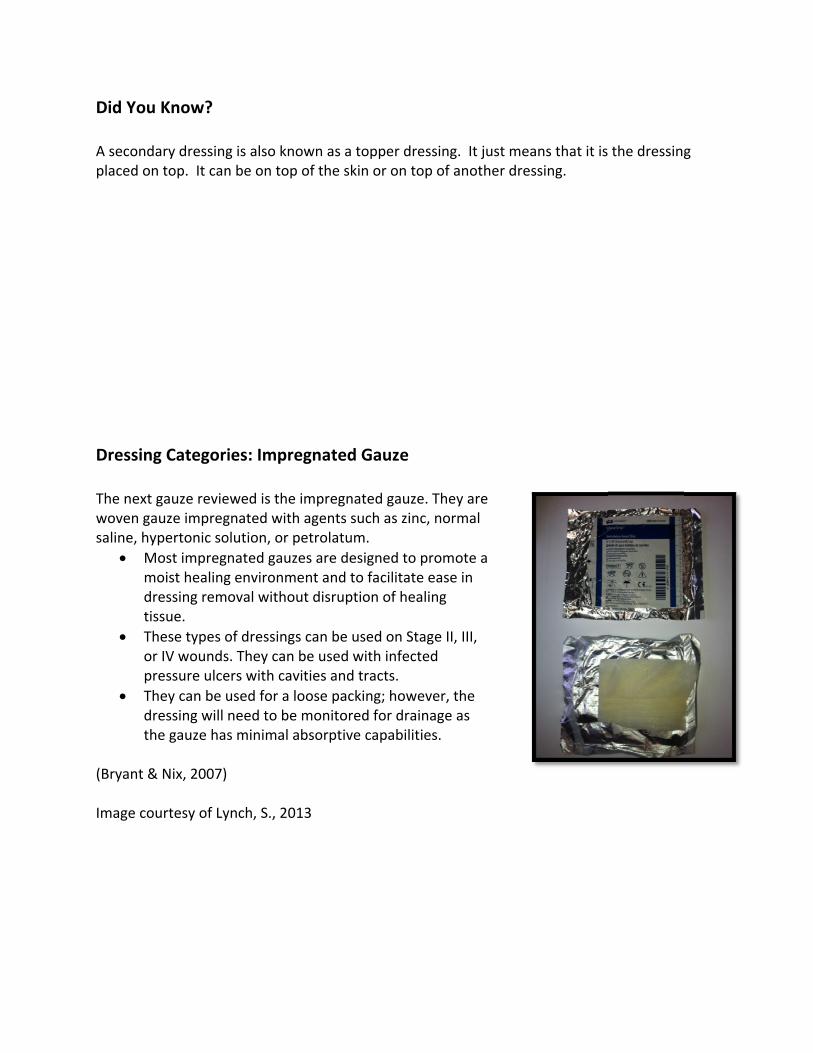
Did You A secondplaced on Dressin The nextwoven gasaline, hy
• Mmdti
• Top
• Tdth
(Bryant & Image co
u Know?
dary dressingn top. It can
g Categori
gauze revieauze impregypertonic soMost impregnmoist healingressing remssue. hese types or IV woundsressure ulcehey can be uressing will he gauze has
& Nix, 2007)
ourtesy of Ly
g is also known be on top o
ies: Impreg
ewed is the ignated with olution, or penated gauzeg environmeoval without
of dressings s. They can bers with cavitused for a loneed to be ms minimal ab
ynch, S., 201
wn as a toppof the skin o
gnated Ga
mpregnatedagents such etrolatum. es are designnt and to fat disruption
can be usedbe used withties and tracoose packingmonitored fobsorptive ca
3
per dressingor on top of a
auze
d gauze. Theas zinc, nor
ned to promocilitate easeof healing
d on Stage II, infected cts. g; however, tor drainage pabilities.
g. It just meaanother dres
y are mal
ote a e in
, III,
the as
ans that it is ssing.
the dressingg
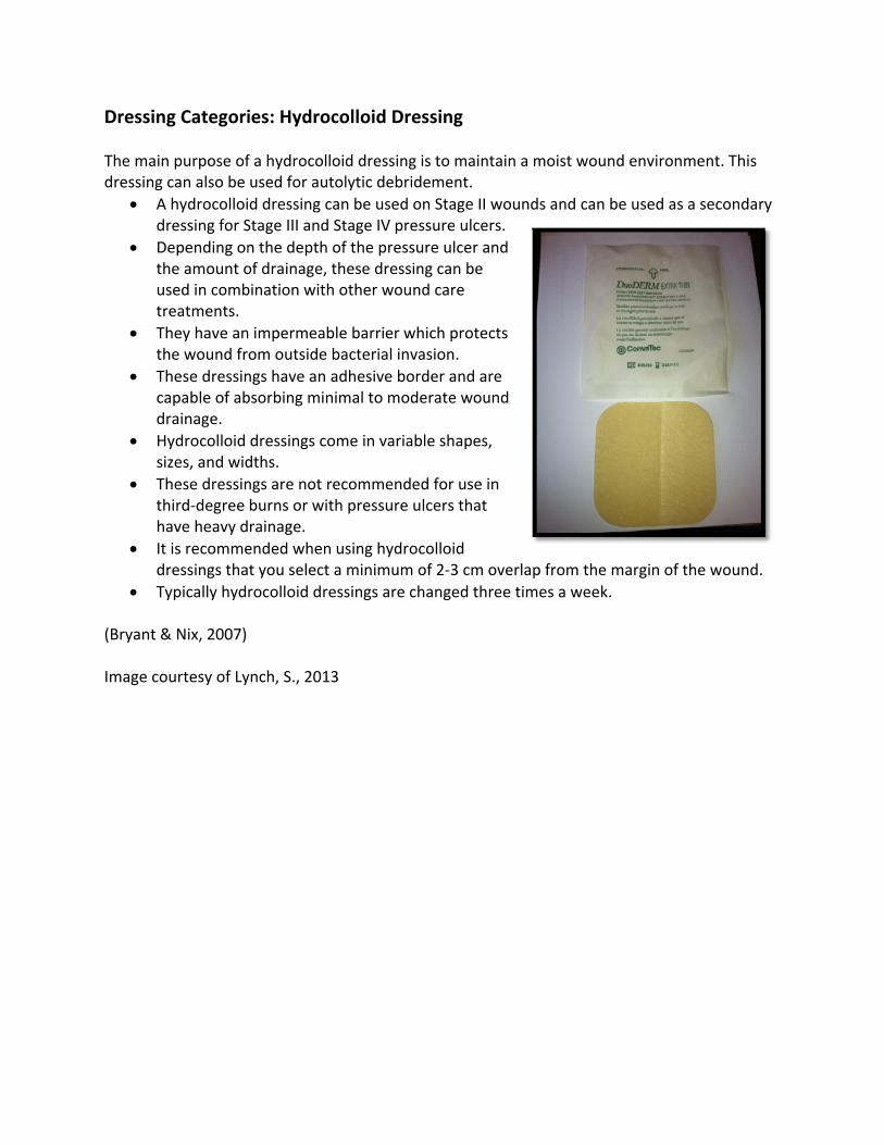
Dressin The maindressing
• Ad
• Dthutr
• Tth
• Tcad
• Hsi
• Tthh
• Itd
• Ty (Bryant & Image co
g Categori
n purpose ofcan also be
A hydrocolloiressing for S
Depending onhe amount osed in combreatments. hey have anhe wound frhese dressinapable of abrainage.
Hydrocolloid izes, and widhese dressinhird‐degree ave heavy dt is recommeressings thaypically hyd
& Nix, 2007)
ourtesy of Ly
ies: Hydro
f a hydrocollused for autd dressing cStage III and n the depth of drainage, bination with
n impermeabom outside ngs have an bsorbing min
dressings codths. ngs are not rburns or witrainage. ended whent you select rocolloid dre
ynch, S., 201
colloid Dre
oid dressingtolytic debrican be used oStage IV preof the pressthese dressih other wou
ble barrier wbacterial invadhesive bonimal to mod
ome in varia
recommendeth pressure
using hydroa minimumessings are c
3
essing
g is to maintadement. on Stage II wessure ulcerssure ulcer aning can be nd care
which protectvasion. order and arederate woun
ble shapes,
ed for use inulcers that
ocolloid of 2‐3 cm ochanged thre
ain a moist w
wounds and s. nd
ts
e nd
n
overlap fromee times a w
wound envir
can be used
the margin week.
ronment. Th
d as a second
of the woun
his
dary
nd.
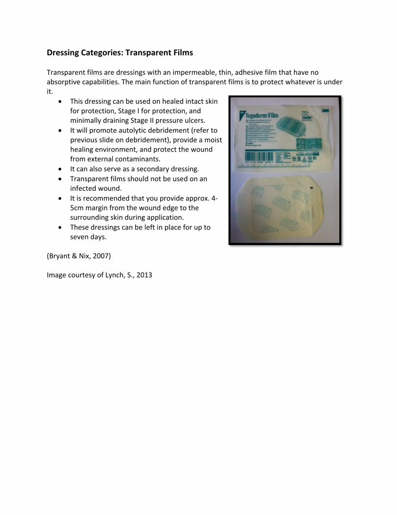
Dressin Transparabsorptivit.
• Tfom
• Itphfr
• It• T
in• It
5su
• Tse
(Bryant & Image co
g Categori
rent films areve capabilitie
his dressing or protectionminimally drat will promotrevious slideealing envirorom externat can also serransparent fnfected wout is recommecm margin furrounding shese dressineven days.
& Nix, 2007)
ourtesy of Ly
ies: Transp
e dressings wes. The main
can be usedn, Stage I foraining Stage te autolytic de on debrideonment, andl contaminarve as a secofilms shouldnd. ended that yfrom the woskin during angs can be le
ynch, S., 201
parent Film
with an impen function of
d on healed r protection,II pressure udebridemenement), provd protect thents. ondary dress not be used
you provide und edge toapplication. eft in place f
3
ms
ermeable, thf transparen
intact skin , and ulcers. nt (refer to vide a moist e wound
sing. d on an
approx. 4‐o the or up to
hin, adhesivent films is to
e film that hprotect wha
ave no atever is undder
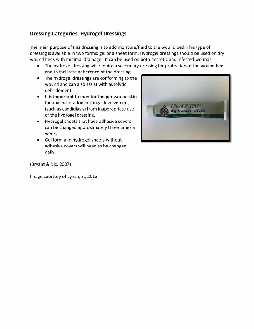
Dressin The maindressing wound b
• Ta
• Twd
• Itfo(so
• Hcaw
• Gad
(Bryant & Image co
g Categori
n purpose ofis available eds with mihe hydrogelnd to facilitahe hydrogelwound and caebridementt is importanor any macesuch as candf the hydrog
Hydrogel shean be changweek. Gel form anddhesive coveaily.
& Nix, 2007)
ourtesy of Ly
ies: Hydro
f this dressinin two formnimal draina dressing wiate adherenc dressings aan also assis. nt to monitoration or fundidiasis) fromgel dressing.ets that havged approxim
hydrogel shers will need
ynch, S., 201
gel Dressin
ng is to add ms; gel or a shage. It can bll require a sce of the drere conformist with autol
r the periwongal involvemm inappropri ve adhesive cmately three
heets withoud to be chan
3
ngs
moisture/fluheet form. Hbe used on bsecondary dessing. ng to the ytic
ound skin ment iate use
covers times a
ut ged
uid to the woHydrogel dreboth necroticressing for p
ound bed. Thssings shoulc and infecteprotection o
his type of d be used oed wounds. f the wound
n dry
d bed
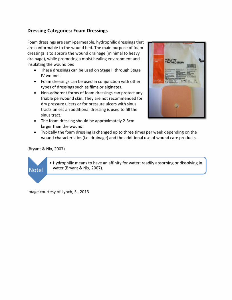
Dressin Foam dreare confodressingsdrainageinsulating
• TIV
• Foty
• Nfrdtrsi
• Tla
• Tyw
(Bryant &
Image co
Note!
g Categori
essings are sormable to ts is to absorb), while promg the woundhese dressinV wounds. oam dressinypes of dressNon‐adherenriable periwory pressure racts unless inus tract. he foam drearger than thypically the wound chara
& Nix, 2007)
ourtesy of Ly
• Hydropwater (
ies: Foam
semi‐permeathe wound bb the woundmoting a mod bed. ngs can be u
ngs can be ussings such ant forms of foound skin. Tulcers or foran additiona
essing shouldhe wound. foam dressicteristics (i.e
ynch, S., 201
philic means(Bryant & Ni
Dressings
able, hydropbed. The maid drainage (moist healing e
sed on Stage
sed in conjus films or algoam dressinhey are not r pressure ulal dressing is
d be approxi
ng is changee. drainage)
3
s to have an ix, 2007).
philic dressinin purpose ominimal to henvironmen
e II through
nction with ginates. gs can proterecommendlcers with sins used to fill
imately 2‐3c
ed up to threand the add
affinity for w
ngs that of foam heavy t and
Stage
other
ect any ded for nus the
cm
ee times perditional use o
water; readil
r week depeof wound ca
y absorbing
nding on theare products
or dissolvin
e s.
g in
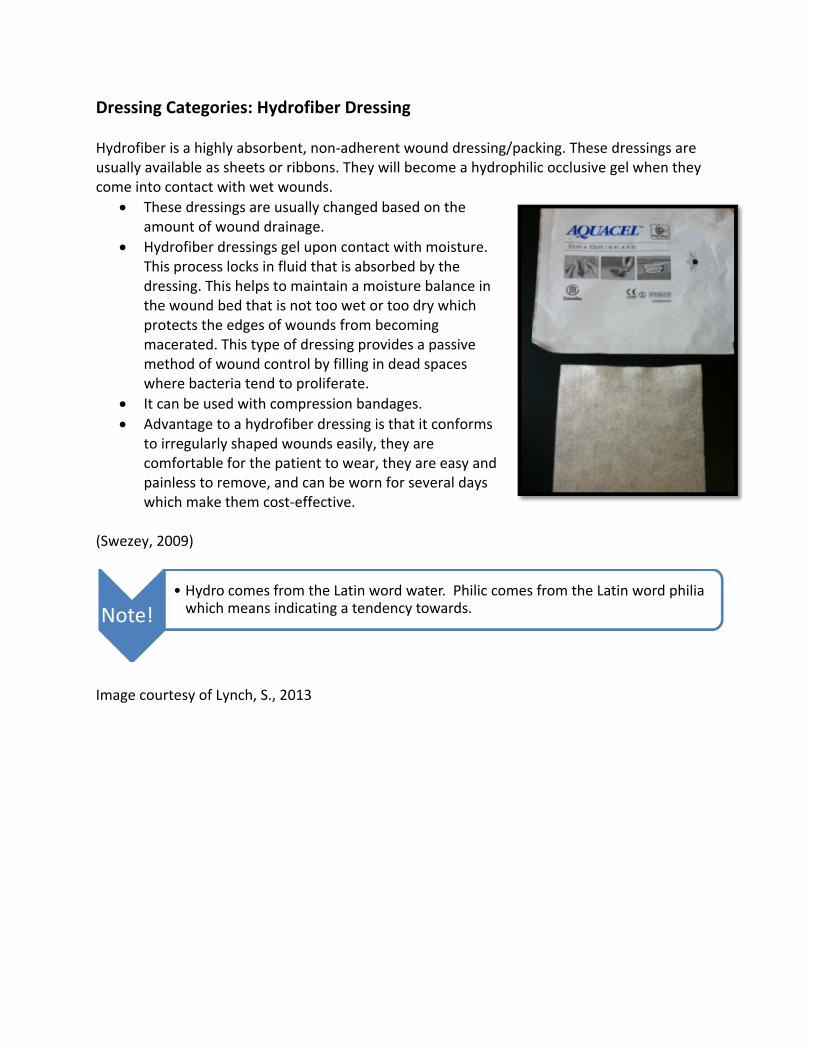
Dressin Hydrofibusually acome int
• Ta
• HTdthpmmw
• It• A
tocopw
(Swezey,
Image co
Note!
g Categori
er is a highlyvailable as sto contact whese dressinmount of woHydrofiber drhis process lressing. Thishe wound berotects the e
macerated. Tmethod of wowhere bactert can be usedAdvantage too irregularly omfortable fainless to re
which make t
2009)
ourtesy of Ly
• Hydro which
ies: Hydro
y absorbent,sheets or ribith wet woungs are usuaound drainaressings gel locks in fluids helps to maed that is noedges of woThis type of dound controria tend to pd with compo a hydrofibeshaped woufor the patieemove, and cthem cost‐ef
ynch, S., 201
comes frommeans indic
fiber Dres
, non‐adherebons. They wunds. lly changed ge. upon contacd that is absoaintain a moot too wet orunds from bdressing prool by filling inroliferate. pression baner dressing isunds easily, ent to wear, can be wornffective.
3
the Latin wcating a tend
sing
ent wound dwill become
based on th
ct with moisorbed by theoisture balanr too dry whbecoming vides a passn dead space
dages. s that it confthey are they are eas
n for several
ord water. Pdency toward
dressing/pac a hydrophil
he
ture. e nce in ich
ive es
forms
sy and days
Philic comesds.
cking. These ic occlusive
s from the La
dressings argel when th
atin word ph
re hey
hilia
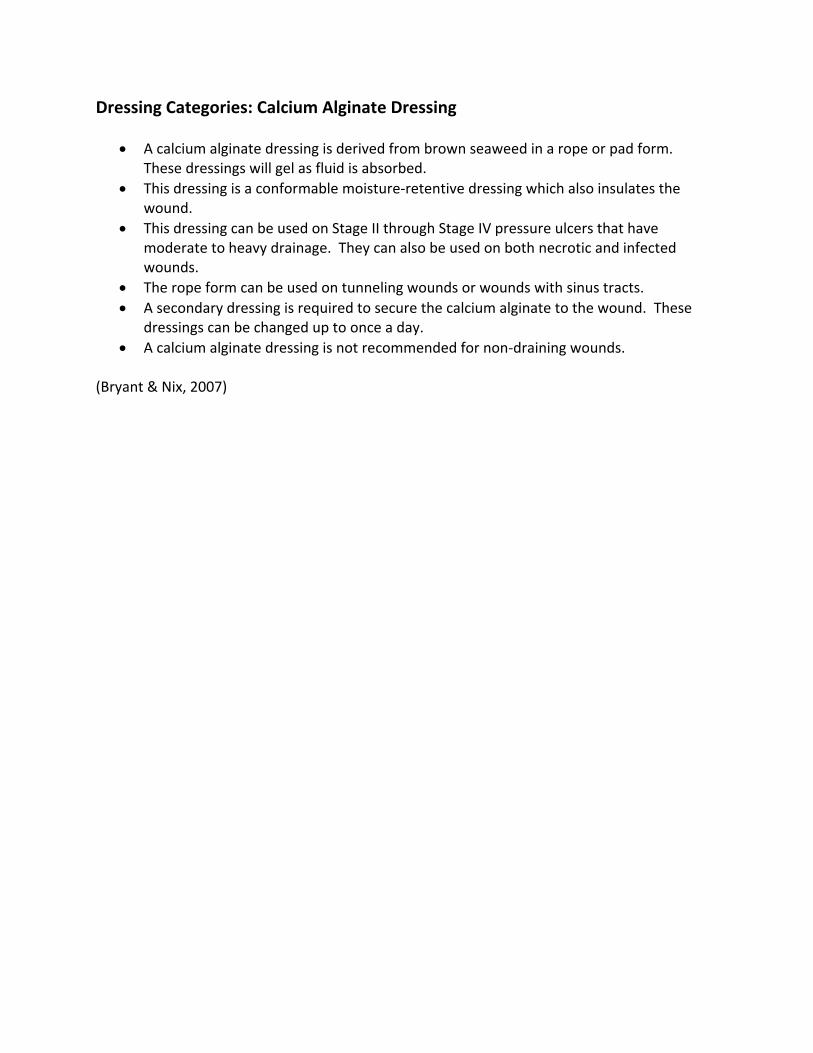
Dressing Categories: Calcium Alginate Dressing
• A calcium alginate dressing is derived from brown seaweed in a rope or pad form. These dressings will gel as fluid is absorbed.
• This dressing is a conformable moisture‐retentive dressing which also insulates the wound.
• This dressing can be used on Stage II through Stage IV pressure ulcers that have moderate to heavy drainage. They can also be used on both necrotic and infected wounds.
• The rope form can be used on tunneling wounds or wounds with sinus tracts. • A secondary dressing is required to secure the calcium alginate to the wound. These
dressings can be changed up to once a day. • A calcium alginate dressing is not recommended for non‐draining wounds.
(Bryant & Nix, 2007)
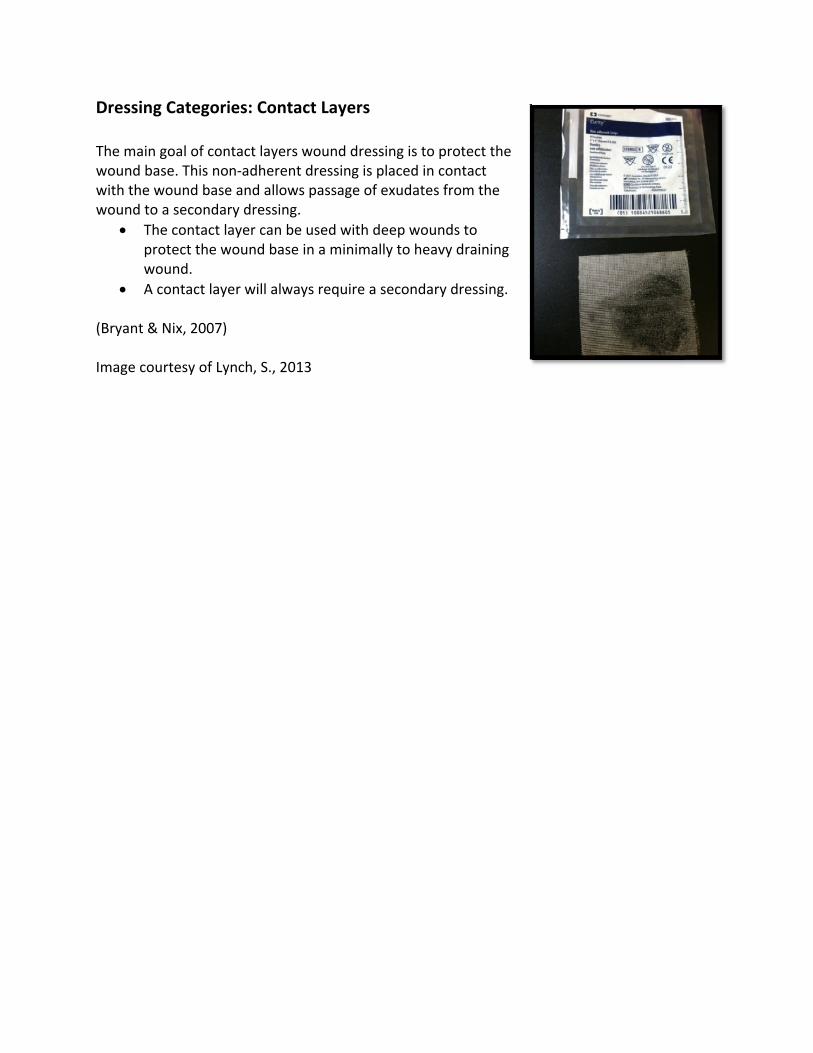
Dressin The mainwound bwith the wound to
• Tpw
• A (Bryant & Image co
g Categori
n goal of conase. This nowound baseo a secondarhe contact larotect the w
wound. A contact laye
& Nix, 2007)
ourtesy of Ly
ies: Contac
ntact layers wn‐adherent e and allowsry dressing. ayer can be wound base i
er will alway
ynch, S., 201
ct Layers
wound dressdressing is p passage of
used with din a minimal
ys require a
3
sing is to proplaced in conexudates fro
eep woundslly to heavy
secondary d
otect the ntact om the
s to draining
dressing.
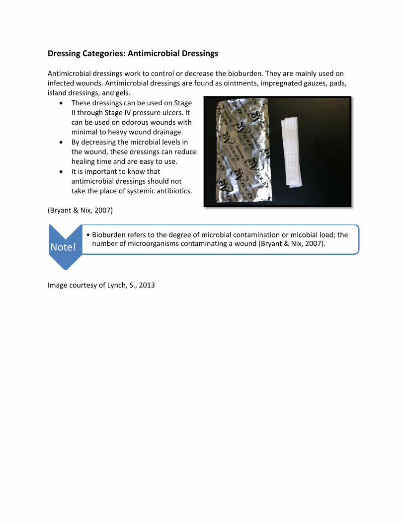
Dressin Antimicroinfected island dr
• TII cam
• Bthh
• Itata
(Bryant &
Image co
Note!
g Categori
obial dressinwounds. Anessings, andhese dressinthrough Staan be used ominimal to hey decreasinghe wound, thealing time t is importanntimicrobialake the place
& Nix, 2007)
ourtesy of Ly
• Bioburnumbe
ies: Antim
ngs work to timicrobial dd gels. ngs can be uage IV pressuon odorous weavy wound g the microbhese dressinand are easynt to know th dressings she of systemi
ynch, S., 201
den refers toer of microor
icrobial Dr
control or ddressings are
sed on Stageure ulcers. Itwounds withdrainage.
bial levels in ngs can reduy to use. hat hould not c antibiotics
3
o the degreerganisms con
ressings
ecrease the e found as o
e t h
uce
s.
e of microbiantaminating
bioburden. ointments, im
al contaminag a wound (B
They are mampregnated
ation or micoBryant & Nix,
ainly used ogauzes, pad
obial load; t, 2007).
n ds,
he
Dressing Categories: Collagen Dressing Collagen dressings are derived from bovine, porcine, or avian sources. They work to accelerate wound repair and stimulate wound healing.
• Collagen dressings will require a secondary dressing. • These wound care dressings can be used on Stage II through Stage IV pressure ulcers.
They can be used with minimal to moderate drainage. • They can be used on infected wounds. • Collagen dressings may absorb slightly. • These dressings are easy to remove and apply. • Collagen dressings are not indicated for patients that have known sensitivity to the
ingredients. • These dressings come in:
o Non‐adherent pouches or vials o Gels loaded into syringes o Pads o Powders o Freeze‐dried sheets
(Bryant & Nix, 2007) Dressing Categories: Honey‐Based Dressings Honey‐based dressings have been found to exert anti‐inflammatory and antibacterial effects without antibiotic resistance. They are able to promote moist wound healing, decrease wound odor, and facilitate debridement.
• Prior to dressing application, the healthcare provider must assess the patient for an allergy to honey, bee products, or bee stings. It is best not to use this product if the patient is allergic.
• Honey is not considered appropriate for dry, necrotic wounds. • Some patients complain of stinging or a burning sensation. • Educate family/patient that honey staining of the skin may be removed with soap and
water. (Pieper, 2009)
Surgery for Wound Closure Along with dressings, treatments for pressure ulcers may include surgery for wound closure. For wound closure on a pressure ulcer, wound closure is most commonly done using one of the following four methods:
1. Linear closure 2. Skin grafts 3. Tissue: Local flaps and distant flaps
Each one of the above surgical interventions will be discussed separately. Linear Closure:
• Linear closure involves bringing the wound edges together and closing those using sutures.
• This type of surgical closure is typically used with a traumatic wound when little tissue is missing.
(Bryant & Nix, 2007) Skin Grafts:
• Commonly, skin grafts are called autografts. • These grafts usually include the epidermis and extend down into the dermis which is
removed from the donor site and placed over a shallow, vascularized pressure ulcer. • Skin grafts will not replace subcutaneous tissue or muscle. It will only replace down to
the dermis level. Because of the depth of the skin graft, it will not provide the cushion that subcutaneous tissue provides over a bony prominence.
(Bryant & Nix, 2007)
Surgery for Wound Closure Tissue Flaps: Tissue flaps are the most common surgical modalities for pressure ulcers. There are two types of tissue flaps that we will discuss; local flaps and free flaps.
1. Local Flaps: The most commonly used flaps are the local flaps. They are categorized according to the anatomical structures involved, the methods used to move the flap and the methods used to retain perfusion of the flap.
Anatomical: o Skin flaps‐involve just the epidermal and possibly the dermal layer. o Fasciocutaneous flaps involve a portion of the epidermis, dermis and the
subcutaneous layer. These flaps provide padding and coverage of the pressure ulcer.
o Myocutaneous flap involve the rotation of all the tissue layers. These flaps provide optimal coverage over a bony prominence and are often used to cover a pressure ulcer.
Methods Used to Move the Flaps: When categorizing tissue flaps according to the method used to move the flap you will find advancement flaps, rotation flaps, and transposition flaps.
Retaining Perfusion: When categorizing tissue flaps according to methods of retaining perfusion you will find random flaps and axial flaps. The random flaps retain the dermal and subdermal vasculature while the axial flaps usually contain and one artery for perfusion (Bryant & Nix, 2007).
2. Free Flaps: With free flaps, the donor site is completely removed from the donor wound and placed on the recipient wound. The microvasculature is carefully connected to the recipient site using special consideration to all of the vessels involved in the wound bed. This flap is not frequently used as it involves microvascular salvation and it is a difficult surgery (Bryant & Nix, 2007).
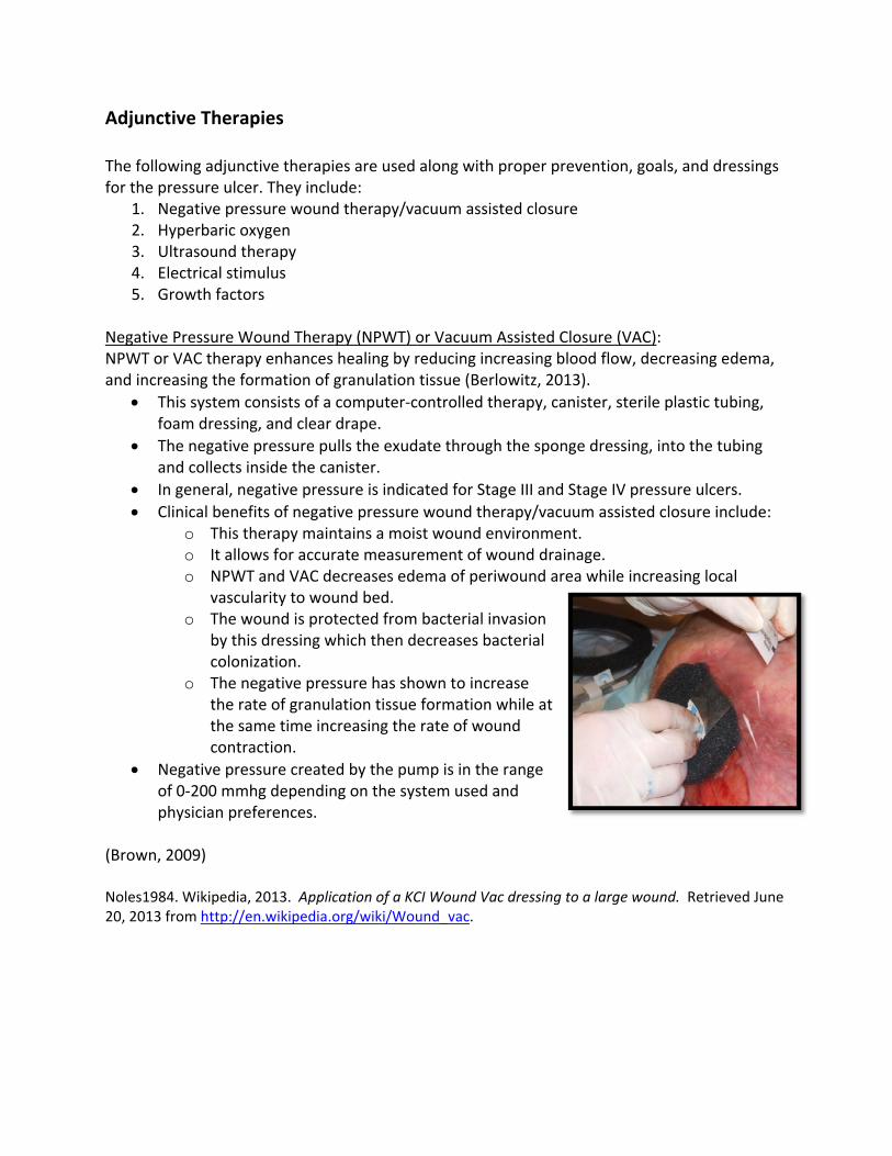
Adjunct The follofor the p
1. N2. H3. U4. E5. G
NegativeNPWT orand incre
• Tfo
• Ta
• In• C
• Nop
(Brown, 2
Noles198420, 2013 f
tive Thera
wing adjuncressure ulce
Negative presHyperbaric oxUltrasound thlectrical stimGrowth facto
e Pressure Wr VAC therapeasing the fohis system coam dressinghe negative nd collects in general, nelinical benef
o This tho It alloo NPWT
vascuo The w
by thicolon
o The nthe rathe sacontra
Negative presf 0‐200 mmhhysician pre
2009)
4. Wikipedia,from http://e
pies
ctive therapier. They inclussure woundxygen herapy mulus ors
Wound Therapy enhances ormation of consists of a g, and clear pressure punside the caegative pressfits of negatherapy mainws for accurT and VAC delarity to wou
wound is prots dressing wization. egative presate of granulame time incaction. ssure createhg dependineferences.
2013. Applicen.wikipedia.o
es are used ude: d therapy/va
py (NPWT) ohealing by rgranulation computer‐cdrape. ulls the exudanister. sure is indicaive pressurentains a moisrate measurecreases edeund bed. tected from
which then d
ssure has shoation tissue creasing the
ed by the pung on the sys
cation of a KCorg/wiki/Wou
along with p
acuum assist
or Vacuum Areducing inctissue (Berlocontrolled th
ate through
ated for Stage wound thest wound enement of woema of periw
bacterial inecreases ba
own to increformation wrate of wou
mp is in the stem used a
CI Wound Vacund_vac.
proper preve
ted closure
Assisted Closreasing blooowitz, 2013)herapy, canis
h the sponge
ge III and Starapy/vacuumnvironment.ound drainawound area
vasion cterial
ease while at und
range nd
c dressing to a
ention, goal
sure (VAC): od flow, decr). ster, sterile p
e dressing, in
age IV pressm assisted c
ge. while increa
a large woun
s, and dress
reasing edem
plastic tubin
nto the tubin
ure ulcers. losure includ
asing local
d. Retrieved
ings
ma,
g,
ng
de:
June
Test Yourself Wound vacuum assisted closure (VAC) is only used on Stage IV and unstageable pressure ulcers.
A. True B. False
The correct answer is false. In general, negative pressure therapy or wound vacuum assisted closing (VAC) is indicated for Stage III and Stage IV pressure ulcers. Adjunctive Therapies Hyperbaric Oxygen Therapy (HBO): What is HBO or HBOT?
• It stands for hyperbaric oxygen therapy. It is a mode of therapy in which the patient breathes 100% oxygen at pressures greater than normal atmospheric pressures.
What does it do? • The research states that HBOT is a treatment for pressure ulcers because during the
treatment, there is a potential to promote healing and reduce bioburden in the wound bed.
How does it do it? • HBOT exerts a bacteriostatic effect on the wound bed by increasing the generation of
oxygen free radicals that damage bacterial membranes DNA strands. Also, the raise in tissue oxygenation enhancing leukocyte activity.
What does the literature say? • HBO has been used, but data is still being collected on the efficacy of the treatment.
(Gray & Ratliff, 2006)
Adjunctive Therapies Ultrasound:
• Research is still being done on the efficacy of non contact low‐frequency ultrasound as a treatment option for suspected deep tissue injury. One case study suggests that the role is preventing the suspected deep tissue injury from progressing to a higher‐stage pressure ulcer (Honaker & Forston, 2011).
• Therapeutic ultrasound delivers energy through mechanical vibrations in the form of sound waves at frequencies above detection by the human ear.
• Low frequency ultrasound has been shown to effectively debride necrotic tissue, eradicate some strains of bacteria from the wound and facilitate wound healing.
• Ultrasound treatment cannot be used near electronic implants/prostheses, on areas of malignancy or over the lower back or abdomen during pregnancy.
(Baranoski, 2012) Electrical Stimulation: With electrical stimulation, direct current is applied to the wound to enhance healing. It promotes the migration and proliferation of fibroblasts (Berlowitz, 2013). Electrical stimulation uses electrical current to stimulate cellular processes involved in wound healing (Baranoski, 2006).
• Use of electrical stimulation is limited in clinical practice. • Electrical stimulation appears to be most effective on Stage III and IV pressure ulcers.
Typically with those pressure ulcers that have failed traditional wound care modalities. The literature suggests that an optimal electrical charge of 300 to 500uA/sec produces positive effects on the pressure ulcer. Determining the optimal charge based on the wounds stage, depth, and drainage need to be studied further (Baranoski, 2012).
Growth Factors: Growth factors are proteins that occur naturally in the human body. These proteins cause cellular growth and cell migration and will assist in wound healing. They are obtained through the use of the body’s platelets and macrophages made chemically or biochemically outside of the body.
• Growth factors are available by prescription only and should be used on clean, granulating wounds only.
• The healthcare provider should be aware not to use growth factors on necrotic wounds or on patients with any neoplasms.
(Brown 2009)
Genera• St• St• St• St• U• S
TreatmenStage I ispreventaskin is nocolor doe How to t
1. M2. M
wm
3. Evco
4. Rtrco
5. Uth
6. Imtha
7. IffrfithC
(NPUAP,
l Overviewtage I tage II tage III tage IV Unstageable uspected de
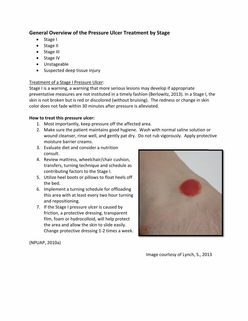
nt of a Stage a warning, ative measurot broken bues not fade w
treat this preMost importaMake sure thwound cleansmoisture barvaluate dietonsult. eview mattrransfers, turontributing fUtilize heel bhe bed. mplement a his area withnd repositiof the Stage I riction, a prolm, foam or he area and hange prote
2010a)
w of the Pr
eep tissue inj
e I Pressure a warning thres are not inut is red or dwithin 30 mi
essure ulcerantly, keep pe patient mser, rinse werier creams. and conside
ress, wheelcning techniqfactors to thoots or pillo
turning scheh at least eveoning. pressure ulcotective dreshydrocolloiallow the skective dressi
ressure Ulc
jury
Ulcer: hat more sernstituted in iscolored (winutes after
r: pressure off aintains gooell, and gent er a nutrition
chair/chair cuque and schehe Stage I. ows to float h
edule for offery two hou
cer is causedssing, transpd, will help pkin to slide eng 1‐2 times
cer Treatm
rious lesionsa timely fash
without bruispressure is a
the affectedod hygiene. tly pat dry. D
n
ushion, edule as
heels off
floading r turning
d by parent protect asily. s a week.
ment by Sta
s may develohion (Berlowsing). The realleviated.
d area. Wash with nDo not rub v
Image co
age
op if appropwitz, 2013). Iedness or ch
normal salinvigorously. A
ourtesy of Ly
riate n a Stage I, tange in skin
e solution oApply protec
ynch, S., 201
the
or ctive
13
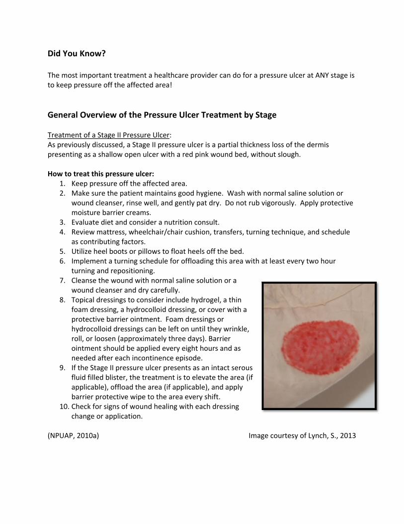
Did You The mostto keep p Genera TreatmenAs previopresentin How to t
1. K2. M
wm
3. Ev4. R
as5. U6. Im
tu7. C
w8. T
fophroon
9. Ifflab
10. Cch
(NPUAP,
u Know?
t important pressure off
l Overview
nt of a Stageously discussng as a shallo
treat this preeep pressur
Make sure thwound cleansmoisture barvaluate dieteview mattrs contributinUtilize heel bmplement a urning and rleanse the w
wound cleansopical dressoam dressingrotective baydrocolloid oll, or loosenintment shoeeded after f the Stage IIuid filled blipplicable), oarrier protecheck for signhange or ap
2010a)
treatment athe affected
w of the Pr
e II Pressure sed, a Stage ow open ulc
essure ulcere off the affe patient mser, rinse werier creams. and consideress, wheelcng factors. oots or pilloturning scheepositioningwound with ser and dry cings to consg, a hydrocoarrier ointmedressings can (approximaould be applieach incont pressure ulster, the treoffload the active wipe tons of woundplication.
a healthcare d area!
ressure Ulc
Ulcer: II pressure ucer with a re
r: fected area.aintains gooell, and gent er a nutritionchair/chair cu
ows to float hedule for offg. normal salincarefully. ider includeolloid dressinent. Foam dan be left onately three died every eigtinence episocer presentsatment is torea (if applico the area evd healing wit
provider ca
cer Treatm
ulcer is a pard pink woun
od hygiene. tly pat dry. D
n consult. ushion, tran
heels off thefloading this
ne solution o
hydrogel, ang, or cover ressings or until they wdays). Barrieght hours anode. s as an intaco elevate thecable), and avery shift. th each dress
n do for a pr
ment by Sta
rtial thicknesnd bed, with
Wash with nDo not rub v
sfers, turnin
e bed. s area with a
or a
thin with a
wrinkle, er nd as
t serous e area (if apply
sing
Imag
ressure ulce
age
ss loss of theout slough.
normal salinvigorously. A
ng technique
t least every
e courtesy o
r at ANY sta
e dermis
e solution oApply protec
e, and sched
y two hour
of Lynch, S.,
ge is
or ctive
ule
2013
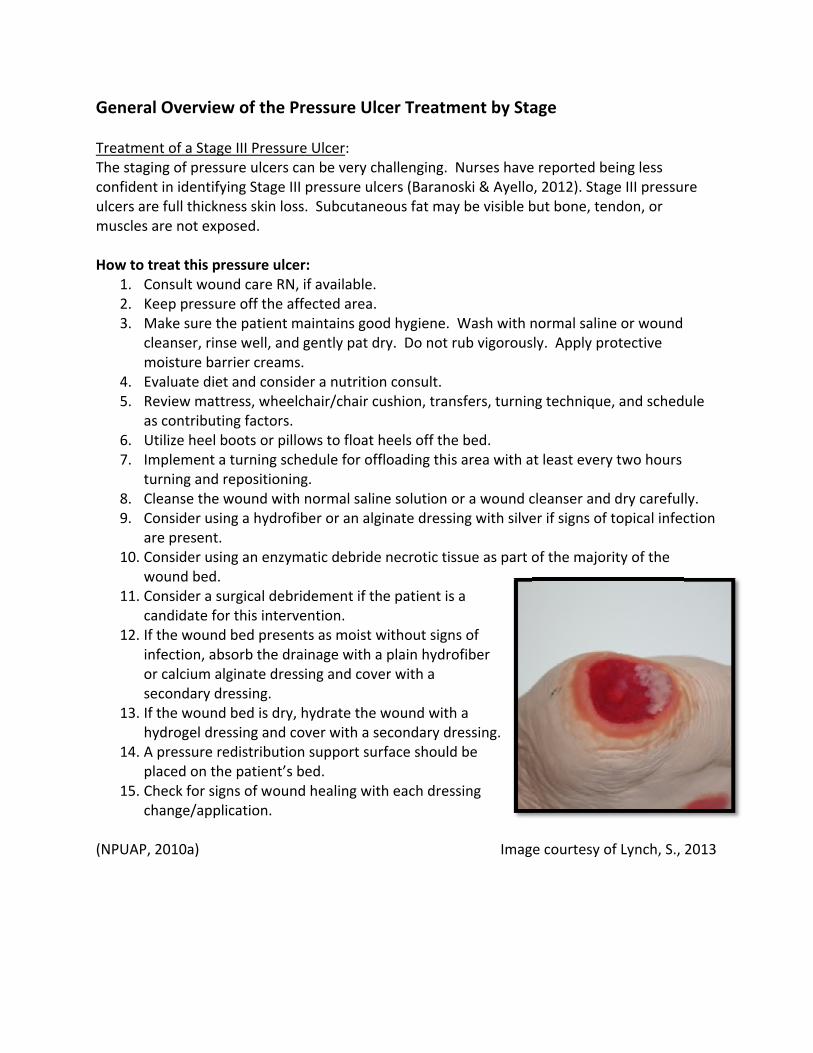
Genera TreatmenThe stagiconfidenulcers aremuscles How to t
1. C2. K3. M
clm
4. Ev5. R
as6. U7. Im
tu8. C9. C
a10. C
w11. C
ca12. If
inose
13. Ifh
14. Ap
15. Cch
(NPUAP,
l Overview
nt of a Stageing of pressut in identifyie full thickneare not expo
treat this preonsult wouneep pressur
Make sure thleanser, rinsmoisture barvaluate dieteview mattrs contributinUtilize heel bmplement a urning and rleanse the wonsider usinre present. onsider usin
wound bed. onsider a suandidate forf the wound nfection, absr calcium algecondary drf the wound ydrogel dres
A pressure relaced on theheck for signhange/appli
2010a)
w of the Pr
e III Pressureure ulcers caing Stage III ess skin lossosed.
essure ulcernd care RN, ie off the affe patient mse well, and grier creams. and consideress, wheelcng factors. oots or pilloturning scheepositioningwound with ng a hydrofib
ng an enzym
urgical debridr this intervebed presentsorb the draginate dressessing. bed is dry, hssing and coedistributione patient’s bns of woundcation.
ressure Ulc
e Ulcer: an be very chpressure ulc. Subcutane
r: if available.fected area.aintains googently pat d er a nutritionchair/chair cu
ows to float hedule for offg. normal salinber or an alg
atic debride
dement if thention. ts as moist winage with aing and cove
hydrate the over with a se support sured. d healing wit
cer Treatm
hallenging. Ncers (Baranoeous fat may
od hygiene. ry. Do not r
n consult. ushion, tran
heels off thefloading this
ne solution oginate dressi
e necrotic tis
he patient is
without signa plain hydroer with a
wound withecondary drrface should
th each dress
ment by Sta
Nurses haveoski & Ayelloy be visible b
Wash with nrub vigorous
sfers, turnin
e bed. s area with a
or a wound cng with silve
sue as part o
a
s of ofiber
a ressing. d be
sing
Imag
age
e reported beo, 2012). Stagbut bone, te
normal salinly. Apply pr
ng technique
t least every
cleanser ander if signs of
of the major
e courtesy o
eing less ge III pressundon, or
e or wound rotective
e, and sched
y two hours
d dry carefultopical infec
rity of the
of Lynch, S.,
re
ule
ly. ction
2013
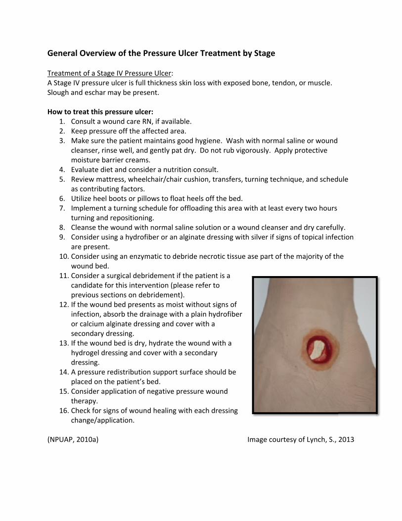
Genera TreatmenA Stage ISlough an How to t
1. C2. K3. M
clm
4. Ev5. R
as6. U7. Im
tu8. C9. C
a10. C
w11. C
cap
12. Ifinose
13. Ifhd
14. Ap
15. Cth
16. Cch
(NPUAP,
l Overview
nt of a StageV pressure und eschar m
treat this preonsult a woeep pressur
Make sure thleanser, rinsmoisture barvaluate dieteview mattrs contributinUtilize heel bmplement a urning and rleanse the wonsider usinre present. onsider usin
wound bed. onsider a suandidate forrevious sectf the wound nfection, absr calcium algecondary drf the wound ydrogel dresressing.
A pressure relaced on theonsider appherapy. heck for signhange/appli
2010a)
w of the Pr
e IV Pressureulcer is full tmay be prese
essure ulcerund care RNe off the affe patient mse well, and grier creams. and consideress, wheelcng factors. oots or pilloturning scheepositioningwound with ng a hydrofib
ng an enzym
urgical debridr this intervetions on debbed presentsorb the draginate dressessing. bed is dry, hssing and co
edistributione patient’s blication of n
ns of woundcation.
ressure Ulc
e Ulcer: hickness skint.
r: N, if availablefected area.aintains googently pat d er a nutritionchair/chair cu
ows to float hedule for offg. normal salinber or an alg
atic to debri
dement if thention (pleasridement).ts as moist winage with aing and cove
hydrate the over with a se
support sured. egative pres
d healing wit
cer Treatm
n loss with e
e.
od hygiene. ry. Do not r
n consult. ushion, tran
heels off thefloading this
ne solution oginate dressi
ide necrotic
he patient is se refer to
without signa plain hydroer with a
wound withecondary
rface should
ssure wound
th each dress
ment by Sta
exposed bon
Wash with nrub vigorous
sfers, turnin
e bed. s area with a
or a wound cng with silve
tissue ase p
a
s of ofiber
a
d be
d
sing
Imag
age
ne, tendon, o
normal salinly. Apply pr
ng technique
t least every
cleanser ander if signs of
part of the m
e courtesy o
or muscle.
e or woundrotective
e, and sched
y two hours
d dry carefultopical infec
majority of th
of Lynch, S.,
ule
ly. ction
he
2013
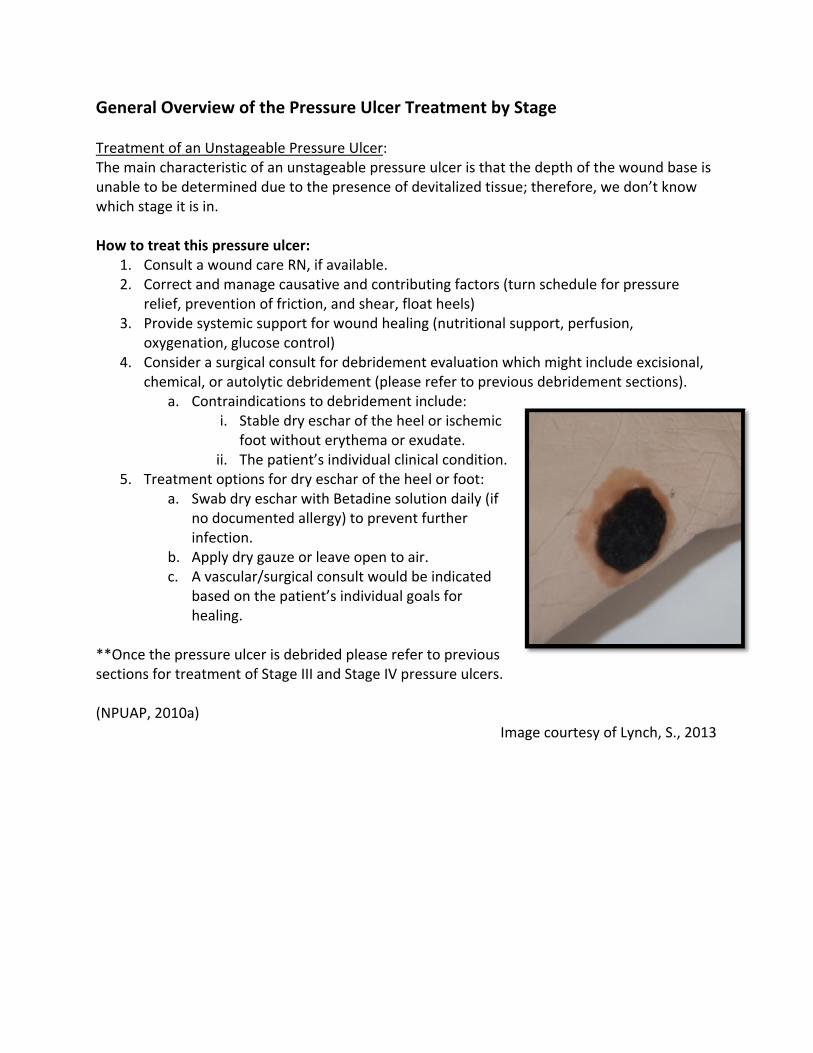
Genera TreatmenThe mainunable towhich sta How to t
1. C2. C
re3. P
o4. C
ch
5. T
**Once tsections (NPUAP,
l Overview
nt of an Unsn characteriso be determage it is in.
treat this preonsult a woorrect and melief, prevenrovide systexygenation, onsider a suhemical, or a
a. Contri.
ii. reatment op
a. Swab no doinfect
b. Applyc. A vasc
basedhealin
the pressurefor treatme
2010a)
w of the Pr
stageable Prestic of an unined due to
essure ulcerund care RNmanage causntion of frictiemic supportglucose con
urgical consuautolytic debaindicationsStable dryfoot withoThe patien
ptions for drdry eschar wcumented aion. dry gauze ocular/surgicad on the pating.
e ulcer is debnt of Stage I
ressure Ulc
essure Ulcerstageable prthe presenc
r: N, if availablesative and coion, and shet for wound ntrol) ult for debridbridement (s to debridemy eschar of thout erythemnt’s individuary eschar of twith Betadinallergy) to pr
or leave openal consult woent’s individ
brided pleasII and Stage
cer Treatm
r: ressure ulcece of devital
e. ontributing fear, float heehealing (nut
dement evalplease referment includehe heel or isca or exudateal clinical cothe heel or fne solution drevent furthe
n to air. ould be indicdual goals fo
e refer to pr IV pressure
ment by Sta
r is that the ized tissue; t
factors (turnels) tritional sup
uation whicr to previouse: chemic e. ondition. foot: daily (if er
cated r
revious ulcers.
Imag
age
depth of thetherefore, w
n schedule fo
port, perfus
h might incls debrideme
e courtesy o
e wound baswe don’t kno
or pressure
sion,
ude excisionnt sections).
of Lynch, S.,
se is ow
nal, .
2013
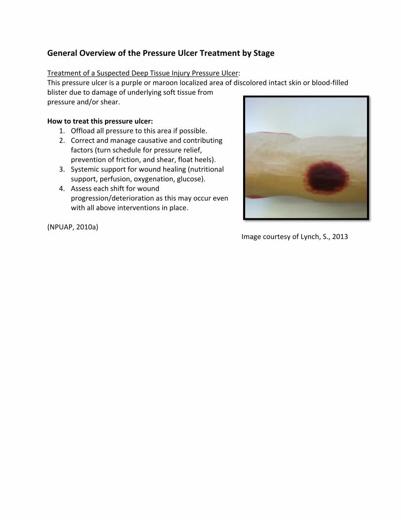
Genera TreatmenThis presblister dupressure How to t
1. O2. C
fap
3. Sysu
4. Apw
(NPUAP,
l Overview
nt of a Suspessure ulcer isue to damag and/or shea
treat this preOffload all prorrect and mactors (turn revention ofystemic supupport, perfAssess each srogression/d
with all above
2010a)
w of the Pr
ected Deep s a purple orge of underlyar.
essure ulceressure to thmanage causschedule forf friction, anport for woufusion, oxygeshift for woudeterioratioe interventio
ressure Ulc
Tissue Injuryr maroon locying soft tiss
r: is area if posative and cor pressure rend shear, floaund healing enation, glucund n as this maons in place.
cer Treatm
y Pressure Ucalized area ue from
ssible. ontributing elief, at heels). (nutritional cose).
y occur even.
ment by Sta
Ulcer: of discolore
n
Image
age
d intact skin
courtesy of
n or blood‐fil
Lynch, S., 20
lled
013
Case Study One Mr. L is a 65 year old gentleman that is admitted to the hospital for lethargy and leukocytosis. Mr. L’s past medical history is significant for embolic stroke six months ago and hypertension. Mr. L is noted to have right sided hemiparesis and tends to roll onto his back when positioned on his side. The patient has been a resident of a skilled nursing facility for approximately six months. Mr. L’s daughter is present with him and reports that he has had a pressure ulcer on his coccyx for approximately two months with no improvement. The referral from the nursing home states that they have been doing wet to dry gauze packing to the coccyx pressure ulcer three times a day.
• WBC 12,000 • Albumin 2.9 • Vital signs are 150/82, 80, 20, 101.2 PO
During his admission assessment you find that Mr. L’s skin is intact except for the pressure ulcer to his coccyx. The pressure ulcer is approximately 6cm x 4cm x 3cm with undermining from 12‐6 0’clock of 2cm. The wound bed is 80% filled with yellow slough and 20% filled with pale pink wound bed. The periwound skin is intact. The periwound skin is also peeling in some areas. The dressing that was removed was saturated with blue/green looking drainage with a fruity odor to it. Mr. L. denies any pain/discomfort during the dressing change. How would you stage this pressure ulcer? This is an unstageable pressure ulcer due to the presence of 80% devitalized tissue. Is the periwound skin infected? Due to the blue/green drainage and the fruity odor and the elevated WBC it appears the wound may be infected. What does blue/green drainage with a fruity odor tell you? Due to the color of the drainage and the odor, pseudomonas is suspected. What is the recommended treatment at this point? Due to the presence of 80% devitalized tissue the wound will not heal without further intervention. Debridement of the wound bed must be done in order to facilitate healing. Also, during the debridement it will be appropriate at that time to obtain a deep wound culture to verify any infection. Surgical debridement would be the at this point the recommended, fastest most appropriate approach.
Case Study One Mr. L. and his daughter agree that surgical intervention for debridement aligns with their goal of healing the wound and Mr. L. is brought to the OR the next day for the procedure. The day after the surgical debridement the doctors’ order reads, “Remove surgical packing and begin NS irrigation and moist to dry dressing changes to coccyx three times a day.” Prior to removing the surgical dressing, what is the most important question you should ask Mr. L.? At this point, it is imperative to assess Mr. L’s pain level. It is also important to discuss with Mr. L that he may experience pain/ discomfort during the dressing change and it may be in his best interest to receive pain medication prior to the dressing change. You pre‐medicate Mr. L. prior to removing the dressing. After removing the dressing you note a large amount of serosanguineous drainage. The wound bed now measures 6.5cm x 5cm x 4cm no undermining noted. The wound bed is 90% beefy pink granulation tissue. The periwound skin remains intact. Although the MD has written for wound care, is there an advanced wound care modality that would be more beneficial for Mr. L? The negative pressure wound therapy modality would be appropriate at this time. The necrotic tissue has been removed and the wound bed is healthy and pink. The NPWT modality will decrease the number of dressing changes, will be less painful for Mr. L., and will be able to contain the amount of drainage that is being produced. What other advanced wound care modalities could you try on this wound? Calcium alginate, hydrofiber, and a foam dressing would all be appropriate. These types of dressings are all capable of absorbing large amounts of exudate and protecting the periwound skin from maceration while at the same time preventing the wound from drying out and providing moist wound healing.
Case Study Two Mrs. C is an 80 year old nursing home resident who is admitted to your hospital after falling and breaking her left hip while ambulating alone to the bathroom. Mrs. C’s past medical history is significant for diabetes, coronary artery disease, and Alzheimer's. Mrs. C is brought immediately to the OR from the emergency room and is admitted to the orthopedic floor later that evening. Once settled after receiving report, the nurse goes into Mrs. C’s room to assess her. Mrs. C is sweating, shaking and crying in pain. Vital signs at that time are:
• BP 140/86 • HR 100 • RR 24 • Temp 99.9 PO • Blood glucose level 142mg/dl
Due to her Alzheimer's disease, Mrs. C is unable to rate her pain level. However, it is clear using objective signs of sweating, shaking, and crying out, that Mrs. C is in pain. The nurse medicates Mrs. C as per MD orders and attempts to reposition her. Mrs. C yells out in pain; therefore, the nurse leaves her on her back and heels on the bed because she is too uncomfortable to be moved. 45 minutes later, it appears that she is sleeping and that her pain level has decreased, the nurse leaves her alone. Throughout the night, the nurse leaves her alone as she is sleeping. At the end of the shift, the nurse goes into Mrs. C’s room and attempts to straighten out Mrs. C’s legs as she is diagonal in the bed. As the nurse gently lifts her affected leg, the nurse notices a 5cm x 5cm purple fluid filled blister. What stage is this pressure ulcer? The purple blister is called a suspected deep tissue injury. Purple or maroon localized area of discolored intact skin or blood‐filled blister due to damage of underlying soft tissue from pressure and/or shear. What do you believe caused this large purple fluid filled blister on the heel of the surgically repaired hip? A suspected deep tissue injury as this was caused by Mrs. C laying in the same position for the entire shift. The pressure of her heel resting on the mattress caused this injury. Furthermore, the pain that she was experiencing caused her to tense her operative hip and press her heel even further into the mattress.
Case Study Two What would you do first for this type of pressure ulcer? The most important intervention is to elevate the heel off the mattress at all times. The heel must be floating in the air. This can be done by placing a pillow under the patient’s calf or by using a boot that suspends the heel in the air. What is the recommended treatment for this pressure ulcer? The best treatment for a suspected deep tissue is to leave the blood filled blister open to air and monitor it every shift. If the blister opens or shows any signs of infection it will need to be unroofed via sharp debridement. If the blister starts to drain but is not showing any signs of infection, the nurse could utilize a foam dressing to contain the drainage and protect the periwound skin from the drainage. How often are foam dressings changed? The foam dressings are usually changed dependent on the amount of drainage. Conclusion When a pressure ulcer happens, it is important to identify and document on the pressure ulcer immediately. Begin working as a team with the primary healthcare provider and/or certified wound RN to guide treatment for the patient’s pressure ulcer. As with healing of all pressure ulcers, no matter what the treatment is, the causative factors must be addressed in order to heal the wound and prevent further deterioration of the wound.
References Armstrong, D. & Myer, A. (2012). Clinical assessment of wounds. Retrieved March 12, 2012, from www.uptodate.com. Baranoski, S. & Ayello, E.A. (2012). Wound Care Essentials (3rd ed.). Ambler, PA: Lippincott, Williams & Wilkins. Baranoski, S. (2006). Raising awareness of pressure ulcer prevention and treatment. Advances in Skin & Wound Care, 19(7): p. 298‐405. Berlowitz, D. (2013). Treatment of Pressure Ulcers. Retrieved from www.uptodate.com. Brown, P. (2009). Quick Reference to Wound Care (3rd ed.). Sudbury, MA: Jones and Bartlett Bryant, R.A. & Nix, D.P. (2007). Acute & Chronic Wounds. St. Louis, MO: Mosby. Gray, M. & Ratliff (2006). Is hyperbaric oxygen therapy effective for the management of chronic wounds? Journal of Wound, Ostomy, and Continence Nursing: 21‐26. Hess, C.T. (2010). Performing a skin assessment. Nursing 2010, 66. Hess, C.T. (2008). Performing a skin assessment. Advances in Skin & Wound Care, 24(8): 392. Hess, C.T. (2005). Clinical Guide: Wound Care (5th ed.). Ambler, PA: Lippincott, Williams & Wilkins. Honaker, J. & Foston, M. (2011). Adjunctive use of noncontact low‐frequency ultrasound for treatment of suspected deep tissue injury: A case study. Journal of Wound, Ostomy, and Continence Nursing, 38(4), p: 394‐403. Little, M.O. (2012). Nutrition and skin ulcers. Ambler, PA: Lippincott, Williams & Wilkins. National Pressure Ulcer Advisory Panel (2010). International Guideline: Prevention and Treatment of Pressure Practice Guideline. Washington DC: National Pressure Ulcer Advisory Panel. National Pressure Ulcer Advisory Panel (2010a). Pressure ulcer treatment: Quick reference guide. Retrieved from http://www.npuap.org/wp‐content/uploads/2012/03/Final_Quick_Treatment_for_web_2010.pdf. Pieper, B. (2009). Honey‐based dressings and wound care volume. Journal of Wound, Ostomy, and Continence Nursing, 36(1), p: 60‐68.
References Rose, B. (2012) Clinical manifestations and diagnosis of edema in adults. Retrieved March 17, 2012, from www.uptodate.com. Sherman, R.A. & Shapiro, C.E. (2007). Maggot therapy for problematic wounds: Uncommon and off‐label applications. Advances in Skin & Wound Care, 20(11): p. 602‐610. Swezey, L. (2009). Wound dressings‐ Hydrofibers. Retrieved from http://ezinearticles.com/?Wound‐Dressings‐‐‐Hydrofibers&id=2642728. Van Anholt, R.D., Sobotka, L., Maijer, E.P., et al. (2010). Specific nutritional support accelerates pressure ulcer healing and reduces wound care intensity in non‐malnourished patients. Nutrition, 26, 867‐872.
Disclaimer This publication is intended solely for the educational use of healthcare professionals taking this course, for credit, from RN.com, in accordance with RN.com terms of use. It is designed to assist healthcare professionals, including nurses, in addressing many issues associated with healthcare. The guidance provided in this publication is general in nature, and is not designed to address any specific situation. As always, in assessing and responding to specific patient care situations, healthcare professionals must use their judgment, as well as follow the policies of their organization and any applicable law. This publication in no way absolves facilities of their responsibility for the appropriate orientation of healthcare professionals. Healthcare organizations using this publication as a part of their own orientation processes should review the contents of this publication to ensure accuracy and compliance before using this publication. Healthcare providers, hospitals and facilities that use this publication agree to defend and indemnify, and shall hold RN.com, including its parent(s), subsidiaries, affiliates, officers/directors, and employees from liability resulting from the use of this publication. The contents of this publication may not be reproduced without written permission from RN.com. Participants are advised that the accredited status of RN.com does not imply endorsement by the provider or ANCC of any products/therapeutics mentioned in this course. The information in the course is for educational purposes only. There is no “off label” usage of drugs or products discussed in this course. You may find that both generic and trade names are used in courses produced by RN.com. The use of trade names does not indicate any preference of one trade named agent or company over another. Trade names are provided to enhance recognition of agents described in the course. Note: All dosages given are for adults unless otherwise stated. The information on medications contained in this course is not meant to be prescriptive or all‐encompassing. You are encouraged to consult with physicians and pharmacists about all medication issues for your patients.