Embed Size (px)
Citation preview

FACULTEIT DIERGENEESKUNDE
Vakgroep Voortplanting, Verloskunde en Bedrijfsdiergeneeskunde
Preservation of fresh bovine semen and utero-tubal junction
insemination in cattle
Steven Verberckmoes
Proefschrift ter verkrijging van de graad van Doctor in de Diergeneeskundige Wetenschappen
(PhD) aan de Faculteit Diergeneeskunde, Universiteit Gent, 2004.
Promotoren: Prof. Dr. A. Van Soom
Prof. Dr. Dr. h. c. A. de Kruif


TABLE OF CONTENTS
List of abbreviations CHAPTER 1 Introduction 7
1.1 State of the art: artificial insemination in cattle 8
1.2 Aims of the study 11
CHAPTER 2 Intra-uterine inseminations in farm animals and humans 15
CHAPTER 3 Collection and composition of cauda epididymal plasma in the bull 43
CHAPTER 4 Storage of fresh bovine semen in a diluent based on the ionic
composition of cauda epididymal plasma (CEP) 63
CHAPTER 5 Comparison of three diluents for the storage of fresh bovine semen 83
CHAPTER 6 Migration of bovine spermatozoa in a synthetic medium and its
relation to in vivo bull fertility 101
CHAPTER 7 Assessment of a new utero-tubal junction insemination device in
dairy cattle 119
CHAPTER 8 Assessment of the Ghent device for utero-tubal junction
insemination in dairy cattle with lower insemination doses 139
CHAPTER 9 Effect of whole blood and serum on bovine sperm quality and in
vitro fertilization capacity 161
CHAPTER 10 General discussion 175
CHAPTER 11 Summary – samenvatting 193
Dankwoord
Curriculum vitae


List of abbreviations ABS acryl butyl styreen AI artificial insemination ATP adenosine triphosphate BSA bovine serum albumin CASA computer assisted sperm analysis CD conventional device CEP cauda epididymal plasma CV coefficient of variance DNA deoxyribonucleic acid FCS fetal calf serum FITC fluorescein isothiocyanate GD Ghent device Hepes N-2-Hydroxyethylpiperazine-N’-2-ethanesulfonic acid HF Holstein Friesian HHA head to head agglutination hpi hours post insemination ICSI intracytoplasmatic sperm injection IUI intra-uterine insemination IVF in vitro fertilization IVM in vitro maturation JC-1 5,5’,6,6’-tetrachloro-1,1’13,3’-tetraethylbenzimidazolyl
carbocyanine iodide LH luteinizing hormone LIN linearity MC methyl cellulose NRR non-return rate PBS phosphate buffered saline PI propidium iodide PMS progressively motile spermatozoa PR pregnancy rate PSA pisum sativum agglutinin PVC polyvinyl chloride r correlation coefficient RBC red blood cells RMS rapid progressively motile spermatozoa ROS reactive oxygen species SD standard deviation SEM standard error of the mean SQA sperm quality analyser SST sperm storage tubules STR straightness TALP tyrode solution supplemented with albumin, lactate and
pyruvate TCM tissue culture medium UTJ utero-tubal junction VAP velocity average pathway VCL velocity curved line VSL velocity straight line


CHAPTER 1:
INTRODUCTION
1.1 State of the art: artificial insemination in cattle
1.2 Aims of the study

8 Chapter 1
1.1 State of the art: artificial insemination in cattle
Artificial insemination (AI) or introduction of semen in the female genital tract by means
of instruments is the first generation of reproductive biotechnologies which was feasible in
cattle. The first commercial AI cooperative was established in 1936 by a Dane, Sorenson
(Foote, 2002). Before the Second World War, most cows in Europe and North America were
fertilized by means of natural service. However, since several cows on different farms were
mated by the same bull, the spread of genital diseases with decreased fertility outcomes was a
constant threat. Moreover, keeping herd bulls was expensive and represented potential danger
for the herd manager (Vishwanath, 2003). Apart from these facts, the limited number of
offspring produced per bull after natural mating made it impossible to set up effective
progeny testing schemes and resulted in a very poor genetic gain. The introduction of AI in
cattle was mainly forced by sanitary reasons, and especially by fertility problems caused by
Campylobacter foetus subspecies venerealis (vibriosis) and Trichomonas foetus. However,
also the control and prevention of non-sexually transmitted diseases such as tuberculosis,
brucellosis and paratuberculosis at the farms benefited from the introduction of AI (Thibier
and Guerin, 2000). Later on, the economic advantage by improved fertility rates and
accelerated genetic progress became the most important motive (Foote et al., 1956; Watson,
1990).
In most developed countries, AI was introduced on a small scale during the 1940s and
1950s and was carried out with fresh semen or semen stored at room temperature.
Notwithstanding the fact that satisfying pregnancy rates can be obtained with low doses of
fresh semen, the high sire utilisation, the inexpensive sperm storage, and the easy use in the
field, the use of fresh semen was seriously restricted by its limited shelf live. In the 1950s the
beneficial effect of glycerol as cryoprotectant in diluents for freezing bovine semen was
discovered (Polge and Rowson, 1952) and further optimization of the freezing-thawing
procedure resulted in the wide spread application of frozen-thawed semen in the 1960s. The
use of frozen-thawed semen facilitated the fast distribution of highly valuable genes, enabled
the development of breeding programmes, and became most successful in the dairy cattle
industry (Chupin and Schuh, 1993). An update survey in 1995 showed that the total number
of doses of semen produced in the developed countries exceeded 200 million with more than
95% of this semen processed as a frozen product (Chupin and Thibier, 1995). The current
world statistics for AI in cattle stand at 232 million doses of semen produced as a frozen

Introduction 9
product and 11.6 million as liquid, with the latter restricted primarily to New Zealand with
smaller amounts used in Africa, France, Australia, Germany, and Eastern Europe (Thibier and
Wagner, 2000).
A recent interesting development in the assisted reproduction of cattle is the possibility to
sort X- and Y-bearing spermatozoa by means of flowcytometry (Seidel, 2003). After sorting
semen, the probability to have a calf of the desired gender after conception is 90%. This is in
contrast to the 51% chance to have a bull calf after conception when non-sorted semen is used
(Seidel, 2003). The possibility to determine the sex of the offspring before conception could
be interesting for the dairy cattle industry. In dairy cattle, only a few bulls which highly
inherit top productivity characteristics are needed for reproductive purposes. The other males
are unprofitable and not wanted. Therefore, the more females are born, the better selection for
productive and reproductive characteristics can be performed and the more milk can be
produced at a farm. The use of sex sorted semen in dairy cattle could also result in a reduction
of dystocia in heifers, and the opportunity to create an extra income out of the production of
cross-bred beef bulls out of cows which are not good enough for production of replacement
heifers. This means that more milk and meat could be produced with fewer animals, which is
an extra advantage for the European dairy farmers, whose production is limited by the number
of animals they may breed to comply with the law concerning environmental protection.
In Europe and the US, sex-sorted bovine semen is already a few years commercially
available, however not widely used. Due to the high costs of the sexing procedure, a dose of
sexed semen contains 1 to 2 million frozen-thawed spermatozoa, which is only 10% of a
conventional insemination dose (Seidel, 2003). Moreover, the sexing procedure has a negative
effect on sperm quality, which results in lower fertility rates (Seidel, 2003).
To improve fertility rates with low doses and less fertile semen (i.e. sex-sorted semen),
two solutions are possible; 1) insemination with fresh instead of frozen-thawed semen, and 2)
semen deposition closer to the site of fertilization.
The use of fresh semen for AI in cattle enables a ten fold reduction of the insemination
dose compared to frozen-thawed semen without a reduction in pregnancy rates (Vishwanath
et al., 1996). The freezing and thawing process has an irreversible impact on the spermatozoa,
both on the recovery of motile, morphologically normal cells and the ensuing pregnancy rates
(Holt, 2000). Bovine semen is the least sensitive of all species to freezing damages, but even
with the best preservation techniques, the optimal cell recovery is just over 50% (Vishwanath
10 Chapter 1
and Shannon, 2000). Many extenders have been developed for liquid storage of semen but, up
to now, none of them is capable of storing spermatozoa for more than 3 days without a drop
in in vivo fertility (Foote, 1978). Development of a diluent which could store semen for 4 to 5
days would be interesting for the cattle breeding industry for several reasons; 1) more
insemination doses could be produced per ejaculate, 2) the work load during sperm collection
and processing would be decreased, and 3) the distribution of liquid semen would be
simplified.
When conventional insemination is performed in cattle, 10 to 15 million frozen-thawed
spermatozoa are deposited into the uterine body, while fertilization occurs in the tuba uterine
(oviduct). Compared to conventional insemination, semen deposition near the utero-tubal
junction could theoretically be performed with 100-times less spermatozoa without a decrease
in fertility due to the reduced loss of spermatozoa by 1) retrograde flow in the cervical mucus
(Larsson and Larsson, 1985; Mitchell et al., 1985; Nelson et al., 1987), 2) decreased
phagocytosis during migration through the uterus (Hawk, 1983), and 3) an increase in
survival time of spermatozoa in the sperm-friendly environment of the isthmus (Suarez,
2001).

Introduction 11
1.2 Aims of the study
To increase the number of insemination doses produced by genetically highly valuable
sire bulls, the use of liquid-stored semen instead of frozen-thawed semen and deposition of
semen near the utero-tubal junction (UTJ) instead of into the uterine body, could be
appropriate alternatives.
Research is needed to increase the time during which semen can be stored in liquid state
whilst maintaining its fertilizing capacity. Since bovine spermatozoa can be stored for several
weeks in the cauda epididymidis without negative effects on their fertilizing capacity, the
cauda epididymal fluid can serve as basis for the development of a sperm diluent to prolong
the shelf life of freshly ejaculated bovine semen. In order to deposit semen near the UTJ, a
special insemination device has to be developed which has no detrimental effect on the sperm
quality and on the endometrium of the cow in oestrus, and which can easily be handled by one
person under field conditions.
In order to achieve these aims, the study comprises the following experiments:
1. Determination of the composition of the cauda epididymal plasma (CEP) of the bull
2. Development of a new diluent (CEP diluent) based on the ionic composition of the
bovine cauda epididymal plasma
3. Comparison of the storage capacity of the CEP diluent with two other diluents for
extended preservation of liquid bovine semen
4. Assessment of a sperm migration assay in a synthetic medium and its relation to in
vivo bull fertility
5. Development of a new insemination device (Ghent device) for utero-tubal junction
insemination in dairy cattle
6. Assessment of the Ghent device for utero-tubal junction insemination in dairy cattle
with full insemination doses
7. Assessment of the Ghent device for utero-tubal junction insemination in dairy cattle
with reduced insemination doses
8. Effect of whole blood and serum on bovine sperm quality and in vitro fertilization
capacity

12 Chapter 1
REFERENCES
Chupin D, Schuh H. Survey of the present status of the use of artificial insemination in
developing countries. World Anim Rev 1993; 74/75: 20-35.
Chupin D, Thibier M. Survey of the present status of the use of artificial insemination in
developing countries. World Anim Rev 1995; 82: 58-68.
Foote RH, Henderson CR, Bratton RW. Testing bulls in artificial insemination centres for
lethals, type and production. Proc 3rd Int Congr Anim Reprod 1956; 3: 49-53.
Foote RH. Extenders and extension of unfrozen semen. In: Salisbury GL, VanDemark NL,
Lodge JR (Eds) Physiology of Reproduction – Artificial insemination of Cattle.
Freeman, San Francisco 1978; pp 442- 493.
Foote RH. The history of artificial insemination: selected notes and notables. Am Soc Anim
Sci 2002; 1-10.
Hawk HW. Sperm survival and transport in the female reproductive tract. J Dairy Sci 1983;
66: 2645-2660.
Holt WV. Basic aspects of frozen storage of semen. Anim Reprod Sci 2000; 62: 3-22.
Larsson B, Larsson K. Distribution of spermatozoa in the genital tract of artificially
inseminated heifers. Acta Vet Scand 1985; 26: 385-395.
Mitchell JR, Senger PL, Rosenberger JL. Distribution and retention of spermatozoa with
acrosomal and nuclear abnormalities in the cow genital tract. Anim Sci 1985; 61: 956-
967.
Nelson VE, Aalseth EP, Hawman CH, Adams GD, Dawon LJ, McNew RW. Sperm discharge
and distribution within the cows reproductive tract and after A.I. Anim Sci 1987; 65:
401.
Polge C, Rowson LEA. Results with bull semen stored at -79°C. Vet Rec 1952; 64: 851-853.
Introduction 13
Seidel GE. Economics of selecting for sex: the most important genetic trait. Theriogenology
2003; 59: 585-598.
Suarez SS. Carbohydrate-mediated formation of the oviductal sperm reservoir in mammals.
Cells tissues organs 2001; 168: 105-112.
Thibier M, Guerin B. Hygienic aspects of storage and use of semen for artificial insemination.
Anim Reprod Sci 2000; 62: 233-251.
Thibier M, Wagner HG. World statistics for artificial insemination in cattle. Proc 14th Int
Congr Anim Reprod 2000; 22: 76 (abstract).
Vishwanath R, Pitt CP, Shannon P. Sperm numbers, semen age and fertility in fresh and
frozen bovine semen. Proc NZ Soc Anim Prod 1996; 56: 31-34.
Vishwanath R, Shannon P. Storage of bovine semen in liquid and frozen state. Anim Reprod
Sci 2000; 62: 23-53.
Vishwanath R. Artificial insemination: the state of the art. Theriogenology 2003; 59, 571-584.
Watson PF. Artificial insemination and the preservation of semen. In: Lamming GE, editor.
Marshall’s physiology of reproduction. Edinburgh: Churchill Livingstone, 1990; pp
747-869.


CHAPTER 2
INTRA-UTERINE INSEMINATION IN FARM ANIMALS AND HUMANS
Adapted from review in Reproduction in Domestic Animals 2004; 39: 1-10.
Verberckmoes S., Van Soom A., de Kruif A.

16 Chapter 2
ABSTRACT
Artificial insemination (AI) is the oldest and currently most common technique in the
assisted reproduction of animals and humans. The introduction of AI in farm animals was
forced by sanitary reasons and the first large scale applications with a commercial goal
were performed in cattle in the late thirties of last century. After the Second World War,
cryopreservation of semen facilitated distribution and AI was mainly performed for
economic reasons, especially in dairy cattle industry. In humans however, AI was
originally performed in cases of physiological and psychological sexual dysfunction, but
later on also in cases of infertility caused by immunological problems. Currently, the most
common indications for intra-uterine insemination (IUI) in humans are idiopathic infertility
and male subfertility. In these cases, IUI is considered as the treatment of the first choice,
before more invasive techniques such as in vitro fertilization (IVF) and intracytoplasmatic
sperm injection (ICSI) are used. In contrast with humans, the quantity and quality of
spermatozoa produced by farm animals is much higher and permits dilution and production
of several insemination doses per ejaculate. However, with the introduction of sex-sorted
semen in farm animals, the same problem of low quality semen as in humans has arisen. In
cattle, pigs and horses, conventional insemination with low numbers of sex-sorted
spermatozoa results in a significant decrease in fertility. To improve the fertility rates with
this semen, new insemination techniques have been developed in order to deposit
spermatozoa closer to the site of fertilization. In sows and mares the advantage of utero-
tubal junction insemination has already been proven, however in cattle it is still under
investigation. In this review, the differences and similarities in the application of AI
between animals and humans are discussed and since AI in farm animals is most successful
in cattle, the situation in this species is elaborated the most.

Intra-uterine insemination in farm animals and humans 17
INTRODUCTION
Artificial insemination (AI) or introduction of semen in the female genital tract by
means of instruments is the first generation of reproductive biotechnologies which was
feasible in farm animals, both historically and in terms of numbers around the world
(Chupin and Thibier, 1995). The first report of artificial insemination (AI) in animals was
made by the Arabs and dates from around 1322. Semen of Arabian stallions was collected
and used for fertilization of mares; however the mechanism of fertilization was still
unknown at that time. The first steps in andrology were made by Van Leeuwenhoek in
1678 (Foote, 2002), who developed a microscope for sperm observation. He was the first
man who could visualise spermatozoa and called them “animalcules”. About 4 centuries
after the Arabs, Spallanzani (1784), in Italy, discovered that a bitch could be impregnated
with the cellular portion of semen. He also demonstrated that cooling and freezing could
inactivate spermatozoa, while they could be reactivated by warming. Around 1900,
Professor Ivanoff was hired by the Russian throne to develop AI in horses. By 1922 he had
developed methods for collecting semen and inseminating horses, cattle, sheep and swine
(Ivanoff, 1922). Semen of stallions was collected by insertion of a sponge into the vagina
of the mare before natural mating, and the sponge was subsequently used for insemination
of other mares. Much of Ivanoffs work was taken over by Milovanov (1938), who
designed artificial vaginas and other items, many similar to those used today. The first
commercial AI cooperative was established in 1936 by a Dane, Sørenson (Foote, 2002).
First, AI in cattle was performed with fresh semen but since 1965 predominantly frozen-
thawed semen is used. Currently, AI is performed in all farm animals: cattle, horses, sheep,
goats, pigs, chickens, turkeys, rabbits, bees, …. However, the technical advancement in AI
has been captured most successfully in dairy cattle (Vishwanath, 2003). During the last
decade, the use of AI in swine production has increased dramatically from about 5% of all
matings in 1990 to about 60 to 70% in 2000 in developed countries (Singleton, 2001; Vyt
et al., 2004). However, in contrast to cattle, frozen-thawed semen is rarely used in pigs
(Singleton, 2001; Vyt et al., 2004).
Originally, AI in farm animals was introduced for sanitary reasons. Later on, the
economic advantage which could be gained by improved fertility rates and accelerated
genetically progress became the most important motive (Foote et al., 1956; Watson, 1990).

18 Chapter 2
Lowering the insemination dose, without a decrease in fertility rate, would increase the
number of inseminations and descendants of highly valuable sires. This would not only be
advantageous for the AI centres but also for farmers.
In humans, AI has been used in clinical medicine for over 200 years in the treatment of
infertile couples. The first documented application of AI was performed in London in the
1770s by John Hunter (Siegler, 1944). A patient with severe hypospadias was advised to
collect the semen (which escaped during the coitus) in a warmed syringe and to inject the
sample into the vagina. In 1873 Dr. Sims reported his findings on 55 inseminations. Intra-
uterine insemination in humans came into widespread clinical use in the 1960's.
Initially, unprepared sperm was used for IUI which sometimes led to serious side
effects such as introduction of infection or painful uterine contractions provoked by
seminal prostaglandins (Rowell and Braude, 2003). These problems were resolved by
preparation of the semen and removal of the seminal plasma before insemination (Martinez
et al., 1993). An alternative for IUI is in vitro fertilization (IVF) with or without
intracytoplasmic sperm injection (ICSI), followed by embryo transfer (Oehninger, 2001).
A great step forward in human assisted reproduction was made by the introduction of ICSI
(Palermo et al., 1992, 1996; Van Steirteghem et al., 1993). ICSI opened new perspectives
in the treatment of extreme male subfertility and made it possible to use epididymal and
testicular sperm (Silber et al., 1995) and even acrosomeless spermatozoa (Lundin et al.,
1994). Currently, IVF with or without ICSI is frequently used in severe cases of infertility
(Oehninger, 2001).
When artificial insemination is performed in animals and humans the cervical-mucus
barrier is bypassed in order to increase the gamete density at the site of fertilization (Allen
et al., 1985). This represents the common factor in AI applied in animals and humans.
However, apart from this factor, there are not many similarities between human and animal
AI. Not only do the anatomical differences interfere with the application, but more
importantly the substantial difference in sperm quality between men and animals leads to
totally different indications for AI. In this review, we will focus on the differences between
farm animals and humans, as far as the indications, semen quality, and outcome of IUI are
concerned.

Intra-uterine insemination in farm animals and humans 19
INDICATIONS FOR AI IN ANIMALS AND HUMANS
In animals, AI was introduced mainly for sanitary reasons. Before the introduction of
AI, cows on different farms were mated by the same bull, which frequently resulted in
spreading of venereal diseases and concomitant fertility problems. In cattle, most of the
fertility problems related to natural mating were due to Campylobacter fetus subspecies
venerealis (vibriosis) and Tritrichomonas foetus, while Taylorella equigenitalis was the
most important infectious agent causing subfertility in mares (Hoffer, 1981; Timoney,
1996; de Kruif, 2003). Also, the control and prevention of non-sexually transmitted
diseases such as tuberculosis, brucellosis and paratuberculosis benefited from the
introduction of AI (Thibier and Guerin, 2000).
Another great advantage of AI in farm animals was the rapid dispersal of valuable
genes and the ability to improve the genetic quality of the livestock, and the reduction of
the number of lethal genes (Foote et al., 1956; Watson, 1990). The introduction of AI in
the pig, poultry and rabbit industry resulted in a rapid improvement of the carcass quality,
and enabled the expansion and specialization of breeding units (Singleton, 2001). In this
way, the application of AI has become a feasible and cost effective technology in the
intensive farm animal industry. In poultry, AI is almost exclusively performed in turkeys
for commercial flock production (Donoghue and Wishart, 2000). The reason for this is the
contrast in size between toms (large white strains can exceed 33 kg) and hens
(approximately 9 kg at the onset of the lay) which hampers natural mating. Moreover the
extreme selection for growth is related to a decrease in fertility, leading to an inevitable
integration of AI in the commercial poultry production (Reddy, 1995; Donoghue and
Wishart, 2000). However, the application of AI in farm animals also has disadvantages.
Probably the most important disadvantage is the limited number of genetic highly valuable
sires (mostly of the same family) which is used on a large scale. This increases the risk of
inbreeding and concomitant genetic defects.
The introduction of glycerol as cryoprotectant for semen, and improvements in the
freezing protocol, made it possible to inseminate livestock species with frozen-thawed
semen. The use of frozen-thawed semen facilitated the fast distribution of valuable genes
and the development of breeding programmes. The development of breeding programmes
based on AI has been most successful in the dairy cattle industry (Chupin and Schuh, 1993;

20 Chapter 2
Chupin and Thibier, 1995). Before a dairy bull is used as a sire, it has to pass several
selection criteria. At first it is used as a “test bull”. During this period, semen is collected,
evaluated and subsequently frozen. A few hundred cows are inseminated with this semen
to determine 1) the fertility of the bull and 2) the production and conformation
characteristics of the bull’s daughters. After three to four years, all data on non-return rates
(fertility) of the test bull, and on production and conformation characteristics of its
daughters, are analysed. At this moment, it is decided whether or not the bull can be used
as sire on large scale for insemination purposes. The application of such breeding
programmes decreases the number of bulls used for reproduction but largely increases the
number of descendants of highly valuable bulls. However, freezing semen is not as
successful in all farm animals as it is in cattle. Semen of horses, pigs, sheep, goats, poultry
and rabbits is more susceptible to freezing damage. In these animals, fertility results with
frozen-thawed semen are much lower than with fresh semen, unless special insemination
techniques are used (Mocé et al., 2003; Armstrong and Evans, 1984; Salamon and
Maxwell, 2000; Donoghue and Wishart, 2000; Johnson et al., 2000; Leboeuf et al., 2000;
Linfor and Meyers, 2002).
In humans, AI was originally only performed in cases of physiological and
psychological dysfunction, such as retrograde ejaculation, hypospadias impotence, and
vaginismus. Afterwards AI was used for the treatment of male infertility due to
immunological causes or uncorrectable semen deficiencies, for a desired pregnancy by
single or lesbian women, or as an alternative source of semen during cycles of assisted
reproductive technology when the original source of semen is unsuitable (Bronson et al.,
1984; Barratt et al., 1992; Wolf et al., 2001). Nowadays, the most common indications for
AI in humans are moderate male subfertility and unexplained infertility (Ombelet et al.,
1995). According to the literature, AI can significantly increase the chance of conception
in idiopathic and moderate male subfertility when compared with the estimated
spontaneous pregnancy rate or pregnancy rate after timed intercourse (Cohlen et al., 2000).
Two important studies in the Netherlands and the United Kingdom (Goverde et al., 2000;
Philips et al., 2000) demonstrated that three cycles of intra-uterine insemination (IUI) offer
the same cumulative ongoing pregnancy rate as in vitro fertilization (IVF), whilst being
more cost-effective.

Intra-uterine insemination in farm animals and humans 21
It is clear that the indications for IUI in humans and farm animals are quite different.
IUI in humans is performed in cases of fertility problems, while AI in animals is performed
for economic reasons. Moreover, both the higher natural fertility of cattle and the stringent
selection criteria of the breeding programmes in animals result in a population of breeding
sires which have a much higher semen quality than the average human sperm donor.
SEMEN QUALITY IN ANIMALS AND HUMANS
Farm animals which are selected for the production of meat or milk are held for
commercial and not for emotional purposes. In such animals (cattle, swine, poultry,
rabbits, sheep and goats), a rigorous selection for fertility has been performed in males
leading to sires with outstanding semen quality. In animals which are kept as companion
animals, such as dogs and horses, selection for fertility has not been as strict as for farm
animals, which has led to a much higher variation in semen quality among stud dogs and
stallions.
The same basic semen analysis is used in humans and animals: evaluation of volume,
concentration, percentage of membrane intact spermatozoa, sperm morphology and
percentage of total and progressively motile spermatozoa. The final goal of sperm
assessment is to predict the fertilizing capacity. In cattle, several studies have already been
performed to find a simple and reliable test to predict in vivo fertility (Larsson and
Rodriguez-Martinez, 2000). Despite the fact that a number of studies have focused on
single sperm traits such as sperm morphology (Barth, 1993), sperm motility (Stalhammar
et al., 1994; Holt et al., 1997), and the presence of intact acrosomes (Cumming, 1995),
none of these traits have been significantly correlated with the in vivo fertility of bulls.
However, combination of the different sperm traits can increase the reliability of the
prediction of the fertilizing capacity (Zhang et al., 1999; Amann and Hammerstedt, 1993;
Farrell et al., 1998; Rodriguez-Martinez, 2003). Moreover, under certain conditions, a
positive association can be found between the number of bovine spermatozoa bound to 0.1
mm² oviductal epithelium and in vivo fertility of bulls (De Pauw et al., 2002). Since this
sperm-oviduct binding assay is new and can only be performed in a well equipped
laboratory, it is not widely used. Up to now, determination of the progressive motility of
the semen sample remains the most important factor to predict the in vivo fertility of the
animal in question. However, since only normally to highly fertile sires are used for AI in

22 Chapter 2
farm animals, the outstanding sperm quality in all of these sires makes it impossible to
predict the relatively small differences in fertility outcomes based on a single sperm trait.
However, in other species such as dogs and horses, the variability in sperm quality and
in vivo fertility is much higher than in farm animals (Loomis, 2001). In horses, a
conventional breeding soundness examination is sufficient to identify stallions that clearly
lack the capacity for adequate fertility. However it does not mean that these stallions are
excluded from breeding (Colenbrander et al., 2003). Just as in cattle, accurate prediction of
the level of fertility and identification of some subfertile stallions is not always possible
(Colenbrander et al., 2003). Like in horses, fertility is not a major selection criterion in dog
breeding, and popular dogs with sperm of very poor quality can be used for breeding
purposes without any restriction. However, in contrast to horses, the average sperm quality
in dogs is very high (Sieger, 1986; Thomassen et al., 2001).
The method of sperm assessment in humans is very similar to that in animals and has
similar limitations. With an exception for donor insemination, male fertility is not a
limiting criterion in human reproduction. Semen of donors is only used if it is of good
quality and free of infectious diseases (American Fertility Society, 1990). Compared to
farm animals, the variation in semen quality in humans is much larger and, as already
stated, the fertility of the man is much lower as well (Table 1). The huge variation in
human sperm quality facilitates the prediction of the fertility by means of sperm traits. The
use of strict morphology criteria (Tygerberg strict criteria) has been proven to be helpful in
predicting IVF (Van Waart et al., 2001) and IUI outcomes (Coetzee et al., 1998). To
achieve acceptable pregnancy rates after IUI, at least 5% of the spermatozoa have to be
morphologically normal (Ombelet et al., 1997; Montanaro-Gauci et al., 2001; Van Waart et
al., 2001). When normal sperm morphology exceeds 14%, the chance of pregnancy after
IUI is 1.8 times higher than when it is lower than 14% (Montanaro Gauci et al., 2001).
Apart from morphology, other parameters, particularly motility and insemination motile
sperm count (IMC), have been shown to be useful in predicting the outcome of IUI. When
sperm motility is higher than 50%, the chance of success after IUI is 2.95 times higher
compared to <50% motility (Montanaro Gauci et al., 2001).

Intra-uterine insemination in farm animals and humans 23
EFFECT OF FREEZING AND SEXING OF SEMEN ON FERTILITY RATES
Before 1950, most inseminations in cattle were performed with fresh semen. However,
fresh semen has a limited shelf-life of only a few days, which seriously restricts the ease of
distribution and use in distant locations (Vishwanath and Shannon, 2000). Many extenders
have been developed for liquid storage of semen but, up to now, none of them is capable of
storing spermatozoa for more than 3 days without a drop in in vivo fertility (Foote, 1978).
The development of a diluent which could store semen for 4 to 5 days would solve the
distribution problem and could increase the number of insemination doses that can be
produced from one ejaculate (De Pauw et al., 2000).
As opposed to fresh semen, cryopreservation of semen is a mean of indefinite sperm
storage. However the freezing and thawing process has an irreversible impact on the
spermatozoa, both on the recovery of motile, morphologically normal cells and the ensuing
pregnancy rates (Holt, 2000). Bovine semen is the least sensitive of all species to freezing
damages, but even with the best preservation techniques, the optimal cell recovery is just
over 50% (Vishwanath and Shannon, 2000). In cattle, similar pregnancy rates can be
obtained with frozen-thawed spermatozoa and with fresh semen (55% per cycle), but
insemination doses need to be 10 times higher when using the former (Shannon, 1978;
Foote and Parks, 1993). Another advantage of frozen semen, besides the ease of
distribution, is that it enables screening of the bull for infectious diseases before its semen
is used in the field. Currently, more than 95% of the semen in cattle is processed as a
frozen product (Chupin and Thibier, 1995), while the use of liquid semen is restricted
primarily to New Zealand with smaller amounts used in Africa, France, Australia,
Germany, and Eastern Europe (Thibier and Wagner, 2000). To maximize the number of
insemination doses produced per sire bull, dilutions are made to determine the optimal
number of spermatozoa per dose. In accordance with the number of spermatozoa required
per insemination dose (5, 10 or 15 x 106 of frozen-thawed spermatozoa respectively) to
achieve acceptable pregnancy rates, a classification can be made between highly,
moderately and lowly fertile sire bulls (Den Daas et al., 1998). With an average production
of 5 to 6 x 10⁹ spermatozoa per ejaculate and an optimal frequency of 6 collections per
week, a production of 30 to 40 x 109 spermatozoa per week can be reached. With a 50-
week-per-year collection schedule and 10 x 106 frozen-thawed spermatozoa per
24 Chapter 2
insemination dose, these sperm numbers can be translated in 200,000 doses of semen for
AI, per bull, per year (Vishwanath, 2003). Due to the higher susceptibility of semen of
other farm animals to freezing damage, the use of frozen-thawed semen is much less
important and mostly not as cost effective as it is in cattle.
It is obvious that the situation is completely different in humans, in whom sperm
quality is much less and in whom a whole ejaculate is required to achieve pregnancy
(Table 1). Except for donor inseminations, fresh semen is used for insemination purposes.
However, due to the poor semen quality, fresh as well as frozen sperm used for IUI is
washed and prepared before use to improve fertilisation rates (Aitken and Clarkson, 1987).
Washing procedures remove prostaglandins, infectious agents, antigenic proteins, non-
motile spermatozoa, leukocytes and immature germ cells. To concentrate the number of
motile spermatozoa, glass-wool filtration or a density gradient such as in a swim-up,
Puresperm® centrifugation (NidaCon International AB, Gothenburg, Sweden) or Percoll®
centrifugation (Pharmacia, Uppsala, Sweden) can be used (Erel et al., 2000; Sakkas et al.,
2000; Tomlinson et al., 2001a,b). In the two latter techniques, cellular debris, non-motile
spermatozoa, and abnormal spermatozoa are trapped at the interface, while motile
spermatozoa with normal head morphology move to the bottom of the tube. In a swim-up,
motile spermatozoa are gathered at the top of the tube. The average pregnancy yield in
humans after IUI with 1 million selected normal motile spermatozoa is approximately 13%
per cycle (Ombelet, 2003). This is much lower than the 55% pregnancy rate per cycle
obtained after IUI in cattle with 10 million unselected frozen-thawed spermatozoa or with
2 million unselected fresh spermatozoa (Foote and Parks, 1993).
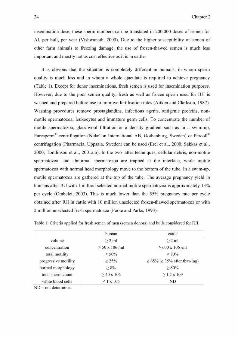
Table 1: Criteria applied for fresh semen of men (semen donors) and bulls considered for IUI.
human cattle volume ≥ 2 ml ≥ 2 ml
concentration ≥ 50 x 106 /ml ≥ 600 x 106 /ml total motility ≥ 50% ≥ 80%
progressive motility ≥ 25% ≥ 65% (≥ 35% after thawing) normal morphology ≥ 8% ≥ 80%
total sperm count ≥ 40 x 106 ≥ 1,2 x 10⁹ white blood cells ≤ 1 x 10⁶ ND
ND = not determined

Intra-uterine insemination in farm animals and humans 25
In humans, the same criteria are used for semen donors as for fertility patients, with an exception
for sperm concentration: ≥ 20 x 106/ml. Data adapted from WHO manual 1999 and A. de Kruif
(personal communication).
Recently, however, a situation comparable to that in humans has arisen in farm
animals. This is due to one of the most important technical advances in sperm processing:
namely sexing of sperm by DNA quantification using flowcytometry. The ability to
separate X- and Y-bearing spermatozoa could be of considerable economic advantage in
the farm animal industry (Seidel, 2003). However, the limited number of spermatozoa that
can be sorted per hour limits its extensive commercial application. Moreover, the low
number of spermatozoa per insemination dose and the lower sperm quality obtained after
the sexing process severely impairs fertility results obtained with sex-sorted semen.
Published data about pregnancy rates obtained with sex-sorted semen in cattle are only
available from heifers under experimental conditions. Pregnancy rates with sex-sorted
semen were significantly lower than with unsexed semen (Table 2) (Seidel et al., 1999).
Moreover, no difference was found between deposition of sex-sorted semen in the uterine
body and uterine horns (Seidel et al., 1999). Since the size of the uterus of older cows is
much larger than in heifers, more sperm loss can be expected and semen deposition near
the UTJ could be advantageous. More research with sexed semen in breeding cows
stressed by heavy lactation, and in superovulated cows, is currently under investigation.
Table 2: Pregnancy rates obtained with sex-sorted semen in the uterine body and horns of heifers.
Treatment / site No. of sperm No. of heifers No. pregnant d 60-63Sexed, frozen / body 1.5 x 10⁶ 27 9 (33%)a Sexed, frozen / body 3.0 x 10⁶ 25 9 (36%)a Sexed, frozen / horns 1.5 x 10⁶ 24 7 (29%)a Sexed, frozen / horns 3.0 x 10⁶ 24 8 (33%)a Control, frozen / body 20 x 106 24 17 (71%)b
a,bMeans without common superscripts differ (P<0.05). From Seidel et al., 1999.
SPERM DISTRIBUTION AND POPULATION OF THE SPERM RESERVOIR
When cattle, sheep, goats and dogs are mated, semen is deposited in or in front of the
cervix uteri. Before oocytes can be fertilized in the ampulla of the oviduct, spermatozoa
have to pass the cervical barrier and migrate through the uterine body to populate the
functional sperm reservoir in the isthmus of the oviduct (Suarez, 1999). During the

26 Chapter 2
transport of the spermatozoa to the site of fertilization, the number of spermatozoa
decreases tremendously (Hunter and Greve, 1998). A great deal is lost by retrograde flow
in the cervical mucus (Larsson and Larsson, 1985; Mitchell et al., 1985; Nelson et al.,
1987) and by phagocytosis during migration through the uterus (Hawk, 1983). Once the
spermatozoa have passed the utero-tubal junction, they enter the isthmus of the oviduct,
where a functional sperm reservoir is established (Hunter and Wilmut, 1982, 1984;
Yanagimachi, 1994; Suarez, 1999). In this sperm friendly environment, spermatozoa are
attached to the apical plasma membrane of the ciliated and secretory epithelial cells until
the moment of ovulation (Suarez et al., 1990). In the isthmus, bovine spermatozoa can
remain arrested for 18 hrs and only detach near the time of ovulation (Hunter and Wilmut,
1984). At the time of activation of the newly-ovulated oocyte, the sperm:egg ratio at the
ampullary-isthmic junction may be close to 1:1 (Hunter and Greve, 1998).
Exceptional kinds of sperm reservoirs are present in poultry and bees and are called
“fossulae spermaticae” and “spermateka” respectively. The fossulae spermaticae are found
in the distal half of the oviduct of all avian species studied to date, sequester and store
spermatozoa which are slowly released over time to insure an adequate population of
spermatozoa at the site of fertilization (Bakst, 1993). However, in contrast to the sperm
reservoir in mammalian farm animals, spermatozoa can be stored for a much longer period
of time. Turkey hens for example, can produce fertilized eggs up to 16 weeks after
insemination when inseminated before onset of egg production (Christensen and Bagley,
1989). The spermateka in bees has a similar function as the fossulae spermaticae in
poultry, but spermatozoa can be stored in this reservoir for 3 to 4 years (Pechhacker, 2003).
Due to the presence of these specific sperm reservoirs in poultry and bees the moment of
insemination is of low importance.
In glaring contrast to poultry and bees, determination of the ideal moment of
insemination in mammalian farm animals and humans is very important. In general, the
ideal moment of insemination in cattle is at 12 hrs after onset of oestrus (Hunter and
Greve, 1997). However, since detection of oestrus is not always easy, determination of the
ideal insemination moment is hampered (Van Eerdenburg et al., 1996). In humans, IUI
insemination is most frequently performed after controlled ovarian hyperstimulation
(COH) and the moment of ovulation can be predicted rather well. Moreover, women can
be monitored very well by means of ultrasound scanning, analysis of luteinizing hormone

Intra-uterine insemination in farm animals and humans 27
(LH), estradiol (E2), and progesterone (P) concentration to determine the best moment of
insemination (Check et al., 1994; Filicori et al., 2003).
When conventional insemination is performed in cattle, horses and pigs, semen is
deposited in the uterine body. In cattle, by-passing the cervix, which is the major barrier to
sperm transport at natural mating, allows a 500-fold reduction of the insemination dose
without a decrease in fertility rate (Senger, 1993). Conventional insemination in cattle is
performed under rectal guidance with a stainless steel Cassou insemination device, which
is covered by a plastic sheet to prevent damage to the female genital tract. In horses and
pigs, IUI can be performed in a rather similar way as in cattle. In ewes, goats and dogs
however, passage of the cervical folds is very difficult and best results for IUI are obtained
with laparoscopy (Hill et al., 1998, Verberckmoes et al., 2001). An alternative for IUI in
these species is intra-vaginal insemination. However at least 10 times as many
spermatozoa are required to obtain similar pregnancy rates as by IUI (Tsutsui et al., 1988;
Linde-Forsberg et al., 1999). Moreover, when intra-vaginal insemination in these species is
performed with frozen-thawed semen, fertility rates decrease tremendously (Armstrong
and Evans, 1984; Salamon and Maxwell, 2000; Thomassen et al., 2001).
In the fifties, a good deal of research in cattle was also performed to compare deep
intra-uterine insemination (in uterine horns) with conventional insemination (in the uterine
body) (Knight et al., 1951; Salisbury and VanDemark, 1951; Stewart and Melrose, 1952;
Olds et al., 1953). However, in most of these studies, which were performed with high
numbers of fresh semen, no significant difference was found between the two insemination
techniques. When deep intra-uterine insemination was performed, semen was deposited
with a rigid insemination device in the uterine horn, but very few experiments were
performed with a flexible insemination device with which semen was deposited near the
utero-tubal junction (Hawk et al., 1988). Theoretically, the insemination dose as used for
conventional insemination in cattle (15 x 106 spermatozoa) could be reduced by at least
100-fold if semen was deposited near the UTJ (Hunter, 2001). This would mainly be due to
the decreased loss of spermatozoa (Hunter, 2001). In the nineties, the possibility to sort
semen of farm animals in X- and Y-bearing spermatozoa led to new interests in deep
insemination (Johnson, 1991; Cran et al., 1993). In pigs and horses, pregnancy cannot be
achieved with low doses of non-sexed semen (5 x 10⁶) or with sex-sorted semen by means
of conventional insemination, but pregnancy can be established with UTJ insemination

28 Chapter 2
(Morris and Allen, 2002; Rath, 2003a). In horses, a distinctive advantage of semen
deposition directly on the ostium uterinum tubae (utero-tubal papilla) rather than in the
uterine body has been shown when the number of spermatozoa was reduced to 3 million
(Morris et al., 2000). To be sure that the semen is deposited on the utero-tubal junction, the
insemination is performed by means of a hysteroscope (Lindsey et al., 2002). To enable
deposition of semen near the utero-tubal junction (UTJ) in sows, specially designed
flexible insemination devices have been developed (Martinez et al., 2001; Grossfeld et al.,
2003). Due to the curvature of the uterine horns in these species at the moment of
insemination, no rigid insemination device can be used for UTJ insemination. In studies by
Rath et al. (2003b) and Grossfeld et al. (2003), sows recently produced piglets after UTJ
insemination with 50 x 106 sex-sorted spermatozoa. This is 2.5% of the conventional
insemination dose of non-sexed semen. In bovines, as in horses and pigs, deep
insemination is advised in cases of low dose insemination (< 5million frozen-thawed
spermatozoa) or with low fertile semen. However, up to now, no beneficial effect of deep
insemination could be established (Seidel et al., 1999). The fact that only heifers were used
in these studies may largely explain the absence of any difference. Since the uterine size is
bigger in older cows than in heifers, it is more likely that a beneficial effect of UTJ
insemination will be observed in older cows.
A remarkable anatomical difference between farm animals and humans is the relatively
bigger uterine body (cavity) and absence of uterine horns in humans. In humans,
inseminations can be performed intravaginally, intracervically, pericervically with a cap,
intra-uterine, intra-tubally or directly intra-peritoneally. However intra-uterine
insemination (IUI) seems to be the method of choice in most studies (Oei et al., 1992;
Ripps et al., 1994; Ombelet et al., 1995; Guzick et al., 1999). IUI is a simple, cheap and
lowly invasive technique by which the inseminate is slowly injected, high up in the uterine
cavity (Peterson et al., 1994). Deposition of semen near the UTJ is not performed in
humans, however clinical trials have been performed in which sperm was directly
deposited into the oviduct (fallopian sperm perfusion, FSP). FSP could be beneficial for
treatment of patients with unexplained infertility, but in other cases of infertility FSP
appeared to be equal to or perhaps inferior to IUI (Karande et al., 1995; Nuojua-Huttunen
et al., 1997; Trout and Kemmann, 1999).

Intra-uterine insemination in farm animals and humans 29
In contrast to farm animals, the UTJ in humans is shaped rather like a funnel and is not
guarded by mucosal folds (Hafez and Black, 1969; Beck and Boots, 1974). So, while in
animals the UTJ may act as a barrier, in humans spermatozoa may be guided right into the
UTJ. Moreover, in humans there is no conclusive evidence for a distinct oviductal sperm
reservoir such as that of farm animals (Williams et al., 1993). Although some human
spermatozoa have been observed to stick to epithelium under certain conditions in vitro
(Pacey et al., 1995), spermatozoa rarely have been observed to bind tightly to oviductal
epithelium in vitro (Yeung et al., 1994; Murray and Smith, 1997).
CONCLUSION
IUI is a simple and successful technique for assisted reproduction in animals and
humans. While IUI in human is performed for reasons of subfertility, mainly caused by
poor sperm quality, IUI in farm animals was originally performed for sanitary reasons but
later especially for economic reasons. However, since the introduction of sex-sorted semen
in farm animals, the same problem of subfertility as in human has arisen. To optimize
fertilization rates with low insemination doses and semen of poor quality, deposition of the
semen near the UTJ has been proven to be successful in sows and mares. As in sows and
mares, UTJ insemination in cattle could be advantageous for insemination with low doses
of semen and with semen of poor quality.

30 Chapter 2
REFERENCES
Aitken RJ, Clarkson JS. Cellular basis of defective sperm function and its association with
the genesis of reactive oxygen species by human spermatozoa. J Reprod Fertil 1987;
81: 459-69.
Allen NC, Herbert CM, Maxson WS, Rogers BJ, Diamond MP, Wentz AC. Intrauterine
insemination: a critical review. Fertil Steril 1985; 44: 568-580.
Amann RP, Hammerstedt RH. In vitro evaluation of sperm quality: an opinion. J Androl
1993; 14: 397-406.
American Fertility Society. New guidelines for the use of semen donor insemination: 1990.
Fertil Steril 1990; 53: 1-13.
Armstrong DT, Evans G. Intrauterine insemination enhances fertility of frozen semen in
superovulated ewes. J Reprod Fertil 1984; 71: 89-94.
Bakst MR. Oviducal sperm storage in poultry: a review. Reprod Fertil Dev 1993; 5: 595-9.
Barratt CLR, Dunphy BC, Mc Leod I, Cooke ID. The poor prognostic value of low to
moderate levels of sperm surface-bound antibodies. Hum Reprod 1992; 7: 95-98.
Barth AD. Factors affecting fertility with artificial insemination. Vet Clin North Am Food
Anim Pract 1993; 9: 275-89.
Beck L.R, Boots LR. The comparative anatomy, histology and morphology of the
mammalian oviduct. In: The oviduct and its function, Johnson AD, Foley CW (Eds).
New York, Academic Press, 1974; pp 2-51.
Bronson R, Cooper G, Rosenfeld D. Sperm antibodies: their role in infertility. Fertil Steril
1984; 42: 171-83.

Intra-uterine insemination in farm animals and humans 31
Check JH, Peymer M, Jairaj P. Single daily monitoring of periovulatory estradiol,
progesterone, and luteinizing sera hormone levels in natural cycles useful for timing
intrauterine insemination. Arch Androl 1994; 32: 151-6.
Christensen VL, Bagley LG. Efficacy of fertilization in artificially inseminated turkey
hens. Poult Sci 1989; 68: 724-9.
Chupin D, Schuh H. Survey of the present status of the use of artificial insemination in
developing countries. World Anim Rev 1993; 74/75: 20-35.
Chupin D, Thibier M. Survey of the present status of the use of artificial insemination in
developing countries. World Anim Rev 1995; 82: 58-68.
Coetzee K, Kruge TF, Lombard CJ. Predictive value of normal sperm morphology: a
structured literature review. Hum Reprod Update 1998; 4: 73-82.
Cohlen BJ, Vandekerckhove P, te Velde ER, Habbema JD. Timed intercourse versus intra-
uterine insemination with or without ovarian hyperstimulation for subfertility in men.
Cochrane Database Syst Rev CD 2000; 360.
Colenbrander B, Gadella BM, Stout TA. The predictive value of semen analysis in the
evaluation of stallion fertility. Reprod Dom Anim 2003; 38: 305-11.
Cran DG, Johnson LA, Miller NG, Cochrane D, Polge C. Production of bovine calves
following separation of X- and Y-chromosome bearing sperm and in vitro
fertilisation. Vet Rec 1993; 132: 40-1.
Cumming IR. Suitability of the intact acrosome method for the prediction of fertility in
bovine artificial insemination. Vet Rec 1995; 136: 289-91.
de Kruif A. Fertility and sterility in domestic animals. Verh K Acad Geneeskd Belg 2003;
65: 189-202.

32 Chapter 2
De Pauw I, Van Soom A, Verberckmoes S, de Kruif A. De overleving van sperma in de
epididymis van de stier: een model voor spermaopslag in vitro? Vlaams
Diergeneeskundig Tijdschrift 2000; 69: 159-167.
De Pauw I, Van Soom A, Laevens H, Verberckmoes S, de Kruif A. Sperm binding to
epithelial oviduct explants in bulls with different non return rates investigated with a
new in vitro model. Biol Reprod 2002; 67: 1073-9.
Den Daas JH, De Jong G, Lansbergen LM, Van Wagtendonk-De Leeuw AM. The
relationship between the number of spermatozoa inseminated and the reproductive
efficiency of individual dairy bulls. J Dairy Sci 1998; 81: 1714-23.
Donoghue AM, Wishart GJ. Storage of poultry semen. Anim Reprod Sci 2000; 62: 213-32.
Erel CT, Senturk LM, Irez T, Ercan L, Elter K, Colgar U, Ertungealp E. Sperm-preparation
techniques for men with normal and abnormal semen analysis. A comparison. J
Reprod Med 2000; 45: 917-22.
Farrell PB, Presicce GA, Brockett CC, Foote RH. Quantification of bull sperm
characteristics measured by computer-assisted sperm analysis (CASA) and the
relationship to fertility. Theriogenology 1998; 49: 871-9.
Filicori M, Cognigni GE, Pocognoli P, Tabarelli C, Ferlini F, Perri T, Parmegiani L.
Comparison of controlled ovarian stimulation with human menopausal gonadotropin
or recombinant follicle-stimulating hormone. Fertil Steril 2003; 80: 390-7.
Foote RH, Henderson CR, Bratton RW. Testing bulls in artificial insemination centres for
lethals, type and production. Proc 3rd Int Congr Anim Reprod 1956; 3: 49-53.
Foote RH. Extenders and extension of unfrozen semen. In: Salisbury GL, VanDemark NL,
Lodge JR (Eds) Physiology of Reproduction – Artificial insemination of Cattle.
Freeman, San Francisco 1978; pp 442- 493.
Intra-uterine insemination in farm animals and humans 33
Foote RH, Parks JE. Factors affecting preservation and fertility of bull sperm: a brief
review. Reprod Fertil Dev 1993; 5: 665-73.
Foote RH. The history of artificial insemination: selected notes and notables. Am Soc
Anim Sci 2002; 1-10.
Goverde AJ, McDonnell J, Vermeiden JP, Schats R, Rutten FF, Schoemaker J. Intrauterine
insemination or in-vitro fertilisation in idiopathic subfertility and male subfertility: a
randomised trial and cost-effectiveness analysis. Lancet 2000; 355: 13-8.
Grossfeld R, Ruiz S, Sieg B, Leigh J, Klinc P, Rath D. Artificial insemination with sexed
semen in pigs. Conference book of the 5th International congress on boar semen
preservation, August 24-27, 2003, Doorwerth, The Netherlands; 62 (abstract).
Guzick DS, Carson SA, Coutifaris C, Overstreet JW, Factor-Litvak P, Steinkampf MP, Hill
JA, Mastroianni L, Buster JE, Nakajima ST, Vogel DL, Canfield RE. Efficacy of
superovulation and intrauterine insemination in the treatment of infertility. National
Cooperative Reproductive Medicine Network. N Engl J Med 1999; 340: 177-83.
Hafez ESE, Black DL. The mammalian uterotubal junction. In: The mammalian oviduct:
comparative biology and methodology, Hafez ESE, Blandeau RJ (Eds) Chicago, The
University of Chicago Press, 1969; pp 85-128.
Hawk HW. Sperm survival and transport in the female reproductive tract. J Dairy Sci
1983; 66: 2645-2660.
Hawk HW, Conley HH, Wall RJ, Whitaker RO. Fertilization rates in superovulating cows
after deposition of semen on the infundibulum, near the uterotubal junction or after
insemination with high numbers of sperm. Theriogenology 1988; 29: 1131-1142.
Hill JR, Thompson JA, Perkins NR. Factors affecting pregnancy rates following
laparoscopic insemination of 28,447 Merino ewes under commercial conditions: a
survey. Theriogenology 1998; 49: 697-709.

34 Chapter 2
Hoffer MA. Bovine campylobacteriosis: a review. Can Vet J 1981; 22: 327-30.
Holt C, Holt WV, Moore HD, Reed HC, Curnock RM. Objectively measured boar sperm
motility parameters correlate with the outcomes of on-farm inseminations: results of
two fertility trials. J Androl 1997; 18: 312-23.
Holt WV. Basic aspects of frozen storage of semen. Anim Reprod Sci 2000; 62: 3-22.
Hunter RHF, Wilmut I. The rate of functional sperm transport into the oviducts of mated
cows. Anim Reprod Sci 1982; 5: 167-173.
Hunter RHF, Wilmut I. Sperm transport in the cow: peri-ovulatory redistribution of viable
cells within the oviduct. Reprod Nutr Dev 1984; 24: 597-608.
Hunter RH, Greve T. Are lower fertility bulls necessarily less fertile? Proposals concerning
insemination procedures. Anim Reprod Sci 1997; 48: 113-21.
Hunter RHF, Greve T. Deep uterine insemination of cattle: a fruitful way forward with
smaller numbers of spermatozoa. Acta Vet Scand 1998; 39: 149-163.
Hunter RHF. New breeding opportunities with deep cornual insemination: exploiting
modern sperm technologies in cattle. Reprod Dom Anim 2001; 36: 217-222.
Ivanoff EI. On the use of artificial insemination for zootechnical puposes in Russia. J Agric
Sci 1922; 12: 244-256.
Johnson LA. Sex selection in swine: altered sex ratios in offspring following surgical
insemination with flow sorted X- and Y-bearing sperm. Reprod Dom Anim 1991; 26:
309-314.
Johnson LA, Weitze KF, Fiser P, Maxwell WM. Storage of boar semen. Anim Reprod Sci
2000; 62: 143-72.
Karande VC, Rao R, Pratt DE, Balin M, Levrant S, Morris R, Dudkeiwicz A, Gleicher N.
A randomized prospective comparison between intrauterine insemination and

Intra-uterine insemination in farm animals and humans 35
fallopian sperm perfusion for the treatment of infertility. Fertil Steril 1995; 64: 638-
40.
Knight CW, Patrick TE, Anderson HW, Branton C. The relation of site of semen deposit to
breeding efficiency of dairy cattle. J Dairy Sci 1951; 34: 199-202.
Larsson B, Larsson K. Distribution of spermatozoa in the genital tract of artificially
inseminated heifers. Acta Vet Scand 1985; 26: 385-395.
Larsson B, Rodriguez-Martinez H. Can we use in vitro fertilization tests to predict semen
fertility? Anim Reprod Sci 2000; 60-61: 327-36.
Leboeuf B, Restall B, Salamon S. Production and storage of goat semen for artificial
insemination. Anim Reprod Sci 2000; 62: 113-41.
Linde-Forsberg C, Strom Holst B, Govette G. Comparison of fertility data from vaginal vs
intrauterine insemination of frozen-thawed dog semen: a retrospective study.
Theriogenology 1999; 52: 11-23.
Lindsey AC, Morris LH, Allen WR, Schenk JL, Squires EL, Bruemmer JE. Hysteroscopic
insemination of mares with low numbers of nonsorted or flow sorted spermatozoa.
Equine Vet J 2002; 34: 128-32.
Linfor JJ, Meyers SA. Detection of DNA damage in response to cooling injury in equine
spermatozoa using single-cell gel electrophoresis. J Androl 2002; 23: 107-13.
Loomis PR. The equine frozen semen industry. Anim Reprod Sci 2001; 68: 191-200.
Lundin K, Sjogren A, Nilsson L, Hamberger L. Fertilization and pregnancy after
intracytoplasmic microinjection of acrosomeless spermatozoa. Fertil Steril 1994; 62:
1266-7.
Martinez AR, Bernardus RE, Vermeiden JPW, Schoemaker J. Basic questions on
intrauterine insemination : an update. Obstet Gynecol Surv 1993; 48: 811-828.

36 Chapter 2
Martinez EA, Vazquez JM, Roca J, Lucas X, Gil MA, Parilla I, Vasquez JL, Day BN.
Succesful non-surgical deep intra-uterine insemination with small numbers of
spermatozoa in sows. Reproduction 2001; 122: 289-96.
Milovanov VK. Isskusstvenoye ossemenebie selsko-khoziasvennykh jivotnykh. (The
artificial insemination of farm animals). Seljhozgiz, Mowcow. 1938.
Mitchell JR, Senger PL, Rosenberger JL. Distribution and retention of spermatozoa with
acrosomal and nuclear abnormalities in the cow genital tract. Anim Sci 1985; 61:
956-967.
Mocé E, Lavara R, Vicente JS. Effect of an asynchrony between ovulation and
insemination on the results obtained after insemination with fresh or frozen sperm in
rabbits. Anim Reprod Sci 2003; 75: 107-18.
Montanaro Gauci M, Kruger TF, Coetzee K, Smith K, Van Der Merwe JP, Lombard CJ.
Stepwise regression analysis to study male and female factors impacting on
pregnancy rate in an intrauterine insemination programme. Andrologia 2001; 33:
135-41.
Morris LHA, Hunter RHF, Allen WR. Hysteroscopic insemination of small numbers of
spermatozoa at the uterotubal junction of preovulatory mares. J Reprod Fert 2000;
118: 95-100.
Morris LHA, Allen WR. An overview of low dose insemination in the mare. Reprod Dom
Anim 2002; 37: 206-210.
Murray SC, Smith TT. Sperm interaction with fallopian tube apical membrane enhances
sperm motility and delays capacitation. Fertil Steril 1997; 68: 351-7.
Nelson VE, Aalseth EP, Hawman CH, Adams GD, Dawon LJ, McNew RW. Sperm
discharge and distribution within the cows reproductive tract and after A.I. Anim Sci
1987; 65: 401.

Intra-uterine insemination in farm animals and humans 37
Nuojua-Huttunen S, Tuomivaara L, Juntunen K, Tomas C, Martikainen H. Comparison of
fallopian tube sperm perfusion with intrauterine insemination in the treatment of
infertility. Fertil Steril 1997; 67: 939-42.
Oehninger S. Place of intracytoplasmatic sperm injection in management of male
infertility. Lancet 2001; 357: 2068-2069.
Oei ML, Surrey ES, McCaleb B, Kerin JF. A prospective, randomized study of pregnancy
rates after transuterotubal and intrauterine insemination. Fertil Steril 1992; 58: 167-
71.
Olds D, Seath DM, Carpenter MC, Lucas HL. Interrelationships between site of
deposition, dosage, and number of spermatozoa in diluted semen and fertility of dairy
cows inseminated artificially. J Dairy Sci 1953; 36: 1031-1035.
Ombelet W, Puttemans P, Bosmans E. Intrauterine insemination: a first-step procedure in
the algorithm of male subfertility treatment. Hum Reprod 1995; 10 (Suppl 1): 90-
102.
Ombelet W, Vandeput H, Van de Putte G, Cox A, Janssen M, Jacobs P, Bosmans E,
Steeno O, Kruger T. Intrauterine insemination after ovarian stimulation with
clomiphene citrate: predictive potential of inseminating motile count and sperm
morphology. Hum Reprod 1997; 12: 1458-63.
Ombelet W. Semen quality and intrauterine insemination. Reproductive BioMedicine
Online 2003; 7: 167-175.
Pacey AA, Hill CJ, Scudamore IW, Warren MA, Barratt CL, Cooke ID. The interaction in
vitro of human spermatozoa with epithelial cells from the human uterine (fallopian)
tube. Hum Reprod 1995; 10: 360-6.
Palermo G, Joris H, Devroey P, Van Steirteghem AC. Pregnancies after intracytoplasmic
injection of single spermatozoon into an oocyte. Lancet 1992; 340: 17-8.

38 Chapter 2
Palermo GD, Cohen J, Rosenwaks Z. Intracytoplasmic sperm injection: a powerful tool to
overcome fertilization failure. Fertil Steril 1996; 65: 899-908.
Pechhacker H. Artificial insemination and mating methods in a honeybee breeding
program. Proceedings AI Vets meeting 2003, Budapest, Hungary, 26-28.
Peterson CM, Hatasaka HH, Jones KP, Poulson AM Jr Carrell DT, Urry RL. Ovulation
induction with gonadotropins and intrauterine insemination compared with in vitro
fertilization and no therapy: a prospective, nonrandomized, cohort study and meta-
analysis. Fertil Steril 1994; 62: 535-44.
Philips Z, Barraza-Llorens M, Posnett J. Evaluation of the relative cost-effectiveness of
treatments for infertility in the UK. Hum Reprod 2000; 15: 95-106.
Rath D. Current perspectives of sperm sorting in domestic farm animals. Proc. of the 19th
scientific meeting of the European embryo transfer association, 12-13 September
Rostock 2003a; 125-128.
Rath D, Ruiz S, Sieg B. Birth of piglets after intra-uterine insemination employing
flowcytometrically sexed boar semen, a case report. Vet Rec 2003b; 152: 400-401.
Reddy RP. Artificial insemination of broilers: economic and management implications. In:
Bakst, MR, Wishart GJ (Eds), Proc 1st Int symp on the artificial insemination of
poultry. Poultry Science Association, Savoy, IL, 1995; pp 73-89.
Ripps BA, Minhas BS, Carson SA, Buster JE. Intrauterine insemination in fertile women
delivers larger number of sperm to the peritoneal fluid than intracervical
insemination. Fertil Steril 1994; 61: 398-400.
Rodriguez-Martinez H. Laboratory semen assessment and prediction of fertility: still
utopia? Reprod Dom Anim 2003; 38, 312-318.
Rowell P, Braude P. Assisted conception. BMJ 2003; 327: 799-801.

Intra-uterine insemination in farm animals and humans 39
Sakkas D, Manicardi GC, Tomlinson M, Mandrioli M, Bizzaro D, Bianchi PG, Bianchi U.
The use of two density gradient centrifugation techniques and the swim-up method to
separate spermatozoa with chromatin and nuclear DNA anomalies. Hum Reprod
2000; 15: 1112-6.
Salamon S, Maxwell WMC. Storage of ram semen. Anim Reprod Sci 2000; 62: 77-111.
Salisbury GW, VanDemark NL. The effect of cervical, uterine and cornual insemination
on fertility of the dairy cow. J Dairy Sci 1951; 34: 68-74.
Seidel GE. Economics of selecting for sex: the most important genetic trait.
Theriogenology 2003; 59: 585-598.
Seidel GE, Schenk JL, Herickhoff LA, Doyle SP, Brink Z, Green RD, Cran DG.
Insemination of heifers with sexed sperm. Theriogenology 1999; 52: 1407-20.
Senger PL. Site of semen deposition and its effect on fertility and sperm retention: a
review. Reprod Fertil Dev 1993; 5: 659-63.
Shannon P. Factors affecting semen preservation and conception rates in cattle. J. Reprod
Fertil 1978; 54: 519-527.
Sieger SWJ. Semen collection and evaluation in the dog. In: Current therapy in
Theriogenology. Morrow DA (Ed.). WB Saunders Company, Philadelphia, 1986; pp
539-544.
Siegler SL. Fertility in women. JB Lippincott, Philadelphia, 1944; pp 403.
Silber SJ, Van Steirteghem AC, Liu J, Nagy Z, Tournaye H, Devroey P. High fertilization
and pregnancy rate after intracytoplasmic sperm injection with spermatozoa obtained
from testicle biopsy. Hum Reprod 1995; 10: 148-52.
Singleton WL. State of the art in artificial insemination of pigs in the United States.
Theriogenology 2001; 56: 1305-10.
Sims JM. In: Uterine surgery. William Wood (Ed.), New York, 1873; pp 365.

40 Chapter 2
Spallanzani L. Dissertations relative to the natural history of animals and vegetables.
Trans. by T Beddoes In: Dissertations relative to the Natural history of animals and
vegetables. J Murray , London, 1784; 2: 195-199.
Stalhammar EM, Janson L, Philipsson J. The impact of sperm motility on non-return rate
in preselected dairy bulls. Reprod Nutr Dev 1994; 34: 37-45.
Stewart DL, Melrose DR. The comparative efficiency of the intra cervical and intra-uterine
methods of insemination in the dairy cow. Vet Rec 1952; 64: 605-607.
Suarez SS, Drost M, Redfern K, Gottlieb W. Sperm motility in the oviduct. In: Bavister
BD, Cummins J, Roldan ERS (eds). Fertilization in mammals. Serono Symposia;
Norwell MA 1990: 111-124.
Suarez SS. Regulation of sperm transport in the mammalian oviduct. In: The male gamete
(C Gagnon Ed.), Cache river press, Vienna, 1999; pp 71-80.
Suarez SS. Carbohydrate-mediated formation of the oviductal sperm reservoir in
mammals. Cells tissues organs 2001; 168: 105-112.
Thibier M, Guerin B. Hygienic aspects of storage and use of semen for artificial
insemination. Anim Reprod Sci 2000; 62: 233-251.
Thibier M, Wagner HG. World statistics for artificial insemination in cattle. Proc 14th Int
Congr Anim Reprod 2000; 22: 76 (abstract).
Thomassen R, Farstad W, Krogenaes A, Fougner JA, Berg KA. Artificial insemination
with frozen semen in dogs: a retrospective study. J Reprod Fertil 2001; 57: 341-6.
Timoney PJ. Contagious equine metritis. Comp Immunol Microbiol Infect Dis 1996; 19:
199-204.
Tomlinson M, Turner J, Powell G, Sakkas D. One-step disposable chambers for sperm
concentration and motility assessment: how do they compare with the World Health
Organization's recommended methods? Hum Reprod 2001a; 16: 121-124.

Intra-uterine insemination in farm animals and humans 41
Tomlinson MJ, Moffatt O, Manicardi GC, Bizzaro D, Afnan M, Sakkas D.
Interrelationships between seminal parameters and sperm nuclear DNA damage
before and after density gradient centrifugation: implications for assisted conception.
Hum Reprod 2001b; 16: 2160-5.
Trout SW, Kemmann E. Fallopian sperm perfusion versus intrauterine insemination: a
randomized controlled trial and metaanalysis of the literature. Fertil Steril 1999; 71:
881-5.
Tsutsui T, Tezuka T, Shimizu T, Murao I, Kawakami E, Ogasa A. Artificial insemination
with fresh semen in beagle bitches. Nippon Juigaku Zasshi 1988; 50: 193-8.
Van Eerdenburg FJ, Loeffler HS, van Vliet JH. Detection of oestrus in dairy cows: a new
approach to an old problem. Vet Q 1996; 18: 52-4.
Van Leeuwenhoek A. De natis è semine genitali animalcules. R Soc (London) Philos Trans
1678; 12: 1040-1043.
Van Steirteghem AC, Nagy Z, Joris H, Liu J, Staessen C, Smitz J, Wisanto A, Devroey P.
High fertilization and implantation rates after intracytoplasmic sperm injection. Hum
Reprod 1993; 8: 1061-6.
Van Waart J, Kruger TF, Lombard CJ, Ombelet W. Predictive value of normal sperm
morphology in intrauterine insemination (IUI): a structured literature review. Hum
Reprod Update 2001; 7: 495-500.
Verberckmoes S, De Pauw I, Vanroose G, Laevens H, de Kruif A. Cervicale insemination
in sheep. Vlaams Diergeneeskundig Tijdschrift 2001; 70: 475-480.
Vishwanath R, Shannon P. Storage of bovine semen in liquid and frozen state. Anim
Reprod Sci 2000; 62: 23-53.
Vishwanath R. Artificial insemination: the state of the art. Theriogenology 2003; 59: 571-
584.

42 Chapter 2
Vyt P, Maes D, Dejonckheere E, Castryck F, Van Soom A. Comparative study on five
different commercial extenders for boar semen. Reprod Dom Anim 2004; 39: 8-12.
Watson PF. Artificial insemination and the preservation of semen. In: Lamming GE,
editor. Marshall’s physiology of reproduction. Edinburgh: Churchill Livingstone,
1990; pp 747-869.
Williams M, Hill CJ, Scudamore I, Dunphy B, Cooke ID, Barratt CLR. Sperm numbers
and distribution within the human fallotubian tube around ovulation. Hum Reprod
1993; 8: 2019-2026.
Wolf DP, Patton PE, Burry KA, Kaplan PF. Intrauterine insemination-ready versus
conventional semen cryopreservation for donor insemination: a comparison of
retrospective results and a prospective, randomized trial. Fertil Steril 2001; 76: 181-
185.
World Health Organisation. WHO laboratory manual for the examination of human semen
and sperm-cervical mucus interaction 1999.
Yanagimachi R. Mammalian fertilization. In: Knobil E, Neill J (eds.) Physiology of
Reproduction. New York: Raven Press, 1994; pp 189-317.
Yeung WS, Ng VK, Lau EY, Ho PC. Human oviductal cells and their conditioned medium
maintain the motility and hyperactivation of human spermatozoa in vitro. Hum
Reprod 1994; 9: 656-60.
Zhang BR, Larsson B, Lundeheim N, Haard MG, Rodriguez-Martinez H. Prediction of
bull fertility by combined in vitro assessments of frozen-thawed semen from young
dairy bulls entering an AI-programme. Int J Androl 1999; 22: 253-60.

CHAPTER 3
COLLECTION AND COMPOSITION OF CAUDA EPIDIDYMAL PLASMA IN THE
BULL

44 Chapter 3
ABSTRACT
When artificial insemination (AI) is performed in cattle, much lower insemination doses
are required with fresh semen than with frozen-thawed semen to obtain similar pregnancy
rates. However, a particular disadvantage of fresh semen is its limited shelf life. Since bovine
spermatozoa can be stored for several weeks in the cauda epididymidis without negative
effects on their fertilizing capacity, the cauda epididymidis is an interesting object to serve as
a model in order to prolong the shelf life of fresh semen. In this study, bovine cauda
epididymal fluid (CEF) was collected in vitro and in vivo, and the biochemical composition
of the cauda epididymal plasma (CEP) was determined. Cauda epididymal fluid was collected
in vitro by micropuncture of epididymides of slaughtered bulls. To evaluate the cannulation
technique for the in vivo collection of CEF, cannulations were first performed in rams and the
technique was subsequently applied in bulls. After centrifugation of the CEF, the CEP was
separated and the biochemical composition of several ions and metabolites in the CEP was
determined in a clinical laboratory. Finally, a new diluent with the same ionic composition,
pH and osmolarity as the bovine CEP was developed and compared with the standard Tris
diluent for extended preservation of fresh ejaculated semen. Storage of fresh ejaculated
bovine semen in a diluent with the same ionic composition as the cauda epididymal plasma
does not bring the ejaculated spermatozoa in a quiescent state nor extend their shelf life.

Collection and composition of cauda epididymal plasma in the bull 45
INTRODUCTION
The widespread use of artificial insemination (AI) in cattle can partially be attributed to
the availability of suitable sperm diluents (Almquist, 1962; Shannon, 1964; Foote, 1970) and
to the fact that small amounts of frozen-thawed spermatozoa (15 millions) can be deposited
beyond the cervical barrier, in the uterus of the cow (Salisbury and Van Demark, 1961)
Although great efforts have been made to optimize the freezing and thawing procedure of
bovine spermatozoa, the cryopreservation process still has detrimental effects on semen
quality (Watson, 1995; Woelders, 1997; Van Wagtendonk-de Leeuw et al., 2000, Frijters and
Luimstra-Mulder, 2003). Vishwanath et al. (1996) showed that in cattle 10 times more frozen-
thawed spermatozoa were required to obtain similar pregnancy rates as with fresh semen. The
ability to obtain good pregnancy rates with low insemination doses would increase the world
wide spreading of genetically superior bulls. This is not only beneficial for the genetic
progress in cattle breeding, but also increases the profits of the AI centres. However, one of
the most important disadvantages of fresh semen is its limited storage capacity. Currently,
fresh semen can be used for AI up to 3 days after collection when it is stored at room
temperature, under N2 gas in Caprogen® diluent (Livestock improvement corporation,
Hamilton, New Zealand) (Vishwanath and Shannon, 1997). Since this storage period is rather
short, more extended semen preservation time would decrease the work of sperm collection
and would facilitate the distribution of fresh semen.
Based on the peculiar capacity of the cauda epididymidis to store bovine epididymal
semen for several weeks without detrimental effects on the fertilizing capacity (Kirillov and
Mozorov, 1936; Cascieri et al., 1976), we wanted to develop a new diluent for fresh
ejaculated bovine semen based on the cauda epididymal conditions. The cauda epididymidis
is characterized by low pH (Carr and Acott, 1984; Acott and Carr, 1984), hyperosmotic
pressure of the fluid (Liu and Foote, 1998), high concentration of spermatozoa (Cascieri et al.,
1976; Amann et al., 1976) low oxygen tension (Free et al., 1979), and low Na / K ratio
(Crabo, 1965). Recently, genes responsible for the production of specific proteins present in
CEP, maturation of epididymal spermatozoa, and secretion and absorption processes in the
epididymis have been thoroughly investigated (Jones et al., 1998). However, only a few
studies have been published concerning the ionic composition of cauda epididymal plasma
(CEP) in bulls (Crabo, 1965; Amann et al., 1963).

46 Chapter 3
Therefore, in this study, we first investigated the biochemical and more specifically, the
ionic composition of the CEP collected by two different techniques. In vitro collection of CEP
was performed by micropuncture of epididymides of slaughtered bulls. In vivo collection by
means of cannulation was first performed on rams, and subsequently used in bulls. The
effectiveness of the newly developed diluent based on the ionic composition of CEP was
compared with the standard Tris diluent in a 6-days storage experiment.
MATERIAL AND METHODS
Exp 1: In vitro collection of epididymal plasma
Testicles of Belgian White-Blue beef bulls (aged 2-2.5) years were collected at the
slaughterhouse, stored in an insulated container, and transported to the laboratory within 2
hours. Four collections of 100 epididymides per collection were performed. After removing
superficial blood and interstitial fluid contamination, the pressure in the epididymal ducts was
increased by clamping pincers on the proximal and distal part of the cauda epididymidis.
Incisions were made in the connective tissue and care was taken to avoid small blood vessels.
The epididymal fluid oozed out and was aspirated into a fine glass pipette and transferred into
a small vial (Figure 1). To separate the sperm fraction from the plasma fraction, the
epididymal fluid was centrifuged for 5 min at 3.214 x g. Subsequently, the superficial
uncontaminated epididymal plasma fraction was removed and frozen at –20°C. Since the
volume of CEP per testicle was much to low for individual analysis, cauda epididymal plasma
of 100 epididymides was pooled.
Figure 1: Aspiration of epididymal plasma with a fine glass pipette from the cauda epididymidis of a bovine testicle.

Collection and composition of cauda epididymal plasma in the bull 47
Exp 2: In vivo collection of epididymal plasma of ram and bull
The institutional ethical committee for animal welfare of Ghent University approved all
procedures involving the animals used in this study. The ram was used as a model for the bull
to test the cannulation technique as previously reported by Amann et al. (1963) and Henault et
al. (1995). Six Suffolk rams aged 1.5 to 2.5 years were used in this experiment, which was
performed during the mating season (September-October). One week before cannulation, the
quality of the ejaculated semen collected by means of an artificial vagina was assessed
microscopically. For each ram, the progressive motility, the percentage of membrane intact
spermatozoa, and the percentage of spermatozoa with normal morphology were at least 80%.
Before the operation, rams were starved for 24 hrs but had access to water ad libitum. An
intra-muscular injection of Xylazine (Xyl-M 2%, VMD, Arendonk, België) (1 ml/100 kg) was
used as tranquillizer and combined with 15 ml of 2% procaine (VMD, Arendonk, België) as
local anaesthesia. When the ram was tranquillized, he was fixed on the operation table.
Subsequently, the scrotum was shaved, cleaned, and disinfected, and a sterile plastic cloth
was used to keep the operation field clean.
A system of three cannulae which were axially movable into each other was used (Figure
2). This set-up was used to dilate the lumen of the ductus deferens without causing damage
and to prevent cracking of the cannulae. To enable daily replacement of the collection vial,
the cannulation set was equipped with a screw cap at one end.
Figure 2: Different parts of the cannulation set: 1) inner tube, 2) outer tube, 3) protecting tube, 4) screw cap, 5) collection vial.
To insert the cannulation system into the ductus deferens, a 6-8 cm incision was made in
the caudal part of the scrotum immediately lateral to the septum scroti. The ductus deferens
was exposed over a distance of 5 cm after making a small incision in the tunica vaginalis, and
1 2
1
3
2
4 5
3
1 2
1
3
2
4 5
3
1 2
1
3
2
4 5
3
1 2
1
3
2
4 5
3

48 Chapter 3
isolated from the adjacent blood vessels and nerves by blunt dissection. A cross section was
made in the ductus deferens and the inner silicone cannula was inserted into the lumen as far
as possible (± 5 cm). An outer silicone cannula was placed completely over the inner cannula,
fixed with glue and sutured to the ductus deferens and the tunica vaginalis by 4-0 silk sutures
(Ethicon®, Neuilly, France). Removal of the inner cannula increased the free space in the
outer cannula, and decreased the risk of obstruction. A protecting cannula (Tygon®, R3603,
Fisher Bioblock Scientific, Tournay, Belgium) with a screw cap was placed over the outer
cannula to prevent cracking. The cauda epididymal effluent was harvested via the indwelling
outer cannula emptying into a sterile collection vial. The collection vial was replaced every
24h and after sexual stimulation of the animal. When signs of infection or discoloration of the
epididymal contents were observed, the cannulation set was removed. The epididymal
effluent was centrifuged for 5 min at 3.214 x g. The uncontaminated epididymal plasma was
removed and frozen at –20°C. The epididymal plasma collected during the whole cannulation
period, was pooled per individual animal.
For the cannulation of the bovine ductus deferens, 9 Holstein bulls aged 1.5 to 2.5 years
were used. As in rams, one week before cannulation, the quality of the ejaculated semen was
assessed and was good for all bulls. The same threshold levels were used as in the ram. The
operation technique applied in the bull was identical to the technique described in the ram. In
contrast to rams, bulls were cannulated at the opposite side when insufficient epididymal
plasma was collected at the first cannulation side.
Biochemical analysis of cauda epididymal plasma
After collection and storage at –20°C, the biochemical composition of CEP of the ram and
bull was analyzed. Cobas® Integra cassettes (Roche, Mannheim, Germany) were used for the
analysis of Na, K, Cl, Ca, Mg, inorganic phosphate, bicarbonate, glucose, lactate and
cholesterol. Osmolarity was evaluated with an osmometer (Fisk Associates®, Massachusetts,
USA), and for the analysis of the pH, pH electrodes (pH 526, TUV®, Wilheim, Germany)
were used.
Storage of fresh bovine semen in Cauda Epididymal Plasma (CEP) diluent
Fresh semen of 2 bulls with a normal sperm quality (progressive motility, percentage
membrane intact spermatozoa, and percentage spermatozoa with normal morphology ≥ 80%)
was collected by means of an artificial vagina in a falcon tube (Becton and Dickinson®, Le

Collection and composition of cauda epididymal plasma in the bull 49
Pont de Claix, France) containing 5 ml of Triladyl diluent, according to the method of De
Pauw et al. (2003). Immediately after collection, the semen quality was evaluated, and
subsequently the semen was centrifuged at 700 x g for 10 min. After centrifugation, the
seminal plasma and Triladyl diluent were removed. Subsequently the fresh semen was stored
in a liquid state at 5°C in the Tris and in a newly developed diluent based on the composition
of the CEP. Both these media contained 10% egg yolk. The sperm was diluted to a
concentration of 10 x 106 spermatozoa / ml and was stored in 1.8 ml cryovials (Nunc®,
Wiesbaden, Germany) for 6 days. The sperm concentration was determined using a Bürker
counting chamber by means of light microscopy (x 400).
The quality of the spermatozoa stored in Tris and in CEP diluent was evaluated before
(t0), and after 2 (t2), 4 (t4), and 6 (t6) days of incubation. Membrane integrity was assessed
by means of eosin-nigrosin staining and light microscopy (x 400). Total motility (TM) and
progressive motility (PM) were assessed subjectively by 2 persons by means of light
microscopy (x 200, 37°C). The Functional Sperm Concentration (FSC), Motile Sperm
Concentration (MSC) and Sperm Motility Index (SMI) were determined in three replicates
with the Sperm Quality Analyzer (SQA II-C®) (Orange Medical, Tilburg, The Netherlands).
The SQA II-C® is provided of a photometric cell that registers the fluctuations in optical
density and converts this information, through mathematical algorithms, digitally to a
numerical output. The FSC represents the cells that are both motile and morphologically
normal (x 106/ml), while the MSC represents the cells that are normally motile (concentration
x % motility) in millions/ml. The SMI is expressing the overall sperm quality, taking into
account three parameters, namely the concentration, the progressive motility, and the
percentage spermatozoa with a normal morphology.
STATISTICAL ANALYSIS
The storage experiment was repeated 3 times. Data were analysed using a linear mixed
effect model, with bull and repetition as random variables, day as group variable and allowing
different standard deviations per stratum (S-plus 2000, Math Soft Inc, Cambridge, USA).
Since residuals were not normally distributed for the FSC and MSC results, a log
transformation was performed. Differences in sperm quality were considered as being
significant when p-values were lower than 0.05.

50 Chapter 3
RESULTS
Exp 1: Collection of epididymal plasma by means of micropuncture from epididymides
derived from the slaughterhouse
When micropuncture was used, the cauda epididymal fluid of 100 epididymides could be
collected by 2 persons within 2 hrs. However, only small amounts (115 ± 50 µL) of
epididymal fluid were harvested per epididymis. The use of clamping pincers increased the
pressure in the epididymal ducts, and facilitated the collection of the epididymal fluid. The
cauda epididymal duct was clearly demarcated and micropuncture could be performed
precisely. After centrifugation of the cauda epididymal fluid, only one fourth of the total
volume was clear plasma which was aspirated and stored at –20°C until analysis.
Exp 2: In vivo collection of epididymal plasma by means of cannulation of the ductus
deferens
In the ram, the cannulation technique of the ductus deferens was easy to perform. The
ductus deferens could be located easily by palpation through the tunica vaginalis. Moreover,
the consistency and straight pathway of the ductus deferens facilitated the insertion of the
cannulation set. In the bull however, the ductus deferens was much more curled and insertion
of the cannulation set up to the cauda epididymidis was more complicated. No epididymal
fluid could be collected in one ram and two bulls which was probably due to trauma or
inflammation caused by the operation. In two other bulls epididymal fluid was collected, but
the amount of plasma obtained after centrifugation was not sufficient for biochemical
analysis. Uncontaminated epididymal fluid could be obtained in rams for an average period of
17.2 ± 4.7 days, whereas this period was reduced to 9.2 ± 4.8 days in the bull. Variation in the
daily collection volume occurred both in rams and bulls. In one bull, no epididymal fluid
could be collected for 2 days due to obstruction of the cannula. Insertion of a small catheter
and sexual stimulation resolved this problem. The average daily production of epididymal
fluid after successful cannulation was 338.4 ± 113.6 µL and 426.3 ± 137.3 µL in rams and
bulls, respectively. The total output of epididymal fluid during the entire collection period
was 3941.0 ± 1020.2 µL and 4108.3 ± 2420.4 µL in the ram and in the bull, respectively.

Collection and composition of cauda epididymal plasma in the bull 51
Analysis of the ionic composition of epididymal plasma
The composition of the new CEP diluent was based on the ionic composition, pH,
osmolarity and Na/K ratio of the bovine CEP as analysed in experiment 1 and 2 (Table1).
Table 1: Composition of the cauda epididymal plasma in the ram and the bull after in vivo cannulation and after micropuncture of epididymides collected in the slaughterhouse.
Average ± SD Units Ram: cannulation Bull: cannulation Bull: micropuncture
Na+ mmol/l 65.8 ± 15.9 42.3 ± 4.0 50.5 ± 6.7 K+ mmol/l 28.4 ± 4.4 28.9 ± 0.9 33.5 ± 4.1 Cl- mmol/l 44.5 ± 12.2 23.0 ± 1.9 38.6 ± 8.1 Ca++ mmol/l 0.65 ± 0.1 0.43 ± 0.1 0.9 ± 0.1 Mg++ mmol/l 2.7 ± 0.6 2.0 ± 0.1 1.5 ± 0.2 P-- mmol/l 17.3 ± 3.4 14.7 ± 7.3 7.1 ± 1.5 Bicarbonate mmol/l 2.6 ± 0.6 2.0 ± 1.7 0.0 Glucose mmol/l 0.08 ± 0.06 0.3 ± 0.1 0.2 ± 0.2 Cholesterol mmol/l 1.7 ± 0.3 1.56 ± 0.35 1.0 ± 0.2 Lactate mmol/l 1.4 ± 0.8 1.50 ± 1.2 12.1 ± 3.6 pH 6.7 ± 0.3 6.7 ± 0.3 6.4 ± 0.2 osm mOsm 347.5 ± 16.4 338.5 ± 23.0 338.7 ± 12.2 Na/K ratio 2.3 1.5 1.5 ND = not determined
The ions were administered to the CEP diluent as salt solutions. The concentration of each
ion (Na+, K+, Cl-, Ca++, P-- and Mg++) in the CEP diluent ranged between the concentrations
found in the bovine CEP after cannulation and micropuncture (Table 2).
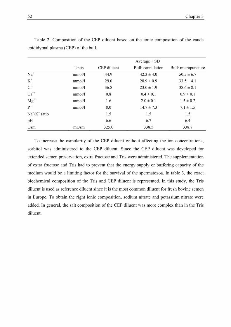
52 Chapter 3
Table 2: Composition of the CEP diluent based on the ionic composition of the cauda
epididymal plasma (CEP) of the bull.
Average ± SD Units CEP diluent Bull: cannulation Bull: micropuncture
Na+ mmol/l 44.9 42.3 ± 4.0 50.5 ± 6.7 K+ mmol/l 29.0 28.9 ± 0.9 33.5 ± 4.1 Cl- mmol/l 36.8 23.0 ± 1.9 38.6 ± 8.1 Ca++ mmol/l 0.8 0.4 ± 0.1 0.9 ± 0.1 Mg++ mmol/l 1.6 2.0 ± 0.1 1.5 ± 0.2 P-- mmol/l 8.0 14.7 ± 7.3 7.1 ± 1.5 Na+/K+ ratio 1.5 1.5 1.5 pH 6.6 6.7 6.4 Osm mOsm 325.0 338.5 338.7
To increase the osmolarity of the CEP diluent without affecting the ion concentrations,
sorbitol was administered to the CEP diluent. Since the CEP diluent was developed for
extended semen preservation, extra fructose and Tris were administered. The supplementation
of extra fructose and Tris had to prevent that the energy supply or buffering capacity of the
medium would be a limiting factor for the survival of the spermatozoa. In table 3, the exact
biochemical composition of the Tris and CEP diluent is represented. In this study, the Tris
diluent is used as reference diluent since it is the most common diluent for fresh bovine semen
in Europe. To obtain the right ionic composition, sodium nitrate and potassium nitrate were
added. In general, the salt composition of the CEP diluent was more complex than in the Tris
diluent.

Collection and composition of cauda epididymal plasma in the bull 53
Table 3: Biochemical composition of the Tris and CEP diluent used in the storage experiment.
Units Tris diluent CEP diluent CaCl mmol/l 0.4 0.8 KCl mmol/l 0 22.0 MgCl mmol/l 0.4 1.6 NaCl mmol/l 11.5 10.0 Na-nitrate mmol/l 0 18.0 K-nitrate mmol/l 0 4.0 Na-bicarbonate mmol/l 11.9 11.9 Na-phosphate mmol/l 0 5.0 K-phosphate mmol/l 0 3.0 glucose mmol/l 11.1 0 fructose mmol/l 55.0 55.0 sorbitol mmol/l 0 5.5 BSA g/l 2.0 2.0 Tris mmol/l 133.7 133.7 genta-S g/l 0.05 0.05 egg yolk % 10.0 10.0 citric acid mmol/l 42.9 42.9 glycine mmol/l 83.4 0 pH 7.5 6.6 Osm mOsm 325 325
Storage of fresh bovine semen in Cauda Epididymal Plasma (CEP) diluent
At the start of the incubation (t0), all spermatozoa expressed a good progressive and total
motility (85%) in both the Tris and CEP diluent. However, a significant decrease over time of
total and progressive motility, and membrane integrity was observed both media (p < 0.05).
No difference in membrane integrity (p = 0.25) and progressive motility (p = 0.57) was found
between spermatozoa stored in Tris and CEP diluent. The total motility decreased more
rapidly for spermatozoa stored in the CEP diluent than in the Tris diluent (p = 0.01).
The first 2 days of incubation, no difference was observed in total motility between
spermatozoa stored in both diluents, while the progressive motility was slightly better for
spermatozoa stored in the CEP diluent. These findings were confirmed by the slightly higher
sperm motility values (SMI, FSC and MSC) obtained with the SQA for spermatozoa stored in
the CEP diluent than in the Tris diluent (Figure 3).

54 Chapter 3
Figure 3: Total motility and sperm motility index (SMI) of fresh semen stored at 5°C in Tris and CEP diluent for 6 days.
From four days of incubation onwards, spermatozoa stored in the the Tris diluent had
higher values for total motility, SMI, FSC, and MSC than spermatozoa stored in the CEP
diluent. While the total motility of spermatozoa stored in the Tris and CEP diluent at t6
remained rather high (65% and 48% respectively), the progressive motility decreased to 25%
and 13% respectively. In the Tris diluent, higher sperm motility parameters were obtained
with the SQA-II C® at t4 and t6 than at t0 and t2. This is in contrast to the total and
progressive motility, which decreased significantly over time in the Tris diluent.
DISCUSSION
In this study, the biochemical composition of the CEP of the bull was evaluated using
both in vitro (micropuncture) and in vivo (cannulation) collection techniques. Rams were
used as model for the in vivo cannulation of the ductus deferens in the bull. Each of the two
collection methods had both advantages and disadvantages.
Using the micropuncture method, large numbers of epididymides can be processed within
a short period of time. However, contamination of the epididymal plasma with interstitial
fluid, blood and epithelial cells may occur. The degree of cellular contamination can be
minimized by means of centrifugation, while contamination with interstitial fluid can be
reduced to less than 5% when the collection method is carried out carefully (Crabo, 1965). In
the present study, the contamination with interstitial fluid was minimized by careful
dissection of the cauda epididymidis, a good clean-up of the surface and an accurate
micropuncture of the cauda epididymal duct using a very fine pipette. Besides contamination,
other disadvantages of the micropuncture method are: 1) the small quantities of CEP per
epididymis, and 2) the fact that samples can be harvested only once. Regarding the small
amount of epididymidal plasma available per epididymis, no extensive individual biochemical
020406080
100
D 0 D 2 D 4 D 6Tota
l mot
ility
(%)
Time (days)
TrisCEP
050
100150200
D 0 D 2 D 4 D 6Time (days)
SMI
020406080
100
D 0 D 2 D 4 D 6Tota
l mot
ility
(%)
Time (days)
TrisCEPTrisCEP
050
100150200
D 0 D 2 D 4 D 6Time (days)
SMI

Collection and composition of cauda epididymal plasma in the bull 55
analysis per bull can be performed. Consequently, when large amounts of epididymal plasma
or spermatozoa of an individual are required, or when a follow up of the characteristics of
epididymal plasma or spermatozoa over time has to be performed, micropuncture is not a
useful collection method (Amann et al., 1963). In these cases, cannulation of the ductus
deferens is a better alternative.
Cannulation of the ductus deferens in the bull was first described by Amann et al. (1963).
Anatomical differences make the cannulation of the ductus deferens easier to perform in the
ram than in the bull, which was also the case in the present study since more complications
were encountered in the bull than in the ram. In two bulls no epididymal fluid could be
collected, while in two other bulls epididymal plasma could be collected, but insufficient to
allow biochemical analysis. Even in the bulls which had been cannulated successfully, the
patency periods of the cannula were shorter than in the ram (9.2 ± 4.8d vs 17.2 ± 4.7d).
However, the daily and total epididymal fluid productions were higher in the bull than in the
ram, which is possible due to the larger size of the bovine testicles. Another advantage is the
fact that during unilateral cannulation of the ductus deferens, ejaculated semen can be
collected simultaneously. This enables the comparison of epididymal sperm and plasma
characteristics with these of ejaculated sperm and plasma, and the effect of addition of
accessory sex gland fluid on epididymal spermatozoa (Henault et al., 1995).
The number of studies in which the ionic composition of the cauda epididymal plasma in
the bull is determined is very limited (Crabo, 1965; Wales et al., 1966; Mann, 1964). In the
ram, the composition of the CEP was first described by Scott et al. (1963). Compared to our
study (Table 2), they found lower concentrations for the different ions, for the Na/K ratio, and
for the urea and total protein concentration. However, the lactate concentrations reported by
Scott et al. (1963) were 5 times higher than in our study. The differences in ion concentration
may be the result of the difference in collection method and analysis technique used to
determine the biochemical composition of the CEP. In the study of Scott et al. (1963) the
surface of the cauda epididymal duct was scraped in order to collect epididymal fluid,
resulting in contamination with interstitial fluid. The interstitial fluid might have a dilution
effect and explain the lower concentration of the different ions in their study. The increased K
concentration in the CEP reported by Scott et al. (1963) can be due to the cell lesions caused
by scraping the surface to collect CEP. The increased lactate concentrations may be due to the
metabolism of glucose present in the CEP. The difference in urea and total protein
concentration might be related with the difference in diet.

56 Chapter 3
In our study, the biochemical composition of the CEP in the bull was examined with the
aim to develop a suitable diluent for prolonged preservation of ejaculated semen. The CEP in
our study as well as in that of Crabo (1965) and Wales et al. (1966) was collected by means of
micropuncture, but nonetheless clear differences in ion concentrations were observed between
our study and the previous ones (Verberckmoes et al., 2001). A great deal of the variation
may be due to the method of biochemical analysis and to the fact that different breeds of bulls
were used.
The composition of the new diluent (CEP diluent) was based on the pH, osmolarity,
concentrations of Na, K, Cl, Ca, Mg and inorganic P, and on the Na/K ratio in the CEP as it
occurs in the bull (Table 2).
The pH of the CEP after cannulation and micropuncture was 6.4 and 6.7 respectively, and
is about the same as in the stallion, the boar, the ram, the dog, and the rabbit (Jones, 1978).
Carr and Acott (1984) however, reported that the pH of the CEP of the bull is as low as 5.8
and in combination with lactate could be responsible for the reversible inhibition of the
motility of the spermatozoa in the cauda epididymidis. This is in contrast with storage
experiments performed earlier in our laboratory with ejaculated semen (De Pauw et al., 2003).
When ejaculated semen was stored in Hepes-TALP at a pH lower than 6, the motility of the
spermatozoa dropped to zero within 24 hrs, and no membrane intact spermatozoa could be
detected after 72 hrs of incubation. When the pH of the storage medium increased from 6 to 8,
no significant difference in sperm motility was observed, while the membrane integrity
decreased significantly (De Pauw et al., 2003). In the present study, no difference in
membrane integrity was observed between spermatozoa stored in Tris (pH = 7.5) vs. CEP
diluent (pH = 6.6), while the motility decreased more rapidly in the CEP diluent.
The slight hypertonicity of the CEP as determined in this study (338 mOsm) is in
agreement with previous findings (Liu and Foote, 1998; Scott et al., 1963; Cragle and
Salisbury, 1959; Levine and Marsh, 1971). Especially in hibernating bats, where a
hypertonicity of 1500 mOsm is reached, this is assumed to be the most important parameter
that enables prolonged preservation of semen as a result of dehydration (Crichton et al.,
1994). The possibility to increase survival of ejaculated bull sperm in hypertonic solutions is
however very limited (De Pauw et al., 2003).
Besides low pH and hypertonicity of the medium, different inorganic and organic
constituents have been alleged to be responsible for maintaining sperm in quiescence (Verma,

Collection and composition of cauda epididymal plasma in the bull 57
2001). We have tried to mimic the inorganic components (Na, K, Cl, Ca, and Mg) of the CEP
in our diluent. Among the inorganic constituents, the effect of different ions as present in the
cauda epididymidis upon keeping epididymal spermatozoa in a quiescent state has been
investigated in several species. Morton et al. (1978) showed that low levels or absence of
calcium was beneficial for sperm quiescence, while Crabo (1965) reported that especially a
low Na/K ratio was required to keep the spermatozoa in a quiescent state. According to
Cragle and Salisbury (1959), a high K/Ca ratio was required besides a low Na/K ratio to
inhibit spermatozoal respiration and glycolysis. However, despite the fact that the ion
concentrations, the Na/K ratio and the K/Ca ratio of our CEP diluent were identical to those
of the CEP, no reduction of the spermatozoal activity was observed (Figure 3). Moreover,
during the first 2 days of storage, even higher sperm motility values were obtained for
spermatozoa stored in the CEP diluent than for spermatozoa stored in the Tris diluent. After 2
days, the Tris diluent was superior to the CEP diluent for sperm survival in vitro.
Sorbitol, which is also present in seminal plasma, was added to the CEP diluent for its
osmotic effect and can serve to a limited extent as a natural substrate for semen under aerobic
condition (Crichton et al., 1994; Clegg et al., 1986). Other organic components of epididymal
fluid, such as glycerylphosphorylcholine and carnitine were not added to our CEP diluent.
Their effect has been studied in rats, with good result on sperm quiescence (Turner and Giles,
1982). However, when carnitine was added to medium conditioned by epididymal cells and
used for bull semen preservation, no beneficial effect on sperm survival could be observed
(Reyes-Moreno et al., 2000).
Since sperm fertility declines much faster than sperm motility (Vishwanath and Shannon,
1997, 2000), and given the fact that after 2 days of incubation sperm motility was lower for
spermatozoa stored in the CEP than in the Tris diluent, it can be concluded that the CEP
diluent is not appropriate for extended preservation of fresh bovine semen. Moreover, despite
the fact that the CEP diluent had the same ionic composition, osmolarity, and pH as the CEP
in the bull, no quiescent state or extended life span of the ejaculated spermatozoa was
acquired. Future experiments to establish a superior sperm diluent for fresh bovine semen are
directed towards the exclusion of nitrate-salts with concomitant modification in ionic
composition of the CEP diluent and to modifications in egg yolk and polyol content, and the
temperature of storage.
58 Chapter 3
REFERENCES
Acott TS, Carr DW. Inhibition of bovine spermatozoa by caudal epididymal fluid: II.
Interaction of pH and a quiescence factor. Biol. Reprod. 1984; 30: 926-935.
Almquist JO. Diluents for bovine sperm XI. Effect of glycerol on fertility and motility of
spermatozoa on homogenized milk and skim milk. J Dairy Sci 1962; 45: 911-916.
Amann RP, Hokanson JF, Almquist JO. Cannulation of the bovine ductus deferens for
quantitative recovery of epididymal spermatozoa. Journal of Reproduction and Fertility
1963; 6: 69.
Amann RP, Johnson L, Thompson DL, Pickett BW. Daily spermatozoal production,
epididymal spermatozoal reserves and transmit time of spermatozoa through the
epididymis of the rhesus monkey. Biol Reprod 1976; 15: 586-592.
Butrimovitz GP, Purdy WC. The determination of zinc in blood plasma by atomic absorption
spectrometry. Anal Chim Acta 1977; 94: 63.
Carr DW, Acott TS. Inhibition of bovine spermatozoa by cauda epididymal fluid: I. Studies of
a sperm motility quiescence factor. Biol. Reprod. 1984; 30: 913-925.
Cascierei M, Amann RP, Hammerstedt RH (1976). Adenine nucleotide changes at initiation
of bull sperm motility. J Biol Chem 1976; 251: 787-793.
Clegg JS, Gallo J, Gordon E. Some structural, biochemical and biophysical characteristics of
L-929 cells growing in the presence of hyperosmotic sorbitol concentrations. Exp Cell
Res 1986; 163: 35-46.
Crabo B. Studies on the composition of epididymal content in bulls and boars. Acta Vet
Scand 1965; 6 (suppl 5): 1-94.
Cragle RG, Salisbury GW. Factors influencing metabolic activity of bull spermatozoa. IV.
PH, osmotic pressure, and the cations, sodium, potassium, and calcium. J Dairy Sci,
1959; 42: 1304-1313.

Collection and composition of cauda epididymal plasma in the bull 59
Crichton EG, Hinton BT, Pallone TL, Hammerstedt RH. Hyperosmolality and sperm storage
in hibernating bats: prolongation of sperm life by dehydration. Am J Physiol 1994;
267:1363-1370.
De Pauw I, Van Soom A, Maes D, Verberckmoes S, de Kruif A. Effect of sperm coating on
the survival and penetrating ability of in vitro stored bovine spermatozoa.
Theriogenology 2003; 59: 1109-1122.
De Pauw I, Van Soom A, Mintiens K, Verberckmoes S, de Kruif A. In vitro survival of
bovine spermatozoa stored at room temperature under epididymal conditions.
Theriogenology 2003; 59: 1093-1107
Foote RH. Fertility of bull semen at high extension rates in Tris buffer extenders. J Dairy Sci
1970; 53: 1475-1477.
Free MJ, Schluntz GA, Jaffe RA. Respiratory gas tensions in tissues and fluids of the male rat
reproductive tract. Biol Reprod 1979; 14: 481-488.
Frijters ACJ, Luimstra-Mulder TA. Time of freezing and the use of semen extenders based on
soybean extracts and egg yolk in cryopreservation of bull semen: effect in vitro and on
fertility in vivo. Proc. A.I. vets meeting 2003 Hungary, cattle session, 6-13.
Henault MA, Killian GJ, Kavanaugh JF and Griel LC Jr. Effect of accessory sex gland fluid
from bulls of differing fertilities on the ability of cauda epididymal sperm to penetrate
zona-free bovine oocytes. Biology of Reproduction 1995; 52: 390-397.
Jones R. Comparative biochemistry of mammalian epididymal plasma. Comp Biochem
Physiol (B) 1978; 61: 365.
Jones RC, Holland MK, Doberska C. The epididymis: cellular and molecular aspects. J
Reprod Fertil 1998; 53 (suppl).
Kirillov VS, Mozorov VA. Duration of survival of bull spermatozoa in an epididymis isolated
from the testis. Anim Breed 1936; 5: 22.

60 Chapter 3
Levine N, Marsh DJ. Micropuncture studies of the electrochemical aspects of fluid and
electrolyte transport in individual seminiferous tubules, the epididymis and the vas
deferens in the rat. J Physiol 1971; 213: 557-570.
Liu Z, Foote RH. Bull sperm motility and membrane integrity in media varying in osmolality.
J Dairy Sci 1998; 81: 1868-1873.
Mann T. The biochemistry of semen and of the male reproductive tract. Methuen, London.
1964.
Morton BE, Sagadraca R, Fraser C. Sperm motility within the mammalian epididymis:
species variation and correlation with free calcium levels in epididymal plasma. Fertil
Steril 1978; 29: 695-698.
Reyes-Moreno C, Gagnon A, Sullivan R, Sirard MA. Addition of specific metabolites to
bovine epididymal cell culture medium enhances survival and motility of cryopreserved
sperm. J Androl 2000; 21: 876-886.
Salisbury GW, Van Demark NL. Physiology of reproduction and artificial insemination of
cattle. WH Freeman (Ed.), San Fransisco, 1961; 415-425.
Scott TW, Wales RG, Wallace JC, White IG. Composition of ram epididymal and testicular
fluid and the biosynthesis of glycerylphosphorylcholine by the rabbit epididymis. J
Reprod Fertil 1963; 6: 49-59.
Shannon P. the effect of diluents containing glycine and glycerol on the fertility of diluted
bovine semen. NZ J Agric Res 1964; 7: 357-363.
Turner TT, Giles RD. Sperm motility –inhibiting factor in rat epididymis. Am J Physiol 1982;
242: 199-203.
Van Wagtendonk-de Leeuw AM, Haring RM, Kaal-Lansbergen LMTE, Den Daas JHG.
Fertility results of bovine semen cryopreserved with egg yolk based or soy bean extract
based extenders. Theriogenology 2000; 54: 57-67.
Collection and composition of cauda epididymal plasma in the bull 61
Verberckmoes S, De Pauw I, Van Soom A, de Kruif A. Ionic composition and osmolarity of
caudal epididymal plasma in the bull. Theriogenology 2001; 55: 449.
Verma RJ. Sperm quiescence in cauda epididymidis: a mini-review. Asian J Androl 2001; 3:
181-183.
Vishwanath R, Pitt CP, Shannon P. Sperm numbers, semen age and fertility in fresh and
frozen bovine semen. Proc NZ Soc Anim Prod 1996; 56: 31-34.
Vishwanath R, Shannon P. Do sperm cells age? A review of the physiological changes in
sperm during storage at ambient temperatures. Reprod Fertil Dev 1997; 9: 321-331.
Vishwanath R, Shannon P. Storage of bovine semen in liquid and frozen state. Anim Reprod
Sci. 2000 Aug 18; 62 (1-3):23-53.
Wales RG, Wallace JC, White IG. Composition of bull epididymal and testicular fluid. J
Reprod Fertil 1966; 12: 139.
Watson PF. Recent developments and concepts in the cryopreservation of spermatozoa and
the assessment of their post thawing function. Reprod Fertil Dev 1995; 7: 871-891.
Woelders H. Fundamentals and recent development in cryopreservation of bull and boar
semen. Vet Q 1997; 19: 135-138.

CHAPTER 4
STORAGE OF FRESH BOVINE SEMEN IN A DILUENT BASED ON THE IONIC
COMPOSITION OF CAUDA EPIDIDYMAL PLASMA (CEP)
Reproduction in Domestic Animals, 2004: 39; 1-7.
Verberckmoes S., Van Soom A., Dewulf J., De Pauw I., de Kruif A.

64 Chapter 4
ABSTRACT
For artificial insemination (AI) in cattle, much lower insemination doses can be applied
when fresh semen is used instead of frozen-thawed semen. However, a particular
disadvantage of fresh semen is its limited shelf life. Since bovine spermatozoa can be stored
for several weeks in the cauda epididymidis without negative effects on their fertilizing
capacity, the cauda epididymidis can serve as a model in order to prolong the shelf life of
fresh semen. First, the ionic composition of the CEP-1 diluent which had the same ionic
composition, pH and osmolarity as the bovine cauda epididymal plasma was modified (CEP-
2) and the storage capacity of the CEP-2 diluent was compared with this of the CEP-1 and
Tris diluent. Subsequently, the effect of addition of different polyols (sorbitol, glycerol,
mannitol) and egg yolk concentrations (5, 10 and 20%) to the CEP 2 diluent was assessed.
Sperm quality decreased rapidly in the CEP-1 diluent. The quality and especially
progressive motility of spermatozoa stored in the CEP-2 diluent were better than in the CEP-1
and Tris diluent. No significant effects of different sugars or egg yolk concentrations on the
quality of fresh bovine semen in the CEP-2 diluent were observed. In conclusion, the CEP-2
diluent with 10% egg yolk and 1g/L sorbitol may be used for extended preservation of fresh
bovine semen at 5°C up to 6 days.

Storage of fresh bovine semen in CEP-1 diluent 65
INTRODUCTION
The widespread use of artificial insemination (AI) in cattle can partly be attributed to the
availability of suitable diluents (Almquist, 1962; Shannon, 1964; Foote, 1970) and to the fact
that similar pregnancy rates can be obtained with small amounts of frozen-thawed semen (±
15 millions) by means of AI as by means of natural mating without the risk of spreading
venereal diseases (Salisbury and Van Demark, 1961). In contrast to natural mating, the
cervical barrier is by-passed when AI is performed, and semen is deposited directly into the
uterine body. In this way, AI in cattle enables a more than 100-fold reduction of the
insemination dose compared to natural mating without a decrease in pregnancy rates (Hunter,
2003). The ability to obtain high pregnancy rates with low insemination doses increases the
number of insemination doses produced by genetically superior sires and accelerates the
genetically progress in livestock. Currently, more than 95% of semen in cattle is processed as
frozen product (Chupin and Thibier, 1995), while less than 5% is processed as fresh semen.
The use of fresh semen is predominantly restricted to New Zealand, and smaller amounts are
used in Africa, France, Australia, Germany, and Eastern Europe (Thibier and Wagner, 2000).
The use of frozen-thawed semen facilitates world wide distribution and enables screening of
the bull for infectious diseases before its semen is used in the field. However, in cattle the
insemination dose for AI can be reduced from 15 to 2 to 4 millions of spermatozoa without a
reduction in pregnancy rates when fresh instead of frozen-thawed spermatozoa are used
(Vishwanath et al., 1996). The much higher insemination dose required with frozen-thawed
than with fresh semen is the result of the detrimental effect of the freezing and thawing
procedure on semen quality (Holt, 2000). When AI is performed with liquid bovine semen in
Europe, semen is traditionally stored at 5°C in a Tris-glycerol based extender which is
normally used for freezing semen (De Leeuw et al., 1993). In New Zealand however, most of
the liquid semen is stored at room temperature, under N2 gas in Caprogen® diluent
(Vishwanath and Shannon, 1997). Semen stored in these diluents, is advised to be used within
3 days. Obviously, the short shelf live is the most important disadvantages of fresh semen.
Therefore, extension of the shelf live of fresh semen would be interesting for the cattle
breeding industry for several reasons; 1) more insemination doses could be produced per
ejaculate, 2) the work load during sperm collection and processing would be decreased, and 3)
the distribution of liquid semen would be simplified.

66 Chapter 4
Inspired by the specific capacity of the bovine cauda epididymidis to store spermatozoa
for several weeks without reduction of their fertilizing capacity (Kirillov and Mozorov, 1936;
Cascieri et al., 1976), we have developed a sperm diluent largely based on the ionic
composition of bovine cauda epididymal fluid. Despite the fact that for in vitro culture of
preimplantation embryos several synthetic culture media have been developed based on the
composition of oviductal fluid, such as ovine synthetic oviduct fluid (SOF) (Tervit et al.,
1972), human tubal fluid (Quinn et al., 1985) and pig zygote medium (Yoshioka et al., 2002),
to our knowledge no such approach has ever been used in the development of a sperm diluent.
To analyse the ionic composition of cauda epididymal fluid we have cannulated the cauda
epididymidis of bulls in vivo and performed micropuncture of slaughterhouse epididymides
(Verberckmoes et al., 2001). In this study we describe the composition of the Cauda
Epididymal Plasma (CEP) diluent, including some of the adjustments which were necessary
to optimize its sperm storage capacity and we compare sperm survival in CEP and Tris
diluent. In addition, the effect of different concentrations of egg yolk and polyols on sperm
survival in the CEP diluent are examined.
MATERIAL AND METHODS
Collection and preparation of semen
Fresh semen of 3 bulls with normal sperm quality (progressive motility, percentage of
membrane intact spermatozoa, and percentage of spermatozoa with normal morphology ≥
80%) was collected by means of an artificial vagina in a falcon tube (Becton and Dickinson®,
Le Pont De Claix, France) containing 5 ml of Triladyl® diluent (Minitüb, Tiefenbach,
Germany), according to the method described by De Pauw et al. (2003a). Immediately after
collection, semen was centrifuged at 700 x g for 10 min. After centrifugation, the seminal
plasma and Triladyl® diluent were removed. The sperm concentration was determined using a
Bürker counting chamber and light microscopy (x 400). Spermatozoa were stored at a
concentration of 10 x 106 /ml in the different sperm diluents. Spermatozoa were incubated for
6 or 8 days in 1.8 ml cryovials (Nunc®, Wiesbaden, Germany) at 5°C in darkness.
Media
Chemicals were obtained from Sigma (Bornem, Belgium) and Life Technologies, Gibco
BRL® products (Merelbeke, Belgium). Triladyl® diluent (Minitüb, Tiefenbach, Germany) was

Storage of fresh bovine semen in CEP-1 diluent 67
used for the dilution and coating of the ejaculated spermatozoa (De Pauw et al., 2003a, b).
Newly developed cauda epididymal plasma (CEP) diluent was used as the basal sperm diluent
(Table 1). The inorganic composition of the CEP-1 medium was calculated according to the
ionic concentration of the cauda epididymal fluid as reported previously (Verberckmoes et al.,
2001). Since preliminary experiments showed that the presence of nitrate in CEP-1 was
detrimental for sperm survival, nitrates were omitted from CEP-2, and Ca, P, and Mg
concentrations were raised. In both CEP-1 and CEP-2, no glucose but fructose and citric acid
were added to the basal medium as energy substrates, at a concentration comparable to that in
TRIS. Sorbitol was added to the CEP-1 and CEP-2 to increase the osmolarity to the same
level as that in cauda epididymal plasma, while BSA was added as a macromolecular
component. TRIS diluent was used for comparison.
Table 1: Composition of sperm diluents.
Tris CEP-1 CEP-2 NaCl (mmol/l) 11.5 10.0 15.0 KCl (mmol/l) - 22.0 7.0 CaCl2(H2O)2 (mmol/l) 0.4 0.8 3.0 MgCl2(H2O)6 (mmol/l) 0.4 1.6 4.0 NaHCO3 (mmol/l) 11.9 11.9 11.9 NaNO3 (mmol/l) - 18.0 - KNO3 (mmol/l) - 4.0 - NaH2PO4 (mmol/l) - 5.0 8.0 KH2PO4 (mmol/l) - 3.0 20.0 Glucose (mmol/l) 11.1 - - Fructose (mmol/l) 55.0 55.0 55.0 Sorbitol (g/l) - 1.0 1.0 BSA (g/l) 2.0 2.0 2.0 Tris (mmol/l) 133.7 133.7 133.7 Gentamycin-S (g)/l 0.05 0.05 0.05 Egg yolk (%) 10.0 10.0 10.0 Citric acid (mmol/l) 42.9 42.9 42.9 Glycine (mmol/l) 83.4 - - pH 7.5 6.6 6.6 Osm (mOsm) 325 325 320

68 Chapter 4
Sperm quality assessment
The sperm quality parameters were evaluated at day 0 (t0), 2 (t2), 4 (t4), 6 (t6), and 8 (t8).
Membrane integrity was determined in 100 spermatozoa by means of nigrosin eosin staining
and light microscopy (x 400). Total motility (TM), progressive motility (PM), and
hyperactivation of the spermatozoa were assessed subjectively on a heated stage (37°C) by 2
persons and by means of light microscopy (x 200). Spermatozoa were estimated as being
hyperactivated when their motility was extremely vigorous and with high curvature flagellar
movements. The Functional Sperm Concentration (FSC), Motile Sperm Concentration (MSC)
and Sperm Motility Index (SMI) were determined objectively in three replicates with the
Sperm Quality Analyzer (SQA II-C®) (Orange Medical, Tilburg, The Netherlands). The SQA
II-C® is equipped with a photometric cell that registers the fluctuations in optical density and
converts this information, through mathematical algorithms, digitally to a numerical output.
The functional sperm concentration (FSC) expresses the number of spermatozoa that are both
motile and morphological normal, while the motile sperm concentration (MSC) expresses the
number of spermatozoa with a normal motility (concentration x motility percentage). The
sperm motility index (SMI) presents the overall sperm quality, taking into account: the
concentration, the progressive motility, and the percentage of spermatozoa with a normal
morphology.
Experiment 1: Quality assessment of spermatozoa stored in the CEP-1 diluent, the CEP-2
diluent, and the Tris diluent
To compare sperm survival in both versions of the CEP diluent with that in the TRIS
diluent, freshly ejaculated semen of 3 bulls was collected, centrifuged and subsequently stored
in the 3 diluents for 6 days at 5°C. Sperm quality was assessed every two days as described
above.
Experiment 2: Effect of sorbitol, glycerol, and mannitol on sperm survival in CEP-2 diluent.
For further experiments, only CEP-2 was used. To determine the effect of different
polyols on the storage capacity of CEP-2 diluent, which contains 1 mg/ml of sorbitol, its
storage capacity was compared with this of CEP-2 diluent without sorbitol, and with CEP-2
diluent in which sorbitol was replaced by the same concentrations of glycerol or mannitol.
Freshly ejaculated semen of the 3 bulls was stored in the 4 diluents for 8 days and sperm
quality was assessed every two days.

Storage of fresh bovine semen in CEP-1 diluent 69
Experiment 3: Quality assessment of spermatozoa stored in CEP-2 diluent with sorbitol and
different egg yolk concentrations.
After collection and centrifugation, freshly ejaculated bovine semen was diluted in CEP-2
diluent with 1 mg/ml of sorbitol and 3 different concentrations of egg yolk: 5%, 10%, and
20%. The diluted semen was stored for 8 days at 5°C, and sperm quality was assessed every 2
days as described before.
STATISTICAL ANALYSIS
All experiments were repeated 4 times. Data were analysed using a linear mixed effect
model, with bull as random variable and allowing different standard deviations per stratum
(S-plus 2000, Math Soft, Inc, Cambridge, USA). An autoregressive correlation structure of
the first order was used to account for correlation in the data. Since residuals were not
normally distributed for the FSC and MSC results, a log transformation was performed.
Differences in sperm quality were considered as being significant when p-values were lower
than 0.05.
RESULTS
Experiment 1: Quality assessment of spermatozoa stored in the CEP-1 diluent, the CEP-2
diluent, and the Tris diluent.
Membrane integrity, total and progressive motility, significantly decreased over time for
spermatozoa stored in each of the 3 diluents (p < 0.01). However, due to the high variation in
SMI, MSC, and FSC values, no decrease over time could be found for these parameters.
Sperm membrane integrity did not differ between the 3 diluents, while significantly higher
total motility values were obtained for spermatozoa stored in Tris diluent than for
spermatozoa stored in the other diluents (p < 0.01) (Figure 1). Notwithstanding no significant
difference in progressive motility was found between the 3 diluents (p = 0.57), progressive
motility of spermatozoa stored in CEP-2 diluent tended to be higher than in both other
diluents (Figure 1). When sperm motility was subjectively evaluated, a remarkably high
percentage of hyperactivated spermatozoa (20 to 30%) was observed in Tris diluent, while no
hyperactivated spermatozoa were observed in CEP-1 and CEP-2 diluent. Significant
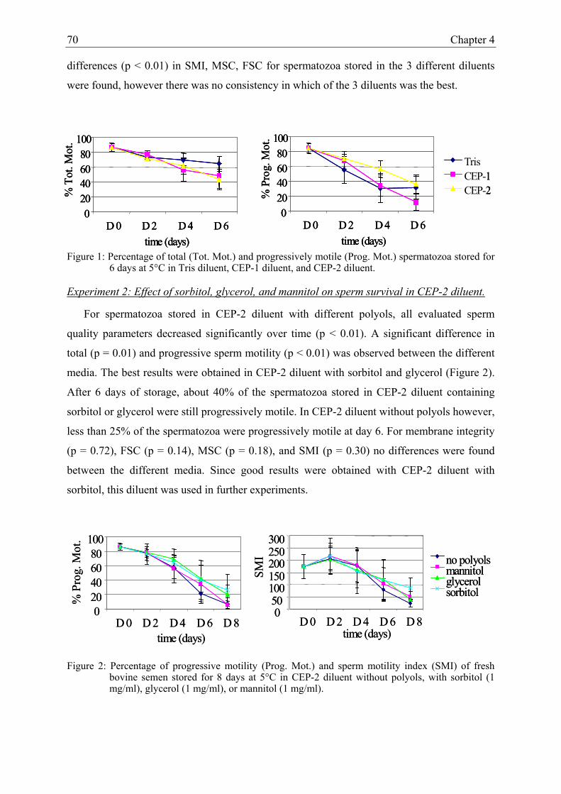
70 Chapter 4
differences (p < 0.01) in SMI, MSC, FSC for spermatozoa stored in the 3 different diluents
were found, however there was no consistency in which of the 3 diluents was the best.
Figure 1: Percentage of total (Tot. Mot.) and progressively motile (Prog. Mot.) spermatozoa stored for 6 days at 5°C in Tris diluent, CEP-1 diluent, and CEP-2 diluent.
Experiment 2: Effect of sorbitol, glycerol, and mannitol on sperm survival in CEP-2 diluent.
For spermatozoa stored in CEP-2 diluent with different polyols, all evaluated sperm
quality parameters decreased significantly over time (p < 0.01). A significant difference in
total (p = 0.01) and progressive sperm motility (p < 0.01) was observed between the different
media. The best results were obtained in CEP-2 diluent with sorbitol and glycerol (Figure 2).
After 6 days of storage, about 40% of the spermatozoa stored in CEP-2 diluent containing
sorbitol or glycerol were still progressively motile. In CEP-2 diluent without polyols however,
less than 25% of the spermatozoa were progressively motile at day 6. For membrane integrity
(p = 0.72), FSC (p = 0.14), MSC (p = 0.18), and SMI (p = 0.30) no differences were found
between the different media. Since good results were obtained with CEP-2 diluent with
sorbitol, this diluent was used in further experiments.
Figure 2: Percentage of progressive motility (Prog. Mot.) and sperm motility index (SMI) of fresh bovine semen stored for 8 days at 5°C in CEP-2 diluent without polyols, with sorbitol (1 mg/ml), glycerol (1 mg/ml), or mannitol (1 mg/ml).
020406080
100
D 0 D 2 D 4 D 6time (days)
% P
rog.
Mot
.
TrisCEP-1CEP-2
020406080
100
D 0 D 2 D 4 D 6time (days)
% T
ot. M
ot.
020406080
100
D 0 D 2 D 4 D 6time (days)
% P
rog.
Mot
.
TrisCEP-1CEP-2
020406080
100
D 0 D 2 D 4 D 6time (days)
% T
ot. M
ot.
020406080
100
D 0 D 2 D 4 D 6time (days)
% P
rog.
Mot
.
TrisCEP-1CEP-2
020406080
100
D 0 D 2 D 4 D 6time (days)
% T
ot. M
ot.
020406080
100
D 0 D 2 D 4 D 6 D 8time (days)
% P
rog.
Mot
.
020406080
100
D 0 D 2 D 4 D 6 D 8time (days)
% P
rog.
Mot
.
SMI
050100150200250300
D 0 D 2 D 4 D 6 D 8time (days)
no polyolsmannitolglycerolsorbitol
SMI
050100150200250300
D 0 D 2 D 4 D 6 D 8time (days)
no polyolsmannitolglycerolsorbitol

Storage of fresh bovine semen in CEP-1 diluent 71
Experiment 3: Quality assessment of spermatozoa stored in CEP-2 diluent with sorbitol and
different egg yolk concentrations.
All evaluated sperm quality parameters decreased significantly over time in CEP-2 diluent
with 5%, 10% and 20% egg yolk (p < 0.01). However, no significant differences in membrane
integrity (p = 0.44), total (p = 0.68) and progressive motility (p = 0.56) were detected between
CEP-2 diluents with different egg yolk concentrations (Figure 3). The percentage of
membrane intact spermatozoa decreased from 91.4 ± 3.6 at t0 to 80.8 ± 9.0, 78.3 ± 10.1, and
75.8 ± 8.9 at t8 in CEP-2 diluent with 5%, 10% and 20% egg yolk respectively. In none of
CEP-2 diluents, hyperactivated spermatozoa were observed and progressive motility was still
40% after 6 days of storage at 5°C (Figure 3). After 6 and 8 days of storage, highest FSC,
MSC, and SMI values were obtained for spermatozoa stored in CEP diluent with 10% egg
yolk (Figure 3).
Figure 3: Percentage of progressive motility (Prog. Mot.) and sperm motility index (SMI) of spermatozoa stored for 8 days at 5°C in CEP-2 diluent with sorbitol and with 5%, 10%, and 20% egg yolk.
DISCUSSION
Despite the fact that we were not able to get the ejaculated spermatozoa in a quiescent
state, our results clearly indicate that our novel CEP diluent is efficient for long term
preservation of bovine spermatozoa. For the first time, semen has been preserved in vitro in a
diluent based on the composition of epididymal fluid. Bovine spermatozoa can be stored in
the cauda epididymidis in a quiescent state for several weeks without reduction of their
fertilizing capacity. The peculiar capacity of the cauda epididymidis to keep spermatozoa in a
quiescent state has been attributed to different conditions present in the cauda epididymal
plasma (CEP). In the studies by Carr and Acott (1984) and Acott and Carr (1984) inhibition
of bovine spermatozoa was attributed to the interaction between pH and a quiescence factor.
Other studies attributed the quiescent state of the epididymal spermatozoa to specific ion
020406080
100
D 0 D 2 D 4 D 6 D 8time (days)
020406080
100
D 0 D 2 D 4 D 6 D 8time (days)
050
100150200250300
D 0 D 2 D 4 D 6 D 8time (days)
5% egg yolk10% egg yolk20% egg yolk
% P
rog.
Mot
.
SMI

72 Chapter 4
concentrations and ion ratios in the CEP. Morton et al. (1978) found a correlation between the
amount of free calcium in the cauda epididymal plasma of mammals and the level of sperm
motility, while Crabo (1965) reported that in bovine especially the low Na/K ratio would be
responsible for keeping spermatozoa in a quiescent state. According to Cragle and Salisbury
(1959), the inhibition of spermatozoal respiration and glycolysis in the bovine epididymis
would be obtained by the high K/Ca ratio in combination with the low Na/K ratio. However,
despite the fact that the pH, the ion concentrations, the Na/K ratio and the K/Ca ratio of CEP-
1 diluent were identical to those of the bovine cauda CEP (Verberckmoes et al., 2001), no
reduction in spermatozoal activity could be observed, indicating that factors other than the
ionic composition of the medium are essential.
Another characteristic of the cauda epididymidis is that spermatozoa are stored at a very
high concentration (1 to 3 x 10⁹ spermatozoa/ml). However, in vitro experiments with
ejaculated semen have shown that better results are obtained at a concentration of 10 x 106
than at 100 or 500 x 106 spermatozoa/ml (De Pauw et al., 2003).
Moreover, a combination of different factors might be responsible for the impracticability
to bring the ejaculated spermatozoa back into a quiescent state. At the moment of ejaculation,
spermatozoa previously stored in the cauda epididymidis are mixed with seminal plasma
secreted by the accessory sex glands (seminal vesicles, prostate gland and Cowper’s gland)
and become increasingly motile. Besides this increase in motility, ejaculated spermatozoa also
differ from the epididymal spermatozoa in intracellular pH (Moore and Hibbitt, 1975;
Hammerstedt et al., 1979), respiration (Lardy and Ghosh, 1952), ATP synthesis and
consumption (Cascieri and Amann 1976), heparin-binding sites (Nass et al. 1990), and the
profile of proteins bound to the plasma membrane (Lee et al., 1985; Shivaji, 1986; Calvete et
al., 1996). All these factors together may be responsible for the irreversible activation of the
quiescent epididymal spermatozoa (Morton et al., 1978; Cragle and Salisbury, 1959; Lardy
and Ghosh, 1952). Notwithstanding the fact that spermatozoa were coated in a Triladyl®
diluent (De Pauw et al., 2003a, b), that the seminal plasma together with the Triladyl® diluent
was removed immediately after collection, and that spermatozoa were incubated in a medium
with a biochemical composition comparable to this of the bovine cauda epididymal plasma,
activation of the quiescent epididymal spermatozoa could not be prevented.
Since the quiescent state of the spermatozoa could clearly not be achieved, special
attention was paid to design a diluent which could preserve progressive motility of

Storage of fresh bovine semen in CEP-1 diluent 73
spermatozoa as long as possible, since this it is one of the best indicators to predict in vivo
fertility (Zhang et al., 1998; Correa et al., 1997; Verberckmoes et al., 2002a).
In CEP-2 diluent the Ca, Mg, and inorganic P concentrations were increased, compared to
CEP-1 diluent. These changes had no effect on sperm membrane integrity and total motility,
however the progressive motility was clearly improved. The beneficial effect of increased Ca,
Mg, and P concentrations in CEP-2 diluent on the progressive motility may be due to the
improved activity of the Ca and Mg dependent enzyme, adenylate cyclase, in the spermatozoa
(Garbers et al., 1973; Garbers and Kopf, 1978; Lapointe et al., 1996). The higher activity of
adenylate cyclase results in increased cAMP concentrations, which makes it possible to raise
and prolong the progressive sperm motility significantly (Mann and Lutwak-Mann, 1981). A
disadvantage of high Ca concentrations in the incubation medium can be that it may cause a
rapid influx of calcium ions, and trigger the acrosome reaction which restricts the shelf life of
the spermatozoa (Zhang et al., 1991; Christensen et al., 1996). Notwithstanding the Ca
concentration in CEP-2 was higher than in CEP-1, the shelf life of the spermatozoa in CEP-2
was increased. This can be explained by the simultaneous increase of Mg, which inhibits the
Ca induced acrosome reaction as shown in guinea pig and hamster spermatozoa (Talbot,
1975). Besides acrosome reaction, high concentrations of Ca can also induce hyperactivation.
However, in this study no hyperactivated spermatozoa were observed in CEP-2 diluent which
may be due to the fact that not only high Ca concentrations, but also sufficient ATP and an
alkaline environment are required to induce hyperactivation (Ho et al., 2002). The higher total
motility for spermatozoa stored in Tris diluent than in CEP-2 diluent, and opposite results for
the progressive motility can be explained by the absence of hyperactivated spermatozoa in
CEP-2 diluent.
When the effect of different polyols was compared, it was obvious that progressive sperm
motility outcomes were highest for spermatozoa diluted in CEP-2 diluent with sorbitol or
glycerol. Originally, sorbitol was added to CEP diluent in order to 1) increase the osmolarity
of the medium to the same level as in the CEP of the bull (338 mOsm), and 2) as thermal
protector, since the CEP diluent was developed for sperm storage at 5 °C. In previous studies
performed in our laboratory, the effect of increasing osmolarity by addition of sorbitol on
sperm quality was examined, and we could conclude that the exact osmolarity of the storage
medium was not that critical. The best results for membrane integrity and sperm motility were
obtained when the osmolarity of the storage medium was about 300 mOsm (De Pauw et al.,
2003b). Besides their osmoregulating capacity, polyols and sugars are also used to stabilize

74 Chapter 4
the spermatozoal membrane by replacing water molecules in the normally hydrated polar
groups during transition through critical temperature zones (Woelders, 1997). Moreover,
under aerobic conditions sorbitol can be used to a limited extent, as an appropriate metabolic
substrate for spermatozoa (Clegg et al., 1986; Crichton et al., 1994). Notwithstanding the fact
that sorbitol and mannitol are isomers, the small differences in structure seemed to affect their
thermal protecting capacity.
When the effect of 5%, 10%, and 20% of egg yolk on the quality of the spermatozoa
stored in CEP-2 diluent was evaluated, no significant differences were observed. Since CEP-2
diluent was developed for extended preservation of fresh bovine semen, and the fact that after
6 days of incubation the best motility results were obtained in CEP-2 diluent with 10% egg
yolk, this concentration will be used in further experiments. Based on results obtained by Van
Demark and Sharma (1957), diluents for bovine semen have included anywhere from 12.5%
to 30% egg yolk. This is much more than in the Caprogen® diluent which is specifically
developed for liquid storage of bovine semen at ambient temperature and contains only 5% of
egg yolk (Shannon and Curson, 1982). However, no difference in fertility was observed when
the egg yolk concentration was decreased from 20% to 5% (Shannon and Curson, 1983). In
freezing media, egg yolk protects spermatozoa from cold shock and freezing damages.
Synthetic liposomes and soya been extracts have been evaluated as substitutes, but none of
them were as good as egg yolk (De Leeuw et al., 1993; Van Wagtendonk-de Leeuw et al.,
2000). In media for liquid storage of semen, the most important effect of egg yolk is probably
binding of seminal plasma fractions that have a detrimental effect on survival of spermatozoa
(De Pauw et al., 2003a, b). Inclusion of macromolecular substances and particularly egg yolk
in media used for liquid semen storage has shown to have a positive effect on sperm survival
(Shannon and Curson, 1972; Al-Somai et al., 1994; Prendergast et al., 1995).
In contrast to the Caprogen® diluent which is used for storage of fresh bovine semen under
N2 gas at 19°C, all experiments with our newly developed CEP-2 diluent were performed at
5°C. Due to the lower metabolism of the spermatozoa at 5°C than at 19°C (Salisbury and Van
Demark, 1961), and 2) the lower risk of overgrowth by contaminants at 5°C than at 19°C
(Yang and Chou, 2000), storage at this low temperature was preferred. Moreover, another
study showed that sperm quality decreased more rapidly when spermatozoa were stored in
CEP-2 diluent at 19°C than at 5°C (Verberckmoes et al., 2002b).

Storage of fresh bovine semen in CEP-1 diluent 75
CONCLUSION
Given the correlation between progressive motility and in vivo fertilizing capacity (Zhang
et al., 1998), and the high percentage of progressively motile spermatozoa (> 40%) up to 6
days of incubation in CEP-2 diluent with 1 mg/ml of sorbitol and 10% egg yolk indicate that
this diluent is suitable for extended preservation of fresh bovine semen at 5°C. Whether these
good in vitro results also can be reached in vivo still needs further investigation.

76 Chapter 4
REFERENCES
Acott TS, Carr DW, 1984: Inhibition of bovine spermatozoa by caudal epididymal fluid: II.
Interaction of pH and a quiescence factor. Biol Reprod 30, 926-35.
Almquist JO, 1962: Diluents for bovine sperm XI. Effect of glycerol on fertility and motility
of spermatozoa on homogenized milk and skim milk. J Dairy Sci 45, 911-916.
Al-Somai N, Vishwanath R, Molan PC, Shannon P, 1994: Anionic and cationic components
from aggregates in bovine seminal plasma and their effects on sperm motility. Mol
Reprod Dev 39, 328-336.
Calvete JJ, Mann K, Sanz ML, Raida M, Topfer-Peterson E, 1996: The primary structure of
BSP-30K, a major lipid-, gelatine-, and heparin-binding glycoprotein of bovine seminal
plasma. FEBS Lett 399, 147-152.
Carr DW, Acott TS. Inhibition of bovine spermatozoa by cauda epididymal fluid: I. Studies of
a sperm motility quiescence factor. Biol Reprod 1984; 30: 913-925.
Cascieri M, Amann RP, Hammerstedt RH, 1976: Adenine nucleotide changes at initiation of
bull sperm motility. J Biol Chem 251, 787-793.
Christensen P, Whitfield CH, Parkinson TJ, 1996: In vitro induction of acrosome reactions in
stallion spermatozoa by heparin and A23187. Theriogenology 45, 1201-1210.
Chupin D, Thibier M, 1995: Survey of the present status of the use of artificial insemination
in developed countries. World Anim Rev 82, 58-68.
Clegg JS, Gallo J, Gordon E, 1986: Some structural, biochemical and biophysical
characteristics of L-929 cells growing in the presence of hyperosmotic sorbitol
concentrations. Exp Cell Res 163, 35-46.
Correa JR, Pace MM, Zavos PM, 1997: Relationship among frozen-thawed sperm
characteristics assessed via the routine semen analysis, sperm functional tests and
fertility of bulls in an artificial insemination program. Theriogenology 48, 721-731.

Storage of fresh bovine semen in CEP-1 diluent 77
Crabo B, 1965: Studies on the composition of epididymal content in bulls and boars. Acta Vet
Scand 6 (suppl 5), 1-94.
Cragle RG, Salisbury GW, 1959: Factors influencing metabolic activity of bull spermatozoa.
IV. PH, osmotic pressure, and the cations, sodium, potassium, and calcium. J Dairy Sci
42, 1304-1313.
Crichton EG, Hinton BT, Pallone TL, Hammerstedt RH 1994: Hyperosmolality and sperm
storage in hibernating bats: prolongation of sperm life by dehydration. Am J Physiol
267, 1363-1370.
De Leeuw FE, De Leeuw AM, Den Daas JHG, Colenbrander B, Verkeij AJ, 1993: Effect of
various cryoprotective agents and membrane stabilizing compounds on bull sperm
membrane integrity after cooling and freezing. Cryobiology 30, 32-44.
De Pauw I, Van Soom A, Maes D, Verberckmoes S, de Kruif A, 2003a: Effect of sperm
coating on the survival and penetrating ability of in vitro stored bovine spermatozoa.
Theriogenology 59, 1009-1122.
De Pauw I, Van Soom A, Mintiens K, Verberckmoes S, de Kruif A, 2003b: In vitro survival
of bovine spermatozoa stored at room temperature under epididymal conditions.
Theriogenology 59, 1093-1107.
Foote RH, 1970: Fertility of bull semen at high extension rates in Tris buffer extenders. J
Dairy Sci 53, 1475-1477.
Garbers DL, First NL, Lardy HA, 1973: The stimulation of bovine epididymal sperm
metabolism by cyclic nucleotide phosphodiesterase inhibitors. Biol Reprod 8, 589.
Garbers DL, Kopf GS 1978: Effect of factors released from eggs and other agents on cyclic
nucleotide concentrations of sea urchin spermatozoa. J Reprod Fertil 52, 135.
Hammerstedt RH, Keith AD, Boltz RC Jr, Todd PW, 1979: Use of amphiphilic spin labels
and whole cell isoelectric focusing to assay charge characteristics of sperm surfaces.
Arch Biochem Biophys 194, 565-580.

78 Chapter 4
Ho HC, Granish KA, Suarez SS, 2002: Hyperactivated motility of bull sperm is triggered at
the axoneme by Ca2+ and not cAMP. Dev Biol 250 (1), 208-217.
Holt WV, 2000: Basic aspects of frozen storage of semen. Anim Reprod Sci 62(1-3), 3-22.
Hunter RH, 2003: Advances in deep uterine insemination: a fruitful way forward to exploit
new sperm technologies in cattle. Anim Reprod Sci 79,157-70.
Kirillov VS, Mozorov VA, 1936: Duration of survival of bull spermatozoa in an epididymis
isolated from the testis. Anim Breed 5, 22.
Lapointe S, Ahmad I, Buhr MM, Sirard MA, 1996: Modulation of post-thaw motility,
survival, calcium uptake, and fertility of bovine sperm by magnesium and manganese. J
Dairy Sci 79, 2163-2169.
Lardy HA, Ghosh D, 1952: Comparative metabolic behaviour of epididymal and ejaculated
mammalian spermatozoa. Ann NY Acad Sci 55, 594-596.
Lee CN, Handrow RR, Lenz RW, Ax RL, 1985: Interactions of seminal plasma and
glycosaminoglycans on acrosome reactions in bovine spermatozoa in vitro. Gamete Res
12, 345-355.
Mann T, Lutwak-Mann C, 1981: Biochemistry of spermatozoa: chemical and functional
correlations in ejaculated semen. Andrological aspects. In: Male Reproductive Function
and Semen. Mann T, Lutwak-Mann C (Eds.). Springer-Verlag, New York, pp 205-217.
Moore HDM, Hibbitt KG, 1975: Isoelectric focusing of boar spermatozoa. J Reprod Fertil 44,
329-332.
Morton BE, Sagadraca R, Fraser C, 1978: Sperm motility within the mammalian epididymis:
species variation and correlation with free calcium levels in epididymal plasma. Fertil
Steril 29, 695-698.
Nass SJ, Miller D, Winer MA, Ax RL, 1990: Male accessory sex glands produce heparin-
binding proteins that bind to cauda epididymal spermatozoa and are testosterone
dependent. Mol Reprod Dev 25, 237-246.

Storage of fresh bovine semen in CEP-1 diluent 79
Prendergast E, Vishwanath R, Shannon P, 1995: Protection of bovine sperm from the
deleterious effects of seminal plasma by charged lipoprotein fractions of egg yolk. Proc.
27th Ann. Conf. Austr Soc Rep Biol Melbourne 27, 50.
Quinn P, Kerin JF, Warnes GM, 1985: Improved pregnancy rate in human in vitro
fertilization with the use of a medium based on the composition of human tubal fluid.
Fertil Steril 44, 493-8.
Salisbury GW, Van Demark NL, 1961: Physiology of reproduction and artificial insemination
of cattle. WH Freeman (Ed.), San Fransisco, 415-425.
Shannon P, 1964: The effect of diluents containing glycine and glycerol on the fertility of
diluted bovine semen. NZ J Agric Res 7, 357-363.
Shannon P, Curson B, 1972: The effect of egg yolk levels on bovine seminal plasma toxins.
Proc. 7th Int Congr Anim Reprod Artif Insem Munich, Germany 4, 1360-1362.
Shannon P, Curson B, 1982: Kinetics of aromatic L-amino acid oxidase and the effect of
catalase on fertility of diluted bovine semen stored at 5°C and ambient temperatures. J
Reprod Fertil 64, 463-467.
Shannon P, Curson B, 1983: Effect of egg yolk levels on the fertility of diluted bovine sperm
stored at ambient temperatures. NZ J Agric Res 26, 187-189.
Shivaji S, 1986: Binding of seminalplasmin to the plasma and acrosomal membranes of
bovine spermatozoa. Fluorescence studies on the changes in the lipid-phase fluidity.
FEBS Lett 196 (2), 255-8.
Talbot P, 1975: The effect of ions on acrosome reaction. In: Eighth annual meeting of society
for the study of reproduction, Fort Collins, Colorado, 28-29.
Tervit HR, Whittingham DG, Rowson LE, 1972: Successful culture in vitro of sheep and
cattle ova. J Reprod Fertil 30, 493-7.
Thibier M, Wagner HG, 2000: World statistics for artificial insemination in cattle. Proc 14th
Int Congr Anim Reprod 22, 76 (abstract).

80 Chapter 4
Van Demark NL, Sharma UD, 1957: Preliminary fertility results from the preservation of
bovine semen at room temperatures. J Dairy Sci 40, 438-439.
Van Wagtendonk-de Leeuw AM, Haring RM, Kaal-Lansbergen LMTE, Den Daas JHG,
2000: Fertility results of bovine semen cryopreserved with egg yolk based or soy bean
extract based extenders. Theriogenology 54, 57-67.
Verberckmoes S, De Pauw I, Van Soom A, de Kruif A, 2001: Ionic composition and
osmolarity of caudal epididymal plasma in the bull. Theriogenology 55, 449.
Verberckmoes S, Van Soom A, De Pauw I, Dewulf J, de Kruif A, 2002a: Migration of bovine
spermatozoa in a synthetic medium and its relation to in vivo bull fertility.
Theriogenology 58, 1027-37.
Verberckmoes S, De Pauw I, Van Soom A, Dewulf J, de Kruif A, 2002b: New CEP-diluter
improves storage capacity of fresh bovine semen. Proceedings XXII World buiatrics
congress, Hannover, Germany, 18-23 August, 87-88.
Vishwanath R, Pitt CP, Shannon P, 1996: Sperm numbers, semen age and fertility in fresh
and frozen bovine semen. Proc NZ Soc Anim Prod 56, 31-34.
Vishwanath R, Shannon P, 1997: Do sperm cells age? A review of the physiological changes
in sperm during storage at ambient temperatures. Reprod Fertil Dev 9, 321-331.
Woelders H, 1997: Fundamentals and recent development in cryopreservation of bull and
boar semen. Vet Q 19, 135-138.
Yang SE, Chou CC, 2000: growth and survival of Escherichia Coli O157:H7 and Listeria
monocytogenes in egg products held at different temperatures. J Food Prot 63, 907-911.
Yoshioka K, Suzuki C, Tanaka A, Anas IM, Iwamura S, 2002: Birth of piglets derived from
porcine zygotes cultured in a chemically defined medium. Biol Reprod 66, 112-9.
Zhang BR, Larsson B, Lundeheim N, Rodriguez-Martinez H, 1998: Sperm characteristics and
zona pellucida binding in relation to field fertility of frozen-thawed semen form dairy AI
bulls. Int J Androl 21, 207-216.

Storage of fresh bovine semen in CEP-1 diluent 81
Zhang J, Muzs LZ, Boyle MS, 1991: Variations in structural and functional changes of
stallion spermatozoa in respons to calcium ionophore A23187. J Reprod Fertil 44, 199-
205.


CHAPTER 5
COMPARISON OF THREE DILUENTS FOR THE STORAGE OF FRESH BOVINE
SEMEN
Theriogenology, 2004 in press.
Verberckmoes S., Van Soom A., Dewulf J., de Kruif A.
84 Chapter 5
ABSTRACT
In New Zealand, 95% of the semen used for artificial insemination in cattle is processed
as liquid semen. Storage of liquid semen for up to 3 days in Caprogen® diluent enables a ten
fold reduction of the insemination dose, compared to frozen-thawed semen, without a
reduction in fertility. In this Caprogen® diluent spermatozoa are stored under N2 gas in the
presence of catalase. However, a new diluent (CEP-2), which was originally based on the
biochemical composition of bovine cauda epididymal plasma, could become an appropriate
alternative to Caprogen®.
In this study, the effect of addition of catalase to bovine spermatozoa stored for 6 days in
CEP-2 diluent under aerobic and anaerobic conditions was evaluated and compared with a
Tris diluent. Additionally, the quality and in vitro fertilizing capacity of fresh bovine semen
stored for 6 days at 5°C in the Triladyl®, CEP-2 (without catalase and N2 gas) and Caprogen®
diluent were compared.
Addition of 4.5 mg/ml catalase to CEP-2 diluent under aerobic and anaerobic conditions
had no effect on sperm quality. Spermatozoa stored in CEP-2 diluent moved faster and
straighter than spermatozoa stored in Triladyl® or Caprogen® diluent. The in vitro fertilization
and polyspermy rates did not differ significantly between spermatozoa stored for 6 days at
5°C in CEP-2 and Caprogen® diluent, but were significantly lower for spermatozoa stored in
Triladyl® diluent.
We can conclude that based on the in vitro results, the CEP-2 diluent is a better diluent
than Triladyl® and a good alternative to the Caprogen® diluent for long term storage of fresh
bovine semen at 5°C. To confirm these promising in vitro results further in vivo experiments
are required.

Comparison of three diluents for the storage of fresh bovine semen 85
INTRODUCTION
Before the optimization of the freezing and thawing procedure, artificial insemination (AI)
in cattle was performed with fresh semen (Foote, 2002). Freshly ejaculated semen was diluted
in an extender ranging from a simple salt solution to a more complex buffered medium
(Foote, 2002). To facilitate long distance distribution of fresh semen, a shelf life of at least 3
days was required. First, a beneficial effect on the survival of fresh diluted semen was
obtained by addition of egg yolk and buffering components such as phosphate, citrate, and
Tris to the extender (Philips, 1939). Secondly, attempts were made to extend the life span of
ejaculated spermatozoa by lowering the metabolic rate of the spermatozoa by decreasing the
storage temperature to 5°C (Salisbury and Van Demark, 1961). Storage at 5°C not only
extended the life span of spermatozoa, it also decreased the risk of overgrowth by
contaminants (Yang and Chou, 2000). However, the use of frozen-thawed semen facilitates
distribution of semen world wide and enables screening of bulls for infectious diseases before
their semen is used in the field. These advantages have resulted in the fact that in the
developed countries, except for New Zealand, most of the semen currently used for AI in
cattle is processed as frozen-thawed semen (Chupin and Thibier, 1995). In rare cases when
liquid semen is used for AI, it is most frequently stored in a Tris based diluent at 5°C. In New
Zealand, the breeding season in cattle is very short and most of the inseminations are
performed with fresh semen. The semen is collected, diluted and stored at ambient
temperature under N2 gas in the Caprogen® diluent. Originally, the Caprogen® diluent was
developed for use at 5°C, but for practical reasons and due to good fertility outcomes after
storage at 15°C to 27°C, it is now mostly used at ambient temperature (Shannon and Curson,
1984). The Caprogen® diluent typically contains catalase and is stored under N2 gas. Catalase
is added to the medium to minimise the level of peroxide generated in the storage medium
(Shannon, 1968), while N2 gas reduces the metabolic activity of the spermatozoa, and reduces
the dissolved O2 level in the medium (Shannon, 1964; Shannon, 1965). Bovine spermatozoa
stored at ambient temperature (15 to 27°C) can be used for three days without a reduction in
fertility rates when 2 million spermatozoa are used per insemination.
An alternative to the Caprogen® diluent could be the newly developed CEP-2 diluent, the
composition of which was originally based on the biochemical composition of bovine Cauda
Epididymal Plasma (CEP) (Verberckmoes et al., 2001). In chapter 5 it was shown that fresh
bovine semen can be stored in this diluent for 6 days at 5°C. Even after 6 days of storage in

86 Chapter 5
the CEP-2 diluent, more than 40% of the spermatozoa were still progressively motile. Since
catalase and N2 have a positive effect on the storage capacity of Caprogen® diluent, the effect
of addition of catalase to CEP-2 diluent under aerobic and anaerobic conditions (under N2
gas) was investigated. Additionally, the quality and in vitro fertilizing capacity of
spermatozoa stored for 6 days at 5°C in CEP-2, Triladyl® and Caprogen® diluent were
compared.
MATERIAL AND METHODS
Media
Chemicals and media were obtained from Sigma (Bornem, Belgium) and Life
Technologies, Gibco BRL® products (Merelbeke, Belgium). Triladyl® (Minitüb, Tiefenbach,
Germany), a commercial Tris based egg yolk diluent, was used for the dilution and coating of
the ejaculated spermatozoa (De Pauw et al., 2003a). The storage media used in this study
were: Tris, Triladyl®, CEP-2, and Caprogen® diluent (LIC, Hamilton, New Zealand). The
composition of the different storage media is presented in Table 1, but the exact composition
of the commercial diluents is not available for publication. Since the Caprogen® diluent has to
be saturated with N2 gas before use and saturation is achieved faster at low temperatures, the
diluent was chilled and subsequently a gentle stream of N2 gas was passed through it for 30
min. After reheating the saturated Caprogen® diluent to 30°C, spermatozoa were suspended
and the airspace present in the storage tube was replaced by N2 gas. Modified bicarbonate
buffered TCM199 supplemented with 20% heat inactivated FBS (Hyclone-cat. No.
SH30071.02) was used as culture medium, and IVF-TALP consisting of bicarbonate buffered
Tyrode solution supplemented with BSA (6 mg/mL) and heparin (25 µl/mL) was used as
fertilization medium.

Comparison of three diluents for the storage of fresh bovine semen 87
Table 1: Composition of Tris and CEP-2 diluent.
Tris CEP-2 NaCl (g/100ml) 0.067 0.088 KCl (g/100ml) - 0.052 CaCl2(H2O)2 (g/100ml) 0.006 0.044 MgCl2(H2O)6 (g/100ml) 0.008 0.081 NaHCO3 (g/100ml) 0.10 0.10 NaH2SO4 (g/100ml) - 0.096 KH2SO4 (g/100ml) - 0.27 Na-citrate (g/100ml) - - Glucose (g/100ml) 0.20 - Fructose (g/100ml) 0.99 0.99 Sorbitol (g/100ml) - 0.1 Glycerol (g/100ml) - - BSA - 0.2 Catalase (g/100ml) - - Trisma-79 (g/100ml) 1.62 1.7 Egg yolk (%) 10 10 Citric acid (g/100ml) 0.90 0.90 Caproic acid (g/100ml) - - Glycine (g/100ml) 0.62 - Antibiotics yes yes
Collection and preparation of semen.
Fresh semen of dairy bulls with normal sperm quality (progressive motility, percentage
membrane intact spermatozoa, and percentage spermatozoa with normal morphology; all
parameters > 80%) was collected by means of an artificial vagina in a falcon tube (Becton and
Dickinson®, France) containing 5 ml of Triladyl® diluent, in accordance with the method
described by De Pauw et al. (2003). Immediately after collection, semen was centrifuged for
10 min at 720 x g, and then seminal plasma and Triladyl® diluent were removed. Finally,
spermatozoa were suspended in the different media. The suspended semen was stored at a
concentration of 25 x 106/ml for 6 days at 5°C in air tight tubes.
Sperm quality assessment.
Sperm quality was determined before cooling (t0) and after 2 (t2), 4 (t4) and 6 days (t6) of
storage. Sperm concentration was determined by means of a Bürker counting chamber and

88 Chapter 5
light microscopy (x 200). Membrane integrity in the first experiment was determined by
means of eosin-nigrosin staining and light microscopy (x 400). Spermatozoa with a white and
red coloured head were defined as having intact and damaged plasma membranes,
respectively. In the second experiment, the fluorescent SYBR-14 and propidium iodide
staining (LIVE/DEAD® Sperm Viability Kit, Molecular Probes, Leiden, The Netherlands)
and fluorescent microscopy (x 400) were used for the determination of the percentage of
membrane intact (MI) spermatozoa. Membrane intact spermatozoa fluoresced green, while
membrane damaged spermatozoa fluoresced red. Acrosome integrity was assessed by means
of Pisum Sativum Agglutinin (PSA) staining with a Leica DMR fluorescence microscope (x
400) (Kawakami et al, 1993). Spermatozoa with a fluorescent green head indicated acrosome
intact spermatozoa. Spermatozoa were considered as acrosome reacted when a fluorescent
band was observed at the equatorial region. For the evaluation of the mitochondrial membrane
potential, spermatozoa were labeled with the fluorescent JC-1 staining (Molecular Probes,
Leiden, The Netherlands). This fluorophore exhibits potential-dependent accumulation in
mitochondria, and can reversibly change its emission from green to orange with increasing
transmembrane electrical potential. Sperm motility was determined subjectively by two
independent trained persons by means of light microscopy (x 200) on a heated stage (37°C).
A distinction was made between total motility (Tot. Mot.) and progressive motility (Prog.
Mot.). Objective evaluation of sperm motility was performed on at least 1000 spermatozoa by
means of a computer assisted sperm analyser (CASA) (Hamilton Thorn Ceros 12.1, Hamilton
Thorne Research, Beverly, USA). Before assessment of sperm motility, samples were
incubated for 5 min in open air at 37°C. Following this, five µL of each sperm aliquot was
inserted in a Leja counting chamber at 37°C. The following instrument settings for the HTR
Ceros 12.1 were: frames acquired, 30 at a frame rate of 60 frames/s; minimum contrast, 20;
minimum cell size (pixels), 5; progressive cells: velocity average pathway (VAP), >50 µm/s;
progressive cells: straightness, >60%. Spermatozoa with a VAP higher than 30 µm/s were
considered motile. The sperm motility parameters determined with the Hamilton Thorn were:
velocity average path (VAP), velocity straight line (VSL), velocity curved line (VCL),
amplitude lateral head (ALH), beat cross frequency (BCF), straightness (STR), linearity (LIN)
and percentage of progressively motile spermatozoa (PMS). The formula for straightness is
[VSL / VAP x 100], and for linearity: [VSL / VCL x 100].

Comparison of three diluents for the storage of fresh bovine semen 89
In vitro fertilization.
Cow ovaries were randomly harvested in a slaughterhouse and transported to the
laboratory within 3 to 4 hours after collection. Follicles of 2 to 6 mm in diameter were
aspirated using an 18-gauge needle and a 10 ml syringe. After being washed in Hepes
buffered TALP medium, the cumulus-oocyte complexes were in vitro matured in 500 µl of
maturation medium at 38.5°C in 5% CO2 in air at 100% humidity for 24-26 hours. After
maturation, oocytes were inseminated with a final sperm concentration of 1 x 10⁶
spermatozoa/ml in 500 µl of fertilization medium.
After fertilization, oocytes were vortexed for 3 min in 2 ml of Hepes-TALP to remove
excess sperm and/or cumulus cells, fixed overnight in 2% paraformaldehyde and 2%
glutaraldehyde in phosphate buffered saline (PBS) solution without calcium and magnesium,
and subsequently stained for 10 min with 1 µg/ml of Hoechst 33342 (Molecular Probes,
Leiden, The Netherlands). The presumed zygotes were mounted in 100% glycerol and
evaluated by means of a Leica DMR fluorescence microscope for fertilization and
polyspermy. Successful fertilization was characterized by the presence of two pronuclei
(normal fertilization). The presence of more than two pronuclei indicated polyspermy.
Experimental design.
Experiment 1: Effect of catalase under aerobic and anaerobic conditions on the storage
capacity of the CEP-2 diluent.
Fresh semen of 3 bulls was collected and diluted in airtight Falcon® tubes containing 4
storage media: 1) CEP-2 diluent, 2) CEP-2 diluent + 4.5 mg/ml catalase (aerobic), 3) CEP-2
diluent + 4.5 mg/ml catalase + N2 (anaerobic), and 4) Tris diluent. The Tris diluent was used
as control medium. The diluted semen was slowly cooled to 5°C and stored for 6 days.
Membrane and acrosome integrity, and sperm motility (objectively and subjectively) were
evaluated every two days.
The experiment was repeated 4 times and the results were analysed using the linear mixed
effect model with the bull as random variable and an autoregressive correlation structure of
the first order (S-Plus, 2000 Math Soft Inc).

90 Chapter 5
Experiment 2: Comparison of sperm quality parameters after storage in Triladyl®, CEP-2,
and Caprogen® diluent.
Fresh semen of 4 bulls was collected as described before and stored in Triladyl®, CEP-2
and Caprogen® diluent for 6 days at 5°C. Membrane and acrosome integrity, and sperm
motility (objectively and subjectively) were evaluated every two days. The mitochondrial
membrane potential of the spermatozoa was evaluated after 6 days of storage in the different
diluents. The experiment was repeated 3 times and the results were analysed using the linear
mixed effect model with the bull as random variable and an autoregressive correlation
structure of the first order (S-Plus, 2000 Math Soft Inc).
Experiment 3: Comparison of in vitro fertilizing (IVF) capacity of spermatozoa stored for 6
days in Triladyl®, CEP-2, and Caprogen® diluent.
Semen of a bull with good sperm quality was collected, prepared and stored in Triladyl®,
CEP-2 and Caprogen® diluent. After 6 days of storage, spermatozoa were centrifuged for 10
min at 720 x g, separated on a discontinuous Percoll® gradient and live spermatozoa were
subsequently used for IVF. To verify if the experiment was affected by external factors, a
control group was included in which IVF was performed with frozen-thawed semen of a bull
with proven IVF rates. For each replicate, matured oocytes were divided in four equal groups
and each group was randomly inseminated with semen of one of the four treatments.
The experiment was repeated 4 times and the results were analyzed using a logistic
regression model with Caprogen® as reference (SPSS 11.0, 2002).
RESULTS
Experiment 1: Effect of catalase under aerobic and anaerobic conditions on the storage
capacity of the CEP-2 diluent.
Except for membrane integrity (p = 0.57), ALH (p = 0.06), and STR (p = 0.43), a
significant (p < 0.01) decrease over time of all evaluated sperm quality parameters was
observed in all media. For membrane integrity, progressive motility, VAP, VSL (Figure 1a),
ALH, BCF (Figure 1b), STR and LIN, a significant (p < 0.05) difference was found between
the different media. However, between the different media no significant difference in total
motility (p = 0.39) and PMS (p = 0.12) (Figure 1c) was found. Except for ALH, the highest
sperm quality values were obtained for spermatozoa stored in different CEP-2 diluents. The

Comparison of three diluents for the storage of fresh bovine semen 91
ALH values were significantly (p < 0.01) higher in Tris diluent than in different CEP-2
diluents. These results indicate that spermatozoa stored in the different CEP-2 diluents moved
faster and in a straighter line than spermatozoa stored in Tris diluent. Moreover, the higher
ALH values in Tris diluent are indicative of hyperactivation of spermatozoa.
c) Progressive motility (PMS; %)
0
20
40
60
80
100
0 2 4 6time (days)
a) Velocity straight line (VSL; µm / s)
0
406080
100120140
0 2 4 6time (days)
b) Beat cross frequency (BCF; Hz)
05
10152025303540
0 2 4 6time (days)
TrisCEPCEP + catalaseCEP + catalase + N2
c) Progressive motility (PMS; %)
0
20
40
60
80
100
0 2 4 6time (days)
a) Velocity straight line (VSL; µm / s)
0
406080
100120140
0 2 4 6time (days)
b) Beat cross frequency (BCF; Hz)
05
10152025303540
0 2 4 6time (days)
TrisCEPCEP + catalaseCEP + catalase + N2
TrisCEPCEP + catalaseCEP + catalase + N2
Figure 1: Effect of the storage medium: 1) CEP-2, 2) CEP-2 + catalase, 3) CEP-2 + catalase + N2, and 4) Tris, on the velocity straight line (VSL) (a), beat cross frequency (BCF) (b), and percentage progressive motile spermatozoa (PMS) (c).
Between spermatozoa stored in CEP-2 diluent, CEP-2 diluent + catalase (aerobic), and
CEP-2 diluent + catalase + N2 (anaerobic), no significant difference in membrane integrity
and sperm motility parameters was observed (p > 0.05). The acrosome integrity at t6 was not
significantly different between the media and ranged between 94% and 97% (p = 0.54).
Experiment 2: Comparison of sperm quality parameters after storage in Triladyl®, CEP-2,
and Caprogen® diluent.
The percentage of membrane and acrosome intact spermatozoa significantly (p < 0.01)
decreased over time from 79.4 ± 13.4% and 98.2 ± 0.8% at t0, to 60.8 ± 16.1% and 94.7 ±
2.2% at t6 respectively. Between diluents, no significant difference in membrane (p = 0.52)
and acrosome (p = 0.48) integrity was observed.
After 6 days of storage, the percentage of spermatozoa with a high mitochondrial
membrane potential did not differ between spermatozoa stored in CEP-2 (97.8 ± 2.8 %) and

92 Chapter 5
Caprogen diluent (99.3 ± 1.5%) (p = 0.74), while it was significantly (p < 0.01) lower for
spermatozoa stored in Triladyl® diluent (80.0 ± 19.0%).
Total and progressive motility (subjectively evaluated) decreased significantly over time
(p < 0.01), but no significant differences were found between different diluents (p = 0.18 and
p = 0.12 respectively) (Figure 2a). After 2 to 4 days of storage, the percentage of motile
spermatozoa in Triladyl® and Caprogen diluent which showed extremely vigorous and
flagellar movements ranged between 65% and 90%, and were defined as being
hyperactivated. Except at day 2 (18%), no hyperactivated spermatozoa were detected in CEP-
2 diluent during the 6 days of storage.
Figure 2: Progressive motility of spermatozoa stored for 6 days at 5°C in Triladyl®, CEP-2, and Caprogen diluent subjectively (a) and objectively (b) determined.
When sperm motility was objectively assessed by means of the Hamilton Thorne, a
significant (p < 0.01) decrease of all evaluated sperm motility parameters over time was
observed except for ALH (p = 0.06). Between diluents, significant differences (p ≤ 0.01) were
obtained for ALH, BCF, STR and LIN, although not for VAP (p = 0.25), VCL (p = 0.28),
PMS (p = 0.62) (Figure 2b), and VSL (p = 0.06). Straightness (STR) and VSL values were
significantly higher (p < 0.01) in CEP-2 than in Triladyl® and Caprogen® diluents, whereas
BCF and LIN values in CEP-2 diluent were not significantly higher than in Triladyl® and
Caprogen® diluent. The higher values for VSL, BCF, STR and LIN for spermatozoa stored in
CEP-2 diluent indicate that they move faster and straighter than spermatozoa stored in other
diluents (Figure 3). The significantly higher values for ALH that have been obtained for
spermatozoa stored in Caprogen® diluent indicate an increased proportion of hyperactivated
spermatozoa.
TriladylCEPCaprogen
TriladylCEP-2Caprogen
a) Progressive motility (subj.) (%)
time (days)
a) Progressive motility (subj.) (%) b) Progressive motility (obj.) (%)
time (days)
b) Progressive motility (obj.) (%)
0
20
40
60
80
0 2 4 60
20
40
60
80
0 2 4 6
TriladylCEPCaprogen
TriladylCEP-2Caprogen
a) Progressive motility (subj.) (%)
time (days)
a) Progressive motility (subj.) (%) b) Progressive motility (obj.) (%)
time (days)
b) Progressive motility (obj.) (%)
0
20
40
60
80
0 2 4 60
20
40
60
80
0 2 4 60
20
40
60
80
0 2 4 60
20
40
60
80
0 2 4 6

Comparison of three diluents for the storage of fresh bovine semen 93
time (days)
0
40
80
120
0 2 4 6
a) Velocity straight line (VSL; µm/s)
010203040
0 2 4 6time (days)
b) Beat cross frequency (BCF; Hz)
0
20
40
60
0 2 4 6time (days)
c) Linearity (LIN; %)
TriladylCEPCaprogen
time (days)
0
40
80
120
0 2 4 6
a) Velocity straight line (VSL; µm/s)
010203040
0 2 4 6time (days)
b) Beat cross frequency (BCF; Hz)
0
20
40
60
0 2 4 6time (days)
c) Linearity (LIN; %)
TriladylCEPCaprogen
TriladylCEPCaprogen
Figure 3: Velocity straight line (VSL) (a), beat cross frequency (BCF) (b), and linearity (LIN) (c) of spermatozoa stored for 6 days at 5°C in Triladyl®, CEP-2, and Caprogen diluent determined by means of Hamilton Thorne.
Experiment 3: Comparison of in vitro fertilizing (IVF) capacity of spermatozoa stored for 6
days in Triladyl®, CEP-2, and Caprogen® diluent.
No significant differences in fertilization and polyspermy rates were observed between
fresh semen stored in CEP and Caprogen® diluents, and frozen-thawed semen (= control
group). However, after storage in Triladyl® diluent, fertilization and polyspermy rates
decreased significantly in comparison with Caprogen® (p < 0.01) (Table 2).
Table 2: Fertilization, penetration and polyspermy rate of fresh bovine semen after 6 days of storage at 5°C in CEP-2, Caprogen® and Triladyl® diluent, and of frozen-thawed semen of a bull with proven IVF rates (control).
Oocytes (N) Normal fertilization (%) Polyspermy (%) Penetration (%) CEP-2 192 96 (50.0%)a 22 (11.5%)a 118 (61.5%)a Caprogen® 211 109 (51.7%)a 31 (14.7)a 140 (66.4)a Triladyl® 207 45 (21.7%)b 6 (2.9%)b 51 (24.6%)b Control 182 105 (57.7%)a 31 (17.0%)a 136 (74.7%)a
a,b values within the same column are significantly different (P < 0.01).

94 Chapter 5
DISCUSSION
In this study, comparable results were obtained for the motility parameters of spermatozoa
stored for 6 days in CEP-2, Caprogen®, and Triladyl® diluent. IVF results were significantly
better after 6 days of storage in CEP-2 and Caprogen® diluent compared to storage in
Triladyl® diluent. To our knowledge, the newly developed CEP-2 diluent is the first medium
which can be used as full alternative to the Caprogen® diluent for prolonged preservation of
fresh bovine semen.
The extended lifespan of spermatozoa in Caprogen® diluent can largely be attributed to
the presence of catalase in the medium and the storage under anaerobic conditions (Shannon,
1964, 1965, 1973). The protective effect of catalase on membrane integrity of spermatozoa is
based on the reduction of the levels of peroxide generated in the storage medium. The
production of reactive oxygen species in aerobic as well as partially aerobic systems is
inevitable (Mann and Lutwak-Mann, 1975, 1981). The main source of peroxide production
during storage of bovine spermatozoa at ambient temperature is the oxidative de-amination of
aromatic amino acids by a membrane-bound aromatic amino acid oxidase (AAAO). The
AAAO enzyme is heat and acid labile and is restricted to dead spermatozoa (Shannon and
Curson, 1972). The storage under N2 gas reduces the oxygen tension and production of
reactive oxygen species (ROS), without affecting the pH of the medium. However, N2 gas
could substantially reduce the metabolic activity of spermatozoa (Shannon, 1964, 1965). In
contrast to Caprogen®, addition of catalase to CEP-2 diluent had no beneficial effect on sperm
quality, even under anaerobic conditions. Storage at 5°C may not only decrease the
production of ROS, but also the enzymatic activity of catalase. The CEP-2 diluent is
developed for storage of bovine semen at 5°C, while the Caprogen® can be used at 5°C, but
proved to be better for semen stored at ambient temperature (Mann and Lutwak-Mann, 1981).
Since spermatozoa were stored at 5°C in the present experiment, less reactive oxygen species
(ROS) might have been produced due to the reduced activity of the AAAO enzyme, which is
temperature dependent (Shannon and Curson, 1972; 1982a, 1982b). Decreased storage
temperatures do not only decrease the metabolic activity of spermatozoa and ROS producing
enzymes, but also decrease the activity of the Na / K pump which copes with the diffusion of
ions across the cell membrane (Sweadner and Goldin, 1980). However, the specific ionic
composition and ion ratios in the CEP-2 diluent may counteract this problem, since membrane
Comparison of three diluents for the storage of fresh bovine semen 95
and acrosome integrity, and the different sperm motility parameters, remained very good
during the 6 days of storage. This is in contrast to the spermatozoa stored in Tris diluent.
Since progressive motility is one of the most reliable parameters to predict the fertilizing
capacity of bovine spermatozoa (Zhang et al., 1998; Correa et al., 1997; Verberckmoes et al.,
2002), special attention was paid to the different motility parameters. The progressive motility
of spermatozoa stored in CEP-2 diluent decreased slowly from 79.4 ± 13.4% before
incubation to 60.8 ± 16.1% after 6 days of incubation at 5°C, independent of the presence of
catalase and N2 in the medium. The motile spermatozoa in CEP-2 diluent also showed a
remarkably good straight line motility, indicated by the high values for straightness (>80%)
and linearity (>50%) during the whole storage period. The velocity straight line (110 µm/s)
and the beat cross frequency (35 Hz) of spermatozoa stored in CEP-2 diluent were twice as
high as those of spermatozoa stored in Tris diluent for 6 days. The increased amplitude of
lateral head displacement of spermatozoa stored in Tris diluent indicated that more
spermatozoa were hyperactivated than in CEP-2 diluent.
In the second experiment, the storage capacity of CEP-2 diluent was compared with two
other diluents which have been used for long term preservation of fresh bovine semen:
Triladyl® (De Pauw et al., 2003b) and Caprogen® (Shannon and Curson, 1984). No significant
differences in membrane and acrosome integrity or in mitochondrial membrane potential were
observed between the three diluents during the 6 days of storage. However, spermatozoa
stored in CEP-2 diluent moved significantly faster (VSL, BCF) and straighter (STR, LIN)
than in Triladyl® and Caprogen® diluent. When sperm motility was evaluated subjectively,
increased percentages of hyperactivated spermatozoa were observed in Caprogen® and
Triladyl® diluents. This finding was confirmed by the increased amplitudes for lateral head
displacement (ALH) that were obtained with the Hamilton Thorne.
Although membrane intact spermatozoa can be motile for an extended period of time, it is
not obvious that they are still capable of fertilizing. In the Illini Variable Temperature (IVT)
diluent e.g., spermatozoa can be stored under CO2 gas for over 90 days at 5°C while
maintaining their motility (> 45%), but after 2 days of storage the fertilizing capacity
decreases sharply (Bartlett and Van Demark, 1962). In this study, in vitro fertilization rates of
50% or more were obtained with frozen-thawed spermatozoa and with spermatozoa stored for
6 days at 5°C in CEP-2 and Caprogen® diluent. Significantly lower fertilization rates were
obtained with spermatozoa stored in Triladyl® diluent. These results indicate that spermatozoa
stored either in CEP-2 or in Caprogen® diluent have the same in vitro fertilizing capacity after

96 Chapter 5
6 days of storage. To determine if the in vivo fertilizing capacity of spermatozoa stored in
CEP-2 diluent is as good as that of spermatozoa stored in Triladyl® or Caprogen® diluent,
more research in the field has to be performed.
In conclusion, the comparable sperm motility and IVF results between spermatozoa stored
for 6 days in CEP-2 and Caprogen® diluent, indicate that the newly developed CEP-2 diluent
can provide an alternative for prolonged preservation of fresh bovine semen. In vivo
experiments are required to confirm the in vitro results.

Comparison of three diluents for the storage of fresh bovine semen 97
REFERENCES
Bartlett FD, Van Demark NL. Effect of diluent composition on survival and fertility of bovine
spermatozoa stored in carbonated diluents. J Dairy Sci 1962; 45: 360-367.
Chupin D, Thibier M. Survey of the present status of the use of artificial insemination in
developed countries. World Anim Rev 1995; 82: 58-68.
Correa JR, Pace MM, Zavos PM. Relationship among frozen-thawed sperm characteristics
assessed via the routine semen analysis, sperm functional tests and fertility of bulls in an
artificial insemination program. Theriogenology 1997; 48: 721-731.
De Pauw I, Van Soom A, Maes D, Verberckmoes S, de Kruif A. Effect of sperm coating on
the survival and penetrating ability of in vitro stored bovine spermatozoa.
Theriogenology 2003a; 59: 1009-1122.
De Pauw I, Van Soom A, Mintiens K, Verberckmoes S, de Kruif A. In vitro survival of
bovine spermatozoa stored at room temperature under epididymal conditions.
Theriogenology 2003b; 59: 1093-1107.
Foote RH. The history of artificial insemination: selected notes and notables. Am Soc Anim
Sci , 2002; 1-10.
Kawakami E, Vandevoort CA, Mahi-Brown CA, Overstreet JW. Induction of acrosome
reactions of canine sperm by homologous zona pellucida. Biol Reprod 1993; 48: 841-
945.
Mann T, Lutwak-Mann C. Biochemical aspects of aging in spermatozoa in relation to motility
and fertilising ability. In: Blandau, JR (Ed), Aging gametes. Karger, Basel, 1975: 122.
Mann T, Lutwak-Mann C. Biochemistry of spermatozoa: chemical and functional correlations
in ejaculated semen. Andrological aspects. In: Male Reproductive Function and Semen.
Mann T, Lutwak-Mann C (Eds.). Springer-Verlag, New York, 1981; pp 205-217.
Philips PH. Preservation of bull semen. J Biol Chem 1939; 130: 415.
98 Chapter 5
Salisbury GW, Van Demark NL. Physiology of reproduction and artificial insemination of
cattle. WH Freeman (Ed.), San Fransisco, 1961; 415-425.
Shannon P. The effect of diluents containing glycine and glycerol on the fertility of diluted
bovine semen. NZ J Agric Res 1964; 7: 357-363.
Shannon P. Contribution of seminal plasma, sperm numbers and gas phase to dilution effects
of bovine spermatozoa. J Dairy Sci 1965; 48: 1357-1361.
Shannon P. Advances in semen dilution. Proc 28th Ann Conf NZ Soc Anim Prod 1968; 28:
23-31.
Shannon P. Factors affecting storage of semen. Proc N Z Soc Anim Prod 1973; 33: 40-48.
Shannon P, Curson B. The effect of egg yolk levels on bovine seminal plasma toxins. Proc. 7th
Int Congr Anim Reprod Artif Insem Munich, Germany 1972; 4: 1360-1362.
Shannon P, Curson B. Kinetics of aromatic L-amino acid oxidase and the effect of catalase on
fertility of diluted bovine semen stored at 5°C and ambient temperatures. J Reprod
Fertil, 1982a; 64: 463-467.
Shannon P, Curson B. Site of aromatic L-amino acid oxidase in dead bovine spermatozoa and
determination of between bull differences in the percentage of dead spermatozoa by
oxidase activity. J Reprod Fertil, 1982b; 64: 469-473.
Shannon P, Curson B. Effect of storage temperature on the viability and fertility of bovine
sperm diluted and stored in Caprogen. N Z J Agric Res 1984; 27: 173-177.
Sweadner KJ, Goldin SM. Activer transport of sodium and potassium ions: mechanism,
function and regulation. N Engl J Med 1980; 302: 777-783.
Verberckmoes S, De Pauw I, Van Soom A, de Kruif A. Ionic composition and osmolarity of
caudal epididymal plasma in the bull. Theriogenology 2001; 55: 449.
Verberckmoes S, Van Soom A, De Pauw I, Dewulf J, de Kruif A. Migration of bovine
spermatozoa in a synthetic medium and its relation to in vivo bull fertility.
Theriogenology 2002a; 58: 1027-37.

Comparison of three diluents for the storage of fresh bovine semen 99
Verberckmoes S, De Pauw I, Van Soom A, Dewulf J, de Kruif A. A new CEP-diluter
improves storage capacity of fresh bovine semen. In: Proceedings of the XXII World
Buiatrics congress. Hannover, Germany; 18-23 August, 2002b. p 87-88.
Verberckmoes S, Van Soom A, Dewulf J, De Pauw I, de Kruif A. Storage of fresh bovine
semen in a diluent based on the ionic composition of cauda epididymal plasma. Reprod
Dom Anim 2004; 39: 1-7.
Yang SE, Chou CC. growth and survival of Escherichia Coli O157:H7 and Listeria
monocytogenes in egg products held at different temperatures. J Food Prot 2000; 63:
907-911.
Zhang BR, Larsson B, Lundeheim N, Rodriguez-Martinez H. Sperm characteristics and zona
pellucida binding in relation to field fertility of frozen-thawed semen form dairy AI
bulls. Int J Androl 1998; 21: 207-216.


CHAPTER 6
MIGRATION OF BOVINE SPERMATOZOA IN A SYNTHETIC MEDIUM AND ITS
RELATION TO IN VIVO BULL FERTILITY
Theriogenology 2002; 58: 1027-1037.
Verberckmoes S, Van Soom A, De Pauw I, Dewulf J, de Kruif A
Chapter 6
102
ABSTRACT
A great deal of research has been performed to predict the in vivo bull fertility by means
of a simple in vitro test. One of such test is the sperm migration test which has been
extensively evaluated in human infertility studies. However, its application to predict bovine
fertility has been very limited, and a clear relation between the sperm migration distance and
in vivo bull fertility has never been demonstrated. A synthetic medium based upon methyl
cellulose (MC) was tested for its suitability to serve as a migration medium for frozen-thawed
bovine spermatozoa. The effects of the concentration of MC, the incubation time, and sperm
concentration on sperm migration capacity was determined. The relation between sperm
migration capacity at different incubation times of the frozen-thawed spermatozoa of five
bulls, and their 56 d non-return rates (NRRs) was assessed in order to evaluate its suitability
as a tool to predict in vivo bull fertility.
The highest repeatability of the sperm migration test (CV = 10.7%) was obtained when
the sperm migration distance of the five vanguard motile spermatozoa was determined at 30
min incubation at 37°C in a migration medium with 1.35% MC. No significant difference in
migration distance was demonstrated when sperm concentrations of 100 x 106 and 150 x 106
spermatozoa/ml were used. Despite the relatively high repeatability of the migration test, no
relation was found between the sperm migration distance and the 56d-NRRs of five sire bulls.
Therefore, the sperm migration test in 1.35% MC cannot be used to predict in vivo bull
fertility accurately.

Assessment of a new UTJ insemination device in dairy cattle 103
INTRODUCTION
During natural mating, spermatozoa must migrate through the cervical mucus to enter the
uterus. Subsequently, spermatozoa are transported mainly by muscular movements of the
uterus to the functional sperm reservoir in the oviduct (Suarez et al., 1990). It is obvious that
when spermatozoa cannot reach the site of fertilization for any reason, they will be unable to
make use of their potential to fertilize (Hunter, 1987; Katz et al., 1989).
To study spermatozoal motility potential, physical barriers to be overcome in vivo can be
mimicked by cervical mucus in sperm migration assays. Cervical mucus from human females
(Reichmann et al., 1973) and from cows (Kummerfeld et al., 1981) has been used for sperm
migration assays to evaluate the fertility of human semen donors in vitro. In humans, a
number of independent studies have shown that the fertilizing capacity of spermatozoa is
correlated with penetration of cervical mucus (Alexander, 1981; Hull et al., 1984). However,
sperm migration distances are influenced by the mucus donor and by the length of time the
mucus was stored before testing (Kummerfeld et al., 1981; Lee et al., 1981). In addition, there
might be technical difficulties with filling capillary tubes with cervical mucus (Tang et al.,
1999). The use of a substitute for cervical mucus can overcome the problem of characteristic
changes in the cervical mucus over time (Aitken et al., 1992). Alternatives that have been
tested in sperm migration assays are hen egg white (Eggert-Kruse et al., 1990; Bostofte et al.,
1992; Engel and Petzoldt, 1999), hyaluronic acid (Neuwinger et al., 1991; Mortimer et al.,
1990; Aitken et al., 1992) and polyacrylamide gel (Lorton et al., 1981; Goldstein et al., 1982;
Parrish and Foote, 1987; Eggert-Kruse et al., 1993). Although sperm migration has been
extensively refined and validated in human infertility studies (Clarcke et al., 1998; Biljan et
al., 1994), its application to predict bovine fertility has been very limited and a clear relation
between the sperm migration distance and in vivo bull fertility has never been demonstrated
(Kummerfeld et al., 1981; Matousek et al., 1989).
The aim of this study was to test a simple sperm migration assay for cattle, which can be
performed easily under field conditions. For this purpose, a synthetic medium based upon
methyl cellulose was evaluated for its suitability as a sperm migration medium. Methyl
cellulose is an unbranched chain polymer and has been extensively used to test effects of
viscosity on sperm movements (Pate and Brokaw, 1980; Suarez and Dai, 1992). The
repeatability of the sperm migration test was optimised and the relation between bovine

Chapter 6
104
fertility as assessed by 56d non-return rates (NRRs) and the migration distance in this
synthetic medium was investigated.
MATERIAL AND METHODS
General Material and Methods
Synthetic migration medium. The synthetic media consisted of a mixture of Hepes-TALP
and methyl cellulose (MC) of 1500 centipoise (cps) (Federa®, Brussels, Belgium). The final
solutions contained 0.27%, 0.68% and 1.35% MC. Solutions were homogenized by shaking,
and stored at 4°C up to 1 week before use. The pH, osmolarity and viscosity of the three
media were determined. Their pH was determined at 20°C by means of a pH meter (pH 526,
TUV®, Weilheim, Germany) and their osmolarity was assessed by determining the freezing
point with an osmometer (Fisk Associates®, Massachusetts, USA). The viscosity of the media
was determined at 37°C by means of the Haake PG 142 viscometer (Haake®, Karlsruhe,
Germany) at four rotation speeds: 64, 128, 256 and 512 rpm (Table 1).
Table 1. pH, osmolarity and viscosity of migration medium 1, 2 and 3 with 0.27%, 0.68% and 1.35% methyl cellulose (MC) respectively, determined at four rotation speeds.
Concentration of MC pH Osmolarity Viscosity (mPas) 64 rpm 128 rpm 256 rpm 512 rpm Medium 1 0.27 % 7.1 295 6.8 5.4 4.5 4.9 Medium 2 0.68 % 7.1 289 16.5 13.4 11.6 9.8 Medium 3 1.35 % 7.1 287 65.5 53.0 53.0 31.4
Treatment of spermatozoa. Frozen bull semen was provided by the Flemish Artificial
Insemination Centre (VRV). The ejaculates were diluted in an egg yolk-Tris-glycerol
extender. The semen was frozen in 0.25 ml straws and maintained in liquid nitrogen at –
196°C until use. Frozen semen of the same batch was used throughout the experiment. To
determine the repeatability of the sperm migration assay, frozen semen of six bulls was used.
The frozen-thawed semen used for the determination of the NRRs originated from the same
batch as the semen used for the sperm migration test.
Sperm migration test. The in vitro migration apparatus consisted of non-heparinised
round microhaematocrite capillaries (Hirschmann, Eberstadt, Germany) which were fixed in
plastic petridishes to facilitate handling. In combination with inverted microscopy, round

Assessment of a new UTJ insemination device in dairy cattle 105
capillaries are as suitable as specially designed flat tubes (Kummerfeld et al., 1981). The
length of the capillaries was 7.5 cm and they had an internal diameter of 1.5 mm. They were
filled with the selected migration medium and closed at one end with putty. At the other end,
10 µL of space was left in which the sperm suspension was injected. The capillaries were pre-
warmed in a humidified chamber at 37°C. Only capillaries free of air bubbles were used. At
time zero (t0), 10 µL of sperm suspension was injected in the open end of the capillary. The
migration distance was determined at different time intervals after injection by means of an
inverted light microscope (Olympus IX70-S8F, Japan) equipped with a heating stage at 37°C
at magnification of x 200 (Figure 1). The capillaries were examined microscopically from the
end where the sperm was injected to the most distant point containing motile spermatozoa.
Since we were interested in spermatozoa with a good migration capacity, the “migration
distance” for the vanguard one and five spermatozoa (including the vanguard one) was
measured (millimetres). The migration distance of the vanguard spermatozoa was measured
from the place of injection to the farthest point of migration.
Figure 1. Determination of sperm migration distance in vitro by means round microhaematocrite capillaries (Hirschmann, Germany), which were fixed in plastic petridishes to facilitate handling, in combination with inverted microscopy.
Experimental Design and Statistical Analysis
Experiment 1: Influence of methyl cellulose concentration on the sperm migration distance.
In this experiment, for each concentration of methyl cellulose (0.27, 0.68 and 1.35% MC)
six replicates per bull were used. The test was performed with frozen-thawed semen of six
bulls. After thawing the semen, one ml of Hepes-TALP was added and the sperm suspension
was centrifuged for 5 min at 514 x g. The supernatant was removed after centrifugation and
the spermatozoa were diluted to obtain a final concentration of 120 x 106 spermatozoa/ml.

Chapter 6
106
Sperm quality was determined before injection into the capillary tubes for the sperm
migration test. The migration distance was determined for the one and five vanguard motile
spermatozoa at 15 and 30 min after injection of the spermatozoa into the capillary tubes and
incubation in the humidified chamber at 37°C. The comparison of the different media and
different spermatozoa concentrations in relation to the sperm migration distance was done by
using ANOVA (SPSS 10, SPSS Inc. Illinois 60611, USA, 1999).
Experiment 2: Repeatability of the sperm migration test.
Semen from six bulls was used to measure migration in 1.35% MC. Per bull, six replicates
of the sperm migration test were performed. In each capillary 120 x 106/ml of spermatozoa
were injected and the migration distance of the one and five vanguard motile spermatozoa
was determined at 15 (t1), 30 (t2), 45 (t3) and 60 min (t4) after incubation in the humidified
chamber at 37°C. The repeatability of the sperm migration test was determined by evaluating
the coefficient of variation for the one and five vanguard motile spermatozoa.
Experiment 3: Influence of sperm concentration on the sperm migration distance.
Semen from two bulls was used to perform the sperm migration assay in a medium with
1.35% methyl cellulose. The frozen-thawed semen was centrifuged for 5 min at 514 x g and
concentrated to 100 x 106 and 150 x 106 spermatozoa/ml. For each concentration, six
replicates per bull were tested. In contrast to the previous experiments, the migration distance
was not determined by assessing the migration point of the one and five vanguard
spermatozoa but was determined by the most distant point containing five motile spermatozoa
per microscopic field at t0 (moment of injection), t1 (15 min) and t2 (30 min) after injection
of the spermatozoa. The influence of the two sperm concentrations (100 x 106 and 150
106/ml) on the sperm migration distance was determined by means of the independent sample
t-test (SPSS 10, SPSS Inc. Illinois 60611, USA, 1999).
Experiment 4: Relation between 4 sperm quality parameters and their predictive value for
NRRs.
Frozen-thawed semen from five bulls with NRRs ranging from 52.8% to 66.7% was used
to determine progressive motility, membrane integrity, sperm concentration and the sperm
migration distance. The spermatozoa used for the sperm migration assay were of the same
batch as those used for the determination of in vivo fertility. The NRRs were determined after
219 ± 16 (mean ± SD) first inseminations in heifers and cows. The inseminations were
performed 12 to18 h after the first estrous signs observed by the farmer. The NRRs were not

Assessment of a new UTJ insemination device in dairy cattle 107
known by the investigators at the time the sperm migration experiments were performed.
Prior to freezing, the semen from the five bulls exhibited minimum values of 80% progressive
motility, 80% membrane intact spermatozoa and a concentration of at least 0.75 x 10⁹
spermatozoa/ml. After thawing, the percentage of progressive motile and membrane intact
spermatozoa and sperm concentration ranged from 25 to 50%, 62 to 86% and 6 to 14 x
106spermatozoa/ml respectively for the five bulls.
The progressive motility of the bull semen was evaluated subjectively at 37°C. Eosin-
nigrosin staining was used for the assessment of membrane integrity. The sperm
concentration was determined by means of a Bürker counting chamber. These three basic
parameters were evaluated by means of light microscopy. The sperm migration capacity was
determined in the Hepes-TALP medium containing 1.35% methyl cellulose. Six replicates of
the sperm migration assay were performed per bull. The migration distance of the five
vanguard spermatozoa was measured at 15 min (t1) and 30 min (t2) after injecting 120 x 106
spermatozoa/ml. The correlation between the sperm migration distance at t2 and the three
other sperm quality parameters, as well as the correlation between the NRRs of the five bulls,
and the progressive motility, membrane integrity and sperm migration distance at t2, was
determined by calculating the correlation coefficient (R). Linear regression was used to
evaluate the predictive properties of the 4 sperm quality parameters on in vivo fertility (NRR),
as well as to evaluate the relation between the sperm migration distance and the three other
sperm quality parameters (SPSS 10, SPSS Inc. Illinois 60611, USA, 1999).
RESULTS
Experiment 1: Influence of methyl cellulose concentration on the sperm migration
distance.
The migration distance of the one and five vanguard spermatozoa after 15 min of
incubation was significantly shorter in the medium with the highest viscosity than in the two
other media. After 30 min of incubation, the migration distance for the one and five vanguard
motile spermatozoa was significantly different for the three media (P<0.05) (Table 2). The
most viscous medium (1.35% MC) yielded the lowest mean sperm migration distance.

Chapter 6
108
Table 2. Migration distance in mm (Mean ± SEM.) for 6 replicates per bull (n = 6) of the vanguard one and five motile spermatozoa after 15 and 30 min incubation in migration medium with three different concentrations of methyl cellulose (MC).
Migration distance at 15 min Migration distance at 30 min Medium vanguard one vanguard five vanguard one vanguard five 0.27 % MC 30.9 ± 0.23a 26.8 ± 0.19 a 44.9 ± 0.27 c 40.4 ± 0.26 c 0.68 % MC 28.6 ± 0.23 a 23.6 ± 0.19 a 38.3 ± 0.27 d 32.8 ± 0.26 d 1.35 % MC 18.3 ± 0.23 b 16.0 ± 0.19 b 25.7 ± 0.27 e 21.4 ± 0.26 e
a,b,c,d,e Values with different superscripts in the same column are significantly different (P < 0.05).
Experiment 2: Repeatability of the sperm migration test.
From experiment 2, only medium 3 was used for the sperm migration assay. The
repeatability of the sperm migration assay in the medium with 1.35% MC for all six bulls is
presented in Table 3. The repeatability of the sperm migration assay increased with increasing
incubation time and was higher for the five vanguard spermatozoa than for the one vanguard
spermatozoon. It was noticed that the number of motile spermatozoa decreased substantially
after > 30 min incubation at 37°C. Therefore, the sperm migration at a later point of time was
not considered reliable despite the low CV. From table 3 it is clear that the highest
repeatability at t2 is obtained for the 5 vanguard spermatozoa (CV = 10.7 ± 3.7).
Table 3. Repeatability of the sperm migration assay determined by the coefficient of variation (CV) for the vanguard one and five motile spermatozoa in migration medium with 1.35% methyl cellulose.
Coefficient of variation Time of incubation vanguard one spermatozoon vanguard five spermatozoa 15 min (t1) 11.4 11.1 30 min (t2) 11.0 10.7 45 min (t3) 10.0 7.5 60 min (t4) 8.1 5.4 Average 10.0 8.5
Experiment 3: Influence of sperm concentration on the sperm migration distance.
Although the five vanguard spermatozoa at t1 and t2 of incubation showed a tendency to
migrate more at a concentration of 150 x 106 spermatozoa/ml than at a concentration of 100 x
106 spermatozoa/ml, this difference was only significant at t1 (P = 0.89 at t0, P = 0.02 at t1, P
= 0.06 at t2) (Figure 2).

Assessment of a new UTJ insemination device in dairy cattle 109
0
5
10
15
20
25
30
35
40
45
0 min 15 min 30 min
mig
ratio
n di
stan
ce (m
m)
100 x 106150 x 106
Figure 2: Influence of sperm concentration on the sperm migration distance at 0, 15 and 30 min after injection of the spermatozoa in the capillaries.
Experiment 4: Relation between 4 different sperm quality parameters and their predictive
value for NRRs.
As is shown in Table 4, sperm concentration (P < 0.01) and membrane integrity (P = 0.05)
were significantly related to the sperm migration distance at t2. Moreover, the sperm
concentration could explain 64% of the variation in migration distance. The correlation
between progressive motility and the sperm migration distance was very low (r = 0.05). As is
presented in Table 5, it is obvious that the progressive motility was the sperm quality
parameter which is most correlated (r = 0.66, P < 0.01) to the NRRs of the five bulls. The
sperm migration distance at t1 and t2 was also significantly related to the NRRs, albeit in a
negative way (β1 -0.50 and -0.43, respectively). The correlation between membrane integrity
and NRRs was negligible (r = 0.05).
Table 4. Relation between progressive motility, membrane integrity, sperm concentration and sperm migration distance of bovine spermatozoa was tested at 30 min of incubation in a migration medium of 1.35% methyl cellulose.
Sperm migration distance at t2 β0 β1 p r Progressive motility 36.48 -0.02 0.81 0.05 Membrane integrity 26.27 0.14 0.05 0.36 Concentration 22.00 0.15 <0.01 0.64
Linear regression was used to determine the regression coefficients (β0 and β1) and the correlation
coefficient (r).
100 x 106 sp/ml
150 x 106 sp/ml
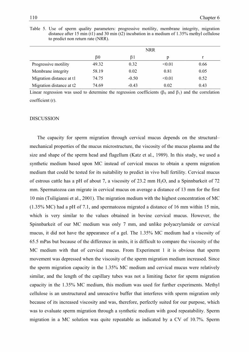
Chapter 6
110
Table 5. Use of sperm quality parameters: progressive motility, membrane integrity, migration distance after 15 min (t1) and 30 min (t2) incubation in a medium of 1.35% methyl cellulose to predict non return rate (NRR).
NRR β0 β1 p r Progressive motility 49.32 0.32 <0.01 0.66 Membrane integrity 58.19 0.02 0.81 0.05 Migration distance at t1 74.75 -0.50 <0.01 0.52 Migration distance at t2 74.69 -0.43 0.02 0.43
Linear regression was used to determine the regression coefficients (β0 and β1) and the correlation
coefficient (r).
DISCUSSION
The capacity for sperm migration through cervical mucus depends on the structural–
mechanical properties of the mucus microstructure, the viscosity of the mucus plasma and the
size and shape of the sperm head and flagellum (Katz et al., 1989). In this study, we used a
synthetic medium based upon MC instead of cervical mucus to obtain a sperm migration
medium that could be tested for its suitability to predict in vivo bull fertility. Cervical mucus
of estrous cattle has a pH of about 7, a viscosity of 23.2 mm H2O, and a Spinnbarkeit of 72
mm. Spermatozoa can migrate in cervical mucus on average a distance of 13 mm for the first
10 min (Tsiligianni et al., 2001). The migration medium with the highest concentration of MC
(1.35% MC) had a pH of 7.1, and spermatozoa migrated a distance of 16 mm within 15 min,
which is very similar to the values obtained in bovine cervical mucus. However, the
Spinnbarkeit of our MC medium was only 7 mm, and unlike polyacrylamide or cervical
mucus, it did not have the appearance of a gel. The 1.35% MC medium had a viscosity of
65.5 mPas but because of the difference in units, it is difficult to compare the viscosity of the
MC medium with that of cervical mucus. From Experiment 1 it is obvious that sperm
movement was depressed when the viscosity of the sperm migration medium increased. Since
the sperm migration capacity in the 1.35% MC medium and cervical mucus were relatively
similar, and the length of the capillary tubes was not a limiting factor for sperm migration
capacity in the 1.35% MC medium, this medium was used for further experiments. Methyl
cellulose is an unstructured and unreactive buffer that interferes with sperm migration only
because of its increased viscosity and was, therefore, perfectly suited for our purpose, which
was to evaluate sperm migration through a synthetic medium with good repeatability. Sperm
migration in a MC solution was quite repeatable as indicated by a CV of 10.7%. Sperm

Assessment of a new UTJ insemination device in dairy cattle 111
samples which were introduced into the capillary at two different concentrations showed a
tendency to migrate further at a concentration of 150 x 106/ml than at a concentration of 100
x 106/ml, although this was only significant at t1 (P = 0.02). This tendency was confirmed in
Experiment 4 where concentration had a significant positive correlation with the sperm
migration distance.
In a number of studies, sperm migration capacity has been linked with other sperm
characteristics such as total motility, sperm morphology, progressive motility, acrosome
integrity and sperm concentration (Hull et al., 1984; Galli et al., 1991). In our study, only the
correlations between sperm migration capacity and progressive motility, membrane integrity
and sperm concentration were determined. In a recent study, Anilkumar et al. (2001) found
that sperm penetration in polyacrylamide gel and bovine cervical mucus was positively
correlated with post-thaw motility (r = 0.81; r = 0.89: P< 0.01) and acrosome integrity (r =
0.88; r = 0.94: P < 0.01) of bovine spermatozoa. However, in contrast to this (Anilkumar et
al., 2001) and other studies (Goldstein et al., 1982; Galli et al., 1991), in our study no
correlation was found between progressive motility and the sperm migration distance (r =
0.05, P = 0.81). In accordance with Galli et al. (1991) and Goldstein et al. (1982) we did
found a positive correlation between the sperm migration distance and membrane integrity (r
= 0.36, P = 0.05) and concentration (r = 0.64, P < 0.01).
Although sperm migration capacity has been linked with different sperm quality
parameters, it has seldom been linked with in vivo fertility in ruminants (Murase et al., 1990).
In contrast to studies in humans (Alexander, 1981; Goldstein et al, 1982), we were not able to
establish a positive correlation between the sperm migration distance in a migration medium
with 1.35% MC and the NRRs of five proven sires. The sperm migration distance at t1 and t2
was even negatively related (β1 -0.50 and -0.43 respectively, P < 0.01 and 0.02 respectively)
to the NRRs. In contrast to this, bull fertility and the sperm migration distance were linked in
a positive way in a previous study (Murase et al., 1990). However, the experimental design in
that study differed from ours in two ways: first, the sperm migration assay was performed in
cervical mucus, and second, fresh spermatozoa were used, whereas the NRRs were obtained
after insemination with frozen-thawed spermatozoa. This means that different ejaculates were
used for the sperm migration assay and for the determination of the NRRs, whereas in our
study the frozen-thawed semen used for the sperm migration test and for the determination of
the NRRs originated from the same batch.
Chapter 6
112
Apart from the experimental design there may be several explanations for the disparity in
results between different bovine studies and between bovine and human experiments. First, it
could be due to differences in characteristics between the media used. Despite the fact that the
MC medium yielded repeatable results it might not be as effective as hyaluronate, cervical
mucus or polyacrylamide gel for sperm migration tests. Hyaluronate and cervical mucus
possess a particular alignment of mucin chains resulting in a higher viscosity and in sperm
movement parallel to the axis of the tube into mucus and hyaluronate solution channels (Tang
et al., 1990). In contrast to cervical mucus, the sperm penetrability in synthetic media such as
MC and hyaluronate is not impaired by the presence of anti-sperm antibodies (Tang et al.,
1990). Polyacrylamide has proved to be more effective than MC when it comes to favouring
migration of hyperactivated spermatozoa, at least in hamsters (Suarez and Dai, 1992).
Whether this property of the medium is really important for studying bovine sperm migration
remains to be determined, since it is known that bull spermatozoa are not hyperactivated at
ejaculation, although they still have to penetrate the cervical mucus at this point of time
(Suarez and Dai, 1992). Despite the fact that frozen-thawed bull spermatozoa are prone to
premature capacitation (Cormier et al., 1997), they do not hyperactivate as readily as hamster
or mouse spermatozoa (Suarez and Dai, 1992; Chamberland et al., 2001), and so it might be
difficult to find a relation between sperm migration and fertility in bulls, even if a more
suitable medium such as polyacrylamide gel was used.
Second, in contrast to humans, in cattle the difference in sperm quality between bulls is a
lot smaller and the number of individuals with very poor sperm quality is very low. For the
evaluation of the sperm migration test in humans, spermatozoa of fertile men (donors of an AI
program) were compared to spermatozoa of patients attending an infertility laboratory
(Alexander, 1981; Insler et al., 1979), whereas in cattle the sperm migration test was
performed with spermatozoa of bulls used for artificial insemination. These bulls have a high
fertility resulting in a narrow range of NRRs, and therefore the number of individuals with
poor sperm quality in cattle is low (Kummerfeld et al., 1981). In the only other comparable
study of cattle a relation was found between fertility and sperm migration, but interestingly, in
this study one bull with 50% NRR and one with 14% NRR were compared with 11 bulls with
NRRs ranging from 60 to 70% (Murase et al., 1990). If the two bulls with the lowest NRRs
were not included in the experiment, no relation could be found between NRR and the sperm
migration distance. Even in the most carefully designed experiment in terms of examining the

Assessment of a new UTJ insemination device in dairy cattle 113
same ejaculate for sperm migration as for NRRs, sperm migration would probably only
discriminate between bulls with extremely varying NRRs.
Finally, the absence of a relation between sperm migration capacity and bull fertility could
be explained by the fact that in dairy cattle, NRRs are determined after artificial insemination.
During insemination the cervical barrier that spermatozoa have to overcome after natural
mating is bypassed and the semen is deposited in the uterine body or in the uterine horns. It
would be more logical to correlate sperm migration tests with fertility data obtained after
natural mating, because then the spermatozoa have to migrate through the mucus before they
reach the site of fertilization. It has been shown that sperm migration through cervical mucus
in vitro could be indicative of the capacity of spermatozoa to populate the oviduct of goats
(Cox et al., 1997). After semen deposition at the ostium uteri externum, spermatozoa from
males with high migration efficiency populated the oviduct more effectively than that from
males with low migration efficiency, but differences were not significant. Apart from these
findings in vivo, no correlation was found between migration efficiency and in vitro
fertilization rates (Cox et al., 1997). This may imply that capillary sperm migration tests may
have value in predicting fertility under in vivo conditions, because the test is able to select
superior sperm samples.
In conclusion, we have shown that sperm migration through MC, albeit a repeatable test,
could not be linked with in vivo bull fertility. Sperm migration through a viscous medium is
only a very limited aspect of sperm behaviour that has to be performed in preparation to
fertilize the oocyte. To obtain more precise prediction of in vivo fertility it is imperative to
combine different in vitro test systems as had been proposed by several authors (Zhang et al.,
1999; Larsson and Rodriguez-Martinez, 2000).

Chapter 6
114
REFERENCES
Aitken RJ, Bowie H, Buckingham D, Harkiss D, Richardson DW, West KM. Sperm
penetration into a hyaluronic acid polymer as a means of monitoring functional
competence. J Androl 1992; 13: 44-54.
Alexander NJ. Evaluation of male infertility with an in vitro cervical mucus penetration test.
Fertil Steril 1981; 36: 201-208.
Anilkumar R, Devanathan TG, Pattabiraman SR, Edwin MJ. Correlation between
spermatozoal characteristics and sperm penetration distance in polyacrylamide gel and
bovine cervical mucus. Theriogenology 2001; 55: 685-691.
Biljan MM, Taylor CT, Manasse PA, Joughin EC, Kingsland CR, Lewis-Jones DI. Evaluation
of different sperm function tests as screening methods for male fertilization potential -
the value of the sperm migration test. Fertil Steril 1994; 62: 591-598.
Bostofte E, Bagger P, Michael A, Stakemann G. The sperm penetration test (P-test) can
predict fecundability in the male partner from infertile couples. Andrologia 1992; 24:
124-129.
Chamberland A, Fournier V, Tardif S, Sirard MA, Sullivan R, Bailey JL. The effect of
heparin on motility parameters and protein phosphorylation during bovine sperm
capacitation. Theriogenology 2001; 55: 823-835.
Clarke GN, Garrett C, Baker GHW. Quantitative sperm mucus penetration: modified
formulae for calculating penetration efficiency. Hum Reprod 1998; 13: 1255-1259.
Cormier N, Sirard MA, Bailey JL. Premature capacitation of bovine spermatozoa is initiated
by cryopreservation. J Androl 1997; 18: 461-468.
Cox JF, Martinez C, Lagos S, Saravia F, Sasmay R. Sperm migration in cervical mucus in
goats. II Relationship with colonization of the oviduct and fertilization efficiency.
Theriogenology 1997;47: 254.

Assessment of a new UTJ insemination device in dairy cattle 115
Eggert-Kruse W, Gerhard I, Tilgen W, Runnebaum B. The use of hens’ egg white as a
substitute for human cervical mucus in assessing human infertility. Int J Androl 1990;
13: 258-266.
Eggert-Kruse W, Schwalbach B, Rohr G, Klinga K, Tilgen W, Runnebaum B. Evaluation of
polyacrylamide gel as substitute for human cervical mucus in the sperm penetration test.
Fertil Steril 1993; 60: 540-549.
Engel S, Petzoldt R. Human sperm penetration in different media. Andrologia 1999; 31: 233-
239.
Galli A, Basetti W, Baldizzi D, Martignoni M, Bornaghi V, Maffi M. Frozen bovine semen
quality and bovine cervical mucus penetration test. Theriogenology 1991; 35: 837-844.
Goldstein MC, Wix LS, Foote RH, Feldschuh R, Feldschuh J. Migration of fresh and
cryopreserved human spermatozoa in polyacrylamide gel. Fertil Steril 1982; 37: 668-
673.
Hull MGR, McLeod FN, Joyce DN, Ray BD, McDermott A. Human in vitro fertilization, in
vivo sperm penetration of cervical mucus and unexplained infertility. Lancet 1984; 2:
245-248.
Hunter RHF. Human fertilization in vivo, with special reference to progression, storage and
release of competent spermatozoa. Hum Reprod 1987; 2: 329-332.
Insler V, Bernstein D, Glezerman M, Misgav N. Correlation of seminal fluid analysis with
mucus penetrating ability of spermatozoa. Fertil Steril 1979; 32: 316-319.
Katz DF, Drobnis EZ, Overstreet JW. Factors regulating mammalian sperm migration through
the female reproductive tract and oocyte vestments. Gamet Res 1989; 22: 443-469.
Kummerfeld HL, Lorton SP, Foote RH. Relationship of spermatozoal migration in cervical
mucus to bovine fertility. J Androl 1981; 2: 103-107.

Chapter 6
116
Larsson B, Rodriguez-Martinez H. Can we use in vitro fertilization tests to predict semen
fertility? Anim Reprod Sci 2000; 60-61, 327-336.
Lee WI, Gaddum-Rosse P, Blandau RJ. Sperm penetration into cervical mucus in vitro. III.
Effect of freezing on estrous bovine cervical mucus. Fertil Steril 1981; 36: 209-213.
Lorton SP, Kummerfelt HT, Foote RH. Polyacrylamide as substitute for cervical mucus in
sperm migration studies. Fertil Steril 1981; 35: 222-225.
Matousek J, Riha J, Srsen V, Veselsky L, Louda F. Penetration of cervical mucus and other
body fluids by bull sperm in capillary tubes. Anim Reprod Sci 1989; 18: 161-166.
Mortimer D, Mortimer ST, Shu MA, Swart R. A simplified approach to sperm–cervical
mucus interaction testing using a hyaluronate migration test. Hum Reprod 1990; 5: 835-
841.
Murase T, Okunda K, Sato K. Assessment of bull fertility using a mucus penetration test and
a human chorionic gonadotrophin stimulation test. Theriogenology 1990; 34: 801-812.
Neuwinger J, Cooper TG, Knuth UA, Nieschlag E. Hyaluronic acid as medium for human
sperm migration tests. Hum Reprod 1991; 6: 396-400.
Parrish JJ, Foote RH. Quantification of bovine sperm separation by a swim up method.
Relationship to sperm motility, integrity of acrosomes, sperm migration in
polyacrylamide gel and fertility. J Androl1987; 8: 259-266.
Pate EF, Brokaw CJ. Movement of spermatozoa in viscous environments. J Exp Biol 1980;
88: 395-397.
Reichmann J, Insler V, Serr DV. A modified in vitro spermatozoal penetration test. Int J Fertil
1973; 18: 241-245.
Suarez SS, Dai X. Hyperactivation enhances mouse sperm capacity for penetrating
viscoelastic media. Biol Reprod 1992; 46: 686-691.

Assessment of a new UTJ insemination device in dairy cattle 117
Suarez SS, Drost M, Redfern K, Gottlieb W. Sperm motility in the oviduct. In: Bavister BD,
Cummins J, Roland ERS (eds): Fertilization in Mammals. Serono Symposia, Norwell,
1990; 111-124.
Tang S, Garrett C, Gordon Baker HW. Comparison of human cervical mucus and artificial
sperm penetration media. Hum Reprod 1999; 14: 2812-2817.
Tsiligianni T, Karagiannidis A, Brikas P, Saratsis P. Physical properties of bovine cervical
mucus during normal and induced (progesterone and/or PGF2α) estrus. Theriogenology
2001; 55: 629-640.
Zhang BR, Larsson B, Lundeheim N, Håård MGH, Rodriguez-Martinez H. Prediction of bull
fertility by combined in vitro assessments of frozen-thawed semen for young dairy bulls
entering an AI-program. Int J Androl 1999; 22: 253-260.


CHAPTER 7
ASSESSMENT OF A NEW UTERO-TUBAL JUNCTION INSEMINATION DEVICE IN
DAIRY CATTLE
Theriogenology 2004; 61: 103-115.
Verberckmoes S., Van Soom A., Dewulf J., de Kruif A.
120 Chapter 7
ABSTRACT
A new artificial insemination device for semen deposition near the utero-tubal junction in
cattle (Ghent device) has been developed at the University of Ghent (Belgium). In this study,
the effect of the new insemination device on sperm quality was evaluated. Moreover, in a
field trial 4064 dairy cows were inseminated by 12 inseminators to examine the efficacy of
the device under field conditions.
The Ghent device is a disposable plastic catheter which can easily follow the curvature of
the uterine horns and thus reach the utero-tubal junction (UTJ). After expulsion of the
inseminate with 0.7 ml or 1.7 ml of air, 19.0% of the insemination dose remained in the
insemination catheter. Sperm loss can be diminished to 9.0% of the original insemination
dose when the insemination catheter is flushed with 0.1 ml of air, followed by 0.6 ml of
physiological saline solution. No toxic effect of the insemination catheter on sperm quality or
fertilizing capacity was found. In the field trial, sperm were inseminated in dairy cattle which
were divided in 3 groups. The first group was inseminated in the uterine body with the
conventional insemination device, the second group in the uterine body with the Ghent
device, and the third group in the tip of both uterine horns with the Ghent device. Each
insemination was performed with 10-15 x 106 frozen-thawed spermatozoa. The pregnancy
rates (PRs) were significantly affected by the insemination technique (P = 0.02), by the
inseminator (P = 0.01), by heifer or cow (P < 0.01), and by the insemination number (P <
0.01). Pregnancy rates obtained with the conventional insemination device (57.6%) were
significantly better than those obtained with the Ghent device in the uterine body (52.7%) (P
< 0.01), but did not differ significantly from those obtained after deep insemination into both
uterine horns (53.8%) (P = 0.27). It can be concluded that the Ghent device is suitable for
utero-tubal junction insemination of dairy cattle under field conditions. Whether the Ghent
device is also suitable for insemination with lower insemination doses is at present under
investigation.

Assessment of a new UTJ insemination device in dairy cattle 121
INTRODUCTION
Currently most commercial artificial inseminations (AI) in cattle are performed into the
uterine body by means of a stainless steel insemination device of Cassou (Noakes et al.,
2001). This rigid device facilitates the passage of the cervix and is covered with a plastic
sheath to minimize transmission of infectious agents by the insemination device. Deposition
of the inseminate into the uterine body of the cow makes it possible to achieve acceptable
pregnancy rates (PRs) with a relatively low dose of frozen-thawed spermatozoa (10-15
million) (Uwland, 1984, Smidt and Zuidberg, 1988; Foote and Kaproth, 1997; Den Daas et
al., 1998).
Lowering the insemination dose without a concomitant reduction in pregnancy rates can
be obtained when fresh diluted semen is used (Shannon and Vishwanath, 1995). However,
using fresh semen complicates distribution logistics and enhances the risk of spreading of
infectious agents, e.g. foot-and-mouth disease virus. Deposition of semen closer to the oviduct
could be an alternative to obtain acceptable pregnancy rates with lower insemination doses,
less fertile semen, such as sex-separated spermatozoa or spermatozoa whose DNA-status may
have been modified by specific treatment (Gandolfi, 2000; Hunter and Greve, 1998). Semen
deposition near the utero-tubal junction will result in a decreased loss of spermatozoa by 1)
retrograde flow in the cervical mucus (Larsson and Larsson, 1985; Mitchell et al., 1985;
Nelson et al., 1987) and 2) phagocytosis during migration through the uterus (Hawk, 1983),
and in an increased survival time of spermatozoa in the sperm-friendly environment of the
isthmus (Suarez, 2001).
A good deal of research, with varying success, has been performed to improve the
pregnancy rates (PRs) in cattle by deposition of spermatozoa closer to the place of
fertilization. In most cases, no difference was found between deep uterine insemination (in
uterine horns) and insemination in the uterine body (Knight et al., 1951; Stewart and Melrose,
1952; Olds et al., 1953; Macpherson, 1968; Hawk and Tanabe, 1986; Williams et al., 1988;
McKenna et al., 1990) In one study, worse PRs were found after deep uterine insemination
(Marshall et al., 1989) while four other research groups obtained better results after deep
uterine insemination (Zavos et al., 1985; López-Gatius et al., 1988, Senger et al., 1988;
Dalton et al., 1999). An explanation for the varying results can be found in the different
insemination doses used, the application of uni- or bilateral insemination, the skill and

122 Chapter 7
experience of inseminators, and the insemination of nulli- and pluriparous cows in natural
heat or after estrus induction. The large variation in experimental set up and the fact that
“deep uterine insemination” is not consistently defined, makes it difficult to compare the
results of these different studies.
However, semen deposition near the utero-tubal junction can not be performed properly
with the conventional rigid insemination device, since the contractility of the uterus in estrous
cows is increased, and the uterine horns are curled (Noakes et al., 2001). Introduction of a
rigid insemination device up to the tip of the uterine horns will enhance the risk of damaging
the friable uterine wall with reduced fertility as a result (Wolff et al., 1990). Substituting the
conventional insemination device for a new instrument which would be useful for UTJ-
insemination meant that a device, which was both rigid enough to penetrate the cervix and
flexible enough to follow the curves of the curled uterus, had to be developed. Moreover, the
device had preferably to be made of disposable materials for sanitary reasons, had to be cheap
to produce, and had to be non-toxic for spermatozoa.
In this article the characteristics of the catheter as well as the effect of the device on sperm
quality and fertilizing ability were investigated. Moreover, a field trial was undertaken to
compare PRs obtained after bilateral utero-tubal junction (UTJ) insemination and those
obtained after insemination with the conventional insemination device. In both cases
inseminations were performed with frozen-thawed semen in dairy cows and heifers in natural
heat. A second goal of this field trial was to evaluate the feasibility of the newly developed
Ghent device under field conditions.
MATERIAL AND METHODS
Semen
Frozen-thawed semen of Holstein Friesian bulls (n = 290) was generously supplied by the
AI-centre, Holland Genetics (The Netherlands). No selection was made in bulls used for the
field trial. The semen was extended in a Tris-egg yolk diluter, packaged in 0.25 ml French
straws (Instruments de Médicine Vétérinaire, l’Aigle, France) frozen and stored in liquid
nitrogen until use. Each straw contained 10-15 x 106 frozen spermatozoa. Before
insemination, semen was thawed for 1 min in 37°C water. For the conventional insemination
method, straws were opened at one end and introduced in the insemination device. For the

Assessment of a new UTJ insemination device in dairy cattle 123
new insemination device, thawed semen was suspended in a vial containing 0.25 ml of 120
mM Na-citrate and subsequently aspirated into a flexible catheter that was introduced in the
Ghent device.
Selection of cows for the insemination trial
Both dairy and dual purpose cows from different dairy farms (n = 1063) were selected for
insemination, if the cows were in good general condition and seen in estrus by the farmer.
Exclusion criteria were: 1) complicated previous parturition, and 2) abnormal, malodorous
vaginal discharge. For data processing a difference was made between nulliparous heifers and
cows.
Inseminators
Inseminations were performed by 12 veterinarians of the Flemish AI-centre (VRV). When
the field trial was finished, the inseminators were asked to fill in a questionnaire to evaluate
the suitability of the Ghent device under field conditions.
Estrus detection and AI
Cows (n = 3428) and nulliparous heifers (n = 636) in natural heat were artificially
inseminated. Estrus detection was performed by the farmer. Animals with behavioral estrus in
the morning were inseminated in the evening, while animals showing signs of standing estrus
in the evening were inseminated the next morning. The conventional and new insemination
techniques were performed under rectal guidance. Before introduction of the Ghent device
into the female genital tract, the inseminator had to 1) thaw the straw, 2) suspend the semen
(0.25 ml) into a vial with 0.25 ml of 120 mM Na-citrate, 3) aspirate the contents into the
catheter by means of a syringe which was connected to the end of the catheter, and finally 4)
introduce and fix the catheter filled with semen in the Ghent device. The syringe used to fill
the catheter remained connected to the catheter and was used to expel the semen. Then the
Ghent device was manipulated through the cervix and when the tip of the insemination device
had entered the uterine body, the inner tube of the device was pushed forward and half of the
insemination dose was deposited in the tip of the left uterine horn. Then the inner tube was
pulled back into the uterine body, and subsequently pushed forward into the tip of the right
uterine horn, where the second half of the insemination dose was deposited. Before the onset
of the field trial each inseminator received 100 Ghent devices to practice.

124 Chapter 7
Characteristics of the Ghent device
The Ghent device consists of 2 hollow plastic tubes, in which a catheter filled with semen
is introduced (Verberckmoes et al., 2002). The outer tube is completely rigid, while the inner
tube consists of a rigid caudal end and a flexible cranial tip. The outer plastic tube can move
independently from the inner tube (Figure 1).
Figure 1: Ghent device with a telescopic, flexible tip.
The dimensions of total device, outer tube, rigid part of the inner tube, flexible part of the
inner tube and the catheter that contains the inseminate are determined in mm (Table 1). The
catheter consists of polyethylene, and the tubes are made of ABS and PVC. The Ghent Device
and the new insemination technique are protected by a patent (01976188.1-2318-EP011181,
Ghent University).
Table 1: Dimensions of the Ghent device.
Length (mm) Internal diameter (mm) External diameter (mm) 1. Outer tube 496.7 3.4 6.2 2a. Inner tube, rigid part 476.1 2.0 3.0 2b. Inner tube, flexible part 223.0 2.5 3.0 3. Insemination catheter 709.8 1.0 1.8
Experiment 1: Determination of the percentage of spermatozoa which remains in the
insemination catheter after insemination.
To determine the number of frozen-thawed spermatozoa which remain in the insemination
catheter after expelling the inseminate, the weight of 10 catheters was determined at different
moments. When the semen was expelled using only air, the weight of the catheter was
assessed 1) before filling, 2) when it was filled with diluted semen, 3) after expelling the
semen with 0.7 ml of air and 4) when the catheter was flushed with an additional 1.0 ml of air.
First the insemination catheter was filled with 0.5 ml of diluted sperm by means of aspiration
with a syringe. The specific gravity of the Tris-egg yolk diluter in which the semen was

Assessment of a new UTJ insemination device in dairy cattle 125
frozen is 1.03 g/ml. The concentration of the spermatozoa in the diluter, determined by means
of a Bürker counting chamber, was 48 x 106 spermatozoa/ml. Since we determined how much
semen remained in the catheter (in g), and since we knew the specific gravity of the sperm
diluter, we were able to calculate the remaining volume of diluted semen in the catheter:
weight (g) / specific gravity (g/ml) = volume (ml). The remaining volume of diluted semen
multiplied by sperm concentration gives the exact number of spermatozoa that remained
attached to the wall of the insemination catheter: volume (ml) x concentration (number/ml) =
exact number. The remaining number of spermatozoa can be expressed as a percentage of the
total insemination dose: number of spermatozoa remaining in the insemination catheter
devided by the number of spermatozoa in insemination dose = % of insemination dose
remaining in the insemination catheter.
When the semen was expelled with 0.1 ml air followed by 0.6 ml of physiological saline
solution, the weight of 10 catheters was determined 1) before filling, 2) when filled with
diluted semen, and 3) after expelling the diluted semen with 0.1 ml of air and 0.6 ml of
physiological saline solution. When the insemination catheter was flushed with 0.1 ml of air
and 0.6 ml of physiological saline solution, 0.56 ml of the flushing medium remained in the
catheter (content of the insemination catheter). Subsequently, the content of the insemination
catheter was expelled by air pressure and the sperm concentration in this medium was
determined by means of a Bürker counting chamber. The remaining volume of physiological
saline solution in the catheter multiplied by the sperm concentration gives the number of
spermatozoa that remained attached to the wall of the insemination catheter and can be
expressed as a percentage of the total insemination dose, agreeing with the previously
described formula.
Experiment 2: Effect of the insemination catheter on sperm quality.
Sperm treatment: Fresh semen of 3 bulls was collected by means of an artificial vagina.
The sperm concentration was determined by means of a Bürker counting chamber and light
microscopy. The spermatozoa were diluted to a concentration of 30 x 106 spermazoa/ml in
physiological saline solution. For each of the 3 bulls, the diluted spermatozoa were incubated
for 1h at 22°C in: 1) 5 insemination catheters and, as a control, 2) in 1 non toxic cryotube
(Nunc®, Wiesbaden, Germany). The sperm quality was determined before and after 1h
incubation in the insemination catheters and cryotubes.

126 Chapter 7
Sperm quality evaluation: The assessed sperm quality parameters were membrane
integrity (MI) and motility and acrosome integrity (AI). The membrane integrity was
determined by means of LIVE/DEAD® Sperm Viability Kit (Molecular Probes) and
fluorescent microscopy. The sperm motility was determined with the Hamilton Thorne
(HTM-Ceros 12.1) on at least 500 spermatozoa, and the following parameters were assessed:
velocity average pathway (VAP), velocity straight line (VSL), velocity curved line (VCL),
straightness (STR), linearity (LIN), percentage of progressive motile spermatozoa (PMS) and
rapid progressively motile spermatozoa (RMS). The acrosome integrity (AI) was assessed by
means of Pisum Sativum Agglutinin (PSA) staining with a Leica DMR fluorescence
microscope (x 400) (Kawakami et al., 1993). Spermatozoa with fluorescent green heads
indicated acrosome intact spermatozoa. Spermatozoa were considered as acrosome-reacted
when a fluorescent band was observed at the equatorial region.
Experiment 3: Field trial.
In a field trial, the effect of insemination technique on PRs was investigated. The
inseminations were performed from the 20st of September until the 1st of November 2001.
Animals were divided at random into 3 groups. In the first group (n = 1416), animals were
inseminated with the conventional insemination device in the uterine body. In the second
group (n = 1334), semen was deposited in the uterine body with the Ghent device. In the third
group (n = 1314), semen was deposited in the tip of both uterine horns with the Ghent device.
Randomisation was performed by computer and the animals included in the field trial at a
given day, were all inseminated with the same insemination technique.
At the moment of insemination the following data were registered by means of a hand
terminal : identity of the cow, identity of the bull used, number of insemination (1st, 2nd, ≥3rd
AI), heifer or cow, identity of the farmer, date of insemination, insemination technique used
and identity of the inseminator. The 56 days non-return rate (NRR) was the percentage of
animals that were not presented for AI during the 56 days after the first AI, and was based on
the data obtained with the hand terminal. The effective pregnancy rates (PRs) were
determined at 45-90 days after last insemination by the inseminators by means of rectal
examination of the animals that were not offered again for AI. During the field trial, the
inseminators were asked to fill in a questionnaire concerning the handiness of the Ghent
device and the time spent per insemination.
Assessment of a new UTJ insemination device in dairy cattle 127
STATISTICAL ANALYSIS
The differences in remaining sperm numbers between different flushing methods, were
compared by means of one-way ANOVA and Scheffé post-hoc tests (SPSS, 10.0 Chicago,
USA). Different sperm quality parameters were compared using a linear mixed effect model
with an autoregressive correlation structure of the first order (S-Plus, 2000; Cambridge,
USA). For the results obtained in the field trial, the effect of insemination technique,
inseminator, parity of the cow, insemination number and week of insemination on the PRs
was analysed by means of univariate and multivariate logistic regression. In the multivariate
analysis, all significant parameters of the univariate analysis (P < 0.05) were included, and all
2-way interactions were tested (SPSS, 10.0, Chicago, USA).
RESULTS
Characteristics of the Ghent device.
The dimensions of the length, the internal and external diameter of the outer tube, the rigid
part of the inner tube, the flexible part of the inner tube and of the catheter that contains the
inseminate are given in Table 1. A small section of the flexible telescopic part of the inner
tube (197.4 mm) protrudes from the outer tube. The content of the insemination catheter is
0.56 ml (π x r² x L). The Luer Lock system at the back of the catheter enables the connection
of a disposable syringe.
Experiment 1: Determination of the number of spermatozoa which remain in the insemination
catheter after insemination.
Although there was a displacement of 0.5 ml of air in the syringe by moving the plunger
backwards, the catheter was loaded with only 0.46 g ± 0.05 g of diluted sperm. Extra flushing
of the catheter with 1 ml of air had no significant effect (P = 0.37) on the remaining number
of spermatozoa in the catheter (Table 2). The remaining number of spermatozoa was
significantly decreased by expulsion of the inseminate with 0.1 ml of air followed by 0.6 ml
of physiological saline solution (P < 0.01) (Table 2).

128 Chapter 7
Table 2: Remaining spermatozoa (mean ± S.D.) after flushing the insemination catheter with 0.7 ml of air. 0.7 + 1.0 ml of air and 0.1 ml of air + 0.6 ml of physiological saline solution.
Number of measurements Remaining spermatozoa (n) Millions % of insemination dose 0.7 ml air n = 10 4.28 ± 0.98a 19.0 0.7 + 1.0 ml air n = 10 3.96 ± 0.85a 18.0 0.1 ml air + 0.6 ml PS n = 10 2.18 ± 0.45b 9.0 ab Different superscripts indicate a significant difference (P < 0.01). PS: physiological saline
Experiment 2: Effect of the insemination catheter on sperm quality.
Of all parameters determined, no significant differences could be found between the
spermatozoa incubated in the insemination catheter and those incubated in the cryotubes (P >
0.05) (Table 3). After 1 h of incubation at 22°C, all sperm motility parameters were decreased
with the exception of velocity curved line (VCL) and amplitude lateral head displacement
(ALH). The small increase in VCL and ALH and decrease in the other sperm motility
parameters indicate an insignificant deterioration of sperm quality during the 1h incubation
period in both containers. For spermatozoa incubated in cryotubes and insemination catheters,
the percentage of membrane-intact spermatozoa decreased insignificantly (P = 0.49) from
66.8% ± 8.8% at T0, to 50.3% ± 11.4% and 41.6% ± 12.1% respectively. The acrosome
integrity (AI) remained unchanged (P = 0.88) at both times, in both containers (97%).
Table 3: Motility parameters (VAP, VSL, VCL, BCF, STR, LIN, PMS, RMS) analysed by means of a Hamilton Thorne analyzer of fresh semen immediately after collection (T0) and after 1h incubation (T1) at 22°C in cryotubes (control) and in the insemination catheters (mean ± S.D.).
Experiment 3: Field trial.
A total of 3428 cows and 636 heifers were inseminated. Rectal examination to confirm
pregnancy could be performed in 3902 animals (96%). Four per cent of the animals could not
be examined, mostly because of culling before examination. As can be deduced from Table 4
the effective PRs were 15% lower than the NRRs.
VAP (µm/s) VSL (µm/s) VCL (µm/s) STR (%) LIN (%) PMS (%) RMS (%)
T0 134.8 ± 11.2 113.0 ± 7.3 205.3 ± 32.5 82.9 ± 2.4 57.6 ± 5.4 51.4 ± 11.6 58.3 ± 11.6T1 control 120.7 ± 19.2 84.5 ± 4.5 220.0 ± 46.0 71.0 ± 8.7 41.0 ± 7.0 30.3 ± 13.6 42.0 ± 9.6 T1 catheter 122.8 ± 6.2 91.6 ± 2.3 220.9 ± 25.3 75.4 ± 4.4 44.3 ± 6.2 39.4 ± 12.1 49.8 ± 13.6P-value 0.98 0.25 0.92 0.79 0.45 0.84 0.93

Assessment of a new UTJ insemination device in dairy cattle 129
Table 4: Non return rate (NRR) and pregnancy results after insemination with the conventional insemination device into the uterine body (CD body), with the Ghent device into the uterine body (GD body) and with the Ghent device into both uterine horns (GD horns).
AI method No. of AIs NRR Pregnant Not pregnant Unknown
CD body 1416 72.0% 57.6%a,b 37.2% 5.2%
GD body 1334 68.0% 52.7%a 43.4% 3.9%
GD horns 1314 69.0% 53.8%b 43.5% 2.7%
Total 4064 70.0% 54.7% 41.3% 4.0% a,b Values with a different superscript in the same column differ significantly (P < 0.05).
The insemination technique, the inseminator, the parity of the cow, and the insemination
number all had a significant effect on the PRs, as shown by univariate logistic regression
(Tables 5, 6).
Table 5: Number of inseminations per inseminator and obtained pregnancy rates with different insemination techniques.
Inseminator No. of inseminations Pregnancy rates (%)
CD body GD body GD horns
1 298 51.6 46.2 53.4
2 375 69.7 51.9 66.4
3 359 50.7 46.9 58.9
4 517 57.9 59.5 52.0
5 371 58.2 48.4 50.5
6 302 64.7 56.3 64.4
7 60 61.5 40.6 53.3
8 242 56.0 42.5 45.7
9 313 58.9 56.0 49.2
10 364 65.9 61.4 55.2
11 393 54.1 50.0 55.7
12 312 44.8 57.0 36.2 CD = Cassou device, GD = Ghent device
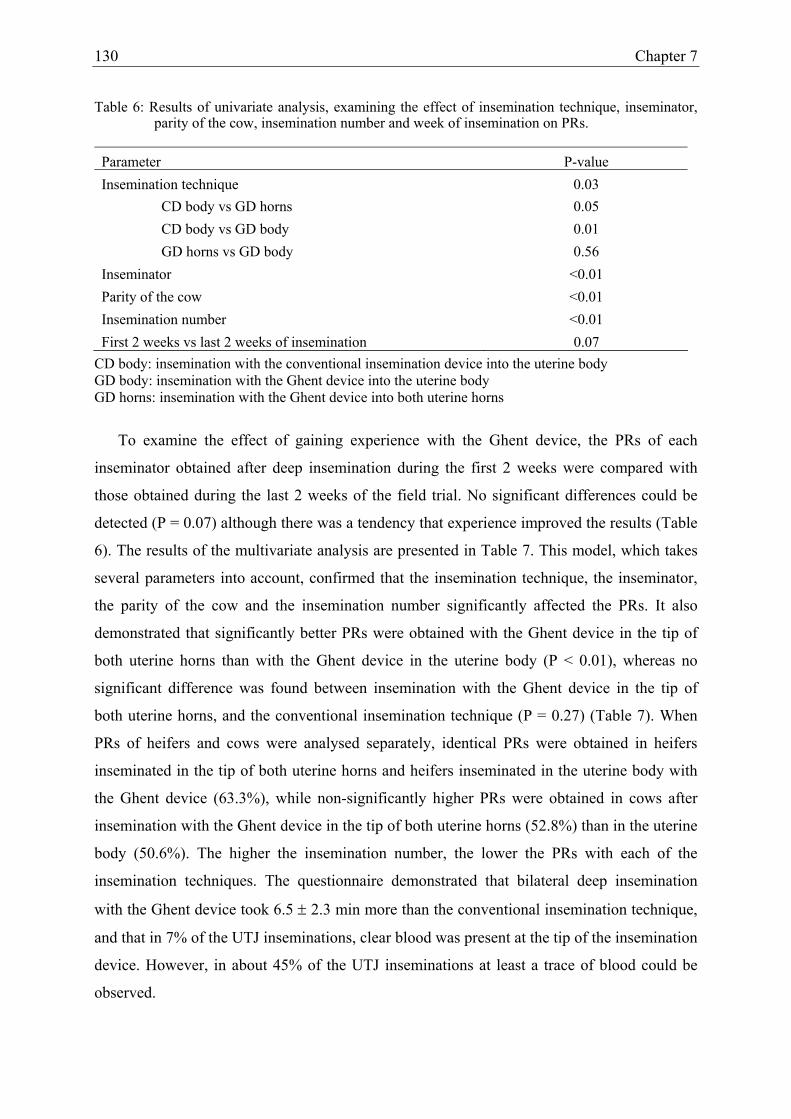
130 Chapter 7
Table 6: Results of univariate analysis, examining the effect of insemination technique, inseminator, parity of the cow, insemination number and week of insemination on PRs.
Parameter P-value Insemination technique 0.03
CD body vs GD horns 0.05 CD body vs GD body 0.01 GD horns vs GD body 0.56
Inseminator <0.01 Parity of the cow <0.01 Insemination number <0.01 First 2 weeks vs last 2 weeks of insemination 0.07
CD body: insemination with the conventional insemination device into the uterine body GD body: insemination with the Ghent device into the uterine body GD horns: insemination with the Ghent device into both uterine horns
To examine the effect of gaining experience with the Ghent device, the PRs of each
inseminator obtained after deep insemination during the first 2 weeks were compared with
those obtained during the last 2 weeks of the field trial. No significant differences could be
detected (P = 0.07) although there was a tendency that experience improved the results (Table
6). The results of the multivariate analysis are presented in Table 7. This model, which takes
several parameters into account, confirmed that the insemination technique, the inseminator,
the parity of the cow and the insemination number significantly affected the PRs. It also
demonstrated that significantly better PRs were obtained with the Ghent device in the tip of
both uterine horns than with the Ghent device in the uterine body (P < 0.01), whereas no
significant difference was found between insemination with the Ghent device in the tip of
both uterine horns, and the conventional insemination technique (P = 0.27) (Table 7). When
PRs of heifers and cows were analysed separately, identical PRs were obtained in heifers
inseminated in the tip of both uterine horns and heifers inseminated in the uterine body with
the Ghent device (63.3%), while non-significantly higher PRs were obtained in cows after
insemination with the Ghent device in the tip of both uterine horns (52.8%) than in the uterine
body (50.6%). The higher the insemination number, the lower the PRs with each of the
insemination techniques. The questionnaire demonstrated that bilateral deep insemination
with the Ghent device took 6.5 ± 2.3 min more than the conventional insemination technique,
and that in 7% of the UTJ inseminations, clear blood was present at the tip of the insemination
device. However, in about 45% of the UTJ inseminations at least a trace of blood could be
observed.

Assessment of a new UTJ insemination device in dairy cattle 131
Table 7: Results of the multivariate analysis, examining the effect of insemination technique, inseminator, parity of the cow and insemination number on PRs.
Parameter P-value Insemination technique 0.02
CD body v.s. GD horns 0.27 CD body v.s. GD body 0.07 GD horns v.s. GD body <0.01
Inseminator 0.01 Inseminator x technique 0.06 Parity of the cow <0.01 Insemination number <0.01
DISCUSSION
In this study, we describe the use of a new artificial insemination device for cattle, which
unlike the conventional Cassou insemination device, can be used for deep uterine
insemination near the utero-tubal junction. Results of the present study indicate that our new
Ghent device permits utero-tubal junction insemination in cattle under field conditions with
satisfactory pregnancy rates. In addition, only a limited proportion of the inseminate is
retained within the catheter, and no negative effect of the device on sperm quality was
observed. This makes the Ghent device a possible useful instrument for artificial insemination
with sexed or low doses of semen. The application of the device for low dose insemination
implicates that a minimal number of spermatozoa may be lost during the insemination
procedure. We have shown that flushing of the insemination catheter with 0.1 ml of air
followed by 0.6 ml of physiological saline solution resulted in a sperm loss which comprised
less than 10% of the initial inseminate. Although further increase of the flushing volume
could reduce the remaining number of spermatozoa in the insemination catheter, we do not
recommend doing so. The higher volume of flushing medium would decrease the number of
spermatozoa entering the oviduct as a result of the increase in sperm loss by retrograde flow.
Therefore, insemination near the UTJ requires low volume insemination doses since it will
increase the percentage of spermatozoa that reach the isthmus of the oviduct, where they are
protected from phagocytosis in the hostile uterine environment (Morris and Allen, 2002). A
higher insemination volume would decrease the number of spermatozoa that enters the
oviduct due to the increased retrograde flow.

132 Chapter 7
As a control for any possible toxic effect of the insemination catheter on the spermatozoa,
we examined sperm motility parameters as assessed by means of computer assisted sperm
analysis. It is known that cytotoxic components can alter the motility of the spermatozoa,
which depends upon intact cellular structures and functions, and which consequently offers an
endpoint for quantitative evaluation of the cytotoxic potential of chemical substances (Seibert
et al, 1989). Spermatozoa were incubated in physiological saline solution which contains no
substances that could act as scavengers for toxic substances. Media containing serum or
chelating agents such as BSA or EDTA are known to overcome (partially) the detrimental
effect of cytotoxic components (Van Langendonckt et al., 1996; Erbach et al., 1995;
Borenfreund, 1986). Even under conditions as described in experiment 2, no differences in
motility parameters (VAP, VSL, VCL, BCF, STR, LIN, PMS, RMS) could be observed for
spermatozoa which had been in contact with the Ghent device for 1 h. Moreover, no effect on
in vivo fertilizing capacity of the same spermatozoa could be detected (data not shown).
Since we know that the Ghent device is not toxic for spermatozoa, and does not result in
important sperm losses, it was somewhat disappointing to obtain no significant difference (P
= 0.27) in PR after utero-tubal junction insemination when compared with conventional
insemination. However, using the multivariate analysis, which is the most correct
representation of the real difference between insemination techniques since all influencing
parameters are accounted for, significantly higher pregnancy rates were observed after
insemination with the Ghent device in the uterine horns than in the uterine body (P < 0.01).
Several explanations for the results obtained with the different insemination techniques are
possible. The lower PRs obtained with the Ghent device than with the conventional
insemination device can be explained by the increased number of manipulations that the
semen had to undergo. After thawing, the semen is suspended in a vial with Na-citrate,
aspirated into a catheter and subsequently introduced in the Ghent device. The extra
manipulations are time consuming and may have a negative effect on the sperm quality. The
fact that the worst results were obtained after insemination in the uterine body with the Ghent
device, can be explained by the technical problems that occur with the Ghent device because
it is not developed for insemination in the uterine body. The Ghent device is longer than the
conventional insemination device, which complicates its fixation in the uterine body. Because
the uterine body of the cow has a length of only 1-3 cm (Nickel et al., 1999) there is an
increased risk of damaging the septum that separates both uterine horns when the
insemination device is not appropriately fixed in the uterine body. Moreover, it was found that

Assessment of a new UTJ insemination device in dairy cattle 133
after using the Ghent device, in some cases blood was sticking on the tip of the device. It is
known that the endometrium is vulnerable at the moment of insemination, because of the
hyperaemic condition of the endometrium during estrus (Dellman and Eurell, 1998). The
blood and shreds of mucosa liberated into the uterine lumen may have a negative effect on the
fertilizing capacity of the spermatozoa. As examined in Chapter 9, blood does not influence
sperm quality, but can cause agglutination of the spermatozoa. Subsequently this
agglutination may result in a reduced number of spermatozoa that can reach the site of
fertilization, resulting in decreased fertilization rates. In humans, it is known that preventing
endometrial damage during deep uterine insemination increases the chance of conception
(Lavie et al., 1997). However, we also have to take into account that the deposition of
spermatozoa closer to the site of fertilization, decreases the chance of contact with blood, and
as a consequence, less chance for the spermatozoa to agglutinate.
Another factor that may have influenced the results is the fact that some experience with
the technique was required to obtain better results. During the preliminary trial period, UTJ-
inseminations were performed after an epidural anaesthesia. Few animals strained and the
female genital tract was easy to manipulate. Despite this advantage, and the fact that an
epidural anaesthesia does not affect sperm transport in the uterus (Thurmon et al., 1996) 11 of
the 12 inseminators did not continue to use epidural anaesthesia after the preliminary trial
period with the Ghent device. The main reason for this was saving time. Interestingly, one
inseminator used epidural anaesthesia for all UTJ-inseminations and obtained 8% better PRs
than with the conventional insemination technique. The importance of gaining experience
with the Ghent device is also shown by the fact that, in the multivariate analysis, the
interaction between inseminator and insemination technique was approaching significance (P
= 0.06). This indicates that some inseminators obtain better results with the Ghent device than
others. Although the inseminators had the impression that inseminations with the Ghent
device were easier to perform when they gained more experience with the device, no
significant difference was found between the PRs of the first 2 and the last 2 weeks of the
field trial. However, increasing the number of inseminations per inseminator could result in a
significant effect.
In conclusion, semen deposition near the utero-tubal junction in cattle by means of a
minimally invasive technique can be performed with the newly developed Ghent device. The
Ghent device is made of disposible materials, has no toxic effect on bovine semen, can easily
be handled by one person, and is suitable under field conditions as proven in the field trial. A

134 Chapter 7
beneficial effect of UTJ insemination with the Ghent device in cattle is expected when low
insemination doses or less fertile semen will be used. The Ghent device could offer the
solution for the application of sex sorted semen under field conditions.

Assessment of a new UTJ insemination device in dairy cattle 135
REFERENCES
Borenfreund E, Puerner JA. Cytotoxicity of metals, metal-metal and metal-chelator
combinations assayed in vitro. Toxicity 1986; 39: 121-134.
Dalton JC, Nadir S, Bame JH, Saacke RG. Effect of deep uterine insemination on
spermatozoal accessibility to the ovum in cattle: a competitive insemination study.
Theriogenology 1999; 51: 883-890.
Dellman HD, Eurell JA.. In Dellmann HD, Eurell JA (eds): Textbook of Veterinary
Histology. Female reproductive system. Baltimore, USA: Williams and Wilkins, 1998;
263-264.
Den Daas JH, De Jong G, Lansberger LM, Van Wagtendonk-De Leeuw AM. The relationship
between the number of spermatozoa inseminated and the reproductive efficiency of
individual dairy bulls. J Dairy Sci 1998; 81: 1714-1723.
Erbach GT, Bhatnagar P, Baltz JM, Biggers JD. Zinc is a possible toxic contaminant of
silicone oil in microdrop cultures of preimplantation mouse embryos. Hum Reprod
1995; 10: 3248-3254.
Foote RH, Kaproth MT. Sperm number inseminated in dairy cattle and non return rates
revisited. J Dairy Sci 1997; 80: 3072-3076.
Gandolfi F. Sperm-mediated transgenesis. Theriogenology 2000; 53: 127-137.
Hawk HW, Tanabe TY. Effect of unilateral cornual insemination upon fertilization rate in
superovulating and single-ovulating cattle. J Anim Sci 1986; 63: 551-560.
Hawk HW. Sperm survival and transport in the female reproductive tract. J Dairy Sci 1983;
66: 2645-2660.
Hunter RHF, Greve T. Deep uterine insemination of cattle: a fruitful way forward with
smaller numbers of spermatozoa. Acta Vet Scand 1998; 39: 149-163.

136 Chapter 7
Kawakami E, Vandevoort CA, Mahi-Brown CA, Overstreet JW. Induction of acrosome
reactions of canine sperm by homologous zona pellucida. Biol Reprod 1993; 48: 841-
945.
Knight CW, Patrick TE, Anderson HW, Branton C. The relation of site of semen deposit to
breeding efficiency of dairy cattle. J Dairy Sci 1951; 34: 199-202.
Larsson B, Larsson K. Distribution of spermatozoa in the genital tract of artificially
inseminated heifers. Acta Vet Scand 1985; 26: 385-395.
Lavie O, Margalioth EJ, Geva-Eldar T, Ben-Chetrit A. Ultrasonographic endometrial changes
after intrauterine insemination: a comparison of two catheters. Fertil Steril 1997; 68:
731-734.
López-Gatius F, Camón-Urgel J, Angulo-Asensio E. Effect of single deep insemination on
transferable embryo recovery rates in superovulated dairy cows. Theriogenology 1988;
30: 877-885.
Macpherson JW. Semen placement effects on fertility in bovines. J Dairy Sci 1968; 51: 807-
808.
Marshall CE, Graves WM, Meador JL, Swain JB, Anderson JI. A fertility comparison of
uterine body and bicornual semen deposition procedures in dairy cattle. J Dairy Sci
1989; 72: 455.
McKenna T, Lenz RW, Fenton SE, Ax RL. Non return rates of dairy cattle following uterine
body or cornual insemination. J Dairy Sci 1990; 73: 1779-83.
Mitchell JR, Senger PL, Rosenberger JL. Distribution and retention of spermatozoa with
acrosomal and nuclear abnormalities in the cow genital tract. Anim Sci 1985; 61: 956-
967.
Morris LHA, Allen WR. An overview of low dose insemination in the mare. Reprod Dom
Anim 2002; 37: 206-210.

Assessment of a new UTJ insemination device in dairy cattle 137
Nelson VE, Aalseth EP, Hawman CH, Adams GD, Dawon LJ, McNew RW. Sperm discharge
and distribution within the cows reproductive tract and after A.I. Anim Sci 1987; 65:
401.
Nickel R, Schummer A, Seiferle E. Algemeine und vergleichende Betrachtungen von die
Gebärmutter. In: Lehrbuch der Anatomie der Haustiere. Parey Buchverlag, Berlin. 1999;
402.
Noakes DE, Parkinson TJ, England GCW. In: Veterinary Reproduction and Obstetrics. 8th
edition. Artificial insemination. W.B. Saunders. 2001; 751-778.
Olds D, Seath DM, Carpenter MC, Lucas HL. Interrelationships between site of deposition,
dosage, and number of spermatozoa in diluted semen and fertility of dairy cows
inseminated artificially. J Dairy Sci 1953; 36: 1031-1035.
Salisbury GW, VanDemark NL. The effect of cervical, uterine and cornual insemination on
fertility of the dairy cow. J Dairy Sci 1951; 34: 68-74.
Seibert H, Kolossa M, Wasserman O. Bovine spermatozoa as an in vitro model for studies on
the cytotoxicity of chemicals. Effect of chlorophenols. Cell biology and toxicology
1989; 5: 315-330.
Senger PL, Becker WC, Davidge ST, Hillers JK, Reeves JJ. Influence of cornual insemination
on conception in dairy cattle. J Anim Sci 1988; 66: 3010-3016.
Shannon P, Vishwanath R. The effect of optimal and suboptimal concentrations of sperm on
the fertility of fresh and frozen bovine semen and a theorethical model to explain the
fertility differences. Anim Reprod Sci 1995; 39: 1-10.
Smidt WJ, Zuidberg C. Bevruchtingsresultaten van inseminaties met diepvriessperma met
verschillende aantallen spermatozoa per dosis bij het MRIJ-vee. Tijdschr Diergeneeskd
1988; 12: 673-676.
Stewart DL, Melrose DR. The comparative efficiency of the intra cervical and intra-uterine
methods of insemination in the dairy cow. Vet Rec 1952; 64: 605-607.

138 Chapter 7
Suarez SS. Carbohydrate-mediated formation of the oviductal sperm reservoir in mammals.
Cells tissues organs 2001; 168: 105-112.
Thurmon JC, Tranquilli WJ, Benson G. In Thurmon JC, Tranquilli WJ, Benson G (eds).
Lumb and Jones’ veterinary anesthesia, third edition. Local and regional anesthetic
techniques: ruminants and swine. Baltimore, USA: Williams and Wilkins, 1996; 495.
Uwland J. Correlation between the number of inseminated spermatozoa and fertility rates in
dairy cattle. Tijdschr Diergeneeskd 1984; 10: 405-412.
Van Langendonckt A, Van Steenbrugge A, Donnay I, Van Soom A, berg U, Semple E,
Grisart B, Mermillod P, Brem G, Massip A, Dessy F. Three years results of in vitro
production of bovine embryos in serum-poor bovine oviduct conditioned medium. An
overview. Reprod Nutr Dev 1996; 36: 493-502.
Verberckmoes S, De Pauw I, Van Soom A, de Kruif A. Technical aspects of a new deep intra-
uterine insemination device in dairy cattle. IIIrd biannual meeting of the Association for
Applied Animal Andrology, 26-28 august, Hungary. 2002; 37.
Williams BL, Gwazdauskas FC, Whittier WD, Pearson RE, Nebel RL. Impact of site of
inseminate deposition and environmental factors that influence reproduction of dairy
cattle. Journal of Dairy Science 1988; 71: 2278-2283.
Wolff H, Politch JA, Martinez A, Haimovici F, Hill JA, Anderson DJ. Leukocytospermia is
associated with poor semen quality. Fertil Steril 1990; 53: 528-536.
Zavos PM, Johns JT, Heersche G, Miksch DE. Site of semen deposition and subsequent
conception in synchronized and artificially inseminated (AI) beef heifers. Journal of
Animal Science 1985; 61: 37.

CHAPTER 8
ASSESSMENT OF THE GHENT DEVICE FOR UTERO-TUBAL JUNCTION
INSEMINATION IN DAIRY CATTLE WITH LOWER INSEMINATION DOSES
Theriogenology 2004, submitted
Verberckmoes S., Van Soom A., Dewulf J., de Kruif A.

140 Chapter 8
ABSTRACT
A new artificial insemination device for semen deposition near the utero-tubal junction
(UTJ) in cattle (Ghent device) was developed at the University of Ghent (Belgium). In this
study, UTJ insemination of dairy cows with the Ghent device was compared with the
conventional insemination technique to evaluate the effect on pregnancy rates after
insemination with different doses of semen.
Three field trials were performed in which the insemination dose was lowered to 8 million
(trial 1), 4 million (trial 2), and finally to 2 million frozen-thawed spermatozoa (trial 3). In
each field trial, cows were divided in 3 groups: the first group was inseminated with a full
insemination dose (12 x 106) in the uterine body with the conventional insemination device,
the second group with a lowered insemination dose in the uterine body with the conventional
insemination device, and the third group with a lowered insemination dose in the tip of both
uterine horns with the Ghent device. Over all experiments, pregnancy rates were significantly
affected by inseminator (p < 0.01), parity of the cow (p < 0.01), bull (p < 0.01) and breed of
the bull (p = 0.03).The fertility rates were not significantly affected by the insemination
technique (p = 0.15), number of insemination (first, second, or third) (p = 0.22), or history of
the bull (p = 0.81). It can be concluded that decreasing the insemination dose from 12 to 2
million frozen thawed spermatozoa had no effect on pregnancy rate in our experiments,
neither with the conventional insemination device, nor with the Ghent device.
UTJ insemination with lower insemination doses 141
INTRODUCTION
Due to the commercialization of sex-sorted semen, new interests have arisen recently for
insemination of cattle with low doses and poorly fertile semen. The application of sex-sorted
semen would increase the efficiency of milk and meat production (Seidel, 2003). Dairy cows
with high genetic value could be used for the production of female offspring, while cows with
lower value could be used for the production of male beef-crossbred calves with high meat
quality and good growth performance. However, an insemination dose of commercially
available sex-sorted semen contains on average 1 - 2 x 10⁶ frozen-thawed spermatozoa, which
is 10% of the conventional insemination dose (10 – 20 x 106 spermatozoa) in cattle.
Moreover, the high speed selection procedure which is used for commercial semen sexing has
a detrimental effect on the sperm quality (Schenk et al., 1999; Garner et al., 2001; Seidel et
al., 2003).
The relation between number of spermatozoa inseminated and bull fertility was first
proposed by Salisbury and Van Demark (1951). This relation was further explained by a
model proposed by Schwartz et al. (1981), which relates the number of spermatozoa to the
probability of conception, based on a Poisson distribution. The validity of the model was
confirmed by a series of field trials (Den Daas, 1992, 1998). These field trials were performed
with the Cassou insemination device in the uterine body with different doses of frozen-thawed
semen, and have shown that bulls differ in their maximum non return rate (NRR) value and
that this maximum is reached as the insemination dose is increased. The required
insemination dose to obtain optimal NRRs was 15, 10 and 7.5 x 10⁶ frozen-thawed
spermatozoa for low, moderate and highly fertile bulls, respectively (Uwland, 1984). Apart
from the insemination dose and bull fertility, other factors such as time of insemination, stage
of estrus of the cow, parity of the cow, number of insemination after calving, and inseminator
do affect NRRs (Macmillan and Curnow, 1977; Verberckmoes et al., 2004a).
A final factor which can affect the optimal insemination dose and related NNRs is the
insemination technique. Most commercial artificial inseminations (AIs) in cattle are
performed into the uterine body by means of the stainless steel Cassou device (Noakes et al.,
2001). However, in horses (Morris and Allen, 2002) and pigs (Rath, 2003), semen deposition
closer to the site of fertilization instead of in the uterine body has been shown to improve
pregnancy rates (PRs) while using lower insemination doses and poorly fertile semen. In

142 Chapter 8
cattle, a good deal of research has been performed to evaluate the effect of deep insemination.
However, in most cases no difference was found between deep uterine insemination (in
uterine horns) and insemination in the uterine body (Knight et al., 1951; Salisbury and Van
Demark, 1951; Steward and Melrose, 1952; Olds et al., 1953; Macpherson, 1968; Hawk and
Tanabe, 1986; Williams et al., 1988; McKenna et al., 1990). In one study, worse PRs were
found after deep uterine insemination (Marshall et al., 1989) while five other research groups
obtained better results after deep uterine insemination (Zavos et al., 1985; Lopez-Gatius et al.,
1988; Senger et al., 1988; Dalton et al., 1999). The large variation in experimental set up and
the fact that “deep uterine insemination” is not consistently defined, makes it difficult to
compare the results of these different studies. Moreover, in none of these studies, has the
effect of deep versus conventional insemination been investigated with low insemination
doses or poorly fertile semen. A limited number of published trials have investigated the
effect of low dose insemination (≤ 2 x 10⁶ frozen-thawed spermatozoa) in lactating dairy
cows has been investigated is limited (Den Daas et al., 1998; Andersson et al., 2004). A
beneficial effect of deep insemination with low dose insemination and insemination with
poorly fertile semen has been shown in both pigs and horses (Morris and Allen, 2002; Rath,
2003). A similar beneficial effect has been predicted for cattle (Hunter and Greve, 1998;
Hunter, 2001; 2003), but this hypothesis has never been proven (Seidel et al., 1999).
In order to enable semen deposition near the UTJ without damaging the endometrium, a
new insemination device (Ghent device) was developed (Verberckmoes et al., 2004a). The
Ghent device is equipped with both a rigid part to penetrate the cervix and a flexible part to
follow the curves of the curled uterus. Moreover, the device is made of non-toxic disposable
materials to keep the price low and to prevent the spread of diseases by using the same
insemination material for the insemination of different cows or at different farms
(Verberckmoes et al., 2004a).
This study evaluated the effect of two different insemination techniques in combination
with gradually decreasing doses of non-sexed semen on pregnancy rates. The insemination
techniques applied were utero-tubal junction (UTJ) insemination with the Ghent device and
conventional insemination in the uterine body with the Cassou device. In a first field trial,
PRs obtained after bilateral UTJ insemination with 8 million of frozen-thawed spermatozoa
were compared with PRs obtained after insemination with the same insemination dose with
the Cassou device in the uterine body. In a second and a third field trial, the insemination dose
was decreased to 4 and 2 million frozen-thawed spermatozoa, respectively. Each trial

UTJ insemination with lower insemination doses 143
included a control group of cows which were inseminated with the conventional insemination
technique with 12 million frozen-thawed spermatozoa.
MATERIAL AND METHODS
Semen
Frozen-thawed semen of 11 Holstein Friesian bulls (5 Red and 6 Black) was generously
supplied by the AI-centre, Holland Genetics (The Netherlands). Only semen from popular
bulls with known NRRs (determined at 56 days post insemination) and which were still alive
at the start of the experiment were used for the field trial. In a preliminary field trial the
“sensitivity” of the different bulls for the decrease of the insemination dose was evaluated.
Based on the results obtained in a preliminary field trial, bulls were divided into three groups
(history of the bull). Group 1 consisted of bulls (n = 4) which were “sensitive” for AI with
decreased insemination doses. The NRRs of these bulls decreased with 2.5 to3.5 % when the
insemination dose was lowered from 20 x 106 to 5 x 10⁶ spermatozoa. Group 2 consisted of
bulls (n = 3) which were not sensitive for AI with decreased insemination doses, and group 3
consisted of bulls (n = 4) with unknown sensitivity for AI with decreased insemination doses.
The semen was collected by means of an artificial vagina, extended in a Tris-egg yolk
diluent with a final glycerol concentration of 7% (w/v) and diluted to final concentrations of
24, 16, 8, and 4 million spermatozoa per mL. To determine the sperm concentration, 65 µl
ejaculated semen was diluted into 9000 µl triton-isoton 0.2% solution, and from this
suspension, again 65 µl was again diluted into 9000µl triton-isoton 0.2% solution by means of
a Hamilton Microlab 500 (Omnilabo, Breda, The Netherlands). The concentration of this final
dilution was assessed by means of a coulter counter (Beckman Coulter, Mijdrecht, The
Netherlands) for each freezing operation and rechecked after thawing.
Subsequently, semen was packaged in 0.5 mL French straws (Instruments de Médicine
Vétérinaire, l’Aigle, France), frozen and stored in liquid nitrogen until use. From one
ejaculate of each bull, 4 groups of straws were produced containing 12, 8, 4, and 2 x 10⁶
frozen spermatozoa per straw. Before insemination, semen was thawed for 1 min in 37°C
water. For the conventional insemination method, straws were opened at one end and
introduced into the insemination device. For the Ghent device, modifications in the filling and
emptying system were performed after each field trial (see further).

144 Chapter 8
Selection of cows for the insemination trial
Both dairy and dual purpose cows from different dairy farms were selected for
insemination, if the cows were in good general condition and seen in estrus by the farmer.
Exclusion criteria were: 1) complicated previous parturition, and 2) abnormal, malodorous
vaginal discharge. Animals were divided at random into 3 groups. In the first group, animals
were inseminated with the conventional insemination device in the uterine body with a full
insemination dose (12 million). In the second group, animals were inseminated in the uterine
body with the conventional insemination device with a lowered insemination dose (8, 4, and 2
x 106). In the third group, the lowered insemination dose (8, 4, and 2 x 10⁶) was deposited in
the tip of both uterine horns, half of the dose left and half of the dose right, with the Ghent
device. Randomisation was performed by computer and the animals included in the field trial
at a given day, were all inseminated with the same insemination technique.
Estrus detection and AI
Cows and nulliparous heifers in natural heat were artificially inseminated. Estrus detection
was performed by the farmer. Animals with behavioural estrus in the morning were
inseminated in the evening, while animals showing signs of standing estrus in the evening
were inseminated the next morning. The conventional and new insemination techniques were
performed under rectal guidance. Before introduction of the Ghent device into the female
genital tract, the inseminator had to thaw the straw and fill the insemination catheter. Then the
Ghent device was manipulated through the cervix and when the tip of the insemination device
had entered the uterine body, the inner tube of the device was pushed forward and half of the
insemination dose was deposited in the tip of the left uterine horn. Then the inner tube was
pulled back into the uterine body, and subsequently pushed forward into the tip of the right
uterine horn, where the second half of the insemination dose was deposited. The semen was
expelled by means of a syringe which was connected on the back side of the Ghent device.
Data collection
At the moment of insemination the following data were registered by means of a hand
terminal: used insemination technique and insemination dose, identity of the cow, identity of
the bull, number of insemination (1st, 2nd, ≥3rd AI), nulliparous heifer or cow, identity of the
farmer, date of insemination, insemination technique used and identity of the inseminator.
The 56 day non-return rate (NRR) was the percentage of animals that were not presented for

UTJ insemination with lower insemination doses 145
AI during the 56 days after the first AI, and was based on the data obtained with the hand
terminal. The effective pregnancy rates (PRs) were determined by the inseminators at 45-90
days after last insemination by means of rectal palpation of the animals that were not
presented again for AI.
Experimental design
Experiment 1: Pregnancy rates obtained with 8 million frozen-thawed spermatozoa
inseminated with the conventional and the Ghent devices, compared to conventional
insemination with 12 million frozen-thawed spermatozoa.
In a first field trial, 269 heifers and 835 cows were inseminated by 8 inseminators. Three
insemination methods were compared; group 1: insemination in the uterine body with the
conventional insemination device with 12 million frozen-thawed spermatozoa, group 2:
insemination in the uterine body with the conventional device with 8 million frozen-thawed
spermatozoa, and group 3: insemination in the tip of both uterine horns with the Ghent device
with 8 million frozen-thawed spermatozoa.
In this field trial, before insemination with the Ghent device, thawed semen was
suspended in a vial and subsequently aspirated into a flexible catheter that was introduced in
the Ghent device (Verberckmoes et al., 2004a).
Experiment 2: Pregnancy rates obtained with 4 million frozen-thawed spermatozoa
inseminated with the conventional and the Ghent devices, compared to conventional
insemination with 12 million frozen-thawed spermatozoa.
In the second field trial, 185 heifers and 683 cows were inseminated by 6 inseminators
and a similar set up as in the first field trial was applied. The three insemination methods in
this field trial were; group 1: insemination in the uterine body with the conventional
insemination device with 12 million frozen-thawed spermatozoa, group 2: insemination in the
uterine body with the conventional insemination device with 4 million frozen-thawed
spermatozoa, and group 3: insemination in the tip of both uterine horns with the Ghent device
with 4 million frozen-thawed spermatozoa.
In contrast to the first experiment, the insemination technique with the Ghent device was
modified to increase the net number of spermatozoa expelled. Instead of emptying the
insemination catheter with air, the catheter was flushed with 0.6 mL of physiological saline
solution, reducing the sperm loss from 19% to 9% (Verberckmoes et al., 2004a).
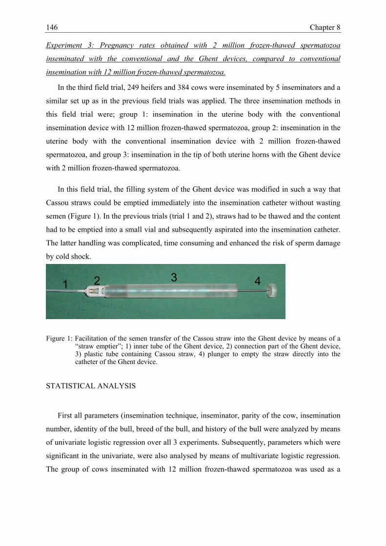
146 Chapter 8
Experiment 3: Pregnancy rates obtained with 2 million frozen-thawed spermatozoa
inseminated with the conventional and the Ghent devices, compared to conventional
insemination with 12 million frozen-thawed spermatozoa.
In the third field trial, 249 heifers and 384 cows were inseminated by 5 inseminators and a
similar set up as in the previous field trials was applied. The three insemination methods in
this field trial were; group 1: insemination in the uterine body with the conventional
insemination device with 12 million frozen-thawed spermatozoa, group 2: insemination in the
uterine body with the conventional insemination device with 2 million frozen-thawed
spermatozoa, and group 3: insemination in the tip of both uterine horns with the Ghent device
with 2 million frozen-thawed spermatozoa.
In this field trial, the filling system of the Ghent device was modified in such a way that
Cassou straws could be emptied immediately into the insemination catheter without wasting
semen (Figure 1). In the previous trials (trial 1 and 2), straws had to be thawed and the content
had to be emptied into a small vial and subsequently aspirated into the insemination catheter.
The latter handling was complicated, time consuming and enhanced the risk of sperm damage
by cold shock.
Figure 1: Facilitation of the semen transfer of the Cassou straw into the Ghent device by means of a “straw emptier”; 1) inner tube of the Ghent device, 2) connection part of the Ghent device, 3) plastic tube containing Cassou straw, 4) plunger to empty the straw directly into the catheter of the Ghent device.
STATISTICAL ANALYSIS
First all parameters (insemination technique, inseminator, parity of the cow, insemination
number, identity of the bull, breed of the bull, and history of the bull were analyzed by means
of univariate logistic regression over all 3 experiments. Subsequently, parameters which were
significant in the univariate, were also analysed by means of multivariate logistic regression.
The group of cows inseminated with 12 million frozen-thawed spermatozoa was used as a
1 2 43

UTJ insemination with lower insemination doses 147
control group within each experiment. Since “bull” and breed of bull were highly correlated,
only the breed of the bull was selected for the multivariate analysis.
RESULTS
The insemination doses of 12, 8, 4, and 2 million effectively consisted of 11.8 ± 0.6 x 10⁶,
8.1 ± 0.5 x 10⁶, 4.0 ± 0.3 x 10⁶, and 2.0 ± 0.2 x 10⁶ spermatozoa, respectively, as evaluated by
Hamilton coulter counter.
Using the univariate logistic regression, no significant difference could be found between
PRs obtained after AI with a full (12 x 106) and a decreased insemination dose (8, 4, and 2 x
106) (p = 0.15) (Table 1).
Table 1: Non return rate (NRR) and pregnancy results after insemination with the conventional insemination device into the uterine body (CD body), with the Ghent device into the uterine body (GD body) and with the Ghent device into both uterine horns (GD horns).
AI method No. of AIs NRR Pregnant (%) Not pregnant (%) Unknown (%)
CD, 12 x 106 390 70.8 49.5 42.3 8.2
CD, 8 x 10⁶ 342 69.2 54.1 41.2 4.7
GD, 8 x 10⁶ 428 68.4 48.4 47.4 4.2
1160 69.5 50.4 43.9 5.7
CD, 12 x 106 344 69.7 45.3 42.7 11.9
CD, 4 x 10⁶ 322 69.8 41.0 43.2 15.8
GD, 4 x 10⁶ 343 65.5 40.2 45.5 14.3
1009 68.4 42.2 43.8 14.0
CD, 12 x 106 225 69.8 51.1 44.0 4.9
CD, 2 x 10⁶ 220 69.5 51.8 40.0 8.2
GD, 2 x 10⁶ 238 69.5 47.9 43.3 8.8
Total 683 69.6 50.2 42.5 7.3 a,b Values with a different superscript in the same column differ significantly (P < 0.05).
Also no significant difference was found between the groups inseminated with decreased
insemination doses either with the conventional or Ghent device (Table 2). PRs in the three

148 Chapter 8
groups of the second experiment were lower than in experiment 1 and 3, albeit not
significantly (p = 0.08). No significant difference was found between cows inseminated for
the first (n = 2165), second (n = 637) or third or more time (n = 50) (p = 0.22). Univariate as
well as multivariate analysis showed that PRs were significantly affected by inseminator,
parity of the cow, bull, and breed of the bull (Table 2).
Table 2: Results of univariate analysis, examining the effect of insemination technique, inseminator, parity of the cow, insemination number, bull, breed of the bull and history of the bull on PRs.
Parameter p-value
Insemination technique 0.15
CD 12 x 106 vs GD 8 x 10⁶ 0.40
CD 12 x 106 vs GD 4 x 10⁶ 0.71
CD 12 x 106 vs GD 2 x 10⁶ 0.90
CD 12 x 106 vs CD 8 x 10⁶ 0.25
CD 12 x 106 vs CD 4 x 10⁶ 0.21
CD 12 x 106 vs CD 2 x 10⁶ 0.38
CD 8 x 10⁶ vs GD 8 x 10⁶ 0.09
CD 4 x 10⁶ vs GD 4 x 10⁶ 0.67
CD 2 x 10⁶ vs GD 2 x 10⁶ 0.42
Inseminator < 0.01
Parity of the cow < 0.01
Insemination number 0.22
Bull < 0.01
Breed of bull 0.03
History of bull 0.81
CD body: insemination with the conventional insemination device into the uterine body GD body: insemination with the Ghent device into the uterine body GD horns: insemination with the Ghent device into both uterine horns History of the bull = “sensitivity” for AI with decreased insemination doses in a preliminary field trial (sensitive, not sensitive, unknown).
The pregnancy rates obtained by the different inseminators varied from 37.8% to 55.5%.
The average PR in heifers (n =782) and cows (n = 2070) was 65.3% and 47.3% respectively
(p < 0.01) (Figure 2).

UTJ insemination with lower insemination doses 149
01020304050607080
CD 12 CD 8 GD 8 CD 4 GD 4 CD 2 GD 2
insemination dose and technique
preg
nanc
y ra
te
heifercow
Figure 2: Effect of insemination dose (12, 8, 4, and 2 x 10⁶ spermatozoa) and insemination technique (conventional device (CD) versus Ghent device (GD)) on PRs in heifers and cows.
PRs obtained by Red Holstein bulls were 4.1% higher than those obtained by Black
Holstein bulls (p < 0.01) (Table 3).
Table 3: Effect of breed of the bull on PRs.
Breed of bull No. of AIs Pregnant (%) Not pregnant (%) Unknown (%)
Black Holstein (n = 6) 1684 45.8 45.1 9.1
Red Holstein (n = 5) 1168 49.9 41.3 8.8
Total 2852 47.4 43.5 9.0
In the present study, no difference in PR was found between bulls which had shown to be
sensitive (n = 4) or insensitive (n = 3) for AI with decreased insemination doses in a
preliminary study (p = 0.81) (Table 4).
Table 4: PRs of bulls divides into three groups in accordance to their sensitivity for AI with decreased insemination doses in a preliminary field trial (history).
History bull No. of AIs Pregnant (%) Not pregnant (%) Unknown (%)
Not sensitive (n = 3) 857 47.5 42.0 10.5
Sensitive (n = 4) 1072 48.0 44.1 7.8
Unknown (n = 4) 923 46.8 44.2 9.0
Total 2852 47.5 43.5 9.0

150 Chapter 8
DISCUSSION
According to Hunter (Hunter and Greve, 1998; Hunter, 2001; 2003), deep uterine
insemination would be a fruitful way forward to improve fertility rates in cattle with low
doses and poorly fertile semen. However, in our study no beneficial effect of bilateral UTJ
insemination with the Ghent device on the PRs could be observed, even when the
insemination dose was decreased from 12 to 2 million frozen-thawed spermatozoa.
Furthermore, no difference was found between semen depositions into the uterine body with
the conventional device and bilateral UTJ insemination with the Ghent device.
In contrast to other studies in which the 56d NRR was used as a fertility parameter, the
fertility assessment in our study was based on effective pregnancy rates (PR) determined by
rectal palpation between 45 and 90 days after last AI. The effective PRs are more accurate and
make it possible to evaluate the fertility based on a relatively low number of cows per group.
In our study, the overestimation of the effective PR by NRR was about 21.0%. This is the
result of not taking the natural breeding and culling after AI into account, which lead to
higher NRRs. The NRR and the percentage of non pregnant cows differed only slightly
between the 3 experiments (Table 1). The variation in PR was larger. This is probably the
result of the relatively high number of unknown data (14.0%) in the second experiment.
Several large scale field trials using the conventional insemination technique with low
insemination doses have been performed in the Netherlands (Uwland, 1984; Smidt and
Zuidberg, 1988; Den Daas et al., 1998). These studies showed that with the conventional
insemination technique a significant decrease in NRR could be obtained only when the
inseminations were performed with less than 10 x 106 frozen-thawed spermatozoa. Therefore
insemination doses of 8, 4 and 2 x 10⁶ frozen-thawed spermatozoa were used in our study.
In the present study we did not only want to investigate the effect of low dose
insemination on the PRs, but we also wanted to evaluate the hypothesis of Hunter, according
to which deep intra-uterine insemination has a beneficial effect on the PRs due to the
reduction of the sperm loss during migration through the uterus and the more efficient
population of the functional sperm reservoir in the isthmus of the oviduct (Hunter and Greve,
1998; Hunter, 2001; 2003). That deep uterine insemination would favour the access of
spermatozoa to the oocyte in cattle was shown by Dalton et al. (1999).

UTJ insemination with lower insemination doses 151
Although the effect of deep insemination has been studied extensively (Knight et al.,
1951; Salisbury and VanDemark, 1951; Stewart and Melrose, 1952; Olds et al., 1953;
Macpherson, 1968; Hawk and Tanabe, 1986; Williams et al., 1988; McKenna et al., 1990;
Marshall et al., 1989; Zavos et al., 1985; López-Gatius et al., 1988; Senger et al., 1988;
Dalton et al., 1999), nobody has been able to clearly demonstrate the beneficial effect of this
technique. In most of these studies, moderate (10 - 20 x 106) to high ≥20 x 106 doses of
frozen-thawed semen were used. Moreover, in the studies performed in the early fifties
relatively high numbers of fresh semen were used (Salisbury and VanDemark, 1951; Stewart
and Melrose, 1952; Olds et al., 1953). It is not surprising that no significant difference was
found between deep and conventional insemination in most of theses studies, since the
number of spermatozoa inseminated was not a limiting factor for fertilization. In our
viewpoint, the beneficial effect of UTJ insemination compared to conventional AI, can only
be demonstrated when the number of spermatozoa inseminated is the limiting factor for
successful fertilization. This has very recently been investigated in a study by Andersson et al.
(2004), in which the effect of low dose insemination (2 x 10⁶) in cattle in combination with
deep and conventional insemination was examined. This study confirmed the hypothesis of
Hunter since no difference was found between deep and conventional AI when 15 x 106
spermatozoa were used, while numerically, although not significantly (p = 0.27), higher PRs
were obtained with 2 x 10⁶ spermatozoa deposited into the horn ipsilateral to the side of the
impending ovulation (Andersson et al., 2004). Similarly, in the present study we were not able
to demonstrate a significant beneficial effect of deep insemination. This can be due to the fact
that, in our study, the insemination doses used, were still not low enough to represent a
limiting factor for fertilization. No significant difference in fertility was observed between AI
with 12, 8, 4 and 2 x 10⁶ spermatozoa. Unexpectedly and in contrast to the other studies
(Uwland, 1984; Smidt and Zuidberg, 1988; Den Daas et al., 1998; Andersson et al., 2004), the
PRs did not decrease in our study. This finding was certainly not due to an incorrect
insemination dose since each batch was checked before and after freezing and thawing. Sperm
quality was also similar in all experiments since all insemination doses were produced from
the same ejaculates and were processed and frozen in the same way and at the same moment.
An important difference between our study and those performed by Uwland (1984), Smidt
and Zuidberg (1988) and Den Daas et al. (1998), is however the fact that in our field trials
inseminations were performed by well trained veterinarians with extensive experience in AI,
whereas in the other studies inseminations were performed by technicians. In studies where
conventional inseminations were performed by technicians, it has been shown that lack of

152 Chapter 8
accuracy of semen deposition was an important reason for reduced fertility (Lopez-Gatius,
2000). In over 50% of such cases, inseminators were not sufficiently trained to deposit semen
into the uterine body, so that intracervical insemination was often performed (Lopez-Gatius,
2000).
Another noticeable difference between our study and investigations by Andersson et al.
(2004) is the fact that PRs did not decrease after deep insemination with 2 x 10⁶ frozen-
thawed spermatozoa. This could be due to the different site of semen deposition, and also to
the insemination technique. In the study of Andersson et al. (2004) an embryo transfer pistolet
with side openings (IMV blue sheets) was used and semen was deposited halfway one uterine
horn and not bilaterally at the UTJ. Moreover semen deposition with rigid devices such as an
embryo pistolet or a conventional insemination device near the UTJ cannot be performed
properly in cows due to the curling of the uterine horns at the moment of insemination
(Noakes et al., 2001). The use of such devices for deep insemination requires extra
manipulations of the uterus and increases the risk of endometrial damage, which may cause a
decrease in fertility (Andersson et al., 2004; Verberckmoes et al., 2004b). Apart from the fact
that UTJ insemination may be more beneficial when low doses or poorly fertile semen is
used, it is also more likely that this beneficial effect will be more obvious in multiparous cows
which have much longer uterine horns than nulliparous heifers. Although in our study
consistently higher PRs were obtained in nulliparous heifers than in multiparous cows, no
beneficial effect of UTJ insemination was found in either heifers or cows. Here again, the
insemination dose seemed to be no limiting factor for successful fertilization, not even in
cows. In contrast to our previous field trial with the Ghent device (Verberckmoes et al.,
2004a), the insemination number had no significant effect on the PRs. This can be explained
by the fact that in the present study 98.3% of all inseminations were first and second
inseminations after calving, while the difference in PR between first (48.4% ± 4.8%) and
second inseminations (45.6% ± 4.8%) after calving was very small.
In the present study each half of the low insemination dose (8, 4 and 2 x 10⁶) was
deposited near the UTJ of each uterine horn with the Ghent device, and similar PRs were
obtained as with the full insemination dose (12 x 106) in the uterine body. However, since
cows are generally single ovulators, only one oocyte per ovulation is released and descends
into the left or right oviduct where fertilization occurs. Determination of the site of ovulation
before insemination would enable semen deposition in the horn ipsilateral to the site of
ovulation. This normally would result in a halving of the insemination dose without a
UTJ insemination with lower insemination doses 153
reduction in fertility. In other single ovulators such as horses and water buffaloes, deposition
of low doses of frozen-thawed semen (1 - 3 x 10⁶) near the UTJ ipsilateral to the site of
ovulation has been shown to be effective (Morris et al., 2000a, 2000b; 2003; Presicce et al.,
2004). However, in contrast to the latter studies in horses and water buffaloes, determination
of the exact moment of insemination in cows is less accurate under field conditions.
Moreover, since no ultrasound examination nor palpation of the ovaries is performed in cattle
at the moment of insemination, the site of ovulation is unknown. Palpation of the ovaries in
cattle is not done in order to avoid rupture of mature follicles. A beneficial effect of UTJ has
also been demonstrated in pigs when low doses of poor fertile semen were used (Martinez et
al. 2001; Grossfeld et al. 2003; Rath et al., 2003). However, since pigs are multiple ovulators,
half of the insemination dose has to be deposited at each of both UTJs.
As in most field trials, a significant difference in PRs was obtained between inseminators
(p< 0.01) and between different bulls (p < 0.01). Some bulls are more fertile than others as
has been clearly shown by Den Daas et al. (1998). This research group demonstrated that the
optimal insemination dose to obtain 95% of the maximal conception rate ranges from 1 x 10⁶
to 11 x 106 frozen-thawed spermatozoa for conventional AI (Den Daas et al., 1998). The
difference in fertility between bulls can only be demonstrated when suboptimal insemination
doses are used. In most studies, no decrease in fertility can be observed when cows are
inseminated with 7.5 x 10⁶ or more frozen-thawed spermatozoa (Smidt and Zuidberg, 1988;
Uwland, 1984). Even when the bulls are divided into 3 groups in accordance with their NRR
(low, moderate or highly fertile) no significant decrease in fertility can be observed when the
insemination dose is decreased from 20 x 106 (n = 5426) to 7.5 x 10⁶ spermatozoa (n = 5165)
(Smidt and Zuidberg, 1988). However, in contrast to our study, Uwland (1984) did find a
significant decrease in fertility for moderately and low fertile bulls when the insemination
dose was decreased to 10 and 7.5 x 10⁶ frozen-thawed spermatozoa, respectively, but not for
highly fertile bulls, not even when the insemination dose was reduced to 3.75 x 10⁶. Despite
the fact that our study included not only highly fertile bulls, but also bulls of unknown fertility
(n = 4) and bulls which had shown to be sensitive for AI with decreased insemination doses (n
= 4), no difference could be demonstrated between the groups of bulls which were suspected
to have a different sensitivity for AI with low insemination doses.
The higher PRs obtained with semen from Red rather than from Black Holstein Friesian
bulls (p = 0.03) is most likely due to a better semen quality, but can also be the result of the

154 Chapter 8
higher fertility of the cows inseminated with this semen. However, based on our field trial, it
is impossible to determine which one of both factors is the most important.
In conclusion, no decrease in fertility was observed when the insemination dose was
decreased from 12 to 2 million frozen-thawed spermatozoa, neither with the Ghent device nor
with the conventional insemination device. Further trials with even lower insemination doses
are required to determine whether UTJ insemination with the Ghent device would be better
than conventional insemination in cattle.

UTJ insemination with lower insemination doses 155
REFERENCES
Andersson M, Taponen J, Koskinen E, Dahlbom M. Effect of insemination with doses of 2 or
15 million frozen-thawed spermatozoa and semen deposition site on pregnancy rates in
dairy cows. Theriogenology 2004; 61: 1582-1588.
Dalton JC, Nadir S, Bame JH, Saacke RG. Effect of a deep uterine insemination on
spermatozoal accessibility to the ovum in cattle: a competitive insemination study.
Theriogenology 1999; 51: 883-890.
Den Daas JH, De Jong G, Lansberger LM, Van Wagtendonk-De Leeuw AM. The relationship
between the number of spermatozoa inseminated and the reproductive efficiency of
individual dairy bulls. J Dairy Sci 1998; 81: 1714-1723.
Den Daas N. Laboratory assessment of semen characteristics. Anim Reprod Sci 1992; 28: 87-
94.
Garner DL, Schenk JL, Seidel Jr GE. Chromatin stability in sex-sorted sperm: In; andrology
in the 21th century, proceedings of the VIIth international congress of andrology,
Montreal, Canada, short communications, 2001: 3-7.
Grossfeld R, Ruiz S, Sieg B, Leigh J, Klinc P, Rath D, 2003: Artificial insemination with
sexed semen in pigs. Conference book of the 5th International congress on boar semen
preservation, Doorwerth, The Netherlands, VI-P62 (abstract).
Hawk HW, Tanabe TY. Effect of unilateral cornual insemination upon fertilization rate in
superovulating and single-ovulating cattle. J Anim Sci 1986; 63: 551-560.
Hawk HW. Sperm survival and transport in the female reproductive tract. J Dairy Sci 1983;
66: 2645-2660.
Hunter RHF, Greve T. Deep uterine insemination of cattle: a fruitful way forward with
smaller numbers of spermatozoa. Acta Vet Scand 1998; 39: 149-163.
156 Chapter 8
Hunter RHF. Advances in deep uterine insemination: a fruitful way forward to exploit new
sperm technologies in cattle. Anim Reprod Sci. 2003; 79: 157-70.
Hunter RHF. New breeding opportunities with deep cornual insemination: exploiting modern
sperm technologies in cattle. Reprod Dom Anim 2001; 36: 217-222.
Knight CW, Patrick TE, Anderson HW, Branton C. The relation of site of semen deposit to
breeding efficiency of dairy cattle. J Dairy Sci 1951; 34: 199-202.
López-Gatius F, Camón-Urgel J, Angulo-Asensio E. Effect of single deep insemination on
transferable embryo recovery rates in superovulated dairy cows. Theriogenology 1988;
30: 877-885.
Lopez-Gatius F. Site of semen deposition in cattle: a review. Theriogenology. 2000; 53: 1407-
1414.
Macmillan KL, Curnow RJ. Factors influencing AB conception rates, XI. Variation in sire
fertility. NZJ Exp Agric 1977; 5: 279-285.
Macpherson JW. Semen placement effects on fertility in bovines. J Dairy Sci 1968; 51: 807-
808.
Marshall CE, Graves WM, Meador JL, Swain JB, Anderson JI. A fertility comparison of
uterine body and bicornual semen deposition procedures in dairy cattle. J Dairy Sci
1989; 72: 455.
Martinez EA, Vazquez JM, Roca J, Lucas X, Gil MA, Parilla I, Vasquez JL, Day BN.
Succesful non-surgical deep intra-uterine insemination with small numbers of
spermatozoa in sows. Reproduction 2001; 122: 289-96.
McKenna T, Lenz RW, Fenton SE, Ax RL. Non return rates of dairy cattle following uterine
body or cornual insemination. J Dairy Sci 1990; 73: 1779-83.
Morris LHA, Allen WR. An overview of low dose insemination in the mare. Reprod Dom
Anim 2002; 37: 206-210.

UTJ insemination with lower insemination doses 157
Morris LHA, Hunter RHF, Allen WR. Hysteroscopic insemination of small numbers of
spermatozoa at the utero-tubal junction of preovulatory mares. J Reprod Fert 2000a; 11:
95-100.
Morris LHA, Tiplady C, Wilsher S, Allen WR. Hysteroscopic insemination of mares with low
numbers of frozen-thawed ejaculated and epididymal spermatozoa. In: Proceedings of
5th international symposium on equine embryo transfer. Havemeyer Foundation
Monograph series 2000b; 3: 4-7.
Morris LHA, Tiplady C, Wilsher S, Allen WR. Pregnancy rates in mares after a single fixed
time hysteroscopic insemination of low numbers of frozen-thawed spermatozoa onto the
uterotubal junction. Equine Vet J 2003; 35: 197-201.
Noakes DE, Parkinson TJ, England GCW. In: Veterinary Reproduction and Obstetrics. 8th
edition. Artificial insemination. W.B. Saunders. 2001; 751-778.
Olds D, Seath DM, Carpenter MC, Lucas HL. Interrelationships between site of deposition,
dosage, and number of spermatozoa in diluted semen and fertility of dairy cows
inseminated artificially. J Dairy Sci 1953; 36: 1031-1035.
Presicce GA, Verberckmoes S, Klinc P, Rath. D. Artificial insemination and conception rate
using sexed semen in mediterranean Italian buffaloes (Bubalus Bubalis) following
ovsynch protocol. Theriogenology 2004, submitted.
Rath D, Ruiz S, Sieg B. Birth of piglets after intra-uterine insemination employing
flowcytometrically sexed boar semen, a case report. Vet Rec 2003; 152, 400-401.
Rath D. Current perspectives of sperm sorting in domestic farm animals. Proc. of the 19th
scientific meeting of the European embryo transfer association, 12-13 September
Rostock 2003; 125-128.
Salisbury GW, VanDemark NL. The effect of cervical, uterine and cornual insemination on
fertility of the dairy cow. J Dairy Sci 1951; 34: 68-74.

158 Chapter 8
Schenk JL, Suh TK, Cran DG, Seidel Jr GE. Cryopreservation of flow-sorted bovine
spermatozoa. Theriogenology 1999; 52: 1375-1391.
Schwartz D, MacDonald PDM, Heuchel V. On the relationship between the number of
spermatozoa per insemination and the probabilityof conception. Reprod Nutr Dev 1981;
21: 979-988.
Seidel GE, Schenk JL, Herickhoff LA, Doyle SP, Brink Z, Green RD, Cran DG. Insemination
of heifers with sexed sperm. Theriogenology 1999; 52: 1407-20.
Seidel Jr GE. Economics of selecting for sex: the most important genetic trait.
Theriogenology 2003; 59: 585-598.
Senger PL, Becker WC, Davidge ST, Hillers JK, Reeves JJ. Influence of cornual insemination
on conception in dairy cattle. J Anim Sci 1988; 66: 3010-3016.
Smidt WJ, Zuidberg C. Bevruchtingsresultaten van inseminaties met diepvriessperma met
verschillende aantallen spermatozoa per dosis bij het MRIJ-vee. Tijdschr Diergeneeskd
1988; 12: 673-676.
Stewart DL, Melrose DR. The comparative efficiency of the intra cervical and intra-uterine
methods of insemination in the dairy cow. Vet Rec 1952; 64: 605-607.
Uwland J. Correlation between the number of inseminated spermatozoa and fertility rates in
dairy cattle. Tijdschr Diergeneeskd 1984; 10: 405-412.
Verberckmoes S, Van Soom A, De Pauw I, Dewulf J, Vervaet C, de Kruif A. Assessment of a
new utero-tubal junction insemination device in dairy cattle. Theriogenology 2004a, 61:
103-115.
Verberckmoes S, Cornillie P, Van Soom A, de Kruif A. Effect of utero-tubal junction
insemination on the integrity of the bovine endometrium. VIIIth annual conference of
ESDAR, Warshau September 2004b; in press.

UTJ insemination with lower insemination doses 159
Williams BL, Gwazdauskas FC, Whittier WD, Pearson RE, Nebel RL. Impact of site of
inseminate deposition and environmental factors that influence reproduction of dairy
cattle. Journal of Dairy Science 1988; 71: 2278-2283.
Zavos PM, Johns JT, Heersche G, Miksch DE. Site of semen deposition and subsequent
conception in synchronized and artificially inseminated (AI) beef heifers. Journal of
Animal Science 1985; 61: 37.


CHAPTER 9
EFFECT OF WHOLE BLOOD AND SERUM ON BOVINE SPERM QUALITY AND IN
VITRO FERTILIZATION CAPACITY
Theriogenology 2004; 61: 25-33.
Verberckmoes S., Van Soom A., De Pauw I., Dewulf J., Rijsselaere T., de Kruif A.

162 Chapter 9
ABSTRACT
Under physiologic conditions, low concentrations of blood may be present in the uterine
fluid of the estrous cow at the moment of insemination. To decrease the insemination dose
and to obtain good insemination results with less fertile semen, more invasive insemination
methods such as utero tubal Junction insemination can be used. However, more invasive
insemination methods increase the risk of damaging the hyperaemic endometrium, with blood
in the uterine fluid as result. In this study, the effect of 0%, 0.15% and 1.5% whole blood and
serum on bovine sperm quality and in vitro fertilizing capacity was evaluated. Sperm quality
as assessed by total motility, progressive motility, membrane integrity and acrosomal status
was not affected by the presence of blood or serum (P > 0.05). However, the in vitro
fertilizing capacity decreased with increasing concentrations of blood and serum (P < 0.01).
The rate of polyspermy increased with increasing concentrations of serum (P < 0.01), but not
with increasing concentrations of blood (P = 0.30). In conclusion, no immediate effect of
blood and serum was visible on several sperm quality parameters, except for an increased
prevalence of head to head agglutination (HHA). However, blood and serum did have a
negative effect on in vitro fertilizing capacity.

Effect of whole blood and serum on bovine sperm quality and in vitro fertilizing capacity 163
INTRODUCTION
In practice, cows are inseminated about 12 h after the onset of behavioral estrus. Cows
observed in estrus in the morning are inseminated the evening of the same day, while cows
observed in estrus in the evening are inseminated the next morning (a.m. / p.m. schedule)
(Noakes et al., 2001a). The standard technique of insemination is to grasp the cervix through
the rectum with one hand. An insemination pipette, containing a semen straw is then passed
through the vagina and manipulated into and through the cervix. The semen is then expelled
into the small uterine body (Noakes et al., 2001a). In these circumstances, presence of blood
on the insemination pipette is very rarely observed.
Utero tubal junction (UTJ) insemination is an alternative to standard inseminations with
possible applications for sexed semen or less fertile semen. A new insemination device has
been developed at our departement and has been used in a field trial (Verberckmoes et al.,
2004). However when inseminations are performed deep in one or both uterine horns, traces
of blood may be present on the tip of the insemination catheter in 45% of inseminations
(Verberckmoes et al., 2002b). It is known that during estrus, the epithelium of the uterus is
very hyperaemic, resulting in capillary haemorrhage and post-estrual bleeding (24-48 hrs post
estrus) (Noakes et al., 2001b). Due to this hyperaemic state of the endometrium, an
insemination device that is introduced deep into the uterine horns may very easily induce
limited bleeding, without causing an inflammatory reaction of the endometrium.
Nevertheless, the presence of blood at the time of insemination may interfere with sperm
quality and subsequent fertilizing ability. Since it is very difficult to investigate the effect of
blood upon fertilization under in vivo conditions, it was the purpose of this study to evaluate
the effect of whole blood and serum on bovine sperm quality and fertilizing capacity in vitro.
Concentrations of blood and serum were chosen in accordance to the concentrations detected
in the uterine fluid of estrous cows (Anderson et al., 1985). Anderson et al. (1985) found 10
million erythrocytes per ml uterine fluid, which equals a 0.15% of whole blood (v:v ratio).
The fertilizing capacity of the spermatozoa was investigated by means of routine in vitro
fertilization in the presence of 0, 0.15 and 1.5% blood and serum.

164 Chapter 9
MATERIALS AND METHODS
Media.
To evaluate the effect of whole blood and serum on sperm quality, spermatozoa were
incubated in isotonic Hepes-TALP medium. This medium contains 10 mM Hepes, 0.2 mM
sodium pyruvate, 10 mM sodium lactate, 2 mM sodium bicarbonate, 10 µg/ml gentamycin
sulphate and 3 mg/ml bovine serum albumin (BSA). Oocytes used for IVF experiments were
incubated in maturation medium consisting of modified bicarbonate buffered TCM199
medium (Gibco BRL, Merelbeke, Belgium) supplemented with 20% heat-inactivated Foetal
Calf Serum (FCS) (N.V. Hyclone Europe S.A., Erembodegem, Belgium), 0.2 mM sodium
pyruvate (Sigma-Aldrich, Bornem, Belgium), 50µg/ml gentamycin sulphate (Gibco BRL,
Merelbeke, Belgium) and 0.4 mM glutamine (Sigma-Aldrich, Bornem, Belgium).
Collection of blood and serum.
Blood was taken from an estrous cow by means of a vacutainer containing 0.125 M Na
Citrate (BD vacutainer systems, Plymouth, UK), resulting in negligible citrate concentration
in the blood cultures. The concentration of red blood cells (RBC) was determined by means of
a Bürker counting chamber after a dilution of 103 in Hepes TALP. One ml of whole blood
contained 6 x 10⁹ red blood cells (RBC). Another blood sample of the same cow was taken by
means of a venoject® (Terumo Europe NV, Leuven, Belgium) for the production of bovine
serum. After 5h of incubation at room temperature, the clotting process was terminated, and
the serum was subsequently separated and stored at 5°C.
Sperm treatment.
For all experiments, frozen-thawed semen from the same bull ejaculate was separated on a
discontinuous Percoll® (Pharmacia, Uppsala, Sweden) gradient (90% and 45% Percoll
solutions in a Hepes buffered salt solution). The concentration of the semen sample was
determined by means of a Bürker counting chamber. For the evaluation of the effect of whole
blood and serum on the sperm quality, spermatozoa were diluted in Hepes TALP containing
0, 0.15 and 1.5% of whole blood or serum. Sperm quality was assessed after 30 min and again
after 5 hrs of incubation at 38.5°C. These time periods were chosen because normally the first
spermatozoa reach the isthmus within 30 min after AI and fertilization occurs a few hours

Effect of whole blood and serum on bovine sperm quality and in vitro fertilizing capacity 165
later (Hawk, 1983). For the evaluation of the effect of whole blood and serum on the IVF
capacity, spermatozoa were diluted in IVF-TALP with 0, 0.15 and 1.5% of whole blood and
the same concentrations of serum.
Oocyte maturation and fertilization.
Cow ovaries were randomly selected in a slaughterhouse and transported to the laboratory
within 3 to 4 hours after collection. Follicles of 2 to 6 mm in diameter were aspirated using an
18-gauge needle and a 10 ml syringe. After being washed in Hepes buffered TALP medium,
the cumulus-oocyte complexes were in vitro matured in 500µl of maturation medium at
38.5°C in 5% CO2 in air at 100% humidity for 24-26 hours. After maturation, oocytes were
inseminated with a final sperm concentration of 1 x10⁶ spermatozoa/ml in 500µl of
fertilization medium.
Sperm quality evaluation.
The assessed sperm quality parameters were percentage of membrane intact spermatozoa,
total and progressive motility, head to head agglutination (HHA) of the spermatozoa and
acrosome reaction. The membrane integrity was determined by means of eosin-nigrosin
staining and light microscopy (x 400). Two experienced people determined the total and
progressive motility, and the HHA subjectively on a heated stage (37°C) using light
microscopy (x 200). The motility was expressed in percentage, while the HHA was given a
score from 0 (= no HHA) to 4 (all spermatozoa show HHA). The acrosome reaction was
assessed before (t0) and after 5h of incubation (t2) by means of Pisum Sativum Agglutinin
(PSA) staining with a Leica DMR fluorescence microscope (x 400) (Kawakami et al., 1993).
Spermatozoa with a fluorescent green head indicated acrosome intact spermatozoa.
Spermatozoa were considered as acrosome reacted when a fluorescent band was observed at
the equatorial region.
Evaluation of the fertilization / polyspermy rate
After fertilization, oocytes were vortexed for 3 min in 2 ml of Hepes-TALP to remove
excess sperm and/or cumulus cells, fixed overnight in 2% paraformaldehyde and 2%
glutaraldehyde in phosphate buffered saline (PBS) solution without calcium and magnesium,
and subsequently stained for 10 min with 1µg/ml of Hoechst 33342 (Molecular Probes,
Leiden, The Netherlands). The zygotes were mounted in 100% glycerol and evaluated by

166 Chapter 9
means of a Leica DMR fluorescence microscope for fertilization and polyspermy. Successful
fertilization was characterized by the presence of two pronuclei. The presence of more than
two pronuclei indicated polyspermy.
Experimental Design and Statistical Analysis
Experiment 1.
Influence of whole blood and serum on sperm quality. After percoll separation, frozen-
thawed semen was diluted in the media with 3 different concentrations of whole blood and
serum to a concentration of 10 x 106/ml. The sperm quality was determined before (t0), after
30 min (t1) and after 5h (t2) incubation at room temperature. This experiment was repeated 4
times. Results of membrane integrity and acrosome reaction were analysed using a linear
mixed effect model (S-Plus 2000, Cambridge, USA) including media with different blood and
serum concentrations as fixed effect and sample as random effect. An autoregressive
correlation structure of the first order (AR 1) was used to correct for dependency between
repeated measurements on the same sample. The HHA was studied and descriptively
recorded.
Experiment 2.
Effect of whole blood / serum on in vitro fertilizing capacity. After percoll separation,
frozen-thawed semen was diluted in the media with 3 different concentrations of whole blood
and serum to a concentration of 2 x 10⁶/ml. Prior to insemination, spermatozoa were
incubated for 30 min at 38.5°C in 5% CO2 in air at maximum humidity. Twenty hours post
insemination, the oocytes were fixed and stained with Hoechst 33342. In vitro fertilizing
capacity and polyspermy were evaluated by means of fluorescence microscopy. This
experiment was repeated 4 times. Results were analysed using logistic regression.
RESULTS
Experiment 1.
Effect of whole blood and serum on sperm quality: incubation of frozen-thawed
spermatozoa for 30 min (data not shown) and 5h, respectively, in medium with blood and
serum at room temperature, did not significantly affect membrane integrity (P = 0.36 and

Effect of whole blood and serum on bovine sperm quality and in vitro fertilizing capacity 167
0.89), total motility (P = 0.56 and 0.82), progressive motility (P = 0.25 and 0.07) or acrosome
reaction (P = 0.57 and 0.24) (Table 1).
Table 1: Effect of 5h incubation (22°C) in media with whole blood and serum on the membrane integrity, total motility, progressive motility and acrosomal integrity of spermatozoa.
Membrane intact (%)
Total motility (%)
Progressive motility (%)
Acrosomal integrity (%)
Whole blood 0% 89.3 a 70.0 a 66.3 a 94.3 a 0.15% 90.7 a 63.8 a 48.8 a 96.0 a 1.5% 73.3 a 60.0 a 42.5 a 95.7 a P = 0.89 P = 0.82 P = 0.07 P = 0.24
Blood serum 0% 88.7 a 73.3 a 70.7 a 95.3 a 0.15% 87.3 a 76.7 a 60.0 a 96.3 a 1.5% 86.3 a 73.3 a 41.7 a 96.7 a P = 0.36 P = 0.56 P = 0.25 P = 0.57
ab Values within the same column with different superscripts are significantly different (P < 0.05).
Increasing concentrations of blood did not affect the progressive motility (P = 0.25), while
there was a trend that the progressive motility decreased with increasing concentrations of
blood (P = 0.07) (Figure 1).
Figure 1: Progressive motility (prog. mot.) of bovine spermatozoa after 0 min, 30 min and 5h incubation in HEPES-Talp (22°C) with 0%, 0.15% and 1.5% of whole blood and serum.
Also a non-significant decrease in total motility was found for the spermatozoa incubated
in increasing concentrations of blood. The amount of head to head agglutination was the same
for blood and serum. However, the amount of agglutination was clearly different between the
3 concentrations (0, 0.15 and 1.5%) (Figure 2). At t0, no agglutination was observed. At t1
and t2, agglutination was observed in medium containing 0.15% and 1.5% blood and serum
(Figure 2).
0
20
40
60
80
100
0 min 30 min 5 h
time
%pr
og. m
ot.
0% blood0.15% blood
1.5% blood
0
20
40
60
80
100
0 min 30 min 5 htime
%pr
og. m
ot.
0% serum
0.15% serum
1.5% serum
0
20
40
60
80
100
0 min 30 min 5 h
time
%pr
og. m
ot.
0% blood0.15% blood
1.5% blood
0
20
40
60
80
100
0 min 30 min 5 h
time
%pr
og. m
ot.
0% blood0.15% blood
1.5% blood
0
20
40
60
80
100
0 min 30 min 5 htime
%pr
og. m
ot.
0% serum
0.15% serum
1.5% serum
0
20
40
60
80
100
0 min 30 min 5 htime
%pr
og. m
ot.
0% serum
0.15% serum
1.5% serum

168 Chapter 9
Figure 2: Amount of agglutination (score 0-4) after 0 min, 30 min and 5h incubation in HEPES-Talp with 0%, 0.15% and 1.5% of whole blood or serum.
Experiment 2.
Effect of whole blood and serum on in vitro fertilizing capacity: increasing concentrations
of blood and serum had a negative effect on the fertilizing capacity (P < 0.01). The
fertilization rate decreased more with increasing serum concentrations than with increasing
blood concentrations. Polyspermy was not significantly influenced by increased blood
concentration (P = 0.30), but increasing serum concentrations significantly increased the
polyspermy rates (P < 0.01) (Table 2).
Table 2: Effect of whole blood and serum on the percentage of normally fertilized oocytes (2 pronuclei) and percentage of polyspermy (> 2 pronuclei).
Number of oocytes Normally ferilization Polyspermy N N % N %
Whole blood 0% 243 152 62.6 a 16 6.6 a 0.15% 272 138 50.7 a 21 7.7 a 1.5% 210 86 41.0 b 9 4.3 a
Blood serum 0% 308 172 56.2 a 22 7.1 a 0.15% 328 140 42.7 a 35 10.7 a 1.5% 327 127 39.1 b 55 16.8 b
ab Values within the same column with different superscripts are significantly different (P < 0.05).
DISCUSSION
It is generally accepted that bovine semen quality can be affected by contaminations such
as blood, urine, dirt or pus (Sigman and Lopez, 1993; Bar-Chama and Fisch, 1993; Chen et
al., 1995; Griggers et al., 2001). However, in such cases the contamination originates from
00,5
11,5
22,5
33,5
4
0 min 30 min 5 h
time
scor
e0% blood / serum
0.15% blood / serum
1.5% blood / serum
00,5
11,5
22,5
33,5
4
0 min 30 min 5 h
time
scor
e0% blood / serum
0.15% blood / serum
1.5% blood / serum

Effect of whole blood and serum on bovine sperm quality and in vitro fertilizing capacity 169
disorders present in the bull’s genital tract. These disorders are mostly related to an infectious
or inflammatory process. Under physiological conditions, low concentrations of blood may be
present in the uterine fluid of the estrous cow at the moment of insemination (Andersen et al.,
1985). However, little information is available on the effect of whole blood on sperm quality.
In our study we found that most parameters that are indicative of bovine sperm quality,
were not affected by the presence of blood or serum, except for a significant effect on head to
head agglutination (HHA). The HHA was more obvious in the presence of 1.5% blood or
serum. In our study, total motility, progressive motility, membrane and acrosome integrity
were not affected by the presence of blood or serum in the incubation medium. In human, 20-
30% serum in an extender for fresh semen had a positive effect on at least one of the motility
parameters (De Lamirande and Gagnon, 1991), which confirms our findings that serum
admixture is not detrimental to sperm motility. However, in the presence of whole blood,
which contains both plasma and cells such as leucocytes and erythrocytes, semen quality can
be negatively affected. In the human, it has been demonstrated that the quality of fresh semen
is negatively affected by the presence of white blood cells that produce reactive oxygen
species, but not in the presence of RBCs (Wolff et al., 1990).
In pigs and dogs, admixture of whole blood (up to 5% and 10% respectively) in fresh
diluted semen is not detrimental to semen quality (Le Roy, 1999; Rijsselaere et al., 2004). In
these species blood may be present in the ejaculate due to trauma of the penis or the prepuce
during semen collection, or to benign prostatic hypertrophy (Keenan, 1998; Johnston et al.,
2001). In contrast, freezing of canine spermatozoa contaminated with blood has a detrimental
effect on the post-thaw semen quality (Rijsselaere et al., 2004). In cattle, where frozen-thawed
semen is used for insemination, contamination of blood during collection is not a problem
because such samples are not used. Anderson et al. (1985) found 9.8 ± 2.8 x 10⁶ RBC/ml in
raw uterine fluid at the day of estrus, which was comparable to the 0.15% concentration we
used. Irrespective of service, many heifers and cows show a bright red sanguineous discharge
at about 24-48 hours after heat. Blood in the uterine lumen originates mainly from the uterine
caruncles, but it is not clearly defined at what moment of estrus the release of the blood into
the uterine cavity occurs (Noakes et al., 2001b). The presence of blood in the uterine lumen is
also more obvious when utero tubal junction (UTJ) insemination is performed, than when
semen is deposited in the uterine body. After DIUI, traces of blood stick on the insemination
pipette in 45% of the inseminations, while blood is seldom observed after classic insemination
in the uterine body (Verberckmoes et al., 2002b).

170 Chapter 9
Presence of blood in the uterine lumen in cattle at the time of insemination can be
considered as a natural phenomenon as it is in dogs (Constantin et al., 1981). Therefore, as
confirmed by our results, it should not be considered harmful to sperm quality. Despite the
lack of effect upon several sperm quality characteristics, the presence of blood and serum did
affect sperm fertilizing ability in a negative way, and serum did increase the number of
polyspermic zygotes.
The effect of blood on in vivo fertility should not be overestimated, since it is known that
fertilization occurs in the oviduct in the absence of blood. The HHA that occured in the
presence of blood and serum, is not necessarily detrimental for the fertilizing capacity of the
spermatozoa. Agglutinated spermatozoa are characterized by a predominance of intact
acrosomal membranes as judged by the presence of the apical ridge (Schroeder et al., 1990).
This was confirmed in our study by means of PSA-staining. The possible negative effect of
HHA on the in vivo fertility could be the result of temporarily mechanical inhibition of the
spermatozoa (Schroeder et al., 1990) and their prolonged exposure to the environment of the
uterus. In rabbits, spermatozoa engage immediately in HHA upon introduction in the uterine
lumen (Senger and Saacke, 1976). In bovine, HHA in the reproductive tract has never been
investigated. The physiological importance of this event, however, is not understood and the
effect of HHA upon in vivo fertility is unknown.
In conclusion, no immediate effect of blood and serum was visible on several sperm
quality parameters, except for an effect on HHA. However, blood and serum did have a
negative effect on in vitro fertilizing capacity. When fresh or frozen semen is deposited into
the uterine body, the presence of blood may interfere with in vivo fertility, if prolonged HHA
prevents the spermatozoa to reach the sperm reservoir. This hypothesis needs further
investigation. Application of a minimally harmful technique, by which spermatozoa are
deposited close to the UTJ could help to reduce fertility problems by minimizing the contact
between inseminated spermatozoa and blood.

Effect of whole blood and serum on bovine sperm quality and in vitro fertilizing capacity 171
REFERENCES
Anderson GW, Gwazdauskas FC, Guise MB. Column chromatography and electrophoresis of
bovine uterine lumenal proteins collected during the estrous cycle. Anim Reprod Sci
1985; 9: 11-24.
Bar-Chama N, Fisch H. Infection and pyospermia in male infertility. World J Urol 1993; 11:
76-81.
Brown DV, Senger PL. Effect of incubation in utero on motility head-to-head agglutination of
ejaculated rabbit spermatozoa. J Reprod Fertil 1982; 66: 283.
Chen D, Scobey ML, Jeyendran RS. Effects of urine on the functional quality of human
spermatozoa. Fertil Steril 1995; 64: 1216-1217.
Constantin A, Meissonier E. In Constantin A, Meissonier E (eds): L’ uterus de la vache,
anatomie, physiologie et pathologie. L’ uterus et le cycle sexuel. Ecole Nationale
Vétérinaire d’Alfort: 1981; 53-78.
De Lamirande E, Gagnon C. Quantitative assessment of the serum-induced stimulation of
human sperm motility. Int J Androl 1991; 14: 11-22.
Griggers S, Paccamonti DL, Thompson RA, Eilts BE. The effect of pH, osmolarity and urine
contamination on equine spermatozoal motility. Theriogenology 2001; 56: 613-622.
Hawk HW. Sperm survival and transport in the female reproductive tract. J Dairy Sci 1983;
66: 2645-2660.
Johnston SD, Root Kustritz MV, Olsen PNS. In Kersey (ed): Canine and feline
theriogenology. Clinical approach to infertility in the male. London: Saunders company,
2001; 370-387.
Kawakami E, Vandevoort CA, Mahi-Brown CA, Overstreet JW. Induction of acrosome
reactions of canine sperm by homologous zona pellucida. Biol Reprod 1993; 48: 841-
945.

172 Chapter 9
Keenan RJK. The infertile male. In: Manual of small animal reproduction and neonatology.
Simpon G. (ed). Shurdington: British small animal veterinary association 1998; 83-93.
LeRoy G. Blood may not affect boar semen. Illinois Veterinary Bulletin 1999; 7.
Noakes DE, Parkinson TJ, England GCW. In Noakes DE, Parkinson TJ, England GCW (eds):
Veterinary Reproduction and Obstetrics. Artificial insemination: W.B. Saunders, 2001a;
758-759.
Noakes DE, Parkinson TJ, England GCW. In Noakes DE, Parkinson TJ, England GCW (eds):
Veterinary Reproduction and Obstetrics. Endogenous and exogenous control of ovarian
cyclicity: W.B. Saunders, 2001b; 20-21.
Rijsselaere T, Van Soom A, Maes D, Verberckmoes, de Kruif A. Effect of blood admixture
on in vitro survival of chilled and frozen-thawed canine spermatozoa. Theriogenology
2004; 61: 1589-1602.
Schroeder AC, Schultz RM, Kopf GS, Taylor FR, Becker RB, Eppig JJ. Fetuin inhibits zona
pellucida hardening and conversion of ZP2 to ZP2f during spontaneous mouse oocyte
maturation in vitro in the absence of serum. Biol Reprod 1990; 43: 891-897.
Senger PL, Saacke RG. Serum induced head-to-head agglutination of bovine spermatozoa. J
Reprod Fertil 1976; 47: 215.
Sigman M, Lopez L. The correlation between round cells and white blood cells in the semen.
J Urol 1993; 149: 1338-1340.
Verberckmoes S, De Pauw I, Van Soom A, Dewulf J, de Kruif A. Assessment of a new deep
intra-uterine insemination device in dairy cattle. Reprod Dom Anim 2002a; 37: 255.
Verberckmoes S, De Pauw I, Van Soom A, de Kruif A. Assessment of feasability of deep
intra-uterine insemination in dairy cattle under field conditions VIth International
Symposium of Reproduction in Domestic Ruminants, Crief, Scotland 2002b; 87.

Effect of whole blood and serum on bovine sperm quality and in vitro fertilizing capacity 173
Verberckmoes S, Van Soom A, De Pauw I, Dewulf J, de Kruif A. Assessment of a new utero-
tubal junction insemination device in dairy cattle under field conditions.
Theriogenology, 2004; 61: 103-115.
Wolff H, Politch JA, Martinez A, Haimovici F, Hill JA, Anderson DJ. Leukocytospermia is
associated with poor semen quality. Fertil Steril 1990; 53: 528-536.


CHAPTER 10
GENERAL DISCUSSION

176 Chapter 10
General discussion
Artificial insemination (AI) is the oldest and currently most common technique in assisted
reproduction of animals. Initially the application of AI in cattle was forced by sanitary reasons
and was performed with fresh semen. However, later economic reasons became the
mainspring and since the optimization of cryopreservation of bovine semen around 1960,
predominantly frozen-thawed semen has been used (Foote, 2002). The use of frozen semen
facilitated distribution and enabled the creation of gene banks. However, despite the fact that
semen of bovines is the least sensitive of all species to freezing damages, and despite the great
efforts that have been made to optimize the freezing and thawing protocol, the optimal cell
recovery is just over 50% (Vishwanath and Shannon, 2000). Currently, AI is performed in all
farm animals: cattle, horses, sheep, goats, pigs, chickens, turkeys, rabbits, bees, …. However,
the technical advancement in AI has been most successful in dairy cattle, which is especially
due to the economic advantage gained when facing improved fertility rates and accelerated
genetically progress (Vishwanath, 2003; Foote et al., 1956; Watson, 1990).
To optimize the use of top bulls, as many cows as possible have to be inseminated with a
minimum number of spermatozoa and without a negative effect on the pregnancy rates.
Therefore, insemination with low doses of semen is very interesting. Moreover, since the
commercialisation of sex-sorted semen, which is damaged by the sexing procedure and
merchandised in doses of 1.5 million frozen spermatozoa (Garner et al., 2001; Schenk et al.,
1999), even more interest has arisen for low dose insemination.
Although the widely spread use of a limited number of highly valuable bulls can
significantly increase the productivity of the herds, it also includes some important risks such
as narrowing of the genetic diversity and rapid dispersal of masked genetic defects. To
preserve the genetic diversity within the dairy cattle population it is important that decent
breeding programs are set up and that the choice of sires is well considered. A regular
alternation between different lines of bulls is therefore recommended. The progress which has
been made in the last decade to trace genetic defects by means of genetic markers is
promising. This method can offer a solution in the prevention of the dispersal of genetic
defects (George et al., 1993; Takeda and Sugimoto, 2003). The combination of a well
considered breeding program and further research to detect genetic defects will enable the use
of a limited number of highly valuable bulls without major problems.

General discussion 177
Two solutions which both have been investigated during our study have been stated to
reduce the insemination dose without negative effects on fertility: 1) the use of fresh instead
of frozen-thawed semen (Shannon, 1978; Foote and Parks, 1993), and 2) semen deposition
close to the site of fertilization (Hunter, 2001).
One of the main restrictions of fresh semen is its limited shelf life. In New Zealand, where
95% of the semen is processed as a fresh product, semen can be stored for about 3 days in
Caprogen® diluent (Thibier and Wagner, 2000). This limited storage period seriously hampers
the ease of distribution. Many extenders have been developed for liquid storage of semen but,
up to now, none of them has been capable of storing spermatozoa for more than 3 days
without a drop in in vivo fertility (Foote, 1978). The development of a diluent which could
store semen for 4 to 5 days would largely overcome the distribution problem (De Pauw et al.,
2000). A new diluent (CEP-1 diluent), which was originally based on the composition of the
bovine cauda epididymal plasma (CEP), has been developed in our laboratory for prolonged
sperm preservation. Despite the fact that the CEP-1 diluent had the same ionic composition
(Na, K, Cl, Ca, P, Mg), osmolarity and pH as the cauda epididymal plasma, no quiescent state
or extended life span of the ejaculated spermatozoa was acquired (Chapter 3). The contact of
the epididymal spermatozoa with the seminal plasma seems to induce an irreversible
activation of the spermatozoa. Moreover, since sperm fertility declines much faster than
sperm motility (Vishwanath and Shannon, 1997; 2000) and given the fact that after 2 days of
incubation sperm motility was lower for spermatozoa stored in CEP-1 than in Tris diluent, it
can be concluded that CEP-1 diluent was not appropriate for extended preservation of fresh
bovine semen.
In order to improve the storage capacity of CEP-1 diluent, the ionic composition was
modified and a new CEP-2 diluent was developed. In CEP-2 diluent the Ca, Mg, and
inorganic P concentrations were increased, compared to CEP-1 diluent. The sperm storage
capacity of CEP-2 diluent was compared with that of CEP-1 and Tris diluent (Chapter 4). The
changes made to CEP-1 diluent had no effect on sperm membrane integrity and total motility,
but the progressive sperm motility was clearly improved. In Chapter 4, also the effect of
different polyols (sorbitol, glycerol, mannitol) and different concentrations of egg yolk were
investigated. However, no difference in sperm quality was observed between spermatozoa
stored in CEP-2 diluent in combination with different polyols and egg yolk concentrations.
178 Chapter 10
Since the extended lifespan of spermatozoa in Caprogen® diluent can largely be attributed
to the presence of catalase in the medium and storage under anaerobic conditions (Shannon,
1964; 1965; 1973), first the effect of catalase under aerobic and anaerobic conditions in CEP-
2 diluent was evaluated. However, in contrast to Caprogen® addition of catalase to CEP-2
diluent had no beneficial effect on sperm quality, neither under aerobic nor under anaerobic
conditions (Chapter 5). This can be due to the low storage temperature which not only may
decrease enzymatic activity of catalase but also reduces the production of ROS to a minimal
level. When only low ROS concentrations are present in the semen diluent, the protective
effect of catalase may become unnoticed.
In Chapter 5 the in vitro storage capacity of CEP-2 diluent was compared with two other
diluents which have been used for long term preservation of fresh bovine semen: Triladyl®
(De Pauw et al., 2003) and Caprogen® (Shannon and Curson, 1984), and the best sperm
motility characteristics were observed for spermatozoa stored in CEP-2 diluent. Spermatozoa
stored in CEP-2 diluent moved significantly faster (VSL, BCF) and straighter (STR, LIN)
than in Triladyl® and Caprogen® diluent. In the last two diluents increased percentages of
hyperactivated spermatozoa were observed during subjective evaluation. This was confirmed
by the increased amplitudes for lateral head displacement (ALH) obtained with Hamilton
Thorne analysis. No significant differences in membrane and acrosome integrity or in
mitochondrial membrane potential were observed between the three diluents during 6 days of
storage. Although spermatozoa with intact membranes can be motile for an extended period
of time, it does not mean that they are still capable of fertilization (Bartlett and Van Demark,
1962). Therefore in vitro fertilization experiments were performed (Chapter 5). Good
fertilization rates (>50% or more) were obtained with frozen-thawed spermatozoa and with
spermatozoa stored for 6 days at 5°C in CEP-2 and Caprogen® diluent. Significantly lower
fertilization rates were obtained with spermatozoa stored in Triladyl® diluent. These results
indicate that spermatozoa stored either in CEP-2 or in Caprogen® diluent have the same in
vitro fertilizing capacity after 6 days of storage. The better sperm motility characteristics of
spermatozoa stored for 6 days in CEP-2 diluent rather than in Triladyl® and Caprogen®
diluent, and comparable IVF results of spermatozoa stored in CEP and Caprogen® diluent,
indicate that the newly developed CEP-2 diluent can provide an alternative for the Caprogen®
diluent which is at present assumed to be the best diluent for fresh bovine semen. To confirm
the promising in vitro results in vivo trials are required. However, even if in vivo fertility is
sufficiently preserved after 4 to 5 days of storage in CEP-2 diluent, the use of fresh bovine

General discussion 179
semen will be of limited interest for most of the developed countries. Although the
distribution problem encountered with fresh semen can be partly solved by using the CEP-2
diluent, there still will remain a sanitary problem. To meet the sanitary requirements in
Europe and North America semen has to be free of a list of pathogenic agents. When fresh
semen is used, it is impossible to determine the sanitary status of the bull before distribution.
This is in contrast to frozen-thawed semen which is kept in quarantine until the sanitary status
of the bull at the moment of collection has been proven to be all right.
The main market for the CEP-2 diluent is expected to be the underdeveloped countries,
where the exact sanitary status of the bull is of low importance and also New Zealand. New
Zealand is an island where import of animals and related risk of import of diseases is very
limited. The sanitary status of all animals is similar within the whole country, which makes
determination of the sanitary status of indigenous bulls redundant. A subsidiary market for the
CEP-2 diluent can be established in the sperm sexing business. Currently only sex-sorted
semen originating from bulls owned by the semen sorting companies is available on the
market. However, when other companies which do not have a semen sorting laboratory have a
highly valuable bull of which the semen would be rewarding to be sexed it becomes very
difficult. Semen has to be collected at the AI centre, transported to the sorting laboratory and
finally distributed to different customers. Freezing semen both before and after sorting would
result in a tremendous decrease in sperm viability, and AI with such semen would be
unsuccessful. To minimize the decrease in sperm quality and loss before sorting, semen could
be collected and transported directly from the AI centre to the sorting laboratory in the CEP-2
diluent. This way of semen handling will result in similar pregnancy rates as currently
obtained with commercially available sex-sorted semen.
A further improvement which could be made to the CEP-2 diluent is the replacement of
egg yolk by synthetic recombinant proteins or phospholipids and to change it into a defined
medium. It is not unlikely that the use of substances of animal origin will be forbidden in the
near future due to risk of microbial or contagious material (e.g. prions) contamination.
Moreover, defined media are easier to standardize. In the Netherlands, field trials with
extenders in which egg yolk was replaced by soy bean extracts have been performed.
However, this resulted in a significant decrease of fertility rates (van Wagtendonk-de Leeuw
et al., 2000).

180 Chapter 10
Despite the fact that good in vitro results were obtained with the CEP-2 diluent, it is not
certain that the in vivo results will also be satisfying. It is generally accepted that in vitro
sperm quality assessment does not always give a good reflection of in vivo fertility. Several
attempts have been made to predict in vivo bull fertility by means of a simple test which
evaluates a single sperm characteristic such as sperm morphology (Barth, 1993), sperm
motility (Stalhammar et al., 1994; Holt et al., 1997), or the presence of intact acrosomes
(Cumming, 1995). Unfortunately none of these traits have been significantly correlated with
in vivo sperm fertility. If a combination of different sperm traits is used, the reliability of the
prediction of the fertilizing capacity can be increased (Amann and Hammerstedt, 1993; Farrell
et al., 1998; Zhang et al., 1999; Rodriguez-Martinez, 2003). Recently a positive association
has been shown between the number of bovine spermatozoa bound in cell cultures to 0.1 mm²
oviductal epithelium and in vivo fertility (De Pauw et al., 2002). However, given the fact that
this sperm-oviduct binding assay can only be performed in a well equipped laboratory, and
that it is only reliable under certain conditions, it is not the Holy Grail for the prediction of in
vivo fertility.
In our research we have investigated whether a correlation could be found between an in
vitro sperm migration assay and in vivo sperm fertility (Chapter 6). In a sperm migration
assay the sperm motility, morphology and capacitation are assessed simultaneously. In
contrast to studies in humans (Alexander, 1981; Goldstein et al., 1982) we were not able to
establish a positive correlation between the sperm migration distance in a migration medium
with 1.35% methyl cellulose (MC) and the NRRs of 5 proven sires. The sperm migration
distance after 15 and 30 min of incubation was even negatively related to the NRRs. In the
present study we also showed that the progressive sperm motility was the best sperm quality
parameter which was most correlated to the in vivo fertility of the bulls.
In the last decade, a great deal of research has been performed to evaluate the usefulness
of flow-cytometry for a rapid and objective sperm assessment. The use of flow-cytometry for
bovine sperm assessment and its value to predict in vivo fertility has been recently reviewed
by Rodriguez-Martinez (2003). The main advantage of this technique is that several
properties can be assessed simultaneously in a large number of spermatozoa. However, this
highly sophisticated sperm analysis does not result in a better prediction of the in vivo fertility
than subjective sperm motility assessment under a simple light microscope (Rodriguez-
Martinez, 2003). Moreover, subjective sperm assessment can be rapidly performed under field
conditions with minimal expenses. In conclusion, determination of the progressive sperm

General discussion 181
motility remains the easiest and most important sperm characteristic which can be used to
predict the in vivo fertility of bulls.
Besides using fresh instead of frozen-thawed semen, a second solution which has been
stated to enable a reduction of the insemination dose without negative effects on fertility, is
semen deposition closer to the oviduct (Hunter and Greve, 1998; Hunter, 2001). Semen
deposition near the utero-tubal junction would result in a decreased loss of spermatozoa
(Hawk, 1983; Larsson and Larsson, 1985; Mitchell et al., 1985; Nelson et al., 1987), and an
increase in survival time of spermatozoa in the sperm-friendly environment of the isthmus
(Suarez, 2001). In the past, the effect of deep insemination has been extensively investigated
with varying success. However, semen deposition near the UTJ has never been performed in
single ovulating cows with an insemination device with a flexible tip. This flexible tip is
assumed to be the main characteristic which is required to enable proper UTJ insemination
without damaging the endometrium. The increased contractility of the uterus and curling of
the uterine horns at the moment of estrus hamper deep insemination with a rigid insemination
device (Noakes et al., 2001). To enable UTJ insemination in a proper way, a new device with
a flexible telescopic tip (Ghent device) has been developed and results obtained with this
device are presented in Chapters 7 and 8. In Chapter 7 we have shown that the Ghent device,
which is made of disposable materials, is both rigid enough to penetrate the cervix and
flexible enough to follow the curves of the curled uterus. Moreover, the device can easily be
handled by one person, is suitable under field conditions, and was proven to be not toxic for
spermatozoa. Determination of the exact place of semen deposition can easily be performed
by means of rectal palpation because the tip of the Ghent device can be easily located through
the uterine wall. Since it has been shown that even conventional AI is not always that easy for
inseminators (Peters et al., 1984), UTJ insemination which requires more skill of the
inseminator may not be useful for do-it-yourself inseminators.
When pregnancy rates (PRs) obtained after AI with a full insemination dose (10-15 x 106)
were compared, it was shown that with the Ghent device significantly higher pregnancy rates
were obtained when semen was deposited near the UTJ than in the uterine body (P < 0.01).
This can be due to the fact that the Ghent device is longer than the conventional insemination
device, which complicates its fixation into the small uterine body (2 to 3 cm long) and
increases the risk of endometrial damage. Especially the septum between both uterine horns is
expected to be damaged. The contact with blood may induce head to head agglutination of the
spermatozoa and hampering of the sperm motility (as discussed in Chapter 9). Moreover,

182 Chapter 10
serious damage to the endometrium may alter the implantation of the embryo after descent
into the uterus about 4 days after fertilization. The fact that no difference was found between
UTJ insemination with the Ghent device and conventional insemination can be caused by the
high insemination dose (10-15 x 106). It is assumed that semen deposition closer to the site of
fertilization will predominantly have a beneficial effect on the pregnancy rates (PRs) when the
number of spermatozoa or sperm viability is the limiting factor for fertilization. A question
which can arise in cases of UTJ insemination with low quality semen is, whether the risk for
congenital defects in the offspring is increased. However, in cases of UTJ insemination, there
is still a natural selection of the most viable spermatozoa since they still have to pass the
mucosal folds of the UTJ on their own forces. If DNA of the spermatozoa is seriously
damaged, motility will be decreased and passage of the mucosal folds will become
impossible. Moreover, even if such spermatozoa have been able to pass the mucosal folds
they still need the capacity to fertilizen before pregnancy is established. Finally, if low quality
semen would induce more congenital defects in the offspring, this would already have been
demonstrated in calves produced after in vitro fertilization, where spermatozoa and oocytes
are brought together with a minimum of barriers.
Whether UTJ insemination could offer a solution for cows with fertility problems has not
been investigated yet. When fertility problems are caused by an impaired uterine micro
environment at the moment of insemination or by hampered sperm transport, e.g. by serosal
adhesions, UTJ insemination could have a beneficial effect on the conception rates. However,
in the majority of cases of subfertility it is more likely that not the number of spermatozoa or
sperm quality are the limiting factors for fertilization, but especially the incorrect timing of
the insemination because of estrus detection problems (Van Eerdenburg et al., 2002) or a low
quality of the oocytes (Lonergan et al., 2001). In these cases, increasing the number of
spermatozoa at the site of fertilization by semen deposition near the UTJ will not improve
conception rates.
To investigate the hypothesis of Hunter (1998) which states that UTJ insemination would
have a beneficial effect on fertility outcomes when the number of spermatozoa is the limiting
factor for fertilization, field trials were performed in which the insemination doses were
gradually decreased (Chapter 8). In these field trials the effect of UTJ insemination with the
Ghent device was compared with uterine body insemination with the conventional device.
However, decreasing the insemination dose from 12 to 2 x 10⁶ frozen-thawed spermatozoa
did not affect the PRs after AI with the Ghent device or with the conventional device. PRs
General discussion 183
obtained after UTJ insemination did not differ significantly from PRs obtained after
conventional AI, even not when the insemination dose was decreased to 2 x 10⁶. These results
indicate that with an insemination dose of 2 x 10⁶ frozen-thawed spermatozoa the limiting
sperm concentration is still not reached yet. This is in contrast to previous studies in which a
clear decrease in fertility was observed at 15, 10 and 7.5 x10⁶ spermatozoa per insemination
dose for low, moderately and highly fertile bulls (Uwland, 1984; Smidt and Zuidberg, 1988;
Den Daas et al., 1998). The only difference between previous studies and the study that we
performed was that in previous studies AIs were performed by technicians while in our study
AIs were performed by veterinarians. This difference could be explained by the lack of
accuracy by technicians to deposit semen into the uterine body so that often intracervical
insemination is performed resulting in decreased fertility. In literature it has been reported that
insufficient training of technicians can be an important reason for decreased fertility after
conventional AI (Lopez-Gatius, 2000).
In order to show the possible beneficial effect of UTJ insemination compared to
conventional AI a further decrease of the insemination dose is probably required.
In all field trials performed with the Ghent device, semen was deposited in the tip of both
uterine horns. However, if the site of ovulation would be known, UTJ insemination could be
performed ipsilaterally at the site of ovulation, and the insemination dose could be halved
without a decrease in fertility. However, determination of the site of ovulation by rectal
palpation can induce rupture of the preovulatory follicle and loss of the oocyte in the
abdomen. Therefore, if ipsilateral insemination is considered, determination of the site of
impending ovulation by ultrasound examination would be advisable. In cattle, Andersson et
al. (2004) obtained similar pregnancy rates with 2 x 10⁶ frozen-thawed spermatozoa after
deposition in the middle of the horn ipsilateral to the site of ovulation with an embryo transfer
pistolet (blue sheet, IMV) as with 15 x 106 frozen-thawed spermatozoa after deposition in the
uterine body. Since semen can be deposited even closer to the UTJ with the Ghent device than
with the embryo transfer pistolet, it is expected that similar pregnancy rates could be obtained
with less than 2 x 10⁶ frozen-thawed spermatozoa after ipsilateral insemination with the
Ghent device. Moreover, in our last field trial we have already shown that deposition of 1 x
106 frozen-thawed spermatozoa in the tip of each uterine horn resulted in similar pregnancy
rates as deposition of 12 x 106 frozen-thawed spermatozoa into the uterine body. In other
single-ovulators such as horses and water buffaloes (Morris et al., 2000; Presicce et al., 2004)
deposition of low doses of frozen-thawed spermatozoa (1 - 3 x 10⁶) near the UTJ ipsilateral to

184 Chapter 10
the site of ovulation has been shown to be effective. In horses for example, similar pregnancy
rates can be obtained with 0.5% of the conventional insemination dose when UTJ
insemination is performed instead of conventional AI. When UTJ insemination ipsilateral to
the site of ovulation would have the same effect in cattle as in horses no decrease in PR may
be expected in cows after insemination with at 0.05 x 10⁶ frozen-thawed spermatozoa (= 0.5%
of the conventional insemination dose). To confirm this hypothesis field trials with less than 2
x 106 frozen-thawed spermatozoa are required. Also in pigs, a beneficial effect of UTJ has
been demonstrated when low doses of poor fertile semen were used (Martinez et al., 2000,
2001; Grossfeld et al., 2003; Rath, 2003b). However, since pigs are multi-ovulators, half of
the insemination dose had to be deposited at the left and half at the right UTJ.
To investigate whether more spermatozoa would be present in the oviduct after UTJ
insemination than after conventional AI, an additional preliminary experiment was performed
in which 7 cows were inseminated with the Ghent device ipsilateral to the site of impending
ovulation and 6 cows with the conventional device in the uterine body. All cows were
inseminated with 12 x 106 frozen-thawed spermatozoa. However due to the low number of
spermatozoa that could be recovered and the high variation between cows, no conclusions
could be drawn from this experiment. Further research on a larger number of cows and with
more accurate techniques to quantify the number of spermatozoa in the oviduct is required to
evaluate this hypothesis. Moreover, the effects of timing of insemination, insemination
technique and administration of different kind of drugs on the sperm population of the
functional sperm reservoir in vivo are interesting topics to investigate more thoroughly. This
research could elucidate the high variation in sperm numbers in the different segments of the
female genital tract after insemination. More research is also required to determine the
specific characteristics of the UTJ in order to clarify the high concentration of spermatozoa in
this specific part of the bovine genital tract after insemination. Lines of thoughts are: specific
structure of the narrow labyrinthine lumen (Hunter et al., 1991; Suarez, 2002), characteristics
of the secretion (Hunter et al., 1991) and sperm receptors present at the UTJ (England and
Pacey, 1998; Suarez, 2002).
In the field trial in which the feasibility of the Ghent device for UTJ insemination was
assessed (Chapter 7), traces of blood were sometimes observed on the tip of the device.
However, anecdotal records of the inseminators showed that the presence of blood on the tip
of the insemination device did not always correspond with non-pregnancy. The presence of
blood into the uterine lumen can be the result of damage to the endometrium, which is

General discussion 185
hyperaemic at the moment of estrus, or can be the result of the late moment of insemination
when blood is present in the uterine lumen, which is known as post-estrual bleeding (Noakes
et al., 2001). In a preliminary trial timed AI was performed in 15 cows. Seven cows were
inseminated with the Ghent device and 8 cows with the conventional device. Twelve hours
after AI all cows were slaughtered and the endometrium was macroscopically and
microscopically examined. However, in none of these cows clear endometrial damage could
be observed macroscopically, and microscopically no increase of interstitial erythrocytes or
neutrophils was detected in any of the cows. These results indicate that it is more likely that
the blood sticking to the Ghent device was probably due to the rather late moment of
insemination (post estrual bleeding) than to endometrial damage caused by the Ghent device.
In Chapter 9 the effect of blood and serum on sperm quality and in vitro fertilizing
capacity is described. Sperm quality was not significantly affected by the presence of blood or
serum. However, blood and serum did have a negative effect on in vitro fertilizing capacity.
The effect of blood on in vivo fertility should not be overestimated, since it is known that
fertilization occurs in the oviduct in the absence of blood. The induced head to head
agglutination (HHA) in the presence of blood and serum, is not necessarily detrimental for the
fertilizing capacity of the spermatozoa. However agglutination of spermatozoa hampers the
progressive motility which is essential to pass through the UTJ, to move from the isthmus to
the ampulla and to penetrate the oocyte (Hawk, 1987). Moreover, when spermatozoa are
agglutinated and inhibited to pass the UTJ, their exposure to the uterine environment is
prolonged. This may have a detrimental effect on spermatozoa since in pigs it has been shown
that neutrophils enter the uterine lumen after insemination but not the lumen of the UTJ nor
the oviductal isthmus (Hunter et al., 1987; Rodriguez-Martinez et al., 1990). On the other
hand, it has been shown that head to head agglutination is only a temporarily mechanical
inhibition of the spermatozoa (Schroeder et al., 1990).

186 Chapter 10
CONCLUDING REMARKS
From the results described in this thesis, the following conclusions can be drawn:
1. CEP-1 diluent with the same ionic composition (Na+, K+, Cl-, Ca++, P--, Mg++)
osmolarity and pH as the cauda epididymal plasma in the bull, has not the ability to
bring ejaculated spermatozoa back into a quiescent state or to extend their life span.
2. CEP-2 diluent is a better diluent than Triladyl® and a good alternative to the
Caprogen® diluent for long term storage of fresh bovine semen at 5°C.
3. The sperm migration test in 1.35% methyl cellulose cannot be used to predict in vivo
bull fertility accurately.
4. No decrease in fertility is observed when the insemination dose is decreased from 12
to 2 million frozen-thawed spermatozoa, neither with the Ghent device nor with the
conventional insemination device.
5. Further trials with even lower insemination doses are required to determine whether
UTJ insemination with the Ghent device would be better than conventional
insemination in cattle.
6. Blood and serum have no clear effect on several sperm quality parameters, except for
head to head agglutination. However, blood and serum do have a negative effect on in
vitro fertilizing capacity of spermatozoa.

General discussion 187
REFERENCES
Alexander NJ. Evaluation of male infertility with an in vitro cervical mucus penetration test.
Fertil Steril 1981; 36: 201-208.
Amann RP, Hammerstedt RH. In vitro evaluation of sperm quality: an opinion. J Androl
1993; 14: 397-406.
Barth AD. Factors affecting fertility with artificial insemination. Vet Clin North Am Food
Anim Pract 1993; 9: 275-89.
Bartlett FD, Van Demark NL. Effect of diluent composition on survival and fertility of bovine
spermatozoa stored in carbonated diluents. J Dairy Sci 1962; 45: 360-367.
Cumming IR. Suitability of the intact acrosome method for the prediction of fertility in
bovine artificial insemination. Vet Rec 1995; 136: 289-91.
De Pauw I, Van Soom A, Laevens H, Verberckmoes S, de Kruif A. Sperm binding to
epithelial oviduct explants in bulls with different non return rates investigated with a
new in vitro model. Biol Reprod 2002; 67: 1073-9.
De Pauw I, Van Soom A, Mintiens K, Verberckmoes S, de Kruif A. In vitro survival of
bovine spermatozoa stored at room temperature under epididymal conditions.
Theriogenology 2003; 59: 1093-1107
De Pauw I, Van Soom A, Verberckmoes S, de Kruif A. De overleving van sperma in de
epididymis van de stier: een model voor spermaopslag in vitro? Vlaams
Diergeneeskundig Tijdschrift 2000; 69: 159-167.
Den Daas JH, De Jong G, Lansberger LM, Van Wagtendonk-De Leeuw AM. The relationship
between the number of spermatozoa inseminated and the reproductive efficiency of
individual dairy bulls. J Dairy Sci 1998; 81: 1714-1723.

188 Chapter 10
England GCW, Pacey AA. Transportation and interaction of dog spermatozoa within the
reproductive tract of the bitch: comparative aspects. Centre for Reproductive Biology
1998; 3: 57-84.
Farrell PB, Presicce GA, Brockett CC, Foote RH. Quantification of bull sperm characteristics
measured by computer-assisted sperm analysis (CASA) and the relationship to fertility.
Theriogenology 1998; 49: 871-9.
Foote RH, Henderson CR, Bratton RW. Testing bulls in artificial insemination centres for
lethals, type and production. Proc 3rd Int Congr Anim Reprod 1956; 3: 49-53.
Foote RH, Parks JE. Factors affecting preservation and fertility of bull sperm: a brief review.
Reprod Fertil Dev 1993; 5: 665-73.
Foote RH. Extenders and extension of unfrozen semen. In: Salisbury GL, VanDemark NL,
Lodge JR (Eds) Physiology of Reproduction – Artificial insemination of Cattle.
Freeman, San Francisco 1978; pp 442- 493.
Foote RH. The history of artificial insemination: selected notes and notables. Am Soc Anim
Sci 2002; 1-10.
Garner DL, Schenk JL, Seidel GE. Chromatin stability in sex sorted sperm. In: Andrology in
the twenty first century. Proc. VIIth Inter. Cong. Androl., Montreal, Canada: Short
communications. Eds B Robaire, H Chemes and CR Morales. Medimond, NJ, USA.
2001: 3-7.
Georges M, Dietz AB, Mishra A, Nielsen D, Sargeant LS, Sorensen LS, Steele MR, Zhao X,
Leipold H, Womack JE. Microsatellite mapping of the gene causing weaver disease in
cattle will allow the study of an associated quantitative trait locus. Proc Natl Acad Sci
USA 1993; 90: 1058-1062.
Goldstein MC, Wix LS, Foote RH, Feldschuh R, Feldschuh J. Migration of fresh and
cryopreserved human spermatozoa in polyacrylamide gel. Fertil Steril 1982; 37: 668-
673.

General discussion 189
Hawk HW. Sperm survival and transport in the female reproductive tract. J Dairy Sci 1983;
66: 2645-2660.
Hawk HW. Transport and fate of spermatozoa after insemination of cattle. J Dairy Sci 1987;
70: 1487-1503.
Holt C, Holt WV, Moore HD, Reed HC, Curnock RM. Objectively measured boar sperm
motility parameters correlate with the outcomes of on-farm inseminations: results of two
fertility trials. J Androl 1997; 18: 312-23.
Hunter RH, Greve T. Are lower fertility bulls necessarily less fertile? Proposals concerning
insemination procedures. Anim Reprod Sci 1997; 48: 113-21.
Hunter RHF, Fléchon B, Fléchon JE. Distribution, morphology and epithelial interactions of
bovine spermatozoa in the oviduct before and after ovulation: a scanning electron
microscope study. Tissue and Cell 1991; 23: 641-656.
Hunter RHF, Greve T. Deep uterine insemination of cattle: a fruitful way forward with
smaller numbers of spermatozoa. Acta Vet Scand 1998; 39: 149-163.
Hunter RHF. New breeding opportunities with deep cornual insemination: exploiting modern
sperm technologies in cattle. Reprod Dom Anim 2001; 36: 217-222.
Larsson B, Larsson K. Distribution of spermatozoa in the genital tract of artificially
inseminated heifers. Acta Vet Scand 1985; 26: 385-395.
Lonergan P, Rizos D, Ward F, Boland MP. Factors influencing oocyte and embryo quality in
cattle. Reprod Nutr Dev 2001; 14: 427-437.
Lopez-Gatius F. Site of semen deposition in cattle: a review. Theriogenology. 2000; 53: 1407-
1414.
Mitchell JR, Senger PL, Rosenberger JL. Distribution and retention of spermatozoa with
acrosomal and nuclear abnormalities in the cow genital tract. Anim Sci 1985; 61: 956-
967.

190 Chapter 10
Nelson VE, Aalseth EP, Hawman CH, Adams GD, Dawon LJ, McNew RW. Sperm discharge
and distribution within the cows reproductive tract and after A.I. Anim Sci 1987; 65:
401.
Noakes DE, Parkinson TJ, England GCW. In: Veterinary Reproduction and Obstetrics. 8th
edition. Artificial insemination. W.B. Saunders. 2001; 751-778.
Peters JL, Senger PL, Rosenberger JL, O’Connor ML. Radiographic evaluation of bovine
artificial inseminating technique among professional and herdsman-inseminators using
0.5 and 0.25-ml French straws. J Anim Sci 1984; 59: 1671-1683.
Presicce GA, Verberckmoes S, Senatore EM, Pascale M, Rath. D. Artificial insemination and
conception rate using sexed semen in mediterranean Italian buffaloes (Bubalus Bubalis)
following ovsynch protocol. Theriogenology 2004, submitted.
Rodriguez-Martinez H, Nicander L, Viring S, Einarsson S, Larsson K. Ultrastructure of the
uterotubal junction in preovulatory pigs. Anat Histol Embryol 1990; 19: 16-36.
Rodriguez-Martinez H. Laboratory semen assessment and prediction of fertility: still utopia?
Reprod Dom Anim 2003; 38, 312-318.
Schenk JL, Suh TK, Cran DG, Seidel GE. Cryopreservation of flow-sorted bovine
spermatozoa. Theriogenology 1999; 52: 1375-1391.
Schroeder AC, Schultz RM, Kopf GS, Taylor FR, Becker RB, Eppig JJ. Fetuin inhibits zona
pellucida hardening and conversion of ZP2 to ZP2f during spontaneous mouse oocyte
maturation in vitro in the absence of serum. Biol Reprod 1990; 43: 891-897.
Shannon P, Curson B. Effect of storage temperature on the viability and fertility of bovine
sperm diluted and stored in Caprogen. N Z J Agric Res 1984; 27: 173-177.
Shannon P. Contribution of seminal plasma, sperm numbers and gas phase to dilution effects
of bovine spermatozoa. J Dairy Sci 1965; 48: 1357-1361.
Shannon P. Factors affecting semen preservation and conception rates in cattle. J. Reprod
Fertil 1978; 54: 519-527.

General discussion 191
Shannon P. Factors affecting storage of semen. Proc N Z Soc Anim Prod 1973; 33: 40-48.
Shannon P. the effect of diluents containing glycine and glycerol on the fertility of diluted
bovine semen. NZ J Agric Res 1964; 7: 357-363.
Smidt WJ, Zuidberg C. Bevruchtingsresultaten van inseminaties met diepvriessperma met
verschillende aantallen spermatozoa per dosis bij het MRIJ-vee. Tijdschr Diergeneeskd
1988; 12: 673-676.
Stalhammar EM, Janson L, Philipsson J. The impact of sperm motility on non-return rate in
preselected dairy bulls. Reprod Nutr Dev 1994; 34: 37-45.
Suarez SS. Carbohydrate-mediated formation of the oviductal sperm reservoir in mammals.
Cells tissues organs 2001; 168: 105-112.
Suarez SS. Formation of a sperm reservoir in the oviduct. Reproduction in Domestic Animals
2002; 37: 140-143.
Takeda H., Sugimoto Y. Construction of YAC/BAC contig map for the BTA 6q21 region
containing a locus for bovine chondrodysplastic dwarfism. Anim Biotechnol 2003; 14:
51-59.
Thibier M, Wagner HG. World statistics for artificial insemination in cattle. Proc 14th Int
Congr Anim Reprod 2000; 22: 76 (abstract).
Uwland J. Correlation between the number of inseminated spermatozoa and fertility rates in
dairy cattle. Tijdschr Diergeneeskd 1984; 10: 405-412.
Van Eerdenburg FJCM, Karthaus D, Taverne MAM, Merics I, Szenci O. The relationship
between estrous behavioral score and time of ovulation in dairy cattle. J Dairy Sci 2002;
85: 1150-1156.
Van Wagtendonk-de Leeuw AM, Haring RM, Kaal-Lansbergen LMTE, Den Daas JHG.
Fertility results of bovine semen cryopreserved with egg yolk based or soy bean extract
based extenders. Theriogenology 2000; 54: 57-67.

192 Chapter 10
Vishwanath R, Shannon P. Do sperm cells age? A review of the physiological changes in
sperm during storage at ambient temperatures. Reprod Fertil Dev 1997; 9: 321-331.
Vishwanath R, Shannon P. Storage of bovine semen in liquid and frozen state. Anim Reprod
Sci. 2000 Aug 18; 62 (1-3):23-53.
Vishwanath R. Artificial insemination: the state of the art. Theriogenology 2003; 59, 571-584.
Watson PF. Artificial insemination and the preservation of semen. In: Lamming GE, editor.
Marshall’s physiology of reproduction. Edinburgh: Churchill Livingstone, 1990; pp
747-869
Zhang BR, Larsson B, Lundeheim N, Haard MG, Rodriguez-Martinez H. Prediction of bull
fertility by combined in vitro assessments of frozen-thawed semen from young dairy
bulls entering an AI-programme. Int J Androl 1999; 22: 253-60.

CHAPTER 11
SUMMARY – SAMENVATTING

194 Chapter 11
Summary
The goal of the AI-industry is to maximize the number of offspring produced by selected
genetically superior bulls. This can be accomplished by decreasing the number of
spermatozoa per insemination dose without a reduction in fertility. Recently it has become
possible to sort bovine semen in X- and Y-bearing spermatozoa by means of flowcytometry.
However, due to the high costs of the sexing procedure, a dose of sexed semen contains 1 to 2
million frozen-thawed spermatozoa, which is only 10% of a conventional insemination dose.
Moreover, the sexing procedure has a detrimental effect on sperm quality. To improve
fertility rates with low dose insemination and less fertile semen (i.e. sex-sorted semen), two
solutions are possible; 1) insemination with fresh instead of frozen-thawed semen, and 2)
semen deposition close to the site of fertilization.
To reach these goals, the two most important aims of this thesis were: 1) to develop a
diluent in which fresh bovine semen could be stored for several days without a reduction in
fertilizing capacity (Chapters 3-5) (Aim 1), and to investigate whether semen deposition near
the utero-tubal junction by means of a new insemination device could improve fertility rates
with low insemination doses (Chapters 7 and 8) (Aim 2). A minor aim of this study was to
determine whether the in vitro results, as obtained for CEP-2 diluent, could be related with in
vivo fertility (Aim 3). Therefore a sperm migration assay was evaluated (Chapter 6). Finally,
the effect of blood and serum on bovine sperm quality was investigated, since in some cases
during the field trial blood was sticking on the insemination device (Chapter 9) (Aim 4).
In Chapter 2, the differences in indications and applications of intra-uterine insemination
in farm animals and humans are described. Moreover, in this chapter it is illustrated that due
to the introduction of flowcytometrical semen sorting in cattle a similar situation of infertility
has arisen as in humans.
In Chapter 3 the methods for in vitro and in vivo collection of cauda epididymal plasma
(CEP) in the ram and bull are discussed, and the biochemical composition of the collected
CEP is given. A semen diluent (CEP-1) with the same ionic composition, pH and osmolarity
as the bovine CEP was developed and compared with the standard Tris diluent for extended
preservation of fresh ejaculated semen. However, spermatozoa stored in the CEP-1 diluent did
not acquire a quiescent state nor had an extended shelf life.

Summary / samenvatting 195
Due to the poor storage results obtained with the CEP-1 diluent, its ionic composition was
modified (CEP-2) and in Chapter 4 the storage capacity of the CEP-2 diluent was compared
with that of CEP-1 and Tris diluent. Subsequently, the effect of addition of different polyols
(sorbitol, glycerol, mannitol) and egg yolk concentrations (5, 10 and 20%) to the CEP-2
diluent was assessed. Finally, CEP-2 diluent with 10% egg yolk and 1g/L sorbitol turned out
to be an appropriate diluent for extended preservation of fresh bovine semen at 5°C.
In New Zealand, where 95% of the semen used for artificial insemination in cattle is
processed as fresh semen, Caprogen® is used as diluent. This Caprogen® diluent is considered
as being the most appropriate diluent for the storage of bovine liquid semen. The main
characteristics of the Caprogen® diluent are: 1) the presence of catalase, and 2) the storage
under N2 gas. Spermatozoa can be stored for 3 days in Caprogen® diluent and used for AI
without a significant decrease in fertility. In order to investigate whether catalase and storage
under N2 gas also have a beneficial effect on the quality and in vitro fertilizing capacity of
spermatozoa stored in CEP-2 diluent, further experiments on fresh semen preservation were
performed (Chapter 5). However, addition of 4.5 mg/ml catalase to CEP-2 diluent under
aerobic and anaerobic conditions had no effect on sperm quality. Spermatozoa stored in CEP-
2 diluent moved faster and straighter than spermatozoa stored in Triladyl® or Caprogen®
diluent. The in vitro fertilizing capacity of spermatozoa stored for 6 days at 5°C in CEP-2 and
Caprogen® diluent was good, while it was significantly worse for spermatozoa stored in
Triladyl® diluent. Based on the in vitro results, the CEP-2 diluent is a better diluent than
Triladyl® and could serve as a good alternative to the Caprogen® diluent for long term storage
of fresh bovine semen at 5°C.
Notwithstanding in vitro sperm assays can give an indication of in vivo fertility, up to
now, no assay has been proven to be successful in predicting in vivo fertility accurately. In
human infertility studies sperm migration assays have been extensively refined and validated,
while the application of sperm migration assays to predict bovine fertility has been very
limited. Moreover, a clear relation between sperm migration distance and in vivo bull fertility
has never been demonstrated. In Chapter 6, a synthetic medium based upon methyl cellulose
was tested for its suitability to serve as a migration medium for frozen-thawed bovine
spermatozoa and the relation between sperm migration capacity and NRR was assessed.
Despite the relatively high repeatability of the sperm migration assay after 30 min incubation
in medium with 1.35% methyl cellulose, no relation was found between the sperm migration

196 Chapter 11
distance and the 56d NRR. Therefore, the evaluated sperm migration assay cannot be used to
predict in vivo bull fertility accurately.
Apart from the use of fresh bovine semen instead of frozen-thawed semen, semen
deposition near the UTJ has been suggested as an alternative technique to increase the number
of descendents of genetically highly valuable bulls. Hence a new artificial insemination
device for semen deposition near the UTJ in cattle (Ghent device) has been developed at our
department. In Chapter 7 this Ghent device and the effect of the insemination catheter on
sperm quality is described. Moreover, in a field trial the efficacy of the Ghent device was
assessed. No toxic effect of the insemination catheter on sperm quality or fertilizing capacity
was found. In the field trial it was shown that the Ghent device was suitable for UTJ
insemination in dairy cattle under field conditions. Moreover, when the Ghent device was
used for AI, better pregnancy rates were obtained after deposition near the UTJ than into the
uterine body.
In Chapter 8, more field trials are described in which the Ghent device is compared to the
conventional Cassou insemination device using decreasing insemination doses (8, 4, and 2
million frozen-thawed spermatozoa). However, no decrease in fertility was observed when the
insemination dose was decreased from 12 to 2 million frozen-thawed spermatozoa, neither
with the Ghent device nor with the conventional insemination device. Field trials with even
lower insemination doses are required to determine whether UTJ insemination with the Ghent
device is effectively better than the conventional insemination method in cattle.
In the field trial in which the practical use of the Ghent device for UTJ insemination was
assessed (Chapters 7 and 8), it was found that in some cases blood was sticking on the tip of
the device. In Chapter 9, the effect of blood and serum on sperm quality and in vitro
fertilizing capacity is described. Sperm quality as assessed by total motility, progressive
motility, membrane integrity and acrosomal status was not affected by the presence of blood
or serum. However, the prevalence of head-to-head agglutination was increased in the
presence of blood and serum. Moreover, the in vitro fertilizing capacity of spermatozoa stored
in medium containing blood or serum was decreased.

Summary / samenvatting 197
The general discussion and conclusions are presented in Chapter 10.
From the results described in this thesis, the following conclusions can be drawn:
1. CEP-1 diluent with the same ionic composition (Na, K, Cl, Ca, P, Mg) osmolarity and
pH as the cauda epididymal plasma in the bull, has not the ability to bring ejaculated
spermatozoa back into a quiescent state or to extend their life span.
2. In vitro results indicate that CEP-2 diluent is a better semen diluent than Triladyl® and
a good alternative to the Caprogen® diluent for long term storage of fresh bovine
semen at 5°C.
3. The sperm migration test in 1.35% methyl cellulose can not be used to predict in vivo
bull fertility accurately.
4. No decrease in fertility was observed in cows when the insemination dose was
decreased from 12 to 2 million frozen-thawed spermatozoa, neither with the Ghent
device nor with the conventional insemination device.
5. Further trials with even lower insemination doses are required to determine whether
UTJ insemination with the Ghent device would give better results than conventional
insemination.
6. Blood and serum have no clear effect on several sperm quality parameters, except for
head to head agglutination. However, blood and serum do have a negative effect on in
vitro fertilizing capacity of spermatozoa.


Samenvatting
Het doel van KI-verenigingen en fokkerij organisaties is het aantal nakomelingen van
genetisch hoogwaardige stieren te maximaliseren. Dit doel zou bereikt kunnen worden door
het aantal spermatozoa per inseminatiedosis te verminderen maar dan mag dit niet ten koste
gaan van de fertiliteit. Onlangs is men erin geslaagd om stierensperma te scheiden in X- en Y-
dragende spermacellen met behulp van flowcytometrie. Toch zijn er enkele nadelen aan het
scheiden van sperma. Wegens de hoge kostprijs van de scheidingsprocedure bevat een
inseminatiedosis slechts 1 à 2 miljoen spermatozoa, wat slechts 10% van de standaard
inseminatiedosis is. Bovendien heeft het scheidingsproces een negatief effect op de
spermakwaliteit. Om het probleem van verminderde fertiliteit na inseminatie met lage
inseminatiedoses of met minder goed vruchtbaar sperma (bv gesekst sperma) op te lossen,
zijn er twee mogelijkheden: 1) inseminatie met vers sperma in plaats van met diepvries
sperma, en 2) sperma deponeren dicht bij de plaats van bevruchting (isthmus van de eileider).
De twee belangrijkste doelstellingen van het in dit proefschrift beschreven onderzoek
waren: 1) de ontwikkeling van een verdunner voor het bewaren van vers stierensperma
gedurende meerdere dagen zonder dat er een vermindering van de bevruchtingscapaciteit
optreedt (Hoofdstuk 3-5), en 2) het onderzoeken of spermadepositie nabij de utero-tubale
junctie met behulp van een nieuwe inseminatiepipet de mogelijkheid biedt om de resultaten
na KI met lage inseminatiedoses te verbeteren (Hoofdstuk 7 en 8). Een bijkomstig doel van
deze studie was na te gaan of de in vitro resultaten die verkregen werden met de CEP-2
verdunner op één of andere manier met in vivo resultaten konden gerelateerd worden.
Hiervoor werd gebruik gemaakt van een spermamigratietest (Hoofdstuk 6). Aangezien er
tijdens de veldproeven af en toe een spoortje bloed aan de pipet kleefde, werd het effect van
bloed en serum op de kwaliteit van het sperma onderzocht (Hoofdstuk 9) (doelstelling 4).
In Hoofdstuk 2 worden de verschillende indicaties en toepassingen voor het uitvoeren
van intra-uteriene inseminatie bij huisdieren en bij de mens beschreven. Bovendien wordt in
dit hoofdstuk geschetst hoe de introductie van het flowcytometrisch scheiden van
stierensperma geleid heeft tot een vergelijkbaar probleem als ook bestaat bij de mens,
namelijk sperma dat van onvoldoende kwaliteit is om de eicel te bevruchten.
In Hoofdstuk 3 worden de verschillende methoden beschreven die kunnen gebruikt
worden om cauda epididymaal plasma (CEP) te verzamelen onder in vitro en in vivo condities

200 Chapter 11
bij de ram en de stier. Eveneens wordt in dit hoofstuk de biochemische samenstelling van het
onderzochte CEP beschreven. Op basis van de ionaire samenstelling, de pH en osmolariteit
van het CEP werd een spermaverdunner (CEP-1) ontwikkeld en vergeleken met de standaard
Tris-verdunner voor de langdurige bewaring van vers geëjaculeerd stierensperma. Toch kon
het sperma dat in de CEP-1 verdunner bewaard werd niet opnieuw in een “rust” stadium
gebracht worden. Eveneens kon de levensduur van het sperma dat in de CEP-1 verdunner
bewaard werd niet verlengd worden.
Wegens de tegenvallende resultaten die verkregen werden met de CEP-1 verdunner, werd
de ionaire samenstelling aangepast (CEP-2) en werd in Hoofdstuk 4 de bewaarcapaciteit van
de CEP-2 verdunner vergeleken met die van de CEP-1 en Tris verdunner. Vervolgens werd
het effect van toevoegen van verschillende polyolen (sorbitol, glycerol, mannitol) en van
verschillende eigeelconcentraties (5, 10 en 20%) aan de CEP-2 verdunner uitgetest. Finaal
bleek de CEP-2 verdunner met 1g/l sorbitol en 10% eigeel een geschikte verdunner te zijn
voor de langdurige bewaring van vers stierensperma bij 5°C.
In Nieuw Zeeland wordt 95% van de koeien geïnsemineerd met vers bewaard sperma. De
bewaring van dit sperma gebeurt in de Caprogen® verdunner. Deze Caprogen® verdunner
wordt beschouwd als de meest complete verdunner voor de bewaring van vers stierensperma.
De belangrijkste kenmerken van de Caprogen® verdunner zijn de aanwezigheid van catalase,
en de bewaring onder N2 gas. In de Caprogen® verdunner kan het sperma gedurende 3 dagen
bewaard worden en vervolgens gebruikt worden voor KI zonder dat er een significante daling
optreedt in de drachtigheidsresultaten. Om na te gaan of catalase en bewaring onder N2 gas
ook een positief effect heeft op de spermakwaliteit en op het in vitro bevruchtend vermogen
van de spermatozoa die in de CEP-2 verdunner bewaard worden, werden enkele experimenten
uitgevoerd (Hoofdstuk 5). Uit deze experimenten bleek dat toevoeging van 4,5 mg/ml
catalase aan de CEP-2 verdunner noch de bewaring onder aërobe of anaërobe omstandigheden
geen effect hadden op de spermakwaliteit. De spermacellen bewaard in de CEP-2 verdunner
bewogen echter wel sneller en rechtlijniger dan deze bewaard in Triladyl® of Caprogen®
verdunner. Het in vitro bevruchtend vermogen van spermacellen die gedurende 6 dagen
bewaard werden bij 5°C in CEP-2 en Caprogen® verdunner was goed, terwijl dit significant
slechter was voor spermacellen die bewaard werden in Triladyl verdunner. Op basis van de in
vitro resultaten kunnen we besluiten dat de CEP-2 verdunner beter is dan de Triladyl
verdunner en dat hij kan dienen als een goed alternatief voor de Caprogen® verdunner in het
kader van de langdurige bewaring van vers stierensperma bij 5°C.

Summary / samenvatting 201
Niettegenstaande het feit dat verschillende in vitro spermatesten een goede indicator
zouden kunnen zijn voor het in vivo bevruchtend vermogen van sperma, is er tot op heden
nog geen enkele die succesvol blijkt te zijn om de in vivo fertiliteit accuraat te voorspellen.
Bij de mens is het gebruik van spermamigratietesten uitgebreid bestudeerd. Het gebruik van
dergelijke spermamigratietesten is bij rundvee daarentegen nooit goed onderzocht. Bovendien
is er bij rundvee nooit een duidelijke correlatie aangetoond tussen de spermamigratieafstand
en het in vivo bevruchtend vermogen van de stier. In Hoofdstuk 6 werd een synthetisch
medium op basis van methylcellulose (MC) getest om na te gaan of dit kon dienen als
migratiemedium voor diepvries-stierensperma. Eveneens werd de relatie tussen de
spermamigratiecapaciteit en de non-return rate (NRR) beoordeeld. Ondanks de relatief hoge
herhaalbaarheid van de spermamigratietest na 30 min incubatie in medium met 1.35% MC,
werd er geen verband gevonden tussen de spermamigratieafstand en de 56-dagen NRR.
Bijgevolg kan hieruit besloten worden dat de spermamigratietest niet kan gebruikt worden
voor het accuraat voorspellen van de in vivo fertiliteit van stierensperma.
Naast het gebruik van vers in plaats van diepvries-stierensperma, wordt verondersteld dat
spermadepositie nabij de UTJ een alternatief zou kunnen zijn om het aantal nakomelingen van
genetisch hoogwaardige stieren te verhogen. Om het effect van UTJ inseminatie uit te testen
werd aan onze vakgroep een nieuwe inseminatiepipet (Ghent device) ontwikkeld. In
Hoodstuk 7 werden het “Ghent device” en het effect van deze pipet op de spermakwaliteit
beschreven. Bovendien werd de efficaciteit van het “Ghent device” onderzocht in een
veldstudie. Uit het onderzoek bleek dat de inseminatiecatheter niet schadelijk was voor de
spermakwaliteit of voor het in vitro bevruchtend vermogen. De veldstudie toonde aan dat het
Ghent device gebruikt kon worden voor het uitvoeren van utero-tubale junctie (UTJ)
inseminaties onder veldomstandigheden. Bovendien werd vastgesteld dat betere
drachtigheidsresultaten werden bekomen na deponeren van sperma nabij de UTJ dan in het
corpus uteri wanneer de inseminaties werden uitgevoerd met het “Ghent device”.
In Hoofdstuk 8 werden meerdere veldproeven met verlaagde inseminatiedoses (8, 4 en 2
miljoen ontdooide diepgevroren spermacellen) beschreven waarin resultaten verkregen met
het “Ghent device” vergeleken werden met die van de conventionele inseminatiepipet. Er
werd geen daling van de fertiliteit waargenomen wanneer de inseminatiedoses verlaagd
werden, noch met het “Ghent device” noch met de conventionele inseminatiepipet. Om na te
gaan of UTJ inseminatie met het “Ghent device” effectief beter is dan inseminatie met de
conventionele inseminatietechniek zijn veldproeven met nog lagere inseminatiedoses nodig.

202 Chapter 11
In de veldproeven waarin de bruikbaarheid van het “Ghent device” voor UTJ inseminatie
onderzocht werd (Hoofdstuk 7 en 8) werd er af en toe een spoortje bloed waargenomen aan
de punt van de inseminatiepipet. Daarom werd het effect van bloed en serum op de
spermakwaliteit en op het in vitro bevruchtend vermogen onderzocht in Hoofdstuk 9. De
spermakwaliteit die beoordeeld werd op basis van totale motiliteit, progressieve motiliteit,
membraanintactheid en acrosoomstatus werd niet aangetast door de aanwezigheid van bloed
of serum. Toch steeg de prevalentie van kop-aan-kop agglutinatie wanneer sperma in contact
kwam met bloed of serum. Bovendien was het in vitro bevuchtend vermogen van
spermacellen die bewaard werden in medium dat bloed of serum bevatte verminderd.
In Hoofdstuk 10 bevat de algemene discussie en de conclusies.
Op basis van de resultaten die in dit onderzoek bekomen werden, kunnen de volgende
conclusies getrokken worden:
1. Met de CEP-1 verdunner die dezelfde ionaire samenstelling (Na+, K+, Cl-, Ca+, P--,
Mg++), osmolariteit en pH heeft als het cauda epididymaal plasma van de stier kunnen
geëjaculeerde spermacellen niet opnieuw in een ruststadium worden gebracht. Ook
kan de overlevensduur van het sperma daarmee niet verlengd worden.
2. Uit in vitro resultaten blijkt dat de CEP-2 verdunner een betere spermaverdunner is
dan Trilayl® en een good alternatief kan zijn voor de Caprogen® verdunner om vers
stierensperma langdurig te bewaren bij 5°C.
3. De spermamigratietest in 1,35% methylcellulose kan niet gebruikt worden om de in
vivo fertiliteit van een stierensperma accuraat te voorspellen.
4. Zelfs met de sterk verlaagde inseminatiedoses werd geen vermindering van het
drachtigheidspercentage vastgesteld, noch met het “Ghent device” noch met de
conventionele inseminatiepipet.
5. Proeven met nog lagere inseminatiedoses zijn nodig om na te gaan of UTJ inseminatie
met het “Ghent device” betere resultaten geeft dan de conventionele
inseminatietechniek.
6. Bloed en serum hebben geen duidelijk effect op de verschillende spermakwaliteits-
parameters. Wel treedt er kop-aan-kop agglutinatie op. Bloed en serum hebben een
negatief effect op het in vitro bevruchtend vermogen van de spermacellen.


Dankwoord
Dankwoord
Zes jaar heb ik erover gedaan om een antwoord te kunnen geven op de vraag: “Hoe
kunnen we nu met minder sperma, meer koeien drachtig krijgen?”. Bovendien moet ik
toegeven dat ik de vraag nu nog niet volledig kan beantwoorden. Met dit onderzoek is volgens
mij echter een stap in de goede richting gezet; een verdunner voor de langdurige bewaring van
vers sperma en een pipet om sperma tot in de top van de baarmoederhoornen te deponeren.
Om al deze onderzoeken tot een goed einde te kunnen brengen heb ik hulp gekregen van heel
wat mensen die ik hier expliciet wil bedanken.
Een eerste woord van dank gaat naar mijn promotor Prof. Dr. A. Van Soom. Zij is de
persoon die mij heeft doen inzien dat je niet kan doctoreren door te werken in de praktijk
maar enkel door degelijk onderzoek te leveren, en door ervoor te zorgen dat dit ook
gepubliceerd wordt. Zij was er steeds om alles na te lezen en om te helpen denken wanneer
mijn eigen hersenen tekort schoten. Ik wil je eveneens bedanken voor het vertrouwen en de
steun, en vooral voor de vrijheid die je mij gaf. Ik vind het knap hoe je er in slaagt om je werk
te combineren met je gezin, en bovendien zo gewoon blijft ondanks al je titels die je reeds bij
elkaar hebt. Ik besef maar al te goed dat ik niet de meest meegaande doctoraatstudent was en
dat ik weigerde als ik gedwongen werd iets te doen. Sorry daarvoor, maar toch bedankt voor
alles.
Eveneens wil ik Prof. Dr. de Kruif, mijn copromotor, bedanken. Hij slaagde erin om
tussen al zijn andere werkzaamheden toch nog steeds de tijd te vinden om mijn artikels te
lezen en van commentaar te voorzien. Van hem heb ik geleerd hoe je een vergadering kan
leiden en uw doel bereiken, zonder dat er al te veel tegenkanting komt van andersgezinden.
Een speciaal woord van dank gaat uit naar de mensen van de Vlaamse Rundveeteelt
Vereniging (VRV) en CRDelta, die niet alleen het onderzoek medefinancierden, maar
eveneens instonden voor het uitvoeren van de veldproeven. Zonder de KI-dierenartsen van de
VRV zou het nooit mogelijk geweest zijn om de veldproeven met de nieuwe inseminatiepipet
uit te voeren. Peter Vercauteren, Piet De Wilde, Jan Vervaet, Jan Coenen, Martin Delva,

Fernand Lagatie, Hugo Gelders, Clement De Craene, Paul Sys, Johan Eekhaut, Jean-Paul De
Coster, Lieven De Muys; collega’s, bedankt voor het vele werk dat jullie voor mij deden. Ik
hoop voor jullie dat de nieuwe pipet die jullie uitgetest hebben een bijdrage kan leveren in de
evolutie van de KI bij rundvee. Jo, hartelijk dank voor het verzamelen en sorteren van de
gegevens.
Verder wil ik ook de veehouders bedanken die hun koeien ter beschikking gesteld hebben
om de efficaciteit van de “Ghent device” te testen.
Dank aan de firma Beldico, die van bij het begin meegewerkt heeft aan de ontwikkeling
van de inseminatiepipet, en voor het gratis ter beschikking stellen van de pipetten voor de
veldproeven. Hopelijk zal het patent op de “Ghent device”, dat jullie nu van ons
overgenomen hebben, ten volle kunnen gevaloriseerd worden.
Mijn collega’s wil ik bedanken voor het gezelschap dat ik van hen mocht ondervinden
tijdens en na de diensturen. Sommige van hen hebben ondertussen de faculteit reeds verlaten
(Ingrid, Geert, Hans, Sanneke, Koen, Yves) en zetten nu hun carriëre elders verder, elk op zijn
manier. Andere van hen doen naarstig verder aan hun eigen onderzoek met het oog op een
doctoraat (Tom VH, Boudewijn, Bart, Jan, Tom R, Sofie, Nathalie, Geert H, Tom M, Tom T,
Jo V, Stefaan, Jo L, Bianca). Ik hoop voor jullie dat het doctoreren vlot verloopt, en dat alles
op tijd af geraakt. Hou echter steeds in gedachte: “Wie schrijft die blijft, en wie niet schrijft, ja
......”. Alhoewel, weet ook dat schrijven alleen niet voldoende is om te blijven. Met de andere
collega’s die op de dienst hun carriëre verder zetten, hoop ik in de toekomst nog verder te
kunnen samenwerken (Jeroen, Geert O, Sarne, Dominiek). Jef, merci voor uw
praktijkervaring met mij te willen delen en vooral voor de prachtige reis naar Ecuador. Een
vaderfiguur zoals jij wordt op de dienst ten zeerste geapprecieerd.
Collega’s, merci voor de samenwerking en de verhelderende gesprekken die we hadden in
het boerderijtje, kolieksken, middenstandshuis of ander oord van verderf.

Bedankt Joke, Griet en Isabel voor het helpen bij de laboproeven. Ook dank aan de andere
mensen van de dienst die rechtstreeks of onrechtstreeks bijgedragen hebben bij het tot stand
komen van mijn thesis. Roger, merci voor al de reanimaties die je op mijn computer hebt
uitgevoerd.
Verder een oprechte dank aan Prof. Dr. P. Simoens (Vakgroep Morfologie, Faculteit
Diergeneeskunde, Merelbeke), Dr. I. Moyaert (VRV), Prof. Dr. G. van der Weyden
(Vakgroep Voortplanting en Bedrijfsdiergeneeskunde, Faculteit Diergeneeskunde, Utrecht) en
de overige leden van de begeleidingscommissie, voor hun kritische en constructieve
opmerkingen tijdens de laatste fasen van het onderzoek.
Dank ook aan de directie, de dierenartsen, en het personeel van de slachthuizen (LAR,
Eeklo, Zele, Dendermonde, Zottegem, en Brugge) omdat zij bereid waren eierstokken en
testikels te collecteren.
De financiële steun die verkregen werd van het vroegere Ministerie van Middenstand en
Landbouw en het Instituut voor de Aanmoediging van Innovatie door Wetenschap en
Technologie in Vlaanderen (IWT, Brussel) wordt ten zeerste gewaardeerd.
Tom en Dieter, je ziet als je niet plooit is er veel mogelijk. Jerom, bij deze heb jij nog een
vat van mij tegoed (cfr. weddenschap opendeurdag faculteit).
Ten slotte wil ook mijn familie en vrienden bedanken. Moe, ge hebt enorm hard gewroet
om den boel draaiende te houden en om ons elk ons goesting te laten doen. Ik hoop dat ge
content zijt met wat er van ons terecht gekomen is. Jammer dat va het niet heeft mogen
meemaken. Kristien, merci voor het eten en de klapkes tussen mijn visites door. Herman,
merci voor het gebruik van uw schapen, als ik er weer enkele nodig had om proeven te doen.
Ge hebt alle twee leuke kadeekes die ons met hun lach en onschuldige vragen bewust maken

van de zin van het leven. Nonkel André, pit, ge woont een eindje van hier, maar in feite toch
zo dichtbij.
Leen, ik heb je leren kennen tijdens mijn doctoraatstudie en ondertussen ben je begonnen
aan je eigen doctoraat. Het begin was moeilijk maar uiteindelijk kwam alles op zijn pootjes
terecht. Je bent en doet zoveel voor mij dat gewoon “dank u” zeggen oneindig veel tekort
schiet. Door het opstarten van onze eigen praktijk gaan we weer een nieuwe uitdaging
tegemoet, maar samen halen we het wel.
DP.


Curriculum vitae
Curriculum vitae
PERSONALIA
Steven Verberckmoes werd geboren op 26 februari 1974 te Hamme. Na het behalen van
het diploma hoger secundair onderwijs aan het Pius-X-College te Zele (Wiskunde-
Wetenschappen) begon hij in 1992 met de studie Diergeneeskunde aan de Universiteit Gent.
Hij behaalde in 1998 het diploma van Dierenarts met onderscheiding. Kort daarna trad hij in
dienst als wetenschappelijk medewerker bij de vakgroep Voortplanting, Verloskunde en
Bedrijfsdiergeneeskunde op het onderzoeksproject getiteld: “Onderzoek naar diepe
inseminatie met lage spermadoses en naar in vivo en in vitro fertiliteit van vers verdund
stierensperma”. Dit project werd gefinancierd door het toenmalige Ministerie van
Middenstand en Landbouw en de Vlaamse Rundvee Verbetering (VRV). Na afloop van het 4
jarig project in 2002, werd een vervolg op dit project gefinancierd door het Instituut voor
Wetenschappelijk en Technologisch onderzoek en VRV-CRDelta. In 2002 behaalde hij het
getuigschrift van “gerechtelijk expert” en in 2003 het getuigschrift van “doctoraatsopleiding
in de diergeneeskundige wetenschappen”.
Curriculum vitae
PUBLICATIES
Publicaties in internationale wetenschappelijke tijdschriften
De Pauw I., Van Soom A., Laevens H., Verberckmoes S., de Kruif A. Sperm binding to
epithelial oviduct explants in bulls with different non-return rates investigated with a new in
vitro model. Biol Reprod 2002; 67: 1073-1079.
Verberckmoes S., Van Soom A., De Pauw I., Dewulf J., de Kruif A. Migration of bovine
spermatozoa in a synthetic medium and its correlation to in vivo bull fertility. Theriogenology
2002; 58: 1027-1037.
De Pauw I., Goff A., Van Soom A., Verberckmoes S., de Kruif A. Hormonal regulation of
bovine secretory proteins derived from caput and cauda epididymal epithelial cell culture.
Journal of Andrology 2003; 24:401-407.
De Pauw I., Van Soom A., Mintiens K., Verberckmoes S., de Kruif A. In vitro survival of
bovine spermatozoa stored at room temperature under epididymal conditions. Theriogenology
2003; 59: 1093-1107.
De Pauw I., Van Soom A., Maes D., Verberckmoes S., de Kruif A. Effect of sperm coating
on the survival and penetrating ability of in vitro stored bovine spermatozoa. Theriogenology
2003; 59: 1109-1122.
Verberckmoes S., De Bosschere H., Van Soom A., Vervaet P., de Kruif A. Case report of an
intra-vaginal fibroma in a sheep. Veterinary Record, April 2003; 566-567.
Verberckmoes S., Van Soom A., De Pauw I., Dewulf J., de Kruif A. Assessment of a new
utero-tubal junction insemination device in dairy cattle under field conditions.
Theriogenology, 2004; 61: 103-115.
Verberckmoes S., Van Soom A., De Pauw I., Dewulf J., Rijsselaere T., de Kruif A.

Curriculum vitae
Influence of whole blood and serum on bovine sperm quality and in vitro fertilization
capacity. Theriogenology, 2004; 61: 25-33.
Rijsselaere T., Van Soom A., Maes D., Verberckmoes S., de Kruif A. Effect of blood
admixture on in vitro survival of chilled and frozen-thawed canine spermatozoa.
Theriogenology, 2004; 61: 1589-1602.
Verberckmoes S., Van Soom A., de Kruif A. Intra-uterine insemination in farm animals and
humans. Reproduction in Domestic Animals 2004; 39: 1-10.
Verberckmoes S., Vandaele L., Van Soom A., El Amiri B., Duchateau L., Sulon J., de Kruif
A., Beckers JF. Use of ovine pregnancy-associated glycoproteins (ovPAGs) for diagnosis of
pregnancy and late embryonic mortality in sheep: a field trial. Reproduction, Nutrition and
Development 2004 submitted.
Vandaele L., Verberckmoes S., El Amiri B., Sulon J., Duchateau L., Van Soom A., Beckers
JF, de Kruif A. The use of a homologous radioimmunoassay (RIA) to evaluate the effect of
maternal and foetal parameters on ovine pregnancy-associated glycoprotein (ovPAG)
concentrations in sheep. Theriogenology 2004 accepted.
Verberckmoes S., Van Soom A., Dewulf J., De Pauw I., de Kruif A. Storage of bovine
semen in a diluent based on the ionic composition of cauda epididymal plasma (CEP)
Reproduction in Domestic Animals 2004; 39: 1-7.
Verberckmoes S., Van Soom A., Dewulf J., de Kruif A. Comparison of three diluents for the
storage of fresh bovine semen. Theriogenology 2004, accepted.
Verberckmoes S., Van Soom A., Dewulf J., de Kruif A. Assessment of the Ghent device for
utero-tubal junction insemination in dairy cattle with lower insemination doses.
Theriogenology 2004, submitted.
Presicce GA., Verberckmoes S., Klinc P., Rath D. Artificial insemination and conception
rate using sexed semen in mediterranean Italian buffaloes (Bubalus Bubalis) following
ovsynch protocol. Theriogenology 2004, submitted.

Curriculum vitae
Publicaties in nationale wetenschappelijke tijdschriften
De Pauw I., Van Soom A., Verberckmoes S., de Kruif A. De overleving van sperma in de
epididymis van de stier: een model voor spermaopslag in vitro? Vlaams Diergeneeskundig
Tijdschrift 2000; 69: 159-167.
Verberckmoes S., De Pauw I., Vanroose G., Laevens H., de Kruif A. Cervicale insemination
in sheep. Vlaams Diergeneeskundig Tijdschrift 2001; 70: 475-480.
Verberckmoes S., Van Soom A., De Pauw I., Van Cruchten S., de Kruif. Freemartinism in
sheep: a case report. Vlaams Diergeneeskundig Tijdschrift 2002; 71: 211-215.
Verberckmoes S., Van Soom A., De Pauw I., Dewulf J., de Kruif A. Diepe intra-uteriene
inseminatie bij melkvee onder praktijkomstandigheden. Vlaams Diergeneeskundig Tijdschrift
2003; 72: 202-209.
Verberckmoes S., Vandaele L., De Cat S., El Amiri B., Sulon J., Duchateau L., de Kruif A.,
Beckers JF, Van Soom A. A new test for early pregnancy diagnosis in sheep: determination of
ovine pregnancy associated glycoprotein (ovPAG) concentration by means of
radioimmunoassay. Vlaams Diergeneeskundig Tijdschrift 2004; 73: 119-127.
Presicce GA., Verberckmoes S., Senatore EM., Dewulf J., Van Soom A. Assessment of a
new utero-tubal junction insemination device in the Mediterranean Italian water buffalo
(Bubalus Bubalis) under field conditions. Bubalus Bubalis, March-April 2004; 58-64.
Abstracts
De Pauw I., Verberckmoes S., Vanroose G., Van Soom A., de Kruif A. Comparison of
eosin/nigrosin and SYBR-14/PI staining for the assessment of the membrane status of bull
sperm. 15e European Embryo Transfert Association, Lyon – Frankrijk, 10 en 11 september
1999. Abstractboek pagina 140.

Curriculum vitae
Verberckmoes S., De Pauw I., Vanroose G., Laevens H., Van Soom A., Dewulf J., de Kruif
A. Influence of ultracentrifugation on motility and membrane integrity of fresh bull sperm.
26th Annual Conference of the IETS, Maastricht – Nederland, 9-11 januari 2000.
Theriogenology 2000, volume 53 pagina 490.
De Pauw I., Verberckmoes S., Mintiens K., Van Soom A., de Kruif A. Comparison of
membrane status of bovine spermatozoa diluted in media with different pH. Fertility 2000
Summer Meeting, Edinburgh – Schotland, 31 juli – 2 augustus 2000. Journal of Reproduction
and Fertility 2000, abstract series number 25, pagina 86 abstract 239.
De Pauw I., Van Soom A., Verberckmoes S., Laevens H., de Kruif A. Binding of bovine
spermatozoa depends on the size of the oviduct vesicles. Fertility 2000 Winter Meeting,
Utrecht – Nederland, 15 en 16 december 2000. Journal of Reproduction and Fertility 2000,
abstract series number 26, pagina 27 abstract 68.
De Pauw I., Verberckmoes S., Van Soom A., Laevens H., de Kruif A. Relationship between
sperm-oviduct binding and non-return rates in six bulls. 27th Annual Conference of the IETS,
Omaha –Nebraska - USA, 13-16 januari 2001, Theriogenology 2001, volume 55 pagina 423.
Verberckmoes S., De Pauw I., Van Soom A., de Kruif. Ionic composition and osmolarity of
caudal epididymal plasma in the bull. 27th Annual Conference of the IETS, Omaha –Nebraska
- USA, 13-16 januari 2001, Theriogenology 2001, volume 55 pagina 449.
Verberckmoes S., Van Soom A., De Pauw I., Laevens H., de Kruif A. Repeatability of
bovine sperm migration assay in a synthetic medium containing methyl cellulose. Vth annual
conference of ESDAR, Wenen 13-15 september 2001, 83.
Verberckmoes S., De Pauw I., Van Soom A., de Kruif A. Correlation between sperm quality
parameters and bovine fertility. Gamete function and interaction leading towards embryo
development, Gent - België, 15 juni 2001. Abstractboek pagina 41.
De Pauw I., Laevens H., Van Soom A., Verberckmoes S., de Kruif A. An in vitro model to
study sperm-oviduct interactions in bulls with different non-return rates. Gamete function and

Curriculum vitae
interaction leading towards embryo development, Gent - België, 15 juni 2001. Abstractboek
pagina 15.
De Pauw I., Goff A., Van Soom A., Verberckmoes S., de Kruif A. Influence of androgens
and hydrocortisone on protein secretion of caput and cauda epididymal epithelium culture in
the bovine. Gamete function and interaction leading towards embryo development, Gent -
België, 15 juni 2001. Abstractboek pagina 18.
Verberckmoes S., De Pauw I., Van Soom A., Dewulf J., de Kruif A. Cannulation of the
ductus deferens in ram and bull. IIIth Belgian workshop on animal endocrinology, Namen,
Begium, oktober 2001; Bioltechnol. Agron. Soc. Environ. 2002; 6 (1): 14.
Verberckmoes S., De Pauw I., Van Soom A., Dewulf J., de Kruif. A New CEP-diluter
improves storage capacity of fresh bovine semen. XXII World buiatrics congress, Hannover,
Germany, 18-23 augustus, 2002, 87-88.
Verberckmoes S., De Pauw I., Van Soom A., Dewulf J., de Kruif A. Assessement of a new
deep intra-uterine insemination device in dairy cattle under field conditions. VIth annual
conference of ESDAR, Parma 12-14 september 2002, Reproduction in Domestic Animals, 37,
4, 255.
Verberckmoes S., De Pauw I., Van Soom A., Dewulf J., de Kruif A. Influence of whole
blood and blood serum on bovine sperm quality. VIth annual conference of ESDAR, Parma
12-14 september 2002, Reproduction in Domestic Animals, 37, 4, 255.
Verberckmoes S., De Pauw I., Van Soom A., Dewulf J., de Kruif A. Influence of whole
blood and blood serum on in vitro fertilizing capacity of bovine spermatozoa. VIth annual
conference of ESDAR, Parma 12-14 september 2002, Reproduction in Domestic Animals, 37,
4, 255.
Verberckmoes S., De Pauw I., Van Soom A., de Kruif A. Assessment of feasibility of deep
intra-uterine insemination in dairy cattle under field conditions. VIth international symposium
of reproduction in domestic ruminants, Crief, Schotland, 14-17 augustus 2002.

Curriculum vitae
De Pauw I., Verberckmoes S., Van Soom A., de Kruif A. Fresh diluted bovine semen can
maintain in vitro fertilizing ability after 4 days of storage at ambient temperature. 28th Annual
Conference of the IETS, Brazilië, 12-15 januari 2002. Theriogenology 2002, volume 57
pagina.
De Pauw I., Goff A., Van Soom A., Verberckmoes S., de Kruif A. Influence of hormones on
protein secretion of caput and caudal epididymal epithelium culture in the bovine. Third
International Conference on the Epididymis, Charlottesville – Virginia – USA, 29 mei – 1
juni 2002.
Verberckmoes S., De Pauw I., Van Soom A., de Kruif A. Technical aspects of a new deep
intra-uterine insemination device in dairy cattle. IIIth biannual meeting of the Association for
Applied Animal Andrology, Hongarije, 26-28 augustus 2002, 37.
Verberckmoes S., De Pauw I., Van Soom A., de Kruif A. Comparison of bovine sperm
quality after 6 days of storage in caprogen and CEP diluter. 29th Annual Conference of the
IETS, Auckland, New Zealand, 11-15 January 2003,Theriogenology 59, 231.
Verberckmoes S., De Pauw I., Van Soom A., Dewulf J., de Kruif A. Effect of catalase and
N2-gas on fresh bovine semen quality during 6 days of storage. 3rd Joint Meeting of the
British Andrology Society, the British Fertility Society and the Society for Reproduction and
Fertility. Aberdeen, Scotland. 13 – 17 July 2003.
Verberckmoes S., De Pauw I., Van Soom A., Dewulf J., de Kruif A. Toxicity test of the
Ghent Device on Bovine Spermatozoa. VIIth annual conference of ESDAR, Dublin 4-6
September 2003. Reproduction in Domestic Animals 2003; 38 (4): 234.
Verberckmoes S., De Pauw I., Van Soom A., Dewulf J., de Kruif A. Effect of the Ghent
Device on bovine Sperm Quality. VIIth annual conference of ESDAR, Dublin 4-6 September
2003. Reproduction in Domestic Animals 2003; 38 (4): 235.
Verberckmoes S., De Pauw I., Van Soom A., Dewulf J., de Kruif A. Low dose insemination
in cattle with the Ghent device. VIIth annual conference of ESDAR, Dublin 4-6 september
2003. Reproduction in Domestic Animals 2003; 38 (4): 235.

Curriculum vitae
Verberckmoes S., Presicce GA., Van Soom A.. Assessment of a new utero-tubal junction
insemination device in the water buffalo under field conditions. 19e European Embryo
Transfer Association, Rostock, Duitsland, 12 en 13 september 2003.
Verberckmoes S., Dewulf J., De Pauw I., Van Soom A., de Kruif A. Assessment of a new
utero-tubal junction insemination device in dairy cattle under field conditions. 10th
International Symposium on Veterinary Epidemiology and Economics (ISVEE 10), 17-21
November, 2003.
Senatore EM., Verberckmoes S., Pascale M., Presicce GA. Deep utero-tubal junction
deposition in Mediterranean Italian Buffaloes using a new artificial insemination device. 30th
Annual Conference of the IETS, Portland, Oregon, USA, Reproduction, Fertility and
Development 16 (2), 133.
Vandaele L., Verberckmoes S., De Cat S., El Amiri B., Sulon J., Duchateau L., Van Soom
A., Beckers JF. Effect of number of lambs, their sex and birth weight on ovine pregnancy-
associated glycoprotein (ovPAG) concentrations. 30th Annual Conference of the IETS,
Portland, Oregon, USA, Reproduction, Fertility and Development 16 (2), 192-193.
Verberckmoes S., Vandaele L., De Cat S., El Amiri B., Sulon J., Duchateau L., Van Soom
A., Beckers JF. Detection of late embryonic mortality by means of ovine pregnancy-
associated glycoproteins. 30th Annual Conference of the IETS, Portland, Oregon, USA,
Reproduction, Fertility and Development 16 (2), 193.
Verberckmoes S., Cornillie P., Van Soom A., de Kruif A. Effect of utero-tubal junction
insemination on the integrity of the bovine endometrium. VIIIth annual conference of ESDAR,
Warshau September 2004. Reproduction in Domestic Animals
Vandaele L., Verberckmoes S., Van Soom A., El Amiri B., Duchateau L., Sulon J., Beckers
JF. Ovine pregnancy-associated glycoprotein profiles in ewes pregnant with singles or twins.
VIIIth annual conference of ESDAR, Warschau September 2004. Reproduction in Domestic
Animals.

Curriculum vitae
Mondelinge presentaties op een internationaal congres:
Verberckmoes S., De Pauw I., Van Soom A., de Kruif A. Correlation between sperm quality
parameters and bovine fertility. Gamete function and interaction leading towards embryo
development, FNRS, Gent - België, 15 juni 2001. Abstractboek pagina 41.
Verberckmoes S., De Pauw I., Van Soom A., de Kruif A. Technical aspects of a new deep
intra-uterine insemination device in dairy cattle. IIIth biannual meeting of the Association for
Applied Animal Andrology, Hongarije, 26-28 augustus 2002, 37.
Verberckmoes S., De Pauw I., Van Soom A., Dewulf J., de Kruif A. Assessement of a new
deep intra-uterine insemination device in dairy cattle under field conditions. VIth annual
conference of ESDAR, Parma 12-14 september 2002, Reproduction in Domestic Animals, 37,
4, 255.
Verberckmoes S., De Pauw I., Van Soom A., Dewulf J., de Kruif A. In vitro fertilizing
capacity of bovine sperm after 6 days of cooled storage in three different diluents. IIIth Joint
meeting of the British andrology society, the British fertility society and the society for
reproduction and fertility. Aberdeen, Scotland. 13-17 July, 2003.
Verberckmoes S., Vandaele L., De Cat S., El Amiri B., Sulon J., Duchateau L., Van Soom
A., Beckers JF. Ovine pregnancy-associated glycoproteins (ovPAGs) as tracer for late
embryonic mortality. FNRS Competence au developpement de l’ovocyte de mammifère et
qualité embryonnaire. 2ème Réunion, 19 septembre 2003, Mons.
Verberckmoes S., Vandaele L., De Cat S., El Amiri B., Sulon J., Duchateau L., Van Soom
A., Beckers JF. Pregnancy diagnosis in sheep by means of ultrasound and ovine pregnancy
associated glycoproteins (ovPAG). 10th International Symposium on Veterinary
Epidemiology and Economics (ISVEE 10), 17-21 November, 2003. (abstract 766)
Verberckmoes S., Van Soom A., Dewulf J., de Kruif A. Low dose insemination in cattle
with the Ghent Device. 15th European A.I. Vets Meeting, 8 to 11 October, 2003, Budapest,
Hungary

Curriculum vitae
Van Soom A., Verberckmoes S. L’ insémination intra-utérine profonde chez les bovines.
Buatrie Frankrijk, Published in : AERA (Association pour l'Etude de la Reproduction
Animale)- Biotechnologies de la reproduction: quoi de neuf- Alfort 2 décembre 2003, p 31-36
Verberckmoes S. Intra-uterine insemination in farm animals and humans (keynote lecture).
Annual conference for the Society for Reproduction and Fertility, 2-7 April 2004, Gent,
Belgium. Published in: Reproduction, abstract series 31, April 2004, pp3.
Verberckmoes S., Van Soom A. Insémination intra-uterine profonde chez la vache. Société
Française pour l’étude de la fertilité. Journée thématique, 17 Mai 2004, Paris, France.