Embed Size (px)
Citation preview
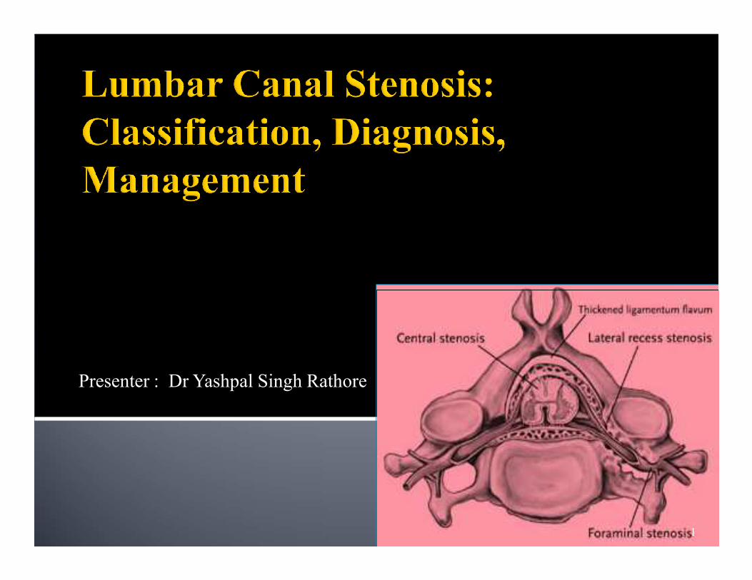
Presenter : Dr Yashpal Singh Rathore
1
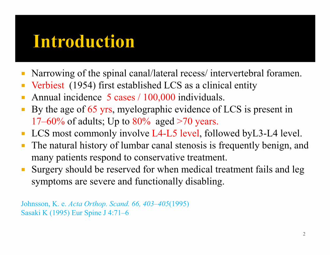
� Narrowing of the spinal canal/lateral recess/ intervertebral foramen.� Verbiest (1954) first established LCS as a clinical entity� Annual incidence 5 cases / 100,000 individuals.� By the age of 65 yrs, myelographic evidence of LCS is present in
17–60% of adults; Up to 80% aged >70 years.LCS most commonly involve L4-L5 level, followed byL3-L4 level.� LCS most commonly involve L4-L5 level, followed byL3-L4 level.
� The natural history of lumbar canal stenosis is frequently benign, and many patients respond to conservative treatment.
� Surgery should be reserved for when medical treatment fails and leg symptoms are severe and functionally disabling.
Johnsson, K. e. Acta Orthop. Scand. 66, 403–405(1995)Sasaki K (1995) Eur Spine J 4:71–6
2
� Etiological Classification� Primary stenosis§ Idiopathic stenosis§ Achondrodysplasia
� Secondary stenosis§ Degenerative § Ossification of the ligamentum flavum & OPLL§ Metabolic or endocrine causes § Infections § Neoplastic§ Rheumatological conditions § Posttraumatic or postoperative stenosis
3
� Anatomical Classification§ Central stenosis (with or without lateral stenosis)§ Isolated lateral stenosis§ Foraminal stenosis
� A patho-morphological classification considers the underlying � A patho-morphological classification considers the underlying pathology such as:§ Hypertrophy of the ligamentum flavum§ Hypertrophy of the facet joints§ Osteophyte formations (spurs)§ Disc herniation§ Synovial facet joint cysts§ Vertebral displacements (anterior/lateral)
4
�
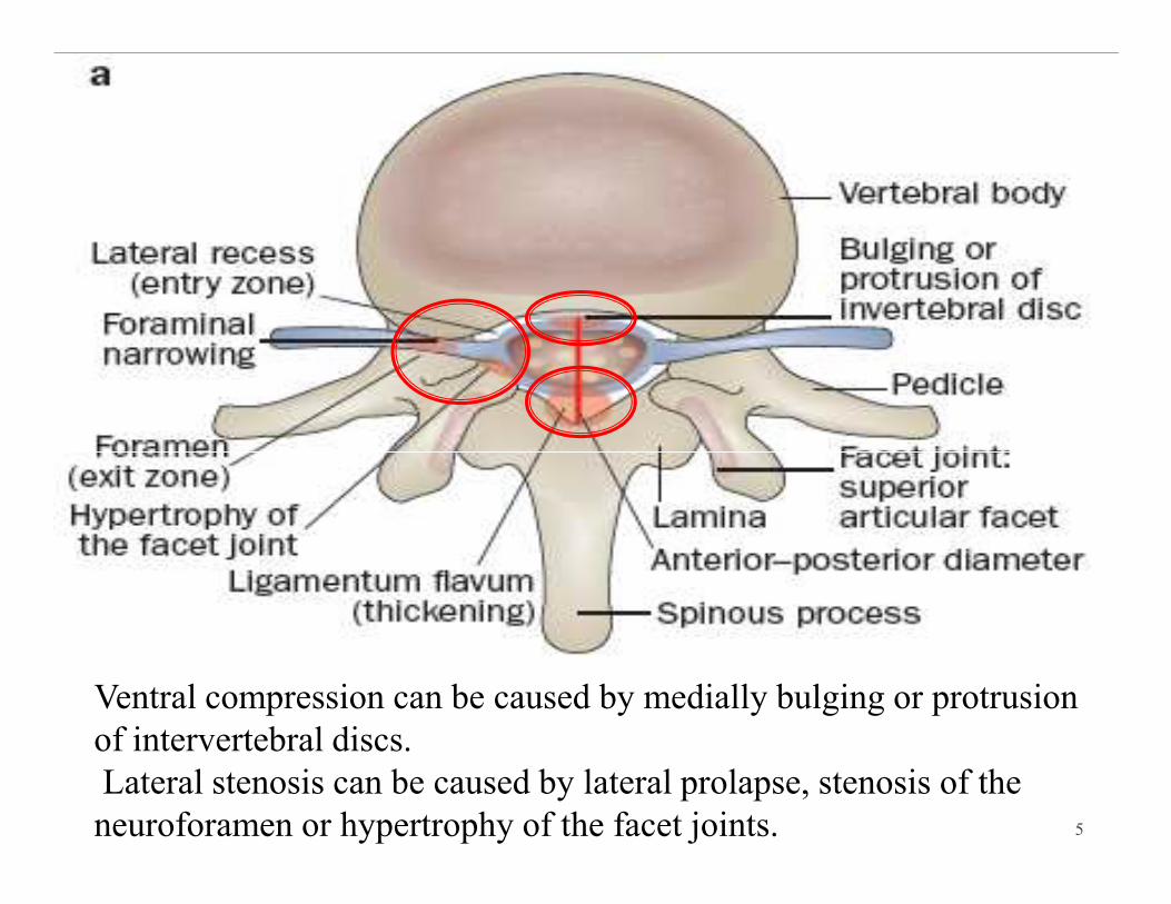
Ventral compression can be caused by medially bulging or protrusion of intervertebral discs.Lateral stenosis can be caused by lateral prolapse, stenosis of the neuroforamen or hypertrophy of the facet joints. 5
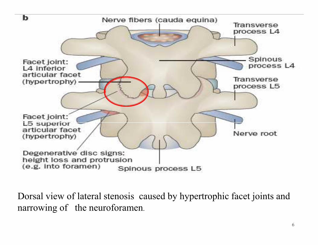
Dorsal view of lateral stenosis caused by hypertrophic facet joints and narrowing of the neuroforamen.
6
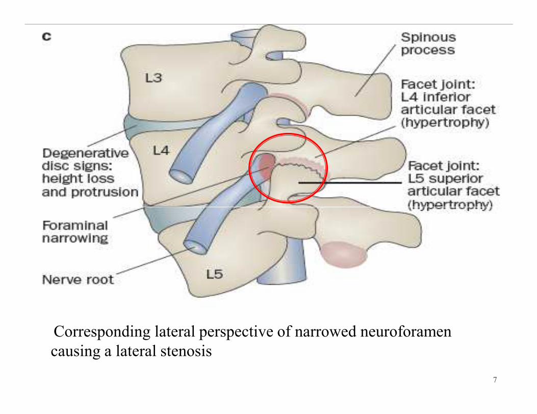
Corresponding lateral perspective of narrowed neuroforamen causing a lateral stenosis
7
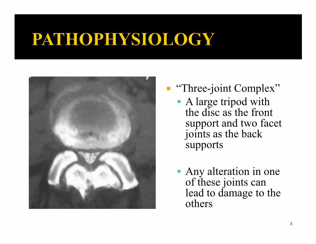
� “Three-joint Complex”§ A large tripod with the disc as the front support and two facet support and two facet joints as the back supports
§ Any alteration in one of these joints can lead to damage to the others
8
• Standing/ walking provokes symptoms
• Pain/weakness
in the legs
• Patients lean forward
while walking to relieve
symptoms
• Sitting or bending
forward relieves
symptoms
9
� Cardinal symptom => Neurogenic claudication§ Numbness, weakness and discomfort in the legs while walking or
prolonged standing,§ Regression of symptoms during sitting and rest .
� Distance decreases with increasing severity of the� Distance decreases with increasing severity of thedegenerative changes
� Radicular claudication� Symptoms can be provoked during walking and prolonged standing but
are localized to a nerve root dermatome� Less frequent symptoms
§ Mechanical low-back pain (worse on activity)§ Atypical leg pain (non-radicular distribution)§ Cauda equina syndrome (very rare)
10
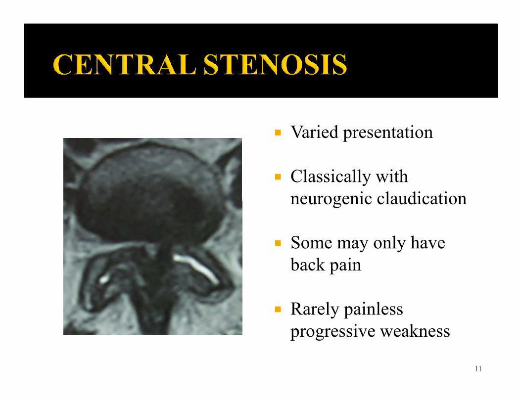
� Varied presentation
� Classically with neurogenic claudicationneurogenic claudication
� Some may only have back pain
� Rarely painless progressive weakness
11
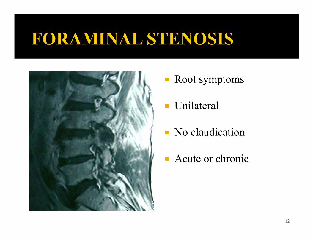
� Root symptoms
� Unilateral
� No claudication
� Acute or chronic
12
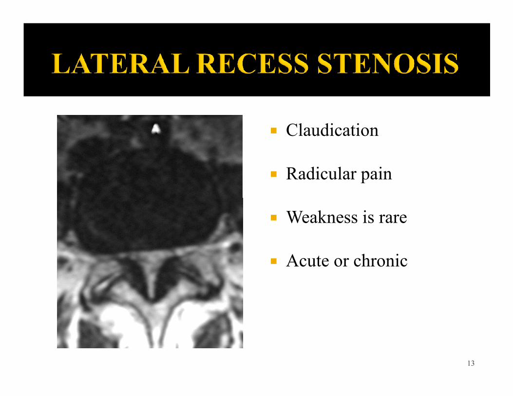
� Claudication
� Radicular pain
� Weakness is rare
� Acute or chronic
13
� The most frequent physical findings § Limited lumbar extension 66–100%§ Sensory deficit 32–58%§ Muscle weakness 18–52%
Straight leg raising 10–90%§ Straight leg raising 10–90%§ Absent knee reflexes 10–50%§ Absent ankle reflexes 50–68%
Katz JN, et al. Rheum. Dis. Clin. North Am. 20:471-483, 1994
§ A reliable assessment of the walking distance is an important parameter for determining the outcome of surgical treatment.
14
� Grade I§ Neurogenic intermittent claudication characterized by a reduced walking distance (caused by pain) and short intermittent sensory-motor deficits that at rest might be unremarkable, but can deteriorate while walking.unremarkable, but can deteriorate while walking.
� Grade II§ Intermittent paresis refers to already persistent sensitivity deficits, loss of reflexes and intermittent paresis.
� Grade III§ Persistent, progressing paresis accompanied by partial regression of pain.
Hufschmidt, A. & Lücking, C. H (eds) Neurologie Compact (Thieme, stuggart, 2006) 15
� Intermittent claudication or vascular claudication
� Radiculopathies or polyneuropathies
� Intraspinal synovial cyst
� Disc prolapse� Disc prolapse
� Tethered cord or spina bifida
� Coxarthrosis or arthrosis of the iliosacral joint
� Abdominal aortic aneurysm
� Neoplasia (for e.g., tumor of myelon, spinal roots, meninges, bones)
� Inflammatory conditions (for e.g., spondylodiscitis, arachnoiditis)
� Dissociative syndromes16
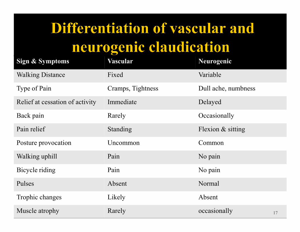
Sign & Symptoms Vascular Neurogenic
Walking Distance Fixed Variable
Type of Pain Cramps, Tightness Dull ache, numbness
Relief at cessation of activity Immediate Delayed
Back pain Rarely OccasionallyBack pain Rarely Occasionally
Pain relief Standing Flexion & sitting
Posture provocation Uncommon Common
Walking uphill Pain No pain
Bicycle riding Pain No pain
Pulses Absent Normal
Trophic changes Likely Absent
Muscle atrophy Rarely occasionally 17
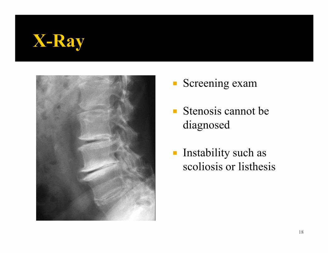
� Screening exam
� Stenosis cannot be diagnoseddiagnosed
� Instability such as scoliosis or listhesis
18
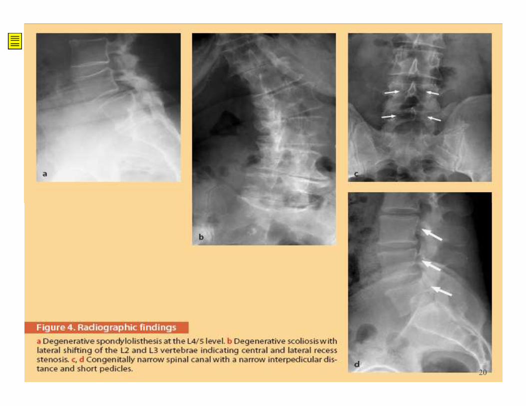
� Findings often associated with spinal stenosis§ Degenerative spondylolisthesis§ Degenerative scoliosis§ Congenitally narrow spinal canal
� Less reliable findings implying lateral recess or foraminal stenosis § Disc space narrowing§ Isthmic spondylolisthesis§ Severe facet osteoarthritis
19
20
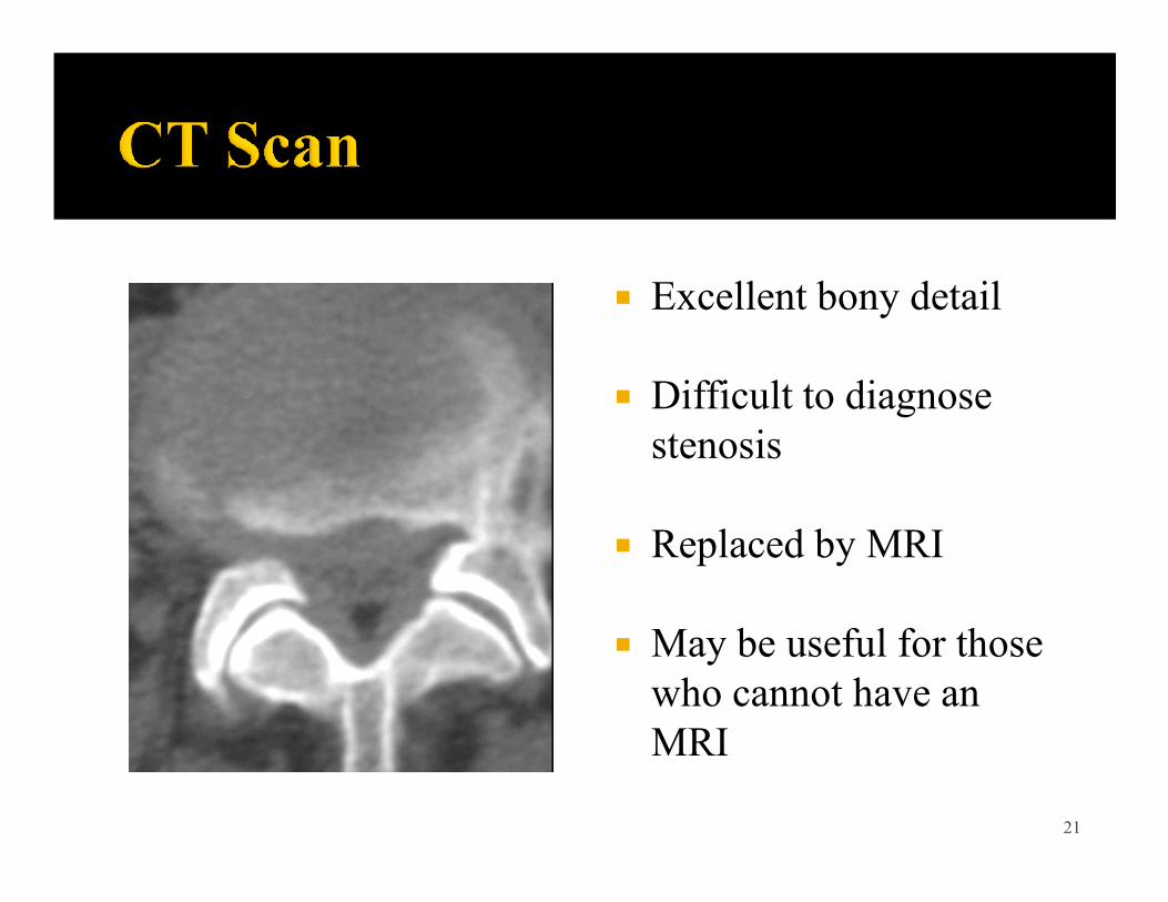
� Excellent bony detail
� Difficult to diagnose stenosisstenosis
� Replaced by MRI
� May be useful for those who cannot have an MRI
21
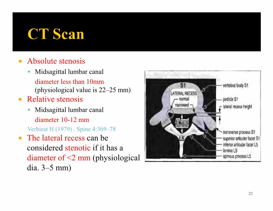
� Absolute stenosis § Midsagittal lumbar canal
diameter less than 10mm(physiological value is 22–25 mm)
� Relative stenosis§ Midsagittal lumbar canal
diameter 10-12 mm Verbiest H (1979) . Spine 4:369–78
� The lateral recess can be considered stenotic if it has a diameter of <2 mm (physiological dia. 3–5 mm)
22
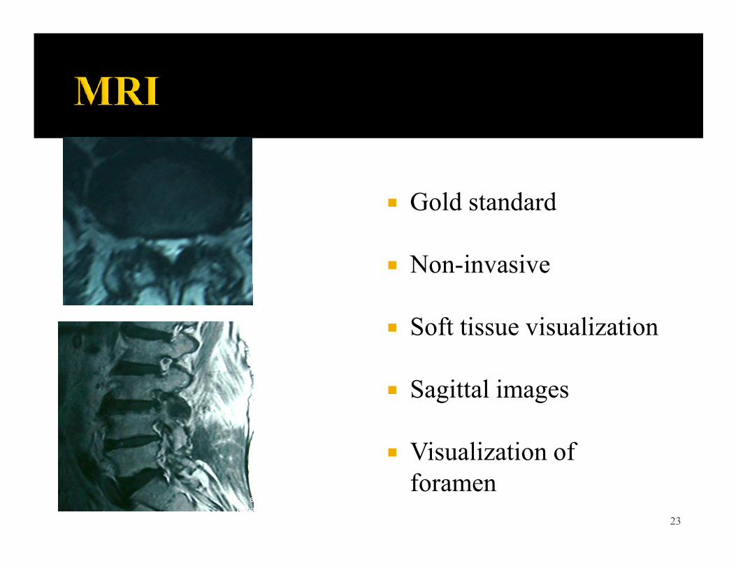
� Gold standard
� Non-invasive
� Soft tissue visualization
� Sagittal images
� Visualization of foramen
23
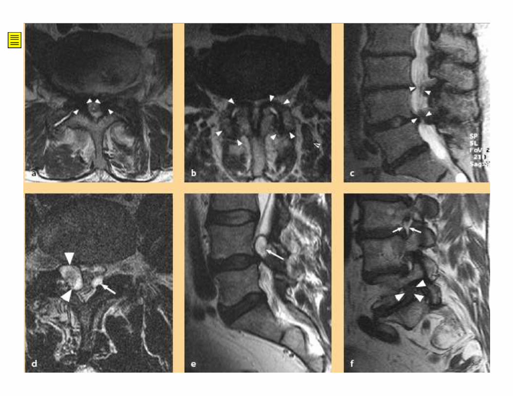
� Characteristic findings of spinal stenosis include:§ Thickened ligamentum flavum § Facet joint hypertrophy § Hourglass appearance of spinal canal on sagittal images § Facet joint synovial cysts § Facet joint synovial cysts § Trefoil appearance of the thecal sac (indicative of spinal lipomatosis)
§ Short pedicles§ Vertebral endplate osteophytes§ Obliteration of the perineural fat in neural foramina in parasaggitalT1WI is indicative of a foraminal stenosis
24
25
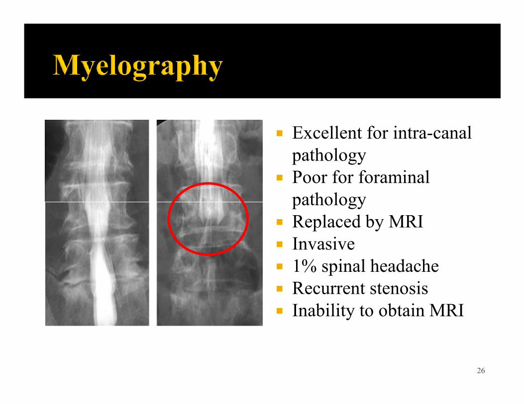
� Excellent for intra-canal pathology
� Poor for foraminal pathologypathology
� Replaced by MRI� Invasive� 1% spinal headache� Recurrent stenosis� Inability to obtain MRI
26
� Excellent visualization of spinal canal
� Excellent for recurrent stenosis
� Invaluable in surgical planning
27
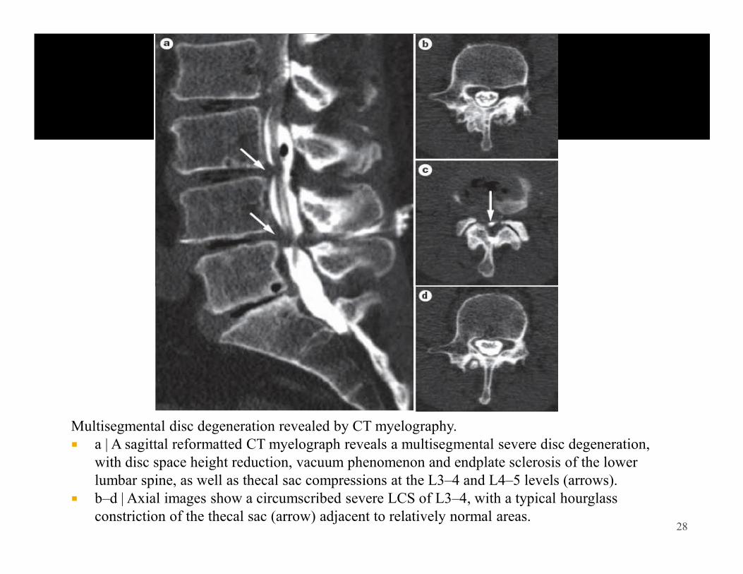
Multisegmental disc degeneration revealed by CT myelography.� a | A sagittal reformatted CT myelograph reveals a multisegmental severe disc degeneration,
with disc space height reduction, vacuum phenomenon and endplate sclerosis of the lower lumbar spine, as well as thecal sac compressions at the L3–4 and L4–5 levels (arrows).
� b–d | Axial images show a circumscribed severe LCS of L3–4, with a typical hourglass constriction of the thecal sac (arrow) adjacent to relatively normal areas.
28
� Neurophysiologic assessment is indicated:§ To confirm the clinical relevance of imaging findings in equivocal cases
§ To identify a peripheral neuropathy
§ To differentiate radiculopathy and mononeuropathy
§ To differentiate non-specific neurological complaints
29
� Steroids§ Effective for acute pain§ Short duration therapy
� Spinal injectionEpidural steroid
� Rest§ Short term activity modification for acute pain
§ Long term activity modification is not § Epidural steroid
§ Transforaminal root block§ Facet joint injection
� Calcitonin has been reported to improve the symptoms of neurogenic claudication
Eskola A etal (1992) Calcif TissueInt 50:400–3
modification is not recommended
� Analgesic§ NSAIDS§ Paracetamol§ Narcotics§ Gabapentin
30
� Avoid extension exercises acutely
� William Flexion � William Flexion Exercises
� Water aerobics
� Strengthening of weak muscle groups
31
� Moderate to severe claudication symptoms
� Significant interference with lifestyle
� Progressive neurological deficits
� Cauda equina syndrome
32
� Decompression (uni-/bilateral laminotomy or laminectomy)
� Decompression with non-instrumented fusion
� Decompression with instrumented fusion
33
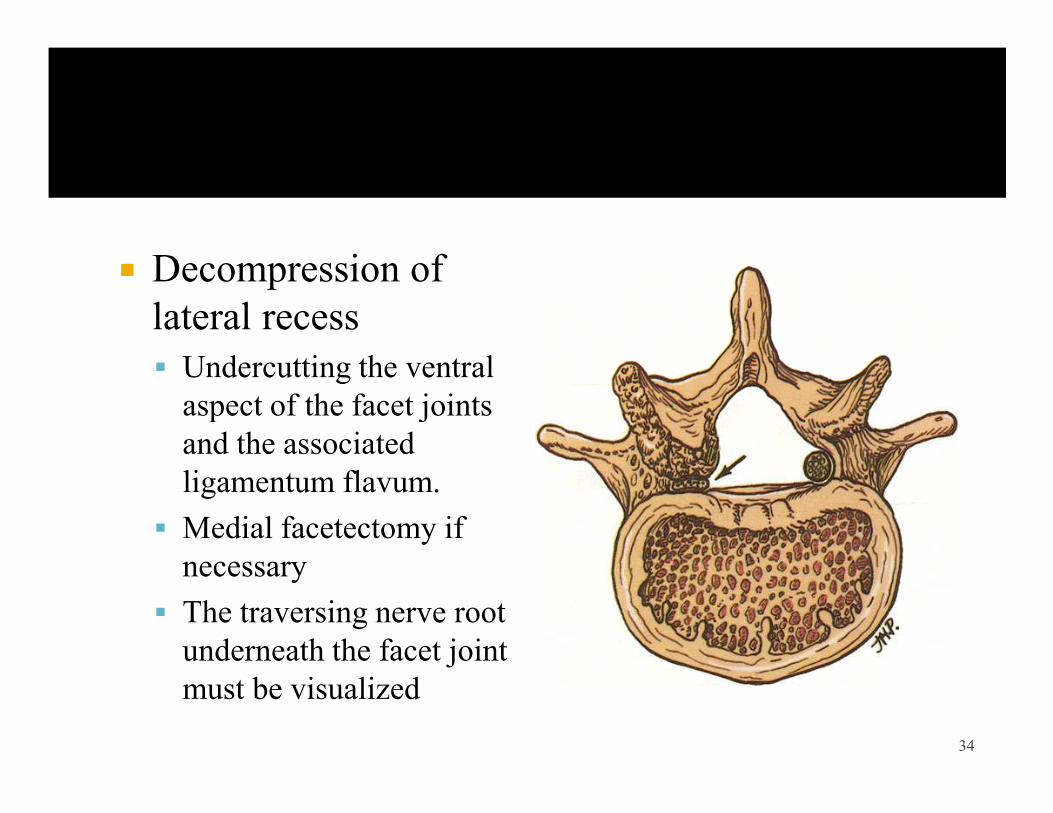
� Decompression of lateral recess§ Undercutting the ventral aspect of the facet joints aspect of the facet joints and the associated ligamentum flavum.
§ Medial facetectomy if necessary
§ The traversing nerve root underneath the facet joint must be visualized
34
� Selective decompression
� Surgical technique of choice in patients presenting with neurogenic claudication without relevant back pain
� Favourable indications § Central stenosis predominantly due to flavum hypertrophy
§ Nerve root claudication due to lateral recess stenosis
§ Absence of degenerative spondylolisthesis and scoliosis
§ Absence of osseous foraminal stenosis 35
� Total laminectomy � Thecal sac cannot be sufficiently decompressed or the access to the foramen is obliterated (foraminal stenosis)
� Laminectomy alone should be avoided in cases with pre-� Laminectomy alone should be avoided in cases with pre-existing instability such as:§ Degenerative spondylolisthesis§ Isthmic spondylolisthesis with secondary degenerative changes
§ Degenerative scoliosis
36
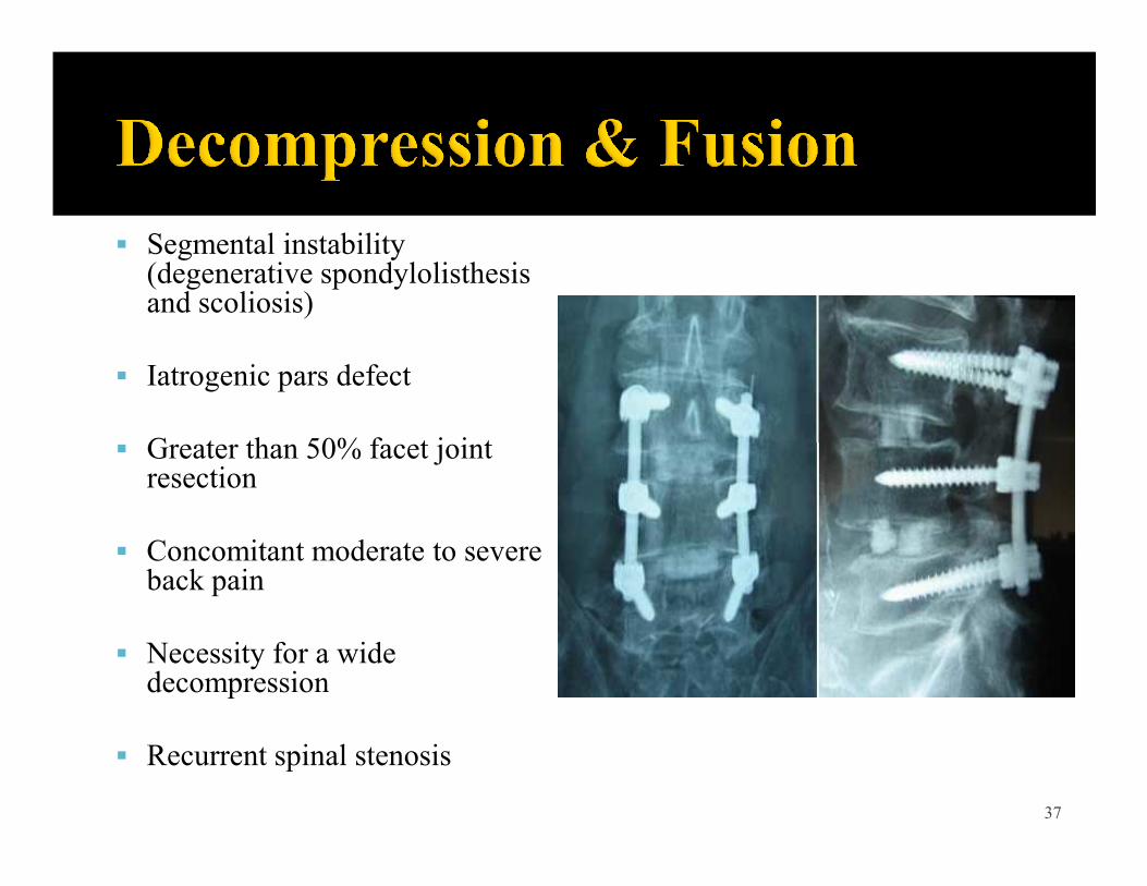
§ Segmental instability (degenerative spondylolisthesis and scoliosis)
§ Iatrogenic pars defect
§ Greater than 50% facet joint § Greater than 50% facet joint resection
§ Concomitant moderate to severe back pain
§ Necessity for a wide decompression
§ Recurrent spinal stenosis
37
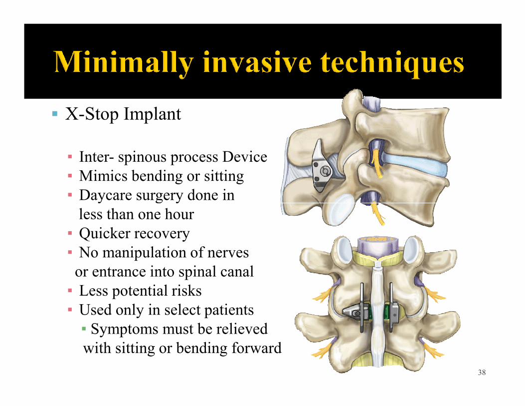
§ X-Stop Implant
▪ Inter- spinous process Device▪ Mimics bending or sitting▪ Daycare surgery done in▪ Daycare surgery done inless than one hour▪ Quicker recovery▪ No manipulation of nervesor entrance into spinal canal▪ Less potential risks▪ Used only in select patients▪ Symptoms must be relievedwith sitting or bending forward
38
� Typical complications of both decompression and fusion surgery § Dura / vessel lacerations § Epidural hematomas
Inadequate decompression with significant residual stenosis§ Inadequate decompression with significant residual stenosis§ Instability§ Re-ossification
� The complication rates for decompression surgery (during and after the surgical procedure) range from 14% to 35%.
Thome, C. et al. J. Neurosurg. Spine 3,129–141 (2005)Benz, r. J. et al Clin. Orthop. Relat. Res. 384, 116–121 (2001)
39
� Unilateral Laminotomy for B/L microdecompression: 520 levels/374pts
� 88% improved VAS 5yr follow up� 0.8% instability � None reoperated� None reoperatedCosta F et.al. JNS-Spine 2007;7(6):579-586
� Clinical results of decompressive laminectomy § Patient satisfaction varies from 57% to 81% with regard to excellent to good results.
Iguchi T etal. J Spinal Disord Tech 2002 ; 15:93–9Iguchi T etal. Spine 2000 ; 25:1754–9
40
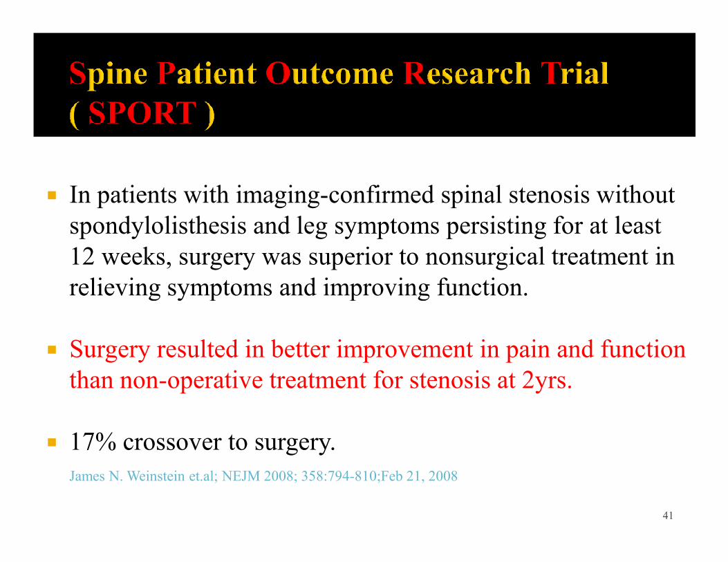
� In patients with imaging-confirmed spinal stenosis without spondylolisthesis and leg symptoms persisting for at least 12 weeks, surgery was superior to nonsurgical treatment in relieving symptoms and improving function.relieving symptoms and improving function.
� Surgery resulted in better improvement in pain and function than non-operative treatment for stenosis at 2yrs.
� 17% crossover to surgery.James N. Weinstein et.al; NEJM 2008; 358:794-810;Feb 21, 2008
41
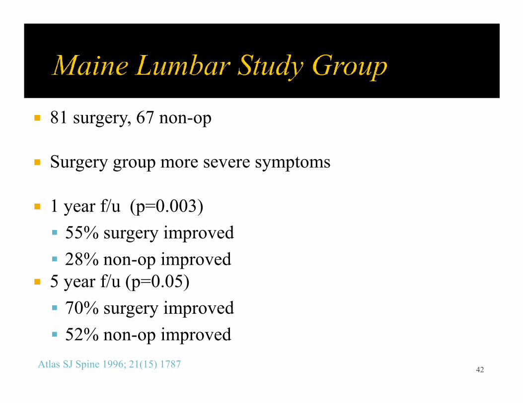
� 81 surgery, 67 non-op
� Surgery group more severe symptoms
� 1 year f/u (p=0.003)� 1 year f/u (p=0.003)§ 55% surgery improved§ 28% non-op improved
� 5 year f/u (p=0.05)§ 70% surgery improved§ 52% non-op improved
Atlas SJ Spine 1996; 21(15) 1787 42
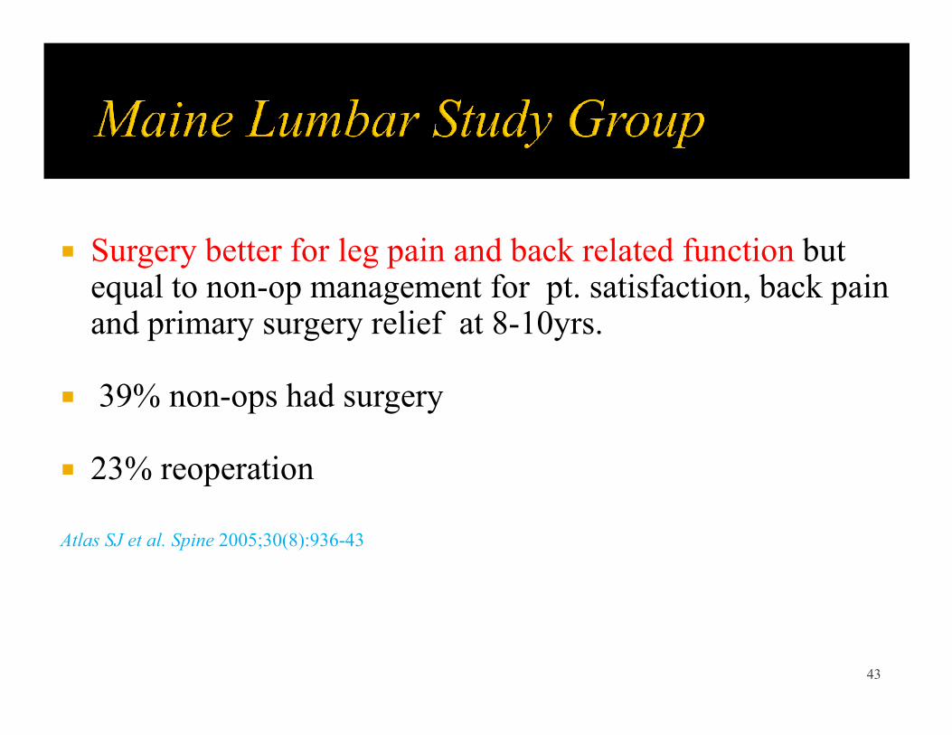
� Surgery better for leg pain and back related function but equal to non-op management for pt. satisfaction, back pain and primary surgery relief at 8-10yrs.
� 39% non-ops had surgery
� 23% reoperation
Atlas SJ et al. Spine 2005;30(8):936-43
43
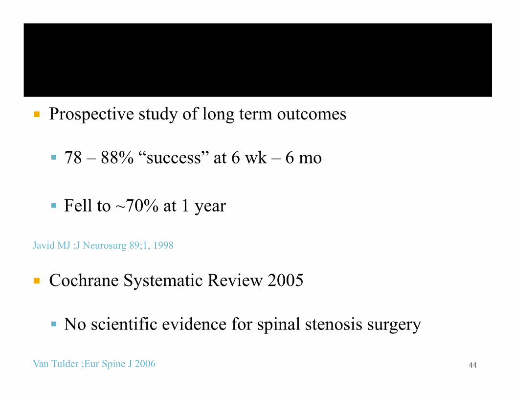
� Prospective study of long term outcomes
§ 78 – 88% “success” at 6 wk – 6 mo
§ Fell to ~70% at 1 year§ Fell to ~70% at 1 year
Javid MJ ;J Neurosurg 89;1, 1998
� Cochrane Systematic Review 2005
§ No scientific evidence for spinal stenosis surgery
Van Tulder ;Eur Spine J 2006 44

THANK YOUTHANK YOUTHANK YOUTHANK YOU
45