Embed Size (px)
Citation preview

Presented by: Dr. Giuseppe Molinaro
Dr. Davide De Biase

VACCINATIONS
ANTIPARASITIC
COMMERCIAL DIET
VOMITING FOR A MONTH
DULLNESS
WEIGHT LOSS
INAPPETANCE
Dog
Spayed Female
LABRADOR RETRIEVER
3 Years old

Physical examination
Temperature: 38,3 °C
Heart/Pulse rate: normal
Respiratory rate: normal
Body condition/State of nutrition: fat (BCS 4/5)
Mentation/Level of consciousness: depressed
Particular signs: none
Integument and skin: normal
Lymph nodes: normal
Mucous membrane color: pink
Capillary refill time (CRT): 2’’
Auscultation: normal
Abdominal palpation: normal

4
CBC
Parameter Result Normal values
RBC 5.39 M/µl (5.50-8.00)
Hgb 11.5 g/dl (12.0-18.0)
Hct 34.8 % (37.0-55.0)
MCV 65 fL (60-76)
MCH 21.4 Pg (20.0-27.0)
MCHC 33.1 g/dL (32.0-38.0)
PLT 350 K/µL (240-400)
WBC 10.8 K/µL (6.0-16.0)
Neutrophils 72% 60-77
Lymphocytes 18% 12-30
Monocytes 6% 3-10
Eosinophils 4% 2-10
Basophile 0% Rari
Chemistry panel
Parameter Result Normal values
AZO 20 mg/Dl (25-35)
CREA 1.13 mg/dL (< 1.8)
GLU 80 mg/dL (60-120)
AST 70 UI/L (5-45)
ALT 356 UI/L (10-47)
GGT 16 UI/L (<5)
F.A. 542 UI/L (<180)
P.T. 6.2 mg/dL (6.0-7.7)
A/G 0.66 (>0.6)
CHOLESTEROL 258 mg/dL (125-250)
TRIGLYCERIDES 95 mg/dL (50-100)
BILIRUBIN TOT 0.1 mg/dL (0.1-0.8)
CALCIUM 8.6 mg/dL (8,4-11)
PHOSPHORUS 6.5 mg/dL (2.5-5)

5
URINE ANALYSES
Parameter Result Normal values
SPECIFIC WEIGHT 1010 (1025-1045)
PH 5 (5,5-7,0)
LEUKOCYTES Absent (ABSENT)
NITRITES Absent (ABSENT)
PROTEIN 2000 mg/ml (ABSENT)
GLUCOSE Absent (ABSENT)
KETONES Absent (ABSENT)
UROBILINOGEN Absent (ABSENT)
BILIRUBIN Absent (ABSENT)
BLOOD Absent (ABSENT)
UPC >10.8 (<0.2)
SEDIMENT EXAMINATION Normal (RARE EPITHELIAL TRANSITIONS CELLS, 0-2 ERYTHROCYTES, 0-3
LEUKOCYTES -FOR MICROSCOPIC FIELD 40X)

Ultrasound
Fine needle aspiration of liver were performed and smears were subsequently stained with MGG (DiffQuick).
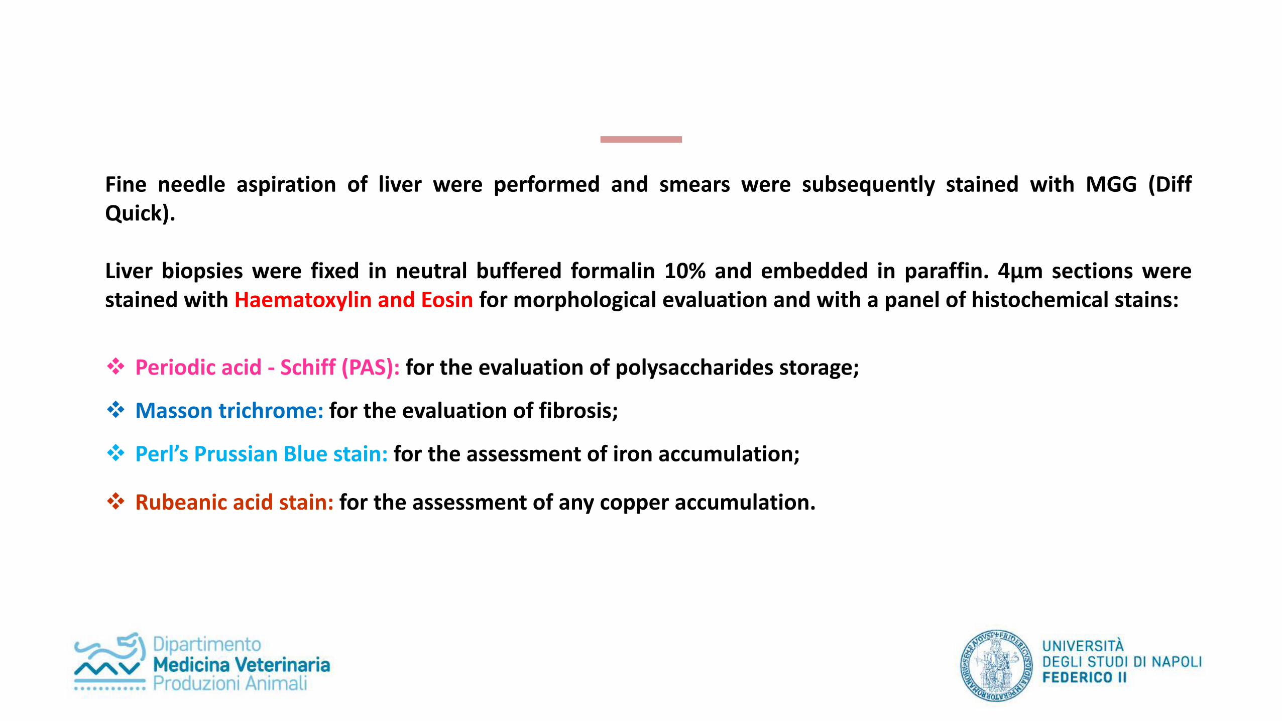
Liver biopsies were fixed in neutral buffered formalin 10% and embedded in paraffin. 4μm sections werestained with Haematoxylin and Eosin for morphological evaluation and with a panel of histochemical stains:
Periodic acid - Schiff (PAS): for the evaluation of polysaccharides storage;
Masson trichrome: for the evaluation of fibrosis;
Perl’s Prussian Blue stain: for the assessment of iron accumulation;
Rubeanic acid stain: for the assessment of any copper accumulation.

Cytology specimen revealed….not so much. We observed large groups of hepatocytes admixed with scatteredinflammatory cells such as macrophages, lymphocytes ad plasma cells (fig. 1-3).
Histopathological findings in sections of the liver specimen stained with HE comprised:
Mild to moderate infiltration by a mixed populations of macrophages (sometimes haemosiderin-laden), lymphocytesand plasma cells (fig. 4-6);
Hepatocytes vacuolation; Small foci of necrosis (fig. 7); Numerous small granulomatous to pyogranulomatous foci scattered randomly through the parenchyma (fig. 8-9).
A mild periportal fibrosis was observed with Masson trichrome stain (fig. 10). A moderate accumulation of PAS positivematerial was detected within the hepatocytes (fig. 11). No copper deposition within the hepatocytes was demonstratedwith rubeanic acid staining, but Perl’s Prussian blue staining revealed haemosiderin (fig. 12-13).

Fig. 1 Diff Quick, 10x
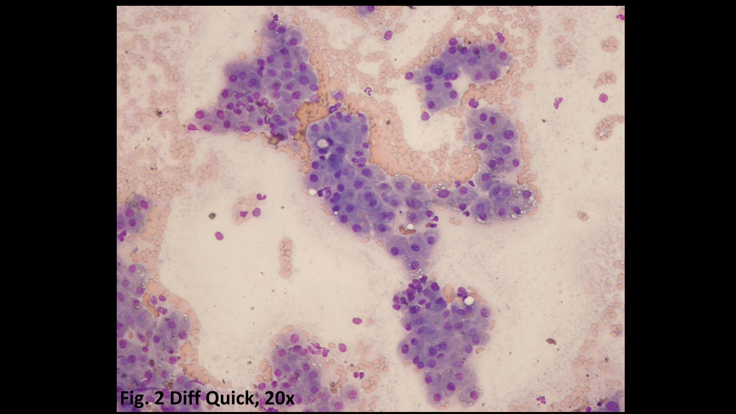
Fig. 2 Diff Quick, 20x

Fig. 3 Diff Quick, 40x

Fig. 4, HE (4x)
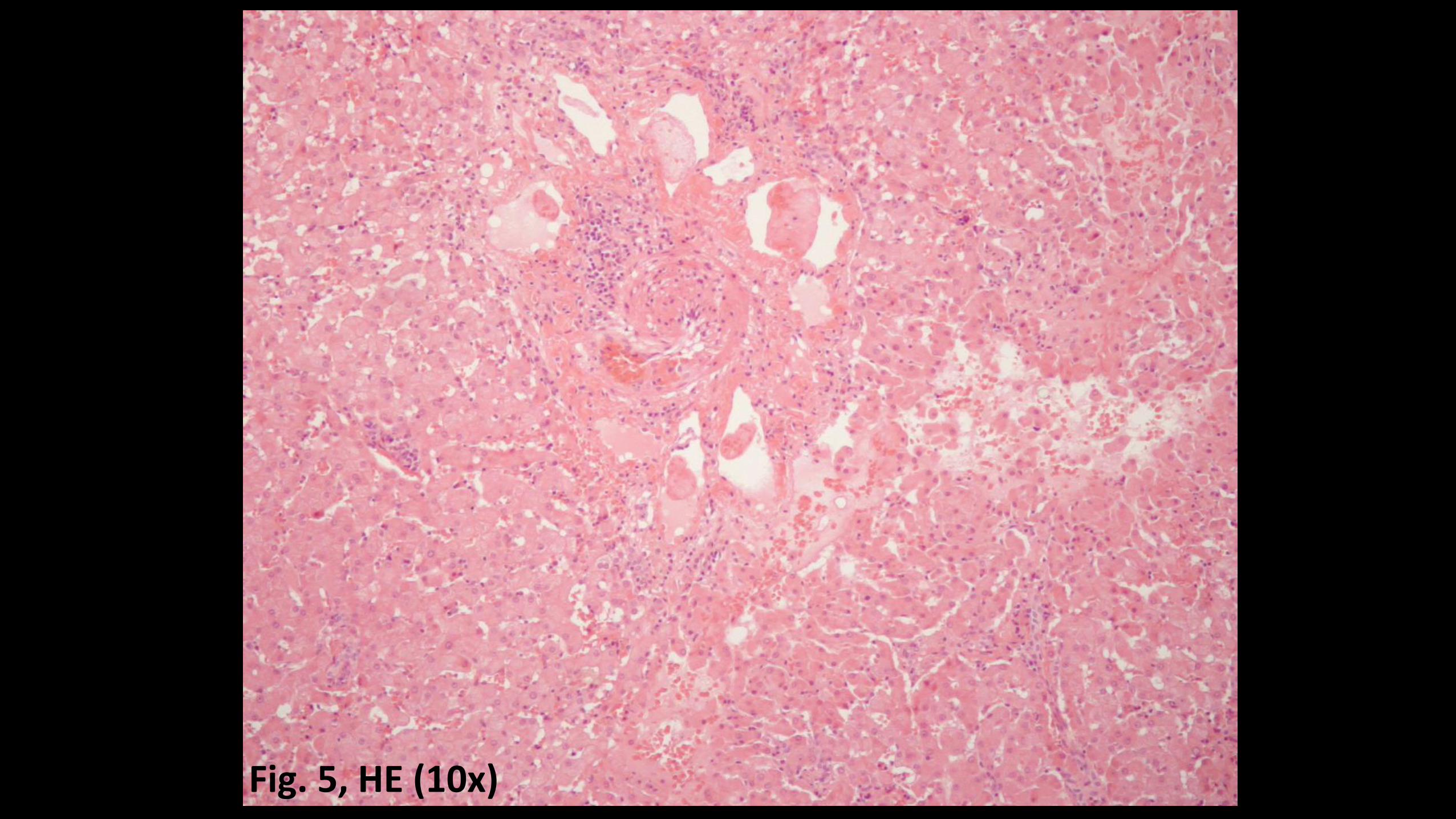
Fig. 5, HE (10x)

Fig. 6, HE (20x)
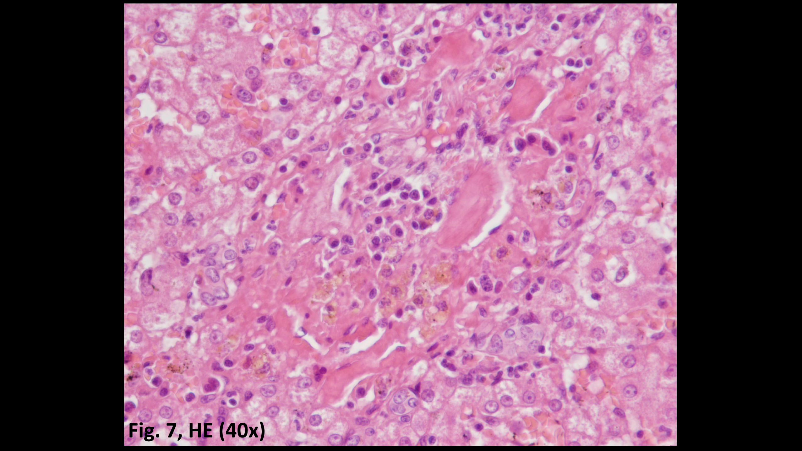
Fig. 7, HE (40x)

Fig. 8, HE (40x)

Fig. 9, HE (40x)

Fig. 10, Masson Trichrome (20x)

Fig. 11, PAS (20x)

Fig. 12, Rubeanic acid stain (10x)

Fig. 13, Perl’s Prussian Blue (40x)
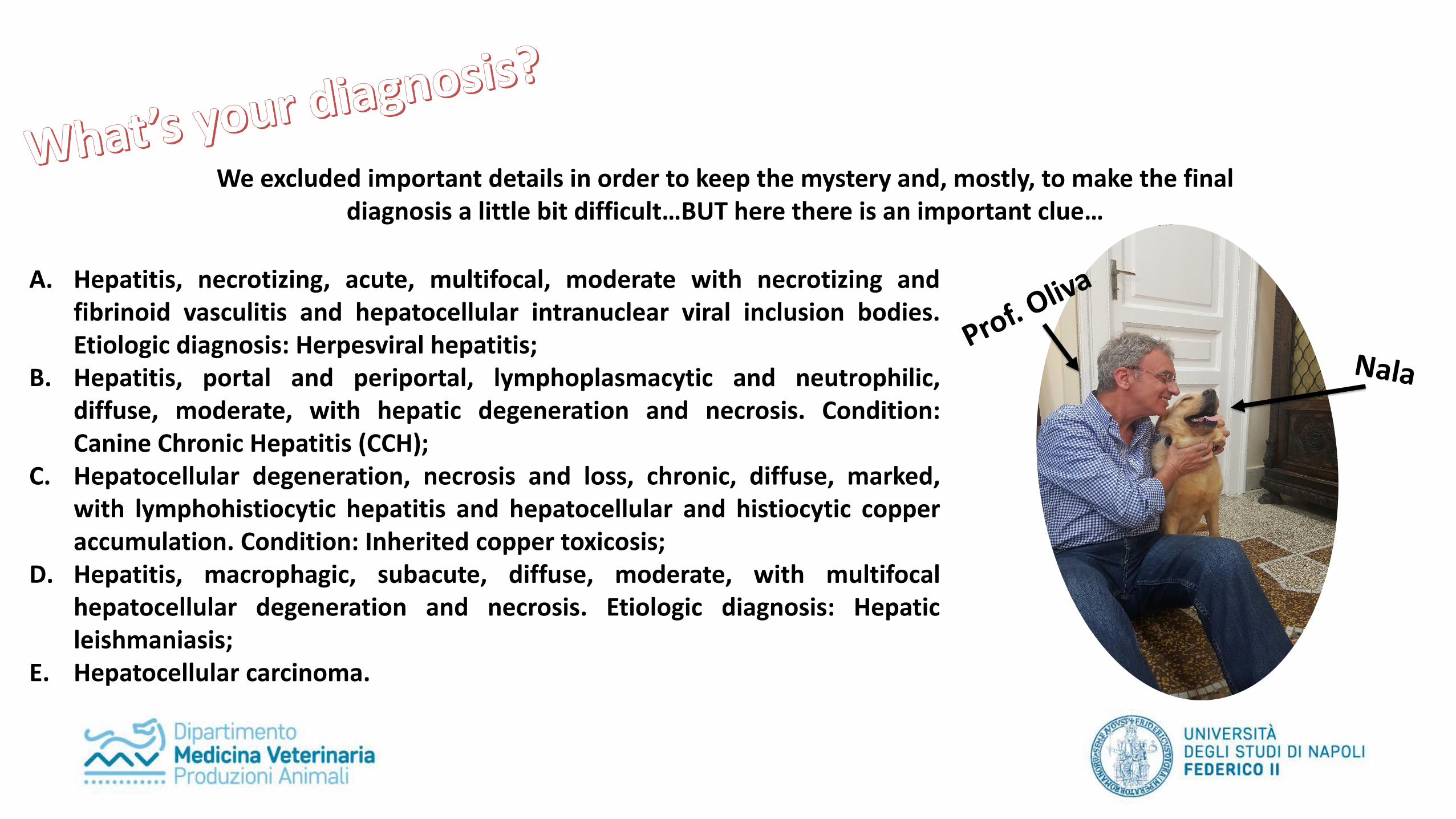
We excluded important details in order to keep the mystery and, mostly, to make the final diagnosis a little bit difficult…BUT here there is an important clue…
A. Hepatitis, necrotizing, acute, multifocal, moderate with necrotizing andfibrinoid vasculitis and hepatocellular intranuclear viral inclusion bodies.Etiologic diagnosis: Herpesviral hepatitis;
B. Hepatitis, portal and periportal, lymphoplasmacytic and neutrophilic,diffuse, moderate, with hepatic degeneration and necrosis. Condition:Canine Chronic Hepatitis (CCH);
C. Hepatocellular degeneration, necrosis and loss, chronic, diffuse, marked,with lymphohistiocytic hepatitis and hepatocellular and histiocytic copperaccumulation. Condition: Inherited copper toxicosis;
D. Hepatitis, macrophagic, subacute, diffuse, moderate, with multifocalhepatocellular degeneration and necrosis. Etiologic diagnosis: Hepaticleishmaniasis;
E. Hepatocellular carcinoma.


![Marco Molinaro Astronomical Data Center Forum 10-11 June ...g-vo.org/cosadie-dcforum/slides/MarcoMolinaro_HowPublishVO.pdf · 6/10/2013 · Marco Molinaro [INAF-OATs IA2] – Astronomical](https://img.pdfslide.us/doc/110x75/60e0767770a05a1578022927/marco-molinaro-astronomical-data-center-forum-10-11-june-g-voorgcosadie-dcforumslidesmarcomolinaro.jpg)