Embed Size (px)
Citation preview

Presentation
A woman brings her 5-year-old son to your office after noticing a “lump” in his abdomen while hugging him. He hasn’t
complained of abdominal pain or nausea and has no changes in his urine or stool. His mom also denies fever, rash, and cold
symptoms.

History• Past Medical Hx: None• Birth Hx: Term infant, SVD, no forceps or vacuum used, mother
had standard prenatal care without any complications, infant was discharged with mother at 48 hours
• Developmental Hx: Normal per Mom• Immunizations: UTD• Family Hx: Mom is healthy, Dad has HTN, 3-year-old sister and
18-month-old brother are healthy, paternal grandfather has DM and HTN
• Social Hx: Lives with parents and 2 younger sibs, goes to pre-kindergarten, has a kitten, no one smokes in the home
• Allergies: Penicillin (rash)• Medications: None

Physical Exam
• Vitals:– T 37 ⁰C– BP 95/54– HR 95– RR 22– O2 100% on room air
• General: non-toxic, alert, interactive
• HEENT: PERRL, EOMI, moist oral mucosa, no exudates
• CV: RRR, no murmur
• Resp: CTAB, no crackles or wheezes
• Abd: Non-distended; firm, nontender, fixed mass on the right that does not cross the midline; normal bowel sounds
• GU: Normal genitalia for age• MS: moves all 4 extremities,
normal tone• Neuro: CN II-XII grossly intact,
reflexes 2+, normal finger-to-nose testing, normal gait

Differential Diagnosis for Abdominal Mass
• Hepatobiliary– Hepatoblastoma– Hepatocellular carcinoma– Choledochal cyst– Benign liver tumor
• Renal– Hydronephrosis– Polycystic kidney– Wilms tumor– Rhabdoid tumor
• Retroperitoneal– Neuroblastoma– Teratoma– Lymphoma– Rhabdomyosarcoma
• GI– Intussusception– Duplication cyst– Constipation– Hirschsprung disease
• GU– Testicular neoplasm– Undescended testicle– Bladder obstruction

Laboratory Tests
Urinary VMA: negUrinary HVA: neg
CBC: 12.08.8 325
36.1
CMP: 138 101 10------------------------< 76
4.0 25 0.8
UA: moderate RBC
AST 32ALT 29Alk phos 125Total protein 7.0Albumin 4.5Total bili 1.1

Imaging
Ultrasound revealed an intraabdominal mass on the right. CT was ordered and is shown below.

Histologic Confirmation of Diagnosis
Biopsy revealed a triphasic combination of stromal (fibrocytic, myxoid, skeletal muscle components) , blastemal (small blue
cells), and epithelial (tubules, glomeruli) cell types. Focal anaplasia, shown on right, is seen in approximately 5%.

Gross Morphology
The mass was subsequently resected. It was large, soft, well-circumscribed, homogenous, and tan in color.

What is your final diagnosis?

Wilms tumor
• Aka nephroblastoma• Embryonal malignancy of the kidney• Most cases are sporadic but 1-2% are familial• May arise in 1 or both kidneys• Associated with the following syndromes:– WAGR (aniridia, GU abnormalities, mental retardation)– Denys-Drash syndrome (early-onset renal failure, male
pseudohermaphrodism)– Beckwith-Wiedemann syndrome (organomegaly,
macroglossia, omphalocele, hemihypertrophy)

Epidemiology
• 2nd most common malignant abdominal tumor in childhood
• Incidence is approximately 8 in 1 million children younger than age 15
• About 500 new cases in North America per year• Peak incidence between ages 2 and 5• Individual with horseshoe kidney have twice the
risk

Initial Presentation
• Many children present with an asymptomatic abdominal mass
• 1/3 have intermittent abdominal pain that may have been exacerbated by trauma
• 1/4 have gross or microscopic hematuria and is usually painless
• 1/4 have HTN• Systemic symptoms may occur, especially if there is
bleeding into the tumor and associated anemia• About 10% will show pulmonary metastases at the time of
diagnosis

Treatment
• National Wilms Tumor Study– Stages I & II w/ favorable histology:
surgical resection followed by 19 weeks of chemotherapy
– Stages I & II w/ anaplastic histology: surgical resection followed by 19 weeks of chemotherapy and radiation therapy
– Stage III: surgical resection followed by 24 weeks of triple drug chemotherapy and radiation therapy
– Stage IV: surgical resection followed by 24 weeks of triple drug chemotherapy plus radiation therapy to the abdomen and lungs for mets
• International Society of Pediatric Oncology– Stage I: pre-op chemotherapy for 4
weeks, surgical resection, post-op chemotherapy for 4 weeks
– Stage II: pre-op chemotherapy for 4 weeks, surgical resection, post-op triple drug chemotherapy for 27 weeks
– Stage III: pre-op chemotherapy for 4 weeks, surgical resection, post-op triple drug chemotherapy for 27 weeks plus radiation therapy
– Stage IV: pre-op triple drug chemotherapy for 6 weeks, surgical resection, post-op triple drug chemotherapy for 9 weeks—if remission is achieved continue to 27 weeks, if not add additional chemotherapeutic agents for an additional 34 weeks and add radiation therapy
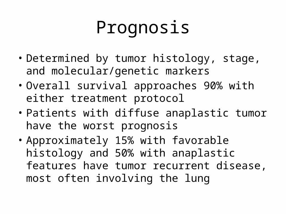
Prognosis
• Determined by tumor histology, stage, and molecular/genetic markers
• Overall survival approaches 90% with either treatment protocol
• Patients with diffuse anaplastic tumor have the worst prognosis
• Approximately 15% with favorable histology and 50% with anaplastic features have tumor recurrent disease, most often involving the lung