Embed Size (px)
Citation preview
Vol. 3, 433-438, March 1997 Clinical Cancer Research 433
Presence of Methylthioadenosine Phosphorylase (MTAP) in
Hematopoietic Stem/Progenitor Cells: Its Therapeutic
Implication for MTAP (-) Malignancies1
John Yu,2 Ayse Batova, Li-en Shao,
Carlos J. Carrera, and Alice L. Yu
Department of Molecular and Experimental Medicine, The ScrippsResearch Institute, La Jolla, California 92037 [J. Y., L-e. S.];Department of Pediatrics, University of California San Diego MedicalCenter, San Diego, California 92103 [A. B., A. L. Y.]; andDepartment of Medicine and Cancer Center, University of California,San Diego, La Jolla, California 92093 [C. J. C.]
ABSTRACT
Methylthioadenosine phosphorylase (MTAP) is important
for the salvage of adenine and methionine. Recently, we foundfrequent deletion of MTAP in T-cell acute lymphoblastic leu-kemia (T-ALL) patients both at diagnosis and at relapse (A.
Batova et aL, Blood, 88: 3083-3090, 1996). In addition, MTAP
deficiency has been reported in other cancers. Thus, MTAPdeficiency in cancer may offer opportunities for developing
selective therapy, which would spare normal cells. It is there-
fore important to document the presence of MTAP activity inhematopoietic stem/progenitor cells. Our approach was to in-vestigate whether hematopoietic stem/progenitor cells can be
rescued from the cytothxicity of an AMP synthesis inhibitor,
L-alanosifle, by 5’-deoxyadenosine, a process that requires
MTAP. Erythroid burst-forming unit, granulocyte/monocyte
colony-forming unit, or granulocyte/erythrocyte/macrophage/
megakaryocyte colony-forming unit progenitors and the prim-itive high proliferative potential colony-forming cells in the
purified CD34� cells were cultured in horse serum-containing
medium, and their colony growth was found to be suppressed
by incubation with 5 p.M or greater concentrations ofL-alaflOSine. However, in the presence of 5-10 ,LM of 5’-deoxya-
denosine, colony formation of hematopoietic stem/primitive
progenitors was restored. On the other hand, 5’-deoxy-5’-
methylthioadenosine, the endogenous substrate of MTAP, wastoxic to hematopoietic stem/progenitors (ffl� < 1 �LM), pre-
sumably due to inhibition of methylation reactions or poly-amine synthesis. We also compared the effects of L-alanosine
Received 9/16/96; revised 12/2/96; accepted 12/13/96.The costs of publication of this article were defrayed in part by the
payment of page charges. This article must therefore be hereby marked
advertisement in accordance with 18 U.S.C. Section 1734 solely to
indicate this fact.I Supported by Grants U10CA28439, MOb RR00827, and FDA 001 129(to A. L. Y.); CA54892 (to C. J. C.); and DK40218 (to J. Y.) and MOIRR00833 from the NIH. A. B. is supported by NIH Training Grant2T32-HLO71O7. This is publication 10160-MEM from The Scripps
Research Institute.2 To whom requests for reprints should be addressed, at Department ofMolecular and Experimental Medicine, NX 3, The Scripps ResearchInstitute, 10550 North Torrey Pines Road, La Jolla, CA 92037.
and 5’-deoxyadenosine on MTAP (+) and MTAP (-) T-ALLcell lines. Treatment of MTAP (+) Molt 4 and MTAP (-)
CEM cell lines with L-alanosine in the presence of S’-deoxya-denosine resulted in killing of MTAP (-), but not MTAP (+)cells. Therefore, our findings demonstrate the presence ofMTAP in human hematopoietic stem/progenitor cells and sup-port the possibffity of targeting MTAP in the design of anenzyme-selective therapy for T-ALL and other MTAP-defi-
cient malignancies.
INTRODUCTION
MTAP3 is an important salvage enzyme for both adenine
and methionine. Specifically, it cleaves MTA, which is gener-
ated during the synthesis of polyamines, into adenine and
5-rnethylthionbose-l-phosphate. Adenine and 5-methylthiori-
bose- I -phosphate are efficiently salvaged to form adenine nu-
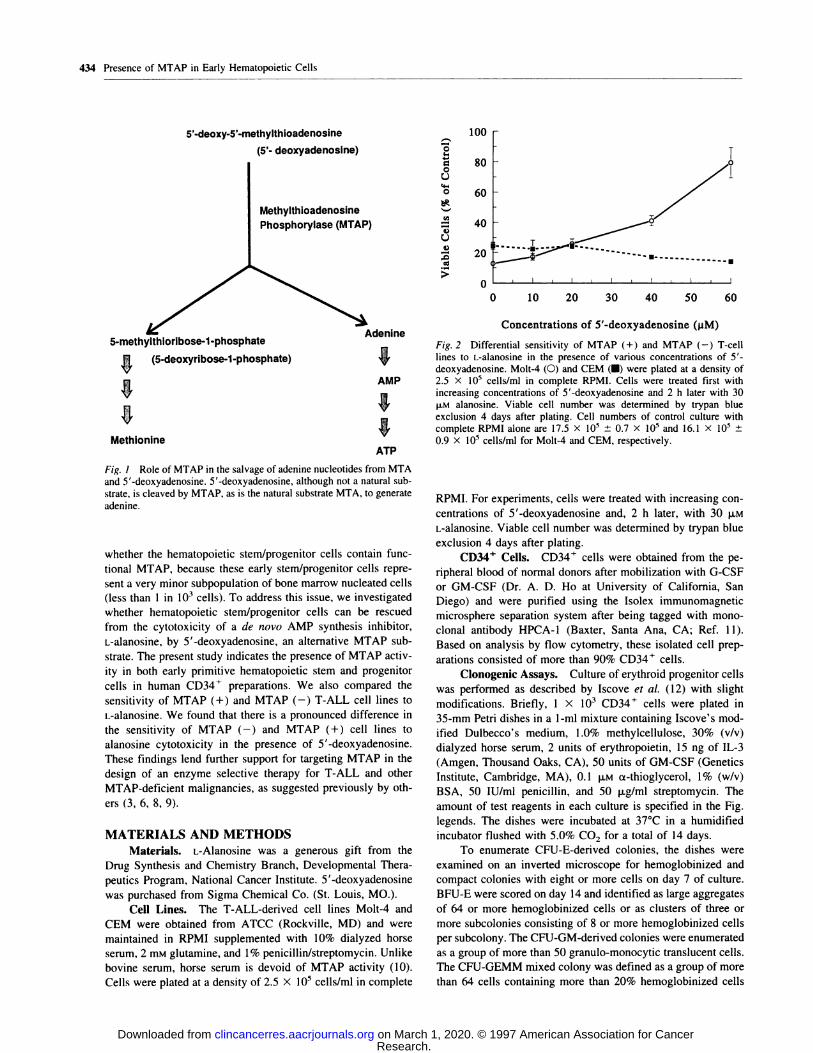
cleotides and methionine, respectively (Fig. I).
Cleavage by MTAP is the sole catabolic pathway for MTA.
MTAP is abundant in many tissues, including most bone mar-
row and peripheral blood nucleated cells (1). On the other hand,
MTAP has been shown to be deficient in some rnurine and
human tumor cells (2, 3). Previously, we reported MTAP en-
zyrne deficiency in one T-ALL and one common ALL patient
among 20 leukemia patients (4). Recently, using quantitative
PCR and Southern blot analyses, we have found frequent dde-
tion of MTAP in T-ALL samples. Deletions that included exon
8 of MTAP occurred in 33.3% of patients at diagnosis and in
39.4% patients at relapse (5). In addition to T-ALL, MTAP
deficiency has been reported at a high frequency in non-small
cell lung cancer (6), glioma (3), isolated cases of rectal adeno-
carcinoma (1), and acute nonlymphoid leukemia (I, 7).
Kamatani et a!. (3) have suggested that MTAP deficiency
in cancer may offer opportunities for developing an enzyme-
selective chemotherapy that would spare normal cells. In MTAP
(-) cancer cells, the salvage of methionine or adenine from
MTA would be blocked, resulting in an increased dependency
on an exogenous supply of these two nutrients. It is conceivable
that the MTAP (-) cancer cells will be more sensitive than
normal cells to the cytotoxicity of either inhibitors of de novo
purine synthesis or of methionine depletion (3, 6, 8, 9). Al-
though MTAP activity has been demonstrated in more than 98%
of nucleated cells in human marrow (3), it remains unclear
3 The abbreviations used are: MTAP, methylthioadenosine phosphoryl-ase; MTA, 5’-deoxy-S’-methylthioadenosine; I-ALL, I-cell acute lym-phoblastic leukemia; ALL, acute lymphoblastic leukemia; IL, interleu-
kin; HPP-CFC, high proliferative potential colony-forming cell; BFU-E,erythroid burst-forming unit; CFU-GM, granubocyte/monocyte colony-forming unit; CFU-GEMM, granulocyte/erythrocyte/macrophage/megakaryocyte colony-forming unit.
Research. on March 1, 2020. © 1997 American Association for Cancerclincancerres.aacrjournals.org Downloaded from
1005’-deoxy-5’-methylthioadefloSifle
(5’- deoxyadenosine)
Methylthioadenosine
Phosphorylase (MTAP)
V�d.
5-methylthiorlbose-1-phosphate enine
,�, (5-deoxyribose-1-phosphate)
03”00
U‘4-
0
C,)
0
U3)
80
60
40
20
0
T
� � U
-4
0 10 20 30 40 50 60
Concentrations of 5’-deoxyadenosine QiM)
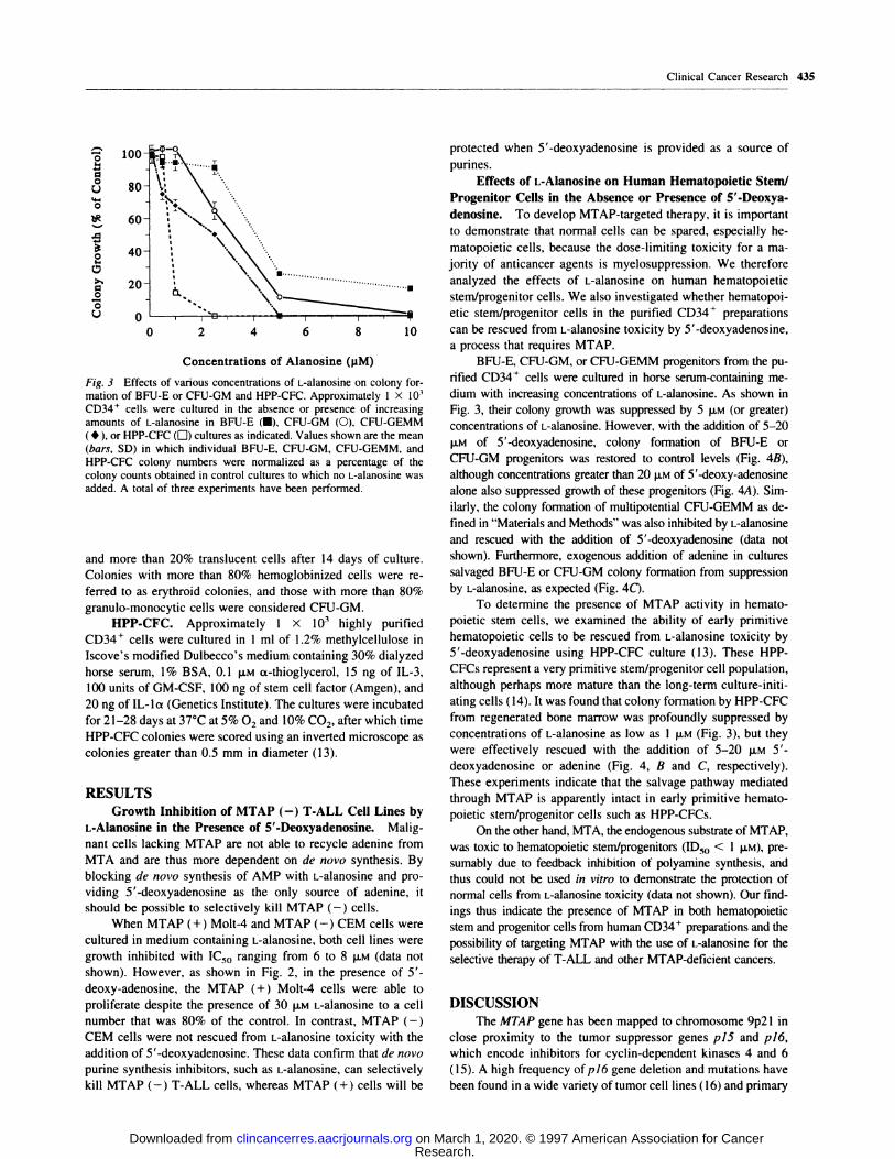
n Fig. 2 Differential sensitivity of MTAP (+) and MTAP (-) I-cellI lines to L-alanosine in the presence of various concentrations of 5’-
deoxyadenosine. Molt-4 (0) and CEM (S) were plated at a density ofAMP 2.5 x l0� cells/mi in complete RPMI. Cells were treated first withn increasing concentrations of 5 ‘-deoxyadenosine and 2 h later with 30t p.M alanosine. Viable cell number was determined by trypan bluen exclusion 4 days after plating. Cell numbers of control culture with
I complete RPMI alone are 17.5 X l0� ± 0.7 X l0� and 16.1 X l0� ±0.9 X l0� cells/ml for Molt-4 and CEM, respectively.
434 Presence of MTAP in Early Hematopoietic Cells
MethionineATP
Fig. I Role of MTAP in the salvage of adenine nucleotides from MTAand 5’-deoxyadenosine. 5’-deoxyadenosine, although not a natural sub-
strate, is cleaved by MTAP, as is the natural substrate MTA, to generateadenine.
whether the hematopoietic stem/progenitor cells contain func-
tional MTAP, because these early stem/progenitor cells repre-
sent a very minor subpopulation of bone marrow nucleated cells
(less than 1 in l0� cells). To address this issue, we investigated
whether hematopoietic stem/progenitor cells can be rescued
from the cytotoxicity of a de novo AMP synthesis inhibitor,
L-alanosine, by 5’-deoxyadenosine, an alternative MTAP sub-
strate. The present study indicates the presence of MTAP activ-
ity in both early primitive hernatopoietic stem and progenitor
cells in human CD34� preparations. We also compared the
sensitivity of MTAP (+) and MTAP (-) T-ALL cell lines to
L-alanoSine. We found that there is a pronounced difference in
the sensitivity of MTAP (-) and MTAP (+) cell lines to
alanosine cytotoxicity in the presence of 5’-deoxyadenosine.
These findings lend further support for targeting MTAP in the
design of an enzyme selective therapy for T-ALL and other
MTAP-deficient malignancies, as suggested previously by oth-
ers (3, 6, 8, 9).
MATERIALS AND METHODS
Materials. L-Alanosine was a generous gift from the
Drug Synthesis and Chemistry Branch, Developmental Thera-
peutics Program, National Cancer Institute. 5’-deoxyadenosine
was purchased from Sigma Chemical Co. (St. Louis, MO.).
Cell Lines. The T-ALL-derived cell lines Molt-4 and
CEM were obtained from ATCC (Rockville, MD) and were
maintained in RPM! supplemented with 10% dialyzed horse
serum, 2 mr�i glutamine, and 1% penicillin/streptornycin. Unlike
bovine serum, horse serum is devoid of MTAP activity (10).
Cells were plated at a density of 2.5 X 10� cells/mi in complete
RPMI. For experiments, cells were treated with increasing con-
centrations of 5’-deoxyadenosine and, 2 h later, with 30 �i.M
L-alanosine. Viable cell number was determined by trypan blue
exclusion 4 days after plating.
CD34� Cells. CD34� cells were obtained from the pe-
ripheral blood of normal donors after mobilization with G-CSF
or GM-CSF (Dr. A. D. Ho at University of California, San
Diego) and were purified using the Isolex irnmunornagnetic
rnicrosphere separation system after being tagged with mono-
cbonal antibody HPCA-l (Baxter, Santa Ana, CA; Ref. 1 1).
Based on analysis by flow cytornetry, these isolated cell prep-
arations consisted of more than 90% CD34� cells.
Clonogenic Assays. Culture of erythroid progenitor cells
was performed as described by Iscove et al. ( 12) with slight
modifications. Briefly, 1 X l0� CD34� cells were plated in
35-mm Petri dishes in a 1-mb mixture containing Iscove’s mod-
ified Dulbecco’s medium, 1 .0% rnethylcellubose, 30% (v/v)
dialyzed horse serum, 2 units of erythropoietin, 15 ng of IL-3
(Arngen, Thousand Oaks, CA), 50 units of GM-CSF (Genetics
Institute, Cambridge, MA), 0. 1 �i.M a-thioglycerol, 1% (w/v)
BSA, 50 lU/mI penicillin, and 50 p.g/ml streptomycin. The
amount of test reagents in each culture is specified in the Fig.
legends. The dishes were incubated at 37#{176}Cin a humidified
incubator flushed with 5.0% CO2 for a total of 14 days.
To enumerate CFU-E-derived colonies, the dishes were
examined on an inverted microscope for hemogbobinized and
compact colonies with eight or more cells on day 7 of culture.
BFU-E were scored on day 14 and identified as large aggregates
of 64 or more hemogbobinized cells or as clusters of three or
more subcolonies consisting of 8 or more hernogiobinized cells
per subcolony. The CFU-GM-derived colonies were enumerated
as a group of more than 50 granulo-rnonocytic translucent cells.
The CFU-GEMM mixed colony was defined as a group of more
than 64 cells containing more than 20% hemogbobinized cells
Research. on March 1, 2020. © 1997 American Association for Cancerclincancerres.aacrjournals.org Downloaded from
100-
80-
0
I-
00U4-
0
,0
0I-
0
0
0U
40-
20- -I U
0
Concentrations of Alanosine (pM)
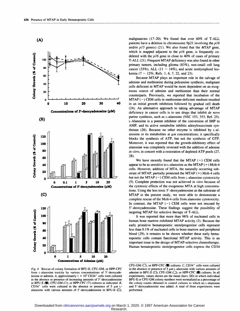
Fig. 3 Effects of various concentrations of L-alanosine on colony for-
mation of BFU-E or CFU-GM and HPP-CFC. Approximately 1 X l0�CD34’ cells were cultured in the absence or presence of increasingamounts of L-alanosine in BFU-E (I), CFU-GM (0), CFU-GEMM( . ), or HPP-CFC (D) cultures as indicated. Values shown are the mean
(bars, SD) in which individual BFU-E, CFU-GM, CFU-GEMM, andHPP-CFC colony numbers were normalized as a percentage of thecolony counts obtained in control cultures to which no L-alanosine was
added. A total of three experiments have been performed.
and more than 20% translucent cells after 14 days of culture.
Colonies with more than 80% hernogbobinized cells were re-
ferred to as erythroid colonies, and those with more than 80%
granubo-monocytic cells were considered CFU-GM.
HPP-CFC. Approximately 1 X l0� highly purified
CD34� cells were cultured in I ml of I .2% methylcellulose in
Iscove’s modified Dulbecco’s medium containing 30% dialyzed
horse serum, 1% BSA, 0.1 �iM a-thioglycerol, 15 ng of IL-3,
100 units of GM-CSF, 100 ng of stern cell factor (Amgen), and
20 ng of IL-la (Genetics Institute). The cultures were incubated
for 21-28 days at 37#{176}Cat 5% 02 and 10% CO2. after which time
HPP-CFC colonies were scored using an inverted microscope as
colonies greater than 0.5 mm in diameter (13).
DISCUSSION
The MTAP gene has been mapped to chromosome 9p2l in
close proximity to the tumor suppressor genes p15 and p16,
which encode inhibitors for cyclin-dependent kinases 4 and 6
(15). A high frequency ofp/6 gene deletion and mutations have
been found in a wide variety oftumor cell lines (16) and primary
Clinical Cancer Research 435
protected when 5’-deoxyadenosine is provided as a source of
purines.
Effects of L-Alanosine on Human Hematopoietic Stem/
Progenitor Cells in the Absence or Presence of 5’-Deoxya-
denosine. To develop MTAP-targeted therapy, it is important
to demonstrate that normal cells can be spared, especially he-
matopoietic cells, because the dose-limiting toxicity for a ma-
jority of anticancer agents is rnyebosuppression. We therefore
analyzed the effects of L-alanosine on human hematopoietic
stem/progenitor cells. We also investigated whether hernatopoi-
etic stem/progenitor cells in the purified CD34� preparations
0 2 4 6 8 10 can be rescued from L-alanosine toxicity by 5’-deoxyadenosine,a process that requires MTAP.
BFU-E, CFU-GM, or CFU-GEMM progenitors from the pu-
rifled CD34� cells were cultured in horse serum-containing me-
diurn with increasing concentrations of L-alanosine. As shown in
Fig. 3, their colony growth was suppressed by 5 p.M (or greater)
concentrations of L-alanOsine. However, with the addition of 5-20
�LM of 5’-deoxyadenosine, colony formation of BFU-E or
CFU-GM progenitors was restored to control levels (Fig. 4B),
although concentrations greater than 20 p.M of 5’-deoxy-adenosine
alone also suppressed growth of these progenitors (Fig. 4A). Sim-
ilarly, the colony formation of multipotential CFU-GEMM as de-
fined in “Materials and Methods” was also inhibited by L-alanosine
and rescued with the addition of 5’-deoxyadenosine (data not
shown). Furthermore, exogenous addition of adenine in cultures
salvaged BFU-E or CFU-GM colony formation from suppression
by L-alanosifle, as expected (Fig. 4C).
To determine the presence of MTAP activity in hemato-
poietic stem cells, we examined the ability of early primitive
hematopoietic cells to be rescued from L-alanosine toxicity by
5’-deoxyadenosine using HPP-CFC culture (13). These HPP-
CFCs represent a very primitive stem/progenitor cell population,
although perhaps more mature than the long-term culture-initi-
ating cells (14). It was found that colony formation by HPP-CFC
from regenerated bone marrow was profoundly suppressed by
concentrations of L-alanosine as low as I p.M (Fig. 3), but they
were effectively rescued with the addition of 5-20 jiM 5’-
deoxyadenosine or adenine (Fig. 4, B and C, respectively).
These experiments indicate that the salvage pathway mediatedRESULTS through MTAP is apparently intact in early primitive hemato-
Growth Inhibition of MTAP (-) T-ALL Cell Lines by poietic stem/progenitor cells such as HPP-CFCs.
L-Alanosine in the Presence of 5’-Deoxyadenosine. Malig- On the other hand, MTA, the endogenous substrate of MTAP,
nant cells lacking MTAP are not able to recycle adenine from was toxic to hematopoietic stem/progenitors (ID50 < 1 p.M), pre-
MTA and are thus more dependent on de novo synthesis. By surnably due to feedback inhibition of polyamine synthesis, and
blocking de novo synthesis of AMP with L-alanosine and pro- thus could not be used in vitro to demonstrate the protection of
viding 5 ‘-deoxyadenosine as the only source of adenine, it normal cells from L-alanOsine toxicity (data not shown). Our find-
should be possible to selectively kill MTAP (-) cells. ings thus indicate the presence of MTAP in both hematopoietic
When MTAP (+) Molt-4 and MTAP (-) CEM cells were stern and progenitor cells from human CD34� preparations and the
cultured in medium containing L-alanosine, both cell lines were possibility of targeting MTAP with the use of L-alanosine for the
growth inhibited with IC50 ranging from 6 to 8 p.M (data not selective therapy of T-ALL and other MTAP-deficient cancers.
shown). However, as shown in Fig. 2, in the presence of 5’-
deoxy-adenosine, the MTAP (+) Molt-4 cells were able to
proliferate despite the presence of 30 �LM L-alanosine to a cell
number that was 80% of the control. In contrast, MTAP (-)
CEM cells were not rescued from L-alanOsine toxicity with the
addition of 5’-deoxyadenosine. These data confirm that de novo
purine synthesis inhibitors, such as L-alanosine, can selectively
kill MTAP (-) T-ALL cells, whereas MTAP (+) cells will be
Research. on March 1, 2020. © 1997 American Association for Cancerclincancerres.aacrjournals.org Downloaded from
(A)
0 10 20 30 40
Concentrations of 5’-deoxyadenoslno (pM)
0 0.1 1 5 10 20Conceatritiofts of 5’-d.oxysdonoslne (pM)
I
I
120
100
$0
60
40
20
020
CFU-GM (El)’ or HPP-CFC (U) cultures. C, CD34� cells were culturedin the absence or presence of 5 p.M L-alanosine with various amounts ofadenine in BFU-E (0), CFU-GM (EJ)’ or HPP-CFC (U) cultures. In allexperiments, values shown are the mean (bars, SD) in which individualBFU-E or CFU-GM colony numbers were normalized as a percentage of
the colony counts obtained in control cultures to which no L-alanosineand 5 ‘-deoxyadenosine was added. A total of three experiments wereperformed.
436 Presence of MTAP in Early Hematopoietic Cells
(B)
I 120
�100
� $0
160
0
40
20
0
(C)
0 1 5 10Concentration.. of Ad.n1�* (pM)
Fig. 4 Rescue of colony formation of BFU-E, CFU-GM, or HPP-CFCfrom L-alanosine toxicity by various concentrations of 5’-deoxyade-nosine or adenine. A, approximately 1 X l0� CD34’ cells were culturedin the absence or presence of increasing amounts of 5’-deoxyadenosinein BFU-E (s), CFU-GM (0), or HPP-CFC (V) cultures as indicated. B,CD34� cells were cultured in the absence or presence of 5 p.M L-
alanosine with various amounts of 5’-deoxyadenosine in BFU-E (0),
malignancies (17-20). We found that over 60% of T-ALL
patients have a deletion in chromosome 9p2l involving the pitS
and/or piS gene(s) (21). We also found that the MTAP gene,
which is mapped adjacent to the pi6 gene, is frequently co-
deleted with the pi6 gene in close to 40% of cases of primary
T-ALL (21). Frequent MTAP deficiency was also found in other
primary tumors, including gliorna (83%), non-small cell lung
cancer (33%), ALL (1 1 - 14%), and acute nonlymphoid leu-
kemia (7 - 12%; Refs. 1, 6, 7, 22, and 23).
Because MTAP plays an important role in the salvage of
adenine and methionine during polyamine synthesis, malignant
cells deficient in MTAP would be more dependent on an exog-
enous source of adenine and methionine than their normal
counterparts. Previously, we reported that incubation of the
MTAP (-) CEM cells in methionine-deficient medium resulted
in an initial growth inhibition followed by gradual cell death
(24). An alternative approach to taking advantage of MTAP
deficiency in cancer cells is to use drugs that inhibit de novo
punne synthesis, such as L-alanosine (NSC 153, 353; Ref. 25).
L-Alanosine is a potent inhibitor of the conversion of IMP to
AMP, and its active metabolite inhibits adenylosuccinate syn-
thetase (26). Because no other enzyme is inhibited by L-al-
anosine or its metabolites at p.� concentrations; it specifically
blocks the synthesis of ATP, but not the synthesis of GTP.
Moreover, it was reported that the growth-inhibitory effect of
alanosine was completely reversed with the addition of adenine
in vitro, in concert with a restoration of depleted ATP pools (27,
28).
We have recently found that the MTAP (-) CEM cells
appear to be as sensitive to L-alanosine as the MTAP (+) Molt-4
cells. However, addition of MTA, the naturally occurring sub-
strate of MTAP, partially protected the MTAP (+) Molt-4 cells
but not the MTAP (-) CEM cells from L-alanosine cytotoxicity
(5). Complete protection was not achieved in vitro because of
the cytotoxic effects of the exogenous MTA at high concentra-
tions. Using the less toxic 5’-deoxyadenosine as the substrate of
MTAP in the present study, we were able to demonstrate a
complete rescue of the Molt-4 cells from alanosine cytotoxicity.
In contrast, the MTAP (-) CEM cells were not rescued by
5’-deoxyadenosine. These findings suggest the possibility of
targeting MTAP for selective therapy of T-ALL.
It was reported that more than 98% of nucleated cells in
human bone marrow exhibited MTAP activity (3). Because the
early primitive hematopoietic stem/progenitor cells represent
less than 0. 1 % of nucleated cells in bone marrow and peripheral
blood (29), it remains to be shown whether these early hema-
topoietic cells contain functional MTAP activity. This is an
important issue in the design of MTAP-selective chemotherapy.
Human hematopoietic stem/progenitor cells express the CD34
Research. on March 1, 2020. © 1997 American Association for Cancerclincancerres.aacrjournals.org Downloaded from
Clinical Cancer Research 437
antigen, but the population of CD34� cells is heterogeneous.
The purified CD34� preparations contain cells that are func-
tionally primitive stem cells (< 1 in 102 CD34’ cells), as well as
more mature, lineage-specific progenitors. In the present study,
we found that marrow cultures of a variety of hematopoietic
lineages were completely protected from alanosine toxicity by
5’-deoxyadenosine. Such findings confirmed the presence of
functional MTAP activity in the committed progenitors. like
erythroid BFU-E, granubocytic/monocytic CFU-GM, or multi-
potent CFU-GEMM (29). In addition, culture of HPP-CFCs,
which requires at least three or more hematopoietic factors for
proliferation and generates colonies as large as 3000-8000 cells
(13), provided specific information on the effect of L-alanosine
on the proliferation and differentiation of the early primitive
HPP-CFCs. The fact that the addition of 5’-deoxyadenosine
rescues these cells from L-alanosine toxicity suggests the pres-
ence of MTAP activity in these early primitive hematopoietic
cells. In the murine model, HPP-CFC has been shown to have a
highly significant correlation with cells capable of repopulating
the bone marrow of lethally irradiated mice and therefore rep-
resents a primitive cell population, closely related to stem cells.
One recent study suggests that there is MTAP “deficien-
cy” in human hematopoietic committed progenitors because
MTA failed to reverse the suppression of the colony forma-
tion of BFU-E, CFU-GM, and CFU-GEMM by methionine
depletion (30). Our observation of a great reduction in the
colony growth of hematopoietic progenitor cells in the pres-
ence of p.� concentrations of MTA is consistent with this
study, as well as previous reports on MTA toxicity in murine
and human hernatopoietic progenitors (3 1 , 32). However, our
study also demonstrated the rescue of alanosine-induced in-
hibition of colony formation of these hernatopoietic progen-
itors by 5’-deoxyadenosine, a process that requires MTAP
activity in these cells. There are two possible explanations for
the back of protection from methionine starvation by MTA.
First, hematopoietic stem/progenitor cells are extremely sen-
sitive to the known MTA toxicity in in vitro marrow culture,
presumably due to inhibition of methylation reactions, as
well as to polyamine synthesis (33-36), which may be im-
portant in proliferation/differentiation of hematopoietic cells.
During normal hematopoiesis, the endogenous MTA pro-
duced under physiological conditions must be rapidly sal-
vaged into adenine and methionine by MTAP to avoid accu-
mulation of toxic levels of MTA. Second, salvage of
methionine from MTA requires the conversion of 5-methyl-
thioribose-i-phosphate into methionine by a series of corn-
plex oxidations via the intermediate 2-keto-4-methylthiobu-
tyrate (37). The apparent lack of MTA rescue in vitro may
therefore be a consequence of defects in the other complex
enzymatic reactions following MTAP reaction, instead of
MTAP deficiency per Se.
Taken together, our results, along with those of previous
studies, suggest that use of specific de novo purine synthesis
inhibitors can selectively kill MTAP (-) cancer cells under
conditions in which MTAP (+) normal cells, including hema-
topoietic stem/progenitor cells, will be protected by using the
endogenous MTA as a source of adenine. Thus, the presence of
a common metabolic defect such as MTAP deficiency can
facilitate development of selective chemotherapy for several
types of cancer, including T-ALL.
REFERENCES
I. Fitchen. J. H., Riscoe, M. K., Dana, B. W., Lawrence, H. J.. and
Ferro, A. J. Methylthioadenosine phosphorylase deficiency in humanleukemias and solid tumors. Cancer Res., 46: 5409-5412, 1986.
2. Toohey, J. I. Methylthioadenosine nucleoside phosphorylase deli-ciency in methylthio-dependent cancer cells. Biochem. Biophys. Res.Commun., 83: 27-35, 1978.
3. Kamatani, N.. Nelson-Rees, W. A., and Carson, D. A. Selectivekilling of human malignant cell lines deficient in methylthioadenosinephosphorylase. a purine metabolic enzyme. Proc. NatI. Acad. Sci. USA,78: 1219-1223, 1981.
4. Kamatani, N., Yu, A. L., and Carson, D. A. Deficiency of methyl-
thioadenosine phosphorylase in human leukemic cells in vito. Blood.60: 1387-1391, 1982.
5. Batova, A., Diccianni, M. B.. Nobori, T., Vu, I., Yu. J., Bridgeman,L., and Yu, A. L. Frequent deletion in the methylthioadenosine phos-phorylase gene in I-cell acute lymphoblastic leukemia: strategies for
enzyme-targeted therapy. Blood, 88: 3083-3090, 1996.
6. Nobori, I., Szinai, I., Amox, D., Parker. B., Obopade, 0. I.,Buchhagen. D. L., and Carson, D. A. Methylthioadenosine phosphoryl-ase deficiency in human non-small cell lung cancers. Cancer Res., 53:
1098-1101, 1993.
7. Traweek, S. I., Riscoe, M. K., Ferro, A. J., Braziel, R. M., Magenis,R. E., and Ftchen, J. H. Methylthioadenosine phosphorylase deficiencyin acute leukemia: pathologic, cytogenetic, and clinical features. Blood,7!: 1568-1573, 1988.
8. Kubota, M., Kamatani, N., and Carson. D. A. Biochemical geneticanalysis of the role of methylthioadenosine phosphorylase in a murine
lymphoid cell line. J. Biol. Chem., 256: 7288-7291, 1983.
9. Christa, L., Thuillier, L., Munier, A., and Perignon, J-L. Salvage of5’-deoxy-methylthioadenosine into purines and methionine by lymphoidcells and inhibition of cell proliferation. Biochim. Biophys. Acta. 803:
7-10, 1984.
10. Kamatani, N., and Carson, D. A. Abnormal regulation of methyl-thioadenosine and polyamine metabolism in methylthioadenosine phos-phorylase-deficient human leukemic cell lines. Cancer Res.. 40. 178-182, 1980.
11 . Lane, I. A., Law, P.. Maruyama, M.. Young, D., Burgess, J..Mullen, M., Mealiffe, M., Terstappen, L. W. M. M., Hardwick. A.,Moubayed, M., Oldham, F., Comngham, R. E. I., and Ho, A. D.Harvesting and enrichment of hematopoietic progenitor cells mobilizedinto the peripheral blood of normal donors by granulocyte-macrophagecolony-stimulating factor (GM-CSF) or G-CSF: potential role in albo-geneic marrow transplantation. Blood, 85: 275-282, 1995.
12. Iscove, N. N., Sieber, F., and Winterhalter, K. H. Erythroidcolony formation in cultures of mouse and human bone marrow:analysis of the requirement for erythropoietin by gel filtration andaffinity chromatography on agarose-concanavalin A. J. Cell Physiol.,83: 309-320, 1974.
13. McNiece. I. K., Stewart, F. M., Deacon, D. M., Temeles, D. S.,Zsebo, K. M., Clark, S. C., and Quesenberry, P. J. Detection of a humanCFC with a high proliferative potential. Blood, 74: 609-612, 1989.
14. McNiece, I. K., Bertoncelbo, I., Kriegler, A. B., and Quesenberry,P. J. Colony-forming cells with high proliferative potential (HPP-CFC).Int. J. Cell Cloning, 8: 146-160, 1990.
15. Kamb, A., Gruis, N. A., Weaver-Feidhaus, J., Lui, Q., Harshman,K., Tavtigian, S. V., Stocker, E., Day, R. S., III, Johnson, B. E., andSkolnick, M. H. A cell cycle regulator potentially involved in genesis ofmany tumor types. Science (Washington DC), 264: 436-440. 1994.
16. Nobori, I., Miura, K., Wu, D. J., Lois, A., Takabayashi, K., andCarson, D. A. Deletions of the cyclin-dependent kinase-4 inhibitor genein multiple human cancers. Nature (Lond.), 368: 753-756, 1994.
17. Jen, J., Harper, J. W., Bigner, S. H., Bigner, D. D., Papadopoulos,N.. Markowtiz, S., Willson. J. K., Kinzler, K. W., and Vogelstein, B.
Research. on March 1, 2020. © 1997 American Association for Cancerclincancerres.aacrjournals.org Downloaded from
438 Presence of MTAP in Early Hematopoietic Cells
Deletion of p16 and p15 genes in brain tumors. Cancer Res., 54:
6353-6358, 1994.
18. Okuda, T., Shurtleff, S. A., Valentine, M. B., Raimondi, S. C.,
Head, D. R., Behm, F., Curcio-Brint, A. M., Liu, Q., Pui, C-H., Sherr,C. J., Beach, D., Look, I., and Downing, J. R. Frequent deletion ofp16INK4aIMTS1 and pl5INK4b/MTS2 in pediatric acute lymphoblas-tic leukemia. Blood, 85: 2321-2330, 1995.
19. Ogawa, S., Hirano, N., Sato, N., Takahashi, T., Hangaishi, A.,
Tanaka, K., Kurokawa, M., Tanaka, T., Mitani, K., and Yazaki, Y.Homozygous loss of the cyclin-dependent kinase 4-inhibitor (p16) genein human leukemias. Blood, 84: 2431-2435, 1994.
20. Cayuela, J. M., Hebert, J., and Sigaux, F. Homozygous MTS1(pl6��(4A) deletion in primary tumor cells of 163 leukemia patients.Blood, 85: 854, 1995.
21. Diccianni, M. B., Batova, A., Yu, J., Xu, I., Pullen, J., Amybon, M.,Polbock, B., and Yu, A. L. Gene status and clinical significance of p15,p16, and p18 in T-cell acute lymphoblastic leukemia. Leuk. Res., in
press, 1997.
22. Obopade, 0. I., Buchhagen, D. L., Malik, K., Sherman, J., Nobori,I., Bader, S., Nau, M. M., Gazdar, A. F., Minna, J. D., and Dizao, M. 0.Homozygous loss of the interferon genes defines the critical region on9p that is deleted in lung cancers. Cancer Res., 53: 2410-2415, 1993.
23. Nobori, I., Karras, J. G., Della Ragione, F., Waltz, I. A., Chen,P. P., and Carson, D. A. Absence of methylthioadenosine phosphorylasein human gliomas. Cancer Res., 51: 3193-3197, 1991.
24. Yu, A. L., Chen, J., Diccianni, M. B., Batova, A., and Yu, J.Exploitation of frequent p16 deletion in the treatment of I-cell acutelymphoblastic leukemia. In: N. G. Abraham, S. Asano, G. Brittinger,and H-J. Schmoll (eds.), Molecular Biology of Hematopoiesis, p. 247.New York: Plenum Press, 1996.
25. Murthy, Y. K., Thiemann, J. E., Coronelbi, C., and Sensi, P. Al-anosine, a new antiviral and antitumour agent isolated from a Strepto-
myces. Nature (Lond.), 211: 1198-1199, 1966.
26. Anadaraj, S. J.,Jayaram, H. N., Cooney, D. A., Tyagi, A. K., Han,N., Thomas, J. H., Chitnis, M., and Montgomery, J. A. Interaction ofL-alanosine (NSC 153, 353) with enzymes metabolizing L-aspartic acid,
L-glutamic acid and their amides. Biochem. Pharmacol., 29: 227-245,1980.
27. Gale, G. R., and Schmidt, G. B. Mode of action of alanosine.Biochem. Pharmacol., 17: 363-368, 1968.
28. Graff, J. C., and Plagemann, P. G. Alanosine toxicity in Novikoff
rat hepatoma cells due to inhibition of the conversion of inosine mono-phosphate to adenosine monophosphate. Cancer Res., 36: 1428-1440,1976.
29. Metcalf, D. The molecular control of cell division, differentiationcommitment and maturation in haemopoietic cells. Nature (Lond.), 339:
27-30, 1989.
30. Schwamborn, J. S., Bergener, P., Dreyer, N., Schondorf, S., andRiscoe, M. K. Methylthioadenosine phosphorylase “deficiency” of hu-man hemopoietic progenitor cells. Acta Haematol. (Basel), 93: 131,1995.
31. Wolford, R. W., Riscoe, M. K., Johnson, L., Ferro, A. J., and
Fitchen, J. H. Effect of 5’-methylthioadenosine (a naturally occurringnucleoside) on murine hematopoiesis. Exp. Hematol., 12: 867-871,1984.
32. Riscoe, M. K., Schwamborn, J., Ferro, A. J., Olson, K. D., andFitchen, J. H. Inhibition of growth but not differentiation of normal andleukemic myeloid cells by methylthioadenosine. Cancer Res., 47:
3830-3834, 1987.
33. Borchardt, R. I. S-adenosyl-L-methionine-dependent macromole-cube methyltransferases. Potential targets for the design of chemother-
apeutic agents. J. Med. Chem., 23: 347-357, 1980.
34. Pajula, R. L., and Raina, A. Methylthioadenosine, a potent inhibitor
of spermine synthase from bovine brain. FEBS Lett., 99: 343-345,
1979.
35. Hibasami, H., Borchardt, R. I., Chen, s. Y., Coward, J. K., andPegg, A. E. Studies of inhibition of rat spermidine synthase and sperm-inc synthase. Biochem. J., 187: 419-428, 1980.
36. Maber, P. A. Inhibition of the tyrosine kinase activity of the fibro-
blast growth factor receptor by the methyltransferase inhibitor 5’-meth-ylthioadenosine. J. Biol. Chem., 268: 4244-4249, 1993.
37. Ghoda, L. Y., Saverese, I. M., Dexter, D. L., Parks, R. E., Jr.,
Trackman, P. C., and Abeles, R. H. Characterization of a defect in the
pathway for converting 5’-deoxy-5’methylthioadenosine to methioninein a subline of a cultured heterogeneous human colon carcinoma. J. Biol.Chem., 259: 6715-6719, 1984.
Research. on March 1, 2020. © 1997 American Association for Cancerclincancerres.aacrjournals.org Downloaded from
1997;3:433-438. Clin Cancer Res J Yu, A Batova, L Shao, et al. for MTAP (-) malignancies.hematopoietic stem/progenitor cells: its therapeutic implication Presence of methylthioadenosine phosphorylase (MTAP) in
Updated version
http://clincancerres.aacrjournals.org/content/3/3/433
Access the most recent version of this article at:
E-mail alerts related to this article or journal.Sign up to receive free email-alerts
Subscriptions
Reprints and
To order reprints of this article or to subscribe to the journal, contact the AACR Publications
Permissions
Rightslink site. Click on "Request Permissions" which will take you to the Copyright Clearance Center's (CCC)
.http://clincancerres.aacrjournals.org/content/3/3/433To request permission to re-use all or part of this article, use this link
Research. on March 1, 2020. © 1997 American Association for Cancerclincancerres.aacrjournals.org Downloaded from