Embed Size (px)
Citation preview
www.elsevier.com/locate/biochempharm
Biochemical Pharmacology 70 (2005) 770–785
Presence of diverse functional P2X receptors
in rat cerebellar synaptic terminals
Cristina Hervas, Raquel Perez-Sen, M Teresa Miras-Portugal *
Department of Biochemistry, Veterinary Faculty, Universidad Complutense de Madrid, Avda. Puerta de Hierro s/n, 28040 Madrid, Spain
Received 18 March 2005; accepted 18 May 2005
Abstract
Studies in individual synaptic terminals have demonstrated the presence of diverse functional P2X receptors in rat cerebellum. No
immunolabelling for P2X1, P2X4, P2X5 and P2X6, and scarce presence of P2X2 were found at the cerebellar synaptic terminals. P2X3
immunolabelling was present in 28% of isolated synaptosomes. At these synaptic terminals, nucleotides as ATP or a,b-meATP induced
Ca2+ transients in the presence of extracellular Ca2+, showing homologous and heterologous receptor desensitization in 60% of cases. Ip5I
10 nM did not block responses to a,b-meATP, but inhibition occurred when antagonist concentrations were equal or higher than 100 nM.
These data agree with the presence of abundant P2X3 homomeric receptors. P2X7 immunolabelling was present in 60% of terminals and
P2X7 receptor hallmarks in Ca2+ responses have been found. BzATP was more potent than ATP and responses were potentiated when
assayed in Mg2+-free medium. EC50 values were, respectively, 39.4 � 0.4 and 0.3 � 0.1 mM for ATP in the presence or absence of Mg2+.
Maximal values of synaptosomal calcium transients, in the presence or absence of Mg2+, were, respectively, 91.6 � 11.9 and
132.9 � 12.9 nM for ATP; and 104.3 � 9.4 and 169.7 � 17.1 nM for BzATP. In addition, Zn2+ inhibited ATP responses in the absence
of Mg2+ and the P2X7 specific antagonist Brilliant Blue G completely blocked these responses in one half of synaptosomes. This study
reports the presence of functional P2X3 and P2X7 receptors at synaptic sites, which provides complexity and regulatory possibilities to the
cerebellar neurotransmission.
# 2005 Elsevier Inc. All rights reserved.
1. Introduction
The role of ATP as a neurotransmitter in both the
peripheral and central nervous system (CNS) is widely
accepted. Extracellular functions of ATP are mediated
through a family of receptors, known as P2, which are
divided into two subclasses: metabotropic G-protein-
coupled receptors (P2Y) and ionotropic receptors (P2X)
[1]. Seven different P2X subunits (P2X1–7) from mamma-
lian species have been identified [2,3]. They assemble to
form functional homomultimeric or heteromultimeric
receptors as demostrated by cDNA expression assays
and pharmacological properties of native receptors [4,5].
ATP and other nucleotides are released after synaptic
terminal stimulation, and once at extracellular medium,
they can activate different nucleotide receptors [6–8].
Pintor and Miras-Portugal [9] showed that synaptic term-
inals isolated from rat midbrain respond to ATP, and also
* Corresponding author. Tel.: +34 1 394 38 94; fax: +34 1 394 39 09.
E-mail address: [email protected] (M.T. Miras-Portugal).
0006-2952/$ – see front matter # 2005 Elsevier Inc. All rights reserved.
doi:10.1016/j.bcp.2005.05.033
diadenosine polyphosphates, by increasing the intrasynap-
tosomal calcium concentration. This was the first demon-
stration of the presence of independent pre-synaptic
ionotropic receptors for nucleotides and dinucleotides in
the CNS. In this model, the effect of ATP appeared to be
mediated by a P2X receptor, which showed P2X2 and P2X3
pharmacological properties [10,11]. Recently, evidences of
functional P2X7 receptors expressed in midbrain synapto-
somes have also been reported [12], contributing with new
data to several reports that attribute a role to pre-synaptic
P2X7 receptors in the CNS. Although this subunit was
cloned at first from brain, no function appeared to be
appointed to P2X7 receptors in neurons, and it was reported
to be mainly restricted to cells involved in immune func-
tions. Nowadays, these receptors have been already
described at many neural locations, such as synaptic
excitatory terminals in the medulla oblongata, spinal cord,
neuromuscular junction, hippocampus or cortex [13–16].
In the last few years, it has been demonstrated that
activation of pre-synaptic P2X receptors can modulate
glutamate, norepinephrine, glycine, acetylcholine and
C. Hervas et al. / Biochemical Pharmacology 70 (2005) 770–785 771
GABA release, that might consequently modify synaptic
efficiency in the CNS, pointing to a regulatory role of P2X
receptors in synaptic neurotransmission [17–21]. Concern-
ing specifically the P2X7 receptors, their activation pro-
motes vesicular release at neuromuscular junction and in a
similar extent glutamate release in the CNS [13,22]. This is
the case of excitatory hippocampal neurons where P2X7
receptors activation evokes glutamate release that subse-
quently modulates GABA release from nearby neurons
[15]. A recent work has suggested pre-synaptic mechan-
isms, such as modulation of action potential width or an
increase in pre-synaptic terminal excitability, underlying
the enhancement of evoked transmitter release by P2X7
receptors activation [23]. In an opposite way, P2X7 recep-
tors mediate depression of synaptic transmission in the
CA3 region of the hippocampus [14], suggesting diverse
roles of P2X7 receptors in synaptic transmission.
Cerebellar structures exhibit high levels of P2X subunits
expression [3]. In cultured granule neurons from rat cer-
ebellum, the presence of P2X1, P2X2, P2X3, P2X4, P2X6
and P2X7, has been confirmed by RT-PCR. In addition, in
cultured Purkinje and granule cells, the activation of P2X
receptors results in calcium transients, dependent on the
presence of extracellular calcium [24–26]. It is noteworthy
the high density of P2X3 subunits in a punctate distribution,
revealed by immunohistochemical data at the cellular
fibers of cerebellar granule cells, where they co-localize
with the synaptic vesicle marker synaptophysin, and those
of P2X1 in a more continuous way along the neural fibers.
Concerning the P2X7 subunit immunolabelling, recent
reports point to the labelling of other related proteins, in
addition to this subunit [27].
The aim of this work has been to study the presence and
functional properties of P2X receptors in pre-synaptic
terminals of rat cerebellum. Immunocytochemical and
microfluorimetric techniques have been employed to char-
acterize the P2X diversity in synaptic terminals isolated
from adult rat cerebellum. Results here reported confirm
that widespread distribution of functional P2X receptors
exists in cerebellum, throughout the synaptic sites. These
receptors respond with calcium transients to ATP and other
more specific analogs of the diverse subtypes, thus pointing
to an important role of P2X receptors in cerebellar neuro-
transmission at the pre-synaptic level.
2. Methods
2.1. Synaptosomal preparation and granule cell
culture
Synaptosomes were obtained from the rat cerebellum of
adult male Wistar rats (6–7 weeks old). The isolation
procedure was the same for synaptosomes used in immu-
nochemical and calcium microfluorimetric assays, both
purified using a Percoll gradient as described by Dunkley
et al. [28].
Primary cultures of cerebellar granule cells (CGC) were
obtained from 7-day-old Wistar rats as previously
described [25]. CGC were seeded at a density of
105 cells/cm2 on poly-lysine coated glass coverslips and
grown in Neurobasal A medium (Gibco, NY, USA) con-
taining 2% B27 supplement (Gibco), 20 mM KCl, 2 mM
glutamine (Sigma, St. Louis, MO, USA), 100 U/mL peni-
cillin, 0.1 mg/mL streptomycin and 0.25 mg/mL ampho-
terycin (Biochrom AG, Berlin, Germany) in a humidified
atmosphere of 5% CO2 at 37 8C.
All the experiments carried out at the Universidad
Complutense of Madrid were performed according to
the guidelines of the International Council for Laboratory
Animal Science (ICLAS).
2.2. Ca2+ measurements in single synaptosomes
Synaptosomal pellets containing 0.5 mg of protein,
obtained by means of a Percoll gradient, were re-sus-
pended in 1 mL hypertonic buffered medium (HBM),
composition (in mM): NaCl, 140; KCl, 5; NaHCO3, 5;
NaH2PO4, 1.2; MgCl2, 1; glucose, 10; HEPES 10; pH 7.4;
and loaded with 5 mM Fura-2 AM (Molecular Probes,
Eugene, OR) for 45 min at 37 8C. After loading, synapto-
somes were placed on coverslips coated with poly-L-lysine
for 45 min at room temperature. This time period is
sufficient for sticking to the substrate. Next, the coverslips
were washed with fresh HBM containing 1.33 mM CaCl2and mounted in a small superfusion chamber on the stage
of a NIKON-TE 200 microscope. The volume of the
chamber accounts for 34 mL and the perfusion speed
was 1.5 mL/min. The synaptosomes were then superfused
continuously with HBM medium. Agonists were applied
during 30 s and a pulse of 30 mM KCl was applied at the
end of each experiment to confirm the functionality and
viability of the synaptosomes under study. Time course
between different applications was at least 1 min. For the
experiments carried out in the absence of Mg2+ ions, the
agonist was diluted in HBM medium in which magnesium
had been replaced by the correspondent concentration of
glucose, as also in the superfusion media. When antago-
nists were assayed, they were applied 2 min before and
kept during agonist stimulation [9,11].
Synaptosomes were imaged through a NIKON TE-200
microscope 100� lens (S Fluor 0.5–1.3 oil iris). Emitted
light was isolated by a dichroic mirror (430 nm) and a
510 nm band pass filter (Omega Optical). The wavelength
of the incoming light was selected with the aid of a
monochromator (12 nm bandwidth, Perkin-Elmer Life
Sciences, Cambridge, UK) set at 340 and 380 nm. These
wavelengths correspond to the fluorescence peaks of
Ca2+-saturated and Ca2+-free Fura-2 solutions. Twelve
bit images were obtained using an Ultrapix 2000
Mono CCD camera controlled by Ultraview PC software

C. Hervas et al. / Biochemical Pharmacology 70 (2005) 770–785772
(Perkin-Elmer Life Sciences, Cambridge, UK) (Fig. 6B).
The exposure time was 50 ms and wavelength change over
time <5 ms. Time course data represent the average light
intensity in a small circle (0.5–1.5 mm diameter). Back-
ground and autofluorescence components were subtracted
at each wavelength and the ratio 340/380 was calibrated
into [Ca2+]i values using the Grynkyevicz’s equation [29].
The variables Rmax, Rmin and b were calculated ‘‘in vitro’’
from the spectra of small Fura-2 droplets in Ca2+-saturated
solution (composition mM: KCl, 100; NaCl, 10; MgCl2, 1;
4-morpholinepropanesulfonic acid (MOPS), 10; CaCl2,
2.5; Fura-2 acid, 0.01) and Ca2+-free solution (composition
mM: KCl 100, NaCl 10, MgCl2 1, MOPS 10, Fura-2 acid
0.01 and EGTA 5), both determined empirically in our
system [11].
Adenosine 50 triphosphate (ATP), alpha,beta-methy-
lene ATP (a,b-meATP), 20-30-O-(4-benzoylbenzoyl)-
adenosine 50-triphosphate (BzATP) and Brilliant Blue
G used in microfluorimetric studies, were all obtained
from Sigma Chemicals. Diinosine pentaphosphate
(Ip5I) was synthesized as indicated in a previous work
[30].
2.3. Immunocytochemical assays
For the immunofluorescence assays, the coverslips con-
taining glued synaptosomes or cultured granule cells were
fixed with 4% paraformaldehyde (PFA) (Sigma) (w/v) for
15 min, washed twice in phosphate-buffered saline (PBS)
and incubated for 1 h in PBS containing 3% bovine serum
albumin (BSA) (Sigma) (w/v), 0.1% Triton X-100 (v/v)
and 5% normal goat serum (Sigma) (v/v). The preparations
were washed twice and incubated with the primary anti-
body for 1 h at 37 8C. Primary antibodies were diluted in
PBS/BSA, and recognized the specified rat proteins: mouse
anti-synaptophysin (1:200) (Sigma), rabbit anti-vesicular
glutamate transporter 1 (VGLUT1) (1:1000) (Synaptic
Systems, Goettingen, Germany), rabbit anti-vesicular glu-
tamate transporter 2 (VGLUT2) (1:500) (Synaptic Sys-
tems), mouse anti-glutamic acid decarboxylase (GAD)
(5 mg/mL; Boehringer Mannheim); rabbit anti-P2X1,
P2X7 (1:100) and guinea-pig anti-P2X2, P2X3 (1:100)
(Chemicon), rabbit anti-P2X4 (1:100) (Alomone Labs,
Jerusalem), rabbit anti-P2X5 (1:500) (a gift from M. Voigt)
and rabbit anti-P2X6 (1:500) (a gift from F. Soto). The
coverslips were then washed three times and incubated for
1 h at 37 8C with the appropriate secondary antibodies:
goat anti-mouse IgG fluorescein isothiocyanate-conju-
gated (FITC) (1:500) (Sigma), goat anti-rabbit IgG tetra-
methylrhodamine isothiocyanate-conjugated (TRICT)
(Sigma) (1:500) and goat anti-guinea pig IgG rhoda-
mine-conjugated (TRICT) (1:500). Finally, preparations
were washed three times and mounted following standard
procedures. Control preparations were made according to
the same protocol, but primary antibodies were replaced by
the same volume of PBS/BSA solution.
The coverslips were viewed with a NIKON TE-200
microscope and a Kappa DX2 camera controlled by Kappa
Image Base Control software. Images were afterwards
analysed with Lucida 3.0 software (Kinetic Imaging,
Nottingham, UK) to subtract no-especific fluorescence
and Paint Shop Pro (Jasc Software) to obtain merged
images.
2.4. Western blotting
Western blot experiments were performed using synap-
tosomal proteins extracted from cerebellar membranes, as
previously described [31]. Membranes were incubated
overnight at 4 8C with an antibody that recognizes P2X7
receptor from Chemicon (Temecula, CA, USA) at 1:500
dilutions. As secondary antibody, incubated 1 h at room
temperature, we used anti-rabbit IgG peroxidase linked
(Amersham Bioscience) at 1:5000.
3. Results
3.1. Immunochemical characterization of rat
cerebellar synaptic terminals
Rat cerebellar synaptosomes were obtained as described
under Section 2 and labelled with an anti-synaptophysin
antibody, a specific marker of synaptic vesicles that allows
identify the total amount of pre-synaptic terminals in our
preparation (Fig. 1A and D). The presence of glutamatergic
terminals was detected with an antibody raised against the
specific vesicular glutamate transporters VGLUT1 and
VGLUT2. This preparation shows a high density of label-
ling with the antibody directed to VGLUT1 (Fig. 1B),
which represents 52 � 2% (n = 3) of the total labelled with
synaptophysin (Fig. 1C). The vesicular glutamate trans-
porter VGLUT2 was found in a lower percentage, account-
ing for 13 � 2% (n = 3) of terminals labelled with
synaptophysin (Fig. 1D–F).
The presence of GABAergic terminals in rat cerebellar
synaptosomal preparations was detected with an antibody
directed against the neural isoform of GAD, the key
enzyme in the biosynthesis of GABA. These terminals
account for 34% of the total labelled with synaptophysin
(n = 3). As it is shown in Fig. 1G–I, there is no co-
localization between terminals marked with VGLUT1
and those labelled with GAD, thus supporting the inde-
pendent existence of excitatory and inhibitory synapto-
somes in cerebellar preparations.
The great abundance of granule cells in cerebellum,
which are glutamatergic, well correlates with the high
number of terminals exhibiting positive labelling to
VGLUT1 and VGLUT2. In a similar way, although in a
lower extent, the presence of GABAergic terminals corre-
lates with Purkinje, and also, stellate, basquet and Golgi
neurons in cerebellar structures.

C. Hervas et al. / Biochemical Pharmacology 70 (2005) 770–785 773
Fig. 1. Immunochemical characterization of glutamatergic and GABAergic terminals in rat cerebellar synaptosomal preparations. (A–F) Co-localization of
VGLUT1 transporters with synaptophysin. Rat cerebellar synaptic terminals were labelled with mouse anti-synaptophysin antibodies and either rabbit anti-
VGLUT1 or rabbit anti-VGLUT2 antibodies. Secondary visualization systems were (FITC)-coupled goat anti-mouse IgG and (TRICT)-conjugated goat anti-
rabbit IgG. FITC- and TRICT-fluorescence images were sequentially collected, as described in Section 2. (A and D) Fluorescence image shows anti-
synaptophysin-FITC immunolabelling; (B and E) respectively, same fields as (A and D), viewed in rhodamine optics reveals anti-VGLUT1 or anti-VGLUT2-
TRICT immunostaining. (C and F) Images of (A and B) or (D and E) are overlaid showing co-localization of synaptophysin/VGLUT1 or synaptophysin/
VGLUT2. (G–I) Negative co-localization of GAD and VGLUT1. (G) Fluorescence image shows anti-GAD-FITC immunolabelling. (H) Same field viewed with
rhodamine optics reveals anti-VGLUT1-TRICT immunostaining. (I) Merge of anti-GAD and anti-VGLUT1 staining is shown. Scale bars = 3 mm (n = 3
experiments; 200–300 synaptosomes analysed by experiment).
3.2. Immunochemical characterization of P2X
receptors in rat cerebellar synaptosomal preparations
The immunological characterization of P2X receptor
subunits present at pre-synaptic level with the antibodies
available for P2X1, P2X2, P2X3, P2X4, P2X5, P2X6 and
P2X7, showed that only the anti-P2X3 and the anti-P2X7
antibodies positively and abundantly labelled rat cerebellar
synaptic terminals (Fig. 2A–F). Much discussion exists
concerning the specificity of P2X7 antibodies [27]. With
regard to this item, when these antibodies were tested
against synaptosomal membranes, two main protein bands
were detected, with molecular weight in the range of what
described for P2X7 (n = 3; Fig. 2G). The degree of co-
localization of P2X3 and P2X7 subunits, with the synaptic
vesicular marker synaptophysin was studied. It was found
C. Hervas et al. / Biochemical Pharmacology 70 (2005) 770–785774
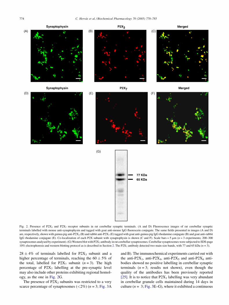
Fig. 2. Presence of P2X3 and P2X7 receptor subunits in rat cerebellar synaptic terminals. (A and D) Fluorescence images of rat cerebellar synaptic
terminals labelled with mouse anti-synaptophysin and tagged with goat anti-mouse IgG fluorescein conjugate. The same fields presented in images (A and D)
are, respectively, shown with guinea pig anti-P2X3 (B) and rabbit anti-P2X7 (E) tagged with goat anti-guinea pig IgG rhodamine conjugate (B) and goat anti-rabbit
IgG rhodamine conjugate (E). Co-localization of each P2X subunit with synaptophysin is shown (C and F). Scale bars = 5 mm (n = 3 experiments; 200–300
synaptosomes analysed by experiment). (G) Western blot with P2X7 antibody in rat cerebellar synaptosomes. Cerebellar synaptosomes were subjected to SDS-page
10% electrophoresis and western blotting protocol as is described in Section 2. The P2X7 antibody detected two main size bands, with 77 and 65 kDa (n = 3).
28 � 4% of terminals labelled for P2X3 subunit and a
higher percentage of terminals, reaching the 60 � 5% of
the total, labelled for P2X7 subunit (n = 3). The high
percentage of P2X7 labelling at the pre-synaptic level
may also include other proteins exhibiting regional homol-
ogy, as the one in Fig. 2G.
The presence of P2X2 subunits was restricted to a very
scarce percentage of synaptosomes (<2%) (n = 3; Fig. 3A
and B). The immunochemical experiments carried out with
the anti-P2X1, anti-P2X4, anti-P2X5 and anti-P2X6 anti-
bodies showed no positive labelling in cerebellar synaptic
terminals (n = 3; results not shown), even though the
quality of the antibodies has been previously reported
[25]. It is to notice that P2X1 labelling was very abundant
in cerebellar granule cells maintained during 14 days in
culture (n = 3; Fig. 3E–G), where it exhibited a continuous

C. Hervas et al. / Biochemical Pharmacology 70 (2005) 770–785 775
Fig. 3. Immunocytochemistry of P2X1 and P2X2 receptor subunits in rat cerebellar granule cells and synaptic terminals. (A and C) Fluorescence images of rat
cerebellar synaptic terminals labelled with mouse anti-synaptophysin and tagged with goat anti-mouse IgG fluorescein conjugate. The same fields presented in
images (A and C) are, respectively, shown with guinea pig anti-P2X2 (B) and rabbit anti-P2X1 (D), tagged with goat anti-guinea pig IgG rhodamine conjugate
(B) or goat anti-rabbit IgG rhodamine conjugate (D). (E) Fluorescence image of rat cerebellar granule cells (14 DIV) labelled with mouse anti-synaptophysin
and tagged with goat anti-mouse IgG fluorescein conjugate. The same field as (E) shown with rabbit anti-P2X1 and tagged with goat anti-rabbit IgG rhodamine
conjugate (F). Co-localization of P2X1 subunit with synaptophysin is shown in (G). Scale bars = 5 mm (n = 3 experiments; 200–300 synaptosomes analysed by
experiment).
distribution on the neuronal prolongations, although it
did not co-distribute with synaptophysin. This fact
could explain the absence of immunolabelling for the
P2X1 at the cerebellar synaptic terminals (n = 3; Fig. 3C
and D).
The immunolabelling of different P2X subunits at the
pre-synaptic terminals indicates the presence of the protein
but does not give information concerning their function-
ality. To approach this specific issue microfluorimetric
techniques were used.
3.3. Ca2+ responses evoked by ATP and a,b-
methylene-ATP in single cerebellar synaptic terminals
Rat cerebellar synaptosomes were tested for their ability
to mobilize Ca2+ after stimulation with ATP or a,b-meATP
in HBM medium which contains Mg2+, as described in
Section 2. In all cases, synaptic terminals were superfused
with a 30 mM KCl pulse at the end of the experiment in
order to confirm synaptosomal functionality. The mean
response in Ca2+ increase in cerebellar synaptic terminals

C. Hervas et al. / Biochemical Pharmacology 70 (2005) 770–785776
once challenged with ATP was 91.6 � 11.9 nM, exhibiting
a pronounced decay after reaching the maximal response.
This ATP response was exclusively due to extracelullar
calcium entry because there was no response when EGTA
was present in the medium (n = 4; Fig. 4A). Once demon-
strated the response to ATP, the desensitization of the pre-
synaptic P2X receptors was studied by applying a second
Fig. 4. Responses to ATP and a,b-meATP in single synaptic terminals from rat cer
superfusion chamber were loaded with Fura-2 AM to monitor changes in the in
(100 mM), in the presence or absence of EGTA and KCl (30 mM), as described in Se
during 30 s each and a time period of 1 min between consecutive stimulations. T
desensitization (C) of the receptors. (D–F) Synaptosomes were challenged with
consecutive stimulations. The three typical responses obtained are shown. In a
experiments; 300–400 synaptosomes analysed by experiment).
pulse of the agonist 1 min later. In this situation, more than
half of the synaptic terminals responding to the first stimulus
of ATP (60%), were not able to respond to the second
application (n = 4; Fig. 4B). This indicates that P2X1 or
P2X3 receptors might be involved in the ATP response.
The existence of terminals containing P2X receptors not
subjected to desensitization is shown in Fig. 4C.
ebellum. (A) Rat cerebellar synaptosomes glued to a coverslip mounted in a
trasynaptosomal Ca2+ concentration induced by agonist stimulation: ATP
ction 2. (B) Synaptosomes were stimulated with two pulses of ATP 100 mM,
wo experimental plots, showing responses with desensitization (B) or no-
a,b-meATP and ATP, for 30 s each, and a time period of 1 min between
ll cases, bars indicate the time of application of each substance (n = 4–5

C. Hervas et al. / Biochemical Pharmacology 70 (2005) 770–785 777
Fig. 5. Effect of Ip5I on the a,b-meATP responses. Intrasynaptosomal
calcium increase induced by 100 mM a,b-meATP was measured in the
presence of Ip5I. Several antagonist concentrations were tested (50 mM, 100
and 10 nM). (A) Time course plot of the intrasynaptosomal calcium
concentration recorded from a representative single synaptic terminal.
The antagonist Ip5I 50 mM was applied 2 min before and during the agonist
stimulation. Synaptosomes were washed for at least 10 min before applying
a second pulse with the agonist a,b-meATP. This time is enough to let
receptors to recover from their desensitization period. At the end, a pulse of
30 mM KCl was applied to test synaptosomal functionality. Stimulation
period is indicated by solid bars. (B and C) An experiment similar to that
showed in (A) but with an antagonist concentration of 100 and 10 nM,
respectively. The number of individual synaptic terminals under study was
300–400 per experiment (n = 3).
As the nucleotide analogue, a,b-meATP, is a more
specific agonist for the homomeric P2X1 and P2X3, or
the heteromeric P2X2/P2X3 receptors, similar experiments
as those with ATP were carried out. The responses recorded
in single synaptic terminals after stimulation with a-b-
meATP are shown in Fig. 4D–F. The maximal calcium
increase was similar to the one induced by ATP and a
similar pronounced decay was observed after reaching the
maximal response. Heterologous desensitization studies
were carried out by applying a second pulse of the agonist
ATP 1 min after the previous application, as it is shown in
Fig. 4D–F (n = 4). More than half of the terminals,
responding to a,b-meATP, were not able to respond to a
second challenge of nucleotide agonist. In addition it is to
emphasize that there are terminals that did not respond to
the first stimulation of a,b-meATP, and as a consequence,
they were not desensitized to a further ATP response. Thus,
pre-synaptic terminals exhibit various possibilities con-
cerning the presence of P2X receptors. As there is no
labelling of P2X1 at the pre-synaptic level, P2X3 is one of
the most probable receptor to be present in these structures,
and capable of desensitization.
To confirm the abundant presence of P2X3 receptors the
inhibitory effect of Ip5I was studied in single synaptic
terminals. As shown in Fig. 5A, 50 mM Ip5I completely
blocked the response to a,b-meATP in isolated synaptic
terminals (n = 3). The same situation occurred when
100 nM Ip5I was employed (Fig. 5B; n = 3). However,
when 10 nM Ip5I was used, a concentration that inhibits
P2X1 but not interferes with P2X3, most of the terminals
were able to respond to a,b-meATP in the presence of the
antagonist (Fig. 5C; n = 3). They were also able to respond
to a second pulse of a,b-meATP 10 min later, once desen-
sitization period had finished.
3.4. Effect of Mg2+ ions on the Ca2+ responses evoked
by ATP in single cerebellar synaptic terminals
Experiments addressed to find functional P2X7 receptors
at the pre-synaptic level were performed. In this way and as
shown in Fig. 6A, the synaptic terminals were identified
according to their responses to ATP in the presence or
absence of Mg2+. It is generally accepted that deprivation
of this ion allows a better response of P2X7 receptors.
There were terminals that only respond to ATP in a
medium with Mg2+, as it was the case of the terminal
labelled as 1 (n = 4; Fig. 6A and B), as could be expected
from the results reported in the former section. There were
also synaptic terminals that responded to ATP in Mg2+-free
medium but not in the presence of this ion, as it was the
case of the one labelled as 2 (Fig. 6A and B), these being
the most abundant. Besides, other synaptic terminals
responded to both pulses of ATP whether in presence or
absence of magnesium ions in the medium and the terminal
labelled as 3 is an example. Finally, there were terminals
that do not show ATP response neither in the presence or
absence of Mg2+ ions. This lack of response could be due to
several factors: first, the absence of nucleotide receptors,
second, a response below the limit of detection, or third, the
presence of receptors being activated by different nucleo-
tide agonists, or exhibiting very specific requirements.
These results confirm the ones reported in previous section

C. Hervas et al. / Biochemical Pharmacology 70 (2005) 770–785778
Fig. 6. Responses to ATP in the absence or presence of Mg2+ ions in single synaptic terminals from rat cerebellum. Rat cerebellar synaptosomes glued to a
coverslip mounted in a superfusion chamber were loaded with Fura-2 AM to monitor changes in the intrasynaptosomal Ca2+ concentration induced by ATP
(100 mM), in the presence or absence of Mg2+, and KCl (30 mM), as described in Section 2. (A) Representative traces show the time course of intrasynaptosomal
calcium concentration recorded from the terminals labelled as 1–3 in the fluorescence image of synaptosomes loaded with Fura-2 (and shown in B). Bars
indicate the time of application of each substance. Scale bar = 3 mm. (C) Calcium concentration increases induced by ATP in the presence or absence of Mg2+ in
synaptic terminals, that correspond, respectively, to the response types 1–3 shown in panel (A). Each bar represents the mean � S.E.M. of four experiments
performed in different synaptosomal preparations, and 300–400 synaptosomes were analysed by preparation. **p < 0.01, very significantly different from the
mean of the response to ATP in population 3, using one tailed Student0s paired test.
concerning the complexity of nucleotide responses at the
pre-synaptic level.
Considering the single synaptic terminals responses
from the experimental situation reported in Fig. 6A, the
terminals responding exclusively to ATP in the absence of
Mg2+ (type 2), accounted for three times higher number
with respect the other types. It is to notice that the high
number of terminals responding to ATP in Mg2+-free
medium supports the abundant existence of functional
P2X7.
C. Hervas et al. / Biochemical Pharmacology 70 (2005) 770–785 779
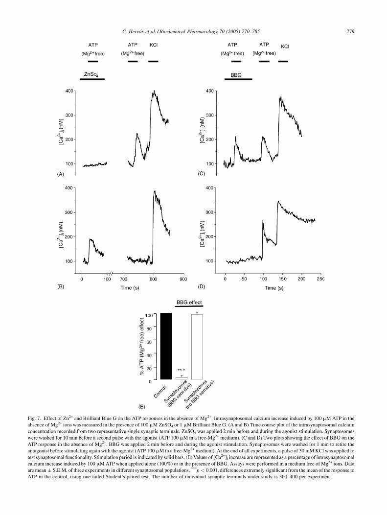
Fig. 7. Effect of Zn2+ and Brilliant Blue G on the ATP responses in the absence of Mg2+. Intrasynaptosomal calcium increase induced by 100 mM ATP in the
absence of Mg2+ ions was measured in the presence of 100 mM ZnSO4 or 1 mM Brilliant Blue G. (A and B) Time course plot of the intrasynaptosomal calcium
concentration recorded from two representative single synaptic terminals. ZnSO4 was applied 2 min before and during the agonist stimulation. Synaptosomes
were washed for 10 min before a second pulse with the agonist (ATP 100 mM in a free-Mg2+ medium). (C and D) Two plots showing the effect of BBG on the
ATP response in the absence of Mg2+. BBG was applied 2 min before and during the agonist stimulation. Synaptosomes were washed for 1 min to retire the
antagonist before stimulating again with the agonist (ATP 100 mM in a free-Mg2+ medium). At the end of all experiments, a pulse of 30 mM KCl was applied to
test synaptosomal functionality. Stimulation period is indicated by solid bars. (E) Values of [Ca2+]i increase are represented as a percentage of intrasynaptosomal
calcium increase induced by 100 mM ATP when applied alone (100%) or in the presence of BBG. Assays were performed in a medium free of Mg2+ ions. Data
are mean � S.E.M. of three experiments in different synaptosomal populations. ***p < 0.001, differences extremely significant from the mean of the response to
ATP in the control, using one tailed Student’s paired test. The number of individual synaptic terminals under study is 300–400 per experiment.

C. Hervas et al. / Biochemical Pharmacology 70 (2005) 770–785780
Fig. 8. Dose–response curves for ATP and BzATP in single synaptic
terminals from rat cerebellum. Dose–responses curves for ATP or BzATP
were obtained both in the presence of 1 mM Mg2+or in the absence of such
ion. Synaptosomes isolated from rat cerebellum and glued to a coverslip
mounted in a superfusion chamber were loaded with Fura-2 AM. Changes in
the intrasynaptosomal Ca2+ concentration induced by graded doses of ATP
in the presence or absence of Mg2+(A), or BzATP in the presence or absence
of Mg2+(B), concentrations ranging from 1 nM to 1 mM, were monitored.
Synaptic terminals were stimulated with 30 mM KCl at the end of the
experiment to test their functionality. Calcium increase values are the
mean � S.E.M. for each agonist concentration of at least three experiments
performed in different synaptosomal preparations (n = 3–4) and number of
individual synaptic terminals tested between 315 and 457.
It is noteworthy to say that the Ca2+ response intensity
elicited by ATP (with or without Mg2+ in the medium)
varied between terminals. Calibration of fluorescence ratio
to get intrasynaptosomal concentration [Ca2+]i (see Section
2), showed a basal value of 85.6 � 7.2 nM. The Ca2+
increase induced by ATP and ATP without Mg2+, at
100 mM concentration, in terminals responding only to
one of these agonists, showed values of 87.4 � 10.9 and
109.4 � 12.7 nM, respectively. However, terminals that
responded to ATP, both in the presence and absence of
Mg2+, exhibit an ATP response in the absence of this ion
significantly higher than the one for ATP with Mg2+, being
183.2 � 17.7 and 96.3 � 12.1 nM, respectively (Fig. 6C).
These values were not modified when ATP-free Mg2+ was
the first agonist to be used in the perfusion system (results
not shown).
As Zn2+ ions have been described as inhibitors of P2X7
receptors, studies were carried out in the presence of
ZnSO4. In Fig. 7A, it can be observed that Zn2+ ions
completely abolished the ATP response in the absence
of Mg2+. These data correspond to a classical P2X7 recep-
tor. However, the terminals responding to Mg2+-free ATP,
when Zn2+ ions were present, did not respond in the
absence of this ion (Fig. 7B). This behaviour has been
reported for P2X3 receptors, where Zn2+ ions improve the
response.
The effect of Brilliant Blue G (BBG), a selective
antagonist for P2X7 receptors, was measured in single
terminals. The antagonist was applied in HBM medium,
and it was kept in contact with synaptic terminals in the
perfusion chamber for a period of 2 min before, and during
the first pulse with the agonist. ATP without Mg2+ was
always employed as the agonist in these experiments. To
check the recovery after inhibitory treatment the terminals
were washed for another minute with HBM media and
challenged again with a second pulse of ATP in the absence
of Mg2+. The inhibition range can only be quantified when
the inhibitory effects are studied in single synaptic term-
inals, with experiments as those reported in Fig. 7C and D,
which shows typical experimental recordings. This tech-
nique has allowed us to identify two different populations
according to the BBG effect. In one population, the
response to 100 mM ATP in the absence of Mg2+ was
completely inhibited by BBG 1 mM, accounting for the
47 � 11% of synaptosomes (n = 3; Fig. 7E). In the other
population, BBG did not have inhibitory effect and corre-
sponds to 50 � 8% of synaptosomes (n = 3; Fig. 7E).
These results as the ones in the previous section points
to the large variety and possibilities of nucleotide receptors
at the pre-synaptic level.
3.5. Dose–response curves for ATP and
BzATP in single terminals
Microfluorimetric techniques have also allowed to carry
out the dose–response curves in single terminals. Synaptic
terminals were challenged with increasing concentrations
of the agonists ATP and BzATP between 1 nM and 1 mM
in the perfusion medium. The experiments were carried out
in the presence or absence of magnesium ions, and the
curves obtained have been represented in Fig. 8 (n = 3–4).
The dose–response curves showed some differences in
affinity and maximum effect with Mg2+ withdrawal. Con-
cerning ATP, EC50 values were 39.4 � 0.4 mM in the
presence of Mg2+ and close to two orders of magnitude
lower, 0.3 � 0.1 mM, in a free-Mg2+ medium. Besides, the
maximum effect for ATP without Mg2+ was higher than
that obtained in the presence of the ion, with calcium
values of 132.8 � 12.9 and 91.6 � 11.9 nM, respectively

C. Hervas et al. / Biochemical Pharmacology 70 (2005) 770–785 781
Table 1
EC50 values, maximal response and Hill numbers for ATP and BzATP concentration–response curves in the absence or presence of Mg2+ in synaptic terminals
from rat cerebellum
EC50 (mM) Maximal response (nM) nH
ATP (+Mg2+) n = 3 (n0 = 315) 39.4 � 0.4 91.6 � 11.9 1.66 � 0.26
ATP (Mg2+ free) n = 4 (n0 = 443) 0.3 � 0.1 132.9 � 12.9 0.63 � 0.10
BzATP (+Mg2+) n = 3 (n0 = 364) 1.4 � 0.5 104.3 � 9.4 1.20 � 0.47
BzATP (Mg2+ free) n = 4 (n0 = 457) 4.2 � 0.9 169.7 � 17.1 0.77 � 0.11
Data are expressed as the EC50 values (concentration of agonist producing 50% of the maximal response) and maximal responses induced by ATP and BzATP in the
different experimental conditions. nH is the Hill coefficient. Data represents the mean � S.E.M. (n = number of experiments; n0 = total of synaptosomes tested).
(Fig. 8A). In a similar way as happened with ATP, the
maximum effect induced by BzATP was higher in free-
Mg2+ conditions, the values being 104.3 � 9.3 and
169.6 � 17.1 nM in the presence or absence of the ion,
respectively (Fig. 8B).
Fig. 9. Correlation between BzATP responses in the absence of Mg2+ ions an
synaptosomes glued to a coverslip mounted in a superfusion chamber were loa
concentration induced by stimulation with two different concentrations of BzATP
applied at the end of the experiment to confirm synaptosomes functionality.
immunocytochemical characterization. (A) Time course of Fura-2 fluorescence e
B. Solid bars indicate the period while each substance was applied. (B) Fluorescen
(B) labelled with anti-P2X7 antibodies and tagged with anti-rabbit-TRICT-co
synaptophysin is shown (n = 3; bar = 5 mm).
Concerning the Hill number both nucleotides in the
presence of Mg2+ exhibit a slight co-operative behaviour
no observable in the absence of the ion. The values of EC50,
maximum effect and Hill number are summarized in
Table 1.
d presence of P2X7 subunits in single synaptic terminals. Rat cerebellar
ded with Fura-2 AM to monitor changes in the intrasynaptosomal Ca2+
(5 and 100 mM), in the absence of Mg2+ ions. A pulse of KCl (30 mM) was
After these studies synaptosomes were fixed with p-formaldehyde for
mission changes recorded for the terminal indicate with an arrow in panel
ce image of synaptophysin-FITC labelled synaptosomes. (C) Same field as
upled secondary antibodies. (D) Co-localization of P2X7 subunit with

C. Hervas et al. / Biochemical Pharmacology 70 (2005) 770–785782
3.6. Correlation between BzATP responses in the
absence of Mg2+ and immunolabelling of P2X7
receptors in single terminals
To determine whether a co-localization between P2X7
immunolabelling and the calcium responses to the better
P2X7 agonist (BzATP in the absence of Mg2+) occurs, we
combined microfluorimetric studies with immunocyto-
chemical assays.
Isolated synaptic terminals loaded with Fura-2 AM were
stuck on microgrid coverslips (square size 55 mm) to
relocate individual synaptosomes after immunostaining
procedures. Synaptosomes were tested for their ability
to mobilize Ca2+ in response to BzATP in the absence
of Mg2+. Two consecutive pulses with the nucleotide, the
first of 5 mM and the second one of 100 mM, were
employed, with 30 s duration and no less than 1 min time
interval between applications (Fig. 9A). The 5 and 100 mM
BzATP concentrations were selected because they corre-
spond to values close to one and twenty times the EC50
value, respectively. After functional studies synaptosomes
were fixed and labelled with antibodies that recognize
specifically either the vesicular protein synaptophysin or
the P2X7 receptor (n = 3; Fig. 9B–D).
These analyses showed that the great majority of the
synaptic terminals responding to BzATP in the absence of
Mg2+, 91 � 6%, were further positively labelled with the
anti-P2X7-subunit antibody.
4. Discussion
The present study demonstrates the existence and large
variety of functional P2X receptors in rat cerebellar synap-
tosomal preparations by means of immunocytochemical
and microfluorimetric techniques.
In cerebellum, 90% of neurons are granule cells, which
contact Purkinje neurons using glutamate as excitatory
neurotransmitter. According with this feature, a high per-
centage that accounts for two thirds of the total number of
synaptic terminals, isolated from rat cerebellum, have
glutamatergic phenotype. This approach is taking into
consideration that in most CNS regions, VGLUT1 and
VGLUT2 expression appears not to overlap, defining
separate populations of synaptic terminals in adult animals
[32,33]. This distribution is consistent with our previous
studies that showed abundant and punctate VGLUTl
and VGLUT2 labelling in cerebellar granule cells [25].
In cerebellum, there are also terminals that exhibit
GABAergic phenotype, labelled with anti-GAD antibo-
dies, accounting for about one-third of the total. This result
agrees with the existence of Purkinje, stellate, basquet and
Golgi neurons in cerebellum, which are GABAergic.
VGLUT1 and GAD labelling does not overlap, showing
that the glutamatergic or GABAergic phenotype does not
co-exist in the same terminal.
Results from immunocytochemical and microfluori-
metric techniques, demonstrated the presence and func-
tionality of various P2X receptors in cerebellar
preparations. Some comments are necessary concerning
the P2X1 subunit, which was not detected by immunola-
belling at the pre-synaptic level. However, in the same
experimental conditions reported here, P2X1 immunola-
belling clearly appears in whole granule cells in culture
with the same antibody, but not exhibiting co-distribution
with the synaptic marker synaptophysin. Presence of P2X1
immunolabelling has also been described in the varicos-
ities of cerebellar parallel fibers and other CNS regions
[3,8,34]. Functional homomeric P2X1 receptors have been
reported in rodent vas deferens and smooth muscle tissue
[5]. These data are relevant concerning the pre-synaptic
calcium responses to a,b-meATP, and the P2X subunits
mediating the response.
By immunolabelling, P2X3 receptor subunits are
detected in a percentage close to one quarter of the total
cerebellar synaptic terminals. The presence of P2X3 sub-
units has been reported in many areas of central nervous
system, such as at the hypothalamic supraoptic nucleus and
solitary tract nucleus, and at the pre-synaptic level of nerve
terminals of sensory pathways to the spinal cord dorsal
horn, and also at the peripheral endings [3,8,35]. Synaptic
terminals from rat midbrain also exhibit abundant labelling
by P2X3 antibodies [31]. It is relevant to emphasize that in
cultured granule cells P2X3 labelling co-distributes with
the vesicular pre-synaptic marker synaptophysin [25]. In
this model, the presence of P2X2 subunit appears more
restricted to the cell soma, as it is also the case for P2X4.
Only scarce terminals are immunolabelled for P2X2 sub-
unit in our model, being P2X4 subunit not detectable at the
pre-synaptic level. Similar abundance and distribution of
P2X2 and P2X3 subunits have been reported in sensory
nerve endings, which is not the case in cerebellar synaptic
terminals [36,37].
The characterization with anti-P2X7 antibodies is sub-
mitted to much controversy, since it has been reported that
the P2X7-KO mice exhibits the same labelling character-
istics in brain tissues for this subunit, as the wild animal
[26]. It is possible that, in cerebellar synaptic terminals, in
addition to the whole length P2X7 labelling protein, other
analogous proteins containing the antigenic epitope could
be responsible for the additional band detected by western
studies. This could explain the abundant presence of more
than half of the total terminals that exhibit P2X7 labelling.
Microfluorimetric studies of calcium responses in single
synaptic terminals are necessary to confirm the existence of
functional P2X receptors at the pre-synaptic level. On the
basis of the P2X subunits present at the synaptic terminals,
the responses to a,b-meATP and the desensitization results
agree with the existence of functional homomeric P2X3
receptors. As, in the presence of Mg2+ ions, more than half
of the ATP and a,b-meATP responding terminals, exhibit
homologous or heterologous desensitization, the abundant

C. Hervas et al. / Biochemical Pharmacology 70 (2005) 770–785 783
presence of P2X3 subunits assembled as homomeric recep-
tors can be inferred at the pre-synaptic level. Moreover, the
existence of no-desensitising nucleotide responses also
indicates the presence of heteromeric P2X2/P2X3 or homo-
meric P2X2 receptors located at the pre-synaptic level
[5,8,35,36,38,39]. However, it is to take into account the
scarce presence of P2X2 subunits detected by immunocy-
tochemistry. Presence of functional P2X3 homomeric and
P2X2/P2X3 heteromeric receptors has also been reported in
midbrain synaptic terminals [31,40]. At this respect, the
inhibitory effect of Ip5I, and the concentration required for
such inhibition confirms the presence of P2X3 receptors at
the pre-synaptic level in rat cerebellum [30]. The presence
of other P2X receptors not yet cloned, or the currently
described but post-transcripcionally modified, needing
more specific functional requirements, are not excluded
at the pre-synaptic level.
In cerebellar synaptic terminals, the abundant presence
of Ca2+ responses to ATP or BzATP that improve when
Mg2+ ions are not present, agrees with the P2X7 phenotype,
and as reported here, more than half of these structures
exhibit P2X7 or P2X7-like immunolabelling. The inhibi-
tory Mg2+ effect can be explained by an allosteric inhibi-
tion mediated by divalent ions binding the receptor, among
other hypothesis [41]. However, it is to emphasize that
dose–response curves showed cooperativity when Mg2+
was present. This effect could be understood as a way to
optimize the effect of the ATP with respect to ATP4�, a less
abundant molecular form in natural conditions.
The fact that BzATP is more potent than ATP in our
cerebellar preparations is another distinctive feature of
P2X7 receptors, according with the effect of BzATP
described by other authors [16]. Although BzATP is an
effective agonist at other P2X receptors, in particular,
P2X1, P2X2 and P2X3, it is only at the P2X7 receptor that
BzATP is more potent than ATP [5].
The inhibitory effect of Zn2+ ions in the absence of Mg2+
agrees with the presence of P2X7 receptors at the synaptic
terminals [41]. Moreover, in terminals where no responses
to ATP were observed, Zn2+ was able to induce a response
to this agonist, indicating that a more favourable situation
for a P2X3 response was being established, thus confirming
once more the presence of P2X3 receptors at the pre-
synaptic level [42].
Studies with Brilliant Blue G, a selective P2X7 antago-
nist [43], well agrees with other works about the P2X7
antagonism, inhibiting calcium influx induced by ATP in
the absence of Mg2+ at concentrations previously reported
to be effective [44,45]. The concentration used is expected
to produce low inhibitory effect on rat P2X2 receptors; in
addition, this subunit is scarcely expressed in the cerebellar
synaptosomes. Moreover, it is to take into account that half
of the responses were completely abolished by BBG, as it
could be expected from the P2X7 properties reported in the
literature [13,22,23,41]. However, no inhibition at all was
observed on the other half of the ATP-Mg2+ free respond-
ing terminals. These data agree, once more, with the great
heterogeneity of P2X receptors present at the nucleotide
responding terminals.
In addition to the functional properties present in P2X7
receptors in cerebellum, their particular subcelullar dis-
tribution also resembles that found in other systems. P2X7,
or its brain analogue, is usually expressed in a dotting
pattern, indicating association with specialized structures
at synapsis [12,14–16,46]. In contrast with other P2X
subunits that can be found pre-synaptically and postsy-
naptically, P2X7 receptors are more prevalent at pre-synap-
tic sites [13,23]. Since the pre-synaptic nerve terminal is a
fundamental regulatory site to control and modulate neu-
rosecretion, the pre-synaptic P2X7, or P2X7-like, charac-
teristic distribution suggests a prominent role of P2X7 in
the signalling of cerebellum.
In addition, P2X7 receptors are mainly targeted to
excitatory terminals of neurons, associated with VGLUT1
and/or VGLUT2 glutamatergic markers. In fact, P2X7
subunits co-localize with both VGLUT1 and VGLUT2
at the granular layer in cerebellum. However, in certain
brainsteam and spinal cord nuclei the P2X7 may be adit-
tionally expressed by subpopulations of cholinergic and
GABAergic/glycinergic terminals, suggesting a role in
neurotransmission in different synaptic terminals [22].
However, it is not clear how the high ATP concentrations
necessary to activate the P2X7 receptor could reach the
synaptic cleft. On one hand, ATP released from damaged
cells or under inflammatory conditions could be a possible
source to achieve high local ATP concentrations, in milli-
molar range, required to activate P2X7 receptors taking
into account that Mg2+ ions are present in extracellular
media surrounding neurons. ATP is also released from
neural tissues under ischemia or anoxia [47]. P2X7 may
also play an important role in excitotoxicity in response to
cellular damage by increasing glutamate release and it
could be a therapeutic target to reduce stress induced cell
death and in neurodegenerative diseases [48,49].
As ATP is released with other neurotransmitters at the
synapsis, this could be the way to achieve the high
concentration of ATP necessary to activate the receptor
in a restricted and small region near the P2X7 receptor.
The fact that P2X7 is expressed close to glutamate
transporters and the evidences that ATP evokes glutamate
release in cerebellar granule cells [50] supports the
hypothesis that P2X7 would be guaranteeing or reinfor-
cing the neurotransmission. The calcium increases
mediated by P2X7 could further induce more glutamate
release or initiate second messenger cascades that might
lead to synapsis reorganization. But in an opposite way,
activation of P2X7 present at mossy fibers in the rat
hippocampus produced a long-lasting inhibition of neu-
rotransmission, maybe as a protection mechanism when
neurotransmission is unusually high [14], suggesting that
P2X7 receptor accomplishes for diverse roles in different
synapsis.

C. Hervas et al. / Biochemical Pharmacology 70 (2005) 770–785784
The pre-synaptic distribution of P2X receptors, mainly
those of P2X3 and P2X7 containing subunits, could indi-
cate a role in synergist action with other neurotransmitter
receptors regulating synaptic transmission efficiency.
Interactions with metabotropic, such as GABAB and ade-
nosine A1 and A2 receptors as also with ionotropic, such as
nicotinic and GABAA receptors, have been described for
the P2X receptors at the pre-synaptic level [37,51–53]. The
cross-talk among P2X and other metabotropic and iono-
tropic receptors may be contributing to long-term plasticity
in response to neuronal activity, at the pre-synaptic level.
Acknowledgements
This work was supported by research grants from the
Spanish Ministry of Science and Technology BFI2002-
03626) and the C.A.M. (SAL/0551/2004). We would like
to thank Dr. J. Gualix for the synthesis and supply of Ip5I.
References
[1] Ralevic V, Burnstock G. Receptors for purines and pyrimidines.
Pharmacol Rev 1998;50:413–92.
[2] North RA, Surprenant A. Pharmacology of cloned P2X receptors.
Annu Rev Pharmacol Toxicol 2000;40:563–80.
[3] Norenberg W, Illes P. Neuronal P2X receptors: localisation and
functional properties. Naunyn-Schmiedebergs Arch Pharmacol
2000;362:324–39.
[4] Torres GE, Egan TM, Voigt MM. Hetero-oligomeric assembly of P2X
receptor subunits. Specificities exist with regard to possible partners. J
Biol Chem 1999;274:6653–9.
[5] North RA. Molecular physiology of P2X receptors. Physiol Rev
2002;82:1013–67.
[6] Edwards FA, Gibb AJ, Colquhoun D. ATP receptor-mediated synaptic
currents in the central nervous system. Nature 1992;359:144–7.
[7] Pankratov Y, Castro E, Miras-Portugal MT, Krishtal O. A purinergic
component of the excitatory postsynaptic current mediated by P2X
receptors in the CA1 neurons of the rat hippocampus. Eur J Neurosci
1998;10:3898–902.
[8] Illes P, Ribeiro JA. Molecular physiology of P2 receptors in the central
nervous system. Eur J Pharmacol 2004;483:5–17.
[9] Pintor J, Miras-Portugal MT. A novel receptor for diadenosine poly-
phosphates coupled to calcium increase in rat midbrain synaptosomes.
Br J Pharmacol 1995;115:895–902.
[10] Gomez-Villafuertes R, Gualix J, Miras-Portugal MT, Pintor J. Ade-
nosine 50-tetraphophate (Ap4), a new agonist on rat midbrain synaptic
terminal P2 receptors. Neuropharmacology 2000;39:2381–90.
[11] Dıaz-Hernandez M, Pintor J, Castro E, Miras-Portugal MT. Indepen-
dent receptors for diadenosine pentaphosphate and ATP in rat mid-
brain single synaptic terminals. Eur J Neurosci 2001;14:918–26.
[12] Miras-Portugal MT, Dıaz-Hernandez M, Giraldez L, Hervas C,
Gomez-Villafuertes R, Sen RP, et al. P2X7 receptors in rat brain:
presence in synaptic terminals and granule cells. Neurochem Res
2003;28:1597–605.
[13] Deuchars SA, Atkinson L, Brooke RE, Musa H, Milligan CJ, Batten
TFC. et al. Neuronal P2X7 receptors are targeted to presynaptic
terminals in the central and peripheral nervous system. J Neurosci
2001;21:7143–52.
[14] Amstrong JN, Brust TB, Lewis RG, MacVicar BA. Activation of
presynaptic P2X7-like receptors depresses mossy fiber-CA3 synaptic
transmission through p38 mitogen-activated protein kinase. J Neurosci
2002;22:5938–45.
[15] Sperlagh B, Kofalvi A, Deuchars J, Atkinson L, Milligan CJ, Buckley
NJ, et al. Involvement of P2X7 receptors in the regulation of neuro-
transmitter release in the rat hippocampus. J Neurochem 2002;81:
1196–211.
[16] Lundy PM, Hamilton MG, Mi L, Gong G, Vair C, Sawyer TW, et al.
Stimulation of Ca2+ influx through ATP receptors on rat brain synapto-
somes: identification of functional P2X7 receptors subtypes. Br J
Pharmacol 2002;135:1616–26.
[17] Gu JG, Macdermott AB. Activation of ATP P2X receptors elicits
glutamate release from sensory neuron synapsis. Nature 1997;389:
749–53.
[18] Hugel S, Schlichter R. Presynaptic P2X receptors facilitate inhibitory
GABAergic transmission between cultured rat spinal cord dorsal horn
neurons. J Neurosci 2000;20:2121–30.
[19] Gomez-Villafuertes R, Gualix J, Miras-Portugal MT. Single GABAer-
gic synaptic terminals from rat midbrain exhibit functional P2X and
dinucleotide receptors, able to induce GABA secretion. J Neurochem
2001;77:84–93.
[20] Dıaz-Hernandez M, Pintor J, Castro E, Miras-Portugal MT. Co-
localisation of functional nicotinic and ionotropic nucleotide receptors
in isolated cholinergic synaptic terminals. Neuropharmacology
2002;42:20–33.
[21] Gualix J, Gomez-Villafuertes R, Dıaz-Hernandez M, Miras-Portugal
MT. Presence of functional ATP and dinucleotide receptors in gluta-
matergic synaptic terminals from rat midbrain. J Neurochem
2003;87:160–71.
[22] Moores TS, Hasdemir B, Vega-Riveroll L, Deuchars J, Parson SH.
Properties of presynaptic P2X(7)-like receptors at the neuromuscular
junction. Brain Res 2005;1034:40–50.
[23] Ireland MF, Noakes PG, Bellingham MC. P2X7-like receptor subunits
enhance excitatory synaptic transmission at central synapses by pre-
synaptic mechanisms. Neuroscience 2004;128:269–80.
[24] Garcıa-Lecea M, Sen RP, Soto F, Miras-Portugal MT, Castro E. P2
receptors in cerebellar neurons: molecular diversity of ionotropic ATP
receptors in Purkinje cells. Drug Dev Res 2001;52:104–13.
[25] Hervas C, Perez-Sen R, Miras-Portugal MT. Co-expression of func-
tional P2X and P2Y nucleotide receptors in single cerebellar granule
cells. J Neurosci Res 2003;73:384–99.
[26] Hervas C. Receptores de nucleotidos en neuronas granulares de
cerebelo de rata. Tipos y senalizacion. Tesis Doctoral. Universidad
Complutense de Madrid; 2004.
[27] Sim JA, Young MT, Sung HY, North RA, Surprenant A. Reanalysis
of P2X7 receptor expression in rodent brain. J Neurosci 2004;24:
6307–14.
[28] Dunkley PR, Jarvie PE, Heath JW, Kidd GJ, Rostas JA. A rapid
method for isolation of synaptosomes on Percoll gradients. Brain Res
1986;372:115–29.
[29] Grynkiewicz G, Poenie M, Tsien RY. A new generation of Ca2+
indicators with greatly improved fluorescence properties. J Biol Chem
1985;260:3440–50.
[30] Pintor J, Gualix J, Miras-Portugal MT. Diinosine polyphosphates, a
group of dinucleotides with antagonistic effects on diadenosine poly-
phosphate receptor. Mol Pharmacol 1996;51:277–84.
[31] Dıaz-Hernandez M, Gomez-Villafuertes R, Hernando F, Pintor J,
Miras-Portugal MT. Presence of different ATP receptors on rat mid-
brain single synaptic terminals. Involvement of the P2X(3) subunits.
Neurosci Lett 2001;301(3):159–62.
[32] Herzog E, Bellenchi GC, Gras C, Bernard V, Ravassard P, Bedet C, et al.
The existence of a second vesicular glutamate transporter specifies
subpopulations of glutamatergic neurons. J Neurosci 2001;21:RC181.
[33] Varoqui H, Schafer MK, Zhu H, Weihe E, Erickson JD. Identification
of the differentiation-associated Na+/PI transporter as a novel vesi-
cular glutamate transporter expressed in a distinct set of glutamatergic
synapses. J Neurosci 2002;22:142–55.

C. Hervas et al. / Biochemical Pharmacology 70 (2005) 770–785 785
[34] Loesch A, Burnstock G. Electron-immunocytochemical localization
of P2X1 receptors in the rat cerebellum. Cell Tissue Res 1998;294:
253–60.
[35] Gu JG, Heft MW. P2X receptor mediated purinergic sensory
pathways to the spinal cord dorsal horn. Purinergic Signall 2004;1:
11–6.
[36] Lewis C, Neidhart S, Holy C, North RA, Buell G, Surprenant A.
Coexpression of P2X2 and P2X3 receptor subunits can account for
ATP-gated currents in sensory neurons. Nature 1995;377:432–5.
[37] Labrakakis C, Tong CK, Weissman T, Torsney C, MacDermott AB.
Localization and function of ATP and GABAA receptors expressed by
nociceptors and other postnatal sensory neurons in rat. J Physiol
2003;549:131–42.
[38] Tsuda M, Koizumi S, Kita A, Shigemoto Y, Ueno S, Inoue K.
Mechanical allodynia caused by intraplantar injection of P2X receptor
agonist in rats: involvement of heteromeric P2X2/3 receptor signaling
in capsaicin-insensitive primary afferent neurons. J Neurosci 2000;20:
RC90.
[39] Jiang LH, Kim M, Spelta V, Bo X, Surprenant A, North RA. Subunit
arrangement in P2X receptors. J Neurosci 2003;23:8903–10.
[40] Gomez-Villafuertes R, Pintor J, Gualix J, Miras-Portugal MT. GABAB
receptor-mediated presynaptic potentiation of ATP ionotropic recep-
tors in rat midbrain synaptosomes. Neuropharmacology 2003;44:311–
23.
[41] Virginio C, Church D, North RA, Surprenant A. Effects of divalent
cations, protons and calmidazolium at the rat P2X7 receptor. Neuro-
pharmacology 1997;36:1285–94.
[42] King BF, Townsend-Nicholson A. Nucleoside and nucleotide recep-
tors. Tocris Rev 2003;23:1–11.
[43] Jiang LH, Mackenzie AB, North RA, Surprenant A. Brilliant Blue G
selectively blocks ATP-gated rat P2X(7) receptors. Mol Pharmacol
2000;58:82–8.
[44] Hibell AD, Thompson KM, Xing M, Humphrey PP. Michel AD
Complexities of measuring antagonist potency at P2X(7) receptor
orthologs. J Pharmacol Exp Ther 2001;296:947–57.
[45] Allgaier C, Reinhardt R, Schadlich H, Rubini P, Bauer S, Reichenbach
A, et al. Somatic and axonal effects of ATP via P2X2 but not P2X7
receptors in rat thoracolumbar sympathetic neurones. J Neurochem
2004;90:359–67.
[46] Atkinson L, Batten TF, Moores TS, Varoqui H, Erickson JD, Deuchars
J. Differential co-localisation of the P2X7 receptor subunit with
vesicular glutamate transporters VGLUT1 and VGLUT2 in rat
CNS. Neuroscience 2004;123:761–8.
[47] Le Feuvre RA, Brough D, Touzani O, Rothwell NJ. Role of P2X7
receptors in ischemic and excitotoxic brain injury in vivo. J Cereb
Blood Flow Metab 2003;23:381–4.
[48] Doble A. The role of excitotoxicity in neurodegenerative disease:
implications for therapy. Pharmacol Ther 1999;81:163–221.
[49] Neary JT, Kang Y, Willoughby KA, Ellis EF. Activation of extra-
cellular signal-regulated kinase by stretch-induced injury in astrocytes
involves extracellular ATP and P2 purinergic receptors. J Neurosci
2003;23:2348–56.
[50] Merlo D, Volonte C. Binding and functions of extracellular ATP in
cultured cerebellar granule neurons. Biochem Biophys Res Commun
1996;225:907–14.
[51] Khakh BS, Zhou X, Sydes J, Galligan JJ, Lester HA. State-dependent
cross-inhibition between transmitter-gated cation channels. Nature
2000;406:405–10.
[52] Dıaz-Hernandez M, Sanchez-Nogueiro J, Pintor J, Miras-Portugal MT.
Interaction between dinucleotide and nicotinic receptors in individual
cholinergic terminals. J Pharmacol Exp Ther 2004;311.
[53] Sokolova E, Nistri A, Giniatullin R. Negative cross-talk between
anionic GABAA and cationic P2X ionotropic receptors of rat dorsal
root ganglion neurons. J Neurosci 2001;21:4958–68.