Embed Size (px)
Citation preview
Tishk International University
Faculty of Pharmacy
Lecture series in Normal Histology for Undergraduate Medical students
PREPARED BY :
Prof.Dr. Hiwa B M Banna , Mphil., Ph.D ( London University)
1
2
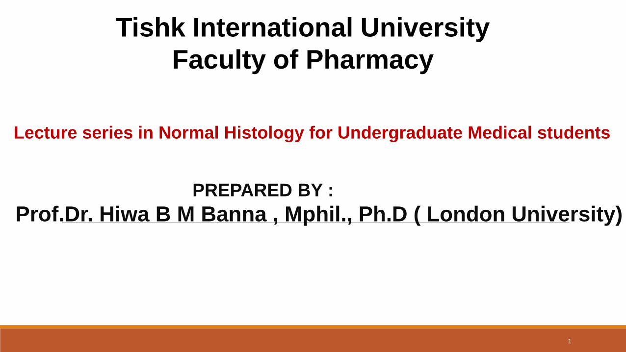
The Integumentary System (skin and its appendages:
Hairs, nail, sebaceous and sweat glands).
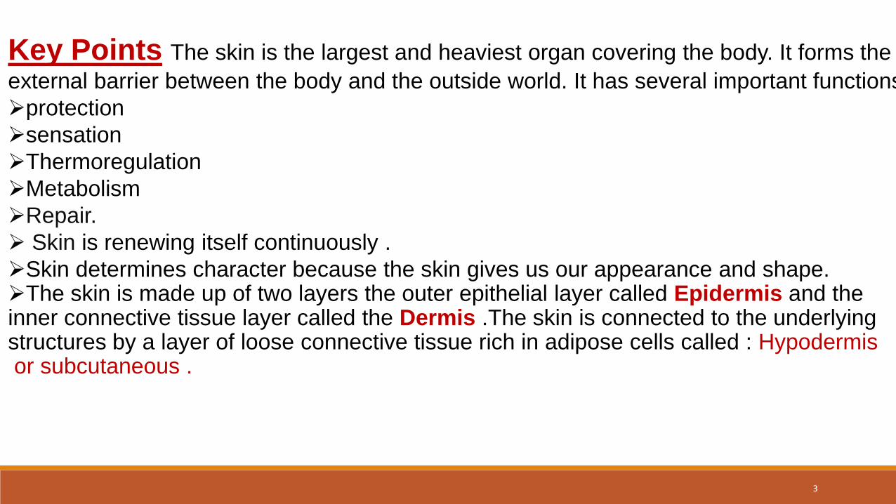
Key Points The skin is the largest and heaviest organ covering the body. It forms the
external barrier between the body and the outside world. It has several important functions:
protection
sensation
Thermoregulation
Metabolism
Repair.
Skin is renewing itself continuously .
Skin determines character because the skin gives us our appearance and shape. The skin is made up of two layers the outer epithelial layer called Epidermis and the inner connective tissue layer called the Dermis .The skin is connected to the underlying structures by a layer of loose connective tissue rich in adipose cells called : Hypodermis or subcutaneous .
3
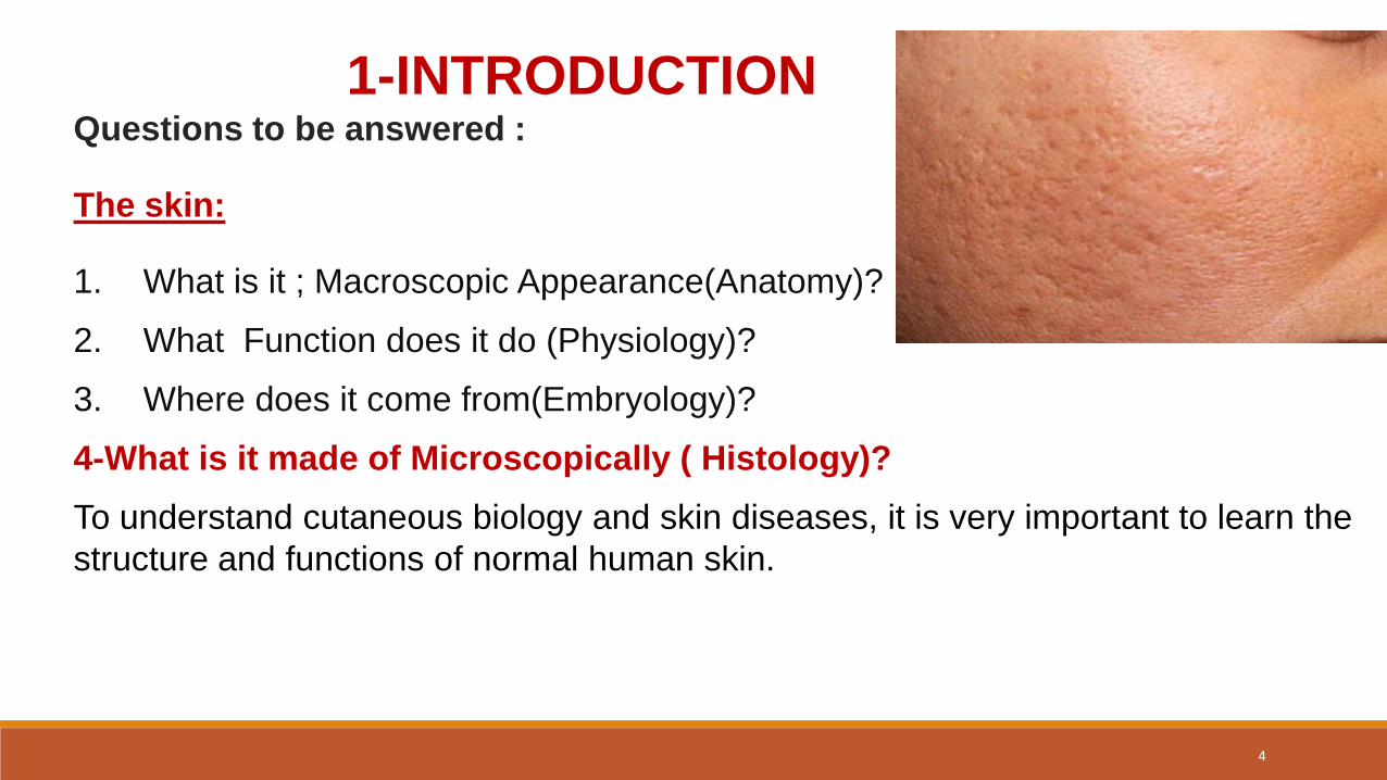
1-INTRODUCTIONQuestions to be answered :
The skin:
1. What is it ; Macroscopic Appearance(Anatomy)?
2. What Function does it do (Physiology)?
3. Where does it come from(Embryology)?
4-What is it made of Microscopically ( Histology)?
To understand cutaneous biology and skin diseases, it is very important to learn the
structure and functions of normal human skin.
4
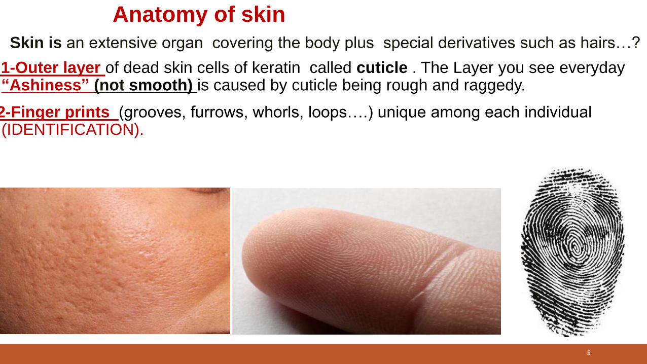
Anatomy of skin
Skin is an extensive organ covering the body plus special derivatives such as hairs…?
1-Outer layer of dead skin cells of keratin called cuticle . The Layer you see everyday“Ashiness” (not smooth) is caused by cuticle being rough and raggedy.
2-Finger prints (grooves, furrows, whorls, loops….) unique among each individual (IDENTIFICATION).
5
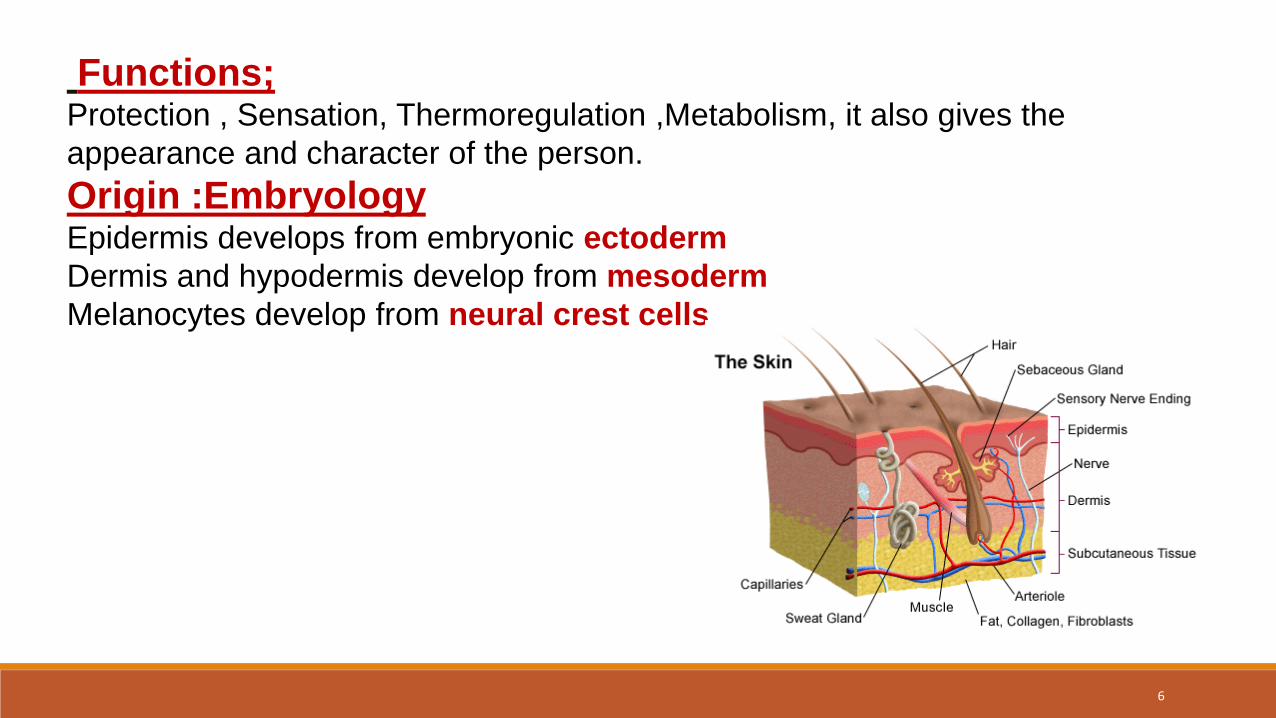
Functions;Protection , Sensation, Thermoregulation ,Metabolism, it also gives the
appearance and character of the person.
Origin :EmbryologyEpidermis develops from embryonic ectoderm
Dermis and hypodermis develop from mesoderm
Melanocytes develop from neural crest cells
6
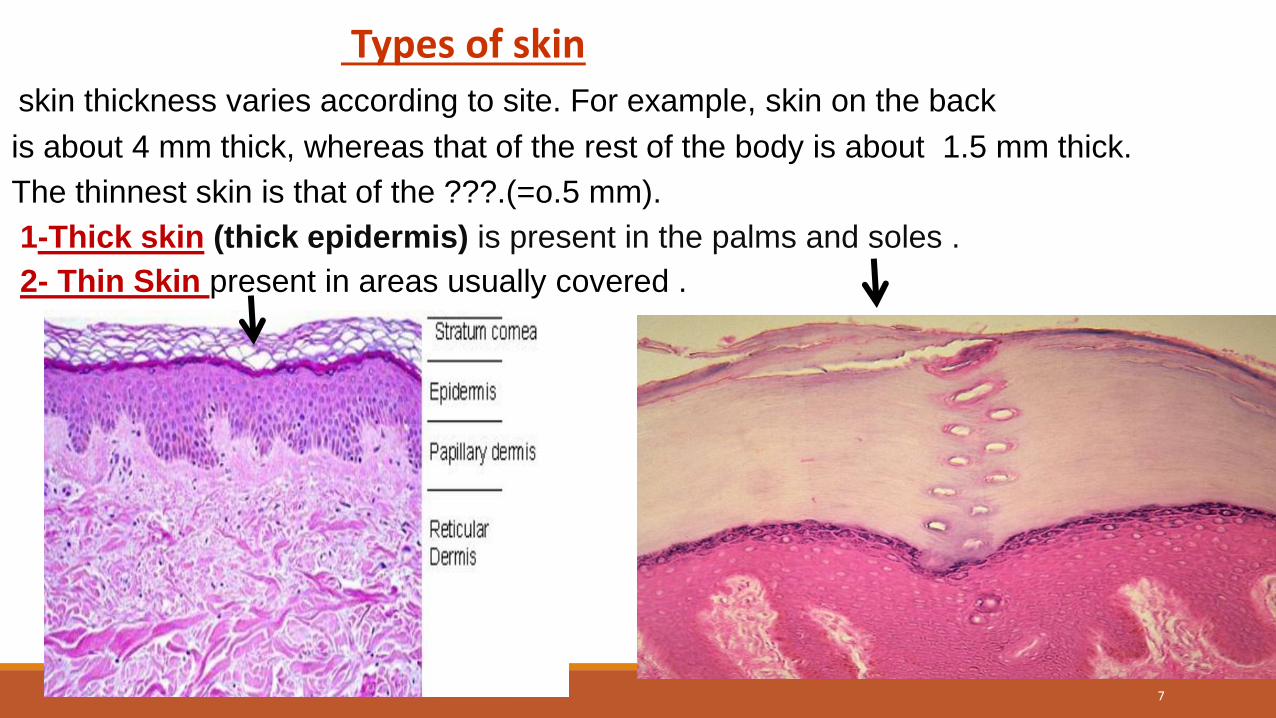
Types of skinskin thickness varies according to site. For example, skin on the back
is about 4 mm thick, whereas that of the rest of the body is about 1.5 mm thick.
The thinnest skin is that of the ???.(=o.5 mm).
1-Thick skin (thick epidermis) is present in the palms and soles .
2- Thin Skin present in areas usually covered .
7
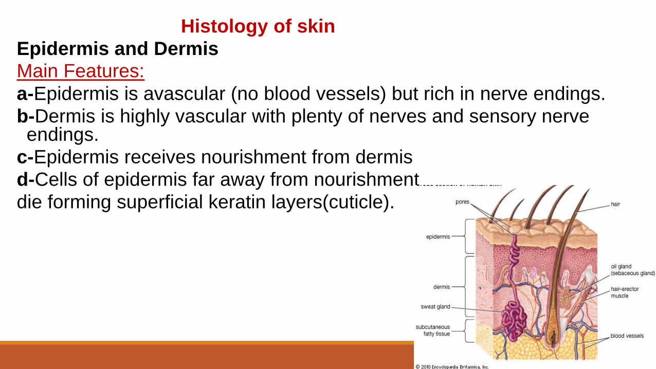
Histology of skin
Epidermis and DermisMain Features:
a-Epidermis is avascular (no blood vessels) but rich in nerve endings.
b-Dermis is highly vascular with plenty of nerves and sensory nerve endings.
c-Epidermis receives nourishment from dermis
d-Cells of epidermis far away from nourishment
die forming superficial keratin layers(cuticle).
8
Microscopic structure: layers of the skin
1- EPIDERMIS - composed of keratinized stratified squamous epithelium it is the outermost protective shield and barrier of the body.
2 - DERMIS - The inner layer or lamina propria of fibro elastic connective tissue(c.t) is highly vascular. Cells of dermis are mainly fibroblasts responsible for elaboration of collagen, elastic and ground substance .
The skin is bind to subjacent structures by HYPODERMIS (sub cut, superficial facia-
loose c.t, mainly fat).
3-The Dermo-epidermal junction= Rete Ridge(peg) system.
(Explain its importance?) , vary in diff. areas of the body ( How & Why).
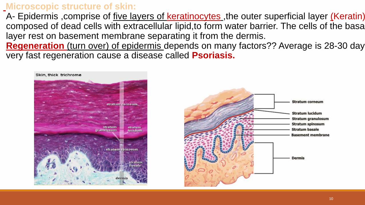
Microscopic structure of skin:A- Epidermis ,comprise of five layers of keratinocytes ,the outer superficial layer (Keratin) composed of dead cells with extracellular lipid,to form water barrier. The cells of the basal layer rest on basement membrane separating it from the dermis.Regeneration (turn over) of epidermis depends on many factors?? Average is 28-30 daysvery fast regeneration cause a disease called Psoriasis.
10
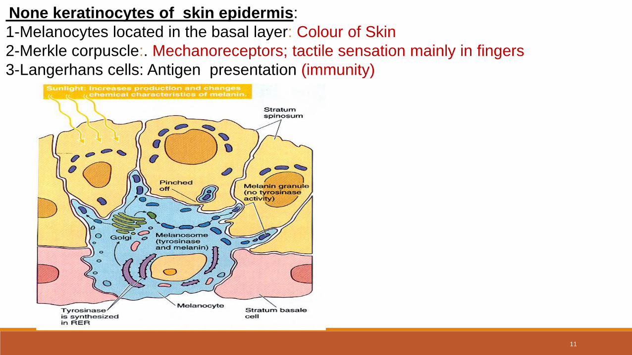
None keratinocytes of skin epidermis:
1-Melanocytes located in the basal layer: Colour of Skin
2-Merkle corpuscle:. Mechanoreceptors; tactile sensation mainly in fingers
3-Langerhans cells: Antigen presentation (immunity)
11
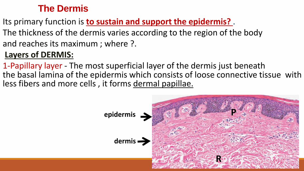
The Dermis
Its primary function is to sustain and support the epidermis? . The thickness of the dermis varies according to the region of the body and reaches its maximum ; where ?.Layers of DERMIS:1-Papillary layer - The most superficial layer of the dermis just beneath the basal lamina of the epidermis which consists of loose connective tissue with less fibers and more cells , it forms dermal papillae.
P
R
dermis
epidermis
12
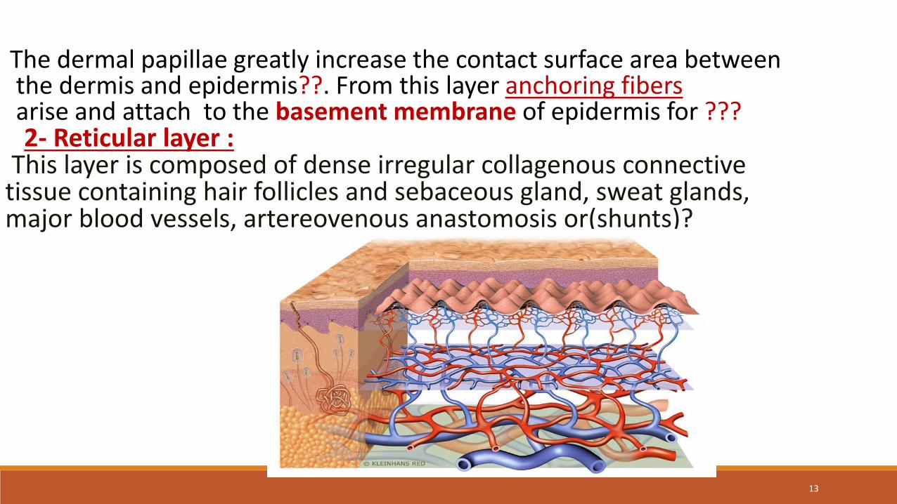
The dermal papillae greatly increase the contact surface area between the dermis and epidermis??. From this layer anchoring fibers arise and attach to the basement membrane of epidermis for ???2- Reticular layer :
This layer is composed of dense irregular collagenous connective tissue containing hair follicles and sebaceous gland, sweat glands, major blood vessels, artereovenous anastomosis or(shunts)?
13
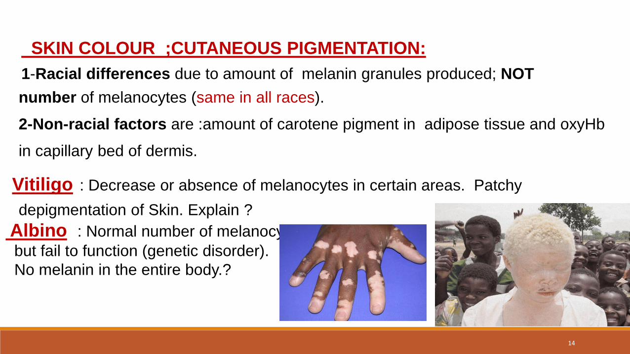
SKIN COLOUR ;CUTANEOUS PIGMENTATION:
1-Racial differences due to amount of melanin granules produced; NOT
number of melanocytes (same in all races).
2-Non-racial factors are :amount of carotene pigment in adipose tissue and oxyHb
in capillary bed of dermis.
Vitiligo : Decrease or absence of melanocytes in certain areas. Patchy
depigmentation of Skin. Explain ?
Albino : Normal number of melanocytes
but fail to function (genetic disorder).
No melanin in the entire body.?
14
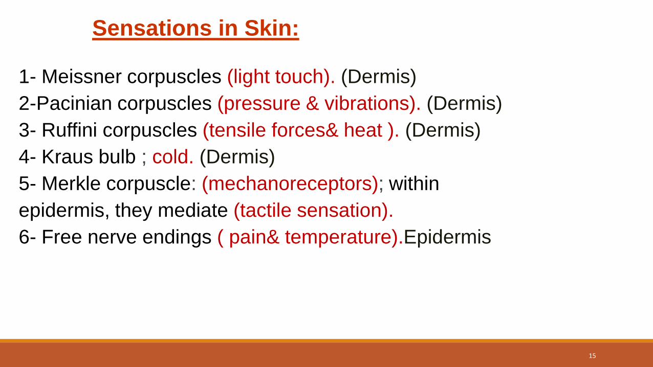
Sensations in Skin:
1- Meissner corpuscles (light touch). (Dermis)
2-Pacinian corpuscles (pressure & vibrations). (Dermis)
3- Ruffini corpuscles (tensile forces& heat ). (Dermis)
4- Kraus bulb ; cold. (Dermis)
5- Merkle corpuscle: (mechanoreceptors); within
epidermis, they mediate (tactile sensation).
6- Free nerve endings ( pain& temperature).Epidermis
15
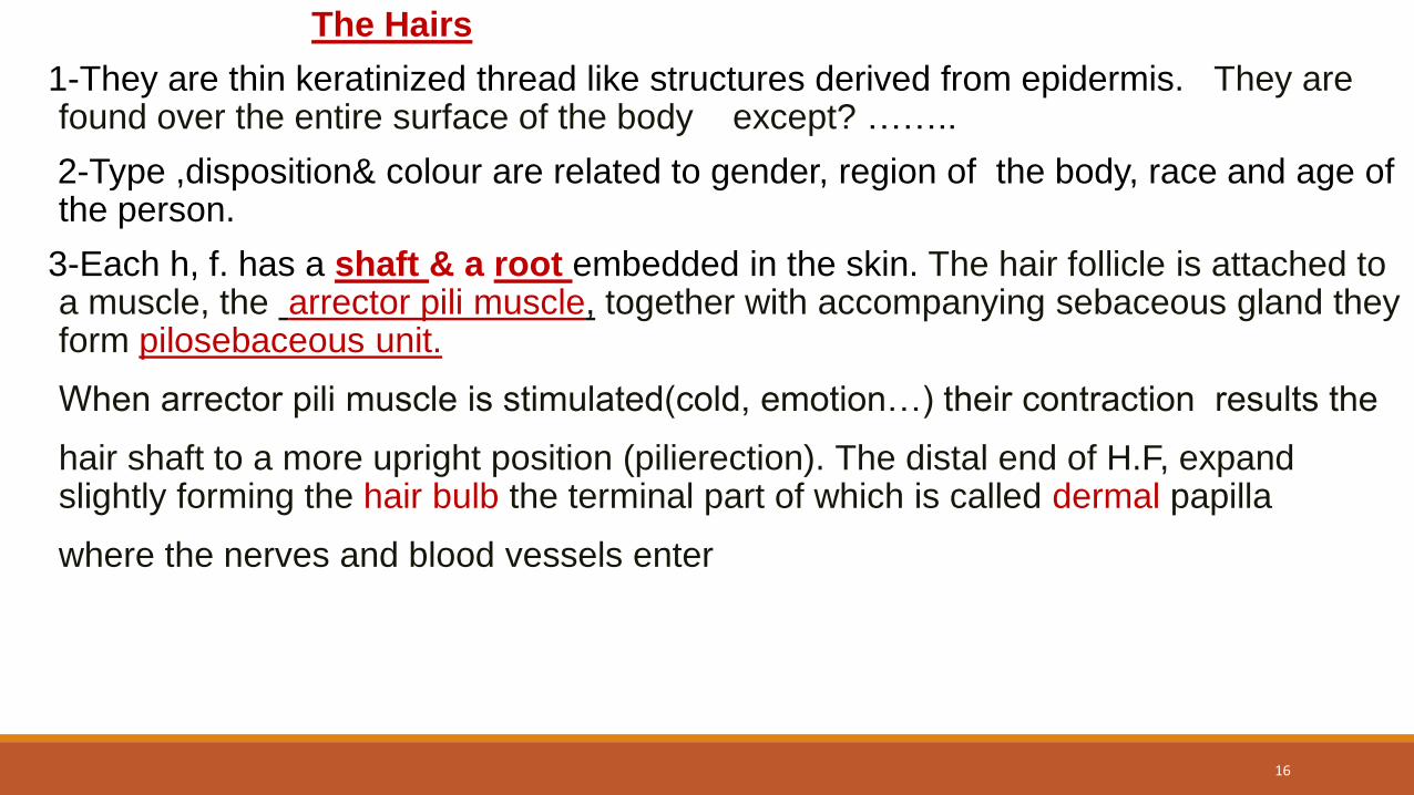
The Hairs
1-They are thin keratinized thread like structures derived from epidermis. They are found over the entire surface of the body except? ……..
2-Type ,disposition& colour are related to gender, region of the body, race and age of the person.
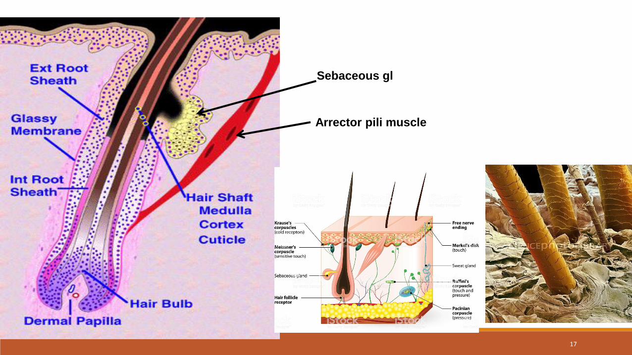
3-Each h, f. has a shaft & a root embedded in the skin. The hair follicle is attached to a muscle, the arrector pili muscle, together with accompanying sebaceous gland they form pilosebaceous unit.
When arrector pili muscle is stimulated(cold, emotion…) their contraction results the
hair shaft to a more upright position (pilierection). The distal end of H.F, expand slightly forming the hair bulb the terminal part of which is called dermal papilla
where the nerves and blood vessels enter
16
17
Sebaceous gl
Arrector pili muscle
Orientation Anatomy of hair follicle:
1-Caucasian hair follicles are oriented obliquely to the skin surface.
2- black persons hair follicles are oriented almost parallel
to the skin surface producing curly hairs.
3-Asian persons have vertically oriented follicles that
produce straight hairs.
These anatomic variations are important considerations
in avoiding alopecia when making incisions in the scalp. ?
18
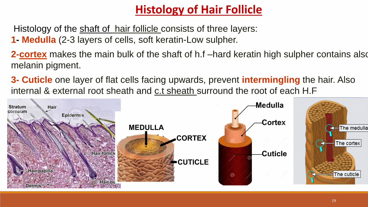
Histology of the shaft of hair follicle consists of three layers:
1- Medulla (2-3 layers of cells, soft keratin-Low sulpher.
2-cortex makes the main bulk of the shaft of h.f –hard keratin high sulpher contains also
melanin pigment.
3- Cuticle one layer of flat cells facing upwards, prevent intermingling the hair. Also
internal & external root sheath and c.t sheath surround the root of each H.F
Histology of Hair Follicle
19
Hair Growth:
Hairs grow in a mosaic way, those at rest coiscent
also called club hair. Explain? The rate or speed of hair growth is about 1.25 centimeters or 0.5 inches per month,
or about 15 centimeters or 6 inches per year. Asian hair grows the fastest, while African hair grows the slowest.
Growth cycles of hair :1- 2- 3- act? psoriasis
Angora Hairs ……….?., Definitive Hairs…?
20
Varieties & sites of Hairs:
Lanugo: fine fetal hairy covering, shed after birth.
Vellus hair: fine body hairs replace lanugo in children and women.
Terminal Hair: Coarse, pigmented and sometime curly hair. Found all over the body
Angora Hair :Terminal hair that grows continuously (scalp, face)
Definitive Hair :Terminal hair that grows to a length then stops (eyelash, eyebrow, pubic, axiliary area)
Scalp, eyebrow and eyelash hairs are thicker.
Ambo sexual hair (pubic & axillae).
Male secondary hair; face and chest.
21
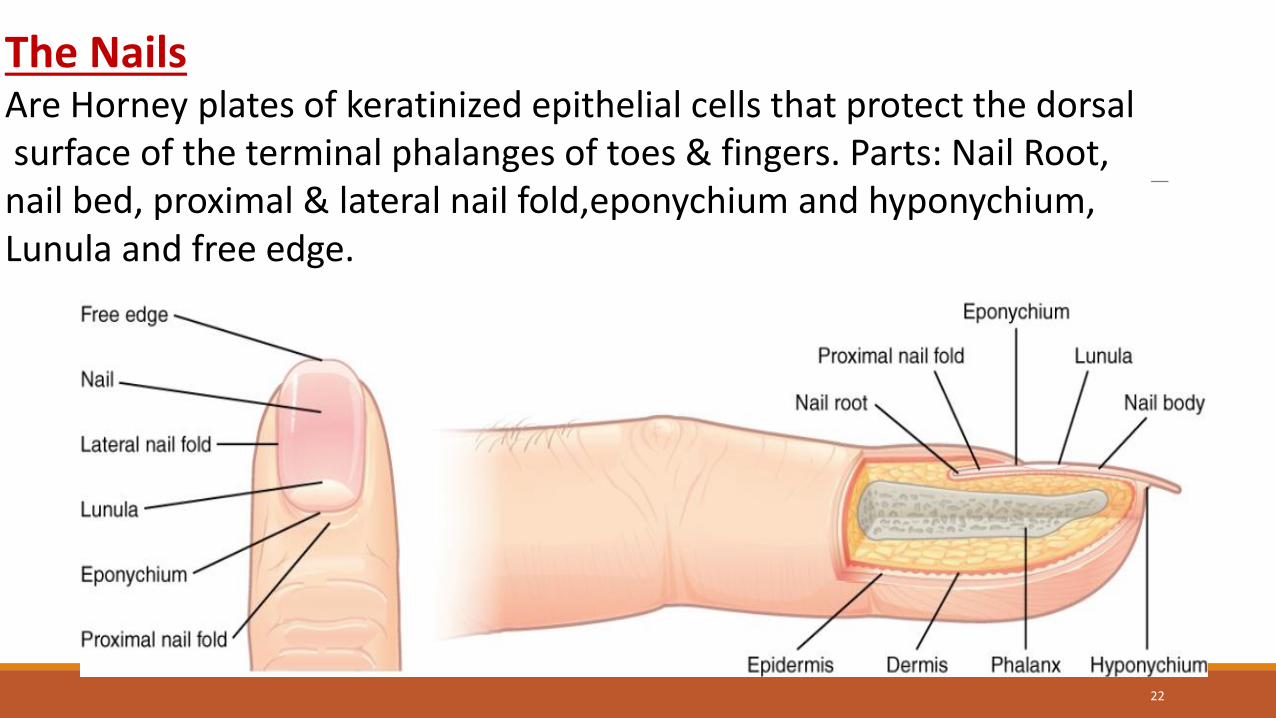
The NailsAre Horney plates of keratinized epithelial cells that protect the dorsalsurface of the terminal phalanges of toes & fingers. Parts: Nail Root, nail bed, proximal & lateral nail fold,eponychium and hyponychium, Lunula and free edge.
22
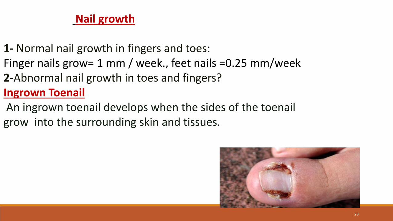
Nail growth
1- Normal nail growth in fingers and toes:Finger nails grow= 1 mm / week., feet nails =0.25 mm/week2-Abnormal nail growth in toes and fingers?Ingrown ToenailAn ingrown toenail develops when the sides of the toenail grow into the surrounding skin and tissues.
23
GLANDS OF THE SKIN; They are of two types:
A- Sebaceous(oil) glands:
B- Sweat Glands(eccrine and apocrine types):
1- Eccrine- merocrine sweat.gl; found almost all over body
2- Apocrine sweat.gl; found in the axillae and groin areas
3- Ceruminous glands which produce ear wax.
4- Mammary glands which produce milk(modified sweat gl)
5-The sweat glands of Moll (eyelid)
24
a- Sebaceous gland:
1- Holocrine gland, pear shaped open on hair follicle by a short duct lined by str.sq.epith. or directly on the surface of the skin in some places like the lips & glans penis. Found all over body except the palms of the hands and soles of the feet.
2-The gland starts functioning after puberty & controlled in men by Testosterone & in women by Androgens?
3- The cells are full of fat droplets ,the secretion of this gland is called :sebum.
If the flow of its secretion is blocked and after bacterial action could cause ACNE?
25
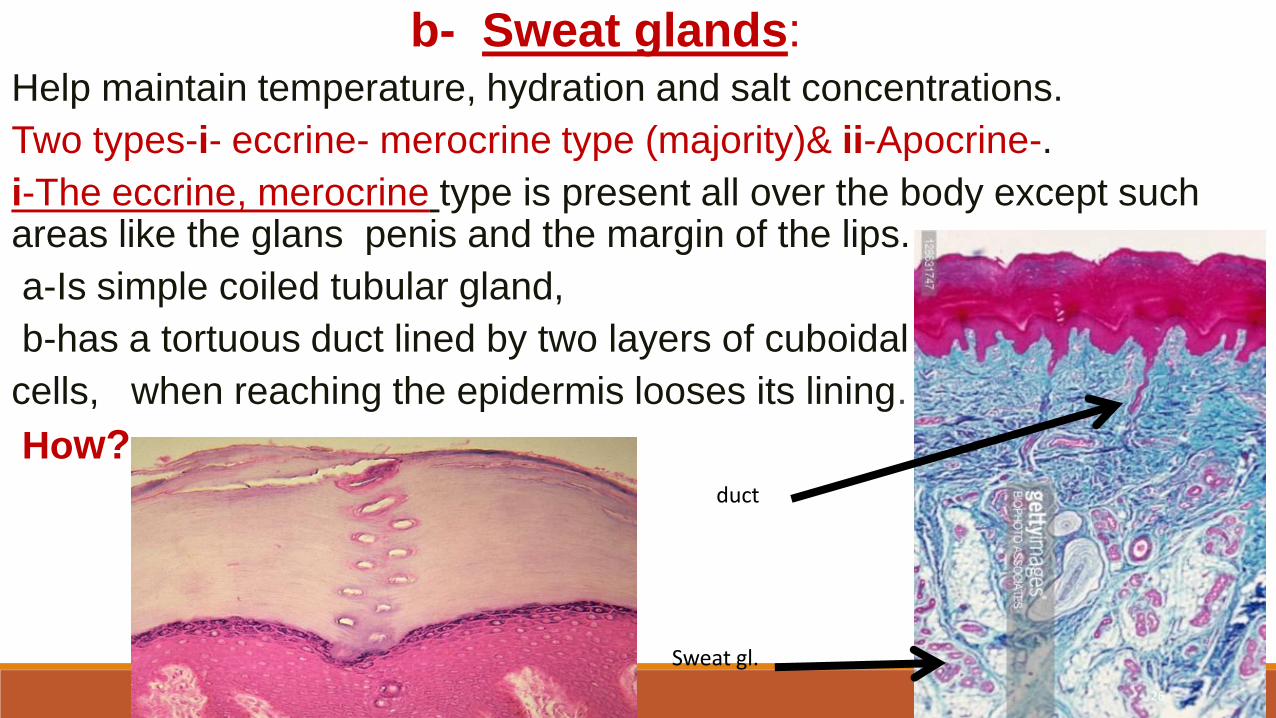
b- Sweat glands:Help maintain temperature, hydration and salt concentrations.
Two types-i- eccrine- merocrine type (majority)& ii-Apocrine-.
i-The eccrine, merocrine type is present all over the body except such areas like the glans penis and the margin of the lips.
a-Is simple coiled tubular gland,
b-has a tortuous duct lined by two layers of cuboidal
cells, when reaching the epidermis looses its lining.
How?
Sweat gl.
duct
26
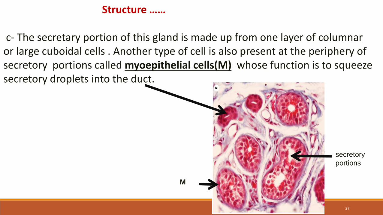
Structure ……
c- The secretary portion of this gland is made up from one layer of columnaror large cuboidal cells . Another type of cell is also present at the periphery of secretory portions called myoepithelial cells(M) whose function is to squeeze secretory droplets into the duct.
M
secretory
portions
27
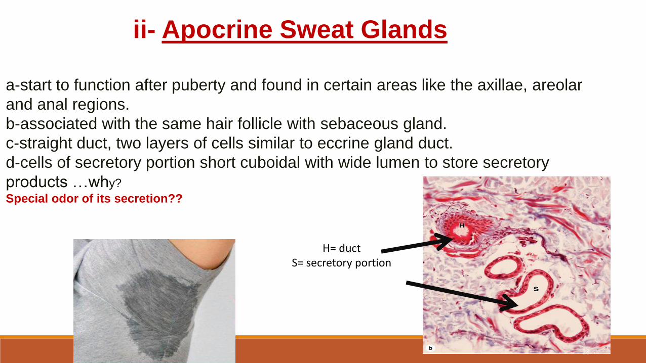
ii- Apocrine Sweat Glands
a-start to function after puberty and found in certain areas like the axillae, areolar
and anal regions.
b-associated with the same hair follicle with sebaceous gland.
c-straight duct, two layers of cells similar to eccrine gland duct.
d-cells of secretory portion short cuboidal with wide lumen to store secretory
products …why?
Special odor of its secretion??
H= ductS= secretory portion
28

Question Time & Summary
All students should participate in repeating the summary of the
major and most important points and ask about the issue
which was not clear 4U?
29

30
References:Wheater's Functional Histology by Barbara Young Elsevier 6TH ed. Churchill Livingstone.edinburghJunqueira's Basic Histology: Text and Atlas, 13th ed / McGraw- Hill.HISTOLOGY A TEXT AND ATLAS by Ross & Pawlina / Lippincott Williams and Wilkins ,Fifth edition.Textbook of Histology by Leslie Gartner pub: Elsevier 4th EDITIONHistology Books, Ebooks & Journals | US Elsevier Healthwww.us.elsevierhealth.com/medicine/histologyNormal histology, with special reference to the ... - Internet Archivehttps://archive.org/details/normalhistologyw00pier. Internet Archive. Di Fiores Atlas of Histology with functional correlations. 12th ed. Wolters Kluwer/Lippincott,& Wilkins Int. www.shutterstock.com alamy stock photo