Embed Size (px)
Citation preview

Governors State UniversityOPUS Open Portal to University Scholarship
All Capstone Projects Student Capstone Projects
Spring 7-1-2017
Preparation of Au/Ag Nanoshells Through aGalvanic Replacement ReactionElizabeth MaschmeyerGovernors State University
Follow this and additional works at: http://opus.govst.edu/capstones
Part of the Analytical Chemistry Commons
For more information about the academic degree, extended learning, and certificate programs of Governors State University, go tohttp://www.govst.edu/Academics/Degree_Programs_and_Certifications/
Visit the Governors State Analytical Chemistry DepartmentThis Project Summary is brought to you for free and open access by the Student Capstone Projects at OPUS Open Portal to University Scholarship. Ithas been accepted for inclusion in All Capstone Projects by an authorized administrator of OPUS Open Portal to University Scholarship. For moreinformation, please contact [email protected].
Recommended CitationMaschmeyer, Elizabeth, "Preparation of Au/Ag Nanoshells Through a Galvanic Replacement Reaction" (2017). All Capstone Projects.306.http://opus.govst.edu/capstones/306

1
Preparation of Au/Ag Nanoshells Through a Galvanic Replacement Reaction
A Project Submitted to:
Governors State University
By: Elizabeth Maschmeyer
In partial fulfillment of the requirement for the Master of Analytical Chemistry
May 2017
Governors State University
University Park, Illinois

2
This is dedicated to everyone in my life who has believed in me to pursue my education from
elementary school to Governors State University.

3
Acknowledgements
My most heartfelt thanks to my research professor who has been there to help me every step of
the way, and my family who never stops believing in me. We also greatly acknowledge our
collaborators for the TEM measurements.

4
Table of Contents
Abstract………………………………………………………………………………………………………………………………..…..5
Introduction…………………………………………………………………………………………………………………….………..6
Kirkendall effect…………………………………………………………….…………………..…………………………….………6
Surface Plasmon Resonance………………………………………………………………..………………………….…..…..8
Galvanic Replacement Reaction………………………………………………………….………………………........……9
Experimental Section………………………………………………………………………………………………………….…...10
Materials…………………………………………………………….……………………………………………………………..……10
Synthesis of Ag Hollow Particles………………………………………………………………………………………………10
Synthesis of Au/Ag Hollow Particle..............................................................................................10
Characterization ………………………………………………………………………………………………….…………..…....12
Results and Discussion…………………………………………………………………………………………………….….…..15
Conclusion…………………………………………………………………………………………………………………….………..24
References………………………………………………………………………………………………………………………..…….25
List of Figures:
Figure 1. Programmable syringe pump used to gradually add small amounts of solution at a
desired rate…………………………………………………………………………………………………………………………..…12
Figure 2. Color change from the Volumes of AgNO3, Glutathione, NaOH, and NaBH4 Used in the
Synthesis of Ag Hollow Particles………………………………………………………………………………………………13

5
Figure 3. UV-Visible absorption spectra of the series of Ag hollow particles obtained by using
different volumes of Glutathione solution: 10 L, 18L, 48L, 78L, 108L, 138L, and
168L…………………………………………………………………………………………………………………………………..….16
Figure 4. Photograph of series of samples illustrating range of colors obtained(top). Normalized
spectra of a series of as-prepared five samples obtained using different…………………………………17
Figure 5. UV-Visible absorption spectra of three different samples: original silver hollow
particles showing SPR peak around 480 nm (orange), hollow silver nanoparticles prepared by
increasing the amounts of reactants by ten times compared to original sample (blue), and the
sample prepared by increasing the amounts of reactants by twenty times (red)…………………….18
Figure 6. TEM image showing pores of the Ag NSs start to collapse as the reaction with HAuCl4
proceeds and eventually resulting in the formation of Au/Ag alloy nanoparticles………………..…18
Figure 7. UV-Visible absorption spectra of Ag nanoshells (left) and Au/Ag alloy nanoshells (right)
produced by galvanic displacement of the pre-formed Ag nanoshells……………………………………..21
Figure 8. TEM images of Ag nanoshells (A) and Au/Ag alloy nanoshells (B) produced by galvanic
displacement of the Ag nanoshells……………………………………………………………………………………….….22
List of Tables
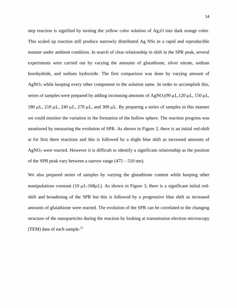
Table 1. Volumes of AgNO3, Glutathione, NaOH, and NaBH4 Used in the Synthesis of Ag Hollow Particles……………………………………………………………………………………………………………………………………17
Table 2. Volumes of AgNO3, Glutathione,NaOH, and NaBH4 Used in the Synthesis of Ag Hollow Particles in larger scale……………………………………………………………………………………………………………19
Table 3. Volumes of AgNO3, Glutathione,NaOH, and NaBH4 Used in the Synthesis of Ag Hollow Particles…………………………………………………………………………………………………………………………………..20

6
Abstract
It generally requires solid metal templates of hollow metal structures with uniform morphology
by the present galvanic replacement mediated-growth approaches. In this project report, we
present a new and simple approach for the controllable synthesis of Au/Ag alloy nanoshells
(NSs) by applying galvanic replacement reaction on hollow Ag template in the presence of gold
salt. High quality thiol-coated spherical shape Au/Ag NSs were produced via this new synthetic
approach and the hollow interior of the Ag template is very well preserved during the galvanic
replacement reaction. Plasmonic property measurements reveal that Au/Ag NSs exhibit strong
absorption peak and more importantly, these can maintain their optical properties even after
several months. These nanoshells were studied by TEM to elucidate their structure. These studies
show that the nanostructures are hollow and do not consist of a Ag core surrounded by a Au
shell. The excellent stability along with the ability to maintain long lasting plasmonic features
make these Au/Ag NSs useful candidates to test for fascinating application in surface
plasmonics, surface-enhanced Raman scattering, chemical and biological sensing and optical
labeling.
Scheme I Schematic illustration of the structural evolution at different stages of the galvanic
replacement reaction between Ag hollow particle and HAuCl4 in an aqueous solution.

7
Introduction
The novel physical properties observed in nanoparticle are attributed to the dramatic
changes in the electronic structure.1Metal nanoparticles play important roles in many areas of
modern science and technology. They have been widely used in photography2, catalysts3,
optoelectronics4, information storage5, and surface enhanced Raman scattering(SERS)6. The
properties of metal nanoparticles are determined by the size, composition, shape,
crystallography, and the type of structure. It is interesting to notice that one could control these
parameters to fine tune the properties of this metal nanoparticle. In addition to that shape control
of nanoparticles is important to understand basic size and shape dependent properties. These
properties have led to the further development of synthetic methods that permit exquisite control
of their dispersity on the nanometer scale7. Several methods can be produced in large quantities
through solution phase methods8. It is possible to control the reaction and diffusion processes of
room temperature to produce polymetallic hollow nanoparticles with very different morphology
and composition by employing simultaneous sequential action of galvanic replacement and the
kirkendall effect9.
The Kirkendall effect occurs when there is a movement of the boundary layer between
two metals. This motion is due to the different diffusion rates of metal atoms. This method was
conceived in 1942 and was verified in 1947.10 The initial experiment used copper and brass
welded to each other and was exposed to higher temperatures. The movement of the boundary
layer was seen between the two since zinc diffuses into copper faster than copper into zinc. The
experiment proved that diffusion of the two metal atoms included defects that help atomic jumps.
Theses defects are called vacancies that are empty lattice sites. Creating the vacancies weakens
the bonding strength of the bond–pad interface. This weakening often causes wire failures that

8
engineers try to prevent. This is done by diffusion layer barriers. Contrary to engineers, chemists
utilize the weakening bond strength effect to create hollow nanostructures. The hollow
nanostructures occur when the vacancies combine into one hollow core.11
Aldinger was the first person to pursue an interest in the hollowing of silver nanoparticles
caused by the Kirkendall effect.12 He used beryllium micro particles covered with Co by
evaporation or Ni by electroplating to form shell particles. He found that vacancies crash at the
shell interface when a structure has any flaws. These imperfections cause the occurrence of
stress cracks that separates the core from the shell. By using the nanoscale, scientists are able to
use the perfection of the nanoparticle and the abundance of single crystal metal nanoparticles.13
This process makes the Kirkendall effect form smooth and uniform-sized hollow compound
nanocrystals. To get pure-phase hollow nanocrystals, an appropriate amount of shell material
compared to the starting crystals is needed. This reaction is less favorable in a solid-solid
reaction. A vapor or solution phase is often more favorable. This is because the source of the
shell material is continuous and sufficient.
In the beginning, the Kirkendall effect came from metal alloys, but it has now taken part
in compound systems in the form of oxides and nitrides14 In these oxides and nitrides, oxidation
or nitridation of the metal element occurs quickly with a fast diffusion rate of the metal through
the compounds. This was shown in a reaction of Co nanocrystals in solution in an elemental S or
O2/Ar ambient that caused the formation of hollow Co3S4 or CoO NPs.15 The experiment has
suggested the size of the ending vacancy was smaller when compared to the beginning Co NP.
The discovery has shown there was an inward growth of the product shell, because of the sulfur
anions and the outfussion of Co cations.

9
The Kirkendall effect method for making hollow nanoparticles(NPs) been beneficial. The
most commonly used procedure uses polystyrene and silica spheres as the sacrificial
model.16Due to the inherent size of the polystyrene and silica spheres, they usually make big
hollow structures. Colloidal metal nanocrystals are able to be made with a high yield. This
results in crystalline hollow NPs even in the quantum range because the colloidal metal
nanocrystals are mixed with the Kirkendall diffusion.Also, the Kirkendall method is a one-step
mass synthesis with almost 100% purity, which is highly favored over the many step purification
routes taken in other methods.17
The plasmonic properties of noble metal nanoparticles are extremely sensitive to their size and
shape. Gold nanospheres have characteristic red color but anisotropic gold nanorods have
different colors. The color is due to collective oscillation of electrons in the conduction band
known as surface plasmonic oscillation. The collective oscillation of the conduction electrons in
resonance with certain frequencies of incident light leads to an excitation known as surface
Plasmon resonance (SPR).11 The resonance condition is established when the frequency of the
light photon match with the natural frequency of the surface electron oscillation. Plasmonic
properties of silver nanoparticles (AgNPs) have been extensively studied for their superior
performances that exceed those of other metals with a surface plasmonic resonance (SPR) in the
visible range like gold.12 The plasmonic response depends on the size, shape, dielectric
environment, and on mutual electromagnetic interactions among particles in close proximity.11
Correlating the NPs plasmonicproperties with their morphology is a fast and easy way for in situ
monitoring of the synthesis by UV–visible spectroscopy.19. This is very useful in the early stages
of wet chemistry synthesis, when many different chemicals are present in solution and especially
sample preparation for transmission electron microscopy analysis.15

10
Galvanic replacement reaction (GRR) was known to be a powerful synthetic technique for
converting solid metal nanostructures into hollow ones. It has been frequently employed to
synthesize hollow Au/Ag nanostructures of various shapes. In this case, the driving force for the
galvanic replacement reaction is the difference in the reduction potential of gold and silver
metals where the deposition of the gold with a higher reduction potential onto a solid template
nanostructure of silver (with a relatively lower reduction potential) occurs. This difference in the
reduction potential drives the oxidation of solid silver template by the gold salt precursor to form
a fully enclosed hollow Au/Ag nanostructure. The most common way of forming Au/Ag
nanostructure is achieved via the reaction between HAuCl4 and Ag nanoparticle template where
the oxidation of Ag nanoparticle template, reduction of AuCl4– and subsequently alloying of the
deposited Au with the remaining Ag in the template occur. The resultant nanoparticles are
expected to be bimetallic and with hollow interiors.16 This approach has been successfully used
to produce hollow bimetallic nanostructures of many different noble metals using solid Ag
nanoparticle template, yet to date there has been no evidence of the forming bimetallic hollow
nanostructures from a hollow Ag nanoparticle template. In addition, to best of our knowledge, no
attempt has been made to synthesize hollow Au/Ag nanostructures, starting from hollow Ag
nanoparticles as sacrificial templates. Attempting to make fully enclosed hollow Au/Ag
nanostructure out of a hollow Ag template rather than a solid Ag template appears to be
challenging since it is required to preserve the hollow template during the galvanic replacement
reaction as the oxidation of remaining Ag template can cause the nanoshell (NS) to etch.
Therefore, the conditions of galvanic replacement need to be adjusted in a way that the
deposition of Au on the hollow Ag template nanoparticle occurs as an effective way of
stabilizing the template too.

11
Based on the previous work done by Sanedrin et al. and Aherne et al., one possible way of
protecting the Ag template against oxidation during the galvanic replacement is by maintaining a
sufficiently high level of reducing agent in the reaction mixture.20-21 This will guarantee that
enough Ag is present in the template as the availability of reducing agent minimize the level of
Ag+ ions in the reaction mixture during the reaction. Taking the advantage of this scenario, we
studied the use of silver hollow spheres as sacrificial templates to generate Au/Ag hollow
spheres with well define shape and hollow structure. This attempt resulted in the formation of
thiol-coated spherical shape Au/Ag hollow particles for the first time by employing galvanic
replacement reaction on spherical shape Ag hollow particle as the sacrificial template.
Here, we have prepared Au/Ag hollow particles that are produced by adding HAuCl4 to
thiol-coated Ag hollow particle solution in the presence of excess reducing agent, ascorbic acid.
Since substantial amount of materials are needed for the gel formation, scaled up reactions were
carried out by increasing the amounts of starting materials by 10times compared to the literature
synthetic method22 while keeping other manipulations the same.
Experimental Section:
I. Materials:
Silver nitrate (99.9%) and sodium borohydride (98%) were purchased from Strem Chemicals. L
glutathione reduced (98%) were purchased from Sigma-Aldrich. Sodium hydroxide was
purchased from VWR, and L(+) ascorbic acid (99%) and HAuCl4 (49.0%Au) were purchased
from Acros.
II. Synthesis of Ag Hollow Particles:
The silver hollow particles were synthesized following a literature synthetic method.22 with
several modifications to prepare bigger particles in a larger scale. In a typical synthesis 2.6mL

12
millipore water was added to a10-mLround bottom flask and it was kept in the freezer for nearly
an hour until ice cubes were formed on the surface. To this ice-cold water, appropriate volumes
of 10 mM silver nitrate and glutathione were added while stirring. After ~4 minutes, 0.1M
sodium hydroxide (0.1M) was poured into the above mixture while vigorous stirring. After ~4
minutes a freshly prepared 10 mM sodium borohydride solution was injected quickly. The
resulting mixture was stirred slowly for nearly an hour.
III. Synthesis of Au/Ag Hollow Particles:
Au/Ag alloy hollow particles were prepared by employing galvanic replacement reaction. To the
as-prepared silver hollow particle solution, ascorbic acid solution (200 μL, 10 mM) was added
and then a solution of HAuCl4 (8 mL, 0.5 mM) was added at a rate of 1 mL min-1 using a syringe
pump. The resulting solution was stirred slowly for few hours. The color change from orange to
bright blue indicates the formation of Au/Ag alloy.
Figure 1.Programmable syringe pump used to gradually add small amounts of solution at a
desired rate.

13
IV. Characterization:
Spectroscopic Instrumentation and Methods:
A PerkinElmer Lambda 35 UV-Vis spectrophotometer was used for optical absorption
measurements on Ag NSs and Au/Ag NSs. The absorption spectrum of the silver seeds solution
was measured without dilution while the silver nanoprism and Ag/Au hollow nanoprism
solutions were diluted 3 in Millipore water and measured from 700 to 300 nm.
Transmission Electron Microscopy (TEM)
The TEM analysis was performed by using JOEL JEM-1230 analytical electron microscope with
Gatan ultra scan 4000 camera operating at a 120-kV acceleration voltage. One drop of
AgNsorAu/Ag Ns solution was added onto a carbon-coated copper TEM grid and the solvent
was allowed to evaporate few hours before introduction to the instrument.
Results and Discussion
Thiol-coated spherical shape Ag NSs were prepared by employing fast reaction diffusion process
as reported in the literature.22 In addition, Ag NSs with different sizes were also prepared by
employing the same synthetic method by changing the ratio of reactants. Since substantial
amount of materials are needed for the property characterization, scaled up reactions were
carried out by increasing the amounts of starting materials by 20 times compared to the reported
procedure while keeping other manipulations same. Ag NSs were produced by the formation of
Ag2O first by reacting AgNO3 with sodium hydroxide in the presence of glutathione followed by
the reduction of preformed Ag2O by freshly prepared sodium borohydride solution. This two-
14
step reaction is signified by turning the yellow color solution of Ag2O into dark orange color.
This scaled up reaction still produce narrowly distributed Ag NSs in a rapid and reproducible
manner under ambient condition. In search of clear relationship in shift in the SPR peak, several
experiments were carried out by varying the amounts of glutathione, silver nitrate, sodium
borohydride, and sodium hydroxide. The first comparison was done by varying amount of
AgNO3 while keeping every other component in the solution same. In order to accomplish this,
series of samples were prepared by adding increasing amounts of AgNO3(90 L,120 L, 150 L,
180 L, 210 L, 240 L, 270 L, and 300 L. By preparing a series of samples in this manner
we could monitor the variation in the formation of the hollow sphere. The reaction progress was
monitored by measuring the evolution of SPR. As shown in Figure 2, there is an initial red-shift
at for first there reactions and this is followed by a slight blue shift as increased amounts of
AgNO3 were reacted. However it is difficult to identify a significant relationship as the position
of the SPR peak vary between a narrow range (475 – 510 nm).
We also prepared series of samples by varying the glutathione content while keeping other
manipulations constant (10 L-168L). As shown in Figure 3, there is a significant initial red-
shift and broadening of the SPR but this is followed by a progressive blue shift as increased
amounts of glutathione were reacted. The evolution of the SPR can be correlated to the changing
structure of the nanoparticles during the reaction by looking at transmission electron microscopy
(TEM) data of each sample.21

15
Figure 2.UV-Visible absorption spectra of the series of Ag hollow particles obtained by using
different volumes of AgNO3 solution: 90 L,120 L, 150 L, 180 L, 210 L, 240 L, 270 L,
and 300 L.
We also prepared another series of samples (Table 1) by varying the AgNO3 (10 L-168L)and
glutathione (18L-100L) at the same time while keeping other manipulations constant. The
positions of the main SPRs of these samples are well separated as can be seen in Figure 4. It has
been reported that the spectral position of the SPR can be tuned by controlling the size of the Ag
nanoprism without any significant variation in thickness.22 Accordingly one can think of the
different peak positions of the samples and the different colors as shown in Figure 4 are due to
different sizes and thicknesses of silver hollow particles prepared according to the conditions
given in the table 1.
0
0.5
1
1.5
2
2.5
3
3.5
4
4.5
5
300 400 500 600 700 800 900 1000
Ab
so
rba
nc
e (
ab
s. u
nit
s)
Wavelength (nm)
90 uL
120 uL
150 uL
180 uL
210 uL
240 uL
300 uL

16
Figure 3.UV-Visible absorption spectra of the series of Ag hollow particles obtained by using
different volumes of glutathione solution: 10 L, 18L, 48L, 78L, 108L, 138L, and 168L.
To characterize the hollow particles produced by this method and to explore the relationship
between nanoparticle dimensions and the position of the main SPR, TEM analysis of statistically
significant numbers of particles from samples need to be carried out and such measurements will
be done in the future.
0
1
2
3
4
5
6
7
8
9
300 400 500 600 700 800 900
Ab
so
rba
nc
e (
ab
s. u
nit
s)
Wavelength (nm)
10 uL
18 uL
48 uL
78 uL
108 uL
138 uL
168 uL
17
Table 1. Volumes of AgNO3, Glutathione,NaOH, and NaBH4 Used in the Synthesis of Ag
Hollow Particles
Sample 1 Sample 2 Sample 3 Sample 4 Sample 5
Water 2.6 mL 2.6 mL 2.6 mL 2.6 mL 2.6 mL
10 mM AgNO3 150 L 150 L 300 L 300 L 500 L
10 mM Glutathione 18L 55 L 60 L 75 L 100 L
0.1 M NaOH 500 L 500 L 500 L 500 L 500L
10 mM NaBH4 180 L 180 L 180 L 180 L 180 L
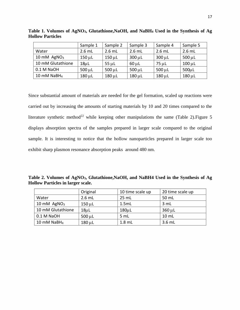
Since substantial amount of materials are needed for the gel formation, scaled up reactions were
carried out by increasing the amounts of starting materials by 10 and 20 times compared to the
literature synthetic method22 while keeping other manipulations the same (Table 2).Figure 5
displays absorption spectra of the samples prepared in larger scale compared to the original
sample. It is interesting to notice that the hollow nanoparticles prepared in larger scale too
exhibit sharp plasmon resonance absorption peaks around 480 nm.
Table 2. Volumes of AgNO3, Glutathione,NaOH, and NaBH4 Used in the Synthesis of Ag
Hollow Particles in larger scale.
Original 10 time scale up 20 time scale up
Water 2.6 mL 25 mL 50 mL
10 mM AgNO3 150 L 1.5mL 3 mL
10 mM Glutathione 18L 180L 360 L
0.1 M NaOH 500 L 5 mL 10 mL
10 mM NaBH4 180 L 1.8 mL 3.6 mL

18
Figure 4. Photograph of series of samples illustrating range of colors obtained (top).
Normalized spectra of a series of as-prepared five samples obtained using different volumes of
AgNO3 and Glutathione(bottom).
0
0.5
1
1.5
2
2.5
3
3.5
4
300 400 500 600 700 800
Ab
so
rba
nc
e (
ab
s. u
nit
s)
Wavelength (nm)
Sample 1
Sample 2
Sample 3
Sample 4
Sample 5

19
Figure 5. UV-Visible absorption spectra of three different samples: original silver hollow
particles showing SPR peak around 480 nm (orange),hollow silver nanoparticles prepared by
increasing the amounts of reactants by ten times compared to original sample (blue), and the
sample prepared by increasing the amounts of reactants by twenty times (red).
The Ag hollow particles grown by this route (scaled up by ten time) display a narrow Surface
Plasmon Resonance (SPR) maximum at ~480 nm. Initially, the galvanic replacement reaction
0
0.5
1
1.5
2
2.5
3
3.5
4
4.5
5
300 400 500 600 700 800 900 1000
Ab
so
rba
nc
e (
ab
s. u
nit
s)
Wavelength (nm)
Original(Orange)
Scaled up by 10times (blue)
Scaled up by 20times (red)
20
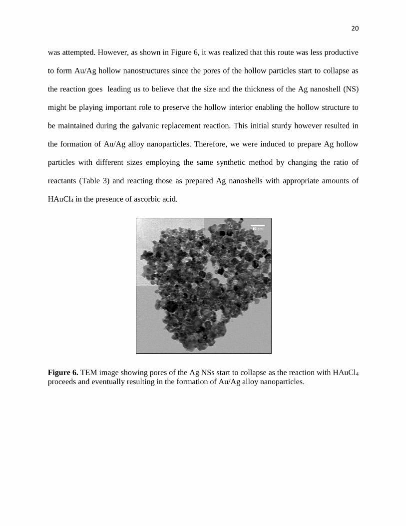
was attempted. However, as shown in Figure 6, it was realized that this route was less productive
to form Au/Ag hollow nanostructures since the pores of the hollow particles start to collapse as
the reaction goes leading us to believe that the size and the thickness of the Ag nanoshell (NS)
might be playing important role to preserve the hollow interior enabling the hollow structure to
be maintained during the galvanic replacement reaction. This initial sturdy however resulted in
the formation of Au/Ag alloy nanoparticles. Therefore, we were induced to prepare Ag hollow
particles with different sizes employing the same synthetic method by changing the ratio of
reactants (Table 3) and reacting those as prepared Ag nanoshells with appropriate amounts of
HAuCl4 in the presence of ascorbic acid.
Figure 6. TEM image showing pores of the Ag NSs start to collapse as the reaction with HAuCl4
proceeds and eventually resulting in the formation of Au/Ag alloy nanoparticles.

21
Table 3. Volumes of AgNO3, Glutathione,NaOH, and NaBH4 Used in the Synthesis of Ag
Hollow Particles
Sample # H2O/ml AgNO3/mL HS-G /mL NaOH/mL NaBH4/mL
Sample 1 25 1.5 0.15 5 1.8
Sample 2 25 0.5 0.05 2.5 1.8
Sample 3 25 0.35 0.05 5 1.8
Since the optical properties are strongly dependent on the structure and composition of the
nanostructure, searching for bimetallic hollow nanostructures with accurately controlled
structures and compositions is very useful to obtain such materials with interesting properties. It
has been demonstrated that surface plasmon resonance peaks of the hollow Au/Ag bimetallic
nanoparticles could be tuned across the visible spectrum region by controlling the Au:Ag ratio in
the galvanic replacement reaction.16 Knowing this fact, we decided to study how the size and
position of the plasmon resonance of our Au/Ag NSs can be controlled through the adjustment of
reaction condition. For this study Ag NSs showing SPR ~550 nm was prepared by employing the
condition given for sample 3 in table 3 where silver nitrate (350 μL, 10 mM), glutathione (50 μL,
10 mM), sodium hydroxide (5 mL, 0.1M) and a freshly prepared sodium borohydride solution
(1.8 mL, 10 mM) were reacted. Attempts were made to prepare Au/Ag alloy nanoshells with
different sizes by adding different amounts of HAuCl4to as prepared Ag NS template solution in
the presence of reducing agent, ascorbic acid (200 μL, 10 mM). By preparing a series of samples

22
in this manner, one can also monitor the progress of the Au/Ag NS formation. At first to Ag NSs
showing SPR ~550 nm, solution of HAuCl4 (8 mL, 0.5 mM) was added at a rate of 1 mL min-1.
The resulting Au/Ag NS sample display a slight progression in color as the SPR red-shifted and
exhibit maximum surface plasmon resonance peak at 570 nm as shown in Figure 7.
Transmission electron micrographs (TEM) show that almost all the individual Ag NSs consist of
significantly larger single hollows in the center of the nanosphere (Figure 8A). The Ag NSs
grown by this route exhibit an average outer diameter of 63.7 ± 10.5 nm and shell thickness of
12.4 ± 2.4 nm and these are much bigger than the Ag NSs prepared by literature synthetic
method where those showing SPR at 480 nm possess the outer diameter of 24.1 ± 4.5 nm and
shell thickness of 5.8 ± 0.9 nm.21 Transmission electron micrographs (TEM) show that in almost
all the individual Au/Ag nanoshells, significantly larger single hollows have been created as
indicated by variation in the degree of contrast between edges and middle of the nanosphere.
400 600 800 1000 1200
0.0
0.5
Absorb
ance
Wavelength (nm)
(A) (B)
Figure 7. UV-Visible absorption spectra of Ag nanoshells (left) and Au/Ag alloy
nanoshells (right) produced by galvanic displacement of the pre-formed Ag
nanoshells.

23
Figure 8. TEM images of Ag nanoshells (A) and Au/Ag alloy nanoshells (B) produced by
galvanic displacement of the Ag nanoshells.
It is interesting to notice that as shown in Figure 8B, Au/Ag nanoshells still retain the
spherical shape after the galvanic displacement reaction and the shell thickness uniformly
distributed around the hollow interior of the particles. This suggests that oxidation of Ag
template initiated uniformly over the entire core of the particle. This is quite a remarkable
finding and indeed this is the first time that such Au/Ag nanoshells have been formed from a Ag
NS template. Based on a calculation from about 100 Au/Ag nanoshells, the outer diameter and
shell thicknesses were obtained as 60.9 ± 10.8 nm and 9.0 ± 1.9 nm, respectively. It is reasonable
to believe that due to the presence of sufficient amount of reducing agent (ascorbic acid), after
initial oxidation of silver from Ag NSs by galvanic replacement reaction, alloy Au/Ag nanoshells
(A) (B)

24
are formed by coreduction of Ag+ and AuCl4– as suggested by Aherne et al.to explain how the
alloying of Ag with Au occur when there is excess ascorbic acid at the low temperatures.21 This
ascorbic acid-mediated co-deposition facilitates the formation of well-defined Au/Ag NSs. Our
ability to form Au/Ag hollow structure using Ag hollow particles as sacrificial template by
employing a general experimental approach allowed us to gain insight regarding the mechanism
of Ag-Au alloying as illustrated in scheme I. Galvanic replacement reaction included two stages.
At the initial stage, replacement reaction starts at specific sites with relatively high surface
energies and then seamless hollow nanostructures with smooth Au−Ag alloy walls were evolved
through an integration of galvanic replacement with alloying.24 Ag atoms also simultaneously
migrate into the Au shell to form a seamless, hollow nanostructure with Au−Ag alloy wall. This
mechanism for galvanic replacement is applicable irrespective of the morphology and
composition of the sacrificial templates as long as the presence of appropriate reduction
potentials difference between the two metals involved.
Conclusion
In conclusion, this work makes it clear that from a hollow Ag nanoparticle template,
chemical synthesis of bimetallic hollow nanostructures with well-controlled shapes, sizes, and
structure is a practical reality. The major requirement seems to be the adjustment of the reaction
conditions to preserve the hollow template during the galvanic replacement reaction. We studied
the wide tunability of the plasmonic features of Ag hollow particles by engineering the size of
the particles through varying the reaction conditions. According to the study of optical property
measurements on Ag hollow samples with increasing glutathione content, it can be seen that
surface plasmon resonance peaks of the Ag hollowparticles prepared with higher glutathione
25
content lie at longer wave lengths and are red-shifted compared to the surface plasmon resonance
of AgNS template and then blue shift. We successfully prepared thiolate-capped Au/Ag alloy
particles in large scale by employing galvanic replacement reaction of Ag hollow particles. Our
success in preparing Au/Ag alloy will be helpful in constructing similar nanostructures such as
Pt/Ag, Pd/Ag bimetallic alloy.

26
References
1. Schwartzberg, A.; Zhang, J. Z. J. Phys. Chem. C 2008, 112, 10323−10337.
2. Lam, D. M-K; Rossitter, B. W; Sci. 1991, 265, 80-90.
3. Lewis, L.N.; Chem. Rev. 1993, 93. 2693-2675.
4. Karmart, P.V. J. Phys. Chem. B. 2002, 106,7729-7735.
5. Murray, C.B.; Sun S.; Doyle H.; Betley T. Mater. Res. Soc. Bull, 2001, 26, 985-1001.
6. Nile S.; Emory R. Science, 1997, 275, 1102-1107.
7. An, K.; Somorjai, G. A. Chem Cat Chem 2012, 4, 1512−1524.
8. Schmid, G. Chem. Rev. 1992, 92, 1709-1711.
9. González, E.; Arbiol, J.; Puntes, V. F. Science. 2011, 334 ,1377–1380.
10. Smigelskas, A.D.; Kirkandall, E.O. Trans. Aime, 2007, 171, 130-135
11. Mulvaney, P. Langmuir. 1996, 12, 788-800 .
12. Xia, Y.; Halas, N.J. MRS Bull, 2005 30, 338–348.
13. Ren, J; Tilley, R. D. J. Am. Chem. Soc. 2007, 129, 3287–3291.
14. Gonzalez, A.L.; Noguez, C.; Ortiz, G.P.; Rodriguez-Gattorno G. J. Phys.
Chem. B, 2005, 109, 17512–17517.
15. Amendola, V.; Bakr, O. M.; Stellacci, F. Plasmonics. 2010, 5, 85–97
16. Xia, Y.; Sun, Y. J. Am. Chem. Soc. 2004, 126, 3892-3901.
17. Sanedrin, R. G.; Georganopoulou, D. G.; Park, S.; Mirkin, C. A. Adv. Mater. 2005, 17,
1027-1031.
18. Ren, J.; Tiley, R.D. J. Am. Chem. Soc. 2007, 129, 3287-3291.
19. Gonzalez, A.L.; Noguez, C.; Ortiz G.P.; Rodriguez-Gattorno G.; J. Phys. Chem. B. 2005,
109, 17512-17517.
20. Sanedrin, R. G.; Georganopoulou, D. G.; Park, S.; Mirkin, C. A. Adv. Mater. 2005, 17,
1027-1031.

27
21. Aherne, D.; Gara, M.; Kelly, J. M.; Gun'ko, Y. K. Adv.Funct. Mater. 2010, 20, 1329-
1338.
22. Moshe, A. B.; Markovich, G. Chem. Mater. 2011, 23, 1239-1245.
23. Aherne, D.; Ledwith, D. M.; Gara. M.; Kelly, J. M. Adv. Funct. Mater. 2008, 18, 2005-
2016.
24. Moshe, A. B. Markovich, G. Chemistry of Materials; 2011, 23, 1239-1245.