Embed Size (px)
Citation preview
1208
Preparation of alginate–chitosan–cyclodextrin micro- andnanoparticles loaded with anti-tuberculosis compoundsAlbert Ivancic1, Fliur Macaev*1, Fatma Aksakal2, Veaceslav Boldescu1,Serghei Pogrebnoi1 and Gheorghe Duca1
Full Research Paper Open Access
Address:1Laboratory of Organic Synthesis and Biopharmaceutics, Institute ofChemistry of ASM, Academiei 3, MD-2028 Chisinau, Moldova and2Department of Chemistry, Faculty of Science, Gebze TechnicalUniversity, Kocaeli, 41400, Turkey
Email:Fliur Macaev* - [email protected]
* Corresponding author
Keywords:chitosan; β-cyclodextrin; density functional theory (DFT); isoconazole;isoniazid; molecular docking; quaternary system; sodium alginate
Beilstein J. Nanotechnol. 2016, 7, 1208–1218.doi:10.3762/bjnano.7.112
Received: 08 June 2016Accepted: 04 August 2016Published: 24 August 2016
This article is part of the Thematic Series "Physics, chemistry and biologyof functional nanostructures III".
Guest Editor: A. S. Sidorenko
© 2016 Ivancic et al.; licensee Beilstein-Institut.License and terms: see end of document.
AbstractThis paper describes the synthesis and application of alginate–chitosan–cyclodextrin micro- and nanoparticulate systems loaded
with isoniazid (INH) and isoconazole nitrate (ISN) as antimycobacterial compounds. Preparation and morphology of the obtained
particles, as well as antimycobacterial activity data of the obtained systems are presented. Docking of isoconazole into the active
site of enoyl–acyl carrier protein reductase (InhA) of Mycobacetrium tuberculosis was carried out in order to predict the binding
affinity and non-covalent interactions stabilizing the InhA–isoconazole complex. To assess these interactions, frontier molecular
orbital calculations were performed for the active site of InhA and isoconazole obtained from docking. Isoconazole was predicted to
be an active inhibitor of InhA with the analysis of the molecular docking and electron density distribution. It has been detected that
alginate–chitosan–cyclodextrin microparticulate systems loaded with INH and ISN are as effective as pure INH applied in higher
dosages.
1208
IntroductionTuberculosis, together with HIV infection, is a leading cause of
death with 1.5 million deaths in 2014 worldwide [1]. The
biggest challenges regarding a successful treatment of tubercu-
losis infections are the necessary high dosages and various side
effects of the majority of the existent antituberculosis drugs, a
long duration of treatment, repellent organoleptic properties,
and the high frequency of administration [1,2]. All of these
often cause reduced compliance of the patient with the treat-
ment regimen. The last factor, alongside with the low quality of
some antituberculosis drugs (insufficient enantiomeric purity)
and reduced bioavailability, cause the development of drug-
resistant (DR-TB), multidrug-resistant (MD-RTB), and exten-
sively drug-resistant (XDR-TB) tuberculosis [1,2].
In order to reduce the duration of treatment and the frequency
and quantity of the administered drugs, and to avoid first-pass
Beilstein J. Nanotechnol. 2016, 7, 1208–1218.
1209
effects and to reduce the side effects, various different micro-
and nanoparticle-based alginate–chitosane–cyclodextrin
systems loaded with antituberculosis drugs for nebulisation
have been proposed. Similar systems have been shown effec-
tive for the delivery of a celecoxib–hydroxypropyl-β-cyclo-
dextrin–PVP complex [3] and for the controlled release of
insulin after oral delivery [4,5]. Since these were mainly admin-
istered through the digestive tract, their sizes were only deter-
mined by bioavailability, and not by aerosol stability as in the
case of inhalational delivery. This fact enabled the authors to
synthesize systems with sizes lower than 500 nm, which are not
possible if high aerosol stability and compositional stability are
important. The application of aerosol-based pharmaceutical
compositions has certain advantages in the treatment of lung
infections. Among others, these are the reduction of the
systemic toxicity, the availability of high drug concentrations at
the site of action, and the avoidance of the first-pass effect [6].
The main challenges connected to this type of formulations are
problems with correct dosage, enzymatic degradation of the
active compound in the lungs and the high cost of production.
These challenges can be addressed with the formulation dis-
cussed in this article.
Previously, advantages of similar formulations have been dis-
cussed by different research groups [7-9]. These include:
• high surface-area-to-volume ratio of the particles [8] leading
to a high rate of dissolution and absorption;
• high potential of the microparticles for penetrating the target
cells (alveolar macrophages in the case of M. tuberculosis lung
infection) [10];
• the ability to maintain a high concentration of the active com-
pound at the site of infection for a longer time, which conse-
quently reduces the frequency of drug administration [10];
• interaction with plasma proteins [11], which has been shown
to influence the biokinetics of the particles [12].
The main challenges in the usage of aerosols with microparti-
cles for inhalation are connected to the loss of the compound
during the inhalation. This problem can be resolved via main-
taining particle sizes in the range of 0.5–5.0 μm. Particles with
diameters below 0.5 μm are mostly lost at exhalation, because
they produce a stable aerosol, which does not precipitate in the
lungs. At the same time, particles with diameters above 5.0 μm
are mainly retained in the oropharynx [13].
The addition of cyclodextrins to the microparticles results in
(i) an increase of stability, solubility, and bioavailability of the
active compounds in the complex; this has been proved for the
delivery of antifungal triazole and imidazole compounds
[14,15]; and (ii) an increased penetration of cell walls. There
have been numerous examples in the literature about this
[2,15,16]. In the case of M. tuberculosis, cyclodextrins extract
the cholesterol deposited in the bacterial cell walls [17], disor-
ganizing their double lipid layer and increasing their penetra-
bility for active compounds [18].
We propose the preparation of chitosane–alginate–cyclodextrin
particles containing the antimycobacterial compounds isoniazid
(INH) and isoconazole nitrate (ISN). INH (4-pyridinecar-
boxylic acid hydrazide, Figure 1, compound 1) is a white crys-
talline powder soluble in water, slightly soluble in ethanol,
chloroform and hardly soluble in ether. INH has antimycobacte-
rial properties and is a first-line agent in the treatment of pulmo-
nary and extrapulmonary tuberculosis [19]. ISN (1-[2-(2,4-
dichlorophenyl)-2-[(2,6-dichlorophenyl)methoxy]ethyl]-1H-
imidazole mononitrate, Figure 1, compound 2) is a white to
light yellow powder soluble in methanol, dimethyl sulfoxide,
slightly soluble in ethanol and hardly soluble in water. ISN has
antifungal activity and is used to treat dermatophytosis and
candidosis (cutaneous and vaginal) [20].
Figure 1: Molecular structures of INH (1) and ISN (2).
The main mechanism of the fungistatic effect of ISN is the inhi-
bition of lanosterol 14a-demethylation in the ergosterol biosyn-
thesis in the fungal membrane. At the same time, in the case of
a prolonged use of ISN, a fungicidal effect can be observed,
which is unrelated to the inhibition of the ergosterol synthesis
but rather involves rapid membrane damage. As antimycotic,
ISN is active against dermatophytes, moulds, yeast, and yeast-
like fungi. It is also active against Corynebacterium minutis-
simum, the microorganism causing erythrasma [21,22]. In addi-
tion to this, ISN has an antibacterial effect against gram-posi-
tive bacteria (B. cereus, C. tuberculostearicum, S. aureus MR,
S. haemolyticus, S. hominis, and S. salivarius) confirmed by nu-
merous reports on its antibacterial activity [21-25]. The antibac-
terial activity of ISN has been suggested to be a result of the
binding of isoconazole to oxidized flavohaemoglobin, which
results in a decomposition of the enzyme-produced superoxide,
a common indicator of cellular stress [21].
Beilstein J. Nanotechnol. 2016, 7, 1208–1218.
1210
ISN, as well as other imidazole antifungals, have been demon-
strated to inhibit testicular 17α-hydroxylase and 17,20-lyase and
have been suggested to be useful in clinical situations requiring
the suppression of androgen production, such as the treatment
of hormone-dependent prostatic cancer [26]. Moreover, this
group has been discovered to be inhibitors of aromatase,
4-hydroxyandrostenedione and aminoglutethimide [27].
We have suggested that isoconazole is an active inhibitor of
enoyl–acyl carrier protein reductase (InhA) from M. tuberculo-
sis. InhA is one of the key enzymes involved in the fatty acid
biosynthesis of the mycobacterium and is an effective antimi-
crobial target. InhA inhibitors are promising candidates for the
development of novel antitubercular agents [28]. In this study,
docking of isoconazole into the active site of InhA was carried
out to predict the binding affinity and non-covalent interactions
between them. Density functional theory (DFT) based calcula-
tions were also performed for the active site of InhA and
isoconazole in order to discuss these interactions with the fron-
tier molecular orbital (FMO) analysis of the electron density
distribution.
The main carriers used in our particles are β-cyclodextrin, sodi-
um alginate, and chitosan. β-Cyclodextrin (β-CD), a cyclic
oligosaccharide consisting of seven D-glucopyranonsyl units.
Because of its size and specific structure it can form inclusion
complexes with preferentially lipophilic low-molecular-weight
compounds. Sodium alginate is a salt of alginic acid, a natu-
rally occurring linear polysaccharide consisting of 1,4-linked
β-D-mannuronate and α-L-guluronate units [29]. Sodium algi-
nate forms gels in the presence of certain bivalent cations, par-
ticularly calcium ions, and entrap other materials in the formed
gel structures [30]. Chitosan is a naturally occurring aminopoly-
saccharide [31] consisting of randomly distributed of β-1,4-
linked D-glucosamine and N-acetyl-D-glucosamine units.
Chitosan is mainly used as carrier for different pharmaceutical
compositions and also shows some antimicrobial activity
[31,32]. These three saccharides are practically non-toxic,
biocompatible and biodegradable.
The aim of the paper is to obtain and analyze the quaternary
systems (systems 1–5): ISN/INH–β-cyclodextrin–algi-
nate–chitosan with potentially enhanced antimycobacterial ac-
tivity and bioavailability, and low toxicity.
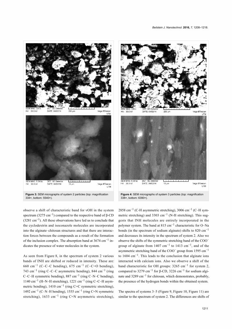
Results and DiscussionMorphology of microparticulate systemsAs one can see from Figure 2, system 1 consists of a mixture
of irregularly shaped micro- and nanoparticles with predomi-
nance of the first. The main size range of the obtained particles
falls in the limits of approximately 0.1–250 μm. The particles
Figure 2: SEM micrographs of system 1 particles (top: magnification331×, bottom: 11770×).
surface is smooth compared to those of systems 2–5 (see below
Figures 3–6), which have many rough edges, probably due to
the excess of β-cyclodextrin that adhered to the surface of algi-
nate–chitosan matrix. The particles of systems 2, 3 and 5, com-
pared to systems 1 and 4, are almost spherical. System 4
contains filamentous formations, characteristic for calcium algi-
nate. Particles with sizes below 100 nm could not be detected
because the obtained images were rather unclear.
Further information was obtained from the analysis of the FTIR
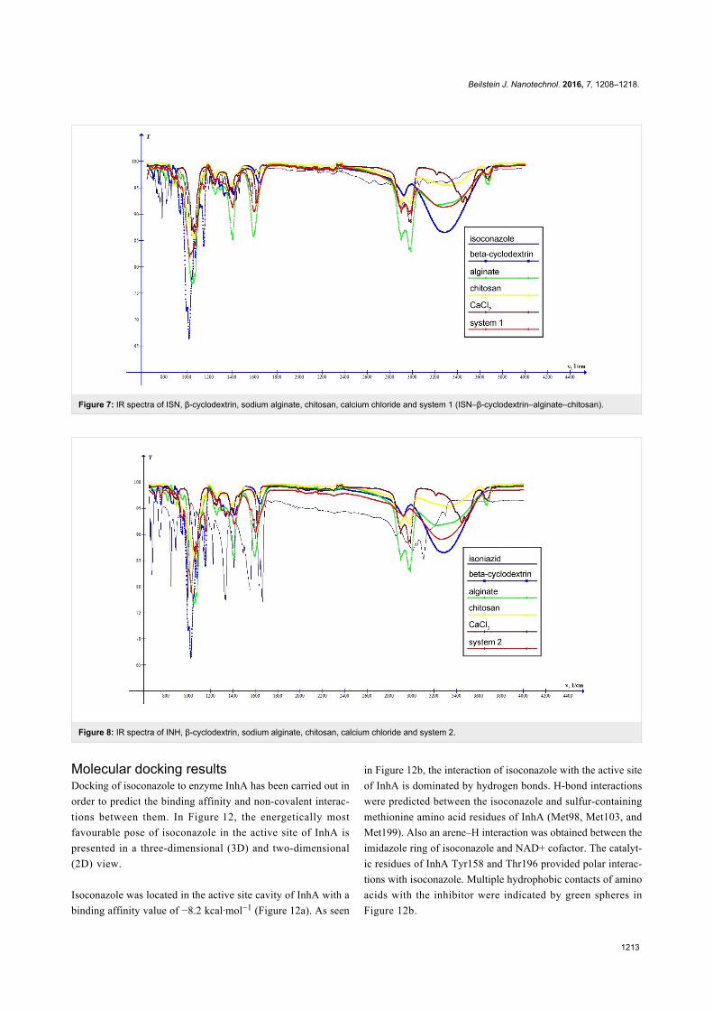
spectra. As one can see from Figure 7, the spectrum of system 1
is mainly the sum of the spectra of its components. However,
some of the absorption bands of the compounds disappear in the
spectrum of the system. These are, for instance, the bands at
754 cm−1 and 2924 cm−1 in the β-CD spectrum, which corre-
spond to the vibrations (δCCO + δCCH) and νasCH, respective-
ly, and the bands at 862 cm−1, 1439 cm−1 and 1585 cm−1 (C=N
bond stretching) in the isoconazole spectrum. Also, one can
Beilstein J. Nanotechnol. 2016, 7, 1208–1218.
1211
Figure 3: SEM micrographs of system 2 particles (top: magnification334×, bottom: 5540×).
observe a shift of characteristic band for νOH in the system
spectrum (3275 cm−1) compared to the respective band of β-CD
(3281 cm−1). All these observations have led us to conclude that
the cyclodextrin and isoconazole molecules are incorporated
into the alginate–chitosan structures and that there are interac-
tion forces between the compounds as a result of the formation
of the inclusion complex. The absorption band at 3674 cm−1 in-
dicates the presence of water molecules in the system.
As seen from Figure 8, in the spectrum of system 2 various
bands of INH are shifted or reduced in intensity. These are:
660 cm−1 (C–C–C bending), 675 cm−1 (C–C=O bending),
743 cm−1 (ring C–C–C asymmetric bending), 844 cm−1 (ring
C–C–H symmetric bending), 887 cm−1 (ring C–N–C bending),
1140 cm−1 (H–N–H stretching), 1221 cm−1 (ring C–C–H asym-
metric bending), 1410 cm−1 (ring C=C symmetric stretching),
1492 cm−1 (C–N–H bending), 1555 cm−1 (ring C=N symmetric
stretching), 1633 cm−1 (ring C=N asymmetric stretching),
Figure 4: SEM micrographs of system 3 particles (top: magnification338×, bottom: 6390×).
2858 cm−1 (C-H asymmetric stretching), 3006 cm−1 (C–H sym-
metric stretching) and 3303 cm−1 (N-H stretching). This sug-
gests that INH molecules are entirely incorporated in the
polymer system. The band at 815 cm−1 characteristic for O–Na
bonds (in the spectrum of sodium alginate) shifts to 820 cm−1
and decreases its intensity in the spectrum of system 2. Also we
observe the shifts of the symmetric stretching band of the COO−
group of alginate from 1407 cm−1 to 1413 cm−1, and of the
asymmetric stretching band of the COO− group from 1595 cm−1
to 1604 cm−1. This leads to the conclusion that alginate ions
interacted with calcium ions. Also we observe a shift of the
band characteristic for OH groups: 3265 cm−1 for system 2,
compared to 3279 cm−1 for β-CD, 3226 cm−1 for sodium algi-
nate and 3289 cm−1 for chitosan, which demonstrates, probably,
the presence of the hydrogen bonds within the obtained system.
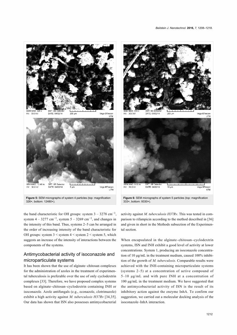
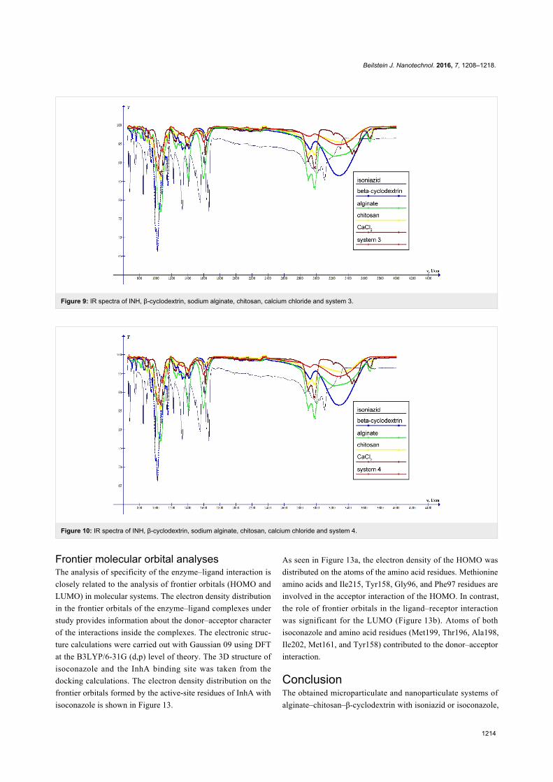
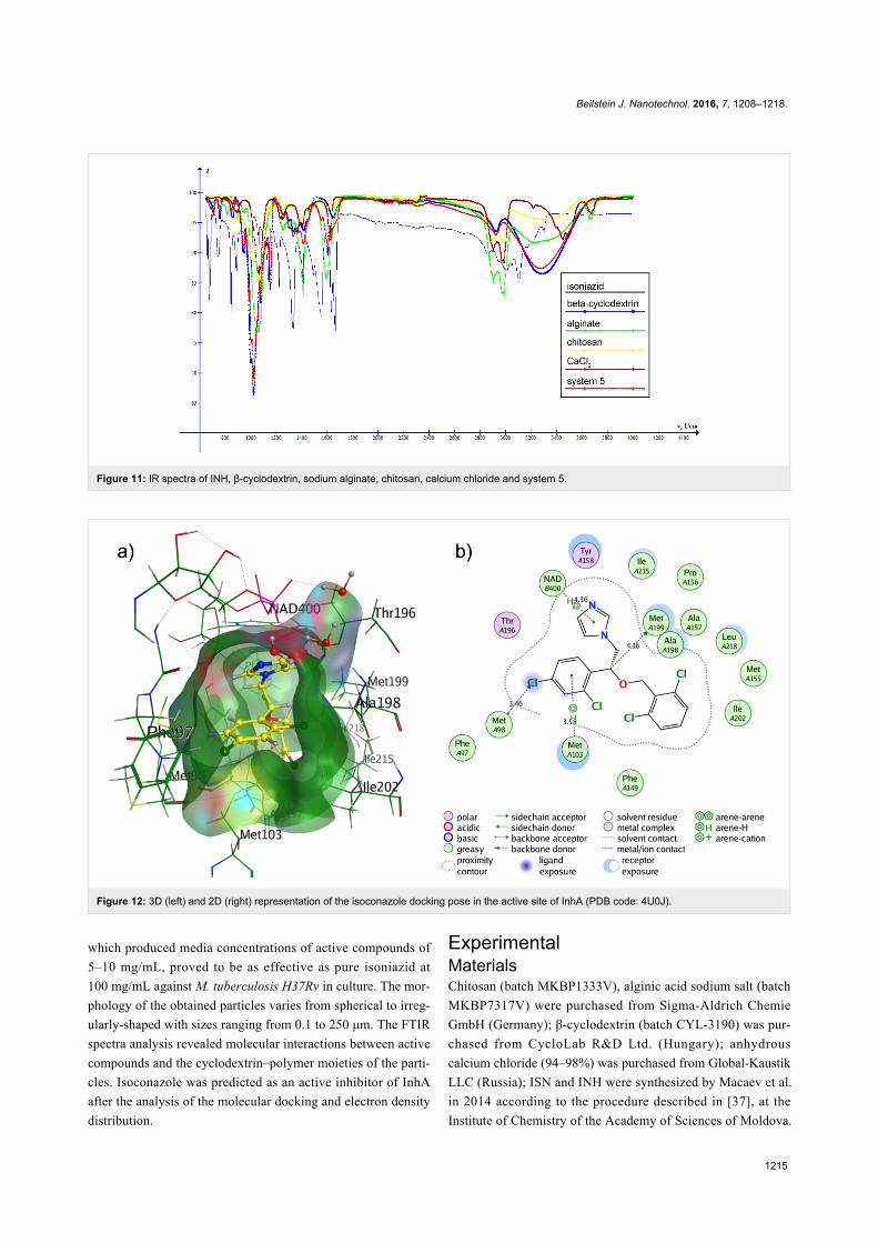
The spectra of systems 3–5 (Figure 9, Figure 10, Figure 11) are
similar to the spectrum of system 2. The differences are shifts of
Beilstein J. Nanotechnol. 2016, 7, 1208–1218.
1212
Figure 5: SEM micrographs of system 4 particles (top: magnification326×, bottom: 12480×).
the band characteristic for OH groups: system 3 – 3278 cm−1,
system 4 – 3277 cm−1, system 5 – 3269 cm−1, and changes in
the intensity of this band. Thus, systems 2–5 can be arranged in
the order of increasing intensity of the band characteristic for
OH groups: system 3 < system 4 < system 2 < system 5, which
suggests an increase of the intensity of interactions between the
components of the systems.
Antimycobacterial activity of isoconazole andmicroparticulate systemsIt has been shown that the use of alginate–chitosan complexes
for the administration of azoles in the treatment of experimen-
tal tuberculosis is preferable over the use of only cyclodextrin
complexes [33]. Therefore, we have proposed complex systems
based on alginate–chitosan–cyclodextrin containing INH or
isoconazole. Azole antifungals (e.g., econazole, clotrimazole)
exhibit a high activity against M. tuberculosis H37Rv [34,35].
Our data has shown that ISN also possesses antimycobacterial
Figure 6: SEM micrographs of system 5 particles (top: magnification333×, bottom: 9330×).
activity against M. tuberculosis H37Rv. This was tested in com-
parison to rifampicin according to the method described in [36]
and given in short in the Methods subsection of the Experimen-
tal section.
When encapsulated in the alginate–chitosan–cyclodextrin
systems, ISN and INH exhibit a good level of activity at lower
concentrations. System 1, producing an isoconazole concentra-
tion of 10 μg/mL in the treatment medium, caused 100% inhibi-
tion of the growth of M. tuberculosis. Comparable results were
achieved with the INH-containing microparticulate systems
(systems 2–5) at a concentration of active compound of
5–10 μg/mL and with pure INH at a concentration of
100 μg/mL in the treatment medium. We have suggested that
the antimycobacterial activity of ISN is the result of its
inhibitory action against the enzyme InhA. To confirm our
suggestion, we carried out a molecular docking analysis of the
isoconazole–InhA interaction.
Beilstein J. Nanotechnol. 2016, 7, 1208–1218.
1213
Figure 7: IR spectra of ISN, β-cyclodextrin, sodium alginate, chitosan, calcium chloride and system 1 (ISN–β-cyclodextrin–alginate–chitosan).
Figure 8: IR spectra of INH, β-cyclodextrin, sodium alginate, chitosan, calcium chloride and system 2.
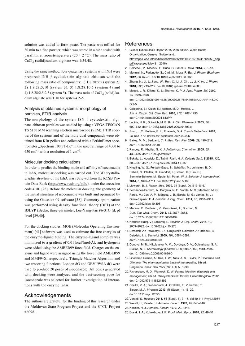
Molecular docking resultsDocking of isoconazole to enzyme InhA has been carried out in
order to predict the binding affinity and non-covalent interac-
tions between them. In Figure 12, the energetically most
favourable pose of isoconazole in the active site of InhA is
presented in a three-dimensional (3D) and two-dimensional
(2D) view.
Isoconazole was located in the active site cavity of InhA with a
binding affinity value of −8.2 kcal·mol−1 (Figure 12a). As seen
in Figure 12b, the interaction of isoconazole with the active site
of InhA is dominated by hydrogen bonds. H-bond interactions
were predicted between the isoconazole and sulfur-containing
methionine amino acid residues of InhA (Met98, Met103, and
Met199). Also an arene–H interaction was obtained between the
imidazole ring of isoconazole and NAD+ cofactor. The catalyt-
ic residues of InhA Tyr158 and Thr196 provided polar interac-
tions with isoconazole. Multiple hydrophobic contacts of amino
acids with the inhibitor were indicated by green spheres in
Figure 12b.
Beilstein J. Nanotechnol. 2016, 7, 1208–1218.
1214
Figure 9: IR spectra of INH, β-cyclodextrin, sodium alginate, chitosan, calcium chloride and system 3.
Figure 10: IR spectra of INH, β-cyclodextrin, sodium alginate, chitosan, calcium chloride and system 4.
Frontier molecular orbital analysesThe analysis of specificity of the enzyme–ligand interaction is
closely related to the analysis of frontier orbitals (HOMO and
LUMO) in molecular systems. The electron density distribution
in the frontier orbitals of the enzyme–ligand complexes under
study provides information about the donor–acceptor character
of the interactions inside the complexes. The electronic struc-
ture calculations were carried out with Gaussian 09 using DFT
at the B3LYP/6-31G (d,p) level of theory. The 3D structure of
isoconazole and the InhA binding site was taken from the
docking calculations. The electron density distribution on the
frontier orbitals formed by the active-site residues of InhA with
isoconazole is shown in Figure 13.
As seen in Figure 13a, the electron density of the HOMO was
distributed on the atoms of the amino acid residues. Methionine
amino acids and Ile215, Tyr158, Gly96, and Phe97 residues are
involved in the acceptor interaction of the HOMO. In contrast,
the role of frontier orbitals in the ligand–receptor interaction
was significant for the LUMO (Figure 13b). Atoms of both
isoconazole and amino acid residues (Met199, Thr196, Ala198,
Ile202, Met161, and Tyr158) contributed to the donor–acceptor
interaction.
ConclusionThe obtained microparticulate and nanoparticulate systems of
alginate–chitosan–β-cyclodextrin with isoniazid or isoconazole,
Beilstein J. Nanotechnol. 2016, 7, 1208–1218.
1215
Figure 11: IR spectra of INH, β-cyclodextrin, sodium alginate, chitosan, calcium chloride and system 5.
Figure 12: 3D (left) and 2D (right) representation of the isoconazole docking pose in the active site of InhA (PDB code: 4U0J).
which produced media concentrations of active compounds of
5–10 mg/mL, proved to be as effective as pure isoniazid at
100 mg/mL against M. tuberculosis H37Rv in culture. The mor-
phology of the obtained particles varies from spherical to irreg-
ularly-shaped with sizes ranging from 0.1 to 250 μm. The FTIR
spectra analysis revealed molecular interactions between active
compounds and the cyclodextrin–polymer moieties of the parti-
cles. Isoconazole was predicted as an active inhibitor of InhA
after the analysis of the molecular docking and electron density
distribution.
ExperimentalMaterialsChitosan (batch MKBP1333V), alginic acid sodium salt (batch
MKBP7317V) were purchased from Sigma-Aldrich Chemie
GmbH (Germany); β-cyclodextrin (batch CYL-3190) was pur-
chased from CycloLab R&D Ltd. (Hungary); anhydrous
calcium chloride (94–98%) was purchased from Global-Kaustik
LLC (Russia); ISN and INH were synthesized by Macaev et al.
in 2014 according to the procedure described in [37], at the
Institute of Chemistry of the Academy of Sciences of Moldova.

Beilstein J. Nanotechnol. 2016, 7, 1208–1218.
1216
Figure 13: 3D view of HOMO (a) and LUMO (b) for the InhA active site interacting with isoconazole.
MethodsBacterial strains and growth conditionsThe procedure was adapted from [36]. Shortly, for the first three
experiments, M. tuberculosis H37Rv inocula were first passaged
in radiometric 7H12 broth (BACTEC 12B) until the growth
index (GI) reached 800–999. For the fourth replicate experi-
ment, H37Rv was grown in 100 mL of Middlebrook 7H9 broth
supplemented with 0.2% glycerol, 10% OADC (oleic acid,
albumin, dextrose, catalase), and 0.05% Tween 80 (Sigma), all
taken in v/v concentrations. Cultures were incubated in 500 mL
nephelometer culture flasks on a rotary shaker at 150 rpm and
37 °C until they reached an optical density of 0.4–0.5 at
550 nm. The, the bacteria were washed and suspended in 20 mL
PBS and passed through an 8 mm pore-size filter to eliminate
clumps. The filtrates were aliquoted, stored at 280 K, and used
within 30 days.
Radiometric susceptibility testFor the radiometric susceptibility test, 1/10 mL of BACTEC
12B-passaged inoculum was introduced into 4 mL of test medi-
um without being diluted. Two-fold drug dilutions were pre-
pared in DMSO and introduced with a 0.5 mL insulin syringe in
a volume of 50 mL. Frozen inocula were subjected to 1:20 dilu-
tion in BACTEC 12B medium, and then 0.1 mL was intro-
duced into the test medium, which yielded 5.0 × 10⁵ CFU per
BACTEC vial. Drug-free control vials were composed to
include only solvent with bacterial inoculum and solvent with a
1:100 dilution of bacterial inoculum (1:100 controls). All vials
were incubated at 37 °C. The GI was determined in a BACTEC
460 instrument until the GI of the 1:100 controls reached at
least 30. The next day, all vials were read. The GI and daily
change in GI (DGI) were recorded for each drug dilution. The
MIC was defined as the lowest concentration for which the DGI
value was smaller than the DGI value of the 1:100 control (10).
If the GI of the test sample was greater than 100, the sample
was considered resistant even if the DGI was less than the DGI
of the 1:100 control.
Preparation of the microparticulate systemsThe quaternary system: ISN–β-cyclodextrin–alginate–chitosan
(system 1) was obtained using kneading, the mass ratio of com-
ponents was 1:2.36:4:4. At the first stage, the binary system
ISN–β-cyclodextrin was obtained by kneading, the molar ratio
of the components was 1:1, and the working temperature was
20 ± 2 °C. In an agate mortar appropriate amounts of β-cyclo-
dextrin and ISN, previously weighed on the electronic analyti-
cal balance model BEL M503i, were added. To the mixture, a
sufficient amount of distilled water to form a paste was added.
The paste was kneaded with a pestle for 90 min: In the first
60 min by adding distilled water to compensate its loss by evap-
oration and maintain the appearance of paste. In the next 30 min
the mixture was milled to a fine powder. The obtained powder
was stored in a sealed tube with parafilm sample, at room tem-
perature (20 ± 2 °C).
A weighed quantity of obtained powder (binary system ISN–β-
cyclodextrin) was transferred in a mortar. Then, chitosan ac-
cording to the proportion 1:2.36:4:4 was added to it, mixed with
the pestle, after which distilled water was added till a paste was
formed. The obtained paste was kneaded for 30 min and the
corresponding amount of sodium alginate was added. The re-
sulting mixture was milled and then 20 mM calcium chloride
Beilstein J. Nanotechnol. 2016, 7, 1208–1218.
1217
solution was added to form paste. The paste was milled for
30 min to a fine powder, which was stored in a tube sealed with
parafilm, at room temperature (20 ± 2 °C). The mass ratio of
CaCl2 (solid)/sodium alginate was 1:34.48.
Using the same method, four quaternary systems with INH were
prepared: INH–β-cyclodextrin–alginate–chitosan with the
following mass ratio of components: 1) 1:8.28:5:5 (system 2);
2) 1:8.28:5:10 (system 3); 3) 1:8.28:10:5 (system 4) and
4) 1:8.28:2.5:2.5 (system 5). The mass ratio of CaCl2 (solid)/so-
dium alginate was 1:10 for systems 2–5.
Analysis of obtained systems: morphology ofparticles, FTIR analysisThe morphology of the system ISN–β-cyclodextrin–algi-
nate–chitosan particles was studied by using a VEGA TESCAN
TS 5130 MM scanning electron microscope (SEM). FTIR spec-
tra of the systems and of the individual compounds were ob-
tained from KBr pellets and collected with a PerkinElmer spec-
trometer „Spectrum 100 FT-IR” in the spectral range of 4000 to
650 cm−1 with a resolution of 1 cm−1.
Molecular docking calculationsIn order to predict the binding mode and affinity of isoconazole
to InhA, molecular docking was carried out. The 3D crystallo-
graphic structure of the InhA was retrieved from the RCSB Pro-
tein Data Bank (http://www.rcsb.org/pdb/), under the accession
code 4U0J [28]. Before the molecular docking, the geometry of
the initial structure of isoconazole was built and optimized by
using the Gaussian 09 software [38]. Geometry optimization
was performed using density functional theory (DFT) at the
B3LYP (Becke, three-parameter, Lee-Yang-Parr)/6-31G (d, p)
level [39,40].
For the docking studies, MOE (Molecular Operating Environ-
ment) [41] software was used to estimate the free energies of
the enzyme–ligand binding. The enzyme–ligand complex was
minimized to a gradient of 0.01 kcal/(mol·Å), and hydrogens
were added using the AMBER99 force field. Charges on the en-
zyme and ligand were assigned using the force field AMBER99
and MMF94X, respectively. Triangle Matcher Algorithm and
two rescoring functions, London dG and GBVI/WSA dG were
used to produce 20 poses of isoconazole. All poses generated
with docking were analyzed and the best-scoring pose for
isoconazole was selected for further investigation of interac-
tions with the enzyme InhA.
AcknowledgementsThe authors are grateful for the funding of this research under
the Moldovan State Program Project and the STCU Project
#6098.
References1. Global Tuberculosis Report 2015, 20th edition, World Health
Organization, Geneva, Switzerland.http://apps.who.int/iris/bitstream/10665/191102/1/9789241565059_eng.pdf (accessed May 31, 2016).
2. Boldescu, V.; Macaev, F.; Duca, G. Chem. J. Mold. 2014, 9, 8–13.3. Mennini, N.; Furlanetto, S.; Cirri, M.; Mura, P. Eur. J. Pharm. Biopharm.
2012, 80, 67–75. doi:10.1016/j.ejpb.2011.08.0024. Zhang, N.; Li, J.; Jiang, W.; Ren, C.; Li, J.; Xin, J.; Li, K. Int. J. Pharm.
2010, 393, 213–219. doi:10.1016/j.ijpharm.2010.04.0065. Moses, L. R.; Dileep, K. J.; Sharma, C. P. J. Appl. Polym. Sci. 2000,
75, 1089–1096.doi:10.1002/(SICI)1097-4628(20000228)75:9<1089::AID-APP1>3.0.CO;2-5
6. Gelperina, S.; Kisich, K.; Iseman, M. D.; Heifets, L.Am. J. Respir. Crit. Care Med. 2005, 172, 1487–1490.doi:10.1164/rccm.200504-613PP
7. Labiris, N. R.; Dolovich, M. B. Br. J. Clin. Pharmacol. 2003, 56,600–612. doi:10.1046/j.1365-2125.2003.01893.x
8. Sung, J. C.; Pulliam, B. L.; Edwards, D. A. Trends Biotechnol. 2007,25, 563–570. doi:10.1016/j.tibtech.2007.09.005
9. Bailey, M. M.; Berkland, C. J. Med. Res. Rev. 2009, 29, 196–212.doi:10.1002/med.20140
10. Pandey, R.; Khuller, G. K. J. Antimicrob. Chemother. 2005, 55,430–435. doi:10.1093/jac/dki027
11. Bekale, L.; Agudelo, D.; Tajmir-Riahi, H. A. Colloids Surf., B 2015, 125,309–317. doi:10.1016/j.colsurfb.2014.11.037
12. Kreyling, W. G.; Fertsch-Gapp, S.; Schäffler, M.; Johnston, B. D.;Haberl, N.; Pfeiffer, C.; Diendorf, J.; Schleh, C.; Hirn, S.;Semmler-Behnke, M.; Epple, M.; Parak, W. J. Beilstein J. Nanotechnol.2014, 5, 1699–1711. doi:10.3762/bjnano.5.180
13. Lipworth, B. J. Respir. Med. 2000, 94 (Suppl. D), S13–S16.14. Fernández-Ferreiro, A.; Bargiela, N. F.; Varela, M. S.; Martínez, M. G.;
Pardo, M.; Ces, A. P.; Méndez, J. B.; Barcia, M. G.; Lamas, M. J.;Otero-Espinar, F. J. Beilstein J. Org. Chem. 2014, 10, 2903–2911.doi:10.3762/bjoc.10.308
15. Macaev, F.; Boldescu, V.; Geronikaki, A.; Sucman, N.Curr. Top. Med. Chem. 2013, 13, 2677–2683.doi:10.2174/15680266113136660194
16. Nardello-Rataj, V.; Leclercq, L. Beilstein J. Org. Chem. 2014, 10,2603–2622. doi:10.3762/bjoc.10.273
17. Brzostek, A.; Pawelczyk, J.; Rumijowska-Galewicz, A.; Dziadek, B.;Dziadek, J. J. Bacteriol. 2009, 191, 6584–6591.doi:10.1128/JB.00488-09
18. Donova, M. N.; Nikolayeva, V. M.; Dovbnya, D. V.; Gulevskaya, S. A.;Suzina, N. E. Microbiology (London, U. K.) 2007, 153, 1981–1992.doi:10.1099/mic.0.2006/001636-0
19. Goodman Gilman, A.; Rall, T. W.; Nies, A. S.; Taylor, P. Goodman andGilman's: The pharmacological basis of therapeutics, 8th ed.;Pergamon Press: New York, NY, U.S.A., 1990.
20. Richardson, M. D.; Warnock, D. W. Fungal infection: diagnosis andmanagement, 4th ed.; Wiley-Blackwell: Oxford, United Kingdom, 2012.doi:10.1002/9781118321492
21. Czaika, V. A.; Siebenbrock, J.; Czekalla, F.; Zuberbier, T.;Sieber, M. A. Mycoses 2013, 56 (Suppl. 1), 16–22.doi:10.1111/myc.12055
22. Veraldi, S. Mycoses 2013, 56 (Suppl. 1), 3–15. doi:10.1111/myc.1205423. Wendt, H.; Kessler, J. Arzneim. Forsch. 1978, 28, 846–848.24. Kessler, H. J. Arzneim. Forsch. 1979, 29, 1344.25. Bosak, I. A.; Kotrekhova, I. P. Probl. Med. Mycol. 2010, 12, 49–51.
Beilstein J. Nanotechnol. 2016, 7, 1208–1218.
1218
26. Ayub, M.; Levell, M. J. J. Steroid Biochem. 1987, 28, 521–531.doi:10.1016/0022-4731(87)90511-5
27. Ayub, M.; Levell, M. J. J. Steroid Biochem. 1988, 31, 65–72.doi:10.1016/0022-4731(88)90207-5
28. He, X.; Alian, A.; Stroud, R.; Ortiz de Montellano, P. R. J. Med. Chem.2006, 49, 6308–6323. doi:10.1021/jm060715y
29. Omar, S.; Ahmad, N.; Ahmad, F. Pertanika 1988, 11, 79–85.30. Kakita, H.; Kamishima, H. J. Appl. Phycol. 2008, 5, 543–549.
doi:10.1007/s10811-008-9317-531. Sonia, T. A.; Sharma, C. P. Chitosan and Its Derivatives for Drug
Delivery Perspective. In Chitosan for Biomaterials I; Jayakumar, R.;Prabaharan, M.; Muzzarelli, R. A. A., Eds.; Advances in PolymerScience, Vol. 243; Springer: Berlin, Germany, 2011; pp 23–54.doi:10.1007/12_2011_117
32. Honarkar, H.; Barikani, M. Monatsh. Chem. 2009, 140, 1403–1420.doi:10.1007/s00706-009-0197-4
33. Ahmad, Z.; Sharma, S.; Khuller, G. K. Nanomedicine 2007, 3,239–243. doi:10.1016/j.nano.2007.05.001
34. Ahmad, Z.; Sharma, S.; Khuller, G. K. FEMS Microbiol. Lett. 2005, 251,19–22. doi:10.1016/j.femsle.2005.07.022
35. Ahmad, Z.; Sharma, S.; Khuller, G. K. FEMS Microbiol. Lett. 2006, 258,200–203. doi:10.1111/j.1574-6968.2006.00224.x
36. Collins, L.; Franzblau, S. G. Antimicrob. Agents Chemother. 1997, 41,1004–1009.
37. Dulcevscaia, G. M.; Kravtsov, V. Ch.; Macaev, F. Z.; Duca, G. G.;Stingachi, E. P.; Pogrebnoi, S. I.; Boldescu, V. V.; Clapco, S. F.;Tiurina, J. P.; Deseatnic-Ciloci, A. A.; Lipkowski, J.; Liu, S.-X.;Decurtins, S.; Baca, S. G. Polyhedron 2013, 52, 106–114.doi:10.1016/j.poly.2012.10.040
38. Gaussian 09; Gaussian, Inc.: Wallingford, CT, U.S.A., 2009.39. Becke, A. D. J. Chem. Phys. 1993, 98, 5648–5652.
doi:10.1063/1.46491340. Lee, C.; Yang, W.; Parr, R. G. Phys. Rev. B 1988, 37, 785–789.
doi:10.1103/PhysRevB.37.78541. Molecular Operating Environment (MOE); Chemical Computing Group
Inc.: Montreal, QC, Canada.
License and TermsThis is an Open Access article under the terms of the
Creative Commons Attribution License
(http://creativecommons.org/licenses/by/4.0), which
permits unrestricted use, distribution, and reproduction in
any medium, provided the original work is properly cited.
The license is subject to the Beilstein Journal of
Nanotechnology terms and conditions:
(http://www.beilstein-journals.org/bjnano)
The definitive version of this article is the electronic one
which can be found at:
doi:10.3762/bjnano.7.112


![Cross-Linked Alginate Film Pore Size Determination Using ...genipin-chitosan alginate membranes [11] have resulted in tremendous improvements in membrane strength. A signifi- cant](https://img.pdfslide.us/doc/110x75/5ff3ad32ce36184d7d3d61da/cross-linked-alginate-film-pore-size-determination-using-genipin-chitosan-alginate.jpg)