Embed Size (px)
Citation preview

Preparation, Characterization, and Surface Modification of Silver Nanoparticles inFormamide
Anjana Sarkar, Sudhir Kapoor,* and Tulsi MukherjeeRadiation Chemistry & Chemical Dynamics DiVision, Bhabha Atomic Research Centre, Mumbai 400 085, India
ReceiVed: December 21, 2004; In Final Form: February 21, 2005
The reduction of silver ions in formamide is shown to take place spontaneously at room temperature withoutaddition of any reductant. The growth of Ag particles was found to be dependent on Ag+ ion concentration.In the absence of any stabilizer, deposition of silver film on the glass walls of the container takes place.However, in the presence of poly(N-vinyl-2-pyrrolidone) (PVP) or colloidal silica (SiO2), which are capableof stabilizing silver nanoparticles by complexing and providing support, a clear dispersion was obtained. Theformation of the silver nanoparticles under different conditions was investigated through UV-visible absorptionspectrophotometry, gas chromatography, and also electron and atomic force microscopic techniques. Atomicforce microscopy results for silver films prepared in the absence of any stabilizer showed the formation ofpolygonal particles with sizes around 100 nm. Transmission electron microscopy results showed that theprepared silver particles in the presence of PVP were around 20 nm. The Ag nanoparticles get oxidized in thepresence of chloroform and toluene. Surface modification of silver film was done in the presence of thetetrasodium salt of ethylenediaminetetraaceticacid (Na4EDTA). It was shown that the reactivity of the silverfilm increased in its presence. The Fermi potential of silver particles in the presence of Na4EDTA seems tolie between-0.33 and-0.446 V vs NHE.
Introduction
A large number of methods have been developed for thesynthesis of metal nanoparticles since the discovery by Faradayon metal particles.1 To stabilize metal nanoparticles, generallysynthetic polymers2-6 and ligands7-13 are widely used. Suchsubstances can also control the reduction rate of metal ions andthe aggregation processes of the metal clusters.3-11 The interac-tion of polymer with metal ions plays a crucial role in controllingthe size and morphology of the particles. El-Sayed et al.12 haveshown that, by changing the capping ratio of metal ions topolymer, one can control the shape of the nanoparticles.
Recently, metals or semiconductor nanoparticles synthesizedby various techniques have found potential application in manyfields such as catalysis, sensors, etc.14-17 In most of theapplications, nanoparticles are used as building blocks towardfunctional nanostructures. The coinage metal nanoparticles suchas silver, gold, and copper are mostly exploited for suchpurposes as they have surface plasmon resonance absorptionin the UV-visible region.3 The surface plasmon band arisesfrom the coherent existence of free electrons in the conductionband due to the small particles size effect, which is dependenton the particle sizes, chemical surrounding, adsorbed specieson the surface, and dielectric constant.3-6 The unique featureof the coinage metal nanoparticles is that a change in theabsorbance or wavelength provides a measure of the particlesize, shape, and interparticle properties. In this, one-dimensionalnanostructures such as wires, rods, etc. have become the focusof intensive research because of their unique applications.14-17
For small particles (<2 nm), the surface plasmon band isstrongly damped due to low electron density in the conductionband. However, as particle size increases, the intensity of the
surface plasmon band increases. It has been suggested that,although both absorbance and scattering contribute to the opticalproperty, the contribution of the latter is relatively insignificantas compared to that of the former for very small nanoparticles(e15 nm).14-23
Recently, Liz-Marzan et al.24-28 have shown that silvernanoparticles can be prepared in DMF without adding anyreductant. It has been suggested that the reduction rate can beenhanced at high temperatures. Because most of the organicreactions take place in organic solvents, it is desirable to developsynthetic methods that lead to the formation of particles havingdifferent morphology in addition to the stabilization of metalnanoparticles in such solvents. In addition, if one can design asynthetic method to prepare different morphological particlesin the same medium without adding reductants from outside,this may have its own important implications.
Formamide is one of the most common solvents used to studyvarious processes such as formation of metal nanoparticles,interaction with alcohols, etc.29-32 It is known that formamide-water and formamide-methanol complexes can serve as modelsystems for protein-water and protein-solvent interactions.Photocatalytic oxidation of volatile organic chemicals (VOCs)constitutes one of the most promising methods for the removalof these pollutants in an enclosed atmosphere.33-35 Generally,metal oxides are being explored for such purposes.33,34As silveris known to have bactericidal properties, it is important to assessits effect on VOCs. In this paper, we describe in detail thepreparation of silver nanoparticles by reduction of silver ionswith formamide in the absence and presence of stabilizers suchas poly(N-vinyl-2-pyrrolidone) (PVP) and SiO2 nanoparticles.In addition, attempts have been made to see the effect of additionof methanol, VOCs, and a complexing agent on the stability ofsilver nanoparticles.
* Corresponding author. Tel.: (+)91-22-25590298. Fax: (+)91-22-25505151. E-mail: [email protected].
7698 J. Phys. Chem. B2005,109,7698-7704
10.1021/jp044201r CCC: $30.25 © 2005 American Chemical SocietyPublished on Web 03/30/2005
Experimental Section
Materials. Silver perchlorate (Aldrich), AgNO3 (BDH, India),tetrasodium salt of ethylenediaminetetraaceticacid (Sigma),methanol, CHCl3 and toluene (UV spectroscopy grade, Spec-trochem, India,), PVP (mol wt.) 360 000 and 40 000) (Sigma),methyl viologen dichloride{paraquat: 1,1′-dimethyl 4,4′-dipyridinium dichloride; MV2+ (Cl-)2} (Aldrich), and SiO2
(LUDOX SM-30, Aldrich) were used as received. IOLAR gradeN2 gas (purity g99.99%) used for purging solutions wasobtained from Indian Oxygen Limited. All solutions wereprepared just before the experiments and kept in dark to avoidany photochemical reactions. Water purified through a Milliporesystem was used.
Film Preparation. The slides were cleaned in perchloric acid.After being rinsed with distilled and Millipore water, they weredried in an oven. The slides were suspended in formamidesolution containing varying concentrations of silver ions. Filmformation takes place with time on the surface of the slide. Theslides were removed after 24 h and dried at room temperature.Care was taken to protect these slides from light both duringand after film formation.
Preparation of Silver Nanoparticles in the Presence ofStabilizer. Dispersions of silver nanoparticles were preparedby mixing the required concentration of PVP or SiO2 informamide followed by addition of silver salt at room temper-ature. With time, the solution gradually turned yellow, andevolution of a stable dispersion of silver nanoparticles wasobserved.
Charge and Size Determination.Zeta potential measure-ments, for determining the size and charge on SiO2 nanoparticlesin formamide and water, were carried out using MalvernInstrument, zeta sizer (nanosizer). No sign of agglomeration and/or precipitation of SiO2 nanoparticles in formamide was foundat least up to 45 days.
Gas Chromatography Experiments.Silver dispersions wereprepared in rectangular quartz cells of dimension 1 cm× 1 cm× 5 cm. The solution was bubbled with nitrogen, and the cellwas closed with a self-sealing septum. The evolved gas wasmeasured using a gas chromatograph after 28 h. For CO2
detection, a Porapak Q-S column and a thermal conductivitydetector were used. Helium was used as a carrier gas. Attemptwas also made to see the evolution of other gases, if any. ForH2 detection, a column with molecular sieve-5A and N2 ascarrier gas was used. The temperatures of the measurementsfor CO2 and H2 were 40 and 32°C, respectively.
Amount of Silver Metal Formed. The amount of silver metalformed during the reduction of silver ions was determined after28 h using a Chemito AA203 atomic absorption spectropho-tometer (AAS). The slit width and lamp current were 0.5 nmand 5 mA, respectively. The silver was detected at 228 nm usingan air acetylene flame.
Characterization. Samples for transmission electron micros-copy (TEM) were prepared by putting a drop of the colloidalsolution on a copper grid coated with a thin amorphous carbonfilm. Samples were dried and kept under vacuum in a desiccatorbefore being put in a specimen holder. TEM characterizationwas carried out using a JEOL JEM-2000FX electron micro-scope. Particle sizes were measured from the TEM micrographsand calculated by taking at least 100 particles. Absorptionmeasurements were carried out on a JascoV-530 spectropho-tometer. The spectra were recorded at room temperature usingeither a 0.2 or a 1 cmquartz cuvette. However, all of the spectrawere later normalized to 1 cm path length of the cuvette. Atomicforce microscopy (AFM) characterization was carried out using
a Solver P-47H microscope. AFM measurements were done bycontact mode using Si3N4 tip, the resonance frequency andspring constant being 100 kHz and 0.6 N/m, respectively.
Dynamic light scattering experiments were done on a Malvern4800 Autosizer employing a 7132 digital correlator. Theintensity correlation function was analyzed by method ofcumulants using the mean and variance of the distribution onthe fitted variable. The diffusion coefficient (D) of the particleis related to the average decay rate (Γ) of the correlation functionby Γ ) Dq2, whereq is the magnitude of the scattering vector(given byq ) 4πn/λ sin θ/2; λ is the wavelength of light,n isthe refractive index, andq is the scattering angle). The meanhydrodynamic coefficient of the particles was obtained fromthe diffusion coefficient using the Stokes-Einstein relationship.The light source was an Ar+ ion laser operated at 514.5 nmand a scattering angleq ) 90°.
Results and Discussion
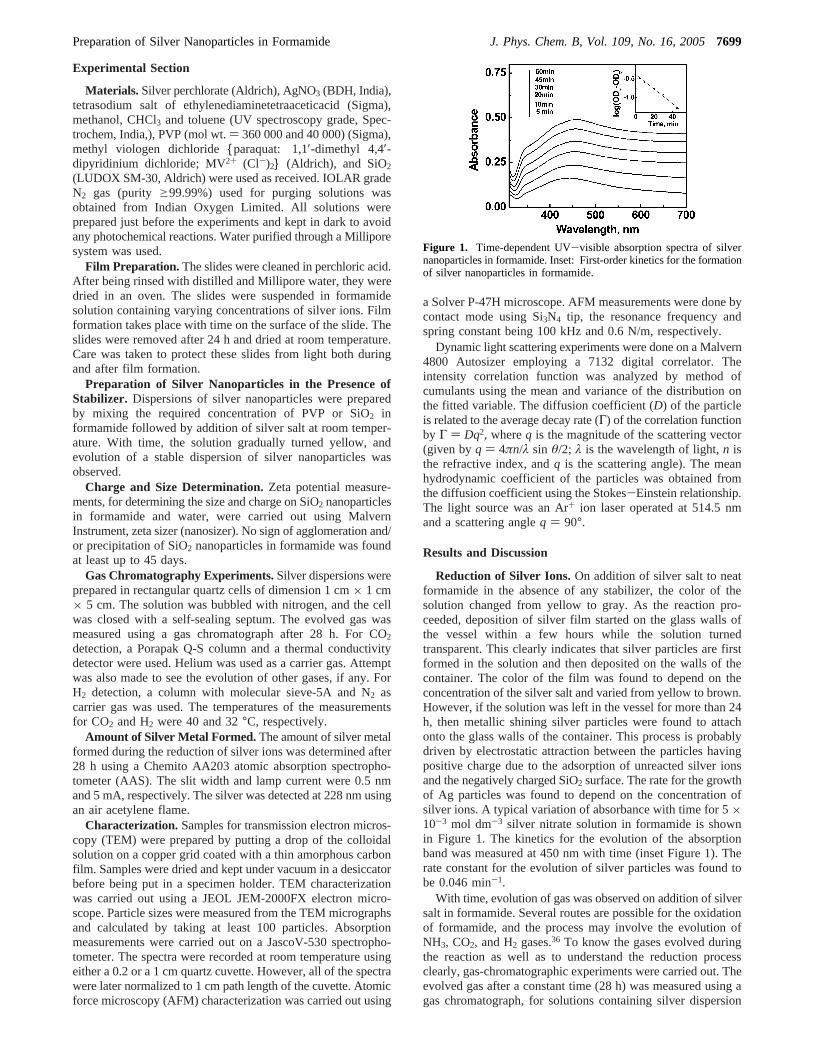
Reduction of Silver Ions.On addition of silver salt to neatformamide in the absence of any stabilizer, the color of thesolution changed from yellow to gray. As the reaction pro-ceeded, deposition of silver film started on the glass walls ofthe vessel within a few hours while the solution turnedtransparent. This clearly indicates that silver particles are firstformed in the solution and then deposited on the walls of thecontainer. The color of the film was found to depend on theconcentration of the silver salt and varied from yellow to brown.However, if the solution was left in the vessel for more than 24h, then metallic shining silver particles were found to attachonto the glass walls of the container. This process is probablydriven by electrostatic attraction between the particles havingpositive charge due to the adsorption of unreacted silver ionsand the negatively charged SiO2 surface. The rate for the growthof Ag particles was found to depend on the concentration ofsilver ions. A typical variation of absorbance with time for 5×10⁻³ mol dm-3 silver nitrate solution in formamide is shownin Figure 1. The kinetics for the evolution of the absorptionband was measured at 450 nm with time (inset Figure 1). Therate constant for the evolution of silver particles was found tobe 0.046 min-1.
With time, evolution of gas was observed on addition of silversalt in formamide. Several routes are possible for the oxidationof formamide, and the process may involve the evolution ofNH3, CO2, and H2 gases.36 To know the gases evolved duringthe reaction as well as to understand the reduction processclearly, gas-chromatographic experiments were carried out. Theevolved gas after a constant time (28 h) was measured using agas chromatograph, for solutions containing silver dispersion
Figure 1. Time-dependent UV-visible absorption spectra of silvernanoparticles in formamide. Inset: First-order kinetics for the formationof silver nanoparticles in formamide.
Preparation of Silver Nanoparticles in Formamide J. Phys. Chem. B, Vol. 109, No. 16, 20057699

and neat formamide. In solution containing silver dispersion, itwas observed that the evolved gas was CO2. The amount ofCO2 evolved was found to be 2.04µmol for a solutioncontaining 10-2 mol dm-3 Ag+ in formamide. The amount ofAg metal formed was determined by AAS after an identicaltime (28 h) to quantify the amount of CO2 evolved. The amountof CO2 liberated was found to be 0.1059 mol and 2.4× 10⁻⁵
mol per mol of Ag metal and formamide, respectively. It isimportant to mention here that CO2 was not observed in absenceof silver ions under identical conditions in neat formamide. Itis pertinent to mention here that during reduction of silver ionsby DMF evolution of CO2 does not occur.24-28 This shows thatthe reduction of silver ions in formamide and DMF followsdifferent mechanisms.
Effect of Stabilizers and Counterion. When a stabilizingagent such as SiO2 or PVP was added to formamide beforeaddition of silver salt, stable clear dispersions of silver colloidswere obtained. However, no adsorption of metallic silver onthe walls of the container was observed in the presence of SiO2
or PVP. Therefore, to restrict the size of the nanoparticles (orto get clear dispersions of Ag nanoparticles) and to study theevolution of silver nanoparticles, different methodologies wereadopted. In the first case, silica nanoparticles (13 nm) were usedto provide support for silver nanoparticles. Figure 2 shows theevolution of silver nanoparticles stabilized by SiO2. On com-parison with the results obtained in Figure 1, it can be notedthat in the presence of silica the rate of formation of silverparticles decreases drastically. For 1× 10⁻² mol dm-3 AgNO3
solution containing 1.5 g/L of SiO2, the rate constant for theevolution of the surface plasmon absorption band of silverparticles was evaluated from the kinetic plot (inset Figure 2)and found to be 0.0086 min-1. It is important to clarify herethat the low absorbance yield in Figure 1 as compared to thatobtained in Figure 2 is due to the fact that in absence of SiO2
silver particles got deposited on the walls of the container.The size of the nanoparticles at a particular ratio of Ag+/
SiO2 was determined by dynamic light scattering. A representa-tive case is shown in Figure 3. The particle size was around 38nm for 1× 10⁻² mol dm-3 AgNO3 solution containing 1.5 g/LSiO2. The variation in the size of the particles with [Ag+]keeping the amount of SiO2 constant was also determined (Table1). It can be noted that as the concentration of Ag+ ionsincreased the size of the particles decreased. This can beexplained by assuming that, to generate small and monodis-persed particles, the nucleation and particle growth processesshould occur on different time scales.37 As the rate of evolutionof silver particles increases with the concentration of silver ions,
it appears that this could be the reason for observing smallerparticles at higher concentration of Ag+ ions.
To confirm the charge on colloidal SiO2 in formamide, itszeta potential was determined. It was noted that the charge oncolloidal SiO2 in formamide becomes more negative (-56.7mV) as compared to that in water (-37.3 mV). The zetapotential,ê, of colloid particles is sensitive to the inverse Debyescreening length (κ) of the medium, which in turn is decidedby the ionic strength and dielectric constant of the medium. Anincrease in dielectric constant decreases the value ofκ, therebyincreasing the effective potential of the particle at the shearplane. This explains the observed increase in the charge ofcolloidal SiO2 in formamide as compared to that in water. Dueto negative charge on the surface of SiO2 particles, theencounters between the particles are inhibited. This helps instabilizing small Ag particles on its surface. The results obtainedcorroborate the earlier findings in aqueous solutions.38,39
Figure 4 shows the surface plasmon absorption band of silvernanoparticles obtained at different times in formamide contain-ing 2 × 10⁻³ mol dm-3 AgNO3 and 1% PVP (wt/v). Theincrease in the intensity of the surface plasmon band at around425 nm indicates the continuous formation of silver particles.After an hour, the relative increase in the growth of the particlesdecreased as can be seen in Figure 4. It can be noticed that thesurface plasmon absorbance band remains symmetric and nosignificant change in the width of the band with time wasobserved. This implies that the size of the nanoparticles remainsmore or less uniform. The surface plasmon absorption bandposition of silver particles in formamide in the presence of PVPwas centered at 425 nm, which was red shifted with respect tothe absorption maximum of silver particles prepared in an
Figure 2. Time-dependent UV-visible absorption spectra of silvernanoparticles in formamide in the presence of 1.5 g/L SiO2. Inset: First-order kinetics for the formation of silver nanoparticles in formamidein the presence of SiO2.
Figure 3. Dynamic light scattering results showing monodispersityand particle size distribution of silver nanoparticles prepared from 1×10⁻² mol dm-3 AgNO3 in formamide containing 1.5 g/L SiO2.
TABLE 1: Effect of Silver Ion Concentration on the Size ofSilver Nanoparticle Containing 1.5 g/L SiO2
[Ag+], mol dm-3 diameter, nm
3 × 10⁻³ 481 × 10-2 38
7700 J. Phys. Chem. B, Vol. 109, No. 16, 2005 Sarkar et al.

aqueous solution. The possible reason for this is the higherrefractive index of formamide as compared to that of water.This is in agreement with the prediction by Mie theory. Kinetictraces showing formation of silver particles in formamide fordifferent Ag+ concentrations in the presence of constant PVP(1.0 wt %/v) are shown in Figure 5. It can be seen from thefigure that the rate of evolution of particles increases with anincrease in silver ion concentration. Similar results were obtainedwhen PVP of low molecular weight (40 000) was used. It shouldbe mentioned here that we could not observe any significantchange in the absorption spectrum when PVP of low molecularweight was used, indicating that particle sizes remain similar.
As discussed earlier, evolution of CO2 gas was also observedduring the reduction of silver ions in the presence of PVP. CO2
gas was measured under identical experimental conditions tosee if there exists any correlation between the concentration ofevolved gas and the growth of the silver plasmon absorptionband. In the presence of PVP, the amount of CO2 evolved wasmeasured to be 1.44µmol after 28 h. It may be noted that theamount of CO2 evolved in the absence of PVP was higher afteran identical time period. This is because the rate of reductionand aggregation is much slower in the presence of PVP than inits absence in formamide. However, it cannot be quantified interms of amount of CO2 evolved per mol of silver metal formed,as some Ag+ ions always remain unreduced in formamidecontaining PVP.
The bulk measurements can provide some evidence aboutthe size of the nanoparticles, but they do not provide exactinformation about the particle size and shape. Atomic force orelectron microscopy is an effective method to provide surfacetopography and phase images. For nanoparticles prepared inthe presence of stabilizers, TEM was used. A typical TEM imagefor a solution containing 1% PVP (wt/v) is shown in Figure 6.The average size of the particles in the presence of 1% PVPwas found to be 25 nm (Figure 6a). At certain places, twinningwas also observed, a phenomenon common for fcc structuresand which is clear from the diffraction pattern (Figure 6b). It isimportant to mention that the samples for TEM were preparedby putting a drop of the silver sol over the carbon-supportedcopper grid and were dried at room temperature. As formamideis highly viscous and has a high boiling point, it took a lot oftime to dry the samples. TEM studies (Figure 6c) revealed thatin the drying process the silver film formation also took placeencompassing many particles, having particular shapes (disktype). Due to the formation of such crystals at certain places, itbecomes difficult to comment on the variation in the size ofthe particles with time.
The morphology of the silver nanoparticles without anystabilizer was probed using AFM. For this, glass slides weresuspended in the solution for a certain period of time (24 h).After the film formation, the slides were taken out and dried inair before being examined under AFM. Figure 7 shows that theparticles were around 100 nm in size having spherical shape.The glass plates (slides) prepared by the above method showedtime-dependent optical properties on the packing density of
Figure 4. Time-dependent UV-visible absorption spectra of silvernanoparticles obtained from reduction of 2× 10⁻³ mol dm-3 AgNO3
in formamide containing 1% PVP (wt/v).
Figure 5. Kinetic traces for the formation of silver nanoparticles informamide with increase in the concentration of silver ion keeping PVPconcentration constant (1 wt %/v).
Figure 6. Transmission electron micrograph of silver nanoparticles in formamide (a) for 5× 10⁻³ mol dm-3 silver ions concentration containing1% PVP (wt/v) [showing twinning of particles]; (b) electron diffraction pattern from a twin particle; and (c) large flat particles with definitegeometric shapes.
Preparation of Silver Nanoparticles in Formamide J. Phys. Chem. B, Vol. 109, No. 16, 20057701
silver particles. This is similar to that as predicted by theMaxwell-Garnett theory and as demonstrated by Ung et al.40
It is important to mention here that films prepared in thepresence and absence of light showed similar results.
To confirm that the above-mentioned results are not due tophotolytic reaction of NO3-, few experiments were also repeatedwith silver perchlorate. No significant difference with silverperchlorate was observed.
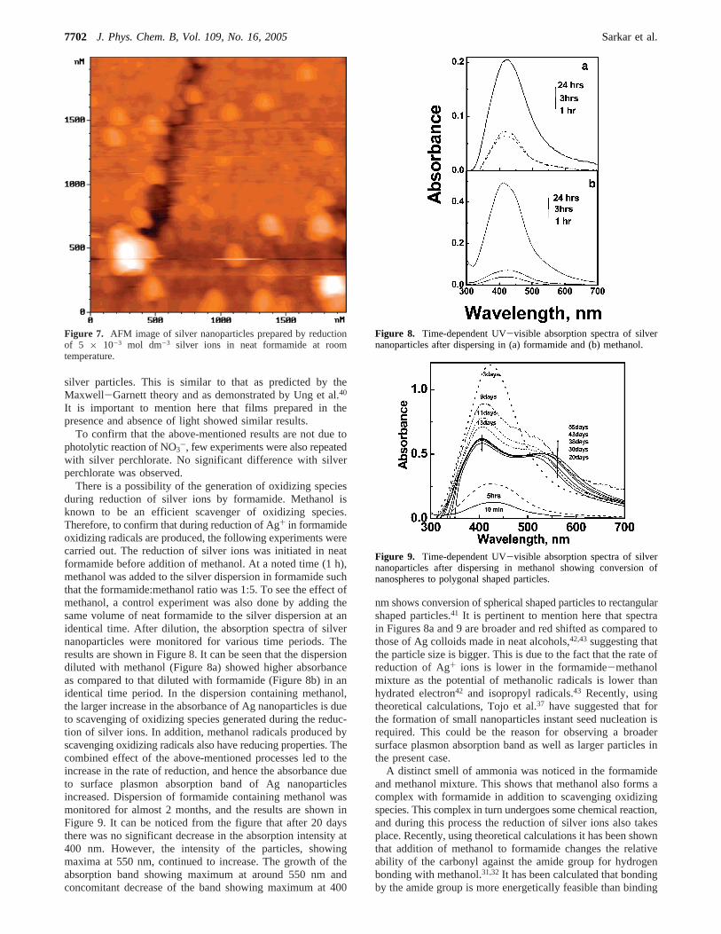
There is a possibility of the generation of oxidizing speciesduring reduction of silver ions by formamide. Methanol isknown to be an efficient scavenger of oxidizing species.Therefore, to confirm that during reduction of Ag+ in formamideoxidizing radicals are produced, the following experiments werecarried out. The reduction of silver ions was initiated in neatformamide before addition of methanol. At a noted time (1 h),methanol was added to the silver dispersion in formamide suchthat the formamide:methanol ratio was 1:5. To see the effect ofmethanol, a control experiment was also done by adding thesame volume of neat formamide to the silver dispersion at anidentical time. After dilution, the absorption spectra of silvernanoparticles were monitored for various time periods. Theresults are shown in Figure 8. It can be seen that the dispersiondiluted with methanol (Figure 8a) showed higher absorbanceas compared to that diluted with formamide (Figure 8b) in anidentical time period. In the dispersion containing methanol,the larger increase in the absorbance of Ag nanoparticles is dueto scavenging of oxidizing species generated during the reduc-tion of silver ions. In addition, methanol radicals produced byscavenging oxidizing radicals also have reducing properties. Thecombined effect of the above-mentioned processes led to theincrease in the rate of reduction, and hence the absorbance dueto surface plasmon absorption band of Ag nanoparticlesincreased. Dispersion of formamide containing methanol wasmonitored for almost 2 months, and the results are shown inFigure 9. It can be noticed from the figure that after 20 daysthere was no significant decrease in the absorption intensity at400 nm. However, the intensity of the particles, showingmaxima at 550 nm, continued to increase. The growth of theabsorption band showing maximum at around 550 nm andconcomitant decrease of the band showing maximum at 400
nm shows conversion of spherical shaped particles to rectangularshaped particles.41 It is pertinent to mention here that spectrain Figures 8a and 9 are broader and red shifted as compared tothose of Ag colloids made in neat alcohols,42,43suggesting thatthe particle size is bigger. This is due to the fact that the rate ofreduction of Ag+ ions is lower in the formamide-methanolmixture as the potential of methanolic radicals is lower thanhydrated electron42 and isopropyl radicals.43 Recently, usingtheoretical calculations, Tojo et al.37 have suggested that forthe formation of small nanoparticles instant seed nucleation isrequired. This could be the reason for observing a broadersurface plasmon absorption band as well as larger particles inthe present case.
A distinct smell of ammonia was noticed in the formamideand methanol mixture. This shows that methanol also forms acomplex with formamide in addition to scavenging oxidizingspecies. This complex in turn undergoes some chemical reaction,and during this process the reduction of silver ions also takesplace. Recently, using theoretical calculations it has been shownthat addition of methanol to formamide changes the relativeability of the carbonyl against the amide group for hydrogenbonding with methanol.31,32It has been calculated that bondingby the amide group is more energetically feasible than binding
Figure 7. AFM image of silver nanoparticles prepared by reductionof 5 × 10⁻³ mol dm-3 silver ions in neat formamide at roomtemperature.
Figure 8. Time-dependent UV-visible absorption spectra of silvernanoparticles after dispersing in (a) formamide and (b) methanol.
Figure 9. Time-dependent UV-visible absorption spectra of silvernanoparticles after dispersing in methanol showing conversion ofnanospheres to polygonal shaped particles.
7702 J. Phys. Chem. B, Vol. 109, No. 16, 2005 Sarkar et al.

by the carbonyl group. Thus, there exists a possibility that, dueto the change in the structure of formamide in the presence ofmethanol, the reaction path for the dissociation and/or oxidationof formamide changes, which in turn affects the agglomerationof the nanoparticles.
Reaction of the Silver Nanoparticles with CHCl3 andToluene in the Presence of Oxygen.To find the possibility ofthe silver nanoparticles reacting with chloroform, particles wereprepared in formamide containing 1% PVP, which was diluted4 times with chloroform. The solution was vigorously shakenfor 30 min. On standing, the color of the silver sol graduallyfaded due to oxidation in the formamide layer. The resultscorroborate the findings reported in aqueous solution;35 however,in N2-bubbled solution the oxidation of particles was very slow.This shows that electron transfer from silver particles isfacilitated in the presence of oxygen. A similar observation wasmade when toluene was added to silver dispersions.
The reaction of silver nanoparticles with CHCl3 is indicativeof the strong reducing power of the particles. As the nano-particles have more surface atoms, the unsaturation at the surfaceis very high. In the presence of an organic molecule, which isan electrophile, the surface atom acquires an excess positivecharge and the rest of the nanoparticles a corresponding negativecharge. In the presence of oxygen, this excess negative chargeis removed and the oxidation of the silver particles can proceed.This could be the reason for observing fast oxidation of silverparticles in an aerated solution as compared to that in a N2-bubbled solution.
Similarly, the reactivity of silver nanoparticles with toluenewas investigated. After addition of toluene to a silver dispersionin formamide, the solution was vigorously shaken for 30 min.On standing, the color of the silver sol gradually faded due tooxidation in the formamide layer. However, in a N2-bubbledsolution, the oxidation of particles was very slow. It is knownthat oxidation of toluene leads to the formation of benzalde-hyde.44,45 As metal particles can act as a sink for electrons, itappears that in the presence of silver particles, there is apossibility of oxidation of toluene, which is facilitated by thepresence of oxygen.
Surface Modification Studies.To further confirm the aboveobservations, we have carried out surface modification studiesof silver films in the presence of Na4EDTA. Silver filmdeposition was carried out as explained earlier. In the absenceof any complexing agent, films were quite stable, and we couldnot see any significant change in the absorption of silver films.However, when a slide of silver film was dipped in the solutionof Na4EDTA, the color of the film changed from brown to pinkdue to adsorption of Na4EDTA on the silver film. Figure 10shows the effect of Na4EDTA on the surface plasmon absorptionband of silver film. The observed change in the intensity andsurface plasmon absorption band is due to the disturbance atthe surface of the electron gas. In the presence of oxygen, theoxidation of silver film takes place. This shows that the reductionpotential of Ag particles is affected in the presence of Na4-EDTA46 and becomes more negative than the O2/O2
-• couple,-0.33 V vs NHE.47 To see whether electron transfer from silverfilm to MV 2+ takes place in the presence of Na4EDTA, a glassslide of silver particles complexed with Na4EDTA was dippedin a N2-bubbled aqueous solution containing methyl viologen(MV2+). No significant change in the color of the slide as wellas no sign of the formation of MV+• radical was observed. Thus,it appears that the reduction potential of Ag particles in thepresence of Na4EDTA lies below that of the MV2+/MV+•
couple,-0.446 V (vs NHE).47 It is important to mention here
that the stability of the film under inert conditions enhanceddrastically as compared to when it was exposed to air. The aboveresults show that Fermi level of Ag particles becomes morenegative in the presence of Na4EDTA, and it seems to liebetween-0.33 and-0.446 V vs NHE.
Conclusion
We have demonstrated the synthesis of silver nanoparticlesin formamide at room temperature without adding any reductantfrom outside. In the presence of a proper stabilizer, nanoparticlesof silver metal can be stabilized. Surface-modified particles (orfilms) show optical properties significantly different from thoseof the primary silver dispersion (or film). The silver nanopar-ticles also act as redox catalysts due to the shift in the Fermilevel toward more negative potential. Moreover, the methoddescribed here presents a simple approach for the formation ofsilver nanoparticles.
References and Notes
(1) Faraday, M.Philos. Trans. R. Soc. London1857, 147, 145.(2) Hirai, H.; Nakao, Y.; Toshima, N.Chem. Lett. 1978, 545.(3) (a) Henglein, A.Chem. ReV. 1989, 89, 1861. (b) Kamat, P. V.Chem.
ReV. 1993, 93, 267.(4) Belloni, J.Radiat. Res.1998, 150, 39.(5) Henglein, A.; Meisel, D.J. Phys. Chem. B1998, 102, 8386.(6) Dimitrijevic, N. M.; Bartels, D. M.; Jonah C. D.; Takahashi, K.;
Rajh, T.J. Phys. Chem. B2001, 105, 954.(7) Marignier, J. L.; Belloni, J.; Delcourt, M. V.; Chevalier, J. P.Nature
1985, 317, 344.(8) Cointet, C. de; Mostafavi, M.; Khatouri, J.; Belloni, J.J. Phys.
Chem. B1997, 101, 3512.(9) Kapoor, S.Langmuir1998, 14, 1021.
(10) Kapoor, S.Langmuir1999, 15, 4365.(11) Kapoor, S.Langmuir2000, 16, 5496.(12) Ahmadi, T. S.; Wang, Z. L.; Green, T. C.; Henglein, A.; El-Sayed,
M. A. Science1996, 272, 1924.(13) Ershov, B. G.; Henglein, A.J. Phys. Chem. 1998, 102, 10663,
10667.(14) Templeton, A. C.; Wuelfing, W. P.; Murray, R. W.Acc. Chem.
Res.2000, 33, 27.(15) Storhoff, J. J.; Mirkin, C. A.Chem. ReV. 2000, 100, 409.(16) Shipway, A. N.; Katz, E.; Willner, I.ChemPhysChem2000, 1, 18.(17) Zhong, C. J.; Maye, M. M.AdV. Mater. 2001, 13, 1507.(18) Henglien, A.Isr. J. Chem.1993, 33, 77.(19) Bruchez, M.; Moronne, M.; Gin, P.; Weiss, S.; Alivisatos, A. P.
Science1998, 281, 2013.(20) Brust, M.; Bethell, D.; Keily, C. J.; Shiffrin, D. J.Langmuir1998,
14, 5425.(21) Geirsig, M.; Pastoriza-Santos, I.; Liz-Marzan, L. M.J. Mater. Chem.
2004, 14, 607.(22) Link, S.; El-Sayed, M. A.J. Phys. Chem. B1999, 103, 8410.(23) Musick, M. D.; Pena, D. J.; Botsko, S. L.; McEvoy, T. M.;
Richardson, T. N.; Natan, M. J.Langmuir1999, 15, 844.
Figure 10. Time-dependent UV-visible absorption spectra of (a) silverfilm obtained by reduction of 1× 10⁻² mol dm-3 silver ions informamide; after dipping the film in an aqueous solution of 5× 10⁻²
mol dm-3 Na4EDTA for (b) 30 min, (c) 70 min, and (d) 200 min.
Preparation of Silver Nanoparticles in Formamide J. Phys. Chem. B, Vol. 109, No. 16, 20057703
(24) Pastoriza-Santos, I.; Liz-Marzan, L. M.Langmuir 1999, 15, 948.(25) Pastoriza-Santos, I.; Liz-Marzan, L. M.Pure Appl. Chem.2000,
72, 83.(26) Pastoriza-Santos, I.; Serra-Rodriguez, C.; Liz-Marzan, L. M.J.
Colloid Interface Sci.2000, 221, 236.(27) Pastoriza-Santos, I.; Liz-Marzan, L. M.Langmuir 2000, 18, 2888.(28) Pastoriza-Santos, I.; Liz-Marzan, L. M.Nano Lett.2002, 2, 903.(29) Han, M. Y.; Quek, C. H.Langmuir2000, 16, 362(30) Han, M. Y.; Quek, C. H.; Huang, W.; Chew, C. H.; Gan, L. M.
Chem. Mater.1999, 11, 1144.(31) Fu, A.-P.; Du, D.-M.; Zhou, Z.-Y.Chem. Phys. Lett.2003, 377,
537.(32) Ojha, A. K.; Srivastava, S. K.; Koster, J.; Shukla, M. K.;
Leszczynski, J.; Asthana, B. P.; Kiefer, W.J. Mol. Struct.2004, 689, 127.(33) Hoffman, M. R.; Martin, S. T.; Choi, W.; Bahnemann, D. W.Chem.
ReV. 1995, 95, 69.(34) Linsebigler, A. L.; Lu, G.; Yates, J. T.Chem. ReV. 1995, 95, 735.(35) (a) Henglein, A.; Linnert, T.; Mulvaney, P.Ber. Bunsen-Ges. Phys.
Chem. 1990, 94, 1449. (b) Henglein, A.; Mulvaney, P.; Linnert, T.Electrochim. Acta1991, 36, 1743.
(36) Yu, J. Y.; Schreiner, S.; Vaska, L.Inorg. Chim. Acta1990, 170,145.
(37) Tojo, T.; Blanco, M. C.; Rivadulla F.; Lopez-Quintela, M. M.Langmuir1997, 13, 1970.
(38) Lawless, D.; Kapoor, S.; Kennepohl, P.; Meisel, D.; Serponne, N.J. Phys. Chem.1994, 98, 9619.
(39) Li, T.; Moen, J.; Morrone, A. A.; Mecholsky, J. J.; Talham, D. R.;Adair, J. H.Langmuir1999, 15, 4328.
(40) Ung, T.; Liz-Marzan, L. M.; Mulvaney, P.Colloids Surf., A2000,202, 119.
(41) Mock, J. J.; Barbic, M.; Smith, D. R.; Schultz, D. A.; Schultz, S.J. Chem. Phys.2002, 116, 6755.
(42) Mostafavi, M.; Dey, G. R.; Francois, L.; Belloni, J.J. Phys. Chem.A 2002, 106, 10184.
(43) Huang, Z.-Y.; Mills, G.; Hajek, B.J. Phys. Chem.1993, 97, 11542.(44) Fuerte, A.; Hemandez-Alonso, M. D.; Maira, A. J.; Martinez-Arias,
A.; Fernandez-Garcia, M.; Conesa, J. C.; Soria, J.; Munuera, G.J. Catal.2002, 212, 1.
(45) Fuerte, A.; Hema´ndez-Alonso, M. D.; Maira, A. J.; Martinez-Arias,A.; Fernandez-Garcia, M.; Conesa, J. C.; Soria, J.Chem. Commun.2001,2178.
(46) Remita, S.; Mostafavi, M.; Delcourt, M. O.J. Phys. Chem.1996,100, 10187.
(47) Wardman, P.J. Phys. Chem. Ref. Data1989, 18, 1637.
7704 J. Phys. Chem. B, Vol. 109, No. 16, 2005 Sarkar et al.