Embed Size (px)
Citation preview
Drug delivery systems that can precisely control the release rate of target drug to aspecific body site have had an enormous impact on the healthcare system. In addition todrug formulations that deliver the drug for a prolonged period of time, it is importantfor efficient therapy to achieve spatial placement of the dosage form in the gastrointesti-nal tract (GIT). Site-specific drug delivery, using novel formulation designs, would im-prove local therapy in the GIT, optimize systemic absorption and minimize prematuredrug degradation (1). Stomach-specific antibiotic drug delivery, for instance, would behighly beneficial in the treatment of Helicobacter pylori infection in peptic ulcer disease(2–4).
413
Acta Pharm. 57 (2007) 413–427 Original research paper
10.2478/v10007-007-0033-5
Preparation and in vitro characterization of gellanbased floating beads of acetohydroxamic acid for
eradication of H. pylori
PARAUVATHANAHALLI SIDDALINGAM
RAJINIKANTH
BRAHMESHWAR MISHRA*
Department of PharmaceuticsInstitute of Technology, Banaras HinduUniversity, Varanasi-221005, India
Accepted October 22, 2007
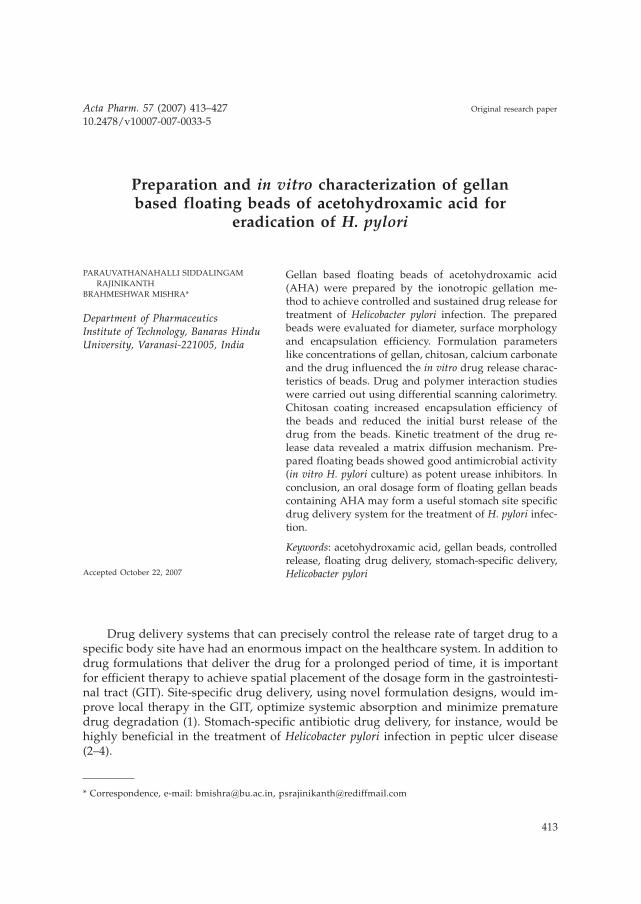
Gellan based floating beads of acetohydroxamic acid(AHA) were prepared by the ionotropic gellation me-thod to achieve controlled and sustained drug release fortreatment of Helicobacter pylori infection. The preparedbeads were evaluated for diameter, surface morphologyand encapsulation efficiency. Formulation parameterslike concentrations of gellan, chitosan, calcium carbonateand the drug influenced the in vitro drug release charac-teristics of beads. Drug and polymer interaction studieswere carried out using differential scanning calorimetry.Chitosan coating increased encapsulation efficiency ofthe beads and reduced the initial burst release of thedrug from the beads. Kinetic treatment of the drug re-lease data revealed a matrix diffusion mechanism. Pre-pared floating beads showed good antimicrobial activity(in vitro H. pylori culture) as potent urease inhibitors. Inconclusion, an oral dosage form of floating gellan beadscontaining AHA may form a useful stomach site specificdrug delivery system for the treatment of H. pylori infec-tion.
Keywords: acetohydroxamic acid, gellan beads, controlledrelease, floating drug delivery, stomach-specific delivery,Helicobacter pylori
* Correspondence, e-mail: [email protected], [email protected]

In recent years, the number of individuals (more than 60% of adults in industriali-zed countries) suffering from acid peptic disease and gastric adenocarcinoma caused byH. pylori has increased (5). Current strategies aimed at eradication of H. pylori infectionfrom patients rely on triple therapy that includes an anti-secretory agent in combinationwith two antibiotics, but these regimes are not wholly effective and patient compliance,side effects and bacterial resistance are major drawbacks of the therapy. This is due tothe fact that the organism remains exclusively on the luminal surface of the gastricmucosa under the mucous gel layer. Thus, the access of antimicrobial drugs to the site isrestricted both from the stomach and from the gastric blood supply (6, 7). Also, theantibiotics are not delivered to the site of infection in effective concentration and in fullyactive form by the conventional drug delivery systems (4). Despite the multi-antibiotictherapy, different therapeutic strategies have been examined to completely eradicate H.pylori from the stomach. Drug delivery to the site of residence in the gastric mucosa mayimprove the efficacy of the current and emerging treatments. Gastric retentive deliverysystems potentially allow increased penetration of the mucus layer and therefore increa-se drug concentration at the site of action (6, 8).
To overcome the problems, we have proposed a concept based on a floating drugdelivery system with floating site-specific drug delivery. It is necessary to design a drugdelivery system that could not only curtail and alleviate the shortcomings of conven-tional drug delivery vehicles, but also deliver the antimicrobial agent to the infected celllines. The drug, acetohydroxamic acid (AHA) inhibits cytoplasmic, which plays an im-portant role in the chemotactic motility of H. pylori (9). As AHA is a small molecule (mo-lecular mass, 75.07), it can permeate intact bacterial cells and effectively inhibit theurease activity of H. pylori. Freely diffusible AHA inhibits over 95% of urease activity af-ter 10 min (10, 11). Gellan gum is a bacterial anionic deacetylated polysaccharide secre-ted by Pseudomonas elodea. Due to its characteristic property of temperature-dependentand cation-induced gelation (12), gellan was selected as a matrix polymer for the prepa-ration of AHA beads in the formulation. The present work is therefore aimed develop-ing of anti-H. pylori agent (antiurease) AHA-loaded gellan floating beads coated withchitosan for stomach-specific drug delivery for eradication of H. pylori.
EXPERIMENTAL
Materials
Acetohydroxamic acid (AHA) and gellan (Gelrite) were purchased from Sigma-Al-drich Chemicals (India). Chitosan was a generous gift sample of the Centre for Fisheriesand Research Institute (India). Brain heart infusion, fetal calf serum, and Campylobacterselective media (Skirrow Supplement) were purchased from Himedia, India. All otherreagents were of analytical grade.
Methods
Preparation of beads. – The beads were prepared by the ionotropic gelation technique.Gellan solution (0.25–1.0%, m/V) was prepared by dissolving the gellan in deionized wa-
414
P. S. Rajinikanth and B. Mishra: Preparation and in vitro characterization of gellan based floating beads of acetohydroxamic acid for
eradication of H. pylori, Acta Pharm. 57 (2007) 413–427.

ter by heating at 90 °C. Different concentrations of the drug (0.1–2.0%, m/V) and calciumcarbonate (0.5–2.0%, m/V) were dissolved/dispersed uniformly in 50 mL of gellan solu-tion below 40 °C m/V under continuous stirring. The stirring was continued after com-plete addition until a uniform dispersion was obtained. The resultant homogeneousbubble free slurry dispersion was dropped through a 21G syringe needle into 100 mL ofcalcium chloride solution (1.5%), which was kept under stirring to improve the mechan-ical strength of the beads and also to prevent aggregation of the formed beads. Immedi-ate formation of small gelled beads took place; after 5 minutes of curing time, the for-med beads were collected by filtration and dried at 40 °C. The gellan beads coated withchitosan were prepared by dropping the gellan slurry containing different amounts ofdrug and calcium carbonate into 100 mL of calcium chloride (1.5%, m/V) with chitosandissolved in it at various concentrations (0.2–0.6%, m/V). Then the beads were collectedand dried as mentioned above.
Morphology and particle size analysis. – Particle size of the prepared beads was deter-mined using an optical microscope (Model BH-2, Olympus, Japan) fitted with a stageand an ocular micrometer. Twenty dried beads were measured for calculating the meandiameter of beads. The shape and surface morphological examination of the surfacestructure of dried beads were carried out by scanning electron microscopy (SEM-JEOLModel 8404, Japan at magnification of 500x).
In vitro floating properties of gellan beads. – The in vitro floating study was performedusing a USP 24 dissolution apparatus II (13) having 500 mL of simulated gastric fluid(SGF, pH 1.2). The medium temperature was kept at 37 ± 0.5 °C. The floating gellan beads(1.0 g beads) were soaked in the dissolution medium and the medium was agitated witha paddle at 50 rpm. After agitation, the beads that floated on the surface of the mediumand those that settled down at the bottom of the flask were recovered separately. Thefloating percentage was estimated by visual observation.
Entrapment efficiency of beads. – The drug content in gellan beads was determined bythe reported method with a slight modification (14). Briefly, the dried beads (100 mg)were allowed to disintegrate in 50 mL of phosphate buffer (pH 7.2) for 4 h. The wholedispersion of beads was sonicated at 125 W for 30 min (Imeco Sonifier, Imeco Ultraso-nics, India) and the solution was filtered through a 0.45-µm filter. Then the polymericdebris was washed twice with fresh solvent (phosphate buffer) to extract any adheringdrug. The drug content of filtrate and washings was determined spectrophotometricallyat 503 nm (Shimadzu, UV/Visible 1601, Japan). Each sample determination was made intriplicate.
Differential scanning calorimetry (DSC). – Differential scanning calorimetry (DSC) wasperformed on pure drug, placebo beads, and drug-loaded beads. DSC measurements weredone on modulated DSC (Q 1000 TA equipped with software Pyris 6.0, USA). About 3.0mg of sample was placed in an aluminium pan and then hermetically sealed with analuminium lid. The thermograms were obtained at a scanning rate of 5 °C min–1 over atemperature range of 40 to 150 °C under an inert atmosphere flushed with nitrogen at arate of 20 mL min–1. All tests were performed twice.
415
P. S. Rajinikanth and B. Mishra: Preparation and in vitro characterization of gellan based floating beads of acetohydroxamic acid for
eradication of H. pylori, Acta Pharm. 57 (2007) 413–427.

Measurement of in vitro drug release. – The release of AHA from the floating beadswas determined using a USP 24 dissolution test apparatus I with a basket (13). A weighedquantity of beads equivalent to 100 mg of AHA was placed in the dissolution basket andthe basket was placed in 500 mL dissolution medium. The dissolution medium having500 mL of simulated gastric fluid (pH 1.2) was maintained at 37 ± 0.5 °C (3). Five mL ofsample was withdrawn at different time intervals and replaced with the same volume offreshly prepared dissolution medium. The drug content was measured at 503 nm. Theseexperiments were conducted in triplicate.
In vivo floating efficiency (X-ray) study. – The in vivo study was carried out by admi-nistering floating beads to rabbits and monitoring them by a radiological method. Sixhealthy albino rabbits of either sex, weighing 2–2.4 kg (2.2 ± 0.3 kg) were used for thepresent study. The animals were housed in individual cages, and the experiments wereconducted in a sanitized room at a temperature maintained at around 24 °C. Food waswithdrawn 12 h prior to the study with water ad libitum. To make the beads radiopaque,1.5 g of barium sulfate was incorporated into polymeric solution (the same formulationcomposition of FBA2 was used to prepare radiopaque beads) and radiopaque beadswere prepared using a similar procedure to that mentioned in the preparation of beads.Twenty beads were administered through oral gastric tube with 25 mL water in fastedstate. Afterwards, the animals were not allowed to eat or drink throughout the study(up to 6 h). The beads loaded with barium sulfate showed the same in vitro buoyancy asthe unloaded units (data not shown). At every hour interval, 10 mL of water was admi-nistered to animals throughout the study. Before taking X-ray photographs, the animalswere placed/held in upright posture. The position of the beads in the rabbit’s stomachwas monitored by X-ray photographs (Siregraph-B, Siemens, Germany) of the gastric re-gion at different time intervals (at 1, 4 and 6 h ) for 6 h. The number of beads that re-mained buoyant on the surface of the gastric content and that of all the beads remaininginside the stomach (buoyant and non-buoyant) were observed visually from the X-rayphotographs.
The protocol of the study was approved by Animals Ethical Committee of the Bana-ras Hindu University (Varanasi, India).
In vitro growth inhibition studies. – The bacterial strain used in this study was origi-nally isolated from a hospitalized human patient (aged 50 years) with gastric ulcer. In vi-tro growth inhibition studies were performed on developed system using a broth cultureof H. pylori. H. pylori broth culture was preformed in a brain-heart infusion containing0.25% yeast extract and 10% fetal calf serum and supplemented with 0.4% Campylobacterselective supplement (Skirrow supplement). H. pylori strain was grown in brucella brothat 37 °C after 7 days in a microaerophilic atmosphere (5% O2, 10% CO2, 85% N2). Growthof the bacteria was monitored by measuring the optical density of broth cultures spec-trophotometrically at 600 nm. The number of bacteria was determined by OD with oneoptimal density unit corresponding to 108 colony-forming unit (CFU) mL–1. The colonieswere identified as H pylori by morphology and urease activity (2). To study the effect offormulations on H. pylori growth inhibition (GI), 10 mL of nutrient broth was inoculatedwith a loopful of the H. pylori from stock culture to make a final culture of 108 CFU mL–1.AHA plain drug and different formulations were added to the tubes and all the tubeswere incubated at 37 °C in a microaerophilic atmosphere. Acetohydroxamic acid was
416
P. S. Rajinikanth and B. Mishra: Preparation and in vitro characterization of gellan based floating beads of acetohydroxamic acid for
eradication of H. pylori, Acta Pharm. 57 (2007) 413–427.

used at a final concentration of 14 mmol L–1 (14 mmol L–1 is approximately four fold ofthe reported MIC50 for H. pylori urease) (10).
The culture containing tubes were shaken at 100 rpm at 37 °C in a microaerobic at-mosphere in an incubator. Then, 100 µL of nutrient broth containing AHA and differentAHA formulations was removed at various time points (4, 8, 12 and 24 h) and serial di-lutions were plated on modified Skirrow’s medium. The agar plates were incubated for4 days at 37 °C under microaerobic conditions in GasPak (BD Diagnostic Systems, USA).The viable cell counts for each sample were calculated by counting the number of colo-nies on the agar plates.
Statistical analysis
Statistical evaluation of the data was performed using the analysis of variance(ANOVA).
RESULTS AND DISCUSSION
Particle size and morphology of beads
The formulation composition and physico-chemical properties of the various bat-ches of the prepared AHA floating beads are shown in Tables I and II, respectively. Thescanning electron micrographs (SEM) of the beads are shown in Figs. 1a and b. The SEM
417
P. S. Rajinikanth and B. Mishra: Preparation and in vitro characterization of gellan based floating beads of acetohydroxamic acid for
eradication of H. pylori, Acta Pharm. 57 (2007) 413–427.
Table I. Formulation variables of the prepared floating beads of acetohydroxamic acid
BatchGellan
concentration(%, m/V)
Chitosanconcentration
(%, m/V)
Calcium carbonateconcentration
(%, m/V)
AHA(%, m/V)
FBA1 0.25 – 1.00 0.50
FBA2 0.50 – 1.00 0.50
FBA3 1.00 – 1.00 0.50
FBA4 0.25 0.40 1.00 0.50
FBA5 0.50 0.40 1.00 0.50
FBA6 1.00 0.40 1.00 0.50
FBA7 0.50 0.20 1.00 0.50
FBA8 0.50 0.60 1.00 0.50
FBA9 0.50 0.40 0.00 0.50
FBA10 0.50 0.40 0.50 0.50
FBA11 0.50 0.40 2.00 0.50
FBA12 0.50 0.40 1.00 0.10
FBA13 0.50 0.40 1.00 1.00
FBA14 0.50 0.40 1.00 2.00

results revealed that all the AHA-loaded floating beads were discrete and spherical inshape with rough outer surface (Figs. 1a and b). However, the chitosan coated AHAbeads had outer rough surface with a number of minor wrinkles (Fig. 1b). This is due tothe formation of a thin layer coat of chitosan over the beads, which caused several wrin-kles during the drying due to dehydration of the chitosan membrane surrounding thebeads (Fig. 1b). The bead diameter varied from 0.72 ± 0.04 to 0.91 ± 0.02 mm for differentbatches. The results indicate that as the amount of gellan and calcium carbonate increa-sed, the size of beads also proportionally increased (Table II). This could be attributed tothe increase in micro-viscosity of the polymeric dispersion due to increasing gellan con-centration, which eventually led to formation of bigger beads. The mean diameter ofprepared beads marginally increased with an increase in drug loading and chitosan con-centration. This could be attributed to drug solubility in water and formation of a thinchitosan coating over the beads due to its ionic interaction, respectively.
In vitro floating properties
The floating ability of the prepared formulations was evaluated in SGF (pH 1.2) us-ing a USP 24 dissolution apparatus II (13). The time the formulation took to emerge onthe medium surface (floating lag time) and the percentage of floating beads on the disso-lution medium surface were evaluated and are shown in Table II. Upon contact with anacidic medium, gelation and cross-linking by Ca2+ ions occurred to provide a gel barrier
418
P. S. Rajinikanth and B. Mishra: Preparation and in vitro characterization of gellan based floating beads of acetohydroxamic acid for
eradication of H. pylori, Acta Pharm. 57 (2007) 413–427.
Table II. Physico-chemical characteristics of the prepared floating beads of acetohydroxamic acid
BatchDiameter(mm)a,b
Entrapmentefficiency (%)a,c
Floating lagtime (min)a,c
Floating ability(%)a,c
Drug content(mg)a,c,d
FBA1 0.72 ± 0.04 44.30 ± 2.49 6.51 ± 0.61 88.25 ± 2.42 42.47 ± 1.98
FBA2 0.79 ± 0.04 48.81 ± 3.98 5.26 ± 1.02 86.41 ± 2.14 45.21 ± 3.14
FBA3 0.86 ± 0.02 56.21 ± 2.52 4.67 ± 0.24 88.24 ± 1.82 57.42 ± 2.14
FBA4 0.78 ± 0.06 56.25 ± 2.65 5.09 ± 1.03 86.26 ± 3.12 55.30 ± 2.47
FBA5 0.85 ± 0.04 65.78 ± 3.51 5.79 ± 1.13 87.23 ± 2.13 67.20 ± 2.62
FBA6 0.89 ± 0.04 73.71 ± 2.80 6.15 ± 1.37 85.82 ± 1.98 74.17 ± 2.43
FBA7 0.74 ± 0.05 61.80 ± 4.12 6.28 ± 0.23 87.49 ± 2.19 58.67 ± 2.98
FBA8 0.88 ± 0.03 58.72 ± 3.56 6.45 ± 1.02 92.36 ± 2.54 57.35 ± 2.61
FBA9 0.85 ± 0.04 59.83 ± 1.65 none none 57.81 ± 1.94
FBA10 0.86 ± 0.05 58.78 ± 4.03 10.41 ± 0.32 71.51 ± 2.78 59.34 ± 2.68
FBA11 0.91 ± 0.02 60.23 ± 2.82 4.89 ± 0.36 100 61.62 ± 2.44
FBA12 0.80 ± 1.01 57.35 ± 2.41 6.12 ± 1.02 86.56 ± 2.63 55.40 ± 2.18
FBA13 0.84 ± 1.01 60.23 ± 2.82 6.07 ± 0.42 87.72 ± 2.12 61.67 ± 2.35
FBA14 0.85 ± 0.10 52.35 ± 2.41 5.61 ± 0.39 87.52 ± 2.79 51.32 ± 2.41
a Mean ± SD.b n = 20.c n = 3.d Drug content of each 100 mg of beads.

at the surface of the formulation. The calcium carbonate effervesced, releasing carbon di-oxide and calcium ions. The released carbon dioxide was entrapped in the gel networkproducing buoyant formulation and then calcium ion reacted with gellan producing across-linked three-dimensional gel network that restricted further diffusion of carbon di-oxide and drug molecules and resulted in an extended period of floating and drug re-lease (16–18). The floating ability of the formulation mainly depended on calcium car-bonate and gellan concentrations. The control beads (without calcium carbonate) sankuniformly in the medium. The beads containing 0.5 to 2.0% of the gas-forming agentdemonstrated good floating ability (71–100% of floating). The floating lag time for thissystem was in the range of 4–10 min. The lowest concentration of calcium carbonatewhich makes the beads float throughout the drug release study was found to be 0.5%(m/V) at all polymer levels.
419
P. S. Rajinikanth and B. Mishra: Preparation and in vitro characterization of gellan based floating beads of acetohydroxamic acid for
eradication of H. pylori, Acta Pharm. 57 (2007) 413–427.
Fig. 1. SEM photograph of the preparedfloating beads of acetohydroxamic acid:a) prepared AHA beads (FBA2), b) sur-face morphology of chitosan coated AHAbeads (FBA2).
a)
b)

On increasing the calcium carbonate concentration, the floating lag time was redu-ced and the duration of floating was increased (Table II). The increase in the amount ofCa2+ and consequently in the amount of CO2 evolved are responsible for the observedreduction in the floating lag time and increased duration of floating. Similarly, an in-crease in the polymer concentration resulted in decreased lag time of the prepared beads(Table II). In SGF, the beads containing 2.0% (m/V) calcium carbonate exhibited a goodfloating ability; about 100% beads floated after the lag time of approximately 4 min (Ta-ble II). On increasing the CaCO3 concentration from 0.75 to 2.5% (m/V), the floating lagtime of AHA beads was reduced from 10 to 4 min (Table II). Almost all the preparedbeads were floating for > 8 h in SGF pH 1.2 (data not shown). Different drug concentra-tion did not produce any significant effect on floating properties of the beads; the beadsremained buoyant even after the buoyancy test period.
Entrapment efficiency
The effects of various formulation parameters on the entrapment efficiency of theprepared floating beads are shown in Table II. The entrapment efficiency of the preparedfloating beads varied from 44.3% for batch FBA1 (gellan 0.25%, m/V, no chitosan coat) to73.7% for batch FBA6 (gellan 1.0%, m/V coated with chitosan 0.4%, m/V). The entrap-ment efficiency increased significantly (p < 0.05) with increasing polymer concentration,as shown in Table II. This is because the increase in the gellan concentration resulted inthe formation of larger beads entrapping more drug. The entrapment efficiency of beadsincreased with increasing the drug loading up to 1.0% (m/V). However, increasing AHAconcentration above 2.0% (m/V) caused a marginal decrease in the incorporation effi-ciency, suggesting that the quantity of gellan becomes insufficient to entrap the drug.The entrapment efficiency was also found to be proportionally increased with increasingchitosan concentration. This is due to the increasing thickness of chitosan coat formedover the beads which may encapsulate a larger amount of drug. This result is well corre-lated with similar results reported earlier for alginate-chitosan beads of timolol maleate(19). The method adopted for the preparation of beads could be responsible for the ob-served higher entrapment efficiency. No significant effect on entrapment efficiency ofbeads was observed with increasing concentration of calcium carbonate (Table II).
DSC studies
In an effort to investigate the possible physical and chemical interactions betweendrug and polymer, we have analyzed: (i) pure acetohydroxamic acid, (ii) placebo beadsand (iii) drug-loaded beads using modulated DSC. The results are displayed in Fig. 2.The DSC thermogram showed a sharp endothermic peak at 89.22 °C for pure AHA asthe melting point of the drug (Fig. 2a). In placebo beads, thermal transition at 246.60 °Ccan be seen, which is attributed to the melting point of the gellan polymer. In the DSCthermogram of the drug-loaded beads, the endothermic peak was observed at 88.32 °Cas the melting point of the drug (Fig. 2c). The evaluation of the thermograms clearly re-vealed no physical interaction between the polymer and the drug in the beads. The anal-ysis of thermograms revealed no physical interaction between the polymer and the drugin the prepared beads.
420
P. S. Rajinikanth and B. Mishra: Preparation and in vitro characterization of gellan based floating beads of acetohydroxamic acid for
eradication of H. pylori, Acta Pharm. 57 (2007) 413–427.

In vitro drug release
The in vitro drug release profiles of gellan floating beads of AHA with different po-lymer concentrations are shown in Figs. 3a and b. The rate and extent of AHA releasefrom floating beads significantly decreased (p < 0.05) with an increase in gellan concen-tration. This could be attributed to the increase of gellan matrix density and in the diffu-sion path length which the drug molecules have to traverse (by formation of biggersized beads). The drug release from these beads was characterized by an initial phase ofhigh release (burst effect) due to good solubility of AHA in water. However, as gelationproceeded (cross-linking of gellan with Ca2+ ions from calcium carbonate), the remain-ing drug was released at a slower rate followed by a phase of moderate release. Thisbi-phasic pattern of release is a characteristic feature of matrix diffusion kinetics (20).The initial burst effect was considerably reduced with the increase in gellan concentra-tion (Fig. 3a). The initial burst effect from batches of chitosan-coated beads (FBA4, FBA5
and FBA6) was considerably reduced when compared to the corresponding batches ofchitosan non-coated beads (FBA1, FBA2 and FBA3), as shown in Fig. 3b. The fact is thatchitosan coating over the beads resulted in better incorporation efficiency and formed athick coating layer around the beads. This could be the reason for the observed decreasein the burst effect.
421
P. S. Rajinikanth and B. Mishra: Preparation and in vitro characterization of gellan based floating beads of acetohydroxamic acid for
eradication of H. pylori, Acta Pharm. 57 (2007) 413–427.
Fig. 2. DSC theromograms of: a) pure acetohydroxamic acid, b) placebo beads, c) acetohydroxamicacid-loaded beads (FBA2).
422
P. S. Rajinikanth and B. Mishra: Preparation and in vitro characterization of gellan based floating beads of acetohydroxamic acid for
eradication of H. pylori, Acta Pharm. 57 (2007) 413–427.
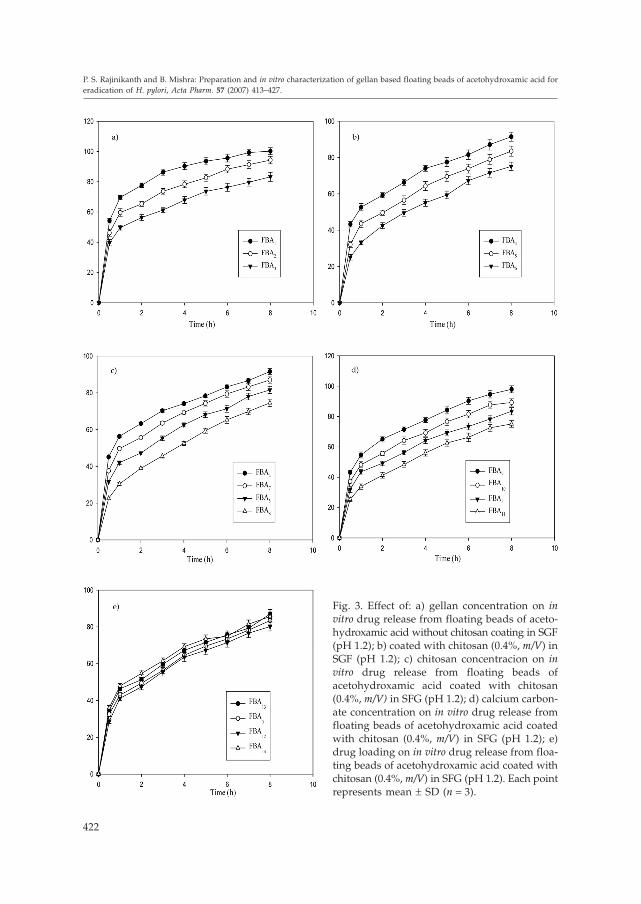
Fig. 3. Effect of: a) gellan concentration on invitro drug release from floating beads of aceto-hydroxamic acid without chitosan coating in SGF(pH 1.2); b) coated with chitosan (0.4%, m/V) inSGF (pH 1.2); c) chitosan concentracion on invitro drug release from floating beads ofacetohydroxamic acid coated with chitosan(0.4%, m/V) in SFG (pH 1.2); d) calcium carbon-ate concentration on in vitro drug release fromfloating beads of acetohydroxamic acid coatedwith chitosan (0.4%, m/V) in SFG (pH 1.2); e)drug loading on in vitro drug release from floa-ting beads of acetohydroxamic acid coated withchitosan (0.4%, m/V) in SFG (pH 1.2). Each pointrepresents mean ± SD (n = 3).
Fig. 3c indicates the effect of chitosan concentration on the release properties ofAHA from gellan beads. An increase in chitosan concentration in beads caused a signifi-cant (p < 0.05) retardation in the drug release of the beads as a result of an increase in thethickness of the coat of chitosan over the beads, thereby increasing the distance traveledby the drug molecule through the chitosan coat. Release rates of AHA from gellan beadswith different amounts of calcium carbonate are shown in Fig. 3d. An increase in cal-cium carbonate content prolonged the release of AHA from the gellan matrix. This effectmay be due to the internal ionotropic gelation effect of calcium carbonate. In acidic me-dium, the calcium carbonate dissolves and the ionized Ca2+ ions then promote internalgelation by cross-linking with the gellan and retarding the drug release from gellan ma-trix (21).
The effect of drug loading on AHA release from the prepared beads is shown in Fig.3e. The results indicate that different drug loadings of beads did not produce any signifi-cant difference in the rate and extent of drug release from beads. However, an initialhigh release was observed with the formulation batch FBA14 with higher drug loading.
In order to investigate the mechanism of drug release, the data were fitted to mod-els representing zero-order, first order and Higuchi’s square root of time (22). The exam-ination of the coefficient of determination (R2) indicated that drug release from the pre-pared beads followed a diffusion controlled mechanism, since the R2 values for Higu-chi’s square root of time (from 0.9661 to 0.997) was always higher compared to zero-or-der (from 0.8604 to 0.9238) and to the first-order ones (from 0.9126 to 0.9613). Since therelease from the prepared beads followed a biphasic profile, it was decided to use amore stringent test in order to distinguish between the mechanisms of drug release. Therelease data were fitted to the Peppas exponential model (23) Mt/M∞ = Ktn , where Mt/M∞is the fraction of drug released after time t, K is the kinetic constant and n is the releaseexponent which characterizes the drug transport mechanism. The n values were in therange of 0.3248–0.5653, indicating that all the prepared formulations followed the Fi-ckian diffusion controlled mechanism of drug release.
In vivo floating efficiency
The results of X-ray photographs of floating beads at different time intervals in rab-bits stomach are shown in Figs. 4a-c. One hour after dosing, the beads showed goodfloatability (~80%); 4 and 6 h after dosing about 60 and 50% of beads were found to bebuoyant on gastric content, respectively, whereas the remaining beads were observed ina lower part of the stomach (Figs. 4b and c). The results clearly indicate that the pre-pared floating beads of AHA remained buoyant for at least 6 h in rabbits stomach andthat they had good floatability in vivo.
In vitro growth inhibition studies
The effect of different drug-loaded, drug-free formulations (placebo) and plain AHAon bacterial growth was investigated at various time intervals for up to 24 hours and theresults are shown in Fig. 5. The antimicrobial effect of formulations and plain drug weredetermined in terms of percentage growth inhibition (GI), which was calculated as theratio of optical density (OD) of a given mixture against that of tubes containing H. pylori
423
P. S. Rajinikanth and B. Mishra: Preparation and in vitro characterization of gellan based floating beads of acetohydroxamic acid for
eradication of H. pylori, Acta Pharm. 57 (2007) 413–427.

alone. Placebo beads (control batch) did not exhibit any significant growth inhibition(Fig. 5).
In order to evaluate the in vitro growth inhibition of formulations against H. pylori,two batches containing different drug concentrations (FBA1 and FBA12) have been se-lected. The growth inhibition of drug loaded formulations was compared with that ofthe drug alone. The percentage GI values for placebo, AHA, FBA1 and FBA12 were 3.6 ±1.2, 69.0 ± 5.6, 56.3 ± 5.6 and 49.9 ± 6.7, respectively after 4 h incubation. After 8 hours ofincubation with formulations FBA1 and FBA12, the bacterial growth was reduced to 68.1± 6.3% and 60.2 ± 5.2%, respectively (Fig. 5), whereas the AHA pure drug inhibited com-pletely the H. pylori growth. The AHA formulations achieved complete growth inhibi-tion only after 12 h of incubation. Continued incubation of H. pylori for up to 24 hours in
424
P. S. Rajinikanth and B. Mishra: Preparation and in vitro characterization of gellan based floating beads of acetohydroxamic acid for
eradication of H. pylori, Acta Pharm. 57 (2007) 413–427.
Fig. 4. X-ray photographs of floating beads of AHA in the gastric region of rabbit after dosing offormulations in the fasted state: a) 1 h after dosing, b) 4 h after dosing, c) 6 h after dosing.
Fig. 5. Percentage of H. pylori growth inhibi-tion (mean ± SD, n = 3). � Placebo beads, �AHA plain drug, � FBA11, � FBA12.

the presence of AHA formulations inhibited completely the bacterial growth. This is dueto the controlled delivery of AHA from formulations, meaning that the microorganismwas exposed to a lower drug concentrations.
The floating beads containing AHA may show more efficacy in in vivo H. pylori clea-rance than pure AHA due to longer residence time of the formulation at the site in stom-ach where H. pylori resided. The results clearly indicate that the formulations showedgood inhibition in in vitro culture. However, the time required for complete inhibitionwas shorter for AHA than for AHA formulations because of the direct exposure of H.pylori to AHA. From the interaction and adsorption of the AHA formulations, the for-mulations effectively targeted the drug on the H. pylori surface. Particularly, AHA inhib-its cytoplasmic urease that plays an important role in the chemostatic motility of H. py-lori. Thus, it can be expected that the floating formulations with selected drug (AHA)will abolish all the mechanisms of H. pylori survival in vivo and may provide better treat-ment efficacy for H. pylori eradication.
CONCLUSIONS
The prepared gellan beads of AHA floated in SGF for a prolonged period of timeand sustained drug release from the beads over a period of at least 8 h. The in vivo float-ing efficiency of beads was satisfactory; beads were retained in rabbits stomach for ex-tended periods. The in vitro H. pylori inhibition study showed good antimicrobial activ-ity of formulations in H. pylori culture.
From the results one can conclude that the gellan based floating beads containingAHA have a promising potential for delivering AHA at the stomach site and may bevery useful for H. pylori eradication.
REFERENCES
1. A. Rubinstein and D. R. Friend, Site-specific Delivery to the Gastrointestinal Tract, in Polymeric Site-Specific Pharmacotherapy (Ed. A. J. Domb), Wiley, Chichester 1994, pp. 267–313.
2. N. Nagahara, Y. Akiyama, M. Tada, M. Nakao, M. Kitano and Y. Ogawa, Mucoadhesive micro-spheres containing amoxicillin for clearance of Helicobacter pylori, Antimicrob. Agents Chemother.42 (1998) 2492–2494.
3. R. Hejazi and M. Amiji, Stomach-specific anti-H. pylori therapy. I. Preparation and characteri-zation of tetracycline-loaded chitosan microspheres, Int. J. Pharm. 235 (2002) 87–94; DOI: 10.1016/S0378-5173(01)00985-1.
4. R. B. Umamaheshwari, S. Jain and N. K. Jain, A new approach in gastroretentive drug deliverysystem using cholestyramine, Drug Deliv. 10 (2003) 151–160; DOI: 10.1080/713840399.
5. B. J. Marshall and J. R. Warren, Unidentified curved bacilli in the stomach of patients with gas-tritis and peptic ulceration, Lancet 1 (1984) 1311–1315; DOI: 10.1016/S0140-6736(84)91816-6.
6. P. S. Rajinikanth, J. Balasubramanium and B. Mishra, Development and evaluation of a novelfloating in situ gelling system of amoxicillin for eradicating of Helicobacter pylori, Int. J. Pharm.335 (2007) 114–122; DOI: 10.1016/j.ijpharm.2006.11.009.
425
P. S. Rajinikanth and B. Mishra: Preparation and in vitro characterization of gellan based floating beads of acetohydroxamic acid for
eradication of H. pylori, Acta Pharm. 57 (2007) 413–427.

7. C. K. Lin, P. I. Hsu and K. H. Lai, One-week quadruple therapy is an effective salvage regimenfor Helicobacter pylori infection in patients after failure of standard triple therapy, J. Clin. Gastro-enterol. 34 (2002) 547–551.
8. R. B. Umamaheshwari, R. Suman and N. K. Jain, Anti Helicobacter pylori effect of mucoadhesivenanoparticle bearing amoxicillin in experimental gerbils, APPS Pharm. Sci. Tech. 5 (2004) article32.
9. A. M. El. Nujumi, C. A. Dorrian, R. S. Chittajallu, W. D. Neithercut, K. E. L. McColl, Effect of in-hibition of Helicobacter pylori urease activity of acetohydroxamic acid on serum gastrin in duo-denal ulcer subjects, Gut 32 (1991) 866–870.
10. H. L. Mobley, L. T. Hu and P. Foxall, Helicobacter pylori urease: Properties and role in patho-genesis, Scand. J. Gastroenterol. Suppl. 187 (1991) 39–46.
11. S. H. Phadnis, M. H. Parlow, M. Levy, D. Iher, C. M. Canldine, J. B. Connors and B. E. Dunn,Surface localization of Helicobacter pylori urease and a heat shock protein homolog requires bac-terial autolysis, Infect. Immun. 64 (1996) 905–912.
12. S. Miyazaki, N. Kawasaki, W. Kudo and D. Attwood, Comparison of in situ gelling formulationfor oral delivery of cimetidine, Int. J. Pharm. 220 (2001) 161–168; DOI: 10.1016/S0378-5173(01)00669-X.
13. United State Pharmacopeia 24 /National Formulary 19, USP Convention, Rockville, MD, 2002.
14. S. A. Agnihotri, S. S. Jawalkar and T. M. Aminabhavi, Controlled release of cephalexin throughgellan gum beads: Effect of formulation parameters on entrapment efficiency, size, and drug re-lease, Eur. J. Pharm. Biopharm. 63 (2006) 249–261; DOI: 10.1016/j.ejpb.2005.12.008.
15. I. Corthesy-Theulaz, N. Portal, M. Glauser, E. Saraga, R. Haas, J. P. Kraehenbushl, A. L. Blemand P. Michetti, Oral immunization with Helicobacter pylori urease B subunit as a treatmentagainst Helicobacter infection in mice, Gastroenterology 109 (1995) 115–121; DOI: 10.1016/0016-5085(95)90275-9.
16. H. Grasdalen and O. Smidsroed, Gellation of gellan gum, Carbohyd. Polym. 7 (1987) 371–393;DOI: 10.1016/0144-8617(87)90004-X.
17. R. Chandrasekaran, L. C. Puigianer, K. L. Joyce and S. Arnotts, The influence of calcium ions,acetate and L-glycerate groups on the gellan double-helix, Carbohyd. Res. 181 (1988) 23–40; DOI:10.1016/0008-6215(88) 84020-5.
18. R. Chandrasekaran and V. G. Thailambad, Cation interactions in gellan: An x-ray study of thepotassium salt, Carbohyd. Polym. 12 (1990) 431–442; DOI: 10.1016/0144-8617(90)90092-7.
19. A. D. Sezer and J. Akbuga, Release characteristics of chitosan treated alginate beads. I. Sustai-ned release of low molecular drug from chitosan treated alginate beads, J. Microencapsul. 16(1999) 195–203; DOI: 10.1080/026520499288636.
20. P. S. Rajinikanth, C. Sankar and B. Mishra, Sodium alginate microspheres of metoprolol tartratefor intranasal systemic delivery: development and evaluation, Drug Deliv. 10 (2003) 21–28; DOI:10.1080/713840323.
21. J. Balasubramaniam and J. K. Pandit, Ion-activated in situ gelling systems for sustained oph-thalmic delivery of ciprofloxacin hydrochloride, Drug Deliv. 10 (2003) 185–191; DOI: 10.1080/713840402.
22. W. I. Higuchi, Analysis of data on the medicament release from ointments, J. Pharm. Sci. 51(1962) 802–804.
23. R. W. Korsmeyer, R. Gurney, E. Doelker, P. Buri and N. A. Peppas, Mechanisms of solute releasefrom porous hydrophilic polymers, Int. J. Pharm. 15 (1983) 25–35; DOI: 10.1016/0378-5173(83)90064-9.
426
P. S. Rajinikanth and B. Mishra: Preparation and in vitro characterization of gellan based floating beads of acetohydroxamic acid for
eradication of H. pylori, Acta Pharm. 57 (2007) 413–427.

S A � E T A K
Priprava i in vitro karakterizacija plutaju}ih zrnaca acetohidroksamskekiseline za iskorjenjivanje H. pylori
PARAUVATHANAHALLI SIDDALINGAM RAJINIKANTH i BRAHMESHWAR MISHRA*
Metodom ionotropskog �eliranja pripravljena su plutaju}a zrnca acetohidroksam-ske kiseline (AHA) na bazi gelana za kontrolirano i usporeno osloba|anje ljekovite tvari,namijenjena za lije~enje infekcija uzrokovanih Helicobacter pylori. Pripravljenim zrncimaprou~avani su dijametar, površinska morfologija i sposobnost inkapsuliranja. Koncen-tracija gelana, kitozana, kalcijeva karbonata i ljekovite tvari utjecala je na osloba|anje invitro. Interakcija izme|u ljekovite tvari i polimera pra}ena je diferencijalnom pretra�nomkalorimetrijom. Oblaganje zrnaca kitozanom pove}alo je u~inkovitost inkapsuliranja ismanjilo po~etno naglo osloba|anje. Osloba|anje ljekovite tvari slijedilo je mehanizamdifuzije matriksa. Plutaju}a zrnca s AHA pokazala su antimikrobno djelovanje in vitrona kulturi H. pylori kao sna�ni inhibitori ureaze. Mo�e se zaklju~iti da su plutaju}a zrncas AHA na bazi gelana pogodna za specifi~nu isporuku u �elucu te korisna u terapiji in-fekcija uzrokovanih H. pylori.
Klju~ne rije~i: acetohidroksamska kiselina, zrnca s gelanom, kontrolirano osloba|anje, plutaju}i pri-pravci za isporuku lijekova, specifi~na isporuka u �elucu, Helicobacter pylori
Department of Pharmaceutics Institute of Technology, Banaras Hindu University Varanasi-221005, India
427
P. S. Rajinikanth and B. Mishra: Preparation and in vitro characterization of gellan based floating beads of acetohydroxamic acid for
eradication of H. pylori, Acta Pharm. 57 (2007) 413–427.