Embed Size (px)
Citation preview

Preparation and characterization of cationic nanofibrillatedcellulose from etherification and high-shear disintegrationprocesses
T. T. T. Ho • T. Zimmermann • R. Hauert •
W. Caseri
Received: 28 March 2011 / Accepted: 9 September 2011 / Published online: 21 September 2011
� Springer Science+Business Media B.V. 2011
Abstract Oat straw cellulose pulp was cationized in
an etherification reaction with chlorocholine chloride.
The cationized cellulose pulp was then mechanically
disintegrated in two process steps to obtain trimethy-
lammonium-modified nanofibrillated cellulose (TMA-
NFC). The materials thus obtained were analyzed by
elemental analysis, X-ray photoelectron spectroscopy
(XPS), X-ray diffraction (XRD), scanning electron
microscopy (SEM) and other techniques. A higher
nitrogen content of TMA-NFC samples was found by
XPS analysis than by elemental analysis, which
indicates that the modification occurred mainly on
the surface of cellulose fibrils. XPS also confirmed the
existence of ammonium groups in the samples. SEM
provided images of very fine network structures of
TMA-NFC, which affirmed the positive effect of ionic
charge on mechanical disintegration process. Accord-
ing to XRD and SEM results, no severe degradation of
the cellulose occurred, even at high reaction temper-
atures. Because of the different properties of the
cationic NFC compared to negatively charged native
cellulose fibers, TMA-NFC may find broad applica-
tions in technical areas, for instance in combination
with anionic species, such as fillers or dyes. Indeed,
TMA-NFC seems to improve the distribution of clay
fillers in NFC matrix.
Keywords Cationic nanofibrillted cellulose �Etherification � High-shear disintegration �Chlorocholine chloride � Dimethylsulfoxide �Trimethylammonium-modified nanofibrillated
cellulose
Introduction
Cellulose is a polysaccharide composed of D-anhy-
droglucopyranose units joined by b-1,4-glucosidic
bonds. It is the most abundant renewable natural
polymer on earth which serves as a primary reinforc-
ing component in plant structures and makes up almost
50% of wood. Due to an increasing demand for
environmental-friendly and biocompatible products in
various applications, such as medicine, cosmetics,
automotive industry, textile, or packaging, cellulose-
based materials are in the focus of numerous studies.
As cellulose belongs to natural fibers, it is associated
with renewability and biodegradability. Further, it is
T. T. T. Ho (&) � T. Zimmermann
Empa, Swiss Federal Laboratories for Materials Science
and Technology, Wood Laboratory, Duebendorf,
Switzerland
e-mail: [email protected]
R. Hauert
Empa, Swiss Federal Laboratories for Materials Science
and Technology, Laboratory for Nanoscale Materials
Science, Duebendorf, Switzerland
W. Caseri
ETH, Swiss Federal Institute of Technology,
Institute for Polymer, Zurich, Switzerland
123
Cellulose (2011) 18:1391–1406
DOI 10.1007/s10570-011-9591-2
characterized by non-abrasiveness, low density and
low cost (Azizi Samir et al. 2005; Bledzki and Gassan
1999).
In plant cell walls, cellulose exists as a system of
fibrils associated by hydrogen bonds. In cellulose
fibrils, highly ordered and disordered regions exist
alternately, namely as crystalline and amorphous
phases. By treating the cell walls chemically, enzy-
matically or mechanically, it is possible to isolate
smaller or wider cellulose fibril aggregates. From such
disintegration processes, nanofibrillated cellulose
(NFC) is obtained. Ionic charges of cellulose could
facilitate the isolation of nanofibrillated cellulose
(Wagberg et al. 2008; Eyholzer et al. 2010). Diameters
of isolated cellulose fibril aggregates are usually
below 100 nm with lengths in the micrometer range.
Therefore they exhibit aspect ratios of at least 50–100
(Turbak et al. 1983; Zimmermann et al. 2004; Tanem
et al. 2006; Hubbe et al. 2008). The cellulose fibrils are
aligned in diverse angles within the cell wall, but after
isolation they form entangled network structures due
to strong hydrogen bonding between hydroxyl groups.
Compared to native cellulose fibers, NFC shows a
much higher grade of homogeneity, higher tensile
strength and modulus, smaller fiber sizes (higher
aspect ratios), high quantity of reactive surface –OH
groups per mass unit of cellulose due to larger specific
surface area, high crystallinity levels, and higher
transparency (Hubbe et al. 2008; Siro and Plackett
2010; Eichhorn et al. 2010). Many studies reported
that NFC can act as a reinforcing component in
polymer composites (Iwamoto et al. 2005; Nogi et al.
2005; Sorrentino et al. 2007; Zimmermann et al. 2005;
Eichhorn et al. 2010; Hubbe et al. 2008; Cheng et al.
2007; Tingaut et al. 2009; Siqueira et al. 2009; Siro
and Plackett 2010). Especially the strength and
stiffness of water-soluble polymers can be increased
significantly by addition of NFC. For instance, com-
posites of poly(vinyl alcohol) and 10% w/w NFC or of
hydroxypropyl cellulose and 20% w/w NFC possess
moduli of elasticity which are 3 times higher com-
pared to that of the neat polymer (Zimmermann et al.
2004). However, increased stiffness and decreased
damping values were also reported for composites of
NFC with apolar polymers like polypropylene,
poly(lactic acid) and poly(caprolactone) (Cheng
et al. 2007; Tingaut et al. 2009; Siqueira et al. 2009).
Further, NFC could also improve barrier properties
due to its relatively high crystallinity (Aulin et al.
2009) in combination with the ability of NFC to form a
dense network (Fendler et al. 2007; Syverud and
Stenius 2009).
Commonly, cellulose fibers are negatively charged
due to the presence of –O- and –COO- groups which
arise from deprotonation of alcohol or small amounts
of carboxylic acid groups, respectively. The anionic
surface charges of cellulose fibers repel other anionic
materials such as inorganic fillers or anionic dyes
which are extensively used in paper-making or textile
industry. Hence, cationized cellulose is used to
facilitate the dispersion of fillers or dyes. In paper-
making industry, the homogeneous dispersion of filler
particles, for instance clay platelets, in cellulose fiber
networks is expected to improve the mechanical
properties of the composites as well as to provide
end-use functions for paper products. In textile
industry, improvement of dye retention or cellulose
dyeability through cationization of cellulose should
help to solve an environmental issue resulting from
otherwise high concentrations of electrolytes needed
in dye baths; and, certainly, to enhance the effective-
ness of the dyeing processes. Cationized cellulose is
also used to react with anionic dyes from waste in
dyeing industry (Abbott et al. 2006). Besides, cationic
cellulose is an ideal polymer used in moisturizers and
conditioners in cosmetics industry (Peffly et al. 2004;
Daly et al. 2010). Moreover, cationic cellulose with
quaternary ammonium groups has antibacterial prop-
erties (Roy et al. 2007) which could be utilized in food
packaging or sanitary materials. Due to these impor-
tant industrial applications, cationized cellulose is
subject of the work described in the following.
Some authors reported the cationization of cellu-
lose; however, the available literature on this topic is
relatively rare. Cationization by absorption of a
cationic polyelectrolyte like polyethylenimine (PEI),
polydiallyldimethylammonium chloride (PDAD-
MAC) or polyallylamine hydrochloride (PAH)
(Alince et al. 1991; de la Orden et al. 2007; Wagberg
et al. 2008) on cellulose exhibits some disadvantages.
For example, polyelectrolytes possibly bind cellulose
fibers together which, in most cases, will cause
agglomeration. Further, polyelectrolytes only physi-
cally adsorb on fiber surfaces, which implies that the
interaction between cellulose fibers and polyelectro-
lyte is probably less sustainable than in chemical
modifications. There are few ways of introduction of
cationic groups in cellulose by chemical reaction.
1392 Cellulose (2011) 18:1391–1406
123

For instance, attaching quaternary ammonium groups
by use of 2,3-epoxypropyl trimethylammonium chlo-
ride (EPTMAC, Fig. 1) (Cai et al. 2003; Hasani et al.
2008; Montazer et al. 2007) or conversion with
hexamethylene diisocyanate followed by reaction with
amines (Stenstad et al. 2008). Conversion with EPT-
MAC provides positive ionic charges to cellulose but
uncontrollable side reactions also proceed (Hasani
et al. 2008). This is as well the case in reactions with
diisocyanates, where isocyanate groups react with
hydroxyl groups to form urethane bonds which subse-
quently react continuously with isocyanates and where
crosslinking can occur due to the bifunctionality of the
diisocyanate. In 2006, Abbott et al. (2006) reported a
cationization reaction of cotton with chlorocholine
chloride (ClChCl, Fig. 1). In contrast to EPTMAC,
undesired side reactions do not proceed with ClChCl.
Ionic liquids were used as solvent and reagent, which
is, however, not applicable in industrial conditions
because of huge consumption of the expensive ionic
liquids per gram of cellulose. Also, the obtained
materials had not been thoroughly characterized.
Most of the above mentioned studies on cationized
cellulose were performed with fibers while nanofibr-
illated cellulose has hardly been considered. There-
fore, in this article, a process to obtain cationized
nanofibrillated cellulose is described, by means of
ClChCl, notably without application of ionic liquids.
The NFC thus obtained, contained trimethylammoni-
um groups and is therefore designated as TMA-NFC.
These materials were characterized with various
methods, and it was also shown that TMA-NFC
improved the dispersibility of clay in cellulose.
Experimental
Materials
Cellulose pulp powder produced from oat straw with a
hemicellulose content of 22.6 and 0.2% rest lignin
(Jelucel OF300, Rosenberg, Germany) was used as
starting raw material. Chlorocholine chloride
(ClChCl) was supplied by Zhengzhou Nongda Bio-
chemical Products Plant (China). Sodium hydroxide
(NaOH) was purchased from Fluka (Switzerland).
Cellulose pulp powder, ClChCl and NaOH were dried
over vacuum at 0.18 mbar overnight at room temper-
ature prior to further processing. Dimethyl sulfoxide
(DMSO) was bought from Roth (Switzerland) and
dried over 4A molecular sieve for 2 days just before
use, ethylacetate from Merck (Germany), methanol
(MeOH) from Thommen-Furler AG (Switzerland) and
methylene blue (3,9-bisdimethylaminophenazothio-
nium chloride) (MB) from Fluka (Switzerland).
The applied clay was montmorillonite EXM 1246
from Sud-Chemie AG (Germany) with a cation
exchange capacity of 106 milliequivalents/100 g.
The employed water was of deionized quality.
Chemical modification
In a typical experiment, 5 g of cellulose pulp and
200 mL of DMSO were introduced in a 500 mL three-
necked round-bottomed flask equipped with an air
condenser and a mechanical stirrer (RW 16 basic, IKA
Werke). The mixture was stirred under N2 atmosphere
for 11 h to swell the cellulose.
Different amounts of NaOH and ClChCl, as well as
various temperatures and reaction times were evalu-
ated. The NaOH content was varied from 2 to 7
hydroxide ions per alcohol group of cellulose, the
ClChCl content also from 2 to 7 ClChCl molecules per
alcohol group of cellulose, the temperature from 60 to
120 �C and the reaction time from 8 to 28 h. The
products from two reaction conditions (see Table 1)
Fig. 1 Chemical structures of ClChCl and EPTMAC
Table 1 Reaction conditions applied for the chemical modi-
fication of cellulose
Reaction
temperature 97.5 �C
Reaction
temperature 120 �C
ChClCha (ratioc) 4.5 4.6
NaOHb (ratioc) 5 2
Time (h) 20 20
a ClChCl was dispersed in DMSO (stock solution ca. 30 g of
ClChCl in 100 mL of DMSO)b NaOH was dispersed in DMSO (stock solution ca. 10 g of
NaOH in 50 mL of DMSO)c The ratios refer to the equivalents of the respective
compound to –OH groups in cellulose pulp
Cellulose (2011) 18:1391–1406 1393
123

were chosen for further characterization and process-
ing steps.
The respective quantities of NaOH and ClChCl
were dispersed in DMSO (see also Table 1), respec-
tively, by using a homogenizer (Ultra-turrax T25
digital, IKA Werke). First, the NaOH/DMSO suspen-
sion was transferred under stirring into the reaction
flask containing the cellulose pulp/DMSO suspension.
Thereafter, the ClChCl/DMSO suspension was added
and the reaction mixture was heated subsequently to
the respective temperatures (Table 1) for 20 h. After
cooling to room temperature, the reaction mixture was
centrifuged (Hettich Laborapparate, Switzerland) in
order to separate excess DMSO. The sediment was
washed with ethylacetate to remove DMSO. Subse-
quently, MeOH and MeOH/H2O (10/9 v/v) were used
in order to remove the by-product choline chloride.
Finally, the product was washed 3 times with deion-
ized water to remove NaCl and traces of other used
solvents. 200 mL of solvent was used for each
washing step. The resulting modified cellulose was
stored in 500 mL of water, and subsequently disinte-
grated to obtain modified NFC suspensions.
Mechanical disintegration
The disintegration process of NFC from the cellulose
pulp powder is the combination of two mechanical
treatment processes using an inline dispersing system
(Megatron MT 3000, Kinematica AG, Switzerland)
for pre-treatment and a high pressure homogenizer
(lab-scale Microfluidizer type M-110Y, Microfluidics
Corporation, USA) for disintegration of the cellulose
material.
In the first process step, 80 g of starting material
cellulose pulp powder was dispersed in 8 L of water in a
10-L thermo-static reactor (swollen for at least 5 days).
After this time, the swollen cellulose suspension was
passed through a closed inline dispersing system
equipped with an ultra turrax (see Fig. 2, left). During
this process, the fibers were divided into smaller parts.
The resulting suspension was treated in a high pressure
homogenizer (see Fig. 2, right) by pumping with high
velocities through fixed-geometry interaction chambers
(Y or Z morphology) with diameters of 400, 200 and
75 lm. Through an E230Z400 lm and a H30Z 200 lm
chamber, the suspension passed for 13 times. After-
wards, the suspension cycled for 12 passes through an
E230Z400 lm and a F20Y75 lm chamber, respectively.
Pressures up to 1000 bar were applied to generate high
shear-stresses to the cellulose fibers (Zimmermann et al.
2004). The concentration of resulting suspension was
only 1.84% w/w or lower due to a high viscosity increase
during the isolation procedure. Thus, in one 8-L batch,
the dry content of cellulose was 80–150 g. Remarkably,
dissimilar to untreated cellulose pulp fibers, the cation-
ized fibers did not need the pre-treatment step in the
inline disperser and could be processed directly in the
high-shear homogenizer.
NFC films and composites with clay films
preparation
A NFC 0.3% w/w suspension from mechanical
disintegration process described above was filtered.
In case of composite film preparation with clay, the
NFC suspension was mixed with an aqueous 5% w/w
clay slurry prior to filtration. The dewatered NFC
Fig. 2 Apparatus for
cellulose disintegration,
consisting of a reactor
coupled to an inline
disperser (left) and a high-
shear homogenizer (right).a 10-L thermo-static reactor.
b Ultra turrax. c Inline
dispersing system. d Air
pump. e Fixed-geometry
interaction chambers (Y or Z
morphology). f Container
for NFC suspension
1394 Cellulose (2011) 18:1391–1406
123

cakes (with and without clay) were then sandwiched
between blotting-papers and dried in a hot press under
loading of 15 MPa at 105 �C for 25 min.
Characterization
Elemental analysis
In order to obtain NFC powders from NFC suspensions
(non-cationized NFC and TMA-NFC, respectively),
these suspensions were dried in an oven at 40 �C with
occasional stirring until powders were left. Subse-
quently, the powders were further dried in an oven
overnight at 105 �C. Microelemental analyses of C, H,
and N were performed by the microelemental labora-
tory of ETH Zurich on a LECO CHN-900 instrument
(Leco Corporation, St. Joseph, MI, USA) for both non-
cationized (starting material cellulose pulp and
disintegrated materials) and TMA-NFC samples.
Acetanilide and caffeine were used as calibration
substances. The combustion products CO2 and H2O
were analyzed quantitatively by infrared spectroscopy
in order to determine the content of carbon and
hydrogen, respectively, in the samples. Nitrogen (N2)
was determined by a thermal conductivity detector.
X-Ray photoelectron spectroscopy
The elements C, O, and N from non-modified and
TMA-NFC films treated at 120 �C (see Table 1) were
investigated with X-Ray photoelectron spectroscopy.
Those films were produced by drying the suspensions
of NFC on aluminium substrates in a vacuum drying
oven overnight at 0.48 mbar and 40 �C.
The spectra were acquired on a Physical Electronics
(PHI) Quantum 2000 photoelectron spectrometer
using monochromated Al-Ka radiation generated from
an electron beam operating at 15 kV and 25 W. The
X-ray beam diameter on the sample specimen was
100 lm. Electrons photo-emitted from cellulose sam-
ples in ultra high vacuum (UHV) were collected and
analyzed by a hemispherical capacitor electron-energy
analyzer equipped with a channel plate and a position-
sensitive detector. These electrons were discriminated
based on their kinetic energy. The binding energy
scale was calibrated for the Au 4f 7/2 electrons to be at
84.0 ± 0.1 eV. The electron take-off angle was 45�and the analyzer was operated in the constant pass
energy mode of 117.4 eV (calibrated to a total
analyzer energy resolution of 1.62 eV for Ag 3d
electrons) for survey scans. The detail scans of the C
1s, O 1s and N 1s signals were measured at an analyzer
pass energy of 58.7 eV resulting in a spectrometer
resolution of 1.05 eV. Compensation of surface
charging during spectra acquisition was obtained by
simultaneous operation of an electron and an argon ion
neutralizer.
Analysis of the XPS spectra was performed using
the MultiPak 6.1A software provided by the instru-
ments manufacturer, Physical Electronics. A Shirley
background subtraction was used to compute the peak
intensities. The atomic concentrations were then
calculated using the predefined sensitivity factors in
the MultiPak 6.1A software.
In cellulose, the O 1s peaks are expected to be at
533.2 eV (Beamson and Briggs 1992). This energy
shift was originating from slight sample surface
charging, which was occurring during XPS analysis
of electrically insulating samples. To compensate for
this surface charging, the binding energies of all spectra
were shifted (by about 2.7 eV) so that the O 1s signal is
at 533.2 eV, the reference position for cellulose.
Methylene blue adsorption
Adsorption of methylene blue was carried out on
starting material cellulose pulp powder and disinte-
grated material suspensions (non-modified NFC and
TMA-NFC from reaction at 120 �C). In order to
prevent the hornification during drying of suspension
to powder form (Eyholzer et al. 2010; Tingaut et al.
2009) which would cause reduction of cellulose fibrils
surface, the disintegrated material for this test was used
in situ as suspension. In the experiments, 39 g of
distilled water and 1 mL of methylene blue solution
were added to 0.016 g of cellulose. The MB solution
was prepared from 0.0163 g of MB (water content
15.5% w/w) in 100 mL of distilled water. In case of
NFC suspension, the amount of added water was
recalculated in order to maintain the same amount of
water (39 g), cellulose (0.016 g) and methylene blue
(1 mL) in all samples. The mixtures were then contin-
uously shaken at 25 �C at 300 rpm for 24 h. The
suspensions were afterwards centrifuged. After centri-
fugation, the optical absorbance of the supernatant
liquids was measured in a CamSpec M302 spectropho-
tometer (Spectronic Camspec Ltd., United Kingdom) at
a wavelength of kmax = 664 nm, employing a 1 cm
Cellulose (2011) 18:1391–1406 1395
123

polystyrene cuvette. Two replicates were performed for
each type of sample. After 5 days, the measurements
were repeated for all samples in order to assure that the
adsorption process was complete.
X-ray diffraction
Cellulose pulp and disintegrated materials (non-mod-
ified NFC and TMA-NFC) in powder form (obtained
from oven-drying as described in Elemental Analysis)
were pressed to form pellets. The powder pellets were
analyzed with a PANalytical (Almelo, Netherland)
X’ Pert Pro diffractometer. The diffractometer was
equipped with a copper anode (Cu Ka) operating at a
wavelength k = 1.5418 A. Cu Ka radiation was
generated at 45 kV and 40 mA and a Ge (111)
monochromator. All XRD spectra were recorded in
the interval 5� \ 2h\ 40� with a step size of 0.033�.
The reflected intensities were defined as functions of
the diffraction angle 2h.
The XRD patterns allowed the calculation of the
crystallinity ratio (CR) (Segal et al. 1959; Thygesen
et al. 2005; Buschlediller and Zeronian 1992):
CR ¼ 1� I1
I2
ð1Þ
where I1 is the intensity of diffraction at the minimum
between 2h = 18 and 19� and I2 the intensity of the
crystalline peak at the maximum between 2h = 22
and 23�.
Viscosity measurements
In general, the cellulose materials in powder form were
dried in an oven for 24 h at 50 �C. A defined amount of
cellulose material (m) was dissolved in cupriethylen-
ediamine (CED) solution. The flow time of this mixture
(t) through a marked distance of a capillary-tube
viscometer was recorded. The intrinsic viscosity was
calculated based on m and t parameters, according to
ISO 5351 (2004) which has been used previously for
the calculation of the degree of polymerization (DP)
(Zimmermann et al. 2010). The intrinsic viscosity, [g]
(mL/g), is related to DP via the Staudinger–Mark–
Houwink equation: g½ � ¼ K� DPa where K and a
depend on the polymer–solvent system. For cellulose
dissolved in CED solution, two different regimes were
found because of formation of superstructures at a DP
of ca. 950 (Gruber and Gruber 1981). Accordingly,
K = 2.28 and a = 0.76 were determined for DP above
ca. 950 and K = 0.42 and a = 1 for DP below ca. 950.
Scanning electron microscopy
Two different ways to prepare the samples for SEM
investigations were employed. For sample preparation
of the starting material, a small amount of dry
cellulose pulp was sprinkled on carbon adhesive tape
fixed on a specimen holder. For preparing samples of
disintegrated materials, a drop of a diluted and ultra-
turrax treated aqueous NFC suspension (0.05% w/w)
was placed on a specimen holder. All samples were
sputter-coated directly with a platinum layer of about
8 nm (BAL-TEC MED 020 Modular High Vacuum
Coating Systems, BAL-TEC AG, Liechtenstein) in Ar
as a carrier gas at 5 9 10-2 mbar. SEM was carried
out using a FEI Nova NanoSEM 230 instrument (FEI,
Hillsboro, Oregon, USA). SEM images were recorded
with an accelerating voltage of 5 kV and a working
distance of 5 mm.
Results
Introduction of cationic groups into cellulose
The process of mechanical disintegration of native
cellulose is described in the Experimental section.
Modification of cellulose by reaction with chlorocholine
chloride (ClChCl, Fig. 1) by Williamson ether synthesis
was performed on cellulose pulp before disintegration,
which is schematically depicted in Fig. 3. Note that the
production of TMA-NFC was carried out without prior
in-line dispersion process as required for disintegration
of non-modified cellulose (see Experimental section)
which saved energy and time.
The conversion of cellulose with ClChCl was
performed in dimethyl sulfoxide (DMSO) as this
solvent is appropriate for the Williamson ether syn-
thesis (Smith et al. 1969). DMSO is a good (non-
derivative) dispersing agent for cellulose and it does
not hydrolyze ClChCl. Besides, DMSO can swell
cellulose even two times better than water (Boluk
2005; Klemm et al. 2004). NaOH was added to convert
alcohol groups of cellulose into more reactive alco-
holate groups (note that water as a solvent would cause
hydrolysis of chlorocholine chloride at alkaline
conditions).
1396 Cellulose (2011) 18:1391–1406
123

Characterizations of TMA-NFC
Elemental analysis
On the basis of elemental analyses, two reaction
temperatures were selected at reaction times of 20 h
for more detailed investigations: 97.5 �C, where the
discolorations of the resulting materials were still
weak (pale yellow) and 120 �C, where the resulting
products were brown but the nitrogen content, indic-
ative for the cellulose-bound ammonium groups, was
considerably higher than after treatment at 97.5 �C
(see Tables 1, 2).
Compared to other methods used for nitrogen
analysis such as the Kjeldahl method (Schwarzinger
et al. 2002) or dye adsorption (Abbott et al. 2006),
microelemental analysis requires only small amounts of
sample (\20 mg) and gives rather accurate results.
Also, dye adsorption is only an indirect method for
nitrogen analysis and therefore dependent on the
reliability of the applied assumptions. Table 2 shows
elemental analysis results of non-modified and modified
cellulose samples. It is evident that the nitrogen contents
increased in the ClChCl treated samples statistically
significant (95% confidence level) with increasing
reaction temperature, implying the double quantity of
ammonium groups per mass unit of cellulose in the
samples modified at 120 �C compared to the samples
modified 97.5 �C. The small quantity of nitrogen which
appears in the non-modified samples was probably due
to natural impurities (e.g. proteins, pectins or wax).
Theoretically, it is also possible to calculate the
quantity of bound ammonium groups based on the
content of the counter ion chloride (Hasani et al.
2008). Yet we found that the chloride content was not
stable enough for the determination of the quantity of
the chlorine. This could be due to (partial) exchange of
chloride by hydroxide or other ions (impurities) during
sample preparation or to the presence of extra chloride
kept in fibers as metal salts which were hardly washed
out.
X-ray photoelectron spectroscopy
In the XPS spectra, the intensity (or the number of
electrons emitted and detected by the analyzer,
Fig. 3 Reaction scheme
for the preparation of
trimethylammonium-
modified nanofibrillated
cellulose (TMA-NFC)
starting from cellulose pulp
and chlorocholine chloride
(ClChCl)
Table 2 Elemental analyses of starting material, non-modified NFC, and TMA-NFC prepared at 97.5 and 120 �C, respectively
(reaction conditions see experimental section)
Sample C (% w/w) H (% w/w) N (% w/w) N/C (%)
Starting cellulose materiala 42.97 ± 0.30 6.19 ± 0.18 0.05 ± 0.03 0.10
Non-modified NFCb 42.81 ± 2.56 6.17 ± 0.21 0.04 ± 0.00e 0.09
TMA-NFC (at T = 97.5 �C)c 43.07 ± 2.28 5.98 ± 0.23 0.13 ± 0.02 0.29
TMA-NFC (at T = 120 �C)d 44.33 ± 0.70 6.06 ± 0.09 0.27 ± 0.05 0.60
Deviations are based on a 95% confidence levela Mean value from 2 determinations of starting cellulose materialb Mean value from 4 determinations of non-modified NFCc Mean value from 10 determinations of 5 different TMA-NFC samples prepared at a temperature of 97.5 �Cd Mean value from 10 determinations of 5 different TMA-NFC samples prepared at a temperature of 120 �Ce All four samples provided the same value (0.04% w/w)
Cellulose (2011) 18:1391–1406 1397
123

respectively, in counts per second) is represented as a
function of the binding energy (eV) these electrons
had in the bulk. XPS survey spectra of non-modified
NFC and TMA-NFC (Fig. 4) show intense O 1s
signals at around 533.2 eV and an intense C 1s peak at
around 286.7 eV. Ca peaks are observed in the spectra
of both non-modified and TMA-NFC, which is
common for natural celluloses derived from cotton
or straw sources (Fras et al. 2005). In addition, in the
spectrum of TMA-NFC, weak N and Cl signals
emerge. The detection limit is below 1 atomic %.
The XPS detail scans of the N 1s peak of non-
modified NFC and TMA-NFC prepared at 120 �C are
illustrated in the inset of Fig. 4. The N 1s spectrum of
NFC without modification shows a weak signal around
400.2 eV while the N 1s spectrum measured after the
modification exhibits a dominating additional peak at
higher binding energy (402.9 eV). This implies the
presence of quaternary ammonium groups introduced
into the cellulose by the reaction with ClChCl. For
comparison, the measured binding energy of the N 1s
electron is comparable to the N 1s position of
402.1 eV found in poly(4-vinylbenzyltrimethylam-
monium chloride) (Beamson and Briggs 1992).
Notably, the C 1s and O 1s signals were charge-
compensated so that the O 1s signal is at the reference
position of cellulose (see Experimental section).
Without charge-compensating, a N 1s position lower
than 403 eV arisen for cationized cellulose containing
other quaternary ammonium chloride groups have
been reported (Glaied et al. 2009; Montplaisir et al.
2008).
Table 3 shows atomic ratios C/O and N/O at the
surface region of non-modified NFC and TMA-NFC
evaluated from XPS detail scans. Generally the values
of the two determinations of a TMA-NFC sample
modified at a temperature of 120 �C were in good
agreement and contained much more nitrogen at the
surface region than the non-modified NFC samples, as
expected when ammonium groups have been intro-
duced. Assuming a H:O ratio of 2:1, as in cellulose,
and neglecting the small amounts of detected calcium
and chlorine, a nitrogen content of 1.6% w/w can be
estimated in the surface region of the ClChCl exposed
samples.
Methylene blue adsorption
Methylene blue (MB) is a cationic dye which was
hypothesized to be adsorbed from aqueous phase on
dispersed cellulose by interaction with –OH groups of
cellulose via H-bonds (Kaewprasit et al. 1998).
Accordingly, the MB adsorption capacities of differ-
ent cellulose samples should represent the number of
available surface –OH groups. Furthermore, it was
assumed that the adsorption of MB (Blasutto et al.
1995; Kaewprasit et al. 1998; Abbott et al. 2006)
should reflect the amount of cationic groups in
cellulose since the cationic MB was supposed to be
repelled when meeting the cationic groups of modified
cellulose. It was then assumed that the degree of
cationization can be determined on the basis that
methylene blue is repelled by the cationic groups of
modified cellulose.
Thus, we exposed various cellulose samples to
methylene blue solutions, using always the same mass
of cellulose and the same quantity of methylene blue
(for details see ‘‘Experimental’’ section). The ratio of
adsorbed methylene blue was calculated from the
Fig. 4 XPS survey spectra of non-modified NFC and TMA-
NFC prepared at 120 �C. Inset: XPS detail spectra of the N 1s
signal
Table 3 Atomic ratios of non-modified NFC and TMA-NFC
prepared at 120 �C (two different analyses of one sample),
evaluated from XPS after Shirley background subtraction
Sample C/O N/O
Non-modified NFC 1.73 0.005
TMA-NFC (at T = 120 �C) 1.94 0.045
TMA-NFC (at T = 120 �C) 1.86 0.047
1398 Cellulose (2011) 18:1391–1406
123

concentration difference of methylene blue in the
aqueous phase before and after adsorption, as mea-
sured by UV/Vis spectroscopy. The amount of
adsorbed methylene blue on starting cellulose fibers,
NFC (non-modified) and TMA-NFC (modified at
120 �C) amounted to 56, 45 and 31%, respectively.
Since the adsorbed quantity of methylene blue is
similar for cellulose fibers and NFC in spite of the
manifold larger specific surface area of NFC, meth-
ylene blue cannot be suited to provide information on
the outer surface of cellulose fibers, in contrast to
opinions in the literature. It appears that methylene
blue has access to the interior of a cellulose fiber due to
swelling with water. The results are, however, com-
patible with the presence of cationic groups on TMA-
NFC which repel methylene blue.
X-ray diffraction
XRD patterns of TMA-NFC, non-modified NFC and
starting cellulose material (Fig. 5) always show four
typical diffraction peaks of cellulose I at 2h of 14.8,
16.6, 22.3, and 34.4�. These peaks correlate respec-
tively with 110, 110, 020, and 004 lattice planes (Sassi
and Chanzy 1995). A narrow peak at 22.3� (020) and a
diffuse peak between 13 and 18� (110, 110) reveal a
quite high degree-of-order structure. The appearance
of a small shoulder in the pattern of TMA-NFC at
2h = 20.6� on the lower side of the (020)-plane might
indicate the presence of cellulose II (Moharram and
Mahmoud 2007). This shoulder can also be assigned to
the (102)-plane (Thygesen et al. 2005; Sassi and
Chanzy 1995).
The resulting crystallinity ratios calculation from
XRD patterns (see Experimental) of starting cellulose
material, non-modified and TMA-NFC samples are
shown in Table 4. The crystallinity of the starting
material (70%) was above that of the nanofibrillated
celluloses including TMA-NFC (62–64%). Therefore,
the impact of chemical modification on the crystallin-
ity of the cellulose fibers was not significant, even for
samples modified with ClChCl at 120 �C. In summary,
the original pattern of cellulose I was clearly observed
also in the TMA-NFC samples, indicating that most of
the fiber crystalline structure was preserved.
Viscosity measurements
Viscosity measurements were used to estimate the
degree of polymerization (DP) of cellulose samples.
The DP values of starting cellulose material, non-
modified NFC and TMA-NFC modified at two differ-
ent temperatures are presented in Table 5. Obviously,
the disintegration process did not affect the DP
considerably as the values for cellulose fibers and
NFC (non-modified) were similar. However, the
chemical modification with ClChCl of cellulose led
to a decrease of DP, which was more accentuated
when the modification was performed at a temperature
of 120 �C than at 97.5 �C.
Scanning electron microscopy
The morphology of cellulose fibers before and after
reaction with ClChCl was investigated with scanning
electron microscopy (SEM). It is clearly evident from
Figs. 6, 7, 8, 9 that the disintegration process wasFig. 5 XRD spectra of starting cellulose material, non-modi-
fied NFC and TMA-NFC prepared at different temperatures
Table 4 Crystallinity ratios (CR) of cellulose pulp used as a
starting material and disintegrated materials (non-cationized
NFC and TMA-NFC), calculated from the intensity minimum
I1 and the intensity maximum I2 of XRD patterns, see text
Sample I1 I2 CR (%)
Starting cellulose material 2,849 9,627 70
Non-modified NFC 3,129 8,715 64
TMA-NFC (at T = 97.5 �C) 3,273 8,750 63
TMA-NFC (at T = 120 �C) 3,455 9,024 62
Cellulose (2011) 18:1391–1406 1399
123
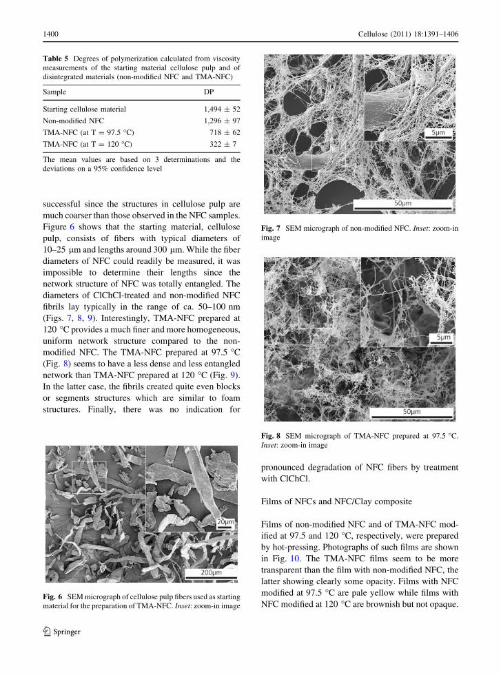
successful since the structures in cellulose pulp are
much coarser than those observed in the NFC samples.
Figure 6 shows that the starting material, cellulose
pulp, consists of fibers with typical diameters of
10–25 lm and lengths around 300 lm. While the fiber
diameters of NFC could readily be measured, it was
impossible to determine their lengths since the
network structure of NFC was totally entangled. The
diameters of ClChCl-treated and non-modified NFC
fibrils lay typically in the range of ca. 50–100 nm
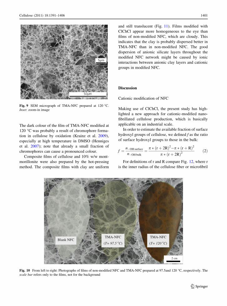
(Figs. 7, 8, 9). Interestingly, TMA-NFC prepared at
120 �C provides a much finer and more homogeneous,
uniform network structure compared to the non-
modified NFC. The TMA-NFC prepared at 97.5 �C
(Fig. 8) seems to have a less dense and less entangled
network than TMA-NFC prepared at 120 �C (Fig. 9).
In the latter case, the fibrils created quite even blocks
or segments structures which are similar to foam
structures. Finally, there was no indication for
pronounced degradation of NFC fibers by treatment
with ClChCl.
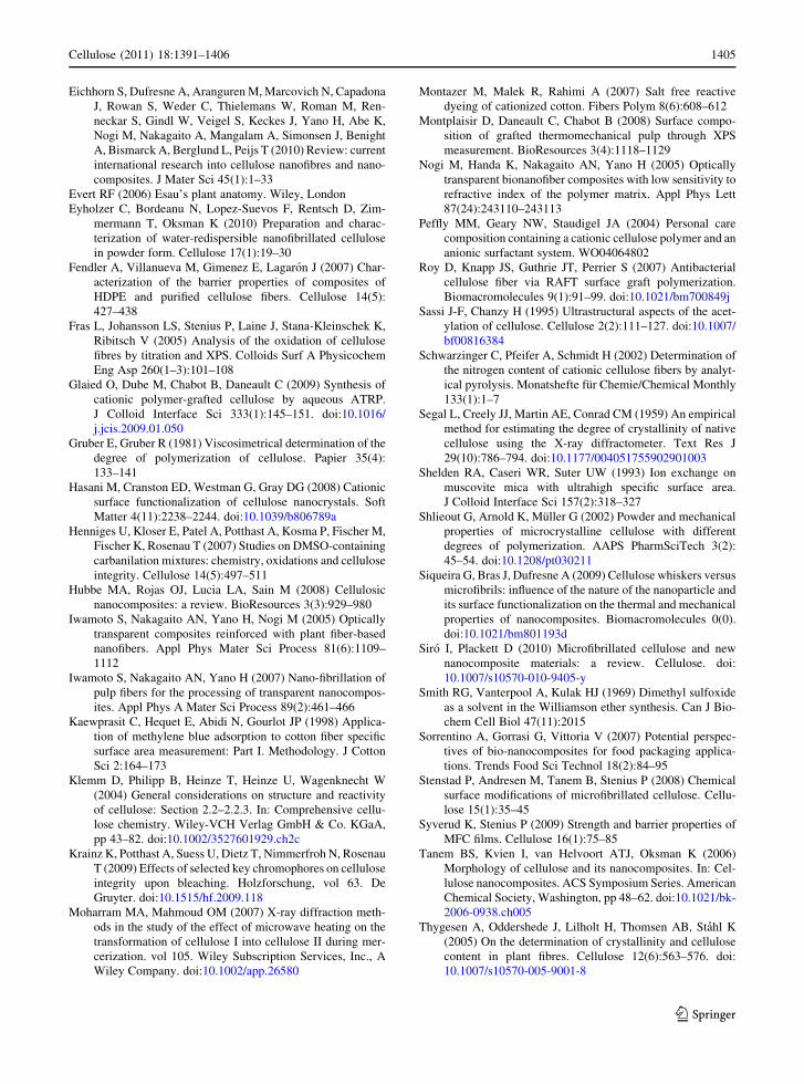
Films of NFCs and NFC/Clay composite
Films of non-modified NFC and of TMA-NFC mod-
ified at 97.5 and 120 �C, respectively, were prepared
by hot-pressing. Photographs of such films are shown
in Fig. 10. The TMA-NFC films seem to be more
transparent than the film with non-modified NFC, the
latter showing clearly some opacity. Films with NFC
modified at 97.5 �C are pale yellow while films with
NFC modified at 120 �C are brownish but not opaque.
Table 5 Degrees of polymerization calculated from viscosity
measurements of the starting material cellulose pulp and of
disintegrated materials (non-modified NFC and TMA-NFC)
Sample DP
Starting cellulose material 1,494 ± 52
Non-modified NFC 1,296 ± 97
TMA-NFC (at T = 97.5 �C) 718 ± 62
TMA-NFC (at T = 120 �C) 322 ± 7
The mean values are based on 3 determinations and the
deviations on a 95% confidence level
Fig. 6 SEM micrograph of cellulose pulp fibers used as starting
material for the preparation of TMA-NFC. Inset: zoom-in image
Fig. 7 SEM micrograph of non-modified NFC. Inset: zoom-in
image
Fig. 8 SEM micrograph of TMA-NFC prepared at 97.5 �C.
Inset: zoom-in image
1400 Cellulose (2011) 18:1391–1406
123
The dark colour of the film of TMA-NFC modified at
120 �C was probably a result of chromophore forma-
tion in cellulose by oxidation (Krainz et al. 2009),
especially at high temperature in DMSO (Henniges
et al. 2007); note that already a small fraction of
chromophores can cause a pronounced colour.
Composite films of cellulose and 10% w/w mont-
morillonite were also prepared by the hot-pressing
method. The composite films with clay are uniform
and still translucent (Fig. 11). Films modified with
ClChCl appear more homogeneous to the eye than
films of non-modified NFC, which are cloudy. This
indicates that the clay is probably dispersed better in
TMA-NFC than in non-modified NFC. The good
dispersion of anionic silicate layers throughout the
modified NFC network might be caused by ionic
interactions between anionic clay layers and cationic
groups in modified NFC.
Discussion
Cationic modification of NFC
Making use of ClChCl, the present study has high-
lighted a new approach for cationic-modified nano-
fibrillated cellulose production, which is basically
applicable on an industrial scale.
In order to estimate the available fraction of surface
hydroxyl groups of cellulose, we defined f as the ratio
of surface hydroxyl groups to those in the bulk:
f ¼ n�OH surface
n�OH bulk
¼ p � rþ 2Rð Þ2�p � rþ Rð Þ2
p � rþ 2Rð Þ2ð2Þ
For definitions of r and R compare Fig. 12, where r
is the inner radius of the cellulose fiber or microfibril
Fig. 9 SEM micrograph of TMA-NFC prepared at 120 �C.
Inset: zoom-in image
Blank NFC TMA-NFC
(T= 97.5 oC)
TMA-NFC
(T= 120 oC)
2 cm
Fig. 10 From left to right: Photographs of films of non-modified NFC and TMA-NFC prepared at 97.5and 120 �C, respectively. The
scale bar refers only to the films, not for the background
Cellulose (2011) 18:1391–1406 1401
123

without the outer layer of glucose unit. The r values for
fibers were derived from own morphological SEM
investigations (Fig. 6), where the cellulose pulp fibers
show diameters in the range of 10–25 lm. The values
of r for microfibrils were taken from reported literature
(Evert 2006). R is the radius of the glucose units on the
surface of the fiber or fibril, with R = 7.95 9
10-10 m (Abbott et al. 2006).
Note that in formula (2), only a half of the –OH
groups of the outer layer cellulose chains was assumed
to be accessible for reaction. As evident from the
schematic cross-section of a cellulose microfibril in
Fig. 12, in 2 glucose molecules (one repeating unit of a
cellulose chain), there are 3 hydroxyl groups on one
and 3 on the opposite side of the molecules. Therefore,
(r ? R) is the radius of the fiber or microfibril without
considering the surface of the outermost cellulose
chains.
The cross-section of a cellulose fiber can be
expressed in the same way as for a cellulose micro-
fibril, only with a different value of r.
Thus in case of a fiber with radius r = 7.5 9
10-6 m, f becomes 2.12 9 10-4. The molar amount
of –OH groups in bulk cellulose, n–OH bulk, is
n�OHbulk ¼3� 1
162:1406¼ 0:0185 mol=g
where one glucose unit has three –OH groups and a
molecular mass of 162.1406 g/mol.
Hence, the available quantity of –OH groups on the
surface of cellulose fibers (possible swelling effect of
the solvent neglected) is
Fig. 11 Photographs of composite films containing 10% w/w montmorillonite and (from left to right) non-modified NFC, TMA-NFC
prepared at 97.5 and at 120 �C, respectively
Fig. 12 Schematic
illustration of the
hierarchical structure of a
cellulose fiber including a
cross-section of a cellulose
microfibril. A cross-section
of a cellulose fiber can be
expressed in the same way
as for a cellulose microfibril,
only with a different value of
the radius r
1402 Cellulose (2011) 18:1391–1406
123

n�OHsurface ¼ f � n�OHbulk
¼ 2:12� 10�4 � 0:0185 mol=g
¼ 3:9� 10�6mol=g
If each surface –OH group of cellulose reacted with
ClChCl, in 1 g cellulose, the maximum molar amount
of trimethyl ammonium groups is 3.9 9 10-6 mol.
This amount is equivalent to a maximum nitrogen
content of Nmax of 0.0055% w/w. This is, however,
much less than the N contents of 0.13 and 0.27% found
after reaction at 97.5 and 120 �C (see Table 2) which
are 24 and 50 times higher than the estimated
maximum nitrogen content at the fiber surface.
Therefore, it appears that the swelling ability of
DMSO (Boluk 2005; Klemm et al. 2004) enabled the
reactants to enter inside the fiber and the reaction with
ClChCl also took place at the surfaces of the fibrils.
When considering a microfibril with a radius
r = 5 9 10-9 m (Evert 2006), n–OH surface becomes
4.2 9 10-3 mol/g, equivalent to Nmax of 5.88% w/w.
This value comes close to that estimated for the
ammonium groups in the surface region by XPS,
which amounted to around 1.6% w/w in the surface
region. This indicates that the modification with
ammonium groups indeed occurred predominantly at
the surfaces of the microfibrils, at relatively high
conversion of the fibrils’ surface –OH groups.
The nitrogen contents (0.27% w/w in the bulk and
1.6% w/w in the surface region) are much higher than
those reported for related modified cellulose fibers
(Abbott et al. 2006), which were estimated on the basis
of methylene blue adsorption. However, as indicated
above in the section Results, quantitative calculations
of surface –OH conversions at cellulose fibers by
means of methylene blue adsorption have to be taken
with care. Note in this context that methylene blue
could also adsorb by exchange with cations (Shelden
et al. 1993), e.g. with the calcium ions detected by
XPS.
In a pre-test, modification of cellulose by reaction
with ClChCl was also performed after disintegration.
However, this method was less successful than
modification before disintegration, apparently since
the fibrils had been modified in situ with ammonium
groups which caused repelling of the positively
charged fibrils. This is in agreement with SEM images
displaying cationized NFC fibrils with finer and more
homogeneous fibril networks and less agglomerates
than non-modifed NFC fibrils (Figs. 7, 8, 9).
Analogous results were reported for cellulose modi-
fied with anionic groups (Eyholzer et al. 2010). When
two routes with interchanged sequence of carboxy-
methylation of cellulose and mechanical disintegra-
tion were applied, samples that were first
carboxymethylated and then disintegrated provided
better homogeneity and as a consequence better water-
redispersibility. Probably, also in this case the elec-
trostatic repulsion between fibrils of alike charges
rendered the disintegration process more efficient
(Wagberg et al. 2008; Eyholzer et al. 2010).
Degradation of TMA-NFC
The NFC itself (i.e. without ClChCl treatment)
appears to undergo little degradation upon disintegra-
tion. The crystalline fraction and the degree of
polymerization decreased only slightly (Table 4, 5).
Yet the color changes (pale yellow and brown,
respectively, for TMA-NFC prepared at 97.5 and
120 �C) indicate some degradation of cellulose under
the action of the chemical treatment. However, as
mentioned above, already small amounts of degrada-
tion products might cause pronounced colorations.
The degree of polymerization (Table 5) became lower
with increasing color intensity of the TMA-NFC.
Under the applied modification conditions, a possible
oxidation reaction with cellulose may take place
through a chain ‘‘peeling’’ process causing a shorten-
ing in chain length. On the other hand, the crystallinity
ratio of NFC did not change significantly upon
chemical modification. No relevant relationships
between crystallinity and degree of polymerization
was also found in other reports (Shlieout et al. 2002).
Finally, there was no evidence from SEM images
(Figs. 7, 8, 9) or elemental analyses (Table 2) for
massive degradation of cellulose, considering the
confidence level of the elemental analyses and the fact
that the carbon content of cellulose is expected to
increase upon reaction with ClChCl. In summary, the
above results show that degradation upon treatment
with ClChCl resulted in materials which still can be
attributed to the class of nanofibrillated cellulose.
The mechanical performance of TMA-NFC is
expected not to be affected strikingly within the
magnitude of the observed decrease in degree of
polymerization (Zimmermann et al. 2010), since
mechanical properties of NFC are related primarily
to the network forming ability of NFC. This view is
Cellulose (2011) 18:1391–1406 1403
123

supported by the preservation of the crystallinity ratio
upon chemical modification (Iwamoto et al. 2007).
Films
Clays belong to the chemical class of layered silicates,
whereat the thickness of the individual silicate layers
amounts to the order of 1 nm. When the thickness of
such particles embedded in a polymer matrix is well
below the wavelength of light, the transmission of
light or the translucence of the resulting films becomes
higher. This is also the case when the cellulose units
become finer. Therefore, because of the finer nanofi-
bril dimensions of TMA-NFC (from SEM observa-
tions), non-modified NFC appeared more opaque to
the eyes than TMA-NFC. In addition, TMA-NFC
films were more homogeneous than films of non-
modified NFC. With the addition of clay particles, the
transparency of the materials decreased. In particular,
the films with non-modified NFC became cloudy, in
contrast to the films with TMA-NFC. This indicates
the presence of clay agglomerates in the films based on
non-modified NFC composites. In composite films of
TMA-NFC, the clay appeared to be better dispersed;
interactions between cationic groups in cationized
NFC and the negatively charged clay surfaces might
cause a dissociation of aggregates of clay and a good
distribution of clay throughout the modified NFC
network.
Conclusions
Nanofibrillated cellulose modified with quaternary
ammonium groups (TMA-NFC) can be prepared by
conversion of cellulose pulp with chlorocholine chlo-
ride (ClChCl), followed by a mechanical disintegra-
tion process. Due to swelling of the cellulose fibers by
the solvent applied for the chemical reaction (DMSO),
chlorocholine chloride had also access to the fibrils in
the interior of the fibers, and a relatively high degree of
surface –OH groups of the fibrils was converted. The
degradation of cellulose induced by the chemical
treatment was moderate, in spite of yellowish or brown
discolorations of the resulting materials. The TMA-
NFC showed a finer network structure and formed
more transparent films than the non-modified materi-
als. Also, clay (montmorillonite) dispersed better in
TMA-NFC than in non-modified nanofibrillated
cellulose.
Acknowledgments We kindly acknowledge the Commission
for Technology and Innovation (CTI) for financial support. We
thank Microanalysis Laboratory at ETH Zurich for conducting
the elemental analyses; Dr. Yoon Songhak for acquiring XRD
spectra; Esther Strub for performing viscosity measurements.
We are very grateful to Steffen Ohr at Cham-Tenero Paper Mills
Inc., Dr. Thomas Geiger and Dr. Philippe Tingaut for their
useful advices and support. Finally, we would like to say thank
Prof. Paul Smith for the helpful discussions.
References
Abbott AP, Bell TJ, Handa S, Stoddart B (2006) Cationic
functionalisation of cellulose using a choline based ionic
liquid analogue. Green Chem 8(9):784–786
Alince B, Petlicki J, van de Ven TGM (1991) Kinetics of col-
loidal particle deposition on pulp fibers 1. Deposition of
clay on fibers of opposite charge. Colloids Surf 59:265–277
Aulin C, Ahola S, Josefsson P, Nishino T, Hirose Y, Osterberg
M, Wagberg L (2009) Nanoscale cellulose films with dif-
ferent crystallinities and mesostructures—their surface
properties and interaction with water. Langmuir 25(13):
7675–7685. doi:10.1021/la900323n
Azizi Samir MAS, Alloin F, Dufresne A (2005) Review of
recent research into cellulosic whiskers, their properties
and their application in nanocomposite field. Biomacro-
molecules 6(2):612–626. doi:10.1021/bm0493685
Beamson G, Briggs D (1992) High resolution XPS of organic
polymers the Scienta ESCA300 database. Wiley, Chichester
Blasutto M, Delben F, Milost R, Painter TJ (1995) Novel cel-
lulosic ethers with low degrees of substitution—I. Prepa-
ration and analysis of long-chain alkyl ethers. Carbohydr
Polym 27(1):53–62
Bledzki AK, Gassan J (1999) Composites reinforced with cel-
lulose based fibres. Prog Polym Sci 24(2):221–274
Boluk Y (2005) Acid–base interactions and swelling of cellu-
lose fibers in organic liquids. Cellulose 12(6):577–593
Buschlediller G, Zeronian SH (1992) Enhancing the reactivity
and strength of cotton fibers. J Appl Polym Sci 45(6):
967–979
Cai X, Riedl B, Ait-Kadi A (2003) Effect of surface-grafted
ionic groups on the performance of cellulose-fiber-rein-
forced thermoplastic composites. J Polym Sci B Polym
Phys 41(17):2022–2032
Cheng Q, Wang S, Rials T, Lee SH (2007) Physical and
mechanical properties of polyvinyl alcohol and polypro-
pylene composite materials reinforced with fibril aggre-
gates isolated from regenerated cellulose fibers. Cellulose
14(6):593–602. doi:10.1007/s10570-007-9141-0
Daly S, Jachowicz J, Bianchini R (2010) Methods and kits
containing charged compounds imparting benefits to hair
and cosmetic products. WO10005906
de la Orden MU, Gonzalez Sanchez C, Gonzalez Quesada M,
Martınez Urreaga J (2007) Novel polypropylene-cellulose
composites using polyethylenimine as coupling agent.
Compos Part A Appl Sci Manuf 38(9):2005–2012
1404 Cellulose (2011) 18:1391–1406
123
Eichhorn S, Dufresne A, Aranguren M, Marcovich N, Capadona
J, Rowan S, Weder C, Thielemans W, Roman M, Ren-
neckar S, Gindl W, Veigel S, Keckes J, Yano H, Abe K,
Nogi M, Nakagaito A, Mangalam A, Simonsen J, Benight
A, Bismarck A, Berglund L, Peijs T (2010) Review: current
international research into cellulose nanofibres and nano-
composites. J Mater Sci 45(1):1–33
Evert RF (2006) Esau’s plant anatomy. Wiley, London
Eyholzer C, Bordeanu N, Lopez-Suevos F, Rentsch D, Zim-
mermann T, Oksman K (2010) Preparation and charac-
terization of water-redispersible nanofibrillated cellulose
in powder form. Cellulose 17(1):19–30
Fendler A, Villanueva M, Gimenez E, Lagaron J (2007) Char-
acterization of the barrier properties of composites of
HDPE and purified cellulose fibers. Cellulose 14(5):
427–438
Fras L, Johansson LS, Stenius P, Laine J, Stana-Kleinschek K,
Ribitsch V (2005) Analysis of the oxidation of cellulose
fibres by titration and XPS. Colloids Surf A Physicochem
Eng Asp 260(1–3):101–108
Glaied O, Dube M, Chabot B, Daneault C (2009) Synthesis of
cationic polymer-grafted cellulose by aqueous ATRP.
J Colloid Interface Sci 333(1):145–151. doi:10.1016/
j.jcis.2009.01.050
Gruber E, Gruber R (1981) Viscosimetrical determination of the
degree of polymerization of cellulose. Papier 35(4):
133–141
Hasani M, Cranston ED, Westman G, Gray DG (2008) Cationic
surface functionalization of cellulose nanocrystals. Soft
Matter 4(11):2238–2244. doi:10.1039/b806789a
Henniges U, Kloser E, Patel A, Potthast A, Kosma P, Fischer M,
Fischer K, Rosenau T (2007) Studies on DMSO-containing
carbanilation mixtures: chemistry, oxidations and cellulose
integrity. Cellulose 14(5):497–511
Hubbe MA, Rojas OJ, Lucia LA, Sain M (2008) Cellulosic
nanocomposites: a review. BioResources 3(3):929–980
Iwamoto S, Nakagaito AN, Yano H, Nogi M (2005) Optically
transparent composites reinforced with plant fiber-based
nanofibers. Appl Phys Mater Sci Process 81(6):1109–
1112
Iwamoto S, Nakagaito AN, Yano H (2007) Nano-fibrillation of
pulp fibers for the processing of transparent nanocompos-
ites. Appl Phys A Mater Sci Process 89(2):461–466
Kaewprasit C, Hequet E, Abidi N, Gourlot JP (1998) Applica-
tion of methylene blue adsorption to cotton fiber specific
surface area measurement: Part I. Methodology. J Cotton
Sci 2:164–173
Klemm D, Philipp B, Heinze T, Heinze U, Wagenknecht W
(2004) General considerations on structure and reactivity
of cellulose: Section 2.2–2.2.3. In: Comprehensive cellu-
lose chemistry. Wiley-VCH Verlag GmbH & Co. KGaA,
pp 43–82. doi:10.1002/3527601929.ch2c
Krainz K, Potthast A, Suess U, Dietz T, Nimmerfroh N, Rosenau
T (2009) Effects of selected key chromophores on cellulose
integrity upon bleaching. Holzforschung, vol 63. De
Gruyter. doi:10.1515/hf.2009.118
Moharram MA, Mahmoud OM (2007) X-ray diffraction meth-
ods in the study of the effect of microwave heating on the
transformation of cellulose I into cellulose II during mer-
cerization. vol 105. Wiley Subscription Services, Inc., A
Wiley Company. doi:10.1002/app.26580
Montazer M, Malek R, Rahimi A (2007) Salt free reactive
dyeing of cationized cotton. Fibers Polym 8(6):608–612
Montplaisir D, Daneault C, Chabot B (2008) Surface compo-
sition of grafted thermomechanical pulp through XPS
measurement. BioResources 3(4):1118–1129
Nogi M, Handa K, Nakagaito AN, Yano H (2005) Optically
transparent bionanofiber composites with low sensitivity to
refractive index of the polymer matrix. Appl Phys Lett
87(24):243110–243113
Peffly MM, Geary NW, Staudigel JA (2004) Personal care
composition containing a cationic cellulose polymer and an
anionic surfactant system. WO04064802
Roy D, Knapp JS, Guthrie JT, Perrier S (2007) Antibacterial
cellulose fiber via RAFT surface graft polymerization.
Biomacromolecules 9(1):91–99. doi:10.1021/bm700849j
Sassi J-F, Chanzy H (1995) Ultrastructural aspects of the acet-
ylation of cellulose. Cellulose 2(2):111–127. doi:10.1007/
bf00816384
Schwarzinger C, Pfeifer A, Schmidt H (2002) Determination of
the nitrogen content of cationic cellulose fibers by analyt-
ical pyrolysis. Monatshefte fur Chemie/Chemical Monthly
133(1):1–7
Segal L, Creely JJ, Martin AE, Conrad CM (1959) An empirical
method for estimating the degree of crystallinity of native
cellulose using the X-ray diffractometer. Text Res J
29(10):786–794. doi:10.1177/004051755902901003
Shelden RA, Caseri WR, Suter UW (1993) Ion exchange on
muscovite mica with ultrahigh specific surface area.
J Colloid Interface Sci 157(2):318–327
Shlieout G, Arnold K, Muller G (2002) Powder and mechanical
properties of microcrystalline cellulose with different
degrees of polymerization. AAPS PharmSciTech 3(2):
45–54. doi:10.1208/pt030211
Siqueira G, Bras J, Dufresne A (2009) Cellulose whiskers versus
microfibrils: influence of the nature of the nanoparticle and
its surface functionalization on the thermal and mechanical
properties of nanocomposites. Biomacromolecules 0(0).
doi:10.1021/bm801193d
Siro I, Plackett D (2010) Microfibrillated cellulose and new
nanocomposite materials: a review. Cellulose. doi:
10.1007/s10570-010-9405-y
Smith RG, Vanterpool A, Kulak HJ (1969) Dimethyl sulfoxide
as a solvent in the Williamson ether synthesis. Can J Bio-
chem Cell Biol 47(11):2015
Sorrentino A, Gorrasi G, Vittoria V (2007) Potential perspec-
tives of bio-nanocomposites for food packaging applica-
tions. Trends Food Sci Technol 18(2):84–95
Stenstad P, Andresen M, Tanem B, Stenius P (2008) Chemical
surface modifications of microfibrillated cellulose. Cellu-
lose 15(1):35–45
Syverud K, Stenius P (2009) Strength and barrier properties of
MFC films. Cellulose 16(1):75–85
Tanem BS, Kvien I, van Helvoort ATJ, Oksman K (2006)
Morphology of cellulose and its nanocomposites. In: Cel-
lulose nanocomposites. ACS Symposium Series. American
Chemical Society, Washington, pp 48–62. doi:10.1021/bk-
2006-0938.ch005
Thygesen A, Oddershede J, Lilholt H, Thomsen AB, Stahl K
(2005) On the determination of crystallinity and cellulose
content in plant fibres. Cellulose 12(6):563–576. doi:
10.1007/s10570-005-9001-8
Cellulose (2011) 18:1391–1406 1405
123

Tingaut P, Zimmermann T, Lopez-Suevos F (2009) Synthesis
and characterization of bionanocomposites with tunable
properties from poly(lactic acid) and acetylated micro-
fibrillated cellulose. Biomacromolecules 11(2):454–464.
doi:10.1021/bm901186u
Turbak AF, Snyder FW, Sandberg KR (1983) Microfibrillated
cellulose, a new cellulose product: properties, uses, and
commercial potential. J Appl Polym Sci Symp 37:815–827
Wagberg L, Decher G, Norgren M, Lindstrom T, Ankerfors M,
Axnas K (2008) The build-up of polyelectrolyte multilay-
ers of microfibrillated cellulose and cationic polyelectro-
lytes. Langmuir 24(3):784–795. doi:10.1021/la702481v
Zimmermann T, Pohler E, Geiger T (2004) Cellulose fibrils for
polymer reinforcement. Adv Eng Mater 6(9):754–761
Zimmermann T, Pohler E, Schwaller P (2005) Mechanical and
morphological properties of cellulose fibril reinforced
nanocomposites. Adv Eng Mater 7(12):1156–1161
Zimmermann T, Bordeanu N, Strub E (2010) Properties of
nanofibrillated cellulose from different raw materials and
its reinforcement potential. Carbohydr Polym 79(4):
1086–1093
1406 Cellulose (2011) 18:1391–1406
123