Embed Size (px)
Citation preview
Thin Solid Films 519 (2010) 1219–1223
Contents lists available at ScienceDirect
Thin Solid Films
j ourna l homepage: www.e lsev ie r.com/ locate / ts f
Preparation and characterization of bio-functionalized iron oxide nanoparticles forbiomedical application
Bharat Bajaj a, B.D. Malhotra b, Sunju Choi a,⁎a Radioisotope Research Division, Basic Science and Technology Department, Korea Atomic Energy Research Institute (KAERI), Yuseong, Daejeon 305-353, Republic of Koreab Department of Science and Technology Center on Biomolecular Electronics, National Physical Laboratory, New Delhi 110012, India
⁎ Corresponding author.E-mail addresses: [email protected] (B. Baja
0040-6090/$ – see front matter © 2010 Elsevier B.V. Aldoi:10.1016/j.tsf.2010.08.072
a b s t r a c t
a r t i c l e i n f oAvailable online 25 August 2010
Keywords:Fe3O4 nanoparticlesMagnetismFunctionalizationDTPA
Amine modified iron oxide (Fe3O4) nanoparticles were synthesized by thermal decomposition method and werefurther used to bio-functionalize by grafting of N-hydroxysuccinimide (NHS) ester of folate and ethylenediami-netetraacetate (EDTA). Fe3O4 nanoparticles of ~22 nm were confirmed from X-ray diffraction (XRD) andtransmission electronmicroscopy (TEM) studies. FT-IR studies indicated twobands at 1515 cm−1and1646 cm−1,which canbe attributed to carboxylic group and the amide linkage respectively, revealing the conjugation of folatewith Fe3O4. The conjugation of the chelating agent showed strong C=O stretch and Fe–O vibrations at 1647 and588 cm−1 respectively. The value of saturationmagnetization for Fe3O4 nanoparticles was found to be 88 emu/g,which further reduced to 18 and 32% upon functionalization with EDTA and NHS ester folate, respectively. Theseamine modified Fe3O4 nanoparticles can also be functionalized with other bifunctional chelators, such as aminoacids based diethylene triamine pentaacetic acid (DTPA), and thus find potential applications in radio-labeling,biosensors and cancer detection, etc.
j), [email protected] (S. Choi).
l rights reserved.
© 2010 Elsevier B.V. All rights reserved.
1. Introduction
Recently functionalized nanomaterials have gained considerableattention due to their unusual optical, electrical and molecularproperties related with the quantum confinement effect, which findnumerous technological applications including those in biomedicalindustries [1–4]. Among these, magnetic nanomaterials have shown asignificant contribution in the field of clinical diagnostics andtherapeutics. This includes magnetic separation of labeled cells andother biological entities, therapeutic drug, gene and radionuclidedelivery, radio frequency methods for the catabolism of tumors viahyperthermia and contrast enhancement agents for magnetic reso-nance imaging applications [5]. Moreover, magnetic nanoparticlesexhibiting a higher specific surface area are used for the precisebinding of larger biomolecules for biosensor applications and selectiveseparation of immobilized biomolecules from a reaction mixturethrough application of the magnetic field [1–5].
Effectiveness of cancer therapy including chemotherapy, surgeryor radiotherapy largely depends on the efficacy of targeting and deathof cancerous cells without affecting healthy cells. Compared to viralsystem, non-viral nanoparticles systems have gained increasinginterest in the clinical therapeutics due to their low toxicity [7].Recently magnetic nanoparticle-folic acid conjugates have shownbetter results for cancer treatment as most of the human cancer
malignancies including ovaries, brain, kidney, breast, renal cellcarcinomas, and brain metastases are found to be frequentlyoverexpressed by the folate receptor [8–10].
Among various magnetic nanomaterials, Fe3O4 nanoparticles havebeen exploited to a larger extent due to their biocompatibility, non-toxicity, easy preparation and high superparamagnetism propertiesthat can be used for drug delivery, tissue repair, separation andpurification of cell populations, cell imaging, targeting and therapy[1]. However, in order to enhance the utility of these particles for suchapplications, they should possess high magnetic saturation, biocom-patibility and should easily conjugate with multifunctional groupssuch as bifunctional chelating agents [1,6].
Bio-functionalized Fe3O4 nanoparticles are a new class of materialswith controlled and tailored properties. The water-dispersibility of(Fe3O4) nanoparticles is an important parameter for use in biology.Sun et al. and Hyeon et al. have synthesized a series of high-quality,monodispersed Fe3O4 nanoparticles soluble in nonpolar solvents [14–16]. There are only few reports on the direct preparation of water-soluble Fe3O4 nanoparticles in a high polar solvent [12]. Recently,preparation of one-step water-soluble amine functionalized Fe3O4
nanoparticles using a single precursor has been reported [11–13]. Theaqueous synthesis of iron oxide colloidal nanoparticles from areduction of a single iron precursor using long-chain amines hasbeen proposed, which provides capping of the amine molecule to thesurface and renders them water dispersible [11]. The amine cappingimparts hydrophilicity on the nanoparticles and thus makes themwater soluble. The water-dispersible Fe3O4 nanoparticles have a hugepotential for further functionalization with biomolecules and
1220 B. Bajaj et al. / Thin Solid Films 519 (2010) 1219–1223
bifunctional chelators while these can be capped with amine groupand amino acid based DTPA due to the abundance of lysine side chainε-amine and N-terminal α-amines.
In this paper, we report the preparation of amine functionalizedFe3O4 nanoparticles via a thermal decomposition method usingreduction of a single precursor. The as synthesized Fe3O4 nanoparti-cles are bio-functionalized by NHS ester of folate and EDTA. The bio-functionalized Fe3O4 nanoparticles may find potential technologicalapplications in biosensing and cancer therapy.
2. Experimental section
2.1. Materials
All chemicals were of reagent grade and obtained from a commercialsource, for e.g. ferrous chloride tetrahydrate (FeCl2.4H2O), dodecylamine,folic acid dihydrate, 1, 3-dicyclohexylcarbodimide, EDTA–dianhydrideand DIPEA (N, N-diisopropylethylamine) were purchased from Sigma-Aldrich. All the solvents (analytical grade) purchased from Sigma-Aldrichwere used without further purification.
2.2. Synthesis and functionalization of Fe3O4 nanoparticles
2.2.1. Synthesis of Fe3O4 nanoparticlesIron oxide nanoparticles (Fe3O4) were synthesized by a one-step
aqueous method from a single iron precursor using long-chain amineas the reducing and surface functionalizing agent according to a
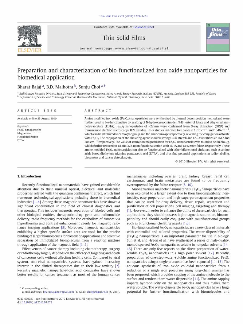
Scheme 1. Proposed mechanism of synthesis and bio-func
method reported in the literature [11]. In brief, amine-capped Fe3O4
nanoparticles were prepared by reducing (0.5 mmol, 0.198 g) offerrous chloride (FeCl2.4H2O) by (2 mmol, 0.7414 g) of dodecylamineundermild heating at 85°–90 °C for 3 h as shown in (Scheme 1, step 1).The synthesis was carried out in two necked glass flask under nitrogenatmosphere and constant magnetic stirring. First, the dodecylaminewas taken in a glass flask and heated until it changes to liquid state, towhich then was added the aqueous solution of ferrous chloride(FeCl2.4H2O), previously degassed by nitrogen bubbling. Here, Fe2+
ions were reduced by amine and simultaneously capped with theamine molecules. The color of the solution changed to deep blackindicating the formation of iron oxide. In order to remove thesupernatant from the solution, an external magnet was applied. Afterdecanting the supernatant, the resulting Fe3O4 nanoparticles werewashed with acetone, water and ethanol several times. The resultingamine modified Fe3O4 nanoparticles were dissolved in water andstored at room temperature.
2.2.2. Synthesis of NHS ester of folic acidNHS ester of folic acid was synthesized as described previously
[17]. In brief 2.5 g of folic acid dihydrate was dissolved in 100 mldimethylsulfoxide (DMSO). A 1.1 molar excess (1.26 g) of NHS anddicyclohexylcarbodiimide (2.26 g) was then added. The reactionmixture was stirred in the dark at room temperature overnight(Scheme 1, step 2). The byproduct dicyclohexylurea was removed byfiltration. The DMSO solution of the NHS–folate was stored at−20 °C.
tionalization of amine stabilized Fe3O4 nanoparticles.
1221B. Bajaj et al. / Thin Solid Films 519 (2010) 1219–1223
2.3. Preparation of folic acid grafted Fe3O4 nanoparticles
To synthesize folate conjugated Fe3O4 nanoparticles, NHS–folate(538.47 mg)was first dissolved in DMSO (1 ml) and then added to thenanoparticle suspension, 10 mg/ml. Subsequently, 174 μl of DIPEA(N, N-diisopropylethylamine) was added as a base. The reactionsolution was shaken at room temperature for 3 h. Unreacted folic acidfrom the folate conjugated iron oxide nanoparticles was separatedusing a magnetic separator and the conjugated Fe3O4 nanoparticleswere washed with DMSO and water, until the traces of free folic acidcompletely removed.
2.4. Preparation of EDTA conjugated Fe3O4 nanoparticle
The conjugation of bifunctional chelator EDTA dianhydride wasconducted by continuous stirring at 40 °C for 24 h [18]. Aminestabilized iron oxide nanoparticles (10 mg/ml) and EDTA dianhydride(1 g/ml) were predissolved in DMSO followed by addition of 0.1 Mbicarbonate buffer (pH 8.5) and DIPEA as a catalyst (Scheme 1, step 3).Unreacted EDTA dianhydride was separated using a magneticseparator. The EDTA conjugated iron oxide nanoparticles werewashed with DMSO and water until the traces of free EDTA wascompletely removed.
2.5. Characterizations
Powder X-ray diffraction (XRD) patterns were acquired using aSiemens D5000 diffractometer (Siemens, Munich, Germany) with Cu
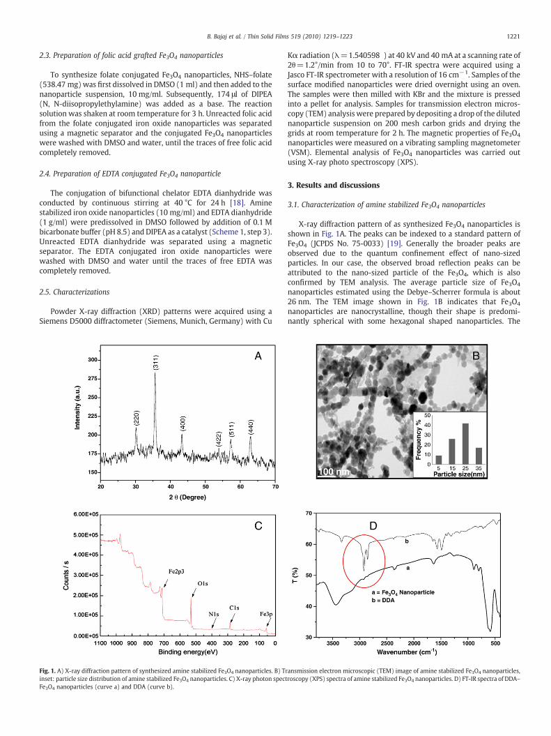
Fig. 1. A) X-ray diffraction pattern of synthesized amine stabilized Fe3O4 nanoparticles. B) Trinset: particle size distribution of amine stabilized Fe3O4 nanoparticles. C) X-ray photon spectFe3O4 nanoparticles (curve a) and DDA (curve b).
Kα radiation (λ=1.540598 ) at 40 kV and 40 mA at a scanning rate of2θ=1.2°/min from 10 to 70°. FT-IR spectra were acquired using aJasco FT-IR spectrometer with a resolution of 16 cm−1. Samples of thesurface modified nanoparticles were dried overnight using an oven.The samples were then milled with KBr and the mixture is pressedinto a pellet for analysis. Samples for transmission electron micros-copy (TEM) analysis were prepared by depositing a drop of the dilutednanoparticle suspension on 200 mesh carbon grids and drying thegrids at room temperature for 2 h. The magnetic properties of Fe3O4
nanoparticles were measured on a vibrating sampling magnetometer(VSM). Elemental analysis of Fe3O4 nanoparticles was carried outusing X-ray photo spectroscopy (XPS).
3. Results and discussions
3.1. Characterization of amine stabilized Fe3O4 nanoparticles
X-ray diffraction pattern of as synthesized Fe3O4 nanoparticles isshown in Fig. 1A. The peaks can be indexed to a standard pattern ofFe3O4 (JCPDS No. 75-0033) [19]. Generally the broader peaks areobserved due to the quantum confinement effect of nano-sizedparticles. In our case, the observed broad reflection peaks can beattributed to the nano-sized particle of the Fe3O4, which is alsoconfirmed by TEM analysis. The average particle size of Fe3O4
nanoparticles estimated using the Debye–Scherrer formula is about26 nm. The TEM image shown in Fig. 1B indicates that Fe3O4
nanoparticles are nanocrystalline, though their shape is predomi-nantly spherical with some hexagonal shaped nanoparticles. The
ansmission electron microscopic (TEM) image of amine stabilized Fe3O4 nanoparticles,roscopy (XPS) spectra of amine stabilized Fe3O4 nanoparticles. D) FT-IR spectra of DDA–
3500 3000 2500 2000 1500 1000 500
40
50
60
70
80
90
100
c = Folic acid
e = EDTA-dianhydride
a
e
c
d
b
T (
%)
Wavenumber (cm-1)
a = Fe3O4 nanoparticles
b = NHS-folate-Fe3O4 nanoparticle
d = EDTA-Fe3O4nanoparticle
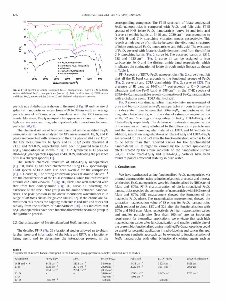
Fig. 2. FT-IR spectra of amine stabilized Fe3O4 nanoparticles (curve a), NHS–folateamine stabilized Fe3O4 nanoparticles (curve b), Folic acid (curve c) EDTA-aminestabilized Fe3O4 nanoparticles (curve d) and EDTA–dianhydride (curve e).
1222 B. Bajaj et al. / Thin Solid Films 519 (2010) 1219–1223
particle size distribution is shown in the inset of Fig. 1B and the size ofspherical nanoparticles varies from ∼10 to 30 nm with an averageparticle size of ∼22 nm, which correlates with the XRD measure-ments. Moreover, Fe3O4 nanoparticles appear in a chain form due tohigh surface area and magnetic dipole–dipole interactions betweenparticles [20,21].
The chemical nature of bio-functionalized amine modified Fe3O4
nanoparticles has been analyzed by XPS measurement. Fe, N, and Opeaks are corrected with reference to the C 1s peak at 284.5 eV. Fromthe XPS measurements, Fe 3p3/2 and Fe 3p1/2 peaks observed at711.0 and 724.8 eV, respectively, have been originated from DDA–Fe3O4 nanoparticles as shown in Fig. 1C. A symmetric N 1s peak forDDA–Fe3O4 nanoparticles appears at 400.0 eV, indicating the presenceof N as a charged species [11].
The surface chemical structure of DDA–Fe3O4 nanoparticles(Fig. 1D, curve a) has been characterized using FT-IR spectroscopy.FT-IR spectra of DDA have also been overlaid for the comparison(Fig. 1D, curve b). The strong absorption peaks at around 588 cm−1
are the characteristics of the Fe–O vibrations, while the transmissionaround 2923 and 2854 cm−1 (Fig. 1D, circle) are well matched withthat from free dodecylamine (Fig. 1D, curve b) indicating theexistence of the free –NH2 group on the amine stabilized nanopar-ticles. The peak position in the above mentioned wavenumber is inrange of all-trans chains-for gauche chains [22]. If the chains are all-trans then this means the capping molecule is rod-like and sticks outradially from the surfaces of nanoparticles [26]. This indicates thatFe3O4 nanoparticles have been functionalizedwith the amino group inthe synthetic process.
3.2. Characterization of bio-functionalized Fe3O4 nanoparticles
The detailed FT-IR (Fig. 2) vibrational studies allowed us to obtainfurther structural information of the folate and EDTA as a functiona-lizing agent and to determine the interaction present in the
Table 1Assignments of infrared bands (correspond to the functional groups present in samples) ob
Assignment Fe3O4–DDA DDA Folate–Fe3O
O–H and N–H 3436 cm−1 3332 cm−1 3440 cm−1
C–H 2923 cm−1 2919 cm−1 2920 cm−1
2854 cm−1 2854 cm−1 2852 cm−1
C=O – – 1646 cm−1
C–O – – 1515 cm−1
Fe–O 588 cm−1 – 588 cm−1
corresponding complexes. The FT-IR spectrum of folate conjugatedFe3O4 nanoparticles is compared with Fe3O4 and folic acid. FT-IRspectra of NHS–folate Fe3O4 nanoparticle (curve b) and folic acid(curve c) exhibit bands at 3440 and 2920 cm−1 corresponding toO–H/N–H and C–H stretching vibration modes respectively. Thisreveals a high degree of similarity between the vibrational structuresof folate conjugated Fe3O4 nanoparticles and folic acid. The existenceof Fe3O4 covered with folate is clearly demonstrated from the shift inC–H stretching bands (Fig. 2, curve b). The observed bands at 1515,588 and 1635 cm−1 (Fig. 2, curve b) can be assigned to ironcarboxylate, Fe–O and the distinct amide band respectively, whichimplicates the conjugation of folate through amide linkage as shownin Table 1.
FT-IR spectra of EDTA–Fe3O4 nanoparticles (Fig. 2, curve d) exhibitthat all the IR band corresponds to the functional groups of Fe3O4
(Fig. 2, curve a) and EDTA dianhydride (Fig. 2, curve e) [23]. Thepresence of IR band at 1647 cm−1 corresponds to C=O stretchvibrations and the Fe–O band at 588 cm−1 in the FT-IR spectra ofEDTA–Fe3O4 nanoparticles reveals conjugation of Fe3O4 nanoparticleswith a chelating agent (EDTA dianhydride).
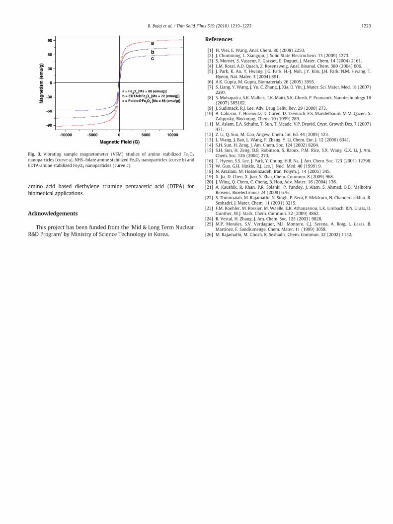
Fig. 3 shows vibrating sampling magnetometer measurement ofpure and bio-functionalize Fe3O4 nanoparticles at room temperaturein a dry state. It can be seen that DDA–Fe3O4 nanoparticles exhibitmagnetic characteristics, with the value of saturation magnetizationas 88, 72 and 56 emu/g corresponding to Fe3O4, EDTA–Fe3O4, andfolate–Fe3O4 respectively. The difference in saturation magnetizationof the samples is mainly attributed to the difference in particle sizeand the layer of nonmagnetic material i.e. EDTA and NHS–folate. Inaddition, saturation magnetizations of folate–Fe3O4 and EDTA–Fe3O4
are reduced to 18% and 32% after the functionalization, which is foundto be better than that reported earlier for the functionalizednanomaterial [8]. It might be caused by the surface spin-cantingeffects created by the amine molecule on the nanoparticle surface[24,25]. These folate–Fe3O4 and EDTA–Fe3O4 particles have beenfound to possess excellent stability in pure water.
4. Conclusions
We have synthesized amine functionalized Fe3O4 nanoparticles viathermal decomposition using reduction of a single precursor and these assynthesized Fe3O4 nanoparticles were bio-functionalized by NHS ester offolate and EDTA. FT-IR characterization of bio-functionalized Fe3O4
nanoparticles revealed the conjugationof nanoparticleswithNHSester offolate and EDTA. XRD measurement showed the formation of themagnetite Fe3O4 phase. The magnetization measurement showed thesaturation magnetization value of 88 emu/g for Fe3O4 nanoparticles,which reduced to about 18% and 32% after the functionalization withEDTA and NHS ester folate, respectively. As high magnetization valuesand smaller particle size (less than 100 nm) are an importantrequirement for biomedical applications, we envisage that such highmagnetization values after functionalization and smaller particle size ofthe present bio-functionalized aminemodified Fe3O4 nanoparticles couldbe useful for potential application in radio-labeling and cancer therapy.This unique synthetic approach can be extended to functionalization ofFe3O4 nanoparticles with other bifunctional chelating agents such as
tained in FT-IR studies.
4 Folic acid EDTA–Fe3O4 EDTA–dianhydride
3436 cm−1 3428 cm−1 3428 cm−1
2920 cm−1 3001 cm−1 2990 cm−1
1690 cm−1 1647 cm−1 1762 cm−1
1608 cm−1 – –
– 588 cm−1 –
-10000 -5000 0 5000 10000
-90
-60
-30
0
30
60
90
a = Fe3O4 [Ms = 88 (emu/g)]b = EDTA@Fe3O4 [Ms = 72 (emu/g)]c = Folate@Fe3O4 [Ms = 56 (emu/g)]
cb
aM
agn
etis
m (
emu
/g)
Magnetic Field (G)
Fig. 3. Vibrating sample magnetometer (VSM) studies of amine stabilized Fe3O4
nanoparticles (curve a), NHS–folate amine stabilized Fe3O4 nanoparticles (curve b) andEDTA-amine stabilized Fe3O4 nanoparticles (curve c).
1223B. Bajaj et al. / Thin Solid Films 519 (2010) 1219–1223
amino acid based diethylene triamine pentaacetic acid (DTPA) forbiomedical applications.
Acknowledgements
This project has been funded from the ‘Mid & Long Term NuclearR&D Program’ by Ministry of Science Technology in Korea.
References
[1] H. Wei, E. Wang, Anal. Chem. 80 (2008) 2250.[2] J. Chumming, L. Xiangqin, J. Solid State Electrochem. 13 (2009) 1273.[3] S. Mornet, S. Vasseur, F. Grasset, E. Duguet, J. Mater. Chem. 14 (2004) 2161.[4] L.M. Rossi, A.D. Quach, Z. Rosenzweig, Anal. Bioanal. Chem. 380 (2004) 606.[5] J. Park, K. An, Y. Hwang, J.G. Park, H.-J. Noh, J.Y. Kim, J.H. Park, N.M. Hwang, T.
Hyeon, Nat. Mater. 3 (2004) 891.[6] A.K. Gupta, M. Gupta, Biomaterials 26 (2005) 3995.[7] S. Liang, Y. Wang, J. Yu, C. Zhang, J. Xia, D. Yin, J. Mater. Sci. Mater. Med. 18 (2007)
2297.[8] S. Mohapatra, S.K. Mallick, T.K. Maiti, S.K. Ghosh, P. Pramanik, Nanotechnology 18
(2007) 385102.[9] J. Sudimack, R.J. Lee, Adv. Drug Deliv. Rev. 29 (2000) 273.
[10] A. Gabizon, T. Horowitz, D. Goren, D. Tzemach, F.S. Mandelbaum, M.M. Qazen, S.Zalippsky, Bioconjug. Chem. 10 (1999) 289.
[11] M. Aslam, E.A. Schultz, T. Sun, T. Meade, V.P. Dravid, Cryst. Growth Des. 7 (2007)471.
[12] Z. Li, Q. Sun, M. Gao, Angew. Chem. Int. Ed. 44 (2005) 123.[13] L. Wang, J. Bao, L. Wang, F. Zhang, Y. Li, Chem. Eur. J. 12 (2006) 6341.[14] S.H. Sun, H. Zeng, J. Am. Chem. Soc. 124 (2002) 8204.[15] S.H. Sun, H. Zeng, D.B. Robinson, S. Raoux, P.M. Rice, S.X. Wang, G.X. Li, J. Am.
Chem. Soc. 126 (2004) 273.[16] T. Hyeon, S.S. Lee, J. Park, Y. Chung, H.B. Na, J. Am. Chem. Soc. 123 (2001) 12798.[17] W. Guo, G.H. Hinkle, R.J. Lee, J. Nucl. Med. 40 (1999) 9.[18] N. Arsalani, M. Hosseinzadeh, Iran. Polym. J. 14 (2005) 345.[19] X. Jia, D. Chen, X. Jiao, S. Zhai, Chem. Commun. 8 (2009) 968.[20] J. Wing, Q. Chem, C. Cheng, B. Hou, Adv. Mater. 16 (2004) 136.[21] A. Kaushik, R. Khan, P.R. Solanki, P. Pandey, J. Alam, S. Ahmad, B.D. Malhotra
Biosens, Bioelectronics 24 (2008) 676.[22] S. Thimmaiah, M. Rajamathi, N. Singh, P. Bera, F. Meldrum, N. Chanderasekhar, R.
Seshadri, J. Mater. Chem. 11 (2001) 3215.[23] F.M. Koehler, M. Rossier, M. Waelle, E.K. Athanassiou, L.K. Limbach, R.N. Grass, D.
Gunther, W.J. Stark, Chem. Commun. 32 (2009) 4862.[24] R. Vestal, H. Zhang, J. Am. Chem. Soc. 125 (2003) 9828.[25] M.P. Morales, S.V. Verdaguer, M.I. Montero, C.J. Serena, A. Roig, L. Casas, B.
Martinez, F. Sandiumenge, Chem. Mater. 11 (1999) 3058.[26] M. Rajamathi, M. Ghosh, R. Seshadri, Chem. Commun. 32 (2002) 1152.


![Toward Theranostic Nanoparticles: CB[7]- …Page 1 of 20 Toward Theranostic Nanoparticles: CB[7]-Functionalized Iron Oxide for Drug Delivery and MRI Farah Benyettou,a Irena Milosevic,b](https://img.pdfslide.us/doc/110x75/5e2f7c57e7d9ee41cd02aee2/toward-theranostic-nanoparticles-cb7-page-1-of-20-toward-theranostic-nanoparticles.jpg)