Embed Size (px)
DESCRIPTION
Preoperative evaluation Indication and contraindication Positioning OR setup. Ass. Prof. Zdravko Perko. OPEN APPROACH Colonoscopy, rectoscopy (surgeon!) Precise measurement Anocutaneus distance biopsy! Barium enema Op strategy MSCT, NMR, EUS. preoperative work-up. - PowerPoint PPT Presentation
Citation preview
Preoperative evaluationIndication and contraindication
PositioningOR setup
Ass. Prof. Zdravko Perko
• OPEN APPROACH
• Colonoscopy, rectoscopy (surgeon!)– Precise measurement– Anocutaneus distance– biopsy!
• Barium enema– Op strategy
• MSCT, NMR, EUS
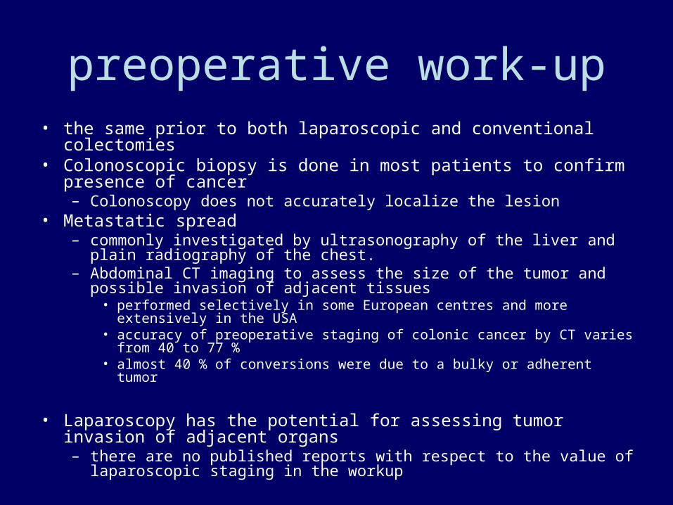
preoperative work-up• the same prior to both laparoscopic and conventional colectomies• Colonoscopic biopsy is done in most patients to confirm presence of
cancer– Colonoscopy does not accurately localize the lesion
• Metastatic spread– commonly investigated by ultrasonography of the liver and plain
radiography of the chest.– Abdominal CT imaging to assess the size of the tumor and possible
invasion of adjacent tissues• performed selectively in some European centres and more extensively in the
USA• accuracy of preoperative staging of colonic cancer by CT varies from 40 to
77 %• almost 40 % of conversions were due to a bulky or adherent tumor
• Laparoscopy has the potential for assessing tumor invasion of adjacent organs– there are no published reports with respect to the value of laparoscopic
staging in the workup
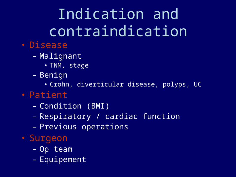
Indication and contraindication
• Disease– Malignant
• TNM, stage
– Benign• Crohn, diverticular disease, polyps, UC
• Patient– Condition (BMI)– Respiratory / cardiac function– Previous operations
• Surgeon– Op team– Equipement
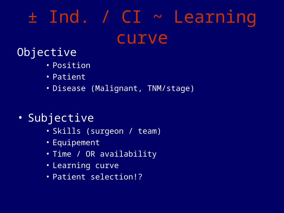
± Ind. / CI ~ Learning curveObjective
• Position• Patient• Disease (Malignant, TNM/stage)
• Subjective• Skills (surgeon / team)• Equipement• Time / OR availability• Learning curve• Patient selection!?
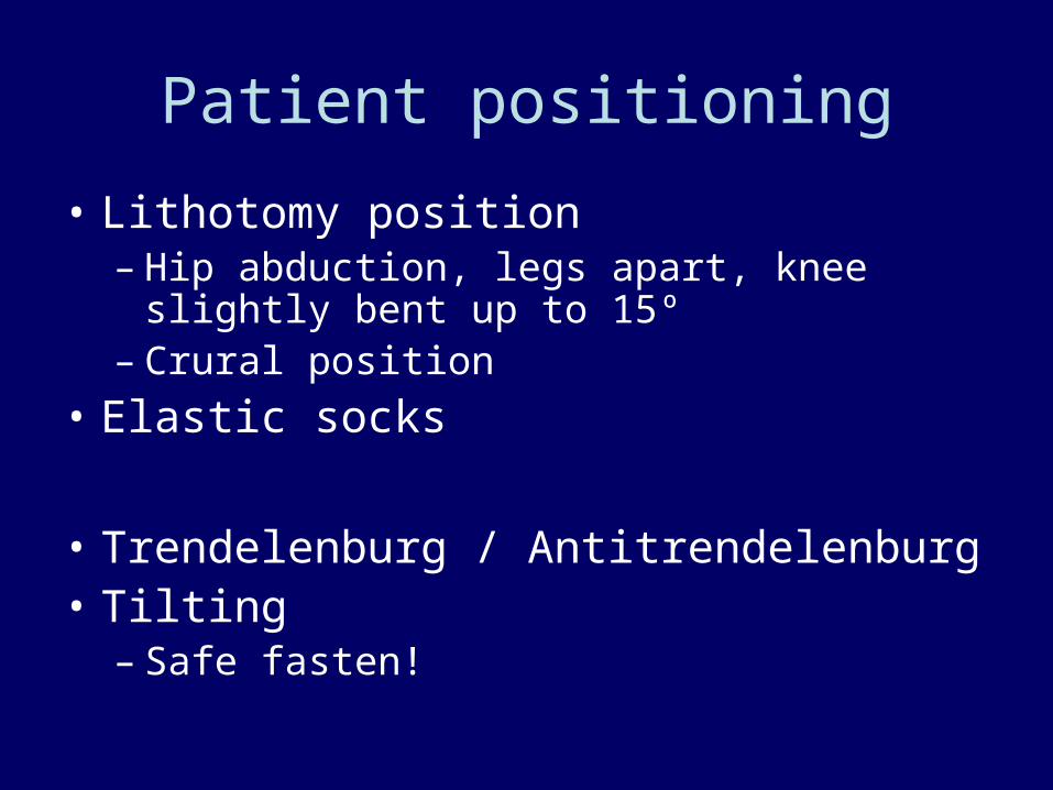
Patient positioning
• Lithotomy position– Hip abduction, legs apart, knee slightly bent
up to 15º– Crural position
• Elastic socks
• Trendelenburg / Antitrendelenburg• Tilting
– Safe fasten!
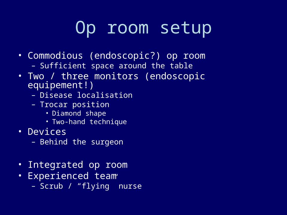
Op room setup
• Commodious (endoscopic?) op room– Sufficient space around the table
• Two / three monitors (endoscopic equipement!)– Disease localisation– Trocar position
• Diamond shape• Two-hand technique
• Devices– Behind the surgeon
• Integrated op room• Experienced team
– Scrub / “flying” nurse
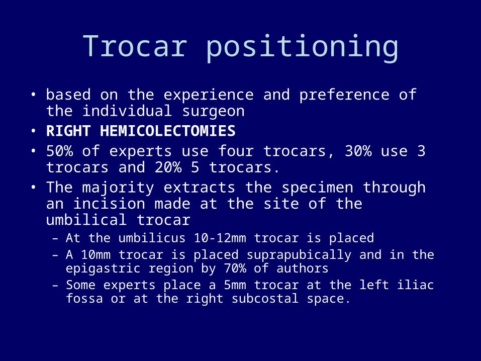
Trocar positioning
• based on the experience and preference of the individual surgeon
• RIGHT HEMICOLECTOMIES• 50% of experts use four trocars, 30% use 3 trocars and
20% 5 trocars.• The majority extracts the specimen through an incision
made at the site of the umbilical trocar– At the umbilicus 10-12mm trocar is placed– A 10mm trocar is placed suprapubically and in the epigastric
region by 70% of authors– Some experts place a 5mm trocar at the left iliac fossa or at the
right subcostal space.

RIGHT HEMICOLECTOMIES

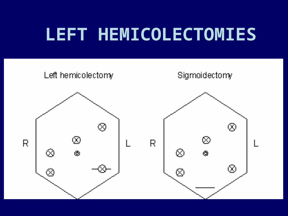
LEFT HEMICOLECTOMIES
• For left hemicolectomy and for sigmoid resection• almost at the same sites• Thirty percent of experts perform these procedures using
the hand-assisted technique• Five trocars are used by over 70% of experts
– A 10-12mm trocar is placed at the umbilicus– two 10mm trocars are placed by 80% of experts in the right iliac
fossa and in the right suprapubic region
• The incision for specimen extraction– the left iliac fossa– suprapubic incision
LEFT HEMICOLECTOMIES

Type of procedure?Preop planning
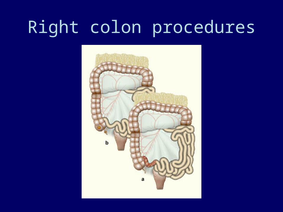
Right colon procedures

Right colonpatient position - op setup
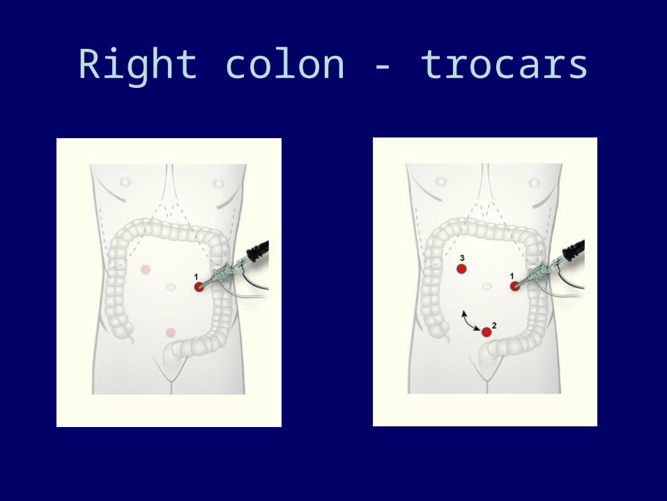
Right colon - trocars
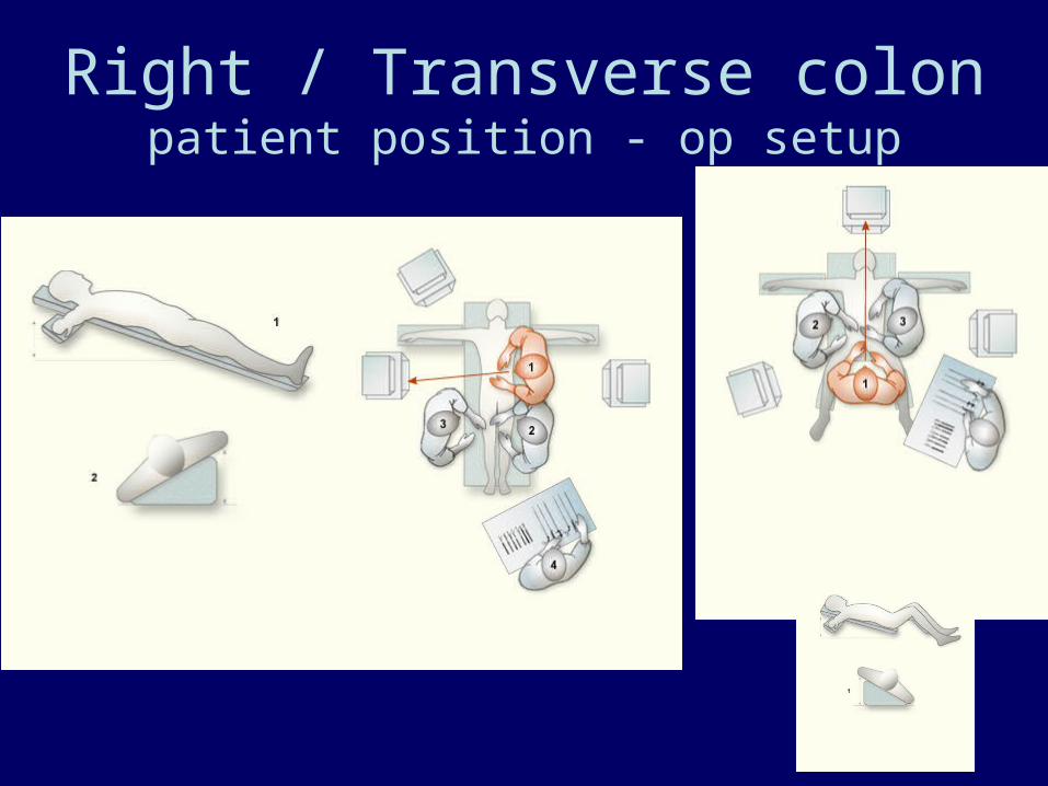
Right / Transverse colonpatient position - op setup
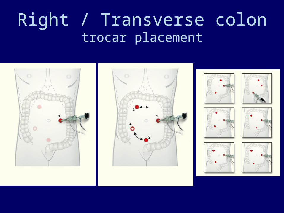
Right / Transverse colontrocar placement
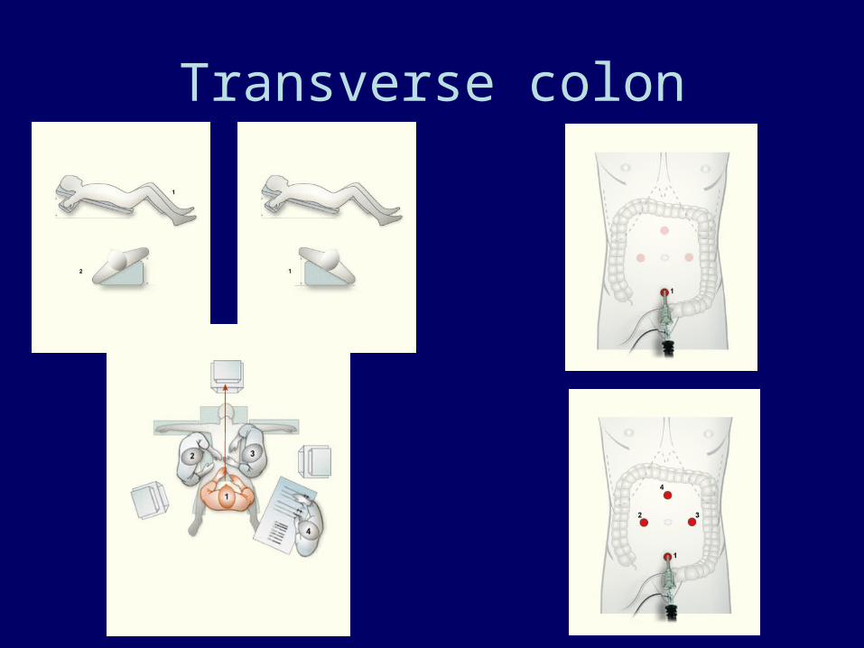
Transverse colon
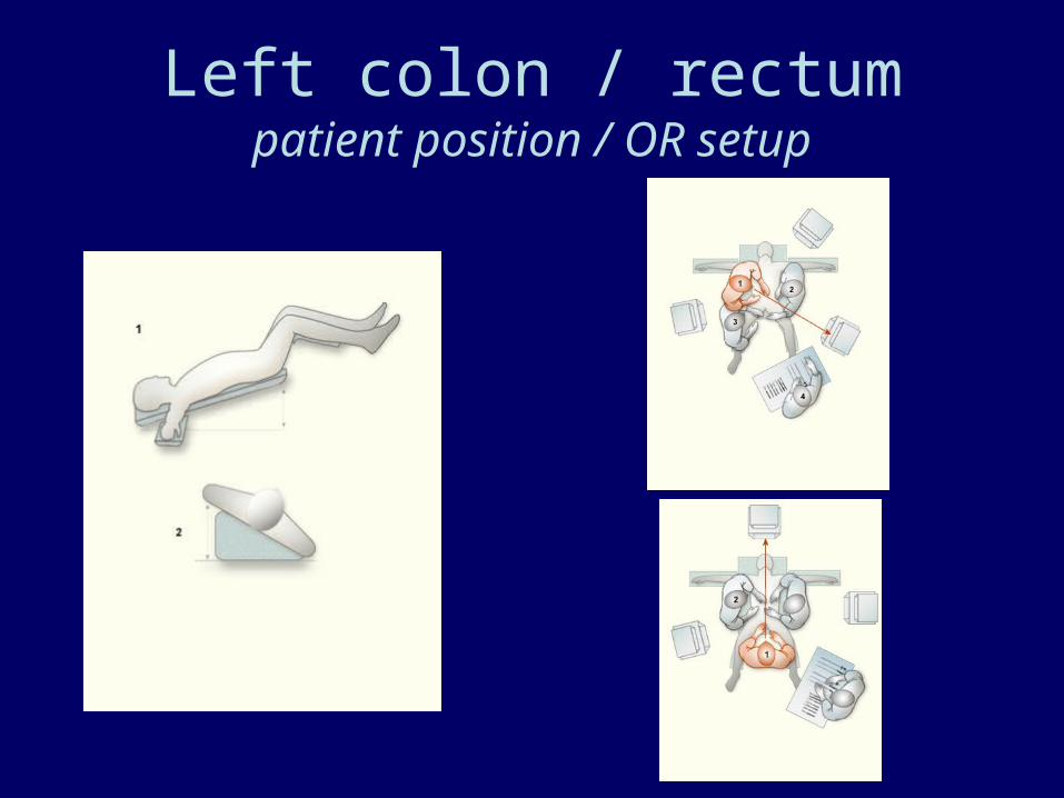
Left colon / rectumpatient position / OR setup

Left colon / rectumtrocar position
Conclusions
• Indication / Contraindication– Open approach?– Objective / subjective / learning curve
• Positioning / OR setup / trocar placement– Based on the experience and preference of the
individual surgeon– Good preop work-up and planning– Avoid surprises and keep flexibility