Embed Size (px)
Citation preview
CROWN 85: Visual Perception:A Window to Brain and Behavior
Lecture 1
1
Crown 85: Visual Perception:
A Window to Brain and Behavior
Lecture 1: Neurons and How They Communicate1 2
in the human body,
what controls ‘thinking’, behavior, movement, perception,
memory, []emotion, ???
brain factoids (from: University of Washington)
3
the language of the brain is ???
4
Your brain is electric. It generates 10 to 12 watts of electricity –enough to power a flashlight.
http://www.morphonix.com/software/education/science/brain/game/specimens/electric_brain.html
5 6
Prelude to Lectures on Visual Perception
CROWN 85: Visual Perception:A Window to Brain and Behavior
Lecture 1
2
7
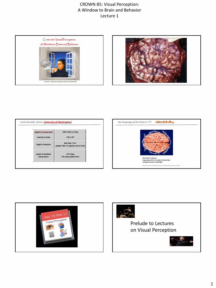
Today:
the Neuron and Electrical Potentials
http://bioserv.fiu.edu/~walterm/Fund_Sp2004/nervous/neuronanim.gif
different regions of the brain are associated with specific behaviors
8
9
Thursday:
Neuroanatomy
Prof/Provost Camps
how does one investigate brain activity and the correlated behavior ??
10
11
next Tuesday:
techniques of neuroscience research
12
CROWN 85: Visual Perception:A Window to Brain and Behavior
Lecture 1
3
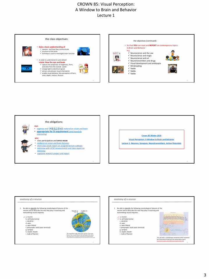
the class objectives:
• Gain a basic understanding of neurons and how they communicate structure of the brain techniques used to investigate brain function
• In order to understand (in some detail)
VISION: How the eye and brain capture the properties of brightness, form,
and color from the outside world change light to electrical signals extract and process visual information enable visual behavior (the perception of form,
color, depth, motion, illusion)
13
the objectives (continued):
• So that YOU can read and REPORT on contemporary topicsin Brain and Behaviore.g. Neuroscience and the Law Neuroscience and magic Neuroscience and art Neurotransmitters and drugs Visual development and amblyopia Mindreading Yadda Yadda Yadda
14
the obligations:
moi: • organize and ~ material on vision and brain
• appropriate for SI requirement (and hopefully interesting)
you:• class participation and OFFICE HOURS
• midterm on vision and brain lectures• short class (oral) report on assigned lecture subtopic• interview with UCSC neuroscientist and class report on
interview• capstone research project and report
15
Crown 85 Winter 2016
Visual Perception: A Window to Brain and Behavior
Lecture 1- Neurons, Synapses, Neurotransmitters, Action Potentials
16
1. Be able to identify the following morphological features of the neuron and to describe the role they play in receiving and transmitting neural impulses.
a. neuronb. cell body (soma)c. dendrited. axone. axon hillockf. presynaptic bulb (axon terminal)g. synapseh. myelin sheathi. node of Ranvier
anatomy of a neuron
17
This magnified image shows two neurons. The nerve fibre of one neuron links to the cell body of the other.http://www.aviva.co.uk/health-insurance/home-of-health/medical-centre/medical-encyclopedia/entry/structure-and-function-nerve-cells/
1. Be able to identify the following morphological features of the neuron and to describe the role they play in receiving and transmitting neural impulses.
a. neuronb. cell body (soma)c. dendrited. axone. axon hillockf. presynaptic bulb (axon terminal)g. synapseh. myelin sheathi. node of Ranvier
anatomy of a neuron
18
The cell wall is a lipid bilayer membrane which separates
the intracellular fluid from the extracellular space.http://fourier.eng.hmc.edu/e180/lectures/signal1/node2.html
CROWN 85: Visual Perception:A Window to Brain and Behavior
Lecture 1
4
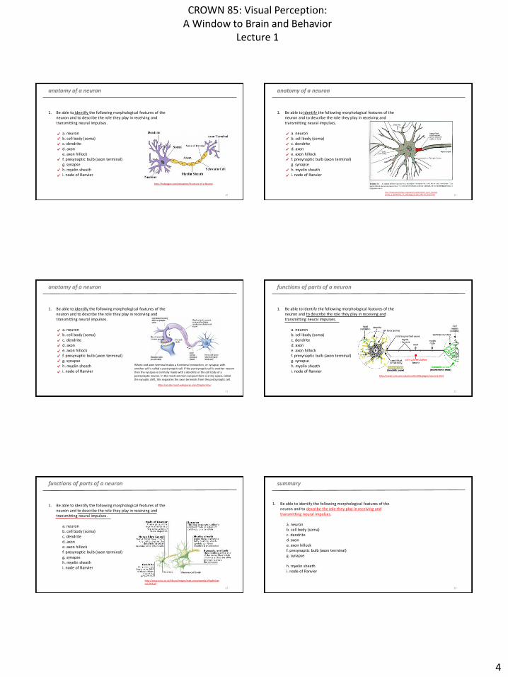
1. Be able to identify the following morphological features of the neuron and to describe the role they play in receiving and transmitting neural impulses.
a. neuronb. cell body (soma)c. dendrited. axone. axon hillockf. presynaptic bulb (axon terminal)g. synapseh. myelin sheathi. node of Ranvier
anatomy of a neuron
19
http://hubpages.com/education/Structure-of-a-Neuron
1. Be able to identify the following morphological features of the neuron and to describe the role they play in receiving and transmitting neural impulses.
a. neuronb. cell body (soma)c. dendrited. axone. axon hillockf. presynaptic bulb (axon terminal)g. synapseh. myelin sheathi. node of Ranvier
anatomy of a neuron
20
http://www.apsubiology.org/anatomy/2010/2010_Exam_Reviews/Exam_3_Review/CH_11_Histology_of_the_Neurons_Axon.htm
1. Be able to identify the following morphological features of the neuron and to describe the role they play in receiving and transmitting neural impulses.
a. neuronb. cell body (soma)c. dendrited. axone. axon hillockf. presynaptic bulb (axon terminal)g. synapseh. myelin sheathi. node of Ranvier
anatomy of a neuron
21
Where and axon terminal makes a functional connection, or synapse, with another cell is called a postsynaptic cell. If the postsynaptic cell is another neuron then the synapse is normally made with a dendrite or the cell body of a postsynaptic neuron. In the most common synapse there is a tiny space, called the synaptic cleft, this separates the axon terminals from the postsynaptic cell.
https://jordan-tesch.wikispaces.com/Chapter+four
1. Be able to identify the following morphological features of the neuron and to describe the role they play in receiving and transmitting neural impulses.
a. neuronb. cell body (soma)c. dendrited. axone. axon hillockf. presynaptic bulb (axon terminal)g. synapseh. myelin sheathi. node of Ranvier
functions of parts of a neuron
22
http://vanat.cvm.umn.edu/neurHistAtls/pages/neuron1.html
1. Be able to identify the following morphological features of the neuron and to describe the role they play in receiving and transmitting neural impulses.
a. neuronb. cell body (soma)c. dendrited. axone. axon hillockf. presynaptic bulb (axon terminal)g. synapseh. myelin sheathi. node of Ranvier
functions of parts of a neuron
23
http://www.aviva.co.uk/library/images/med_encyclopedia/cfhg464nercel_003.gif
summary
24
(basic cell of brain and peripheral nervous system)
(contains nucleus with RNA and metabolic components)
(processes that collect inputs from other neurons)
(process[es] that provides output to other neurons [and muscles])
(junction of soma and axon where action potentials originate [sums inputs])
(process at tip of axon where neurotransmitters are stored)
(junction between two neurons which allows transmission of signals from
one neuron to another)
(insulating sheath that promotes transmission of action potentials)
(gaps in myelin sheath allowing contact with extracellular space allowing
regeneration of action potential)
1. Be able to identify the following morphological features of the neuron and to describe the role they play in receiving and transmitting neural impulses.
a. neuron b. cell body (soma)c. dendrited. axone. axon hillockf. presynaptic bulb (axon terminal)g. synapse
h. myelin sheathi. node of Ranvier
CROWN 85: Visual Perception:A Window to Brain and Behavior
Lecture 1
5
understand the basic functioning of the neural action potential
25
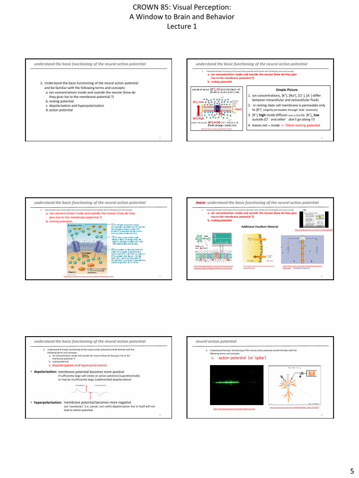
2. Understand the basic functioning of the neural action potential
and be familiar with the following terms and concepts:
a. ion concentrations inside and outside the neuron (how do they give rise to the membrane potential ?)
b. resting potentialc. depolarization and hyperpolarizationd. action potential
understand the basic functioning of the neural action potential
26
2. Understand the basic functioning of the neural action potential and be familiar with the following terms and concepts:
a. ion concentrations inside and outside the neuron (how do they give rise to the membrane potential ?)
b. resting potential
1. ion concentrations, [K+], [Na+], [Cl], [A] differ between intracellular and extracellular fluids
2. in resting state cell membrane is permeable only to [K+] (slightly permeable through ‘leak’ channels)
3. [K+]i high inside diffuses (only a little) to [K+]o lowoutside (Cl and other don’t go along !!)
4. leaves net inside 70mV=resting potentialhttp://csls-text.c.u-tokyo.ac.jp/active/05_03.html
Simple Picture
-70mV
[K+]i=high
[K+]o=low
understand the basic functioning of the neural action potential
27
2. Understand the basic functioning of the neural action potential and be familiar with the following terms and concepts:
a. ion concentrations inside and outside the neuron (how do they give rise to the membrane potential ?)
b. resting potentiaL
http://classes.midlandstech.edu/carterp/Courses/bio210/chap03/lecture1.htm
more: understand the basic functioning of the neural action potential
28
2. Understand the basic functioning of the neural action potential and be familiar with the following terms and concepts:
a. ion concentrations inside and outside the neuron (how do they give rise to the membrane potential ?)
b. resting potential
Additional Excellent Material
http://sites.sinauer.com/neuroscience5e/animations02.01.html
http://www.getbodysmart.com/ap/nervoussystem/neurophysiology/restingpotentials/menu/menu.html
http://sites.sinauer.com/neuroscience5e/animations02.02.html ADVANCED=CHEM 1C
https://www.youtube.com/watch?v=JApn3gRr8Q8
understand the basic functioning of the neural action potential
29
2. Understand the basic functioning of the neural action potential and be familiar with the
following terms and concepts:a. ion concentrations inside and outside the neuron (how do they give rise to the
membrane potential ?)b. resting potential
c. depolarization and hyperpolarization
• depolarization:
• hyperpolarization:
membrane potential becomes more positiveif sufficiently large will create an action potential (suprathreshold)or may be insufficiently large (subthreshold depolarization)
membrane potential becomes more negativecan ‘counteract’ (i.e. cancel, sum with) depolarization but in itself will not lead to action potential
neural action potential
30
2. Understand the basic functioning of the neural action potential and be familiar with the
following terms and concepts:
c. action potential (or ‘spike’)
http://ep.yimg.com/ca/I/yhst-31600583429934_2260_31723678https://faculty.washington.edu/chudler/flash/son1.html
CROWN 85: Visual Perception:A Window to Brain and Behavior
Lecture 1
6
action potential
31
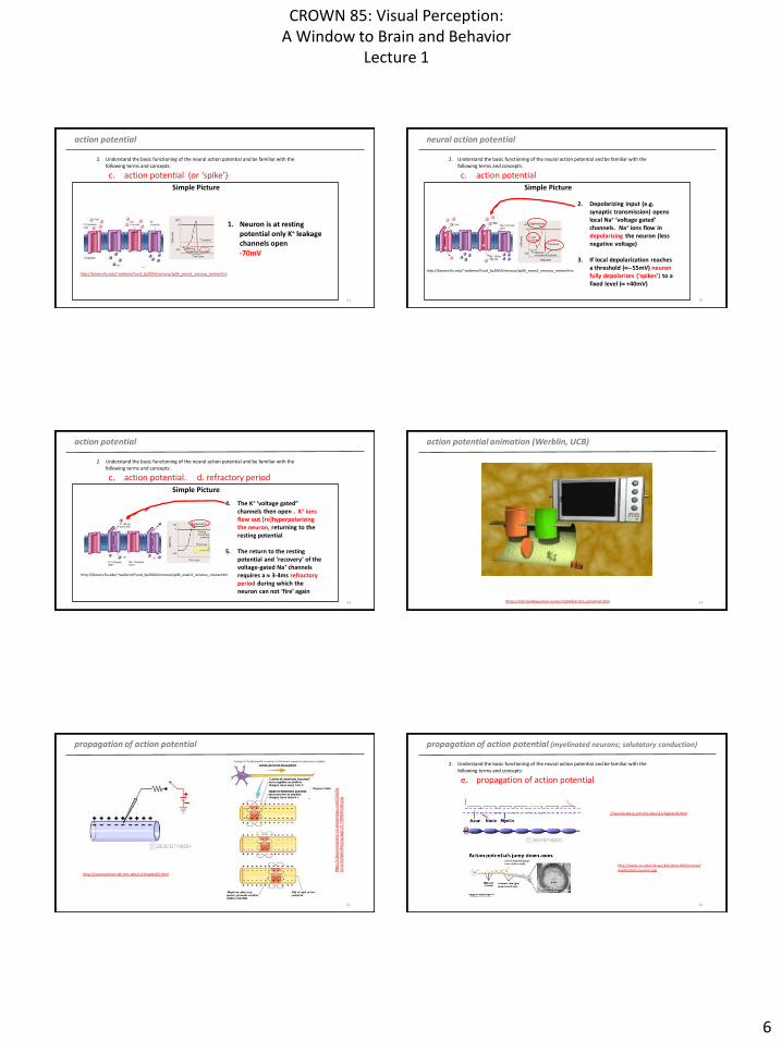
2. Understand the basic functioning of the neural action potential and be familiar with the
following terms and concepts:
c. action potential (or ‘spike’)
Simple Picture
1. Neuron is at resting potential only K+ leakage channels open-70mV
http://bioserv.fiu.edu/~walterm/Fund_Sp2004/nervous/sp06_exam2_nervous_review.htm
neural action potential
32
2. Understand the basic functioning of the neural action potential and be familiar with the
following terms and concepts:
c. action potential
Simple Picture
2. Depolarizing input (e.g. synaptic transmission) opens local Na+ ‘voltage gated’ channels. Na+ ions flow in depolarizing the neuron (less negative voltage)
3. If local depolarization reaches a threshold (55mV) neuron fully depolarizes (‘spikes’) to a fixed level ( +40mV)
http://bioserv.fiu.edu/~walterm/Fund_Sp2004/nervous/sp06_exam2_nervous_review.htm
action potential
33
2. Understand the basic functioning of the neural action potential and be familiar with the
following terms and concepts:
c. action potential. d. refractory period
Simple Picture
4. The K+ ‘voltage gated” channels then open . K+ ions flow out [re]hyperpolarizing the neuron, returning to the resting potential
5. The return to the resting potential and ‘recovery’ of the voltage-gated Na+ channels requires a 3-4ms refractory period during which the neuron can not ‘fire’ again
http://bioserv.fiu.edu/~walterm/Fund_Sp2004/nervous/sp06_exam2_nervous_review.htm
action potential animation (Werblin, UCB)
34https://mcb.berkeley.edu/courses/mcb64/action_potential.html
propagation of action potential
35
htt
ps:
//cl
assc
on
nec
tio
n.s
3.a
maz
on
aws.
com
/54
4/f
lash
card
s/6
66
54
4/p
ng
/ap
p13
17
789
045
502
.pn
g
http://neuroscience.uth.tmc.edu/s1/chapter03.html
propagation of action potential (myelinated neurons; salutatory conduction)
36
http://neuroscience.uth.tmc.edu/s1/chapter03.html
http://www.uic.edu/classes/bios/bios100/lectures/myelinated_neurons.jpg
2. Understand the basic functioning of the neural action potential and be familiar with the
following terms and concepts:
e. propagation of action potential
CROWN 85: Visual Perception:A Window to Brain and Behavior
Lecture 1
7
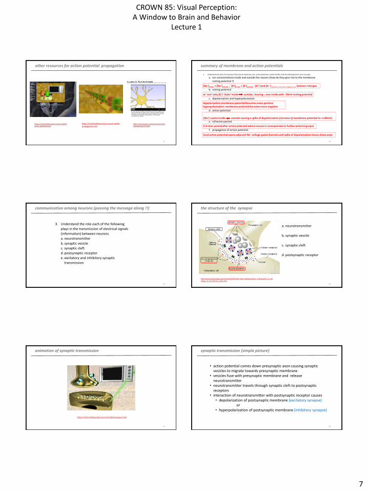
other resources for action potential propagation
37
https://mcb.berkeley.edu/courses/mcb64/propagation.html
https://mcb.berkeley.edu/courses/mcb64/action_potential.html
https://mcb.berkeley.edu/courses/mcb64/action_potential.html
http://sites.sinauer.com/neuroscience5e/animations02.03.html
local membrane depolarization, above threshold level, by stimulus causes ‘voltage gated’ Na+ pores to open;[Na+] rushes inside outside causing a spike of depolarization (increase of membrane potential to +40mV)
summary of membrane and action potentials
38
2. Understand the basic functioning of the neural membrane and action potentials and be familiar with the following terms and concepts:
a. ion concentrations inside and outside the neuron (how do they give rise to the membrane
resting potential ?)
b. resting potential
c. depolarization and hyperpolarization
d. action potential
e. refractory period
f. propagation of action potential
[Na+]inside < [Na+]outside ; [K+]inside > [K+]outside ; [Cl] and [A ] proteins and other negative ions balance +charges
depolarization: membrane potential becomes more positivehyperpolarization: membrane potential becomes more negative
at ‘rest’ only [K+] ‘leaks’ inside outside ; leaving ions inside with -70mV resting potential
3-4 msec period after action potential where neuron is unresponsive to further polarizing input
local action potential opens adjacent Na+ voltage-gated channels and spike of depolarization moves down axon
communication among neurons (passing the message along !!)
39
3. Understand the role each of the following
plays in the transmission of electrical signals
(information) between neurons
a. neurotransmitter
b. synaptic vesicle
c. synaptic cleft
d. postsynaptic receptor
e. excitatory and inhibitory synaptic
transmission
the structure of the synapse
40
http://www.apsubiology.org/anatomy/2010/2010_Exam_Reviews/Exam_3_Review/CH_11_Histology_of_the_Neurons_Axon.htm
a. neurotransmitter
b. synaptic vesicle
c. synaptic cleft
d. postsynaptic receptor
animation of synaptic transmission
41
https://mcb.berkeley.edu/courses/mcb64/synapse.html
synaptic transmission (simple picture)
42
• action potential comes down presynaptic axon causing synapticvesicles to migrate towards presynaptic membrane
• vesicles fuse with presynaptic membrane and release neurotransmitter
• neurotransmitter travels through synaptic cleft to postsynaptic receptors
• interaction of neurotransmitter with postsynaptic receptor causes• depolarization of postsynaptic membrane (excitatory synapse)
or• hyperpolarization of postsynaptic membrane (inhibitory synapse)
CROWN 85: Visual Perception:A Window to Brain and Behavior
Lecture 1
8
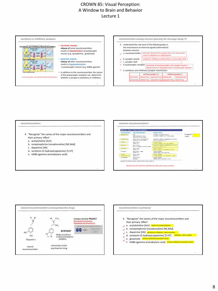
excitatory vs inhibitory synapses
43
• EXCITATORY SYNAPSE:release of some neurotransmitters results in depolarization of postsynaptic neuron (e.g. epinephrine, glutamate)
• INHIBITORY SYNAPSE:release of other neurotransmitters results in hyperpolarizationin postsynaptic neuron (e.g. GABA, glycine)
• In addition to the neurotransmitter the nature of the postsynaptic receptors can determinewhether a synapse is excitatory or inhibitoryhttp://faculty.southwest.tn.edu/rburkett/A&P1%20Muscle%20Physiology.htm
communication among neurons (passing the message along !!)
44
3. Understand the role each of the following plays in
the transmission of electrical signals (information)
between neurons
a. neurotransmitter
b. synaptic vesicle
c. synaptic cleft
d. postsynaptic receptor
e. excitatory and inhibitory synaptic transmission
chemicals released from synapse that cause postsynapticneuron to depolarize or hyperpolarize
‘containers’ holding neurotransmitters in presynaptic bulb
space between pre- and postsynaptic membranes
interaction of neurotransmitter with receptor results in depolarization or hyperpolarization of postsynaptic dendrite
excitatory synapse (+) Inhibitory synapse (-)
presynaptic depolarizing hyperpolarizing depolarizing hyperpolarizing
postsynaptic depolarizing hyperpolarizing hyperpolarizing depolarizing
neurotransmitters
45
4. “Recognize” the names of the major neurotransmitters and
their primary ‘effect’
a. acetylcholine [Ach]
b. norepinephrine (noradrenaline) [NE,NAd]
c. dopamine [DA]
d. serotonin (5-hydroxytryptamine) [5-HT]
e. GABA (gamma-aminobutyric acid)
common neurotransmitters
46
http://bioserv.fiu.edu/~walterm/Fund_Sp2004/nervous/sp06_exam2_nervous_review.htm
“recognize”know:
naturalneurotransmitter
chemically similarpsychoactive drug
POSSIBLE CAPSTONE PROJECT:
DRUG INTERACTIONS: SYNAPTIC
TRANSMISSION AND OTHER EFFECTS
natural neurotransmitters and psychoactive drugs neurotransmitters (summary)
48
4. “Recognize” the names of the major neurotransmitters and
their primary ‘effect’
a. acetylcholine [Ach]
b. norepinephrine (noradrenaline) [NE,NAd]
c. dopamine [DA]
d. serotonin (5-hydroxytryptamine) [5-HT]
e. glutamate
f. GABA (gamma-aminobutyric acid)
neuron to muscle excitatory
excitatory or inhibitory , role in emotions
inhibitory , role in moods
primary inhibitory transmitter in brain
primary excitatory transmitter in brain
CROWN 85: Visual Perception:A Window to Brain and Behavior
Lecture 1
9
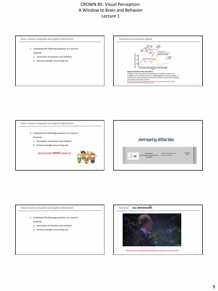
how a neuron integrates and signals information
49
5. Understand the following properties of a neuron’s
response
a. Summation of excitation and inhibition
b. Stimulus strength versus firing rate
integration of neuronal signals
50
Signal summation at the axon hillockA single neuron can receive both excitatory and inhibitory inputs from multiple neurons. All these inputs are added together at the axon hillock. If the EPSPs are strong enough to overcome the IPSPs and reach the threshold of excitation, the neuron will fire.https://www.boundless.com/biology/textbooks/boundless-biology-textbook/the-nervous-system-35/how-neurons-communicate-200/signal-summation-764-11997/
how a neuron integrates and signals information
51
5. Understand the following properties of a neuron’s
response
a. Summation of excitation and inhibition
b. Stimulus strength versus firing rate
the first student REPORT: January 11
52
short report by: William Yates
how a neuron integrates and signals information
53
5. Understand the following properties of a neuron’s
response
a. Summation of excitation and inhibition
b. Stimulus strength versus firing rate
overview
54
http://www.hhmi.org/biointeractive/molecular-mechanism-synaptic-function
CROWN 85: Visual Perception:A Window to Brain and Behavior
Lecture 1
10
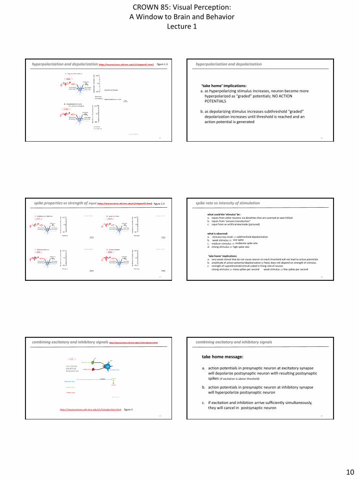
hyperpolarization and depolarization (http://neuroscience.uth.tmc.edu/s1/chapter01.html)
55
figure 1.3 hyperpolarization and depolarization
56
‘take home’ implications:a. as hyperpolarizing stimulus increases, neuron become more
hyperpolarized as “graded” potentials; NO ACTION POTENTIALS
b. as depolarizing stimulus increases subthreshold “graded” depolarization increases until threshold is reached and an action potential is generated
spike properties vs strength of input (http://neuroscience.uth.tmc.edu/s1/chapter01.html)
57
figure 1.4 spike rate vs intensity of stimulation
58
what is observed:a. stimulus too small b. weak stimulus c. medium stimulus d. strong stimulus
what could the ‘stimulus’ be :a. inputs from other neurons via dendrites that are summed at axon hillockb. inputs from ‘sensory transduction” c. input from an artificial electrode (pictured)
‘take home’ implications:a. very weak stimuli that do not cause neuron to reach threshold will not lead to action potentialsb. amplitude of action potential depolarization is fixed, does not depend on strength of stimulusc. strength of suprathreshold stimuli coded in firing-rate of neuron
strong stimulus many spikes per second weak stimulus few spikes per second
subthreshold depolarizationone spike
moderate spike rate
high spike rate
combining excitatory and inhibitory signals http://neuroscience.uth.tmc.edu/s1/introduction.html
59
http://neuroscience.uth.tmc.edu/s1/introduction.html figure 5
combining excitatory and inhibitory signals
60
take home message:
a. action potentials in presynaptic neuron at excitatory synapsewill depolarize postsynaptic neuron with resulting postsynapticspikes (if excitation is above threshold)
b. action potentials in presynaptic neuron at inhibitory synapsewill hyperpolarize postsynaptic neuron
c. if excitation and inhibition arrive sufficiently simultaneously,they will cancel in postsynaptic neuron

CROWN 85: Visual Perception:A Window to Brain and Behavior
Lecture 1
11
61
Finis Lecture 1
62