Embed Size (px)
Citation preview
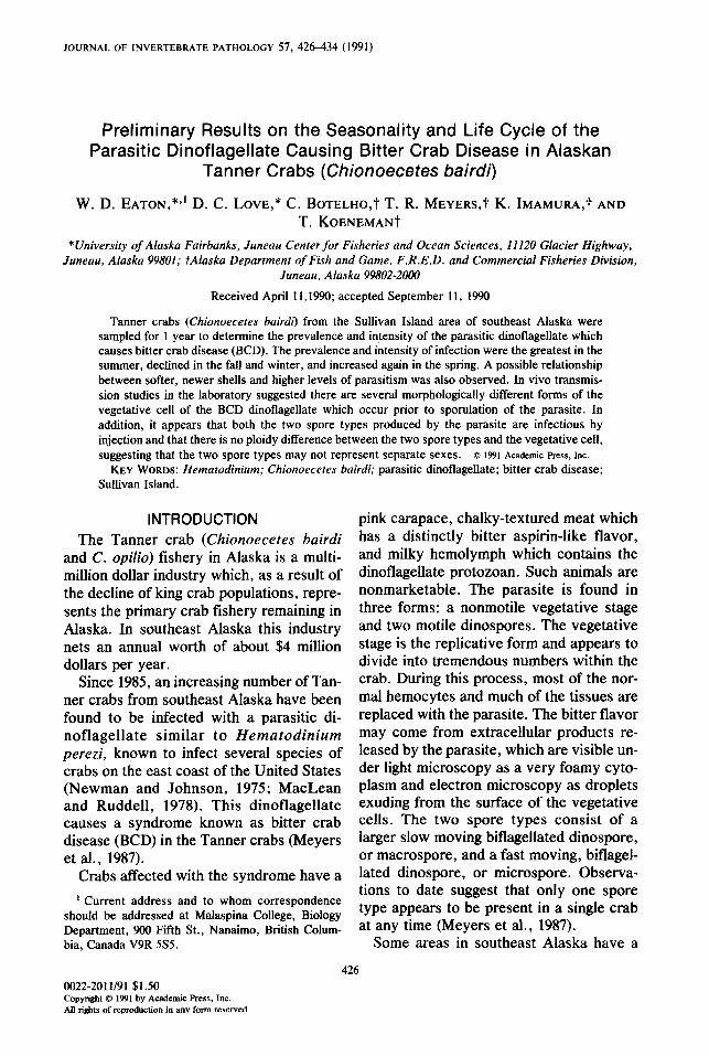
JOURNAL OF INVERTEBRATE PATHOLOGY 57, 426-434 (1991)
Preliminary Results on the Seasonality and Life Cycle of the Parasitic Dinoflagellate Causing Bitter Crab Disease in Alaskan
Tanner Crabs (Chionoecetes bairdi)
W.D. EATON, *J D.C. LOVE,* ~.BOTELHO,? T.R. MEYERS,? K. IMAMun&t AND T. KOENEMANt
*University of Alaska Fairbanks, Juneau Center for Fisheries and Ocean Sciences, 11120 Glacier Highway, Juneau, Alaska 99801; tAlaska Department of Fish and Game, F.R.E.D. and Commercial Fisheries Division,
Juneau, Alaska 99802-2ooO
Received April 11,199O; accepted September 11, 1990
Tanner crabs (Chionoecetes bairdi) from the Sullivan Island area of southeast Alaska were sampled for 1 year to determine the prevalence and intensity of the parasitic dinoflagellate which causes bitter crab disease (BCD). The prevalence and intensity of infection were the greatest in the summer, declined in the fall and winter, and increased again in the spring. A possible relationship between softer, newer shells and higher levels of parasitism was also observed. In vivo transmis- sion studies in the laboratory suggested there are several morphologically different forms of the vegetative cell of the BCD dinoflagellate which occur prior to sporulation of the parasite. In addition, it appears that both the two spore types produced by the parasite are infectious by injection and that there is no ploidy difference between the two spore types and the vegetative cell, suggesting that the two spore types may not represent separate sexes. 8 FBI Academic PESS, IW.
KEY WORDS: Hematodinium; Chionoecetes bairdi; parasitic dinoflagellate; bitter crab disease; Sullivan Island.
INTRODUCTION The Tanner crab (Chionoecetes bairdi
and C. opilio) fishery in Alaska is a multi- million dollar industry which, as a result of the decline of king crab populations, repre- sents the primary crab fishery remaining in Alaska. In southeast Alaska this industry nets an annual worth of about $4 million dollars per year.
Since 1985, an increasing number of Tan- ner crabs from southeast Alaska have been found to be infected with a parasitic di- noflagellate similar to Hematodinium perezi, known to infect several species of crabs on the east coast of the United States (Newman and Johnson, 1975; MacLean and Ruddell, 1978). This dinoflagellate causes a syndrome known as bitter crab disease (BCD) in the Tanner crabs (Meyers et al., 1987).
Crabs affected with the syndrome have a
’ Current address and to whom correspondence should be addressed at Malaspina College, Biology Department, 900 Fifth St., Nanaimo, British Colum- bia, Canada V9R SSS.
pink carapace, chalky-textured meat which has a distinctly bitter aspirin-like flavor, and milky hemolymph which contains the dinoflagellate protozoan. Such animals are nonmarketable. The parasite is found in three forms: a nonmotile vegetative stage and two motile dinospores. The vegetative stage is the replicative form and appears to divide into tremendous numbers within the crab. During this process, most of the nor- mal hemocytes and much of the tissues are replaced with the parasite. The bitter flavor may come from extracellular products re- leased by the parasite, which are visible un- der light microscopy as a very foamy cyto- plasm and electron microscopy as droplets exuding from the surface of the vegetative cells. The two spore types consist of a larger slow moving bi&gellated dinospore, or macrospore, and a fast moving, biflagel- lated dinospore, or microspore. Observa- tions to date suggest that only one spore type appears to be present in a single crab at any time (Meyers et al., 1987).
Some areas in southeast Alaska have a
426 0022-2011/91 $1.50 Copyright 0 1991 by Academic PRSS, Inc. All rights of retw3duction in anv form reserved
BCD IN ALASKAN TANNER CRABS 427
prevalence of BCD as high as 95% in the Tanner crabs. In 1985, a high prevalence of BCD was found in crabs from the Sullivan Island area in the upper Lynn Canal. In 1986, 1987, and 1988 more intensive sam- pling was conducted and the disease was found in numerous locations in southeast Alaska. During this sampling period some areas negative for the disease in 1986 were positive for BCD in 1987 and 1988, and some regions positive for BCD in 1986 had a higher prevalence in 1988 (Meyers, 1990).
Appropriate management decisions con- cerning BCD and the Tanner crab industry can only be made after the biology and pathogenicity of the parasite are under- stood. The purpose of this study was to de- termine if a seasonal increase in prevalence and intensity of infection occurs in crabs. In addition, attempts were made to eluci- date the life cycle of the dinoflagellate.
MATERIALS AND METHODS
Seasonality
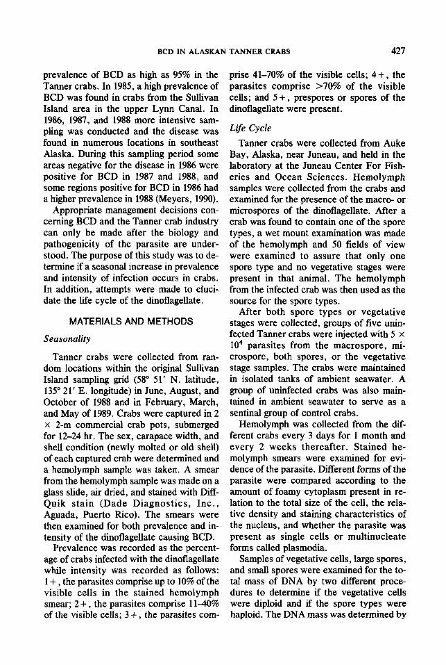
Tanner crabs were collected from ran- dom locations within the original Sullivan Island sampling grid (58’ 5 1’ N. latitude, 135” 21’ E. longitude) in June, August, and October of 1988 and in February, March, and May of 1989. Crabs were captured in 2 x 2-m commercial crab pots, submerged for 12-24 hr. The sex, carapace width, and shell condition (newly molted or old shell) of each captured crab were determined and a hemolymph sample was taken. A smear from the hemolymph sample was made on a glass slide, air dried, and stained with Diff- Quik stain (Dade Diagnostics, Inc., Aguada, Puerto Rico). The smears were then examined for both prevalence and in- tensity of the dinoflagellate causing BCD.
Prevalence was recorded as the percent- age of crabs infected with the dinoflagellate while intensity was recorded as follows: 1 + , the parasites comprise up to 10% of the visible cells in the stained hemolymph smear; 2 + , the parasites comprise 1140% of the visible cells; 3 + , the parasites com-
prise 41-70% of the visible cells; 4 + , the parasites comprise ~70% of the visible cells; and 5 + , prespores or spores of the dinoflagellate were present.
Life Cycle
Tanner crabs were collected from Auke Bay, Alaska, near Juneau, and held in the laboratory at the Juneau Center For Fish- eries and Ocean Sciences. Hemolymph samples were collected from the crabs and examined for the presence of the macro- or microspores of the dinoflagellate. After a crab was found to contain one of the spore types, a wet mount examination was made of the hemolymph and 50 fields of view were examined to assure that only one spore type and no vegetative stages were present in that animal. The hemolymph from the infected crab was then used as the source for the spore types.
After both spore types or vegetative stages were collected, groups of five unin- fected Tanner crabs were injected with 5 x
lo4 parasites from the macrospore, mi- crospore, both spores, or the vegetative stage samples. The crabs were maintained in isolated tanks of ambient seawater. A group of uninfected crabs was also main- tained in ambient seawater to serve as a sentinal group of control crabs.
Hemolymph was collected from the dif- ferent crabs every 3 days for 1 month and every 2 weeks thereafter. Stained he- molymph smears were examined for evi- dence of the parasite. Different forms of the parasite were compared according to the amount of foamy cytoplasm present in re- lation to the total size of the cell, the rela- tive density and staining characteristics of the nucleus, and whether the parasite was present as single cells or multinucleate forms called plasmodia.
Samples of vegetative cells, large spores, and small spores were examined for the to- tal mass of DNA by two different proce- dures to determine if the vegetative cells were diploid and if the spore types were haploid. The DNA mass was determined by

428 EATON ET AL.
the potassium acetate precipitation method described by Davis et al. (1980) and the ethidium bromide spectrofluorometry method described by Bentle et al. (1981). In each instance, the dinoflagellate cells were pelleted at 2000g for 20 min, washed with STE buffer, and pelleted again. The sam- ples were then resuspended in a lysing buffer (0.2 M Tris-Cl, pH 8.5,O. 1 M EDTA, 0.1 M NaCl, 1% SDS, 100 p&&l proteinase K) and the two procedures were com- pleted.
RESULTS
Seasonal@
The prevalence and intensity of the BCD dinoflagellate in Sullivan Island Tanner crabs were the highest in the summer months of June and August of 1988, then decreased in October 1988 and February 1989, and finally began to increase in March and May of 1989 (Table 1). When a x2 anal- ysis (Zar, 1974) was performed on this data, it was found that there was a statistically significant relationship between month and prevalence and intensity of infection (P < 0.001). These data correlate well with pre- liminary samples collected from Tanner crabs in the Sullivan Island area in 1987 by the Alaska Department of Fish and Game (Table 1; Meyers et al., 1990) which
TABLE 1 SEASONALITYOFINFECTIONOFBI~~ER CRAB
DISEASE IN ALASKAN TANNER CRABS COLLECTED FROM THE SULLIVAN ISLAND AREA, NORTHERN
LYNNCANAL,ALASKA
Month crabs Percentage Number Mean collected infected positive intensity”
March 1987’ October 19876 June 1988 August 1988 October 1988 February 1989 March 1989 May 1989
37 61
; 55 46
17146 1.9 + 0.3’ 42/69 1.8 f 0.4
2801333 2.8 + 0.4 99/100 3 ” 0.2 xi/102 2.2 f 0.3 46000 2 f 0.2 741138 2.2 + 0.4 57l95 2.4 2 0.2
a Mean intensity was calculated from all BCD-infected crabs collected during a particular month.
b Samples conducted by the Alaska Department of Fish and Game.
c Standard deviation.
showed that 37% (1.9 + intensity) and 61% (1.8 + intensity) of the crabs examined in March and October of 1987, respectively, were infected by the parasite.
No relationship was found among cara- pace width, sex of crab, and infection rates. However, a relationship was discovered between shell condition and parasitism (Ta- ble 2). From June 1988 through February 1989, 610 crabs collected had new or softer shells and 63 crabs collected had older or harder shells. Data on the shell condition of crabs collected from other months were not available. About 81% (494/610) of the crabs with newer, softer shells were infected with the dinoflagellate, while only 24% (U/63) of the crabs with older, harder shells were in- fected. When a x2 analysis was performed on the data, the relationship between newer shells and an increased incidence of infec- tion proved statistically significant (P < 0.001). At the time of submission of this work, Meyers et al. (1990) had also submit- ted results which corroborate the apparent relationship between shell condition and parasitism.
Life Cycle
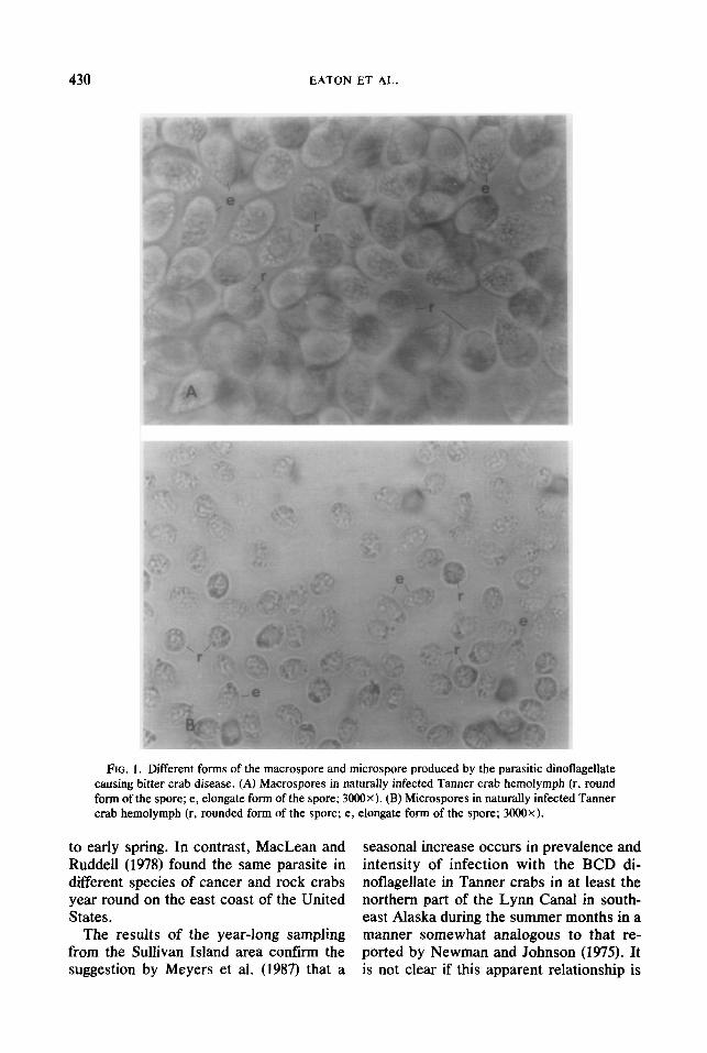
Although Meyers et al. (1987) described both the large and small spore types as oval (15.2 x 11.4 km, n = 8; 12.0 x 4.4 pm, n = 5; respectively), we found the earliest mo- tile form of the different spore types to be circular. Following removal from the crabs, both the macrospore and the microspore types were first observed as round, motile cells with two flagella and were 12.3 and 7.9 pm in diameter (n = lOO), respectively. Six hours after removal from the crabs the mo- tile spore types had elongated to 13.3 x
11.4 pm and 9.5 x 7.6 pm (n = IOO), re- spectively. After 12 hr, the motile spore types had elongated to 17.1 x 9.9 pm and 11.8 x 4.8 p,rn (n = lOO), respectively (Fig. 1).
Several different forms of the parasite were observed in crabs during the injection study, suggesting a continuum from small, dense, slow replicating forms to large, dif-

BCD IN ALASKAN TANNER CRABS 429
TABLE 2 THE RELATIONSHIP BETWEEN THE NUMBER OF TANNER CRABS HAVING A NEW SHELL OR OLD SHELL AND THE INCIDENCE OF INFECTION WITH THE PARASITIC DINOFLAGELLATE, FROM SULLIVAN ISLAND, ALASKA
June August 1988 1988
October 1988
February 1988 Total
New shell Old shell
91% (2724297) 99% (92/93) 62% (X/91) 4% (631129) 81% (494/610) 22% (8/36) 100% (7r7) 0% (O/l 1) 0% (O/9) 24% (15163)
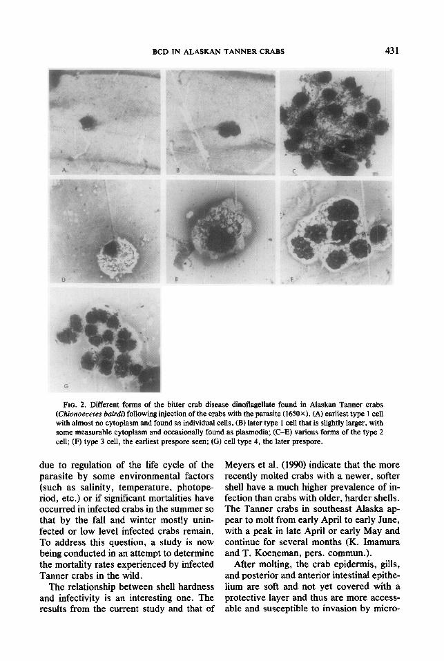
fuse, rapidly replicating forms to prespores and spores. None of the control crabs de- veloped an infection. The earliest forms of the parasite (type 1 cells; Fig. 2) were small, round, vegetative cells (6-11 pm in diame- ter) that occurred individually or in plasmo- dia composed of two to eight cells, contain- ing very little cytoplasm (no more than a 2-pm region of the total cell) and a dense basophilic nucleus (suggesting slow cell di- vision). Only a few of these forms were ob- served in crabs injected with both spores from 25 to 63 days postinjection. These were small cells with almost no cytoplasm and were present as individual cells. From day 77 to day 99, type 1 cells were present at 1 + levels and were generally larger with some measurable cytoplasm and were oc- casionally present in plasmodia of two to eight cells (Table 3).
Later stages of the parasite (type 2 cells; Fig. 2) found in all injection groups were larger vegetative cells (12-20 km in diame- ter) with an eccentric, eosinophilic, less dense nucleus (suggesting more rapid cell division) and extensive amounts of foamy cytoplasm (a 4- to 8-p,m region of the total cell). These forms were found individually or in plasmodia containing up to 30 cells and were seen in crabs injected with the different spores from about 100 to 370 days postinjection (Table 3). A few of these forms of the parasite were present in crabs 7 days following injection with the vegeta- tive stage. However, this probably repre- sented residual cells remaining after inocu- lation rather than replicating parasites.
The type 2 cells appeared to develop into two types of prespores (type 3 and 4 cells; Fig. 2), the earliest of which were present
as plasmodia, with pleomorphic, eosino- philic, diffuse nuclei containing extensive foamy cytoplasm which, immediately be- fore sporulation, developed into smaller cells in plasmodia with little or no foamy cytoplasm, and extremely dense basophilic nuclei. The parasite developed into spores in two of the five crabs with prespore infec- tions, while the other three crabs with pre- spore infections died before the parasite de- veloped into spores (Table 3). The crab in- jected originally with both spore types contained both the macro- and the mi- crospore in the hemolymph and the crab injected with the microspore contained the macrospore in the hemolymph. The differ- ent spore type morphologies were previ- ously described by Meyers et al. (1987).
Estimates of the mass of DNA in the dif- ferent cell types showed there to be no more than 8 p,g DNA/lo6 cells difference in DNA mass between the vegetative cells and the two spore types. In addition, the results of the potassium acetate and ethid- ium bromide procedures used to estimate the mass of DNA were similar as there was no more than a 7 pg DNA/lo6 cells differ- ence when a single sample was examined by the two procedures (Table 4). Although there was some variation between proce- dures, there appears to be a similar mass of DNA in the two spore types and the vege- tative cell, suggesting a similarity in ploidy.
DISCUSSION
A seasonal occurrence of H. perezi was reported by Newman and Johnson (1975) in the parasitic dinoflagellate in blue crabs on the east coast of the United States from late spring to early winter, but not during winter
EATON ET AL.
FIG. 1. Different forms of the macrospore and microspore produced by the parasitic dinoflagellate causing bitter crab disease. (A) Macrospores in naturally infected Tanner crab hemolymph (r, round form of the spore; e, elongate form of the spore; 3000X). (B) Microspores in naturally infected Tanner crab hemolymph (r, rounded form of the spore; e, elongate form of the spore; 3000X).
to early spring. In contrast, MacLean and Ruddell (1978) found the same parasite in different species of cancer and rock crabs year round on the east coast of the United States.
The results of the year-long sampling from the Sullivan Island area confirm the suggestion by Meyers et al. (1987) that a
seasonal increase occurs in prevalence and intensity of infection with the BCD di- noflagellate in Tanner crabs in at least the northern part of the Lynn Canal in south- east Alaska during the summer months in a manner somewhat analogous to that re- ported by Newman and Johnson (1975). It is not clear if this apparent relationship is
BCD IN ALASKAN TANNER CRABS 431
FIG. 2. Different forms of the bitter crab disease dinoflagellate found in Alaskan Tanner crabs (Chionoeceres bair& following injection of the crabs with the parasite (1650X). (A) earliest type 1 cell with almost no cytoplasm and found as individual cells, (B) later type 1 cell that is slightly larger, with some measurable cytoplasm and occasionally found as plasmodia; (C-E) various forms of the type 2 cell; (F) type 3 cell, the earliest prespore seen; (G) cell type 4, the later prespore.
due to regulation of the life cycle of the parasite by some environmental factors (such as salinity, temperature, photope- riod, etc.) or if significant mortalities have occurred in infected crabs in the summer so that by the fall and winter mostly unin- fected or low level infected crabs remain. To address this question, a study is now being conducted in an attempt to determine the mortality rates experienced by infected Tanner crabs in the wild.
The relationship between shell hardness and infectivity is an interesting one. The results from the current study and that of
Meyers et al. (1990) indicate that the more recently molted crabs with a newer, softer shell have a much higher prevalence of in- fection than crabs with older, harder shells. The Tanner crabs in southeast Alaska ap- pear to molt from early April to early June, with a peak in late April or early May and continue for several months (K. Imamura and T. Koeneman, pers. commun.).
After molting, the crab epidermis, gills, and posterior and anterior intestinal epithe- lium are soft and not yet covered with a protective layer and thus are more access- able and susceptible to invasion by micro-
432 EATON ET AL.
TABLE 3 FORMS OF THE PARASITIC DINOFLAGELLATE OBSERVED, TIME OF DEVELOPMENT, AND INTENSITY OF
INFECTION FOLLOWING INJECTION OF TANNER CRABS WITH THE SPORE TYPE, BOTH SPORE TYPES, OR THE VEGETATIVE STAGE OF THE PARASITE
Injection Days Number Intensity of group” postinjection infected Cell type infection
Both spore typesb 25-63 415 Type 1 Few 71-99 415 Type 1 1+
112-365 213 Type 2 l-3+ 379-393 l/2 Types 3 and 4 4+
399 l/2 Both spore types 5+ Macrospore” 126-399 214 Type 2 2-3 +
419 212 Type 3 4+ Microspored 140-379 215 Type 2 2-3 +
399 212 Type 4 4+ 419 l/2 Macrospore 5+
Vegetative stage’ 7-63 S/S Type 2 Few 71-305 112 Type 2 2-3 +
LI Five crabs were injected in each group. b Two crabs with 2 + BCD infections died at 200 days postinjection and one crab with a 3 + BCD infection died
at 305 days postinjection. c One crab died immediately after injection, two crabs died due to water system failure, and the final two crabs
died with 4 + BCD prespore infections. d One crab with a 3 + BCD infection died at 263 days postinjection and two crabs with 3 + BCD infections died
at 305 days postinjection. c Two crabs with 2 + BCD infections died at 168 days postinjection, two crabs with 3 + BCD infections died
due to a water system failure, and one crab with a 3 + infection died at 319 days.
bial pathogens. Spores of the parasitic di- noflagellate have been observed within crabs from the Sullivan Island area in the mid- to late summer (T. R. Meyers and W. D. Eaton, unpubl.) and from the Auke Bay area as early as mid-May (D. C. Love
TABLE 4 ESTIMATES OF THE AMOUNT OF DNA IN
PRESPWES, SPORES, AND VEGETATIVE STAGES OF THE PARASITIC DINOFLAGELLATE CAUSING BITTER
CRAB DISEASE IN TANNER CRABS
Amount of DNA
Stage Potassium of the acetate
parasite precipitation”
Ethidium bromide
fluorescenceb
Vegetative 30 pg DNA’ 34 Fg DNA Macrospore 35 pg DNA 42 pg DNA Microspore 29 pag DNA 37 pg DNA
0 Potassium acetate precipitation procedure for es- timation of the mass of DNA in cells.
’ Ethidium bromide fluorescence procedure for es- timation of the mass of DNA in cells.
c Amounts are reported as pg of DNA/lo6 cells.
and W. D. Eaton, unpubl.). Thus the pe- riod of overlap of dinoflagellate sporulation and Tanner crab molting is minimal.
Although some transmission of the para- site may occur as the spores enter the crab during molting, there must also be another mechanism of infection. Meyers et al. (1990) suggests that transmission of the vegetative stages may occur at this time. It may also be that during molting, crabs are somewhat stressed and presumably immu- nosuppresed so that low level infections may be exacerbated into the disease state during this time. In any case, the question of parasitism and shell condition needs fur- ther examination.
The results from this study suggest that the parasite appears to metamorphose from a small, slowly replicating cell with a dense, basophilic nucleus into a larger cell with ex- tensive foamy cytoplasm and a diffuse eo- sinophilic nucleus that is rapidly undergo- ing cell division (as suggested by its eosin- ophilic nature), then into the pleomorphic
BCD IN ALASKAN TANNER CRABS 433
early prespore to the smaller, denser pre- spore, and finally to the two spore types, which initially are round, motile, billagel- lated forms that elongate into the final spore shapes. This work is being repeated using larger numbers of animals and the waterborne route of infection.
The data from this work also suggest that both spore types are infectious by injection and that each can individually result in spore formation as the final step in the life cycle of the parasite. Although it is un- known at this time whether the spore types and vegetative stages are diploid, haploid, or possibly aneuploid, it appears that the mass of the DNA in all three forms of the parasite are similar enough to suggest that the ploidy of all three forms is similar. Thus, the two spore types may not, in fact, represent two different sexes, both of which would be required for a successful infection.
Although some genera (such as Mero- dinium and Coccidinium) of parasitic di- noflagellates are thought to produce a large (macrospore) and small (microspore) spore, other genera (such as Cochlosyndinium) are thought to produce only one size spore (Chatton, 1952). If two different spore types are produced by a single species of dinoflagellate, but are not necessary for a successful infection to occur, this raises some interesting questions as to the selec- tive advantage of a single species of di- noflagellate producing two morphologically different, diploid spore types, both of which are infectious. It may be that we are seeing in the Tanner crabs two different but related species of dinoflagellates or two dif- ferent forms of the same species, both of which are capable of developing into spores. A more in-depth comparison of the DNA of the two spore types is necessary to address this problem.
It is not certain at this time if the parasite from Alaskan Tanner crabs is the same as that found in crabs from the east coast of the United States. Although the uninucle- ate and plasmodial masses of cells de- scribed by MacLean and Ruddell (1978)
and Newman and Johnson (1975) seem sim- ilar to those described by Meyers et al. (1987) and from the current study, there are still some differences observed. No dino- spores have been observed in crabs in- fected with Hematodinium from the east coast, while dinospores have been found in infected Tanner crabs from Alaska. The plasmodia seen from the eastern infections are much smaller than those observed in crabs from Alaska. In addition, Newman and Johnson (1975) describe the presence of highly motile vermiform multinucleate bodies up to 64 pm in diameter containing up to 12 nuclei in some of the eastern crabs. This form of the parasite has not been ob- served in either the naturally infected Tan- ner crabs from the wild or the artificially infected crabs in the laboratory. A side by side comparison of the morphology and DNA homology of both the east coast and west coast parasites is necessary in order to determine if these are the same or different dinoflagellates .
It is quite evident that there is much more to be learned about the seasonality and life cycle of this dinoflagellate. The life cycle of the parasite presents a number of interest- ing biological questions which are being ad- dressed in future studies on BCD. The question of seasonality of infection is still a concern. If a true seasonality is associated with the infection, then the Tanner crab fishing season can be moved to a time when the prevalence and intensity of infection are the lowest, so that there is a greater chance that those animals harvested will be mar- ketable .
ACKNOWLEDGMENTS
This work was supported by Alaska Sea Grant Proj- ect NA82AA-D-SG041. I thank the Alaska Depart- ment of Fish and Game Pathology Lab and the Com- mercial Fisheries Division for their assistance in this endeavor.
REFERENCES
BENTLE, L. A., DUTTA, A., AND METCOFF, J. 1981. The sequential enzymatic determination of DNA and RNA. Anal. Biochem., 116, 5-16.
CHAT-TON, E. 1952. Classe’ des Dinoflagelles ou Peri-

434 EATON ET AL.
diniens. In “T&e de Zoologie” (P. P. Grasse,’ Ed.) Vol. l(1) pp. 309-406. Masson, Paris.
DAVIS, R. W., THOMAS, M., CAMERON, J., ST. JOHN, T. P., SCHERER, S., AND PADGETT, R. A. 1980. In “Methods in Enzymology” (L. Grossman and K. Moldave, Eds.) Vol. 65, pp. 404-411. Academic Press, New York.
MACLEAN, S. A., AND RUDDELL, C. L. 1978. Three new crustacean hosts for the parasitic dinoflagellate Hematodinium perezi (Dinoflagellata: Syndinidae). J. Parasitol.) 64, 158-160.
MEYERS, T. R., KOENEMAN, T. M., BOTELHO, C., AND SHORT, S. 1987. Bitter crab disease: A fatal dinoflagellate infection and marketing problem for Alaskan Tanner crabs, Chionoecetes bairdi. Dis. Aquat. Org., 3, 195-216.
MEYERS, T. R. 1990. Diseases caused by protistans and metazoans. In “Diseases of Marine Animals” (0. Kinne, Ed.) Vol III, Biologische Anstalt Helgo- land, Hamburg, Germany.
MEYERS, T. R., BOTELHO, C., EATON, W., KOENE- MAN, T. M., AND IMAMURA, K. 1990. The distribu- tion of the bitter crab dinoflagellate syndrome in Alaskan Tanner crabs, Chionoecetes bairdi. 9, 37- 43.
NEWMAN, N. W., AND JOHNSON, C. A. 1975. A dis- ease of blue crabs (Callinectes sapidus) caused by a parasitic dinoflagellate, Hematodinium sp. .I. Para- sitol., 61, 554457.
ZAR, J. H. 1974. “Biostatistical Analysis,” pp. 62-65. Prentice-Hall, Englewood Cliffs, New Jersey.


![Sweet&Bittero Sweet & Bitter Sweet Bitter *Ota E') r ...yokohamashakyo.sakura.ne.jp/sblo_files/nagatsuta/... · Sweet&Bittero Sweet & Bitter Sweet Bitter *Ota E') r +ÃZSweet&Bitter]](https://img.pdfslide.us/doc/110x75/5fc88504d86f533ff96954fb/sweetbittero-sweet-bitter-sweet-bitter-ota-e-r-sweetbittero.jpg)


![Seasonality PM Group[1]](https://img.pdfslide.us/doc/110x75/577cd3441a28ab9e789703ef/seasonality-pm-group1.jpg)