Embed Size (px)
Citation preview

Preliminary information to user groups:
MRI at AMI
Toni Auranen
AMI-Centre
O.V. Lounasmaa Laboratory
School of Science, Aalto University
AMI-Centre
July, 2012

AMI-Centre is an infrastructure providing services for its users
Key personnel:
• Simo Vanni, Scientific Director, [email protected]
• Toni Auranen, Technical Director, [email protected]
• Marita Kattelus, Radiographer, [email protected]
• Ari Laiho, Physicist, [email protected]
Otakaari 5 I (Magnethouse), Espoo

AMI-Centre maintains the operation of a 3 T MR-device used for MRI and fMRI
For the needs of research groups at:
• Aalto University (AU)
• University of Helsinki (HY)
• Hospital district of Helsinki and Uusimaa (HUS)
• Other users (Other academic, industry etc.)
More info?
• Check our webpage: http://ami.tkk.fi
• Join AMI mailing list
• Come for a visit
3 T GE Signa (2002 – 8/2011)
3 T Siemens Skyra (11/2011 –)

Standard clinical scanner
• MR scanner: whole-body Siemens MAGNETOM Skyra 3T
• RF-coils:
• 32-channel head array
• 20-channel head-neck array
• Flex coils (large and small)
• Body coil (transmitting)
1. Versatile stimulus- and response systems
2. Daytime measurement times available
3. Quality assurance, assistance & consultation for fMRI
4. Constant development of the environment and knowhow

Overview – topics to be covered
� fMRI data acquisition
� Stimulation systems
� Subject monitoring & Safety
� Quality control
� Future and Summary

Scanning at AMI – requirements
1. Users must have passed the AMI AMI
CentreCentre’’s Safety Courses Safety Course
2. Users must read and follow AMI CentreAMI Centre’’s s
Safety and Operations instructionsSafety and Operations instructions
3. All studies involving human subjects,
must acquire a supporting ethical ethical
statementstatement
4. Apply a research permission research permission from AMI
5. Username/Password for the reservation reservation
calendar calendar and AMI computer networkand AMI computer network

Learn to use: Equipment
• Magnet and magnet console• Invisible to users: Electronics, amplifiers, cabinets,
signal processing hardware, …
• What you need: console operation, subject positioning, etc.
• Stimulus systems, accessories
• AMIserver, AMI computer network
• Data transfer
• Remote use of AMI network
• Data analysis
From: www.siemens.com

Measurements – general
• Reservations
• Learn the rules and follow them!
• Changes are informed at AMI-news list
• Keep things where they belong
• Stick to the schedule!
• Separate preparation area (you can freely use it 30 min.
prior and 30 min. after your reserved time)
• One person from the group must be responsible
for that session
• Radiographer help – Marita Kattelus
• Please, notify AMI personnel immediately if
something is missing/broken etc.

Reservations
• Cancellation policy• Cancellation within 48 hours before the measurement is not acceptable without a force major
reason (such as subject falling ill etc.)
• Prereservations are recommended• These prereservations must be canceled two weeks before the measurement if you plan not to use
them
• Rush hour rule• If you reserve more than 7 weekday hours (9-16) within one week, you should announce it in AMI-
news list ([email protected]), so that if within 24 hours another research group asks for the same
hours, you need to modify your reservation
• If you plan to reserve more than 20 hours per month, please contact AMI scientific director
beforehand
• Remember these:• Free 2 pilot hours for new groups/projects
• You can practise freely with the stimulus computer, eye-movement camera etc. when there are no
reservations (if you use the scanner, you must make a valid reservation)

At the console
• In principle, data acquisition is
straightforward
• Plan well, imaging parameters, …
• Reserve time for piloting (phantom &
human subject)
• AMI personnel will help with paracticalities
and how to use the console
• Do it yourself vs. Use radiographer’s
assistance
• Fill in scanner log while scanning
From: www.siemens.com

Still at the console
• Create, modify, and save your research
protocol using the console• Localizer scan
• Variable number of functional and anatomical scans
• Monitor your subject
• Check for artefacts during scanning
• Standard DICOM images• MOSAIC format
• Backups are not provided by AMI (2-week grace period)
• Transfer your data or burn it on CD/DVD
From: www.siemens.com

The most valuable thing – Data
• Data transfered to AMIserver from the console automatically
• Kept in AMIserver only for two weeks
• On the console the same or even less
• Back-up your data as soon as possible!
• Stimulus computer, EEG-computer, and other computers in the console room
• Not backed-up day-by-day basis…
• Someone might change or accidentally delete something
• Copy your logfiles and other data immediately (USB-sticks, portable HDs)

Measurements – general (cont.)
• Remember to prepare your studies well!• Practice (you will save time)
• Pre-screen your subjects• Contraindications for MR, Safety questionnaire (in the webpages)
• Ask for help if needed, we are happy to assist you!

Differences between GE and Siemens
• Other than the console operations itself, things are roughly the
same as before• If you knew how to scan with GE, you will with Siemens as well
• AMI Centre has ready-made protocols and we will gladly assist you
to put together your own protocol
• What are the minimum requirements from you:• Know your paradigm well, have your stimuli and a working Presentation script
• Basic parameters: TR, TE, flip angle, # of slices, slice orientations, matrix size etc.
• We’ll help you choose the other parameters (and these as well) if needed!
• Note that if you want to optimize your paradigm, you should do
background research on similar studies/paradigms yourself

Differences between GE and Siemens (cont.)
• GRE-EPI, SE-EPI
• One fMRI trigger pulse per volume (GE: one per slice)
• Dummies don’t send pulses and are not included (GE: you could choose this)
• You may want to add more dummies than the scanner does, check your paradigm!
• Less time-points with Siemens’ SE-EPI (sequence programming required)
• GE – T1 SPGR, BRAVO / Siemens – MPRAGE
• Current T1 anatomicals work well with e.g. Freesurfer’s automatic process flow
• T2 anatomicals
• No surprises, 3D sequence now possible (SPACE)
• Siemens DTI sequences
• Current images look good and it is easy to get some nice tracts on the console

Differences between GE and Siemens (cont.)
• FOV, matrix sizes, slice thickness etc.
• No surprises
• You should be able to get to at least to the same as before
• DICOM data format / Scanner coordinate system
• Siemens Mosaic format
• Most software recognize these from header information
• Signal-to-noise ratio
• Siemens 32ch coil at least 1.9 times better than our old 16ch coil with GE
• Even more so when closer to the surface of the head
• Magnet homogeinity
• GE was a long bore magnet � more homogeneous

Your options with fMRI
1. Coils• 32-channel head (A+P or P alone)
• 20-channel head-neck (12ch for head; A+P or P alone)
• Flex coils, body coil (transmitting coil)
2. Other measurement modalities• EEG, GSR, eye-tracking
• pulse, respiration, ECG, EMG, motion sensors
3. Stimulation• Visual, auditory, somatosensory (pneumatic, pain/laser)
• Presentation, E-Prime, Psychtoolbox
4. Response devices• Response buttons, trackball, joystick
5. Coming soon:• Insert earphones (already operational)
• Blood pressure (already operational)
• Active-noise cancellation and an optical microphone (in test use)
• Custom-made 30-channel coil (in test use)
• New projector
• (EEG update, grip-force, …)

Simultaneously with fMRI: EEG, GSR
� Electroencephalography (EEG) / Galvanic skin response (GSR)
• BrainProducts GmbH
• Guidance & Instructions available
� EEG only with the 20ch coil
� Limitations with used sequences and measurement times

Simultaneously with fMRI: Eye-tracking
� Eye-movement and pupil size• SR-Research EyeLink 1000 for fMRI
� Guidance and instructions
available
� More robust than our old system• Approx. 5 minutes to set up
From: http://www.sr-research.com

Simultaneously with fMRI: Biopac
� Electrocardiography (ECG)
� Pulse plethysmography (PPG) x 2
� Respiration signal (RSP)
• Biopac Systems, Inc.
• Guidance and instructions available
• Other measurements available by adding ”blocks” to the system
• Blood pressure sensor just arrived
• EDA/GSR possibly in the future, CO2, …
From: www.biopac.com

Simultaneously with fMRI: Others
� ECG, respiration, and pulse sensors with scanner integrated devices
• Good for monitoring purposes
� Movement sensors (accelerometers)
• Custom-made at BRU
� Electromyography (EMG)
� If you need something else, ask us!

Practice makes perfect
• You can and should practice your
measurements with phantoms
• Especially important if you have difficulty
in your paradigm (ECG, EEG, …)
• Although you need to reserve the
scanner, it will pay off in the future
• You can practice with the other
measurements devices for free
• Also: 2 hours of free phantom piloting with the
scanner for new studies/groups
From: www.phantomlab.com

Stimulus delivery & paradigm design
� Presentation software (Neurobehavioral Systems)
• Timing: Scanner pulses sent to Presentation (and other computers)
• Two computers, one for backup use
• Newest software versions
� E-Prime (and Psychtoolbox) can be used as well
� Researcher’s own laptops and software can also be used
• May need some special arrangements
� You can test the stimulus system when there are no scanner
reservations for free!
• AMI Centre does not prepare or implement your paradigm!

Auditory and Visual stimulation
� External high-quality audio card (E-MU 1616m)
� Waveforms transformed to sounds and relayed via
tubing (Unides ADU2a)
� Scanner noise can be further dampened via
isolation and padding
� Data projector (Christie Vista X3)
• Stimuli reflected using surface mirrors
• Luminance output values measured for calibration
• Remember to turn off when finished!
� Eye sight correction (MediGoggles)
� New projector system in development

Somatosensory stimulation
� Pneumatic tactile stimulator
� Both rarely used � may require some extra
setup-time and checks
� Laser stimulator

Subject response
� Two 4-button optical response boxes (Photon control)
� Optical trackball with two buttons (Current Designs)
� Optical joystick (Current Designs)
� Need any of these? Sliders, wheels, … contact us!
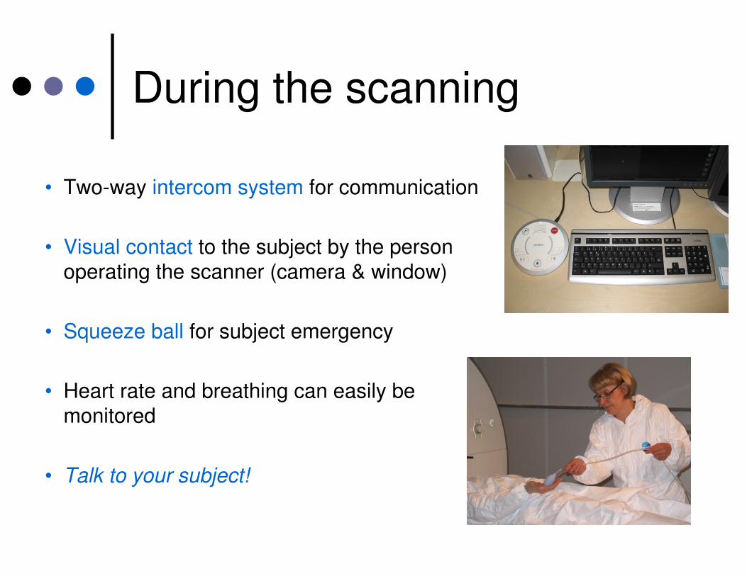
During the scanning
• Two-way intercom system for communication
• Visual contact to the subject by the person
operating the scanner (camera & window)
• Squeeze ball for subject emergency
• Heart rate and breathing can easily be
monitored
• Talk to your subject!

Safety first! – Safety courses are organized several times a year
� Superconducting magnet: always on
� Attracts ferromagnetic objects!
� Danger may be inside subject’s body
(implants, pacemaker etc.)
� RF-power may be focused, heating
� SAR (specific abrorption rate) monitoring
� Acoustic noise
� Helium, claustrofobia, contrast agents, …

Safety first! – Safety courses are organized several times a year
� Superconducting magnet: always on
� Attracts ferromagnetic objects!
� Danger may be inside your body
(implants, pacemaker etc.)
� RF-power may be focused, heating
� SAR (specific abrorption rate) monitoring
� Acoustic noise
� Helium, claustrofobia, contrast agents, …

Safety precautions
� You are in charge!
� Access limited to the MRI unit
• Shielded room is strongly restricted
� Safety course must be passed
� Safety questionnaire and screening
� Measurement clothes, metal detector, test magnet, …
� Double hearing protection for subjects

Safety precautions – there can’t be too many
� Responsible researcher (or medical doctor) has to make sure
that the measurement situation is proper (ethics committee)
� Items that are allowed inside the shielded room are labeled as
such – MRI safe – all the others are not!
� Oxygen level monitoring, emergency ventilation
� Advanced medical life support equipments and medicines,
automatic defibrillator, and oxygen
� All abnormal subject sensations and near accidents must be
reported to AMI personnel!

Daily quality routines
� Stability of the scanner is checked every morning
� Operation of squeeze ball and defibrillator ensured
� All the equipment checked for faults or breaking
� Helium level, temperatures, etc. monitored
� Equipment and pharmaceuticals in the first-aid kit checked
constantly
� In case of problems with the scanner hardware, Siemens
service should be notified immediately

• fMRI (& physiological data)
• Different anatomical contrasts
• T1, T2
• Diffusion tensor imaging (DTI)
and tractography
• Other body parts (minor)
• Clinical cases (minor)
What? – Mostly fMRI and anatomical images
(c) Mika Seppä (c) Mika Seppä

Current or recent research topics by different groups measuring @AMI
� Human visual- and auditory-systems
� Somatosensory and motor studies, pain studies
� Attention, memory, language processing
� Anatomical reference images for MEG source localization (and fMRI)
� Decision making, neuroeconomics
� Movies, music, natural stimulation
� Physiological measurements

AMI in near future
� User training, Safety courses
� Constant development of stimulus systems and measurement
environment
� Sequence programming• 3 people have already been to the Siemens’ IDEA/ICE course
� Methods development projects
� There are preliminary plans that a 7T machine would be
purchased in 5–7 years time• Ultra-high field magnets will most likely be the standard in basic research
• AMI will be actively involved

Practicalities – am I ready?
• Research idea and funding?
• Ethical statement?
• Aalto, HY, HUS, …
• AMI permission?
• Safety course?
• Study the following:
• Safety and Operations
Instructions
• Your favorite MR physics & MR in practice book
• Reservation calendar account
• AMI computer network user
account

Safe and successful fMRI
measurements
� Plan and pilot your studies well
� Develop good working practices
� Instruct your MR co-workers
� Select and prepare carefully your
patients/subjects
� Follow the instructions and procedures
� Ask, if in doubt!

Questions and answers?
AMI-CentreO.V. Lounasmaa Laboratory
School of Science, Aalto Universityhttp://ami.aalto.fi