Embed Size (px)
Citation preview

DOI 10.1378/chest.93.3.522 1988;93;522-526Chest
P Barriot, B Riou and P Viars hemothorax.Prehospital autotransfusion in life-threatening
http://chestjournal.chestpubs.org/content/93/3/522
can be found online on the World Wide Web at: The online version of this article, along with updated information and services
) ISSN:0012-3692http://chestjournal.chestpubs.org/site/misc/reprints.xhtml(without the prior written permission of the copyright holder.reserved. No part of this article or PDF may be reproduced or distributedChest Physicians, 3300 Dundee Road, Northbrook, IL 60062. All rights
ofbeen published monthly since 1935. Copyright1988by the American College is the official journal of the American College of Chest Physicians. It hasChest
© 1988 American College of Chest Physicians by guest on May 31, 2012chestjournal.chestpubs.orgDownloaded from

522 Prehospltal Autotranstusion In Life-threatenIng Hemcthorac (Barrict, R!ou, Wars)
Prehospital Autotranstusion inLife-Threatening HemothoraxPatrick Barriot, M.D.;5 Bruno Riots, M.D.;t and Pierre Viarst
Eighteen patients with life-threatening traumatic hemo-
thorax received prehospital autotransfusion using a simple
new device. During transfer to the hospital, they received3.9±0.5 L ofcolloid fluid and 4.1±0.6 L of autotransfused
blood, without anticoagulation. Hemorrhagic blood was notcoagulable, had a hematocrit of 20±4 percent, few plato-
lets, and low fibrinogen levels. Five patients died fromirreversible hemorrhagic shock. Thirteen patients werealive upon admission to the hospital, underwent emergencysurgery, and were discharged alive. During autotransfu-
sion, hematocrit decreased from 24±3 to 19±3 percent,and systolic arterial pressure increased from 78 ± II to
utotransfusion from hemothorax was first performed
by Elmendorf’ during the First World War on a
soldier with traumatic hemothorax; then it was used
sporadically as a life-saving procedure.2 Symbas3 de-
scribed a modffied autotransfusion technique for trau-
matic hemothorax which was simple, safe, and easy to
practice. He autotransfused up to 400 patients with
traumatic hemothorax in a trauma center, without any
noticeable complication.
In France, the development of prehospital emer-
gency care units with fully equipped ambulances and
on-site emergency physicians has surely improved the
management ofsevere hemorrhagic shock. Technique
of massive fluid loading using a wide-gauge central
venous catheter, inserted using the Seldinger method,
and extensive use of Medical Antishock Trousers
(MAST) have been developed for treatment of severe
traumatic hemorrhagic shock in our unit.4 Neverthe-
less, prehospital management of patients with life-
threatening hemothorax remained unsatisfactory. In
these patients, the flow rate ofhemorrhage is often too
high, and MAST have no hemostatic effect upon
hemorrhage above the diaphragm. Moreover, the po-
tential for MAST application to exacerbate bleeding
above the diaphragm has led to reservations about its
use in the treatment of thoracic injuries.5 Most of our
85eMce de Sante de la Brigade des Sapeurs Pompiers de Paris,France.
tD#{233}partementd’Anesth#{233}sie-B#{233}animation, HOpitaldela PitiC, Paris,France.Presented at the 53rd Annual Scientific Assembly, AmericanCollege of Chest Physicians, Atlanta, October 26-30, 1987.
Manuscript received July 29; accepted forpublication September14.Reprint requests: DE Barriot, BSPP, 47 rue Saint-Fargeau, 75020Thris, France
88±12 mm Hg. Upon admission to the hospital, plateletcount was 90,800± 21,400/cu mm, pmthrombin time 48±3
percent, partial thromboplastin time 197 ± 18 percent,plasma free hemoglobin levels 21± 7 mg/i#{174}ml, and serumpotassium levels 3.6± 0.5 mmol/L. No serious complicationcould be related to autotransfusion considered to be crucial
to patien& survival. The preliminary results of this studysuggest that autotransfusion might be developed in theprehospital setting since it appears simple and safe, andrepresents the only hope of survival for patients with life-threatening hemothorax.
patients with life-threatening hemothorax died before
reaching the hospital or experienced severe hemodilu-
tion that probably accounted for some deaths in the
operating room during emergency surgery.
Prehospital autotransfusion of life-threatening he-
mothorax appeared possible in our unit, but, in the
prehospital setting, a very simple device is required,
without any aspirator or pump, that could be installed
very quickly and which permits a high flow rate of auto-
transfusion. We have designed a simple device for pre-
hospital autotransfusion, and conducted a prospective
clinical study ofprehospital autotransfusion in cases of
life-threatening hemothorax.
PATIENTS AND METHODS
Description ofthe Prehospital Emergency Care Unit
This unit Is part ofthe Fire Brigade ofParis. Emergency calls arereceived at a switchboard office staffed by a chief physician. Sixemergency care ambulances with physicians trained in emergency
medicine are based at five different sites in Paris and its suburbs. Foreach patient, a standardized data sheet is filled out by the physician,which provides some details about the case history, clinical findings,and treatment administered. The prehospital physician inlbrms the
chiefphysician by radio about the clinical status ofthe patient andthe type of surgical treatment required. Then, the chief physicianlooks for a hospital team that can be ready to care for the patient andprovide effective and rapid treatment ‘fransfertime to the hospital of
a patient with multiple trauma usually ranges from 30 to 60 min, but
it is often greater especially for patients with major thoracic trauma.As amatteroffact, even in alarge urban area, itis sometimes difficultto find a hospital team immediately able to perform emergencythoracotomy and cardiopulmonary bypass for major thoracic trauma.
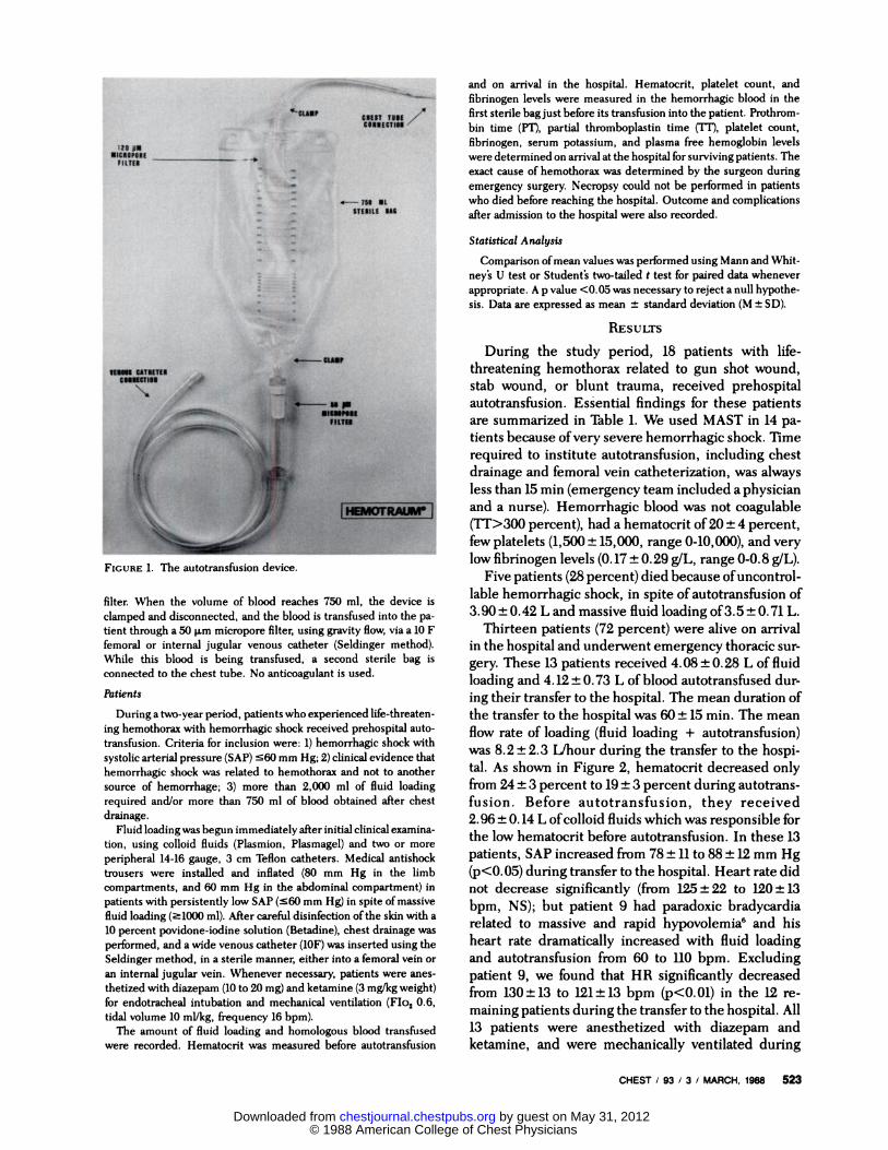
Description of the Autotransfuslon Device (Hemotraum)
A 28 to 30 F plastic chest tube is inserted into the affected pleural
space and connected to the autotransfusion device (Fig 1). Thehemorrhagic blood fills a 750 ml sterile bag via a 120 p.m micropore
© 1988 American College of Chest Physicians by guest on May 31, 2012chestjournal.chestpubs.orgDownloaded from
flUUU$ UTNTEUSNNITIN
N
flu.
4. �1
CHEST / 93 / 3 / MARCH, igee 523
hi �USICUINIE _______________________
FILTh - -,
�..su.pCPU? TUECIUUICTIN
.-7iI UI$ThiIII sas
and on arrival in the hospital. Hematocrit, platelet count, andfibrinogen levels were measured in the hemorrhagic blood in the
first sterile bag just before its transfusion into the patient. Prothrom-
bin time (P’F), partial thromboplastin time (TT), platelet count,fibrinogen, serum potassium, and plasma free hemoglobin levels
were determined on arrival at the hospital for surviving patients. The
exact cause of hemothorax was determined by the surgeon during
emergency surgery. Necropsy could not be performed in patients
who died before reaching the hospital. Outcome and complications
after admission to the hospital were also recorded.
Statistical Analysis
FIGURE 1. The autotransfusion device.
filter. When the volume of blood reaches 750 ml, the device is
clamped and disconnected, and the blood is transfused into the pa-
tient through a 50 p.m micropore filter, using gravity flow, via a 10 F
femoral or internal jugular venous catheter (Seldinger method).While this blood is being transfused, a second sterile bag is
connected to the chest tube. No anticoagulant is used.
Patients
During a two-year period, patients who experienced life-threaten-
ing hemothorax with hemorrhagic shock received prehospital auto-
transfusion. Criteria for inclusion were: 1) hemorrhagic shock withsystolic arterial pressure (SAP) �60 mm Hg; 2)clinical evidence that
hemorrhagic shock was related to hemothorax and not to another
source of hemorrhage; 3) more than 2,000 ml of fluid loading
required and/or more than 750 ml of blood obtained after chest
drainage.
Fluidloadingwas begun immediately after initial clinical examina-tion, using colloid fluids (Plasmion, Plasmagel) and two or more
peripheral 14-16 gauge, 3 cm Teflon catheters. Medical antishock
trousers were installed and inflated (80 mm Hg in the limbcompartments, and 60 mm Hg in the abdominal compartment) in
patients with persistently low SAP (�60 mm Hg) in spite of massive
fluid loading (�1000 ml). After careful disinfection ofthe skin with a10 percent povidone-iodine solution (Betadine), chest drainage was
performed, and a wide venous catheter (1OF) was inserted using theSeldinger method, in a sterile manner, either into a femoral vein oran internal jugular vein. Whenever necessary, patients were anes-thetized with diazepam (10 to 20mg) and ketamine (3 mg/kg weight)
for endotracheal intubation and mechanical ventilation (F1o2 0.6,
tidal volume 10 ml/kg, frequency 16 bpm).
The amount of fluid loading and homologous blood transfusedwere recorded. Hematocrit was measured before autotransfusion
Comparison ofmean values was performed using Mann and Whit-ney’s U test or Student’s two-tailed t test for paired data whenever
appropriate. A p value <0.05 was necessary to reject a null hypothe-
sis. Data are expressed as mean ± standard deviation (M ± SD).
RESULTS
During the study period, 18 patients with life-
threatening hemothorax related to gun shot wound,
stab wound, or blunt trauma, received prehospital
autotransfusion. Essential findings for these patients
are summarized in Table 1. We used MAST in 14 pa-
tients because ofvery severe hemorrhagic shock. lime
required to institute autotransfusion, including chest
drainage and femoral vein catheterization, was always
less than 15 mm (emergency team included a physician
and a nurse). Hemorrhagic blood was not coagulable
(TT>300 percent), had a hematocrit of2O ± 4 percent,
few platelets (1,500 ± 15,000, range 0-10,000), and very
low fibrinogen levels (0. 17 ± 0.29 gIL, range 0-0. 8 gIL).
Five patients (28 percent) died because of uncontrol-
lable hemorrhagic shock, in spite ofautotransfusion of
3.90 ± 0.42 L and massive fluid loading of3.5 ± 0.71 L.
Thirteen patients (72 percent) were alive on arrival
in the hospital and underwent emergency thoracic sur-
gery. These 13 patients received 4. 08 ± 0.28 L of fluid
loading and 4. 12 ± 0. 73 L of blood autotransfused dur-
ing their transfer to the hospital. The mean duration of
the transfer to the hospital was 60 ± 15 mm. The mean
flow rate of loading (fluid loading + autotransfusion)
was 8.2 ± 2.3 Lfhour during the transfer to the hospi-
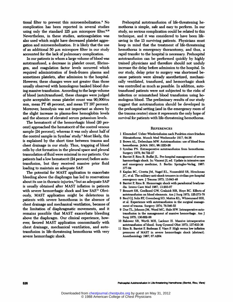
tal. As shown in Figure 2, hematocrit decreased only
from 24 ± 3 percent to 19 ± 3 percent during autotrans-
fusion. Before autotransfusion, they received
2. 96 ± 0. 14 L ofcolloid fluids which was responsible for
the low hematocrit before autotransfusion. In these 13
patients, SAP increased from 78 ± II to 88 ± 12 mm Hg
(p’(O.05) during transfer to the hospital. Heart rate did
not decrease significantly (from 125 ± 22 to 120±13
bpm, NS); but patient 9 had paradoxic bradycardia
related to massive and rapid hypovolemia6 and his
heart rate dramatically increased with fluid loading
and autotransfusion from 60 to 110 bpm. Excluding
patient 9, we found that HR significantly decreased
from 130 ± 13 to 121 ± 13 bpm (p<O. 01) in the 12 re-
maining patients during the transfer to the hospital. All
13 patients were anesthetized with diazepam and
ketamine, and were mechanically ventilated during
© 1988 American College of Chest Physicians by guest on May 31, 2012chestjournal.chestpubs.orgDownloaded from
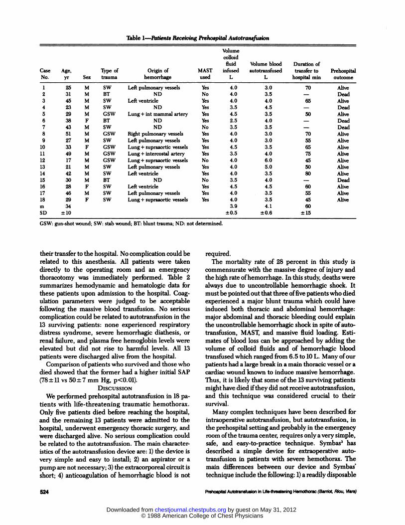
Table 1-P�dienta Receiving Prehospild Autotransfusion
524 Prehospltsi Autoiransfuslon In Life-threatenIng Hemothorac (Barrlct RIou, Wars)
Volume
colloid
CaseNo.
Age,yr Sex
Type oftrauma
Origin ofhemorrhage
MASTused
fluid
infusedL
Volume blood
autotransfusedL
Duration of
transfer tohospital miss
Prehospitaloutcome
1 25 M SW Left pulmonary vessels Yes 4.0 3.0 70 Alive2 31 M BT ND No 4.0 3.5 - Dead3 45 M SW Left ventricle Yes 4.0 4.0 65 Alive4 23 M SW ND Yes 3.5 4.5 - Dead
5 29 M GSW Lung + int mammal artery Yes 4.5 3.5 50 Alive6 38 F BT ND Yes 2.5 4.0 - Dead7 43 M SW ND No 3.5 3.5 - Dead8 51 M GSW Bight pulmonary vessels Yes 4.0 3.0 70 Alive9 27 M SW Left pulmonary vessels Yes 4.0 3.0 55 Alive
10 33 F GSW Lung + supraaortic vessels Yes 4.5 3.5 65 Alive11 49 M GSW Lung + intercostal artery Yes 3.5 4.0 75 Alive12 17 M GSW Lung + supraaortic vessels No 4.0 6.0 45 Alive13 21 M SW Left pulmonary vessels Yes 4.0 5.0 50 Alive
14 42 M SW Left ventricle Yes 4.0 3.5 80 Alive15 30 M BT ND No 3.5 4.0 - Dead16 28 F SW Leftventricle Yes 4.5 4.5 60 Alive17 46 M SW Left pulmonary vessels Yes 4.0 3.5 55 Alive18 29 F SW Lung + supraaortic vessels Yes 4.0 3.5 45 Alive
m 34 3.9 4.1 60SD ±10 ±0.5 ±0.6 ±15
GSW: gun-shot wound; SW: stab wound; BT: blunt trauma; ND: not determined.
their transfer to the hospital. No complication could be
related to this anesthesia. All patients were taken
directly to the operating room and an emergency
thoracotomy was immediately performed. Table 2
summarizes hemodynamic and hematologic data for
these patients upon admission to the hospital. Coag-
ulation parameters were judged to be acceptable
following the massive blood transfusion. No serious
complication could be related to autotransfusion in the
13 surviving patients: none experienced respiratory
distress syndrome, severe hemorrhagic diathesis, or
renal failure, and plasma free hemoglobin levels were
elevated but did not rise to harmful levels. All 13
patients were discharged alive from the hospital.
Comparison ofpatients who survived and those who
died showed that the former had a higher initial SAP
(78±11 vs 50±7 mm Hg, p<O.Ol).
DISCUSSION
We performed prehospital autotransfusion in 18 pa-
tients with life-threatening traumatic hemothorax.
Only five patients died before reaching the hospital,
and the remaining 13 patients were admitted to the
hospital, underwent emergency thoracic surgery, and
were discharged alive. No serious complication could
be related to the autotransfusion. The main character-
istics ofthe autotransfusion device are: 1) the device isvery simple and easy to install; 2) an aspirator or a
pump are not necessary; 3) the extracorporeal circuit is
short; 4) anticoagulation of hemorrhagic blood is not
required.
The mortality rate of 28 percent in this study is
commensurate with the massive degree of injury and
the high rate ofhemorrhage. In this study, deaths were
always due to uncontrollable hemorrhagic shock. It
must be pointed out that three offive patients who died
experienced a major blunt trauma which could have
induced both thoracic and abdominal hemorrhage:
major abdominal and thoracic bleeding could explain
the uncontrollable hemorrhagic shock in spite of auto-
transfusion, MAST, and massive fluid loading. Esti-
mates of blood loss can be approached by adding the
volume of colloid fluids and of hemorrhagic blood
transfused which ranged from 6.5 to 10 L. Many of ourpatients had a large break in a main thoracic vessel or a
cardiac wound known to induce massive hemorrhage.
Thus, it is likely that some ofthe 13 surviving patients
might have died ifthey did not receive autotransfusion,
and this technique was considered crucial to their
survival.
Many complex techniques have been described for
intraoperative autotransfusion, but autotransfusion, in
the prehospital setting and probably in the emergency
room ofthe trauma center, requires only a very simple,
safe, and easy-to-practice technique. Symbas3 has
described a simple device for extraoperative auto-
transfusion in patients with severe hemothorax. The
main differences between our device and Symbas’
technique include the following: 1) a readily disposable
© 1988 American College of Chest Physicians by guest on May 31, 2012chestjournal.chestpubs.orgDownloaded from

30r
0
Table 2-Hemodynamic and Hematologic Data in P�itients Surviving Autotransfusion upon Admission to the Hospital
CHEST I 93 I 3 I MARCH. 1988 525
CaseNo.
SAPmm Hg
HRbpm
Hematocrit%
Platelets/cu mm
Fibrinogeng/L
PT%
U%
Plasma free
hemoglobinmg/100 ml
Serumpotassium
mmollL
1 90 120 15 100,000 0.80 60 206 20 3.43 90 120 20 80,000 1.00 50 187 25 3.85 110 130 15 70,000 1.30 40 203 18 2.98 100 120 18 100,000 0.90 50 177 15 3.7
9 90 110 20 70,000 1.20 40 203 23 4.110 80 140 20 120,000 1.00 60 190 10 3.311 90 100 15 90,000 1.30 30 228 16 3.512 60 110 25 110,000 0.80 50 206 21 2.813 90 130 20 60,000 1.00 40 163 17 4.514 90 120 23 100,000 0.70 40 190 22 4.216 80 140 20 80,000 0.80 60 218 30 3.817 100 120 20 130,000 1.10 50 213 28 2.818 80 100 18 70,000 1.40 50 176 34 3.9m 88 120 19 90,800 1.02 48 197 21 3.6
SD ±12 ±13 ±3 ±21,400 ±0.43 ±3 ±18 ±7 ±0.5
SAP: systolic arterial pressure; HR: heart rate; fl: prothrombin time; �VF: partial thromboplastin time; Tfis expressed as percentage of control.
S �
I-
00
(5
E2oa)
I
151
BATFicuax 2. Hematocrit before (B) and after autotransfusion uponadmission the hospital (AT). (A) individual values; (0) mean ± SD.
sterile one-use package; 2) use ofa plastic bag easier to
handle in the prehospital setting than a chest bottle; 3)
use ofa wide-gauge central venous catheter that allowsa higher flow rate ofautotransfusion; 4) two micropore
filters 120 and 50 �m, respectively.
An experimental study7 suggested that full systemic
heparimzation may enhance preservation of the hem-
orrhagic blood during autotransfusion and thus, these
authors7 recommended heparinization during intra-
operative autotransfusion. Reul et al� preferred CPD
anticoagulation in the autotransfusion device which
appeared to be as efl�ctive in the prevention of platelet
aggregation and clot formation in the filter, and to
decrease problems of hemostasis, compared to sys-
temic heparinization. Symbas3 did not use anticoag-
ulants at all in extraoperative autotransfusion of hemo-
thorax. Our study confirms previous experimental and
clinical results from Symbas: hemorrhagic blood from
hemothorax had few platelets, low fibrinogen levels
and does not clot. The extracorporeal circuit of our
device is short and the time ofcontact between blood
and the sterile bag is also shortened by the high flow
rate of autotransfusion, and hemorrhagic blood was
incoagulable. It is possible that anticoagulation of
hemorrhagic blood in massive autotransfusion could
induce hemorrhagic diathesis (heparin overdosage,
citrate intoxication with CPD); as a matter offact, the
amount of anticoagulant used increases as the amount
ofhemorrhagic blood autotransfused increases. There-
fore, we suggest that systemic or local anticoagulation
is not required in massive autotransfusion for life-
threatening hemothorax.
Microembolization ofplatelet aggregates and partic-
ulate debris is involved in the development ofthe acute
respiratory distress syndrome. A 125 �am micropore flu-ter is usually used in the autotransfusion apparatus, but
some authors have recommended the use of an addi-
© 1988 American College of Chest Physicians by guest on May 31, 2012chestjournal.chestpubs.orgDownloaded from
526 Prehospltal Autotranafuslon In Life-threatenIng Hemothorac (Barrlcd, RIou, Wars)
tional filter to prevent this microembolization.8 No
complication has been reported in several studies
using only the standard 125 �tm micropore filter.9’#{176}
Nevertheless, in these studies, anticoagulation was
also used which might have decreased platelet aggre-
gation and microembolization. It is likely that the use
of an additional 50 �am micropore filter in our study
accounted for the lack of pulmonary complication.
In our patients in whom a large volume ofblood was
autotransfused, a decrease in platelet count, fibrino-
gen, and coagulation factor levels occurred which
required administration of fresh-frozen plasma and
sometimes platelets, after admission to the hospital.
However, these changes were not greater than those
usually observed with homologous banked blood dur-
ing massive transfusion. According to the large volume
of blood (auto)transfused, these changes were judged
quite acceptable: mean platelet count was 90,000/cu
mm, mean PT 48 percent, and mean TT 197 percent.
Moreover, hemolysis was not important as shown by
the slight increase in plasma-free hemoglobin levels
and the absence of elevated serum potassium levels.
The hematocrit of the hemorrhagic blood (20 per-
cent) approached the hematocrit ofthe control venous
sample (24 percent), whereas it was only about half of
the control sample in Symbas’ study.3 Most likely, this
is explained by the short time between trauma and
chest drainage in our study. Thus, trapping of blood
cells by clot formation in the pleural space and pleural
transudation offluid were minimal in our patients. Our
patients had a low hematocrit (24 percent) before auto-
transfusion, but they received massive prior fluid
loading to maintain an adequate SAP
The potential for MAST application to exacerbate
bleeding above the diaphragm has led to reservations
about its use in thoracic injuries,2 but an adequate SAP
is usually obtained after MAST inflation in patients
with severe hemorrhagic shock and low SAP.U Obvi-
ously, MAST application might be deleterious in
patients with severe hemothorax in the absence of
chest drainage and mechanical ventilation, because of
the limitation of diaphragmatic movements, and it
remains possible that MAST exacerbate bleeding
above the diaphragm. Our clinical experience, how-
ever, favored MAST application concomitantly with
chest drainage, mechanical ventilation, and auto-
transfusion in life-threatening hemothorax with very
severe hemorrhagic shock.
Prehospital autotransfusion of life-threatening he-
mothorax is simple, safe and easy to perform. In our
study, no serious complication could be related to this
technique, and it was considered to have been life-
saving in the 13 surviving patients. Physicians must
keep in mind that the treatment of life-threatening
hemothorax is emergency thoracotomy, and thus, a
rapid transfer to the hospital is necessary. Prehospital
autotransfusion can be performed quickly by highly
trained physicians and therefore should not unduly
increase the delay before admission to the hospital. In
our study, delay prior to surgery was shortened be-
cause patients were already anesthetized, mechani-
cally ventilated, transfused, and hemorrhagic shock
was controlled as much as possible. In addition, auto-
transfused patients were not subjected to the risks of
infection or mismatched blood transfusion using ho-
mologous blood. The preliminary results of our study
suggest that autotransfusion should be developed in
the prehospital setting (and in the emergency room of
the trauma center) since it represents the only hope of
survival for patients with life-threatening hemothorax.
REFERENCES
1 Elmendorf. Ueber Wiederinfusion nach Punktion eines frischenH#{228}matothorax. Munch Med Wochenschr 1917; 64:36
2 Brown AL, Debenham MW. Autotmansfusion: use ofblood from
hemothorax. JAMA 1931; 96:1223-283 Symbas PN. Extraoperative autotransfusion from hemothorax.
Surgery 1978; 84:722-274 Barriot P. Riou B, Buffat JL Pre-hospital management of severe
haemorrhagic shock. In: VincentJL ed. Update in intensive careand emergency medicine, 3. Berlin: Springler-Verlag, 1987:377-84
5 Kaplan BC, Civetta JM, Nagel EL, Nussenfeld SR, HirschmanJC, et al. The military anti-shock trousers in civilian pre-hospitalemergency care. J ‘li-auma 1973; 13:843-48
6 BarriOt P, Riou B. Hemorragic shock with paradoxical bradycar-dia. Intens Care Med 1987; 13:203-07
7 Bennett SH, Geelhoed GW, Gralnick HR. Hoye RC. Effects ofautotransfusion on blood elements. Am J Surg 1973; 125:273-78
8 Reid GJ, Solis RT, Greenberg SD, Mattox KL, Whisennand HH,
et al Experience with autotransfusion in the surgical manage-ment of trauma. Surgery 1974; 76:546-55
9 Due TL, Johnson JM, Wood MC, Hale HW. Intraoperative auto-
transfusion in the management of massive hemorrhage. Am JSurg 1975; 130:652-58
10 Rakower SR. Worth MH, Lackner H. Massive intraoperativeautotransfusion of blood. Surg Gynecol Obst 1973; 137:633-36
11 Riou B, Barriot P. Bodenan P. Viars P. High versus low inflation
pressures of MAST in severe hemorrhagic shock (abstract).Anesthesiology 1987; 67:A204
© 1988 American College of Chest Physicians by guest on May 31, 2012chestjournal.chestpubs.orgDownloaded from

DOI 10.1378/chest.93.3.522 1988;93; 522-526Chest
P Barriot, B Riou and P ViarsPrehospital autotransfusion in life-threatening hemothorax.
May 31, 2012This information is current as of
http://chestjournal.chestpubs.org/content/93/3/522Updated Information and services can be found at:
Updated Information & Services
http://chestjournal.chestpubs.org/content/93/3/522#related-urlsThis article has been cited by 2 HighWire-hosted articles:
Cited Bys
http://www.chestpubs.org/site/misc/reprints.xhtmlonline at: Information about reproducing this article in parts (figures, tables) or in its entirety can be foundPermissions & Licensing
http://www.chestpubs.org/site/misc/reprints.xhtmlInformation about ordering reprints can be found online:
Reprints
the right of the online article.Receive free e-mail alerts when new articles cite this article. To sign up, select the "Services" link to
Citation Alerts
slide format. See any online figure for directions. articles can be downloaded for teaching purposes in PowerPointCHESTFigures that appear in Images in PowerPoint format
© 1988 American College of Chest Physicians by guest on May 31, 2012chestjournal.chestpubs.orgDownloaded from