Embed Size (px)
Citation preview
Proc. Natl. Acad. Sci. USAVol. 93, pp. 5141-5145, May 1996Immunology
Preferential induction of a Th1 immune response and inhibition ofspecific IgE antibody formation by plasmid DNA immunizationEYAL RAZ*, HELEN TIGHE*, YUKIO SATO*, MARIPAT CORR*, JEAN A. DUDLER*, MARK ROMAN*, SUsAN L. SWAINt,HANS L. SPIEGELBERGt, AND DENNIS A. CARSON**Department of Medicine and The Sam and Rose Stein Institute for Research on Aging, tCancer Center, and tDepartment of Pediatrics, University of California,San Diego, 9500 Gilman Drive, La Jolla, CA 92093-0663
Communicated by Frank J. Dixon, The Scripps Research Institute, La Jolla, CA, January 11, 1996 (received for review October 15, 1995)
ABSTRACT We compared the antigen-specific antibodyisotypes and lymphokine secretion by CD4+ T cells in BALB/cmice immunized intradermally with either Escherichia coli13-galactosidase (13-gal) or plasmid DNA (pDNA) encoding,8-gal in a cytomegalovirus-based expression vector (pCMV-LacZ). pCMV-LacZ induced mainly IgG2a, whereas 13-gal insaline or alum induced IgGI and IgE 1-gal-specific antibodies.In addition, splenic CD4+ T helper (Th) cells isolated frompDNA-immunized mice secreted interferon-y but not inter-leukin (IL)-4 and IL-5, whereas Th cells from 13-gal-injectedmice secreted IL-4 and IL-5 but not interferon-y after in vitrostimulation with antigen. Together these data demonstratethat pDNA immunization induced a T helper type 1 (Thl)response, whereas protein immunization induced a T helpertype 2 (Th2) response to the same antigen. Interestingly,priming of mice with pCMV-LacZ prevented IgE antibodyformation to a subsequent i.p. 13-gal in alum injection. Thiseffect was antigen-specific, because priming with pCMV-LacZdid not inhibit IgE anti-ovalbumin antibody formation. Mostimportantly, intradermal immunization with pCMV-LacZ(but not pCMV-OVA) of 13-gal in alum-primed mice caused a66-75% reduction of the IgE anti-,B-gal titer in 6 weeks. Also,pCMV-LacZ induced specific IgG2a antibody titers and in-terferon-y secretion by Th cells in the 13-gal in alum-primedmice. The data demonstrate that gene immunization inducesa Th, response that dominates over an ongoing protein-induced Th2 response in an antigen-specific manner. Thissuggests that immunization with pDNA encoding for allergensmay provide a novel type of immunotherapy for allergicdiseases.
inflammatory reaction at the injection site despite the induc-tion of a cellular (cytotoxic lymphocytes) and humoral (IgGantibodies) immune response to the encoded antigens. Whenwe analyzed the specific IgG antibodies for their subclassdistribution, we found that the IgG antibodies were almostentirely of the IgG2a subclass. This was surprising becausemice immunized with protein antigens, in contrast to viral orbacterial antigens, produce predominantly IgGl (7) and oftenIgE (8) antibodies. IgG2a antibody formation is dependent oninterferon-y (IFN-,y) as an IgM-to-IgG2a switch factor and isbelieved to be typical for a T helper (Th) type 1 (Th1) response(9). In contrast, IgGl and particularly IgE antibody productiondepends on interleukin (IL)-4 secreted by Th2 cells (10). Toinvestigate the possibility that i.d. immunization with pDNAinduced a Th, response to the gene product rather than theexpected Th2 response, we studied the primary immune re-sponse of BALB/c mice to i.d. pCMV-LacZ injections ascompared with injections of the protein (3-gal in saline or alum.The data showed that pCMV-LacZ induces a Th, and that(3-gal in saline or alum induces a Th2 response to the sameantigen. Therefore, we also investigated the secondary im-mune response of pDNA-primed mice to ,3-gal in alum and theresponse of ,B-gal in alum-primed mice to pCMV-LacZ todetermine whether one of these two modes of immunizationdominates over the other in a secondary immune response. Adominant response by immunization with pDNA could beuseful for switching an undesired Th2 response to a morefavorable Th, response in allergic and certain parasitic disor-ders.
Intramuscular (i.m.) or intradermal (i.d.) injection of "naked"plasmid DNA (pDNA) encoding for viral or other proteinantigens results in uptake of the pDNA by the muscle (1-5) orskin cells (6) and subsequent synthesis of the protein by thesecells. The transfected cells process the antigen and present theimmunogenic peptides on major histocompatibility complexclass I and/or class II molecules, depending on the cell type.This results in both a cellular and humoral immune responseto the antigen (1-6). Immunization with pDNA encoding anantigen (gene vaccination) has generated great interest byinvestigators searching for better vaccination methods, espe-cially for inducing cellular immune responses to viral infec-tions, including HIV-1 (4). Recently, we showed that i.d.injection of pDNA encoding influenza nucleoprotein (NP) orEscherichia coli ,B-galactosidase ((3-gal) in a cytomegalovirus(CMV)-based expression vector (pCMV-NP and pCMV-LacZ, respectively) led to prolonged expression of intracellularantigen by dermal keratinocytes, fibroblasts, and cells with themorphology of Langerhans cells and macrophages (6). We alsoshowed that expression of these antigens does not cause an
METHODSMice. Female BALB/c mice were purchased from The
Jackson Laboratory, maintained in the University of Califor-nia, San Diego, Animal Facility accredited by the AmericanAssociation for the Accreditation of Laboratory Animal Care,and used at 6-8 weeks of age.pDNA Preparation. The construction pCMV-based vectors
has been described (6). pCMV-LacZ contains the CMV IElpromotor-intron, the simian virus 40 t-intron, the E. coli LacZcDNA, and the simian virus 40 polyadenylylation site. ThepCMV-OVA vector encodes hen egg ovalbumin (OVA)cDNA, and the plasmid backbone is identical to pCMV-LacZ.pDNA was purified using a Qiagen megaprep kit (Qiagen,Chatsworth, CA) and was stored at -20°C in 10mM Tris.HCl/0.1 mM EDTA, pH 8.0. The endotoxin content was reduced byextraction with Triton X-114 (Sigma) to 0.5-5 ng per 1 mg ofpDNA (11), as determined by limulus amoebocyte lysate assay(Sigma). Before inoculation, pDNA was precipitated in 100%ethanol, washed with 70% ethanol, and dissolved in pyrogen-free saline.
Abbreviations: pDNA, plasmid DNA; i.d., intradermal; ,B-gal, j3-galac-tosidase; IFN-,y, interferon--y; CMV, cytomegalovirus; OVA, ovalbu-min; Th, T helper.
The publication costs of this article were defrayed in part by page chargepayment. This article must therefore be hereby marked "advertisement" inaccordance with 18 U.S.C. §1734 solely to indicate this fact.
5141
Dow
nloa
ded
by g
uest
on
Janu
ary
24, 2
022
Proc. Natl. Acad. Sci. USA 93 (1996)
Immunization. pCMV-LacZ or pCMV-OVA was eitherinjected i.d. at the base of the tail or scratched into the shavedskin of the lower back with a tyne skin test applicator (Con-naught Laboratories). The applicators were washed extensivelyin distilled water, soaked overnight in 0.5% SDS, rinsed withwater, soaked overnight in 0.1 M NaOH, rinsed with water, anddried at 37°C for 8 hr. Fifty micrograms of pDNA dissolved in6 ,ul of saline was dripped onto the spikes of the tyneapplicator. Mice received two applications (total of 100 ,tg ofDNA) per inoculation. Both i.d. and tyne applicator pDNAinoculations were given once a week for 3 weeks. For pDNAinjections, 100 ,ug of pDNA in 50 gl of saline was injected i.d.three times at weekly intervals. Three weekly injections werepreviously found to induce a more reproducible response thana single injection (6). For primary immunization with protein,10 ,xg of 3-gal (Calbiochem) dissolved in 50 ,ul of saline or 1ptg in 50 ,ul of saline containing 3 mg of alum was injected i.d.once at the base of the tail. For booster injections, 1 ,tg of 13-galin 0.5 ml of saline containing 3 mg of alum was injected i.p.OVA, 2 jig in 0.5 ml of saline containing 3 mg of alum, wasinjected i.p. for primary or booster injections.Antibody Measurements. Anti-,3-gal antibodies of the IgG
subclasses IgGl, IgG2a, IgG2b, and IgG3 were measured byELISA. Microtiter plates were coated overnight with 5 ,tg of13-gal per ml of borate-buffered saline (BBS; pH 9.2) and thenwashed with BBS, and nonspecific binding sites were thenblocked with 1% bovine serum albumin in BBS. After washingtwice in BBS/0.5% Tween 20 (Sigma), serum samples diluted1:40 and then 1:4 for 8 steps in phosphate-buffered saline (pH7.4) were added to the wells. After overnight incubation at 4°C,the plates were washed with BBS/Tween 20 and incubatedwith alkaline phosphatase-labeled goat anti-mouse IgGI,IgG2a, IgG2b, or IgG3 (Southern Biotechnology Associates,Birmingham, AL) for 2 hr at room temperature. The plateswere washed with BBS/Tween 20, and a solution of p-nitrophenyl phosphate (1 mg/ml; Boehringer Mannheim) wasadded. Absorbance at 405 nm was read 1 hr after addition ofthe substrate. Each plate included a previously screenedstandard serum that contained a high titer of anti-13-galantibodies. The results are expressed in units per ml, calculatedbased on the units/ml of the standard serum, and represent themean + SE of four animals in each group.The IgE anti-1-gal or anti-OVA antibodies were measured
by a radioimmuno-allergosorbent test as described (12).Briefly, 96-well polyvinyl plates were coated with 10 ,tg of13-gal per ml ofBBS (pH 9.2) for 1 hr at room temperature. Theplates were washed, and nonspecific sites were blocked with5% nonfat milk in BBS for 2 hr. Sera diluted at 1:10 and 1:20were added overnight, and after the plates were washed,
A IgG2a B IgGl
125I-radiolabeled purified goat anti-IgE antibodies were addedfor 4 hr. The plates were washed, and the radioactivity boundto the wells was measured with a scintillation counter. Serialdilutions of a standard serum consisting of a pool of 13-gal inalum-immunized mice was included on each plate to ensurereproducible cpm. Also, to determine the effect of competitionof IgG antibodies for antigen, sera of groups of four mice werepooled and passed over 0.5 ml of protein G-Sepharose (Phar-macia), which removed essentially all IgG2a, IgG2b, and IgG3and "90% of the IgGl. Retesting of the IgG-depleted serumand comparison with unabsorbed serum showed higher cpmfor the IgG-absorbed sera but no qualitative differences in theIgE antibody titers.Lymphokine Assays. In each set of experiments, mice were
killed 1 week after the last antibody determination for quan-titation of the lymphokines secreted by CD4+ T cells after13-gal stimulation in vitro (13). Three days before sacrifice, themice were injected i.v. with 10 Ag of 1-gal in 50 ,lI of saline.Spleens were removed and teased to prepare single-cell sus-pensions in RPMI 1640 medium supplemented with 10%heat-inactivated fetal bovine serum. The cells were enrichedfor CD4+ T cells by treatment with monoclonal antibodies toCD8 and CD56 and complement as described (14). Thisprocedure resulted in removal of >95% of the CD8+ cells asshown by FACS (Becton Dickinson) analyses. The CD4+ Tcells were cultured at 2 x 10⁶ cells per ml with an equal numberof irradiated (30 grays) T cell-depleted BALB/c spleen cells inRPMI 1640 medium containing 10% heat-inactivated bovineserum, 2mM L-glutamine, 5 x 10⁻⁵ M 2-mercaptoethanol, 1%penicillin-streptomycin, and 20 gg of 13-gal per ml. After 36 or72 hr of culture, the IFN-y, IL-4, and IL-5 levels weredetermined in the supernatants by ELISA assays (15, 16) withanti-IL-4 antibodies (llBl1 and BVD6 24G2; PharMingen),anti-IFN-y antibodies (R46A2 and XMG1.2), and anti-IL-5antibodies (TRFK4 and TRFK5).
Statistical Analyses. The data were analyzed for statisticalsignificance by ANOVA.
RESULTSThe Primary Immune Response ofBALB/c Mice to pCMV-
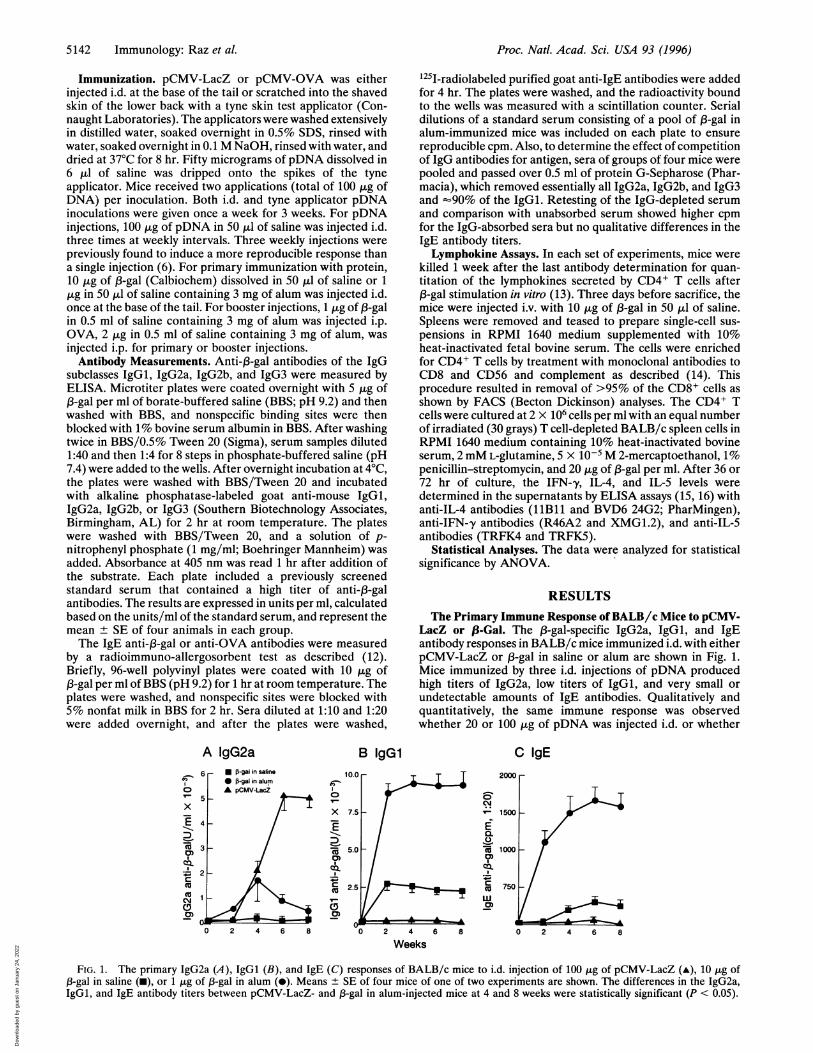
LacZ or ,8-Gal. The 1B-gal-specific IgG2a, IgGl, and IgEantibody responses in BALB/c mice immunized i.d. with eitherpCMV-LacZ or 13-gal in saline or alum are shown in Fig. 1.Mice immunized by three i.d. injections of pDNA producedhigh titers of IgG2a, low titers of IgGl, and very small orundetectable amounts of IgE antibodies. Qualitatively andquantitatively, the same immune response was observedwhether 20 or 100 jig of pDNA was injected i.d. or whether
C IgE1.0-
'.5 _ 8 _if.5~~~~~~~C
10.
&.t- coz
.0~~~~.5 ct
w0)
0 2 4 6 8
Weeks
2000
0 2 4 6 8
FIG. 1. The primary IgG2a (A), IgGl (B), and IgE (C) responses of BALB/c mice to i.d. injection of 100 ,g of pCMV-LacZ (-), 10 ,tg of,B-gal in saline (m), or 1 j,g of 13-gal in alum (0). Means ± SE of four mice of one of two experiments are shown. The differences in the IgG2a,IgGl, and IgE antibody titers between pCMV-LacZ- and 1-gal in alum-injected mice at 4 and 8 weeks were statistically significant (P < 0.05).
* 5-gal in saline* 5-gal in alumA pCMV-LacZ
x
0)
CDcJ
(9
0)
10.
0
X 7.
zx 5.
C._E 2.
C)
5142 Immunology: Raz et al.
6 r
Dow
nloa
ded
by g
uest
on
Janu
ary
24, 2
022
Proc. Natl. Acad. Sci. USA 93 (1996) 5143
pDNA was scratched into the skin with a tyne device (data notshown). In contrast to the pDNA-immunized mice, miceinjected with the protein ,3-gal either in saline or in alumproduced high IgGl and IgE antibody titers and significantlyless IgG2a antibodies. ,3-Gal in saline induced a lower IgE titerthan 13-gal in alum; however, the qualitative nature of the im-mune response (IgGl and IgE antibody formation) was thesame. IgG2b and IgG3 antibodies were also measured in theserum of these mice. pDNA-immunized mice produced moreIgG2b than the protein-immunized mice, whereas both pDNAand protein immunization induced only low titers of IgG3antibodies (data not shown).
Because IgG2a antibody formation is typical for a Th,response and IgGl and IgE antibody production results froma Th2 immune response (9), these isotype profiles suggestedthat pDNA and protein induced different Th cell responses tothe same antigen. To confirm this hypothesis, we determinedthe 13-gal-induced IFN-,y, IL-4, and IL-5 secretion by splenicCD4+ T cells from the two groups of mice. As shown in Table1 (experiments A-D), in vitro 13-gal-stimulated CD4+ T cellsfrom naive animals did not secrete lymphokines, whereasCD4+ T cells from pCMV-LacZ-immunized mice secretedIFN-,y and no detectable IL-4 or IL-5. In contrast, CD4+ Tcells from 13-gal in saline- or 13-gal in alum-immunized micesecreted IL-4 and IL-5 and no detectable IFN-,y. These datacorroborate the isotype-restricted IgG2a or IgGl and IgEantibody responses shown in Fig. 1, and they confirm thatpCMV-LacZ induced a Th1 immune response and 13-galinduced a Th2 immune response to the same antigen.The Response to a Secondary (3-Gal in Alum Immunization
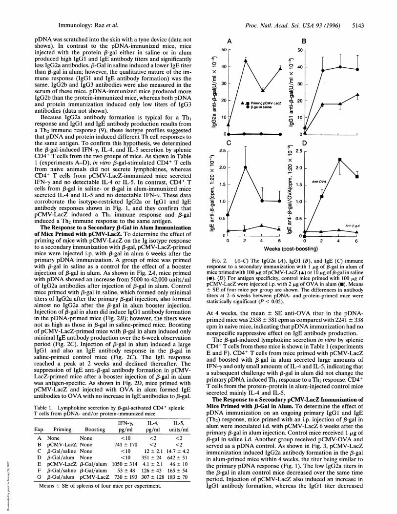
of Mice Primed with pCMV-LacZ. To determine the effect ofpriming of mice with pCMV-LacZ on the Ig isotype responseto a secondary immunization with 13-gal, pCMV-LacZ-primedmice were injected i.p. with 1-gal in alum 6 weeks after theprimary pDNA immunization. A group of mice was primedwith 13-gal in saline as a control for the effect of a boosterinjection of 13-gal in alum. As shown in Fig. 24, mice primedwith pDNA showed an increase from 5000 to 42,000 units/mlof IgG2a antibodies after injection of 13-gal in alum. Controlmice primed with 1-gal in saline, which formed only minimaltiters of IgG2a after the primary 13-gal injection, also formedalmost no IgG2a after the 13-gal in alum booster injection.Injection of 13-gal in alum did induce IgGl antibody formationin the pDNA-primed mice (Fig. 2B); however, the titers werenot as high as those in 13-gal in saline-primed mice. Boostingof pCMV-LacZ-primed mice with 13-gal in alum induced onlyminimal IgE antibody production over the 6-week observationperiod (Fig. 2C). Injection of 13-gal in alum induced a largeIgGl and also an IgE antibody response in the 13-gal insaline-primed control mice (Fig. 2C). The IgE responsereached a peak at 2 weeks and declined thereafter. Thesuppression of IgE anti-13-gal antibody formation in pCMV-LacZ-primed mice after a booster injection of 13-gal in alumwas antigen-specific. As shown in Fig; 2D, mice primed withpCMV-LacZ and injected with OVA in alum formed IgEantibodies to OVA with no increase in IgE antibodies to 1-gal.
Table 1. Lymphokine secretion by 13-gal-activated CD4+ splenicT cells from pDNA- and/or protein-immunized mice
IFN-y, IL-4, IL-5,Exp. Priming Boosting pg/ml pg/ml units/mlA None None <10 <2 <2B pCMV-LacZ None 741 ± 170 <2 <2C 13-Gal/saline None <10 12 ± 2.1 14.7 ± 4.2D 13-Gal/alum None <10 351 ± 24 642 + 51E pCMV-LacZ 13-Gal/alum 1050 + 314 4.1 + 2.1 46 ± 10F 13-Gal/saline 1B-Gal/alum 53 ± 48 126 ± 43 165 + 54G 13-Gal/alum pCMV-LacZ 730 ± 193 307 ± 128 183 + 70
Means + SE of spleens of four mice per experiment.
A50
0)
v-x
ED
')CD
x
cJco
E
04
0CD
0w0)
S~
B50 r
08x
0)C-
.-I
cQv-
0
0)
D0
x0cM
C.0
-i0)
4._C
UJ
0 2 4
Weeks (post-boosting)
FIG. 2. (A-C) The IgG2a (A), IgGl (B), and IgE (C) immuneresponse to a secondary immunization with 1 jig of 13-gal in alum ofmice primed with 100 ,tg of pCMV-LacZ (A) or 10 jig of 13-gal in saline(-). (D) For antigen specificity, control mice primed with 100 ,g ofpCMV-LacZ were injected i.p. with 2 ,ug of OVA in alum (-). Means± SE of four mice per group are shown. The differences in antibodytiters at 2-6 weeks between pDNA- and protein-primed mice werestatistically significant (P < 0.05).
At 4 weeks, the mean ± SE anti-OVA titer in the pDNA-primed mice was 2358 ± 581 cpm as compared with 2241 ± 338cpm in naive mice, indicating that pDNA immunization had nononspecific suppressive effect on IgE antibody production.The 13-gal-induced lymphokine secretion in vitro by splenic
CD4+ T cells from these mice is shown in Table 1 (experimentsE and F). CD4+ T cells from mice primed with pCMV-LacZand boosted with j3-gal in alum secreted large amounts ofIFN-,y and only small amounts of IL-4 and IL-5, indicating thata subsequent challenge with 13-gal in alum did not change theprimary pDNA-induced Th, response to a Th2 response. CD4+T cells from the protein-protein in alum-injected control micesecreted mainly IL-4 and IL-5.The Response to a Secondary pCMV-LacZ Immunization of
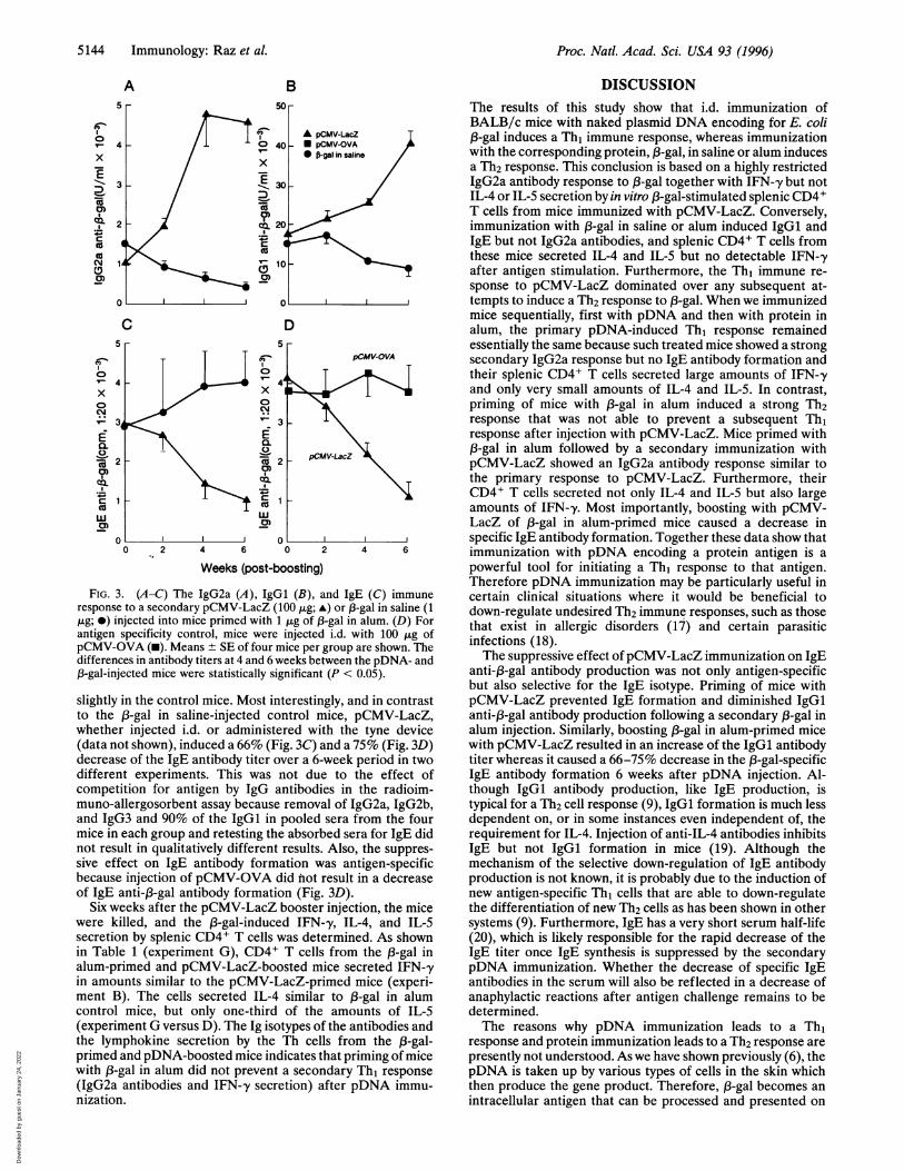
Mice Primed with (-Gal in Alum. To determine the effect ofpDNA immunization on an ongoing primary IgGI and IgE(Th2) response, mice primed with an i.p. injection of 13-gal inalum were inoculated i.d. with pCMV-LacZ 6 weeks after theprimary 13-gal in alum injection. Control mice received 1 jig of,B-gal in saline i.d. Another group received pCMV-OVA andserved as a pDNA control. As shown in Fig. 3, pCMV-LacZimmunization induced IgG2a antibody formation in the 1B-galin alum-primed mice within 4 weeks, the titer being similar tothe primary pDNA response (Fig. 1). The low IgG2a titers inthe 13-gal in alum control mice decreased over the same timeperiod. Injection of pCMV-LacZ also induced an increase inIgGl antibody formation, whereas the IgGl titer decreased
Immunology: Raz et al.
Dow
nloa
ded
by g
uest
on
Janu
ary
24, 2
022
Proc. Natl. Acad. Sci. USA 93 (1996)
A
x
'Eas
N
CM,
0)
. -
E
co
cs
CD
al)
B50 r
40
x
E 30
D'a)co2
C)'r- 10
c
A pCMV-LacZ_ pCMV-OVA0 ft-gal in saline
D
x
Ef0.
CC._
Cuf
I
a)
4 6 0 2
Weeks (post-boosting)4 6
FIG. 3. (A-C) The IgG2a (A), IgGl (B), and IgE (C) immuneresponse to a secondary pCMV-LacZ (100 jig; A) or 13-gal in saline (1,ug; *) injected into mice primed with 1 jig of l3-gal in alum. (D) Forantigen specificity control, mice were injected i.d. with 100 ,ug ofpCMV-OVA (-). Means ± SE of four mice per group are shown. Thedifferences in antibody titers at 4 and 6 weeks between the pDNA- and,3-gal-injected mice were statistically significant (P < 0.05).
slightly in the control mice. Most interestingly, and in contrastto the 13-gal in saline-injected control mice, pCMV-LacZ,whether injected i.d. or administered with the tyne device(data not shown), induced a 66% (Fig. 3C) and a 75% (Fig. 3D)decrease of the IgE antibody titer over a 6-week period in twodifferent experiments. This was not due to the effect ofcompetition for antigen by IgG antibodies in the radioim-muno-allergosorbent assay because removal of IgG2a, IgG2b,and IgG3 and 90% of the IgGl in pooled sera from the fourmice in each group and retesting the absorbed sera for IgE didnot result in qualitatively different results. Also, the suppres-sive effect on IgE antibody formation was antigen-specificbecause injection of pCMV-OVA did not result in a decreaseof IgE anti-,3-gal antibody formation (Fig. 3D).
Six weeks after the pCMV-LacZ booster injection, the micewere killed, and the 3-gal-induced IFN--y, IL-4, and IL-Ssecretion by splenic CD4+ T cells was determined. As shownin Table 1 (experiment G), CD4+ T cells from the 13-gal inalum-primed and pCMV-LacZ-boosted mice secreted IFN-'yin amounts similar to the pCMV-LacZ-primed mice (experi-ment B). The cells secreted IL-4 similar to 13-gal in alumcontrol mice, but only one-third of the amounts of IL-S(experiment G versus D). The Ig isotypes of the antibodies andthe lymphokine secretion by the Th cells from the 13-gal-primed and pDNA-boosted mice indicates that priming of micewith 13-gal in alum did not prevent a secondary Th1 response(IgG2a antibodies and IFN--y secretion) after pDNA immu-nization.
DISCUSSIONThe results of this study show that i.d. immunization ofBALB/c mice with naked plasmid DNA encoding for E. coli(3-gal induces a Th, immune response, whereas immunizationwith the corresponding protein, (3-gal, in saline or alum inducesa Th2 response. This conclusion is based on a highly restrictedIgG2a antibody response to 13-gal together with IFN-,y but notIL-4 or IL-5 secretion by in vitro 13-gal-stimulated splenic CD4+T cells from mice immunized with pCMV-LacZ. Conversely,immunization with 13-gal in saline or alum induced IgGl andIgE but not IgG2a antibodies, and splenic CD4+ T cells fromthese mice secreted IL-4 and IL-5 but no detectable IFN-yafter antigen stimulation. Furthermore, the Th1 immune re-sponse to pCMV-LacZ dominated over any subsequent at-tempts to induce a Th2 response to 13-gal. When we immunizedmice sequentially, first with pDNA and then with protein inalum, the primary pDNA-induced Th, response remainedessentially the same because such treated mice showed a strongsecondary IgG2a response but no IgE antibody formation andtheir splenic CD4+ T cells secreted large amounts of IFN-'yand only very small amounts of IL-4 and IL-5. In contrast,priming of mice with 13-gal in alum induced a strong Th2response that was not able to prevent a subsequent Th,response after injection with pCMV-LacZ. Mice primed with13-gal in alum followed by a secondary immunization withpCMV-LacZ showed an IgG2a antibody response similar tothe primary response to pCMV-LacZ. Furthermore, theirCD4+ T cells secreted not only IL-4 and IL-5 but also largeamounts of IFN--y. Most importantly, boosting with pCMV-LacZ of 83-gal in alum-primed mice caused a decrease inspecific IgE antibody formation. Together these data show thatimmunization with pDNA encoding a protein antigen is apowerful tool for initiating a Th, response to that antigen.Therefore pDNA immunization may be particularly useful incertain clinical situations where it would be beneficial todown-regulate undesired Th2 immune responses, such as thosethat exist in allergic disorders (17) and certain parasiticinfections (18).The suppressive effect ofpCMV-LacZ immunization on IgE
anti-83-gal antibody production was not only antigen-specificbut also selective for the IgE isotype. Priming of mice withpCMV-LacZ prevented IgE formation and diminished IgGlanti-,B-gal antibody production following a secondary 13-gal inalum injection. Similarly, boosting 13-gal in alum-primed micewith pCMV-LacZ resulted in an increase of the IgGl antibodytiter whereas it caused a 66-75% decrease in the 13-gal-specificIgE antibody formation 6 weeks after pDNA injection. Al-though IgGl antibody production, like IgE production, istypical for a Th2 cell response (9), IgGl formation is much lessdependent on, or in some instances even independent of, therequirement for IL-4. Injection of anti-IL-4 antibodies inhibitsIgE but not IgGl formation in mice (19). Although themechanism of the selective down-regulation of IgE antibodyproduction is not known, it is probably due to the induction ofnew antigen-specific Th, cells that are able to down-regulatethe differentiation of new Th2 cells as has been shown in othersystems (9). Furthermore, IgE has a very short serum half-life(20), which is likely responsible for the rapid decrease of theIgE titer once IgE synthesis is suppressed by the secondarypDNA immunization. Whether the decrease of specific IgEantibodies in the serum will also be reflected in a decrease ofanaphylactic reactions after antigen challenge remains to bedetermined.The reasons why pDNA immunization leads to a Th,
response and protein immunization leads to a Th2 response arepresently not understood. As we have shown previously (6), thepDNA is taken up by various types of cells in the skin whichthen produce the gene product. Therefore, 13-gal becomes anintracellular antigen that can be processed and presented on
5144 Immunology: Raz et aL
Dow
nloa
ded
by g
uest
on
Janu
ary
24, 2
022
Proc. Natl. Acad. Sci. USA 93 (1996) 5145
major histocompatibility class I molecules like other intracel-lular antigens (21). Major histocompatibility complex classI-presented (3-gal thus may preferentially induce CD8+ anti-gen-specific T cells (6) secreting IFN-y, as well as otherlymphokines that promote Th1 cell differentiation and down-regulate the differentiation of CD4+ Th2 cells. Another pos-sibility is that pDNA uptake induces IL-12 secretion byantigen-presenting cells and/or keratinocytes in the skin.Keratinocytes, as well as macrophages, have been shown toproduce IL-12 (22, 23) and IL-12 induces IFN--y secretionwhich plays an important role in Tho-to-Th1 cell differentia-tion. Such a model would be similar to infection of the skin withherpes simplex virus, which also elicits a Th, response to thevirus (24). In a recent study, the immune response to intra-muscular vaccination with pDNA encoding a herpes simplexvirus glycoprotein was compared with the immune response tolive or attenuated herpes simplex virus (5). All three immu-nization procedures elicited predominantly IgG2a antibodiesand IFN-y-producing CD4+ T cells; however, the ratio ofIgG2a to IgGl antibodies for pDNA-immunized animals was3- and 6-fold higher than live or attenuated virus immuniza-tion, respectively. Although herpes simplex virus by itselfinduces a Th1 response, these experiments show that pDNAimmunization is the most effective inducer of IgG2a antibodiesto the viral glycoprotein.The fact that a preexisting Th2 response to [3-gal in alum was
unable to prevent a Th, response to a secondary immunizationwith pCMV-LacZ suggests that pDNA immunization mayhave a clinical application for disorders resulting from adominant Th2 response. Strains of mice that are resistant toLeishmania infections produce a Th, response, whereas sus-ceptible strains show a Th2 response (25). Manipulations suchas IFN-y injections that "switch" the Th2 to a Th, response insusceptible strains make such mice resistant to Leishmaniainfection (26). A similar situation may occur in humans withsome types of parasitic infections such as filariasis and inpersons with allergies. Nonatopic humans form a Th, whereasatopic patients form a Th2 response to allergens (27). Classicalimmunotherapy appears to slowly change the Th2 to a Thbresponse in the patients (28, 29). However, the allergen mustbe injected subcutaneously over a long period of time. Fur-thermore, injection of native antigen as is used in conventionalimmunotherapy may cause severe anaphylactic reactions.Therefore, transfecting skin cells with pDNA encoding forallergens may be a safer and more efficient form of immuno-therapy. Because pDNA remains in the transfected cells for along time (6), only a few injections of pDNA would benecessary. Furthermore, the fact that scratching the skin witha tyne applicator to which the pDNA was applied resulted inthe same transfection of the skin cells as i.d. injection of pDNAcould make this form of immunotherapy easily applicable andcost effective. For these reasons it will be crucial to investigatein primates whether immunization with pDNA-encoding al-lergens will induce IgG antibodies and down-regulate anypreexisting or new IgE anti-allergen antibody formation. Thisexperiment will help to determine whether allergen genevaccination may be applicable for the treatment of allergicdisorders in humans.
The authors thank P. Charos, G. Huston, and L. Beck for theirexcellent technical assistance, Arash Ronaghy and Nils Dahlgren fortheir devoted help, and Mrs. Linda Galbreath for preparing themanuscript. This work was supported by the National Institutes of
Health Grants A137305 and AR41897 and the CIBA-Geigy Corpo-ration.
1. Wolff, J. A., Malone, R. W., Williams, P., Chong, W., Ascadi, G.,Jani, A. & Felgner, P. L. (1990) Science 247, 1465-1468.
2. Ulmer, J. B., Donnelly, J. J., Parker, S. E. M., Rhodes, G. H.,Felgner, P. L., Dwarki, V. J., Gromkowski, S. H., Randall-Deck,R. R., DeWitt, C. M., Friedman, A., Hawe, L. A., Laender, K. R.,Martinez, D., Perry, H. C., Shiver, J. W., Montgomery, D. L. &Lium, M. A. (1993) Science 259, 1745-1749.
3. Fynan, E. F., Webster, R. G., Fuller, D. H., Haynes, J. R., San-toro, J. C. & Robinson, H. L. (1993) Proc. Natl. Acad. Sci. USA90, 1478-1482.
4. Wang, B., Ugen, K. E., Srikantan, V., Agadjanyan, M. G., Dang,K., Refaeli, Y., Sato, A. L., Boyer, J., Williams, W. V. & Weiner,D. B. (1993) Proc. Natl. Acad. Sci. USA 90, 4156-4160.
5. Manickan, E., Rouse, R. J. D., Zu, Z., Wire, W. S. & Rouse, B. T.(1995) J. Immunol. 155, 259-265.
6. Raz, E., Carson, D. A., Parker, S. E. M., Parr, T. B., Abai, A. M.,Aichinger, G., Gromkowski, S. H., Singh, M., Lew, D., Yankauc-kas, M. A., Baird, S. M. & Rhodes, G. H. (1994) Proc. Natl. Acad.Sci. USA 91, 9519-9523.
7. Coutelier, J. P., Logt, V.-D., Heessen, W. A., Vink, A. & Van-Snick, J. (1988) J. Exp. Med. 168, 2373-2378.
8. Beck, L. & Spiegelberg, H. L. (1989) Cell. Immunol. 123, 1-8.9. Mosmann, T. R. & Coffman, R. L. (1989) Annu. Rev. Immunol.
7, 145-173.10. Coffman, R. L., Ohara, J., Bond, M. W., Carty, J., Zlotnik, A. &
Paul, W. E. (1986) J. Immunol. 136, 4538-4541.11. Aida, Y. & Pabst, M. J. (1990)J. Immunol. Methods 132,191-195.12. Lebrun, P. & Spiegelberg, H. L. (1987) J. Immunol. 139, 1459-
1464.13. Swain, S. L., Weinberg, A. D. & English, M. (1990) J. Immunol.
144, 1788-1799.14. Bradley, L. M., Duncan, D. D., Tonkonogy, S. & Swain S. L.
(1991) J. Exp. Med. 174, 547-559.15. Croft, M., Carter, L., Swain, S. L. & Dutton, R. W. (1994)J. Exp.
Med. 180, 1715-1728.16. Graham, B. S., Henderson, G. S., Tang, Y. W., Lu, X., Neuzil,
K. M. & Colley, D. G. (1993) J. Immunol. 151, 2032-2040.17. Maggi, E., Del Prete, G., Macchia, D., Parronchi, P., Tiri, A.,
Chretien, I., Ricci, M. & Romagnani, S. (1988) Eur. J. Immunol.18, 1045-1052.
18. Sher, A. & Coffman, R. L. (1992) Annu. Rev. Immunol. 10,385-409.
19. Finkelman, F. D., Katona, I. M., Urban, J. F., Snapper, C. M.,Ohara, J. & Paul, W. E. (1987) Proc. Natl. Acad. Sci. USA 83,9675-9679.
20. Haba, S., Ovary, Z. & Nisonoff, A., (1985) J. Immunol. 139,1459-1465.
21. Schwartz, R. H. (1985) Annu. Rev. Immunol. 3, 237-261.22. Hsieh, C. S., Macatonia, S. E. M., Tripp, C. S., Wolf, S. F.,
O'Garra, A. & Murphy, K. M. (1993) Science 260, 547-549.23. Aragane, Y., Riemann, H., Bhardwaj, R. S., Schwarz, A., Sawada,
Y., Yamada, H., Luger, T. A., Kubin, M., Trinchieri, G. &Schwarz, T. (1994) J. Immunol. 153, 5366-5372.
24. Smith, P. M., Wolcott, R. M., Chervenak, R. & Jennings, S. R.(1994) Virology 202, 76-88.
25. Heinzel, F. P., Sadick, M. D., Mutha, S. S. & Locksley, R. M.(1991) Proc. Natl. Acad. Sci. USA 88, 7011-7018.
26. Morris, L., Troutt, A. B., Handman, E. & Kelso, A. (1992) J.Immunol. 149, 2715-2721.
27. Wierenga, E. A., Snoek, M., de Groot, C., Chretien, I., Bos, J. D.,Jansen, H. M. & Kapsenberg, M. (1990) J. Immunol. 144, 465 1-4656.
28. Jutel, M., Skribic, D., Pichler, W. J. & Muller, U. R. (1995) J.Allergy Clin. Immunol. 95, 308 (abstr.).
29. Akoum, H., Tsicopoulos, A., Vorng, H., Joseph, M., Capron, A.& Tonel, A. B. (1995) J. Allergy Clin. Immunol. 95, 306 (abstr.).
Immunology: Raz et al.
Dow
nloa
ded
by g
uest
on
Janu
ary
24, 2
022