Embed Size (px)
Citation preview
Resuscitation (2005) 67S1, S1—S2
Preface
This supplement of Resuscitation contains the Euro-pean Resuscitation Council (ERC) Guidelines forResuscitation 2005. It is derived from the 2005International Consensus Conference on Cardiopul-monary Resuscitation and Emergency Cardiovascu-lar Care Science with Treatment Recommendationsproduced by the International Liaison Committeeon Resuscitation (ILCOR) published simultaneouslyin an issue of Resuscitation.
co-chairman, for thanks and praise. He is univer-sally respected and popular, and has proved to bea wonderful ambassador for Europe. His scientificcredibility and understanding are beyond doubt andhis integrity, dedication, sheer hard work, patienceand meticulous attention to detail and sensitivitieshave won the admiration of all. He has led the Con-sensus on Science process on our behalf, and hasbeen the lead co-ordinator in producing the Euro-
The European representatives at that Confer-ence, held in Dallas in January 2005, more thanpulled their weight in the process of producing theConsensus on Science conclusions arising as a resultof presentations and debate. Their names are listedat the end of this Foreword, and the resuscitationcommunity in Europe and beyond is most gratefulto them for their talent, dedication and selflesshard work. In addition, they, and many others fromEurope, also produced worksheets addressing theevidence for and against every conceivable detailof resuscitation theory and practice.
The ERC Guidelines contain recommendationsthat, by consensus of the European representatives,
pean Guidelines.Finally we thank our publishers, Elsevier, through
the Publishing Editor for Resuscitation, Anne Lloydand her colleagues, for their professionalism, tol-erance and patience in these endeavours.
Representatives from Europe at theInternational Consensus Conferenceheld in Dallas, USA, in January 2005
Hans-Richard Arntz (Germany), Dennis Azzopardi(UK), Jan Bahr (Germany), Gad Bar-Joseph (Israel),
are suitable for European practice in the light oftoday’s conclusions agreed in the Consensus on Sci-etmtoda
sMZon
Peter Baskett (UK), Michael Baubin (Austria),Dominique Biarent (Belgium), Bob Bingham (UK),BSC((EFrHKPmM
Cound
nce. As with the Consensus on Science document,hey represent an enormous amount of work byany people who have worked against the clock
o produce the Guidelines for Europe. Each sectionf the Guidelines has been masterminded and coor-inated by the leaders of the ERC working groupsnd areas of special interest.
Such ventures do not happen without leader-hip, and we are grateful to Vinay Nadkarni, Billontgomery, Peter Morley, Mary Fran Hazinski, Arnoaritsky, and Jerry Nolan for guiding the Consensusn Science process through to completion. It wouldot be invidious to single out Jerry Nolan, the ILCOR
0300-9572/$ — see front matter © 2005 European Resuscitationoi:10.1016/j.resuscitation.2005.10.001
ernd Bottiger (Germany), Leo Bossaert (Belgium),teven Byrne (UK), Pierre Carli (France), Pascalassan (France), Sian Davies (UK), Charles DeakinUK), Burkhard Dirks (Germany), Volker DoergesGermany), Hans Domanovits (Austria), Christophich (Germany), Lars Ekstrom (Sweden), Peterenici (Italy), F. Javier Garcia-Vega (Spain), Hen-ik Gervais (Germany) Anthony Handley (UK), Johanerlitz (Sweden), Fulvio Kette (Italy), Rudolphoster (Netherlands), Kristian Lexow (Norway),erttu Lindsberg (Finland), Freddy Lippert (Den-ark), Vit Marecek (Czech Republic), Koenraadonsieurs (Belgium), Jerry Nolan (UK), Narcisco
cil. All Rights Reserved. Published by Elsevier Ireland Ltd.

S2 Preface
Perales (Spain), Gavin Perkins (UK), Sam Rich-mond (UK), Antonio Rodriquez Nunez (Spain), StenRubertsson (Sweden), Sebastian Russo (Germany),Jas Soar (UK), Eldar Soreide (Norway), Petter Steen(Norway), Benjamin Stenson (UK), Kjetil Sunde(Norway), Caroline Telion (France), Andreas Thier-
bach (Germany), Christian Torp Pederson (Den-mark), Volker Wenzel (Austria), Lars Wik (Norway),Benno Wolke (Germany), Jonathan Wyllie (UK),David Zideman (UK).
Peter BaskettDavid Zideman
Resuscitation (2005) 67S1, S7—S23
European Resuscitation Council Guidelines forResuscitation 2005Section 2. Adult basic life support and use ofautomated external defibrillators
Anthony J. Handley, Rudolph Koster, Koen Monsieurs, Gavin D. Perkins,Sian Davies, Leo Bossaert
Bptploat(im
I
SduafitaEadi
0d
asic life support (BLS) refers to maintaining airwayatency and supporting breathing and the circula-ion, without the use of equipment other than arotective device.1 This section contains the guide-ines for adult BLS by lay rescuers and for the usef an automated external defibrillator (AED). Itlso includes recognition of sudden cardiac arrest,he recovery position and management of chokingforeign-body airway obstruction). Guidelines forn-hospital BLS and the use of manual defibrillatorsay be found in Sections 3 and 4b.
ntroduction
udden cardiac arrest (SCA) is a leading cause ofeath in Europe, affecting about 700,000 individ-als a year.2 At the time of the first heart rhythmnalysis, about 40% of SCA victims have ventricularbrillation (VF).3—6 It is likely that many more vic-ims have VF or rapid ventricular tachycardia (VT)
effectively.9 Many victims of SCA can survive ifbystanders act immediately while VF is still present,but successful resuscitation is unlikely once therhythm has deteriorated to asystole.10 The opti-mum treatment for VF cardiac arrest is immediatebystander CPR (combined chest compression andrescue breathing) plus electrical defibrillation. Thepredominant mechanism of cardiac arrest in victimsof trauma, drug overdose, drowning, and in manychildren is asphyxia; rescue breaths are critical forresuscitation of these victims.
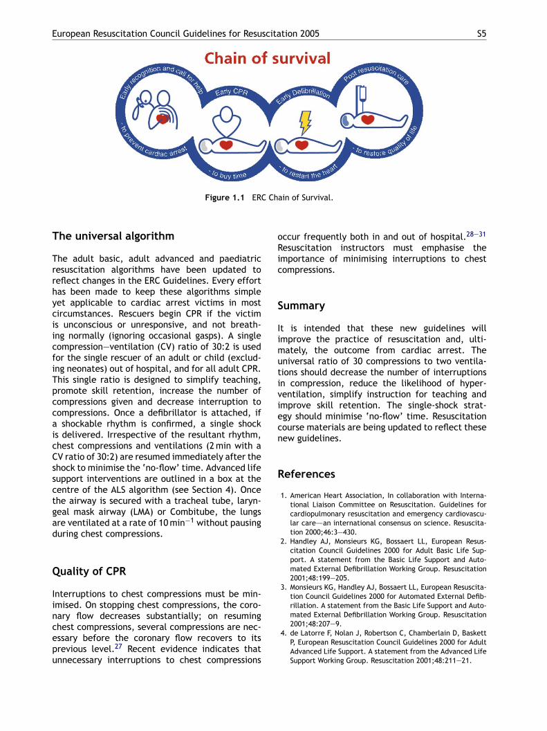
The following concept of the Chain of Survivalsummarises the vital steps needed for success-ful resuscitation (Figure 1.1). Most of these linksare relevant for victims of both VF and asphyxialarrest.11
1. Early recognition of the emergency and call-ing for help: activate the emergency medicalservices (EMS) or local emergency response sys-tem, e.g. ‘‘phone 112’’.12,13 An early, effectiveresponse may prevent cardiac arrest.
t the time of collapse but, by the time the firstCG is recorded, their rhythm has deteriorated tosystole.7,8 VF is characterized by chaotic, rapidepolarisation and repolarisation. The heart loses
2. Early bystander CPR: immediate CPR can doubleor triple survival from VF SCA.10,14—17
3. Early defibrillation: CPR plus defibrillationwithin 3—5 min of collapse can produce survival
Counc
ts coordinated function and stops pumping blood
300-9572/$ — see front matter © 2005 European Resuscitationoi:10.1016/j.resuscitation.2005.10.007
rates as high as 49—75%.18—25 Each minute of
il. All Rights Reserved. Published by Elsevier Ireland Ltd.
S8 A.J. Handley et al.
delay in defibrillation reduces the probability ofsurvival to discharge by 10—15%.14,17
4. Early advanced life support and post-resuscitation care: the quality of treatmentduring the post-resuscitation phase affectsoutcome.26
In most communities, the time from EMS call toEMS arrival (response interval) is 8 min or longer.27
During this time the victim’s survival is dependenton early initiation by bystanders of the first threeof the links of the Chain of Survival.
Victims of cardiac arrest need immediate CPR.This provides a small but critical blood flow to theheart and brain. It also increases the likelihoodthat a defibrillatory shock will terminate VF andenable the heart to resume an effective rhythm andeffective systemic perfusion. Chest compression isespecially important if a shock cannot be deliveredsooner than 4 or 5 min after collapse.28,29 Defibril-lation interrupts the uncoordinated depolarisation-repolarisation process that occurs during VF. Ifthe heart is still viable, its normal pacemakersthen resume their function and produce an effec-tive rhythm and resumption of circulation. In the
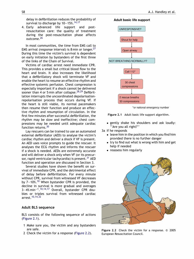
Figure 2.1 Adult basic life support algorithm.
• gently shake his shoulders and ask loudly:‘‘Are you all right?’’
3a If he responds• leave him in the position in which you find him
provided there is no further danger• try to find out what is wrong with him and get
help if needed• reassess him regularly
FE
first few minutes after successful defibrillation, therhythm may be slow and ineffective; chest com-pressions may be needed until adequate cardiacfunction returns.30
Lay rescuers can be trained to use an automatedexternal defibrillator (AED) to analyse the victim’scardiac rhythm and deliver a shock if VF is present.An AED uses voice prompts to guide the rescuer. Itanalyses the ECG rhythm and informs the rescuerif a shock is needed. AEDs are extremely accurateand will deliver a shock only when VF (or its precur-sor, rapid ventricular tachycardia) is present.31 AEDfunction and operation are discussed in Section 3.
Several studies have shown the benefit on sur-vival of immediate CPR, and the detrimental effectof delay before defibrillation. For every minutewithout CPR, survival from witnessed VF decreasesby 7—10%.10 When bystander CPR is provided, thedecline in survival is more gradual and averages3—4% min−1.10,14,17 Overall, bystander CPR dou-bles or triples survival from witnessed cardiacarrest.10,14,32
Adult BLS sequence
BLS consists of the following sequence of actions(Figure 2.1).
1 Make sure you, the victim and any bystandersare safe.
2 Check the victim for a response (Figure 2.2).
igure 2.2 Check the victim for a response. © 2005uropean Resuscitation Council.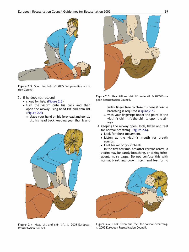
European Resuscitation Council Guidelines for Resuscitation 2005 S9
Figure 2.3 Shout for help. © 2005 European Resuscita-tion Council.
3b If he does not respond• shout for help (Figure 2.3)• turn the victim onto his back and then
open the airway using head tilt and chin lift(Figure 2.4)◦ place your hand on his forehead and gently
tilt his head back keeping your thumb and
FR
Figure 2.5 Head tilt and chin lift in detail. © 2005 Euro-pean Resuscitation Council.
index finger free to close his nose if rescuebreathing is required (Figure 2.5)
◦ with your fingertips under the point of thevictim’s chin, lift the chin to open the air-way
4 Keeping the airway open, look, listen and feelfor normal breathing (Figure 2.6).• Look for chest movement.• Listen at the victim’s mouth for breath
sounds.• Feel for air on your cheek.
In the first few minutes after cardiac arrest, avictim may be barely breathing, or taking infre-quent, noisy gasps. Do not confuse this withnormal breathing. Look, listen, and feel for no
igure 2.4 Head tilt and chin lift. © 2005 Europeanesuscitation Council.
F©
igure 2.6 Look listen and feel for normal breathing.2005 European Resuscitation Council.
S10 A.J. Handley et al.
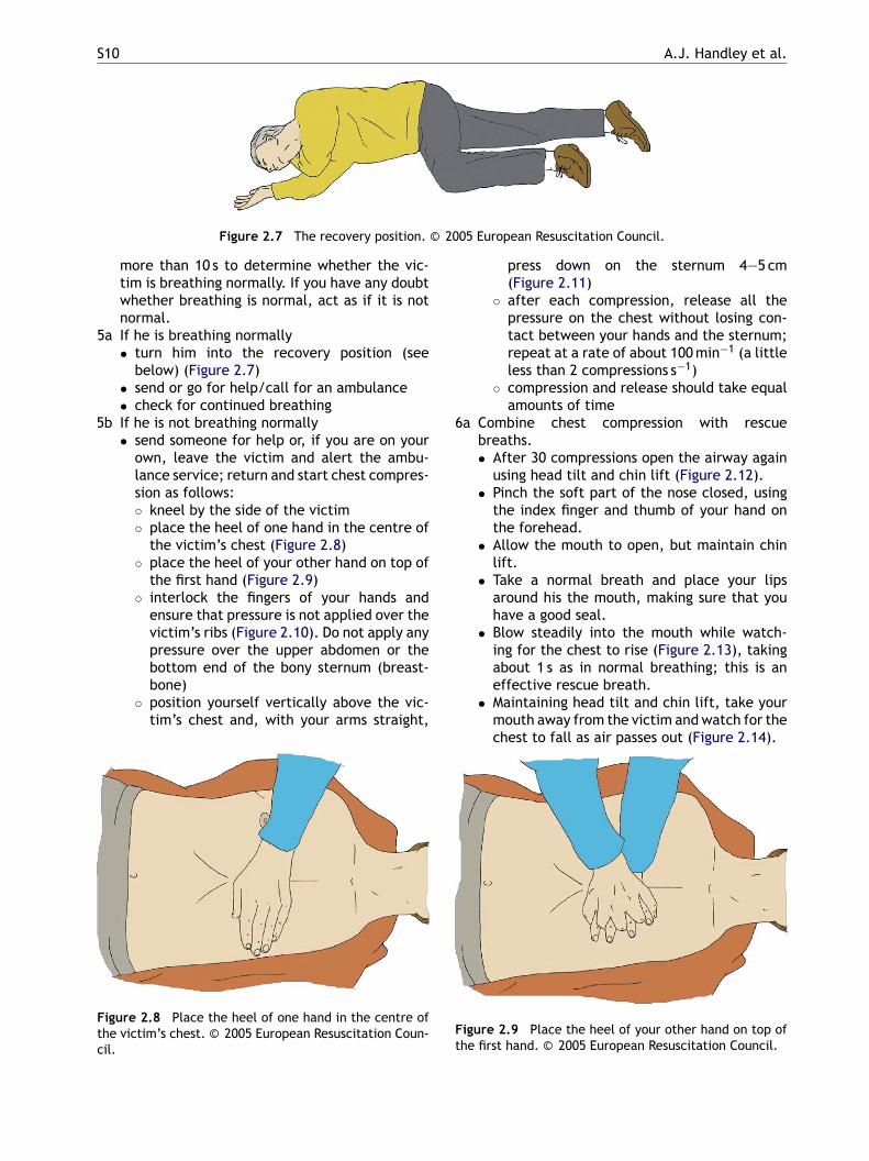
Figure 2.7 The recovery position. © 2005 European Resuscitation Council.
more than 10 s to determine whether the vic-tim is breathing normally. If you have any doubtwhether breathing is normal, act as if it is notnormal.
5a If he is breathing normally• turn him into the recovery position (see
below) (Figure 2.7)• send or go for help/call for an ambulance• check for continued breathing
5b If he is not breathing normally• send someone for help or, if you are on your
own, leave the victim and alert the ambu-lance service; return and start chest compres-sion as follows:◦ kneel by the side of the victim◦ place the heel of one hand in the centre of
the victim’s chest (Figure 2.8)◦ place the heel of your other hand on top of
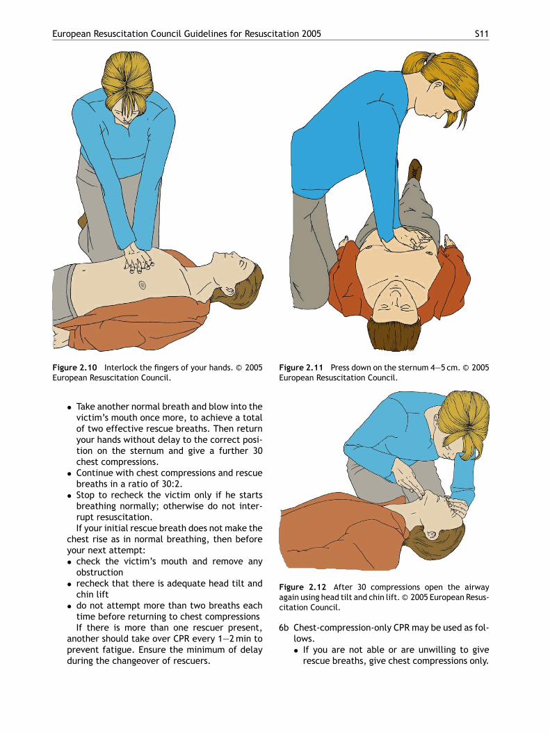
the first hand (Figure 2.9)◦ interlock the fingers of your hands and
ensure that pressure is not applied over thevictim’s ribs (Figure 2.10). Do not apply anypressure over the upper abdomen or thebottom end of the bony sternum (breast-bone)
press down on the sternum 4—5 cm(Figure 2.11)
◦ after each compression, release all thepressure on the chest without losing con-tact between your hands and the sternum;repeat at a rate of about 100 min−1 (a littleless than 2 compressions s−1)
◦ compression and release should take equalamounts of time
6a Combine chest compression with rescuebreaths.• After 30 compressions open the airway again
using head tilt and chin lift (Figure 2.12).• Pinch the soft part of the nose closed, using
the index finger and thumb of your hand onthe forehead.
• Allow the mouth to open, but maintain chinlift.
• Take a normal breath and place your lipsaround his the mouth, making sure that youhave a good seal.
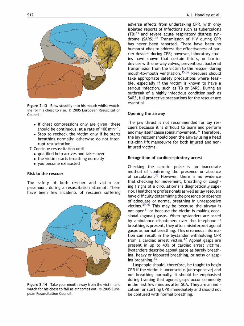
• Blow steadily into the mouth while watch-ing for the chest to rise (Figure 2.13), takingabout 1 s as in normal breathing; this is aneffective rescue breath.
◦ position yourself vertically above the vic-tim’s chest and, with your arms straight,
Figure 2.8 Place the heel of one hand in the centre ofthe victim’s chest. © 2005 European Resuscitation Coun-cil.
• Maintaining head tilt and chin lift, take yourmouth away from the victim and watch for thechest to fall as air passes out (Figure 2.14).
Figure 2.9 Place the heel of your other hand on top ofthe first hand. © 2005 European Resuscitation Council.
European Resuscitation Council Guidelines for Resuscitation 2005 S11
Figure 2.10 Interlock the fingers of your hands. © 2005European Resuscitation Council.
• Take another normal breath and blow into thevictim’s mouth once more, to achieve a totalof two effective rescue breaths. Then returnyour hands without delay to the correct posi-tion on the sternum and give a further 30chest compressions.
• Continue with chest compressions and rescuebreaths in a ratio of 30:2.
• Stop to recheck the victim only if he startsbreathing normally; otherwise do not inter-rupt resuscitation.If your initial rescue breath does not make the
chest rise as in normal breathing, then beforeyour next attempt:• check the victim’s mouth and remove any
obstruction• recheck that there is adequate head tilt and
chin lift• do not attempt more than two breaths each
time before returning to chest compressionsIf there is more than one rescuer present,
another should take over CPR every 1—2 min toprevent fatigue. Ensure the minimum of delayduring the changeover of rescuers.
Figure 2.11 Press down on the sternum 4—5 cm. © 2005European Resuscitation Council.
Figure 2.12 After 30 compressions open the airwayagain using head tilt and chin lift. © 2005 European Resus-citation Council.
6b Chest-compression-only CPR may be used as fol-lows.• If you are not able or are unwilling to give
rescue breaths, give chest compressions only.
S12 A.J. Handley et al.
Figure 2.13 Blow steadily into his mouth whilst watch-ing for his chest to rise. © 2005 European ResuscitationCouncil.
• If chest compressions only are given, theseshould be continuous, at a rate of 100 min−1.
• Stop to recheck the victim only if he startsbreathing normally; otherwise do not inter-rupt resuscitation.
7 Continue resuscitation until• qualified help arrives and takes over• the victim starts breathing normally• you become exhausted
Risk to the rescuer
The safety of both rescuer and victim areparamount during a resuscitation attempt. Therehave been few incidents of rescuers suffering
adverse effects from undertaking CPR, with onlyisolated reports of infections such as tuberculosis(TB)33 and severe acute respiratory distress syn-drome (SARS).34 Transmission of HIV during CPRhas never been reported. There have been nohuman studies to address the effectiveness of bar-rier devices during CPR; however, laboratory stud-ies have shown that certain filters, or barrierdevices with one-way valves, prevent oral bacterialtransmission from the victim to the rescuer duringmouth-to-mouth ventilation.35,36 Rescuers shouldtake appropriate safety precautions where feasi-ble, especially if the victim is known to have aserious infection, such as TB or SARS. During anoutbreak of a highly infectious condition such asSARS, full protective precautions for the rescuer areessential.
Opening the airway
The jaw thrust is not recommended for lay res-cuers because it is difficult to learn and performand may itself cause spinal movement.37 Therefore,the lay rescuer should open the airway using a headtilt-chin lift manoeuvre for both injured and non-
Figure 2.14 Take your mouth away from the victim andwatch for his chest to fall as air comes out. © 2005 Euro-pean Resuscitation Council.
injured victims.
Recognition of cardiorespiratory arrest
Checking the carotid pulse is an inaccuratemethod of confirming the presence or absenceof circulation.38 However, there is no evidencethat checking for movement, breathing or cough-ing (‘signs of a circulation’) is diagnostically supe-rior. Healthcare professionals as well as lay rescuershave difficulty determining the presence or absenceof adequate or normal breathing in unresponsivevictims.39,40 This may be because the airway isnot open41 or because the victim is making occa-sional (agonal) gasps. When bystanders are askedby ambulance dispatchers over the telephone ifbreathing is present, they often misinterpret agonalgasps as normal breathing. This erroneous informa-tion can result in the bystander withholding CPRfrom a cardiac arrest victim.42 Agonal gasps arepresent in up to 40% of cardiac arrest victims.Bystanders describe agonal gasps as barely breath-ing, heavy or laboured breathing, or noisy or gasp-ing breathing.43
Laypeople should, therefore, be taught to beginCPR if the victim is unconscious (unresponsive) andnot breathing normally. It should be emphasisedduring training that agonal gasps occur commonlyin the first few minutes after SCA. They are an indi-cation for starting CPR immediately and should notbe confused with normal breathing.
European Resuscitation Council Guidelines for Resuscitation 2005 S13
Initial rescue breaths
During the first few min after non-asphyxial cardiacarrest the blood oxygen content remains high, andmyocardial and cerebral oxygen delivery is limitedmore by the diminished cardiac output than a lackof oxygen in the lungs. Ventilation is, therefore,initially less important than chest compression.44
It is well recognised that skill acquisition andretention is aided by simplification of the BLSsequence of actions.45 It is also recognized thatrescuers are frequently unwilling to carry outmouth-to-mouth ventilation for a variety of rea-sons, including fear of infection and distaste for theprocedure.46—48 For these reasons, and to empha-sise the priority of chest compressions, it is recom-mended that in adults CPR should start with chestcompression rather than initial ventilation.
Ventilation
During CPR the purpose of ventilation is to maintainadequate oxygenation. The optimal tidal volume,respiratory rate and inspired oxygen concentrationto achieve this, however, are not fully known. Theci
1
2
3
4
5
rw
rise, but to avoid rapid or forceful breaths Thisrecommendation applies to all forms of ventilationduring CPR, including mouth-to-mouth and bag-valve-mask (BVM) with and without supplementaryoxygen.
Mouth-to-nose ventilation is an effective alter-native to mouth-to-mouth ventilation.57 It may beconsidered if the victim’s mouth is seriously injuredor cannot be opened, the rescuer is assisting a vic-tim in the water, or a mouth-to-mouth seal is diffi-cult to achieve.
There is no published evidence on thesafety, effectiveness or feasibility of mouth-to-tracheostomy ventilation, but it may be usedfor a victim with a tracheostomy tube or trachealstoma who requires rescue breathing.
To use bag-mask ventilation requires consider-able practice and skill.58,59 The lone rescuer hasto be able to open the airway with a jaw thrustwhile simultaneously holding the mask to the vic-tim’s face. It is a technique that is appropriateonly for lay rescuers who work in highly specialisedareas, such as where there is a risk of cyanide poi-soning or exposure to other toxic agents. Thereare other specific circumstances in which non-
urrent recommendations are based on the follow-ng evidence:
. During CPR, blood flow to the lungs is sub-stantially reduced, so an adequate ventilation-perfusion ratio can be maintained with lowertidal volumes and respiratory rates thannormal.49
. Not only is hyperventilation (too many breathsor too large a volume) unnecessary, but it isharmful because it increases intrathoracic pres-sure, thus decreasing venous return to the heartand diminishing cardiac output. Survival is con-sequently reduced.50
. When the airway is unprotected, a tidal volumeof 1 l produces significantly more gastric disten-tion than a tidal volume of 500 ml.51
. Low minute-ventilation (lower than normal tidalvolume and respiratory rate) can maintaineffective oxygenation and ventilation duringCPR.52—55 During adult CPR, tidal volumes ofapproximately 500—600 ml (6—7 ml kg−1) shouldbe adequate.
. Interruptions in chest compression (for exam-ple to give rescue breaths) have a detrimentaleffect on survival.56 Giving rescue breaths overa shorter time will help to reduce the durationof essential interruptions.
The current recommendation is, therefore, forescuers to give each rescue breath over about 1 s,ith enough volume to make the victim’s chest
healthcare providers receive extended training infirst aid which could include training, and retrain-ing, in the use of bag-mask ventilation. The samestrict training that applies to healthcare profession-als should be followed.
Chest compression
Chest compressions produce blood flow by increas-ing the intrathoracic pressure and by directly com-pressing the heart. Although chest compressionsperformed properly can produce systolic arterialpressure peaks of 60—80 mmHg, diastolic pressureremains low and mean arterial pressure in thecarotid artery seldom exceeds 40 mmHg.60 Chestcompressions generate a small but critical amountof blood flow to the brain and myocardium andincrease the likelihood that defibrillation will besuccessful. They are especially important if the firstshock is delivered more than 5 min after collapse.61
Much of the information about the physiology ofchest compression and the effects of varying thecompression rate, compression-to-ventilation ratioand duty cycle (ratio of time chest is compressedto total time from one compression to the next) isderived from animal models. However, the conclu-sions of the 2005 Consensus Conference62 includedthe following:
(1) Each time compressions are resumed, the res-cuer should place his hands without delay ‘‘inthe centre of the chest’’.63
S14 A.J. Handley et al.
(2) Compress the chest at a rate of about100 min−1.64—66
(3) Pay attention to achieving the full compressiondepth of 4—5 cm (for an adult).67,68
(4) Allow the chest to recoil completely after eachcompression.69,70
(5) Take approximately the same amount of timefor compression and relaxation.
(6) Minimise interruptions in chest compression.(7) Do not rely on a palpable carotid or femoral
pulse as a gauge of effective arterial flow.38,71
There is insufficient evidence to support a spe-cific hand position for chest compression during CPRin adults. Previous guidelines have recommended amethod of finding the middle of the lower half ofthe sternum by placing one finger on the lower endof the sternum and sliding the other hand down toit.72 It has been shown that for healthcare profes-sionals the same hand position can be found morequickly if rescuers are taught to ‘‘place the heelof your hand in the centre of the chest with theother hand on top’’, provided the teaching includesa demonstration of placing the hands in the middleof the lower half of the sternum.63 It is reasonableto extend this to laypeople.
mouth ventilation in unknown victims of cardiacarrest.46,48 Animal studies have shown that chestcompression-only CPR may be as effective as com-bined ventilation and compression in the first fewminutes after non-asphyxial arrest.44,79 In adults,the outcome of chest compression without venti-lation is significantly better than the outcome ofgiving no CPR.80 If the airway is open, occasionalgasps and passive chest recoil may provide some airexchange.81,82 A low minute-ventilation may be allthat is necessary to maintain a normal ventilation-perfusion ratio during CPR.
Laypeople should, therefore, be encouraged toperform compression-only CPR if they are unableor unwilling to provide rescue breaths, althoughcombined chest compression and ventilation is thebetter method of CPR.
CPR in confined spaces
Over-the-head CPR for single rescuers and straddleCPR for two rescuers may be considered for resus-citation in confined spaces.83,84
Compression rate refers to the speed at whichcompressions are given, not the total number deliv-ered in each minute. The number delivered isdetermined by the rate, but also by the numberof interruptions to open the airway, deliver res-cue breaths and allow AED analysis. In one out-of-hospital study rescuers recorded compressionrates of 100—120 min−1 but, the mean number ofcompressions was reduced to 64 min−1 by frequentinterruptions.68
Compression—ventilation ratio
Insufficient evidence from human outcome studiesexists to support any given compression:ventilationratio. Animal data support an increase in the ratioabove 15:2.73—75 A mathematical model suggeststhat a ratio of 30:2 would provide the best compro-mise between blood flow and oxygen delivery.76,77
A ratio of 30 compressions to two ventilations isrecommended for the single rescuer attemptingresuscitation on an adult or child out of hospi-tal. This should decrease the number of inter-ruptions in compression, reduce the likelihoodof hyperventilation,50,78 simplify instruction forteaching and improve skill retention.
Compression-only CPR
Healthcare professionals as well as lay rescuersadmit to being reluctant to perform mouth-to-
Recovery position
There are several variations of the recovery posi-tion, each with its own advantages. No single posi-tion is perfect for all victims.85,86 The positionshould be stable, near a true lateral position withthe head dependent, and with no pressure on thechest to impair breathing.87
The ERC recommends the following sequenceof actions to place a victim in the recoveryposition:
• Remove the victim’s spectacles.• Kneel beside the victim and make sure that both
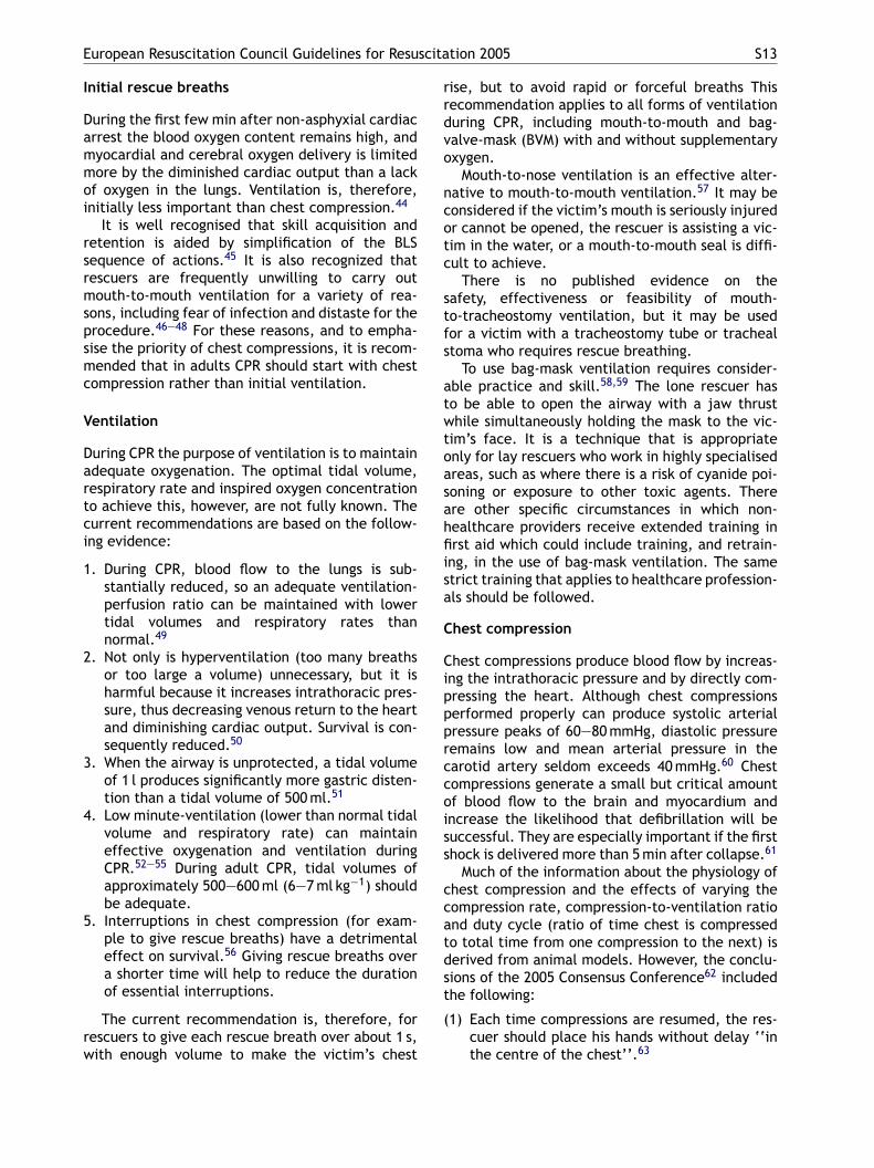
legs are straight.• Place the arm nearest to you out at right angles to
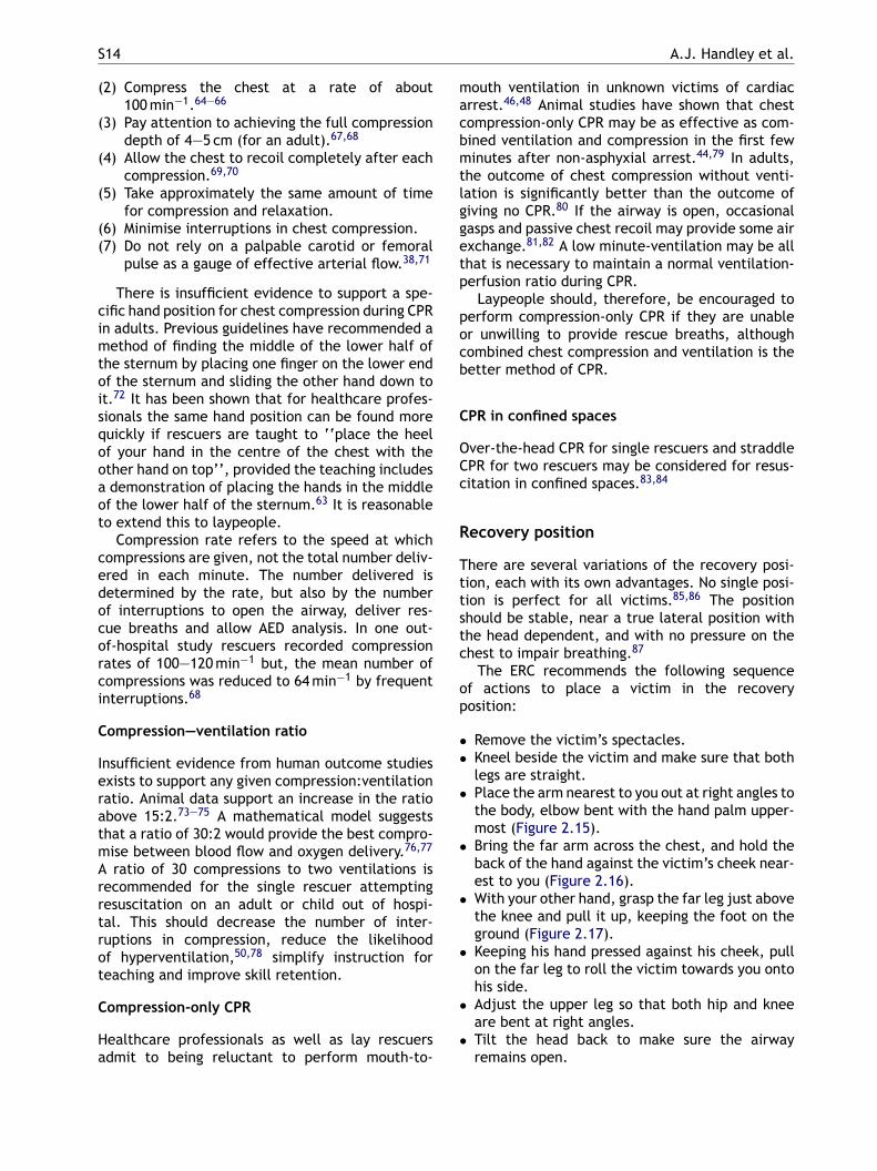
the body, elbow bent with the hand palm upper-most (Figure 2.15).
• Bring the far arm across the chest, and hold theback of the hand against the victim’s cheek near-est to you (Figure 2.16).
• With your other hand, grasp the far leg just abovethe knee and pull it up, keeping the foot on theground (Figure 2.17).
• Keeping his hand pressed against his cheek, pullon the far leg to roll the victim towards you ontohis side.
• Adjust the upper leg so that both hip and kneeare bent at right angles.
• Tilt the head back to make sure the airwayremains open.
European Resuscitation Council Guidelines for Resuscitation 2005 S15
Figure 2.15 Place the arm nearest to you out at rightangles to his body, elbow bent with the hand palm upper-most. © 2005 European Resuscitation Council.
Figure 2.16 Bring the far arm across the chest, and holdthe back of the hand against the victim’s cheek nearestto you. © 2005 European Resuscitation Council.
Figure 2.17 With your other hand, grasp the far leg justabove the knee and pull it up, keeping the foot on theground. © 2005 European Resuscitation Council.
Figure 2.18 The recovery position. © 2005 EuropeanResuscitation Council.
• Adjust the hand under the cheek, if necessary, tokeep the head tilted (Figure 2.18).
• Check breathing regularly.
If the victim has to be kept in the recovery posi-tion for more than 30 min turn him to the oppositeside to relieve the pressure on the lower arm.
Foreign-body airway obstruction (choking)
Foreign-body airway obstruction (FBAO) is anuncommon but potentially treatable cause of acci-dental death.88 Each year approximately 16,000adults and children in the UK receive treatment inan emergency department for FBAO. Fortunately,less than 1% of these incidents are fatal.89 Thecommonest cause of choking in adults is airwayobstruction caused by food such as fish, meat orpoultry.89 In infants and children, half the reportedepisodes of choking occur while eating (mostly con-fectionery), and the remaining choking episodesoccur with non-food items such as coins or toys.90
Deaths from choking are rare in infants and chil-dren; 24 deaths a year on average were reportedin the UK between 1986 and 1995, and over half oft
hese children were under 1 year.90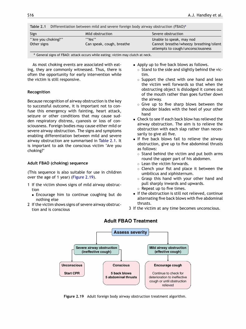
S16 A.J. Handley et al.
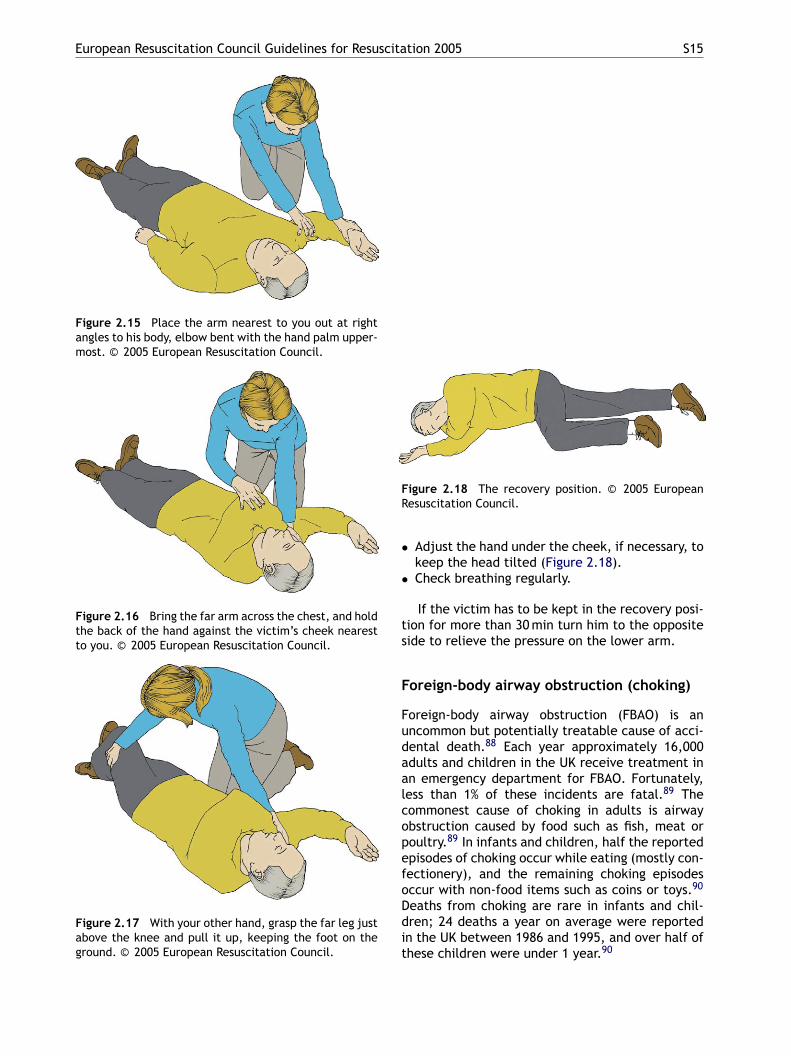
Table 2.1 Differentiation between mild and severe foreign body airway obstruction (FBAO)a
Sign Mild obstruction Severe obstruction
‘‘Are you choking?’’ ‘‘Yes’’ Unable to speak, may nodOther signs Can speak, cough, breathe Cannot breathe/wheezy breathing/silent
attempts to cough/unconsciousnessa General signs of FBAO: attack occurs while eating; victim may clutch at neck.
As most choking events are associated with eat-ing, they are commonly witnessed. Thus, there isoften the opportunity for early intervention whilethe victim is still responsive.
Recognition
Because recognition of airway obstruction is the keyto successful outcome, it is important not to con-fuse this emergency with fainting, heart attack,seizure or other conditions that may cause sud-den respiratory distress, cyanosis or loss of con-sciousness. Foreign bodies may cause either mild orsevere airway obstruction. The signs and symptomsenabling differentiation between mild and severeairway obstruction are summarised in Table 2.1. Itis important to ask the conscious victim ‘Are youchoking?’
Adult FBAO (choking) sequence
(This sequence is also suitable for use in childrenover the age of 1 year) (Figure 2.19).
1 If the victim shows signs of mild airway obstruc-
• Apply up to five back blows as follows.◦ Stand to the side and slightly behind the vic-
tim.◦ Support the chest with one hand and lean
the victim well forwards so that when theobstructing object is dislodged it comes outof the mouth rather than goes further downthe airway.
◦ Give up to five sharp blows between theshoulder blades with the heel of your otherhand
• Check to see if each back blow has relieved theairway obstruction. The aim is to relieve theobstruction with each slap rather than neces-sarily to give all five.
• If five back blows fail to relieve the airwayobstruction, give up to five abdominal thrustsas follows:◦ Stand behind the victim and put both arms
round the upper part of his abdomen.◦ Lean the victim forwards.◦ Clench your fist and place it between the
umbilicus and xiphisternum.◦ Grasp this hand with your other hand and
pull sharply inwards and upwards.
3
rway
tion• Encourage him to continue coughing but do
nothing else2 If the victim shows signs of severe airway obstruc-
tion and is conscious
Figure 2.19 Adult foreign body ai
◦ Repeat up to five times.• If the obstruction is still not relieved, continue
alternating five back blows with five abdominalthrusts.
If the victim at any time becomes unconscious.
obstruction treatment algorithm.
European Resuscitation Council Guidelines for Resuscitation 2005 S17
• Support the victim carefully to the ground.• Immediately activate EMS.• Begin CPR (from 5b of the adult BLS sequence).
Healthcare providers, trained and experiencedin feeling for a carotid pulse, should initiatechest compressions, even if a pulse is presentin the unconscious choking victim.
FBAO causing mild airway obstruction
Coughing generates high and sustained airway pres-sures and may expel the foreign body. Aggressivetreatment, with back blows, abdominal thrusts andchest compression, may cause potentially seriouscomplications and could worsen the airway obstruc-tion. It should be reserved for victims who havesigns of severe airway obstruction. Victims withmild airway obstruction should remain under con-tinuous observation until they improve, as severeairway obstruction may develop.
FBAO with severe airway obstruction
The clinical data on choking are largely retrospec-
documented harm to the victim96,99 or rescuer.91
Therefore, avoid use of a blind finger sweep andmanually remove solid material in the airway onlyif it can be seen.
Aftercare and referral for medical review
Following successful treatment for FBAO, foreignmaterial may nevertheless remain in the upper orlower respiratory tract and cause complicationslater. Victims with a persistent cough, difficultyswallowing or the sensation of an object being stillstuck in the throat, should therefore be referred fora medical opinion.
Abdominal thrusts can cause serious internalinjuries, and all victims treated with abdomi-nal thrusts should be examined for injury by adoctor.91
Resuscitation of children (see alsoSection 6) and victims of drowning (seealso Section 7c)
tive and anecdotal. For conscious adults and chil-dren over 1 year with a complete FBAO, case reportsdemonstrate the effectiveness of back blows or‘slaps’, abdominal thrusts and chest thrusts.91
Approximately 50% of episodes of airway obstruc-tion are not relieved by a single technique.92 Thelikelihood of success is increased when combina-tions of back blows or slaps, and abdominal andchest thrusts are used.91
A randomised trial in cadavers93 and twoprospective studies in anaesthetised volunteers94,95
showed that higher airway pressures can be gener-ated using chest thrusts compared with abdominalthrusts. Since chest thrusts are virtually identicalto chest compressions, rescuers should be taughtto start CPR if a victim of known or suspectedFBAO becomes unconscious. During CPR, each timethe airway is opened the victim’s mouth should bequickly checked for any foreign body that has beenpartly expelled. The incidence of unsuspectedchoking as a cause of unconsciousness or cardiacarrest is low; therefore, during CPR routinelychecking the mouth for foreign bodies is notnecessary.
The finger sweep
No studies have evaluated the routine use of a fingersweep to clear the airway in the absence of visibleairway obstruction,96—98 and four case reports have
Both ventilation and compression are importantfor victims of cardiac arrest when the oxygenstores become depleted—–about 4—6 min after col-lapse from VF and immediately after collapsefrom asphyxial arrest. Previous guidelines tried totake into account the difference in pathophysiol-ogy, and recommended that victims of identifiableasphyxia (drowning; trauma; intoxication) and chil-dren should receive 1 min of CPR before the lonerescuer left the victim to get help. The majorityof cases of SCA out of hospital, however, occur inadults, and are of cardiac origin due to VF. Theseadditional recommendations, therefore, added tothe complexity of the guidelines while affectingonly a minority of victims.
It is important to be aware that many childrendo not receive resuscitation because potential res-cuers fear causing harm. This fear is unfounded;it is far better to use the adult BLS sequence forresuscitation of a child than to do nothing. Forease of teaching and retention, therefore, laypeo-ple should be taught that the adult sequence mayalso be used for children who are not responsiveand not breathing.
The following minor modifications to the adultsequence will, however, make it even more suitablefor use in children.
• Give five initial rescue breaths before startingchest compressions (adult sequence of actions,5b).
S18 A.J. Handley et al.
• A lone rescuer should perform CPR for approxi-mately 1 min before going for help.
• Compress the chest by approximately one thirdof its depth; use two fingers for an infant under1 year; use one or two hands for a child over 1year as needed to achieve an adequate depth ofcompression.
The same modifications of five initial breaths, and1 min of CPR by the lone rescuer before gettinghelp, may improve outcome for victims of drown-ing. This modification should be taught only tothose who have a specific duty of care to poten-tial drowning victims (e.g. lifeguards). Drowning iseasily identified. It can be difficult, on the otherhand, for a layperson to determine whether car-diorespiratory arrest is a direct result of traumaor intoxication. These victims should, therefore, bemanaged according to the standard protocol.
Use of an automated externaldefibrillator
Section 3 discusses the guidelines for defibrillation
• push shock button as directed (fully auto-matic AEDs will deliver the shock automat-ically)
• continue as directed by the voice/visualprompts
5b If no shock indicated• immediately resume CPR, using a ratio of 30
compressions to 2 rescue breaths• continue as directed by the voice/visual
prompts6 Continue to follow the AED prompts until
• qualified help arrives and takes over• the victim starts to breathe normally• you become exhausted
CPR before defibrillation
Immediate defibrillation, as soon as an AEDbecomes available, has always been a key ele-ment in guidelines and teaching, and considered ofparamount importance for survival from ventricu-lar fibrillation. This concept has been challengedbecause evidence suggests that a period of chestcompression before defibrillation may improve sur-vlsop
ptvgbctrtpltaee
V
I‘utss
using both automated external defibrillators (AEDs)and manual defibrillators. However, there are somespecial considerations when an AED is to be used bylay or non-healthcare rescuers.
Standard AEDs are suitable for use in childrenolder than 8 years. For children between 1 and 8years use paediatric pads or a paediatric mode ifavailable; if these are not available, use the AED asit is. Use of AEDs is not recommended for childrenless than 1 year.
Sequence for use of an AED
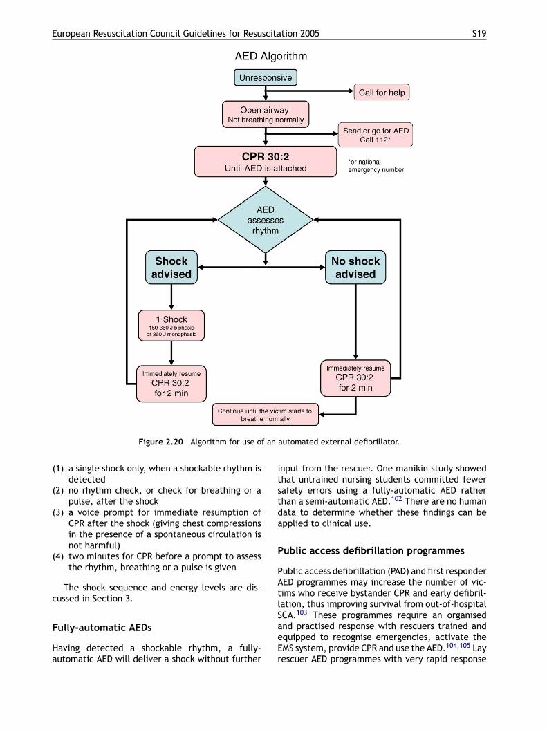
See Figure 2.20.
(1) Make sure you, the victim, and any bystandersare safe.
(2) If the victim is unresponsive and not breathingnormally, send someone for the AED and to callfor an ambulance.
(3) Start CPR according to the guidelines for BLS.(4) As soon as the defibrillator arrives
• switch on the defibrillator and attach theelectrode pads. If more than one rescuer ispresent, CPR should be continued while thisis carried out
• follow the spoken/visual directions• ensure that nobody touches the victim while
the AED is analysing the rhythm5a If a shock is indicated
• ensure that nobody touches the victim
ival when the time between calling for the ambu-ance and its arrival exceeds 5 min.28,61,100 Onetudy101 did not confirm this benefit, but the weightf evidence supports a period of CPR for victims ofrolonged cardiac arrest before defibrillation.
In all of these studies CPR was performed byaramedics, who protected the airway by intuba-ion and delivered 100% oxygen. Such high-qualityentilation cannot be expected from lay rescuersiving mouth-to-mouth ventilation. Secondly, theenefit from CPR occurred only when the delay fromall to the availability of a defibrillator was greaterhan 5 min; the delay from collapse to arrival of theescuer with an AED will rarely be known with cer-ainty. Thirdly, if good bystander CPR is already inrogress when the AED arrives, it does not seemogical to continue it any further. For these reasonshese guidelines recommend an immediate shock,s soon as the AED is available. The importance ofarly uninterrupted external chest compression ismphasised.
oice prompts
n several places, the sequence of actions statesfollow the voice/visual prompts’. The prompts aresually programmable, and it is recommended thathey be set in accordance with the sequence ofhocks and timings for CPR given in Section 2. Thesehould include at least:
European Resuscitation Council Guidelines for Resuscitation 2005 S19
Figure 2.20 Algorithm for use of an automated external defibrillator.
(1) a single shock only, when a shockable rhythm isdetected
(2) no rhythm check, or check for breathing or apulse, after the shock
(3) a voice prompt for immediate resumption ofCPR after the shock (giving chest compressionsin the presence of a spontaneous circulation isnot harmful)
(4) two minutes for CPR before a prompt to assessthe rhythm, breathing or a pulse is given
The shock sequence and energy levels are dis-cussed in Section 3.
Fully-automatic AEDs
Having detected a shockable rhythm, a fully-automatic AED will deliver a shock without further
input from the rescuer. One manikin study showedthat untrained nursing students committed fewersafety errors using a fully-automatic AED ratherthan a semi-automatic AED.102 There are no humandata to determine whether these findings can beapplied to clinical use.
Public access defibrillation programmes
Public access defibrillation (PAD) and first responderAED programmes may increase the number of vic-tims who receive bystander CPR and early defibril-lation, thus improving survival from out-of-hospitalSCA.103 These programmes require an organisedand practised response with rescuers trained andequipped to recognise emergencies, activate theEMS system, provide CPR and use the AED.104,105 Layrescuer AED programmes with very rapid response
S20 A.J. Handley et al.
times in airports,22 on aircraft23or in casinos,25 anduncontrolled studies using police officers as firstresponders,106,107 have achieved reported survivalrates as high as 49—74%.
The logistic problem for first responder pro-grammes is that the rescuer needs to arrive notjust earlier than the traditional EMS, but within5—6 min of the initial call, to enable attempteddefibrillation in the electrical or circulatory phaseof cardiac arrest.108 With longer delays, the survivalcurve flattens;10,17 a few minutes’ gain in time willhave little impact when the first responder arrivesmore than 10 min after the call27,109 or when a firstresponder does not improve on an already shortEMS response time.110 However, small reductionsin response intervals achieved by first-responderprogrammes that have an impact on many residen-tial victims may be more cost effective than thelarger reductions in response interval achieved byPAD programmes that have an impact on fewer car-diac arrest victims.111,112
Recommended elements for PAD programmesinclude:
• a planned and practised response
tality and Morbidity Statistics in Europe. Eur Heart J1997;18:1231—48.
3. Cobb LA, Fahrenbruch CE, Olsufka M, Copass MK. Chang-ing incidence of out-of-hospital ventricular fibrillation,1980—2000. JAMA 2002;288:3008—13.
4. Rea TD, Eisenberg MS, Sinibaldi G, White RD. Incidence ofEMS-treated out-of-hospital cardiac arrest in the UnitedStates. Resuscitation 2004;63:17—24.
5. Vaillancourt C, Stiell IG. Cardiac arrest care andemergency medical services in Canada. Can J Cardiol2004;20:1081—90.
6. Waalewijn RA, de Vos R, Koster RW. Out-of-hospital car-diac arrests in Amsterdam and its surrounding areas:results from the Amsterdam resuscitation study (ARREST)in ‘Utstein’ style. Resuscitation 1998;38:157—67.
7. Cummins R, Thies W. Automated external defibrillators andthe Advanced Cardiac Life Support Program: a new initia-tive from the American Heart Association. Am J Emerg Med1991;9:91—3.
8. Waalewijn RA, Nijpels MA, Tijssen JG, Koster RW. Preven-tion of deterioration of ventricular fibrillation by basic lifesupport during out-of-hospital cardiac arrest. Resuscitation2002;54:31—6.
9. Page S, Meerabeau L. Achieving change through reflec-tive practice: closing the loop. Nurs Educ Today2000;20:365—72.
10. Larsen MP, Eisenberg MS, Cummins RO, Hallstrom AP.Predicting survival from out-of-hospital cardiac arrest: agraphic model. Ann Emerg Med 1993;22:1652—8.
11. Cummins RO, Ornato JP, Thies WH, Pepe PE. Improving
• training of anticipated rescuers in CPR and use ofthe AED• link with the local EMS system• programme of continuous audit (quality improve-
ment)
Public access defibrillation programmes are mostlikely to improve survival from cardiac arrestif they are established in locations where wit-nessed cardiac arrest is likely to occur.113 Suit-able sites might include those where the proba-bility of cardiac arrest occurring is at least oncein every 2 years (e.g., airports, casinos, sportsfacilities).103 Approximately 80% of out-of-hospitalcardiac arrests occur in private or residentialsettings;114 this fact inevitably limits the overallimpact that PAD programmes can have on survivalrates. There are no studies documenting effective-ness of home AED deployment.
References
1. Recommended guidelines for uniform reporting of datafrom out-of-hospital cardiac arrest: the ‘Utstein style’.Prepared by a Task Force of Representatives from theEuropean Resuscitation Council, American Heart Asso-ciation. Heart and Stroke Foundation of Canada, Aus-tralian Resuscitation Council. Resuscitation 1991;22:1—26.
2. Sans S, Kesteloot H, Kromhout D. The burden of cardio-vascular diseases mortality in Europe. Task Force of theEuropean Society of Cardiology on Cardiovascular Mor-
survival from sudden cardiac arrest: the ‘‘chain of sur-vival’’ concept. A statement for health professionals fromthe Advanced Cardiac Life Support Subcommittee and theEmergency Cardiac Care Committee, American Heart Asso-ciation. Circulation 1991;83:1832—47.
12. Calle PA, Lagaert L, Vanhaute O, Buylaert WA. Do victimsof an out-of-hospital cardiac arrest benefit from a trainingprogram for emergency medical dispatchers? Resuscitation1997;35:213—8.
13. Curka PA, Pepe PE, Ginger VF, Sherrard RC, Ivy MV,Zachariah BS. Emergency medical services priority dis-patch. Ann Emerg Med 1993;22:1688—95.
14. Valenzuela TD, Roe DJ, Cretin S, Spaite DW, LarsenMP. Estimating effectiveness of cardiac arrest interven-tions: a logistic regression survival model. Circulation1997;96:3308—13.
15. Holmberg M, Holmberg S, Herlitz J. Factors modifying theeffect of bystander cardiopulmonary resuscitation on sur-vival in out-of-hospital cardiac arrest patients in Sweden.Eur Heart J 2001;22:511—9.
16. Holmberg M, Holmberg S, Herlitz J, Gardelov B. Survivalafter cardiac arrest outside hospital in Sweden. SwedishCardiac Arrest Registry. Resuscitation 1998;36:29—36.
17. Waalewijn RA, De Vos R, Tijssen JGP, Koster RW. Survivalmodels for out-of-hospital cardiopulmonary resuscitationfrom the perspectives of the bystander, the first responder,and the paramedic. Resuscitation 2001;51:113—22.
18. Weaver WD, Hill D, Fahrenbruch CE, et al. Use of the auto-matic external defibrillator in the management of out-of-hospital cardiac arrest. N Engl J Med 1988;319:661—6.
19. Auble TE, Menegazzi JJ, Paris PM. Effect of out-of-hospital defibrillation by basic life support providers oncardiac arrest mortality: a metaanalysis. Ann Emerg Med1995;25:642—58.
20. Stiell IG, Wells GA, DeMaio VJ, et al. Modifiable factorsassociated with improved cardiac arrest survival in a mul-ticenter basic life support/defibrillation system: OPALS
European Resuscitation Council Guidelines for Resuscitation 2005 S21
Study Phase I results. Ontario Prehospital Advanced LifeSupport. Ann Emerg Med 1999;33:44—50.
21. Stiell IG, Wells GA, Field BJ, et al. Improved out-of-hospitalcardiac arrest survival through the inexpensive optimiza-tion of an existing defibrillation program: OPALS studyphase II. Ontario Prehospital Advanced Life Support. JAMA1999;281:1175—81.
22. Caffrey S. Feasibility of public access to defibrillation. CurrOpin Crit Care 2002;8:195—8.
23. O’Rourke MF, Donaldson E, Geddes JS. An airline cardiacarrest program. Circulation 1997;96:2849—53.
24. Page RL, Hamdan MH, McKenas DK. Defibrillation aboarda commercial aircraft. Circulation 1998;97:1429—30.
25. Valenzuela TD, Roe DJ, Nichol G, Clark LL, Spaite DW,Hardman RG. Outcomes of rapid defibrillation by secu-rity officers after cardiac arrest in casinos. N Engl J Med2000;343:1206—9.
26. Langhelle A, Nolan JP, Herlitz J, et al. Recommended guide-lines for reviewing, reporting, and conducting researchon post-resuscitation care: the Utstein style. Resuscitation2005;66:271—83.
27. van Alem AP, Vrenken RH, de Vos R, Tijssen JG, Koster RW.Use of automated external defibrillator by first respondersin out of hospital cardiac arrest: prospective controlledtrial. BMJ 2003;327:1312—7.
28. Cobb LA, Fahrenbruch CE, Walsh TR, et al. Influenceof cardiopulmonary resuscitation prior to defibrillation inpatients with out-of-hospital ventricular fibrillation. JAMA1999;281:1182—8.
38. Bahr J, Klingler H, Panzer W, Rode H, Kettler D. Skillsof lay people in checking the carotid pulse. Resuscitation1997;35:23—6.
39. Ruppert M, Reith MW, Widmann JH, et al. Checkingfor breathing: evaluation of the diagnostic capability ofemergency medical services personnel, physicians, med-ical students, and medical laypersons. Ann Emerg Med1999;34:720—9.
40. Perkins GD, Stephenson B, Hulme J, Monsieurs KG. Birming-ham assessment of breathing study (BABS). Resuscitation2005;64:109—13.
41. Domeier RM, Evans RW, Swor RA, Rivera-Rivera EJ, Fred-eriksen SM. Prospective validation of out-of-hospital spinalclearance criteria: a preliminary report. Acad Emerg Med1997;4:643—6.
42. Hauff SR, Rea TD, Culley LL, Kerry F, Becker L,Eisenberg MS. Factors impeding dispatcher-assisted tele-phone cardiopulmonary resuscitation. Ann Emerg Med2003;42:731—7.
43. Clark JJ, Larsen MP, Culley LL, Graves JR, Eisenberg MS.Incidence of agonal respirations in sudden cardiac arrest.Ann Emerg Med 1992;21:1464—7.
44. Kern KB, Hilwig RW, Berg RA, Sanders AB, Ewy GA.Importance of continuous chest compressions duringcardiopulmonary resuscitation: improved outcome dur-ing a simulated single lay-rescuer scenario. Circulation2002;105:645—9.
45. Handley JA, Handley AJ. Four-step CPR—–improving skillretention. Resuscitation 1998;36:3—8.
46. Ornato JP, Hallagan LF, McMahan SB, Peeples EH, Rostafin-
29. Wik L, Myklebust H, Auestad BH, Steen PA. Retention ofbasic life support skills 6 months after training with anautomated voice advisory manikin system without instruc-tor involvement. Resuscitation 2002;52:273—9.
30. White RD, Russell JK. Refibrillation, resuscitation andsurvival in out-of-hospital sudden cardiac arrest victimstreated with biphasic automated external defibrillators.Resuscitation 2002;55:17—23.
31. Kerber RE, Becker LB, Bourland JD, et al. Automatic exter-nal defibrillators for public access defibrillation: recom-mendations for specifying and reporting arrhythmia analy-sis algorithm performance, incorporating new waveforms,and enhancing safety. A statement for health professionalsfrom the American Heart Association Task Force on Auto-matic External Defibrillation, Subcommittee on AED Safetyand Efficacy. Circulation 1997;95:1677—82.
32. Holmberg M, Holmberg S, Herlitz J. Effect of bystandercardiopulmonary resuscitation in out-of-hospital cardiacarrest patients in Sweden. Resuscitation 2000;47:59—70.
33. Heilman KM, Muschenheim C. Primary cutaneous tubercu-losis resulting from mouth-to-mouth respiration. N Engl JMed 1965;273:1035—6.
34. Christian MD, Loutfy M, McDonald LC, et al. Possible SARScoronavirus transmission during cardiopulmonary resusci-tation. Emerg Infect Dis 2004;10:287—93.
35. Cydulka RK, Connor PJ, Myers TF, Pavza G, Parker M.Prevention of oral bacterial flora transmission by usingmouth-to-mask ventilation during CPR. J Emerg Med1991;9:317—21.
36. Blenkharn JI, Buckingham SE, Zideman DA. Prevention oftransmission of infection during mouth-to-mouth resusci-tation. Resuscitation 1990;19:151—7.
37. Aprahamian C, Thompson BM, Finger WA, Darin JC. Experi-mental cervical spine injury model: evaluation of airwaymanagement and splinting techniques. Ann Emerg Med1984;13:584—7.
ski AG. Attitudes of BCLS instructors about mouth-to-mouthresuscitation during the AIDS epidemic. Ann Emerg Med1990;19:151—6.
47. Brenner BE, Van DC, Cheng D, Lazar EJ. Determinants ofreluctance to perform CPR among residents and applicants:the impact of experience on helping behavior. Resuscita-tion 1997;35:203—11.
48. Hew P, Brenner B, Kaufman J. Reluctance of paramedicsand emergency medical technicians to perform mouth-to-mouth resuscitation. J Emerg Med 1997;15:279—84.
49. Baskett P, Nolan J, Parr M. Tidal volumes which are per-ceived to be adequate for resuscitation. Resuscitation1996;31:231—4.
50. Aufderheide TP, Sigurdsson G, Pirrallo RG, et al.Hyperventilation-induced hypotension during cardiopul-monary resuscitation. Circulation 2004;109:1960—5.
51. Wenzel V, Idris AH, Banner MJ, Kubilis PS, Williams JLJ.Influence of tidal volume on the distribution of gasbetween the lungs and stomach in the nonintubatedpatient receiving positive-pressure ventilation. Crit CareMed 1998;26:364—8.
52. Idris A, Gabrielli A, Caruso L. Smaller tidal volume is safeand effective for bag-valve-ventilation, but not for mouth-to-mouth ventilation: an animal model for basic life sup-port. Circulation 1999;100(Suppl. I):I-644.
53. Idris A, Wenzel V, Banner MJ, Melker RJ. Smaller tidal vol-umes minimize gastric inflation during CPR with an unpro-tected airway. Circulation 1995;92(Suppl.):I-759.
54. Dorph E, Wik L, Steen PA. Arterial blood gases with 700ml tidal volumes during out-of-hospital CPR. Resuscitation2004;61:23—7.
55. Winkler M, Mauritz W, Hackl W, et al. Effects of half thetidal volume during cardiopulmonary resuscitation on acid-base balance and haemodynamics in pigs. Eur J Emerg Med1998;5:201—6.
56. Eftestol T, Sunde K, Steen PA. Effects of interruptingprecordial compressions on the calculated probability of
S22 A.J. Handley et al.
defibrillation success during out-of-hospital cardiac arrest.Circulation 2002;105:2270—3.
57. Ruben H. The immediate treatment of respiratory failure.Br J Anaesth 1964;36:542—9.
58. Elam JO. Bag-valve-mask O2 ventilation. In: Safar P, ElamJO, editors. Advances in cardiopulmonary resuscitation:the Wolf Creek Conference on Cardiopulmonary Resusci-tation. New York, NY: Springer-Verlag, Inc.; 1977. p. 73—9.
59. Dailey RH. The airway: emergency management. St. Louis,MO: Mosby Year Book; 1992.
60. Paradis NA, Martin GB, Goetting MG, et al. Simultaneousaortic, jugular bulb, and right atrial pressures during car-diopulmonary resuscitation in humans. Insights into mech-anisms. Circulation 1989;80:361—8.
61. Wik L, Hansen TB, Fylling F, et al. Delaying defibrillation togive basic cardiopulmonary resuscitation to patients without-of-hospital ventricular fibrillation: a randomized trial.JAMA 2003;289:1389—95.
62. International Liaison Committee on Resuscitation. Inter-national consensus on cardiopulmonary resuscitation andemergency cardiovascular care science with treatment rec-ommendations. Resuscitation 2005:67.
63. Handley AJ. Teaching hand placement for chestcompression—–a simpler technique. Resuscitation2002;53:29—36.
64. Yu T, Weil MH, Tang W, et al. Adverse outcomes of inter-rupted precordial compression during automated defibril-lation. Circulation 2002;106:368—72.
65. Swenson RD, Weaver WD, Niskanen RA, Martin J, DahlbergS. Hemodynamics in humans during conventional and
74. Dorph E, Wik L, Stromme TA, Eriksen M, Steen PA. Qualityof CPR with three different ventilation:compression ratios.Resuscitation 2003;58:193—201.
75. Dorph E, Wik L, Stromme TA, Eriksen M, Steen PA. Oxygendelivery and return of spontaneous circulation with ven-tilation:compression ratio 2:30 versus chest compressionsonly CPR in pigs. Resuscitation 2004;60:309—18.
76. Babbs CF, Kern KB. Optimum compression to ventila-tion ratios in CPR under realistic, practical conditions:a physiological and mathematical analysis. Resuscitation2002;54:147—57.
77. Fenici P, Idris AH, Lurie KG, Ursella S, Gabrielli A. What isthe optimal chest compression—ventilation ratio? Curr OpinCrit Care 2005;11:204—11.
78. Aufderheide TP, Lurie KG. Death by hyperventilation: acommon and life-threatening problem during cardiopul-monary resuscitation. Crit Care Med 2004;32:S345—51.
79. Chandra NC, Gruben KG, Tsitlik JE, et al. Observations ofventilation during resuscitation in a canine model. Circula-tion 1994;90:3070—5.
80. Becker LB, Berg RA, Pepe PE, et al. A reappraisal of mouth-to-mouth ventilation during bystander-initiated cardiopul-monary resuscitation. A statement for healthcare profes-sionals from the Ventilation Working Group of the Basic LifeSupport and Pediatric Life Support Subcommittees, Amer-ican Heart Association. Resuscitation 1997;35:189—201.
81. Berg RA, Kern KB, Hilwig RW, et al. Assisted ventilationdoes not improve outcome in a porcine model of single-rescuer bystander cardiopulmonary resuscitation. Circula-tion 1997;95:1635—41.
experimental methods of cardiopulmonary resuscitation.Circulation 1988;78:630—9.
66. Kern KB, Sanders AB, Raife J, Milander MM, Otto CW,Ewy GA. A study of chest compression rates during car-diopulmonary resuscitation in humans: the importanceof rate-directed chest compressions. Arch Intern Med1992;152:145—9.
67. Abella BS, Alvarado JP, Myklebust H, et al. Quality ofcardiopulmonary resuscitation during in-hospital cardiacarrest. JAMA 2005;293:305—10.
68. Wik L, Kramer-Johansen J, Myklebust H, et al. Quality ofcardiopulmonary resuscitation during out-of-hospital car-diac arrest. JAMA 2005;293:299—304.
69. Aufderheide TP, Pirrallo RG, Yannopoulos D, et al. Incom-plete chest wall decompression: a clinical evaluationof CPR performance by EMS personnel and assessmentof alternative manual chest compression—decompressiontechniques. Resuscitation 2005;64:353—62.
70. Yannopoulos D, McKnite S, Aufderheide TP, et al. Effectsof incomplete chest wall decompression during cardiopul-monary resuscitation on coronary and cerebral perfusionpressures in a porcine model of cardiac arrest. Resuscita-tion 2005;64:363—72.
71. Ochoa FJ, Ramalle-Gomara E, Carpintero JM, Garcia A, Sar-alegui I. Competence of health professionals to check thecarotid pulse. Resuscitation 1998;37:173—5.
72. Handley AJ, Monsieurs KG, Bossaert LL. European Resusci-tation Council Guidelines 2000 for Adult Basic Life Support.A statement from the Basic Life Support and AutomatedExternal Defibrillation Working Group(1) and approved bythe Executive Committee of the European ResuscitationCouncil. Resuscitation 2001;48:199—205.
73. Sanders AB, Kern KB, Berg RA, Hilwig RW, Heiden-rich J, Ewy GA. Survival and neurologic outcomeafter cardiopulmonary resuscitation with four differentchest compression-ventilation ratios. Ann Emerg Med2002;40:553—62.
82. Berg RA, Kern KB, Hilwig RW, Ewy GA. Assisted ventila-tion during ‘bystander’ CPR in a swine acute myocardialinfarction model does not improve outcome. Circulation1997;96:4364—71.
83. Handley AJ, Handley JA. Performing chest compressions ina confined space. Resuscitation 2004;61:55—61.
84. Perkins GD, Stephenson BT, Smith CM, Gao F. A compari-son between over-the-head and standard cardiopulmonaryresuscitation. Resuscitation 2004;61:155—61.
85. Turner S, Turner I, Chapman D, et al. A comparative studyof the 1992 and 1997 recovery positions for use in the UK.Resuscitation 1998;39:153—60.
86. Handley AJ. Recovery position. Resuscitation1993;26:93—5.
87. Anonymous. Guidelines 2000 for cardiopulmonary resus-citation and emergency cardiovascular care—–an interna-tional consensus on science. Resuscitation 2000;46:1—447.
88. Fingerhut LA, Cox CS, Warner M. International compara-tive analysis of injury mortality. Findings from the ICE oninjury statistics. International collaborative effort on injurystatistics. Adv Data 1998;12:1—20.
89. Industry DoTa. Choking. In: Home and leisure accidentreport. London: Department of Trade and Industry; 1998,p. 13—4.
90. Industry DoTa. Choking risks to children. London: Depart-ment of Trade and Industry; 1999.
91. International Liaison Committee on Resuscitation. Part2. Adult basic life support. 2005 international consensuson cardiopulmonary resuscitation and emergency cardio-vascular care science with treatment recommendations.Resuscitation 2005;67:187—200.
92. Redding JS. The choking controversy: critique of evidenceon the Heimlich maneuver. Crit Care Med 1979;7:475—9.
93. Langhelle A, Sunde K, Wik L, Steen PA. Airway pressure withchest compressions versus Heimlich manoeuvre in recentlydead adults with complete airway obstruction. Resuscita-tion 2000;44:105—8.
European Resuscitation Council Guidelines for Resuscitation 2005 S23
94. Guildner CW, Williams D, Subitch T. Airway obstructedby foreign material: the Heimlich maneuver. JACEP1976;5:675—7.
95. Ruben H, Macnaughton FI. The treatment of food-choking.Practitioner 1978;221:725—9.
96. Hartrey R, Bingham RM. Pharyngeal trauma as a result ofblind finger sweeps in the choking child. J Accid Emerg Med1995;12:52—4.
97. Elam JO, Ruben AM, Greene DG. Resuscitation of drowningvictims. JAMA 1960;174:13—6.
98. Ruben HM, Elam JO, Ruben AM, Greene DG. Investigation ofupper airway problems in resuscitation. 1. Studies of pha-ryngeal X-rays and performance by laymen. Anesthesiology1961;22:271—9.
99. Kabbani M, Goodwin SR. Traumatic epiglottis followingblind finger sweep to remove a pharyngeal foreign body.Clin Pediatr (Phila) 1995;34:495—7.
100. Eftestol T, Wik L, Sunde K, Steen PA. Effects of cardiopul-monary resuscitation on predictors of ventricular fibrilla-tion defibrillation success during out-of-hospital cardiacarrest. Circulation 2004;110:10—5.
101. Jacobs IG, Finn JC, Oxer HF, Jelinek GA. CPR before defibril-lation in out-of-hospital cardiac arrest: a randomized trial.Emerg Med Australas 2005;17:39—45.
102. Monsieurs KG, Vogels C, Bossaert LL, Meert P, Calle PA.A study comparing the usability of fully automatic ver-sus semi-automatic defibrillation by untrained nursing stu-dents. Resuscitation 2005;64:41—7.
103. The Public Access Defibrillation Trial Investigators.Public-access defibrillation and survival after out-of-
1
105. Priori SG, Bossaert LL, Chamberlain DA, et al. Policy state-ment: ESC-ERC recommendations for the use of auto-mated external defibrillators (AEDs) in Europe. Resuscita-tion 2004;60:245—52.
106. White RD, Bunch TJ, Hankins DG. Evolution of a community-wide early defibrillation programme experience over 13years using police/fire personnel and paramedics as respon-ders. Resuscitation 2005;65:279—83.
107. Mosesso Jr VN, Davis EA, Auble TE, Paris PM, Yealy DM. Useof automated external defibrillators by police officers fortreatment of out-of-hospital cardiac arrest. Ann Emerg Med1998;32:200—7.
108. Weisfeldt M, Becker L. Resuscitation after cardiac arrest.A 3-phase time-sensitive model. JAMA 2002;288:3035—8.
109. Groh WJ, Newman MM, Beal PE, Fineberg NS, Zipes DP. Lim-ited response to cardiac arrest by police equipped withautomated external defibrillators: lack of survival bene-fit in suburban and rural Indiana—–the police as responderautomated defibrillation evaluation (PARADE). Acad EmergMed 2001;8:324—30.
110. Sayre M, Evans J, White L, Brennan T. Providing automatedexternal defibrillators to urban police officers in additionto fire department rapid defibrillation program is not effec-tive. Resuscitation 2005;66:189—96.
111. Nichol G, Hallstrom AP, Ornato JP, et al. Potential cost-effectiveness of public access defibrillation in the UnitedStates. Circulation 1998;97:1315—20.
112. Nichol G, Valenzuela T, Roe D, Clark L, Huszti E, Wells GA.Cost effectiveness of defibrillation by targeted respondersin public settings. Circulation 2003;108:697—703.
1
1
hospital cardiac arrest. N Engl J Med 2004;351:637—46.
04. Priori SBL, Chamberlain D, Napolitano C, Arntz HR, KosterR, Monsieurs K, Capucci A, Wellens H. Policy Statement:ESC-ERC recommendations for the use of AEDs in Europe.Eur Heart J 2004;25:437—45.
13. Becker L, Eisenberg M, Fahrenbruch C, Cobb L. Public loca-tions of cardiac arrest: implications for public access defib-rillation. Circulation 1998;97:2106—9.
14. Becker DE. Assessment and management of cardiovascularurgencies and emergencies: cognitive and technical con-siderations. Anesth Progress 1988;35:212—7.
Resuscitation (2005) 67S1, S3—S6
European Resuscitation Council Guidelines forResuscitation 2005Section 1. Introduction
Jerry Nolan
It is five years since publication of the Guide-lines 2000 for Cardiopulmonary Resuscitation (CPR)aEoaiarhmicefr
othatc
sus on treatment recommendations. The processfor the latest resuscitation guideline update began
nd Emergency Cardiovascular Care (ECC).1 Theuropean Resuscitation Council (ERC) based itswn resuscitation guidelines on this document,nd these were published as a series of papersn 2001.2—7 Resuscitation science continues todvance, and clinical guidelines must be updatedegularly to reflect these developments and adviseealthcare providers on best practice. In betweenajor guideline updates (about every five years),
nterim advisory statements can inform the health-are provider about new therapies that might influ-nce outcome significantly;8 we anticipate thaturther advisory statements will be published inesponse to important research findings.
The guidelines that follow do not define thenly way that resuscitation should be achieved;hey merely represent a widely accepted view ofow resuscitation can be undertaken both safelynd effectively. The publication of new and revisedreatment recommendations does not imply thaturrent clinical care is either unsafe or ineffective.
in 2003, when ILCOR representatives establishedsix task forces: basic life support; advanced car-diac life support; acute coronary syndromes; pae-diatric life support; neonatal life support; and aninterdisciplinary task force to address overlappingtopics, such as educational issues. Each task forceidentified topics requiring evidence evaluation, andappointed international experts to review them.To ensure a consistent and thorough approach, aworksheet template was created containing step-by-step directions to help the experts documenttheir literature review, evaluate studies, determinelevels of evidence and develop recommendations.10
A total of 281 experts completed 403 worksheets on276 topics; 380 people from 18 countries attendedthe 2005 International Consensus Conference onECC and CPR Science with Treatment Recommen-dations (C2005), which took place in Dallas inJanuary 2005.11 Worksheet authors presented theresults of their evidence evaluations and pro-posed summary scientific statements. After discus-sion among all participants, these statements were
refined and, whenever possible, supported by treat-msboCon
Consensus on science
The International Liaison Committee on Resuscita-tion (ILCOR) was formed in 1993.9 Its mission isto identify and review international science andknowledge relevant to CPR, and to offer consen-
0300-9572/$ — see front matter © 2005 European Resuscitation Coudoi:10.1016/j.resuscitation.2005.10.002
ent recommendations. These summary sciencetatements and treatment recommendations haveeen published in the 2005 International Consensusn Cardiopulmonary Resuscitation and Emergencyardiovascular Care Science with Treatment Rec-mmendations (CoSTR).12
cil. All Rights Reserved. Published by Elsevier Ireland Ltd.

S4 Jerry Nolan
From science to guidelines
The resuscitation organisations forming ILCOR willpublish individual resuscitation guidelines that areconsistent with the science in the consensus docu-ment, but will also consider geographic, economicand system differences in practice, and the avail-ability of medical devices and drugs. These 2005ERC Resuscitation Guidelines are derived from theCoSTR document but represent consensus amongmembers of the ERC Executive Committee. TheERC Executive Committee considers these new rec-ommendations to be the most effective and eas-ily learned interventions that can be supportedby current knowledge, research and experience.Inevitably, even within Europe, differences in theavailability of drugs, equipment, and personnel willnecessitate local, regional and national adaptationof these guidelines.
Demographics
Ischaemic heart disease is the leading cause ofdeath in the world.13—17 Sudden cardiac arrest is
Table 1.1 Out-of-hospital cardiopulmonary arrests(21,175) by aetiology.19
Aetiology Number (%)
Presumed cardiac disease 17451 (82.4)
Non-cardiac internal aetiologies 1814 (8.6)Lung disease 901 (4.3)Cerebrovascular disease 457 (2.2)Cancer 190 (0.9)Gastrointestinal haemorrhage 71 (0.3)Obstetric/paediatric 50 (0.2)Pulmonary embolism 38 (0.2)Epilepsy 36 (0.2)Diabetes mellitus 30 (0.1)Renal disease 23 (0.1)
Non-cardiac external aetiologies 1910 (9.0)Trauma 657 (3.1)Asphyxia 465 (2.2)Drug overdose 411 (1.9)Drowning 105 (0.5)Other suicide 194 (0.9)Other external 50 (0.2)Electric shock/lightning 28 (0.1)
includes prevention of conditions leading to thecardiopulmonary arrest, early CPR, early activa-tion of the emergency services and early advancedlife support. In hospital, the importance of earlyrecognition of the critically ill patient and activa-tion of a medical emergency team (MET) is now wellaccepted.23 Previous resuscitation guidelines haveprovided relatively little information on treatmentof the patient during the post-resuscitation carephase. There is substantial variability in the waycomatose survivors of cardiac arrest are treatedin the initial hours and first few days after returnof spontaneous circulation (ROSC). Differences intreatment at this stage may account for some ofthe interhospital variability in outcome after car-diac arrest.24 The importance of recognising crit-ical illness and/or angina and preventing cardiacarrest (in- or out-of-hospital), and post resuscita-tion care has been highlighted by the inclusion ofthese elements in a new four-ring Chain of Sur-vival. The first link indicates the importance ofrecognising those at risk of cardiac arrest and call-ing for help in the hope that early treatment canprevent arrest. The central links in this new chaindepict the integration of CPR and defibrillation astiti(
responsible for more than 60% of adult deathsfrom coronary heart disease.18 Based on data fromScotland and from five cities in other parts ofEurope, the annual incidence of resuscitation forout-of-hospital cardiopulmonary arrest of cardiacaetiology is 49.5—66 per 100,000 population.19,20
The Scottish study includes data on 21,175 out-of-hospital cardiac arrests, and provides valuableinformation on aetiology (Table 1.1). The incidenceof in-hospital cardiac arrest is difficult to assessbecause it is influenced heavily by factors such asthe criteria for hospital admission and implementa-tion of a do-not-attempt-resuscitation (DNAR) pol-icy. In a general hospital in the UK, the incidenceof primary cardiac arrest (excluding those withDNAR and those arresting in the emergency depart-ment) was 3.3/1000 admissions;21 using the sameexclusion criteria, the incidence of cardiac arrestin a Norwegian University hospital was 1.5/1000admissions.22
The Chain of Survival
The actions linking the victim of sudden cardiacarrest with survival are called the Chain of Sur-vival. They include early recognition of the emer-gency and activation of the emergency services,early CPR, early defibrillation and early advancedlife support. The infant-and-child Chain of Survival
he fundamental components of early resuscitationn an attempt to restore life. The final link, effec-ive post resuscitation care, is targeted at preserv-ng function, particularly of the brain and heartFigure 1.1).25,26
European Resuscitation Council Guidelines for Resuscitation 2005 S5
Figure 1.1 ERC Chain of Survival.
The universal algorithm
The adult basic, adult advanced and paediatricresuscitation algorithms have been updated toreflect changes in the ERC Guidelines. Every efforthas been made to keep these algorithms simpleyet applicable to cardiac arrest victims in mostcircumstances. Rescuers begin CPR if the victimis unconscious or unresponsive, and not breath-ing normally (ignoring occasional gasps). A singlecompression—ventilation (CV) ratio of 30:2 is usedfor the single rescuer of an adult or child (exclud-ing neonates) out of hospital, and for all adult CPR.This single ratio is designed to simplify teaching,promote skill retention, increase the number ofcompressions given and decrease interruption tocompressions. Once a defibrillator is attached, ifa shockable rhythm is confirmed, a single shockis delivered. Irrespective of the resultant rhythm,chest compressions and ventilations (2 min with aCV ratio of 30:2) are resumed immediately after theshock to minimise the ‘no-flow’ time. Advanced lifesupport interventions are outlined in a box at thecentre of the ALS algorithm (see Section 4). Oncethe airway is secured with a tracheal tube, laryn-
occur frequently both in and out of hospital.28—31
Resuscitation instructors must emphasise theimportance of minimising interruptions to chestcompressions.
Summary
It is intended that these new guidelines willimprove the practice of resuscitation and, ulti-mately, the outcome from cardiac arrest. Theuniversal ratio of 30 compressions to two ventila-tions should decrease the number of interruptionsin compression, reduce the likelihood of hyper-ventilation, simplify instruction for teaching andimprove skill retention. The single-shock strat-egy should minimise ‘no-flow’ time. Resuscitationcourse materials are being updated to reflect thesenew guidelines.
References
1. American Heart Association, In collaboration with Interna-tional Liaison Committee on Resuscitation. Guidelines for
geal mask airway (LMA) or Combitube, the lungsare ventilated at a rate of 10 min−1 without pausingduring chest compressions.
Quality of CPR
Interruptions to chest compressions must be min-imised. On stopping chest compressions, the coro-nary flow decreases substantially; on resumingchest compressions, several compressions are nec-essary before the coronary flow recovers to itsprevious level.27 Recent evidence indicates thatunnecessary interruptions to chest compressions
cardiopulmonary resuscitation and emergency cardiovascu-lar care—–an international consensus on science. Resuscita-tion 2000;46:3—430.
2. Handley AJ, Monsieurs KG, Bossaert LL, European Resus-citation Council Guidelines 2000 for Adult Basic Life Sup-port. A statement from the Basic Life Support and Auto-mated External Defibrillation Working Group. Resuscitation2001;48:199—205.
3. Monsieurs KG, Handley AJ, Bossaert LL, European Resuscita-tion Council Guidelines 2000 for Automated External Defib-rillation. A statement from the Basic Life Support and Auto-mated External Defibrillation Working Group. Resuscitation2001;48:207—9.
4. de Latorre F, Nolan J, Robertson C, Chamberlain D, BaskettP, European Resuscitation Council Guidelines 2000 for AdultAdvanced Life Support. A statement from the Advanced LifeSupport Working Group. Resuscitation 2001;48:211—21.
S6 Jerry Nolan
5. Phillips B, Zideman D, Garcia-Castrillo L, Felix M, Shwarz-Schwierin U, European Resuscitation Council Guidelines2000 for Basic Paediatric Life Support. A statement fromthe Paediatric Life Support Working Group. Resuscitation2001;48:223—9.
6. Phillips B, Zideman D, Garcia-Castrillo L, Felix M, Shwarz-Schwierin V, European Resuscitation Council Guidelines2000 for Advanced Paediatric Life Support. A statementfrom Paediatric Life Support Working Group. Resuscitation2001;48:231—4.
7. Phillips B, Zideman D, Wyllie J, Richmond S, van ReemptsP, European Resuscitation Council Guidelines 2000 for NewlyBorn Life Support. A statement from the Paediatric Life Sup-port Working Group. Resuscitation 2001;48:235—9.
8. Nolan JP, Morley PT, Vanden Hoek TL, Hickey RW. Therapeu-tic hypothermia after cardiac arrest. An advisory statementby the Advancement Life support Task Force of the Inter-national Liaison committee on Resuscitation. Resuscitation2003;57:231—5.
9. The Founding Members of the International Liaison Commit-tee on Resuscitation. The International Liaison Committeeon Resuscitation (ILCOR)—–past, present and future. Resus-citation 2005;67:157—61.
10. Morley P, Zaritsky A. The evidence evaluation process for the2005 International Consensus on Cardiopulmonary Resuscita-tion and Emergency Cardiovascular Care Science With Treat-ment Recommendations. Resuscitation 2005;67:167—70.
11. Nolan JP, Hazinski MF, Steen PA, Becker LB. Controversialtopics from the 2005 International Consensus Conference onCardiopulmonary Resuscitation and Emergency Cardiovascu-
1
1
1
1
1
17. Levi F, Lucchini F, Negri E, La Vecchia C. Trends in mor-tality from cardiovascular and cerebrovascular diseases inEurope and other areas of the world. Heart 2002;88:119—24.
18. Zheng ZJ, Croft JB, Giles WH, Mensah GA. Sudden car-diac death in the United States, 1989 to 1998. Circulation2001;104:2158—63.
19. Pell JP, Sirel JM, Marsden AK, Ford I, Walker NL, Cobbe SM.Presentation, management, and outcome of out of hospitalcardiopulmonary arrest: comparison by underlying aetiology.Heart 2003;89:839—42.
20. Herlitz J, Bahr J, Fischer M, Kuisma M, Lexow K, ThorgeirssonG. Resuscitation in Europe: a tale of five European regions.Resuscitation 1999;41:121—31.
21. Hodgetts TJ, Kenward G, Vlackonikolis I, et al. Incidence,location and reasons for avoidable in-hospital cardiac arrestin a district general hospital. Resuscitation 2002;54:115—23.
22. Skogvoll E, Isern E, Sangolt GK, Gisvold SE. In-hospital car-diopulmonary resuscitation. 5 years’ incidence and survivalaccording to the Utstein template. Acta Anaesthesiol Scand1999;43:177—84.
23. The MERIT study investigators. Introduction of the medicalemergency team (MET) system: a cluster-randomised con-trolled trial. Lancet 2005;365:2091—7.
24. Langhelle A, Tyvold SS, Lexow K, Hapnes SA, Sunde K, SteenPA. In-hospital factors associated with improved outcomeafter out-of-hospital cardiac arrest. A comparison betweenfour regions in Norway. Resuscitation 2003;56:247—63.
25. Langhelle A, Nolan J, Herlitz J, et al. Recommended guide-lines for reviewing, reporting, and conducting research on
2
2
2
2
3
3
lar Care Science with treatment recommendations. Resusci-tation 2005;67:175—9.
2. International Liaison Committee on Resuscitation. 2005International Consensus on Cardiopulmonary Resuscitationand Emergency Cardiovascular Care Science with TreatmentRecommendations. Resuscitation 2005;67:157—341.
3. Murray CJ, Lopez AD. Mortality by cause for eight regionsof the world: global burden of disease study. Lancet1997;349:1269—76.
4. Sans S, Kesteloot H, Kromhout D. The burden of cardiovas-cular diseases mortality in Europe. Task Force of the Euro-pean Society of Cardiology on Cardiovascular Mortality andMorbidity Statistics in Europe. Eur Heart J 1997;18:1231—48.
5. Kesteloot H, Sans S, Kromhout D. Evolution of all-causesand cardiovascular mortality in the age-group 75—84 yearsin Europe during the period 1970—1996; a comparison withworldwide changes. Eur Heart J 2002;23:384—98.
6. Fox R. Trends in cardiovascular mortality in Europe. Circula-tion 1997;96:3817.
post-resuscitation care: The Utstein style. Resuscitation2005;66:271—83.
6. Perkins GD, Soar J. In hospital cardiac arrest: missing linksin the chain of survival. Resuscitation 2005;66:253—5.
7. Kern KB, Hilwig RW, Berg RA, Ewy GA. Efficacy of chestcompression-only BLS CPR in the presence of an occludedairway. Resuscitation 1998;39:179—88.
8. Wik L, Kramer-Johansen J, Myklebust H, et al. Quality ofcardiopulmonary resuscitation during out-of-hospital cardiacarrest. JAMA 2005;293:299—304.
9. Abella BS, Alvarado JP, Myklebust H, et al. Quality of car-diopulmonary resuscitation during in-hospital cardiac arrest.JAMA 2005;293:305—10.
0. Abella BS, Sandbo N, Vassilatos P, et al. Chest compressionrates during cardiopulmonary resuscitation are suboptimal:a prospective study during in-hospital cardiac arrest. Circu-lation 2005;111:428—34.
1. Valenzuela TD, Kern KB, Clark LL, et al. Interruptions of chestcompressions during emergency medical systems resuscita-tion. Circulation 2005;112:1259—65.

Resuscitation (2005) 67S1, S25—S37
European Resuscitation Council Guidelines forResuscitation 2005Section 3. Electrical therapies: Automatedexternal defibrillators, defibrillation,cardioversion and pacing
Charles D. Deakin, Jerry P. Nolan
I
TuaapuIad
iteifitaac
iiev
d
ntroduction
his section presents guidelines for defibrillationsing both automated external defibrillators (AEDs)nd manual defibrillators. All healthcare providersnd lay responders can use AEDs as an integral com-onent of basic life support. Manual defibrillation issed as part of advanced life support (ALS) therapy.n addition, synchronised cardioversion and pacingre ALS functions of many defibrillators and are alsoiscussed in this section.
Defibrillation is the passage across the myocard-um of an electrical current of sufficient magnitudeo depolarise a critical mass of myocardium andnable restoration of coordinated electrical activ-ty. Defibrillation is defined as the termination ofbrillation or, more precisely, the absence of ven-ricular fibrillation/ventricular tachycardia (VF/VT)t 5 s after shock delivery; however, the goal ofttempted defibrillation is to restore spontaneousirculation.
Defibrillator technology is advancing rapidly. AED
the rhythm while CPR is in progress is required toprevent unnecessary delays in CPR. Waveform anal-ysis may also enable the defibrillator to calculatethe optimal time at which to give a shock.
A vital link in the chain of survival
Defibrillation is a key link in the Chain of Survivaland is one of the few interventions that have beenshown to improve outcome from VF/VT cardiacarrest. The previous guidelines, published in 2000,rightly emphasised the importance of early defib-rillation with minimum delay.1
The probability of successful defibrillation andsubsequent survival to hospital discharge declinesrapidly with time2,3 and the ability to deliverearly defibrillation is one of the most importantfactors in determining survival from cardiacarrest. For every minute that passes followingcollapse and defibrillation, mortality increases7%—10% in the absence of bystander CPR.2—4 EMS
nteraction with the rescuer through voice promptss now established, and future technology maynable more specific instructions to be given by
systems do not generally have the capability todeliver defibrillation through traditional paramedicresponders within the first few minutes of a call,a
Coun
oice prompt. The ability of defibrillators to assess
0300-9572/$ — see front matter © 2005 European Resuscitationoi:10.1016/j.resuscitation.2005.10.008
nd the alternative use of trained lay responders
cil. All Rights Reserved. Published by Elsevier Ireland Ltd.
S26 C.D. Deakin, J.P. Nolan
to deliver prompt defibrillation using AEDs is nowwidespread. EMS systems that have reduced time todefibrillation following cardiac arrest using trainedlay responders have reported greatly improvedsurvival-to-discharge rates,5—7 some as high as75% if defibrillation is performed within 3 min ofcollapse.8 This concept has also been extended toin-hospital cardiac arrests where staff, other thandoctors, are also being trained to defibrillate usingan AED before arrival of the cardiac arrest team.When bystander CPR is provided, the reduction insurvival rate is more gradual and averages 3%—4%per minute from collapse to defibrillation;2—4
bystander CPR can double2,3,9 or treble10 sur-vival from witnessed out-of-hospital cardiacarrest.
All healthcare providers with a duty to performCPR should be trained, equipped, and encouragedto perform defibrillation and CPR. Early defibrilla-tion should be available throughout all hospitals,outpatient medical facilities and public areas ofmass gathering (see Section 2). Those trained inAED use should also be trained to deliver at leastexternal chest compressions before the arrival ofALS providers, to optimise the effectiveness of early
Automated external defibrillators have beentested extensively against libraries of recordedcardiac rhythms and in many trials in adults18,19
and children.20,21 They are extremely accurate inrhythm analysis. Although AEDs are not designed todeliver synchronised shocks, all AEDs will recom-mend shocks for VT if the rate and R-wave mor-phology exceed preset values.
In-hospital use of AEDs
At the time of the 2005 Consensus Confer-ence, there were no published randomised trialscomparing in-hospital use of AEDs with manualdefibrillators. Two lower level studies of adultswith in-hospital cardiac arrest from shockablerhythms showed higher survival-to-hospital dis-charge rates when defibrillation was providedthrough an AED programme than with manual defib-rillation alone.22,23 A manikin study showed thatuse of an AED significantly increased the likelihoodof delivering three shocks, but increased the timeto deliver the shocks when compared with manualdefibrillators.24 In contrast, a study of mock arrestsin simulated patients showed that use of monitor-
defibrillation.
Automated external defibrillators
Automated external defibrillators are sophisti-cated, reliable computerised devices that use voiceand visual prompts to guide lay rescuers and health-care professionals to safely attempt defibrillationin cardiac arrest victims. Automated defibrillatorshave been described as ‘‘. . . the single greatestadvance in the treatment of VF cardiac arrest sincethe development of CPR.’’11 Advances in technol-ogy, particularly with respect to battery capacity,and software arrhythmia analysis have enabled themass production of relatively cheap, reliable andeasily operated portable defibrillators.12—15 Use ofAEDs by lay or non-healthcare rescuers is coveredin Section 2.
Automated rhythm analysis
Automated external defibrillators have micropro-cessors that analyse several features of the ECG,including frequency and amplitude. Some AEDs areprogrammed to detect spontaneous movement bythe patient or others. Developing technology shouldsoon enable AEDs to provide information aboutfrequency and depth of chest compressions dur-ing CPR that may improve BLS performance by allrescuers.16,17
ing leads and fully automated defibrillators reducedtime to defibrillation when compared with manualdefibrillators.25
Delayed defibrillation may occur when patientssustain cardiac arrest in unmonitored hospital bedsand in outpatient departments. In these areas sev-eral minutes may elapse before resuscitation teamsarrive with a defibrillator and deliver shocks.26
Despite limited evidence, AEDs should be consid-ered for the hospital setting as a way to facilitateearly defibrillation (a goal of <3 min from collapse),especially in areas where staff have no rhythmrecognition skills or where they use defibrillatorsinfrequently. An effective system for training andretraining should be in place. Adequate numbers ofstaff should be trained to enable achievement ofthe goal of providing the first shock within 3 min ofcollapse anywhere in the hospital. Hospitals shouldmonitor collapse-to-first-shock intervals and resus-citation outcomes.
Strategies before defibrillation
Safe use of oxygen during defibrillation
In an oxygen-enriched atmosphere, sparking frompoorly applied defibrillator paddles can cause afire.27—32 There are several reports of fires beingcaused in this way, and most have resulted in
European Resuscitation Council Guidelines for Resuscitation 2005 S27
significant burns to the patient. The risk of fire dur-ing attempted defibrillation can be minimised bytaking the following precautions.
• Take off any oxygen mask or nasal cannulae andplace them at least 1 m away from the patient’schest.
• Leave the ventilation bag connected to the tra-cheal tube or other airway adjunct. Alterna-tively, disconnect any bag-valve device from thetracheal tube (or other airway adjunct such asthe laryngeal mask airway, combitube or laryn-geal tube), and remove it at least 1 m from thepatient’s chest during defibrillation.
• If the patient is connected to a ventilator, forexample in the operating room or critical careunit, leave the ventilator tubing (breathing cir-cuit) connected to the tracheal tube unless chestcompressions prevent the ventilator from deliv-ering adequate tidal volumes. In this case, theventilator is usually substituted for a ventila-tion bag, which can itself be left connected ordetached and removed to a distance of at least1 m. If the ventilator tubing is disconnected,ensure it is kept at least 1 m from the patientor, better still, switch the ventilator off; mod-
Shaving the chest
Patients with a hairy chest have air trappingbeneath the electrode and poor electrode-to-skinelectrical contact. This causes high impedance,reduced defibrillation efficacy, risk of arcing(sparks) from electrode to skin and electrodeto electrode and is more likely to cause burnsto the patient’s chest. Rapid shaving of thearea of intended electrode placement may benecessary, but do not delay defibrillation if ashaver is not immediately available. Shaving thechest per se may reduce transthoracic impedanceslightly and has been recommended for elective DCcardioversion.35
Paddle force
If using paddles, apply them firmly to the chestwall. This reduces transthoracic impedance byimproving electrical contact at the electrode—skininterface and reducing thoracic volume.36 Thedefibrillator operator should always press firmlyon handheld electrode paddles, the optimal forcebeing 8 kg in adults37 and 5 kg in children aged1 38
mbipprmtttf
E
Npcataritf
ma(si
ern ventilators generate massive oxygen flowswhen disconnected. During normal use, whenconnected to a tracheal tube, oxygen from a ven-tilator in the critical care unit will be vented fromthe main ventilator housing well away from thedefibrillation zone. Patients in the critical careunit may be dependent on positive end expiratorypressure (PEEP) to maintain adequate oxygena-tion; during cardioversion, when the spontaneouscirculation potentially enables blood to remainwell oxygenated, it is particularly appropriate toleave the critically ill patient connected to theventilator during shock delivery.
• Minimise the risk of sparks during defibrillation.Theoretically, self-adhesive defibrillation padsare less likely to cause sparks than manual pad-dles.
The technique for electrode contact withthe chest
Optimal defibrillation technique aims to delivercurrent across the fibrillating myocardium in thepresence of minimal transthoracic impedance.Transthoracic impedance varies considerably withbody mass, but is approximately 70—80 � inadults.33,34 The techniques described below aim toplace external electrodes (paddles or self-adhesivepads) in an optimal position using techniques thatminimise transthoracic impedance.
—8 years when using adult paddles ; 8-kg forceay be attainable only by the strongest mem-ers of the cardiac arrest team, and therefore its recommended that these individuals apply theaddles during defibrillation. Unlike self-adhesiveads, manual paddles have a bare metal plate thatequires a conductive material placed between theetal and patient’s skin to improve electrical con-
act. Use of bare metal paddles alone creates highransthoracic impedance and is likely to increasehe risk of arcing and to worsen cutaneous burnsrom defibrillation.
lectrode position
o human studies have evaluated the electrodeosition as a determinant of return of spontaneousirculation (ROSC) or survival from VF/VT cardiacrrest. Transmyocardial current during defibrilla-ion is likely to be maximal when the electrodesre placed so that the area of the heart that is fib-illating lies directly between them, i.e., ventriclesn VF/VT, atria in atrial fibrillation (AF). Therefore,he optimal electrode position may not be the sameor ventricular and atrial arrhythmias.
More patients are presenting with implantableedical devices (e.g., permanent pacemaker,
utomatic implantable cardioverter defibrillatorAICD)). MedicAlert bracelets are recommended foruch patients. These devices may be damaged dur-ng defibrillation if current is discharged through
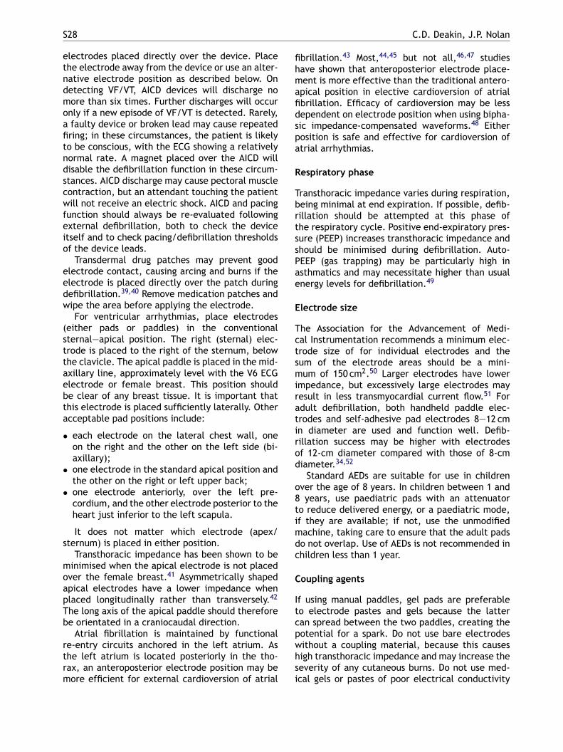
S28 C.D. Deakin, J.P. Nolan
electrodes placed directly over the device. Placethe electrode away from the device or use an alter-native electrode position as described below. Ondetecting VF/VT, AICD devices will discharge nomore than six times. Further discharges will occuronly if a new episode of VF/VT is detected. Rarely,a faulty device or broken lead may cause repeatedfiring; in these circumstances, the patient is likelyto be conscious, with the ECG showing a relativelynormal rate. A magnet placed over the AICD willdisable the defibrillation function in these circum-stances. AICD discharge may cause pectoral musclecontraction, but an attendant touching the patientwill not receive an electric shock. AICD and pacingfunction should always be re-evaluated followingexternal defibrillation, both to check the deviceitself and to check pacing/defibrillation thresholdsof the device leads.
Transdermal drug patches may prevent goodelectrode contact, causing arcing and burns if theelectrode is placed directly over the patch duringdefibrillation.39,40 Remove medication patches andwipe the area before applying the electrode.
For ventricular arrhythmias, place electrodes(either pads or paddles) in the conventional
fibrillation.43 Most,44,45 but not all,46,47 studieshave shown that anteroposterior electrode place-ment is more effective than the traditional antero-apical position in elective cardioversion of atrialfibrillation. Efficacy of cardioversion may be lessdependent on electrode position when using bipha-sic impedance-compensated waveforms.48 Eitherposition is safe and effective for cardioversion ofatrial arrhythmias.
Respiratory phase
Transthoracic impedance varies during respiration,being minimal at end expiration. If possible, defib-rillation should be attempted at this phase ofthe respiratory cycle. Positive end-expiratory pres-sure (PEEP) increases transthoracic impedance andshould be minimised during defibrillation. Auto-PEEP (gas trapping) may be particularly high inasthmatics and may necessitate higher than usualenergy levels for defibrillation.49
Electrode size
The Association for the Advancement of Medi-ctsmiratirod
o8timdc
C
Itcpwhsi
sternal—apical position. The right (sternal) elec-trode is placed to the right of the sternum, belowthe clavicle. The apical paddle is placed in the mid-axillary line, approximately level with the V6 ECGelectrode or female breast. This position shouldbe clear of any breast tissue. It is important thatthis electrode is placed sufficiently laterally. Otheracceptable pad positions include:
• each electrode on the lateral chest wall, oneon the right and the other on the left side (bi-axillary);
• one electrode in the standard apical position andthe other on the right or left upper back;
• one electrode anteriorly, over the left pre-cordium, and the other electrode posterior to theheart just inferior to the left scapula.
It does not matter which electrode (apex/sternum) is placed in either position.
Transthoracic impedance has been shown to beminimised when the apical electrode is not placedover the female breast.41 Asymmetrically shapedapical electrodes have a lower impedance whenplaced longitudinally rather than transversely.42
The long axis of the apical paddle should thereforebe orientated in a craniocaudal direction.
Atrial fibrillation is maintained by functionalre-entry circuits anchored in the left atrium. Asthe left atrium is located posteriorly in the tho-rax, an anteroposterior electrode position may bemore efficient for external cardioversion of atrial
al Instrumentation recommends a minimum elec-rode size of for individual electrodes and theum of the electrode areas should be a mini-um of 150 cm2.50 Larger electrodes have lower
mpedance, but excessively large electrodes mayesult in less transmyocardial current flow.51 Fordult defibrillation, both handheld paddle elec-rodes and self-adhesive pad electrodes 8—12 cmn diameter are used and function well. Defib-illation success may be higher with electrodesf 12-cm diameter compared with those of 8-cmiameter.34,52
Standard AEDs are suitable for use in childrenver the age of 8 years. In children between 1 andyears, use paediatric pads with an attenuator
o reduce delivered energy, or a paediatric mode,f they are available; if not, use the unmodifiedachine, taking care to ensure that the adult padso not overlap. Use of AEDs is not recommended inhildren less than 1 year.
oupling agents
f using manual paddles, gel pads are preferableo electrode pastes and gels because the latteran spread between the two paddles, creating theotential for a spark. Do not use bare electrodesithout a coupling material, because this causesigh transthoracic impedance and may increase theeverity of any cutaneous burns. Do not use med-cal gels or pastes of poor electrical conductivity
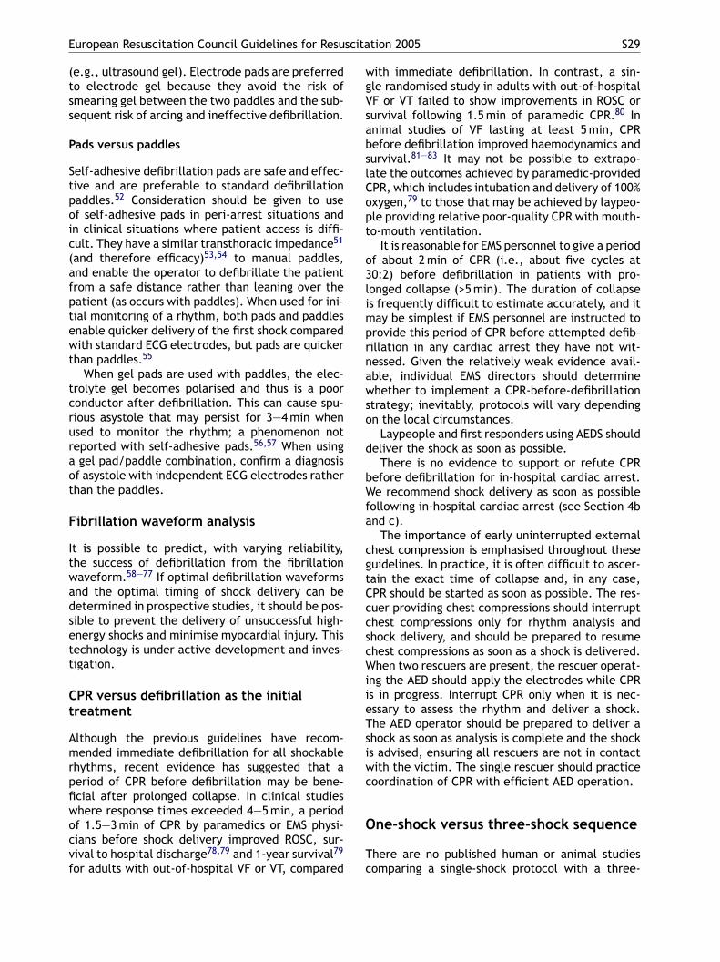
European Resuscitation Council Guidelines for Resuscitation 2005 S29
(e.g., ultrasound gel). Electrode pads are preferredto electrode gel because they avoid the risk ofsmearing gel between the two paddles and the sub-sequent risk of arcing and ineffective defibrillation.
Pads versus paddles
Self-adhesive defibrillation pads are safe and effec-tive and are preferable to standard defibrillationpaddles.52 Consideration should be given to useof self-adhesive pads in peri-arrest situations andin clinical situations where patient access is diffi-cult. They have a similar transthoracic impedance51
(and therefore efficacy)53,54 to manual paddles,and enable the operator to defibrillate the patientfrom a safe distance rather than leaning over thepatient (as occurs with paddles). When used for ini-tial monitoring of a rhythm, both pads and paddlesenable quicker delivery of the first shock comparedwith standard ECG electrodes, but pads are quickerthan paddles.55
When gel pads are used with paddles, the elec-trolyte gel becomes polarised and thus is a poorconductor after defibrillation. This can cause spu-rious asystole that may persist for 3—4 min whenuraot
F
Itwadsett
Ct
Amrpfiwocvf
with immediate defibrillation. In contrast, a sin-gle randomised study in adults with out-of-hospitalVF or VT failed to show improvements in ROSC orsurvival following 1.5 min of paramedic CPR.80 Inanimal studies of VF lasting at least 5 min, CPRbefore defibrillation improved haemodynamics andsurvival.81—83 It may not be possible to extrapo-late the outcomes achieved by paramedic-providedCPR, which includes intubation and delivery of 100%oxygen,79 to those that may be achieved by laypeo-ple providing relative poor-quality CPR with mouth-to-mouth ventilation.
It is reasonable for EMS personnel to give a periodof about 2 min of CPR (i.e., about five cycles at30:2) before defibrillation in patients with pro-longed collapse (>5 min). The duration of collapseis frequently difficult to estimate accurately, and itmay be simplest if EMS personnel are instructed toprovide this period of CPR before attempted defib-rillation in any cardiac arrest they have not wit-nessed. Given the relatively weak evidence avail-able, individual EMS directors should determinewhether to implement a CPR-before-defibrillationstrategy; inevitably, protocols will vary dependingon the local circumstances.
d
bWfa
cgtCccscWiieTsiwc
O
Tc
sed to monitor the rhythm; a phenomenon noteported with self-adhesive pads.56,57 When usinggel pad/paddle combination, confirm a diagnosis
f asystole with independent ECG electrodes ratherhan the paddles.
ibrillation waveform analysis
t is possible to predict, with varying reliability,he success of defibrillation from the fibrillationaveform.58—77 If optimal defibrillation waveformsnd the optimal timing of shock delivery can beetermined in prospective studies, it should be pos-ible to prevent the delivery of unsuccessful high-nergy shocks and minimise myocardial injury. Thisechnology is under active development and inves-igation.
PR versus defibrillation as the initialreatment
lthough the previous guidelines have recom-ended immediate defibrillation for all shockable
hythms, recent evidence has suggested that aeriod of CPR before defibrillation may be bene-cial after prolonged collapse. In clinical studieshere response times exceeded 4—5 min, a periodf 1.5—3 min of CPR by paramedics or EMS physi-ians before shock delivery improved ROSC, sur-ival to hospital discharge78,79 and 1-year survival79
or adults with out-of-hospital VF or VT, compared
Laypeople and first responders using AEDS shouldeliver the shock as soon as possible.
There is no evidence to support or refute CPRefore defibrillation for in-hospital cardiac arrest.e recommend shock delivery as soon as possible
ollowing in-hospital cardiac arrest (see Section 4bnd c).
The importance of early uninterrupted externalhest compression is emphasised throughout theseuidelines. In practice, it is often difficult to ascer-ain the exact time of collapse and, in any case,PR should be started as soon as possible. The res-uer providing chest compressions should interrupthest compressions only for rhythm analysis andhock delivery, and should be prepared to resumehest compressions as soon as a shock is delivered.hen two rescuers are present, the rescuer operat-
ng the AED should apply the electrodes while CPRs in progress. Interrupt CPR only when it is nec-ssary to assess the rhythm and deliver a shock.he AED operator should be prepared to deliver ahock as soon as analysis is complete and the shocks advised, ensuring all rescuers are not in contactith the victim. The single rescuer should practiceoordination of CPR with efficient AED operation.
ne-shock versus three-shock sequence
here are no published human or animal studiesomparing a single-shock protocol with a three-
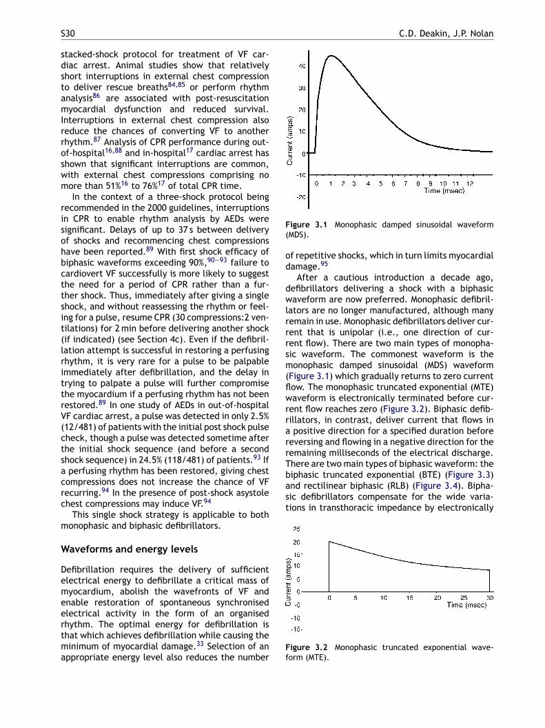
S30 C.D. Deakin, J.P. Nolan
stacked-shock protocol for treatment of VF car-diac arrest. Animal studies show that relativelyshort interruptions in external chest compressionto deliver rescue breaths84,85 or perform rhythmanalysis86 are associated with post-resuscitationmyocardial dysfunction and reduced survival.Interruptions in external chest compression alsoreduce the chances of converting VF to anotherrhythm.87 Analysis of CPR performance during out-of-hospital16,88 and in-hospital17 cardiac arrest hasshown that significant interruptions are common,with external chest compressions comprising nomore than 51%16 to 76%17 of total CPR time.
In the context of a three-shock protocol beingrecommended in the 2000 guidelines, interruptionsin CPR to enable rhythm analysis by AEDs weresignificant. Delays of up to 37 s between deliveryof shocks and recommencing chest compressionshave been reported.89 With first shock efficacy ofbiphasic waveforms exceeding 90%,90—93 failure tocardiovert VF successfully is more likely to suggestthe need for a period of CPR rather than a fur-ther shock. Thus, immediately after giving a singleshock, and without reassessing the rhythm or feel-ing for a pulse, resume CPR (30 compressions:2 ven-
Figure 3.1 Monophasic damped sinusoidal waveform(MDS).
of repetitive shocks, which in turn limits myocardialdamage.95
After a cautious introduction a decade ago,defibrillators delivering a shock with a biphasicwaveform are now preferred. Monophasic defibril-lators are no longer manufactured, although manyremain in use. Monophasic defibrillators deliver cur-rent that is unipolar (i.e., one direction of cur-rent flow). There are two main types of monopha-sic waveform. The commonest waveform is themonophasic damped sinusoidal (MDS) waveform(Figure 3.1) which gradually returns to zero currentflow. The monophasic truncated exponential (MTE)waveform is electronically terminated before cur-rent flow reaches zero (Figure 3.2). Biphasic defib-rillators, in contrast, deliver current that flows ina positive direction for a specified duration beforereversing and flowing in a negative direction for theremaining milliseconds of the electrical discharge.There are two main types of biphasic waveform: thebiphasic truncated exponential (BTE) (Figure 3.3)and rectilinear biphasic (RLB) (Figure 3.4). Bipha-sic defibrillators compensate for the wide varia-tions in transthoracic impedance by electronically
Ff
tilations) for 2 min before delivering another shock(if indicated) (see Section 4c). Even if the defibril-lation attempt is successful in restoring a perfusingrhythm, it is very rare for a pulse to be palpableimmediately after defibrillation, and the delay intrying to palpate a pulse will further compromisethe myocardium if a perfusing rhythm has not beenrestored.89 In one study of AEDs in out-of-hospitalVF cardiac arrest, a pulse was detected in only 2.5%(12/481) of patients with the initial post shock pulsecheck, though a pulse was detected sometime afterthe initial shock sequence (and before a secondshock sequence) in 24.5% (118/481) of patients.93 Ifa perfusing rhythm has been restored, giving chestcompressions does not increase the chance of VFrecurring.94 In the presence of post-shock asystolechest compressions may induce VF.94
This single shock strategy is applicable to bothmonophasic and biphasic defibrillators.
Waveforms and energy levels
Defibrillation requires the delivery of sufficientelectrical energy to defibrillate a critical mass ofmyocardium, abolish the wavefronts of VF andenable restoration of spontaneous synchronisedelectrical activity in the form of an organisedrhythm. The optimal energy for defibrillation isthat which achieves defibrillation while causing theminimum of myocardial damage.33 Selection of anappropriate energy level also reduces the number
igure 3.2 Monophasic truncated exponential wave-orm (MTE).
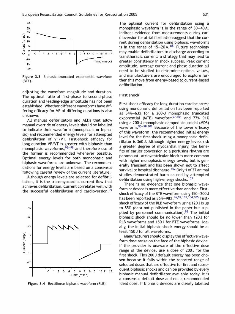
European Resuscitation Council Guidelines for Resuscitation 2005 S31
Figure 3.3 Biphasic truncated exponential waveform(BTE).
adjusting the waveform magnitude and duration.The optimal ratio of first-phase to second-phaseduration and leading-edge amplitude has not beenestablished. Whether different waveforms have dif-fering efficacy for VF of differing durations is alsounknown.
All manual defibrillators and AEDs that allowmanual override of energy levels should be labelledto indicate their waveform (monophasic or bipha-sic) and recommended energy levels for attempteddefibrillation of VF/VT. First-shock efficacy forlong-duration VF/VT is greater with biphasic thanmonophasic waveforms,96—98 and therefore use ofthe former is recommended whenever possible.Optimal energy levels for both monophasic andbiphasic waveforms are unknown. The recommen-dations for energy levels are based on a consensusfollowing careful review of the current literature.
Although energy levels are selected for defibril-lation, it is the transmyocardial current flow thatachieves defibrillation. Current correlates well withthe successful defibrillation and cardioversion.99
The optimal current for defibrillation using amonophasic waveform is in the range of 30—40 A.Indirect evidence from measurements during car-dioversion for atrial fibrillation suggest that the cur-rent during defibrillation using biphasic waveformsis in the range of 15—20 A.100 Future technologymay enable defibrillators to discharge according totransthoracic current: a strategy that may lead togreater consistency in shock success. Peak currentamplitude, average current and phase duration allneed to be studied to determine optimal values,and manufacturers are encouraged to explore fur-ther this move from energy-based to current-baseddefibrillation.
First shock
First-shock efficacy for long-duration cardiac arrestusing monophasic defibrillation has been reportedas 54%—63% for a 200-J monophasic truncatedexponential (MTE) waveform97,101 and 77%—91%using a 200-J monophasic damped sinusoidal (MDS)waveform.96—98,101 Because of the lower efficacyof this waveform, the recommended initial energylevel for the first shock using a monophasic defib-rafipwessd
fshstpbRal
fIrfissqbai
Figure 3.4 Rectilinear biphasic waveform (RLB).illator is 360 J. Although higher energy levels riskgreater degree of myocardial injury, the bene-
ts of earlier conversion to a perfusing rhythm arearamount. Atrioventricular block is more commonith higher monophasic energy levels, but is gen-rally transient and has been shown not to affecturvival to hospital discharge.102 Only 1 of 27 animaltudies demonstrated harm caused by attemptedefibrillation using high-energy shocks.103
There is no evidence that one biphasic wave-orm or device is more effective than another. First-hock efficacy of the BTE waveform using 150—200 Jas been reported as 86%—98%.96,97,101,104,105 First-hock efficacy of the RLB waveform using 120 J is upo 85% (data not published in the paper but sup-lied by personnel communication).98 The initialiphasic shock should be no lower than 120 J forLB waveforms and 150 J for BTE waveforms. Ide-lly, the initial biphasic shock energy should be ateast 150 J for all waveforms.
Manufacturers should display the effective wave-orm dose range on the face of the biphasic device.f the provider is unaware of the effective doseange of the device, use a dose of 200 J for therst shock. This 200 J default energy has been cho-en because it falls within the reported range ofelected doses that are effective for first and subse-uent biphasic shocks and can be provided by everyiphasic manual defibrillator available today. It isconsensus default dose and not a recommended
deal dose. If biphasic devices are clearly labelled
S32 C.D. Deakin, J.P. Nolan
and providers are familiar with the devices they usein clinical care, there will be no need for the default200 J dose. Ongoing research is necessary to firmlyestablish the most appropriate initial settings forboth monophasic and biphasic defibrillators.
Second and subsequent shocks
With monophasic defibrillators, if the initial shockhas been unsuccessful at 360 J, second and sub-sequent shocks should all be delivered at 360 J.With biphasic defibrillators there is no evidence tosupport either a fixed or escalating energy proto-col. Both strategies are acceptable; however, if thefirst shock is not successful and the defibrillator iscapable of delivering shocks of higher energy, itis rational to increase the energy for subsequentshocks. If the provider is unaware of the effectivedose range of the biphasic device and has used thedefault 200 J dose for the first shock, use eitheran equal or higher dose for second or subsequentshocks, depending on the capabilities of the device.
If a shockable rhythm (recurrent ventricular fib-rillation) recurs after successful defibrillation (withor without ROSC), give the next shock with the
Blind defibrillation
Delivery of shocks without a monitor or an ECGrhythm diagnosis is referred to as ‘‘blind’’ defibril-lation. Blind defibrillation is unnecessary. Handheldpaddles with ‘‘quick-look’’ monitoring capabilitieson modern manually operated defibrillators arewidely available. AEDs use reliable and proven deci-sion algorithms to identify VF.
Spurious asystole and occult ventricularfibrillation
Rarely, coarse VF can be present in some leads, withvery small undulations seen in the orthogonal leads,which is called occult VF. A flat line that may resem-ble asystole is displayed; examine the rhythm intwo leads to obtain the correct diagnosis. Of moreimportance, one study noted that spurious asystole,a flat line produced by technical errors (e.g., nopower, leads unconnected, gain set to low, incor-rect lead selection, or polarisation of electrolytegel (see above)), was far more frequent than occultVF.120
There is no evidence that attempting to defib-rcfir
P
TugtmTiabtiVtwtlaosi
cni
energy level that had previously been successful.
Other related defibrillation topics
Defibrillation of children
Cardiac arrest is less common in children. Aetiologyis generally related to hypoxia and trauma.106—108
VF is relatively rare compared with adult cardiacarrest, occurring in 7%—15% of paediatric and ado-lescent arrests.108—112 Common causes of VF inchildren include trauma, congenital heart disease,long QT interval, drug overdose and hypothermia.Rapid defibrillation of these patients may improveoutcome.112,113
The optimal energy level, waveform and shocksequence are unknown but, as with adults, bipha-sic shocks appear to be at least as effective as,and less harmful than, monophasic shocks.114—116
The upper limit for safe defibrillation is unknown,but doses in excess of the previously recommendedmaximum of 4 J kg−1 (as high as 9 J kg−1) havedefibrillated children effectively without signifi-cant adverse effects.20,117,118 The recommendedenergy level for manual monophasic defibrillationis 4 J kg−1 for the initial shock and for subsequentshocks. The same energy level is recommended formanual biphasic defibrillation.119 As with adults, ifa shockable rhythm recurs, use the energy level fordefibrillation that had previously been successful.
illate true asystole is beneficial. Studies inhildren121 and adults122 have failed to show bene-t from defibrillation of asystole. On the contrary,epeated shocks will cause myocardial injury.
recordial thump
here are no prospective studies that evaluatese of precordial (chest) thump. The rationale foriving a thump is that the mechanical energy ofhe thump is converted to electrical energy, whichay be sufficient to achieve cardioversion.123
he electrical threshold of successful defibrillationncreases rapidly after the onset of the arrhythmia,nd the amount of electrical energy generated fallselow this threshold within seconds. A precordialhump is most likely to be successful in convert-ng VT to sinus rhythm. Successful treatment ofF by precordial thump is much less likely: in allhe reported successful cases, the precordial thumpas given within the first 10 s of VF.123 Although
hree case series124—126 reported that VF or pulse-ess VT was converted to a perfusing rhythm by
precordial thump, there are occasional reportsf thump causing deterioration in cardiac rhythm,uch as rate acceleration of VT, conversion of VTnto VF, complete heart block or asystole.125,127—132
Consider giving a single precordial thump whenardiac arrest is confirmed rapidly after a wit-essed, sudden collapse and a defibrillator is notmmediately to hand. These circumstances are
European Resuscitation Council Guidelines for Resuscitation 2005 S33
most likely to occur when the patient is monitored.Precordial thump should be undertaken immedi-ately after confirmation of cardiac arrest and onlyby healthcare professionals trained in the tech-nique. Using the ulnar edge of a tightly clenchedfist, a sharp impact is delivered to the lower half ofthe sternum from a height of about 20 cm, followedby immediate retraction of the fist, which createsan impulse-like stimulus.
Cardioversion
If electrical cardioversion is used to convert atrialor ventricular tachyarrhythmias, the shock must besynchronised to occur with the R wave of the elec-trocardiogram rather than with the T wave: VF canbe induced if a shock is delivered during the rel-ative refractory portion of the cardiac cycle.133
Synchronisation can be difficult in VT because ofthe wide-complex and variable forms of ventriculararrhythmia. If synchronisation fails, give unsynchro-nised shocks to the unstable patient in VT to avoidprolonged delay in restoring sinus rhythm. Ventric-ular fibrillation or pulseless VT requires unsynchro-ntc
A
BmAr
M
ArmAeclto2
B
Mdlft
randomised study comparing escalating monophasicenergy levels to 360 J and biphasic energy levels to200 J found no difference in efficacy between thetwo waveforms.137 An initial shock of 120—150 J,escalating if necessary, is a reasonable strategybased on current data.
Atrial flutter and paroxysmalsupraventricular tachycardia
Atrial flutter and paroxysmal SVT generallyrequire less energy than atrial fibrillation forcardioversion.138 Give an initial shock of 100-Jmonophasic or 70—120 J biphasic waveform. Givesubsequent shocks using stepwise increases inenergy.99
Ventricular tachycardia
The energy required for cardioversion of VTdepends on the morphological characteristics andrate of the arrhythmia.139 Ventricular tachycardiawith a pulse responds well to cardioversion usinginitial monophasic energies of 200 J. Use biphasicenergy levels of 120—150 J for the initial shock.Ga
P
CboaapWEtpo
R
ised shocks. Conscious patients must be anaes-hetised or sedated before attempting synchronisedardioversion.
trial fibrillation
iphasic waveforms are more effective thanonophasic waveforms for cardioversion ofF100,134,135; when available, use a biphasic defib-illator in preference to a monophasic defibrillator.
onophasic waveforms
study of electrical cardioversion for atrial fib-illation indicated that 360-J MDS shocks wereore effective than 100-J or 200-J MDS shocks.136
lthough a first shock of 360-J reduces overallnergy requirements for cardioversion, 360 J mayause greater myocardial damage than occurs withower monophasic energy levels, and this must beaken into consideration. Commence cardioversionf atrial fibrillation using an initial energy level of00 J, increasing in a stepwise manner as necessary.
iphasic waveforms
ore data are needed before specific recommen-ations can be made for optimal biphasic energyevels. First-shock efficacy of a 70-J biphasic wave-orm has been shown to be significantly greater thanhat with a 100-monophasic waveform.100,134,135 A
ive stepwise increases if the first shock fails tochieve sinus rhythm.139
acing
onsider pacing in patients with symptomaticradycardia refractory to anticholinergic drugs orther second-line therapy (see Section 4f). Immedi-te pacing is indicated, especially when the block ist or below the His—Purkinje level. If transthoracicacing is ineffective, consider transvenous pacing.henever a diagnosis of asystole is made, check the
CG carefully for the presence of P waves, becausehis may respond to cardiac pacing. Do not attemptacing for asystole; it does not increase short-termr long-term survival in or out of hospital.140—148
eferences
1. American Heart Association in collaboration with Inter-national Liaison Committee on Resuscitation. Guide-lines 2000 for Cardiopulmonary Resuscitation and Emer-gency Cardiovascular Care, Part 6: Advanced Cardiovas-cular Life Support: Section 2: Defibrillation. Circulation2000;102(Suppl.):I90—4.
2. Larsen MP, Eisenberg MS, Cummins RO, Hallstrom AP.Predicting survival from out-of-hospital cardiac arrest: agraphic model. Ann Emerg Med 1993;22:1652—8.
3. Valenzuela TD, Roe DJ, Cretin S, Spaite DW, LarsenMP. Estimating effectiveness of cardiac arrest interven-tions: a logistic regression survival model. Circulation1997;96:3308—13.
S34 C.D. Deakin, J.P. Nolan
4. Waalewijn RA, de Vos R, Tijssen JGP, Koster RW. Survivalmodels for out-of-hospital cardiopulmonary resuscitationfrom the perspectives of the bystander, the first responder,and the paramedic. Resuscitation 2001;51:113—22.
5. Myerburg RJ, Fenster J, Velez M, et al. Impact ofcommunity-wide police car deployment of automatedexternal defibrillators on survival from out-of-hospital car-diac arrest. Circulation 2002;106:1058—64.
6. Capucci A, Aschieri D, Piepoli MF, Bardy GH, IconomuE, Arvedi M. Tripling survival from sudden cardiacarrest via early defibrillation without traditional edu-cation in cardiopulmonary resuscitation. Circulation2002;106:1065—70.
7. van Alem AP, Vrenken RH, de Vos R, Tijssen JG, Koster RW.Use of automated external defibrillator by first respondersin out of hospital cardiac arrest: prospective controlledtrial. BMJ 2003;327:1312.
8. Valenzuela TD, Bjerke HS, Clark LL, et al. Rapid defibrilla-tion by nontraditional responders: the Casino Project. AcadEmerg Med 1998;5:414—5.
9. Swor RA, Jackson RE, Cynar M, et al. Bystander CPR,ventricular fibrillation, and survival in witnessed, unmon-itored out-of-hospital cardiac arrest. Ann Emerg Med1995;25:780—4.
10. Holmberg M, Holmberg S, Herlitz J. Effect of bystandercardiopulmonary resuscitation in out-of-hospital cardiacarrest patients in Sweden. Resuscitation 2000;47:59—70.
11. Monsieurs KG, Handley AJ, Bossaert LL. European Resus-citation Council Guidelines 2000 for Automated ExternalDefibrillation. A statement from the Basic Life Support and
ysis in infants and children. Ann Emerg Med 2003;42:185—96.
21. Cecchin F, Jorgenson DB, Berul CI, et al. Is arrhythmiadetection by automatic external defibrillator accurate forchildren? Sensitivity and specificity of an automatic exter-nal defibrillator algorithm in 696 pediatric arrhythmias.Circulation 2001;103:2483—8.
22. Zafari AM, Zarter SK, Heggen V, et al. A program encour-aging early defibrillation results in improved in-hospitalresuscitation efficacy. J Am Coll Cardiol 2004;44:846—52.
23. Destro A, Marzaloni M, Sermasi S, Rossi F. Automatic exter-nal defibrillators in the hospital as well? Resuscitation1996;31:39—43.
24. Domanovits H, Meron G, Sterz F, et al. Successful automaticexternal defibrillator operation by people trained only inbasic life support in a simulated cardiac arrest situation.Resuscitation 1998;39:47—50.
25. Cusnir H, Tongia R, Sheka KP, et al. In hospital cardiacarrest: a role for automatic defibrillation. Resuscitation2004;63:183—8.
26. Kaye W, Mancini ME, Richards N. Organizing and imple-menting a hospital-wide first-responder automated exter-nal defibrillation program: strengthening the in-hospitalchain of survival. Resuscitation 1995;30:151—6.
27. Miller PH. Potential fire hazard in defibrillation. JAMA1972;221:192.
28. Hummel IIIrd RS, Ornato JP, Weinberg SM, Clarke AM. Spark-generating properties of electrode gels used during defib-rillation. A potential fire hazard. JAMA 1988;260:3021—4.
29. Fires from defibrillation during oxygen administration.
Automated External Defibrillation Working Group (1) andapproved by the Executive Committee of the EuropeanResuscitation Council. Resuscitation 2001;48:207—9.12. Cummins RO, Eisenberg M, Bergner L, Murray JA. Sensitivityaccuracy, and safety of an automatic external defibrillator.Lancet 1984;2:318—20.
13. Davis EA, Mosesso Jr VN. Performance of police firstresponders in utilizing automated external defibrillationon victims of sudden cardiac arrest. Prehosp Emerg Care1998;2:101—7.
14. White RD, Vukov LF, Bugliosi TF. Early defibrillation bypolice: initial experience with measurement of criticaltime intervals and patient outcome. Ann Emerg Med1994;23:1009—13.
15. White RD, Hankins DG, Bugliosi TF. Seven years’ experi-ence with early defibrillation by police and paramedicsin an emergency medical services system. Resuscitation1998;39:145—51.
16. Wik L, Kramer-Johansen J, Myklebust H, et al. Quality ofcardiopulmonary resuscitation during out-of-hospital car-diac arrest. JAMA 2005;293:299—304.
17. Abella BS, Alvarado JP, Myklebust H, et al. Quality ofcardiopulmonary resuscitation during in-hospital cardiacarrest. JAMA 2005;293:305—10.
18. Kerber RE, Becker LB, Bourland JD, et al. Automatic exter-nal defibrillators for public access defibrillation: recom-mendations for specifying and reporting arrhythmia analy-sis algorithm performance, incorporating new waveforms,and enhancing safety. A statement for health professionalsfrom the American Heart Association Task Force on Auto-matic External Defibrillation, Subcommittee on AED Safetyand Efficacy. Circulation 1997;95:1677—82.
19. Dickey W, Dalzell GW, Anderson JM, Adgey AA. The accu-racy of decision-making of a semi-automatic defibrillatorduring cardiac arrest. Eur Heart J 1992;13:608—15.
20. Atkinson E, Mikysa B, Conway JA, et al. Specificity andsensitivity of automated external defibrillator rhythm anal-
Health Devices 1994;23:307—9.30. Lefever J, Smith A. Risk of fire when using defibrillation in
an oxygen enriched atmosphere. Medical Devices AgencySafety Notices 1995;3:1—3.
31. Ward ME. Risk of fires when using defibrillators in an oxygenenriched atmosphere. Resuscitation 1996;31:173.
32. Theodorou AA, Gutierrez JA, Berg RA. Fire attributableto a defibrillation attempt in a neonate. Pediatrics2003;112:677—9.
33. Kerber RE, Kouba C, Martins J, et al. Advance prediction oftransthoracic impedance in human defibrillation and car-dioversion: importance of impedance in determining thesuccess of low-energy shocks. Circulation 1984;70:303—8.
34. Kerber RE, Grayzel J, Hoyt R, Marcus M, Kennedy J.Transthoracic resistance in human defibrillation. Influenceof body weight, chest size, serial shocks, paddle sizeand paddle contact pressure. Circulation 1981;63:676—82.
35. Sado DM, Deakin CD, Petley GW, Clewlow F. Comparisonof the effects of removal of chest hair with not doing sobefore external defibrillation on transthoracic impedance.Am J Cardiol 2004;93:98—100.
36. Deakin CD, Sado DM, Petley GW, Clewlow F. Differen-tial contribution of skin impedance and thoracic volumeto transthoracic impedance during external defibrillation.Resuscitation 2004;60:171—4.
37. Deakin C, Sado D, Petley G, Clewlow F. Determining theoptimal paddle force for external defibrillation. Am J Car-diol 2002;90:812—3.
38. Deakin C, Bennetts S, Petley G, Clewlow F. What is the opti-mal paddle force for paediatric defibrillation? Resuscitation2002;55:59.
39. Panacek EA, Munger MA, Rutherford WF, Gardner SF. Reportof nitropatch explosions complicating defibrillation. Am JEmerg Med 1992;10:128—9.
40. Wrenn K. The hazards of defibrillation through nitroglycerinpatches. Ann Emerg Med 1990;19:1327—8.
European Resuscitation Council Guidelines for Resuscitation 2005 S35
41. Pagan-Carlo LA, Spencer KT, Robertson CE, Dengler A, Bir-kett C, Kerber RE. Transthoracic defibrillation: importanceof avoiding electrode placement directly on the femalebreast. J Am Coll Cardiol 1996;27:449—52.
42. Deakin CD, Sado DM, Petley GW, Clewlow F. Is the ori-entation of the apical defibrillation paddle of impor-tance during manual external defibrillation? Resuscitation2003;56:15—8.
43. Kirchhof P, Borggrefe M, Breithardt G. Effect of electrodeposition on the outcome of cardioversion. Card Electro-physiol Rev 2003;7:292—6.
44. Kirchhof P, Eckardt L, Loh P, et al. Anterior-posterior ver-sus anterior-lateral electrode positions for external car-dioversion of atrial fibrillation: a randomised trial. Lancet2002;360:1275—9.
45. Botto GL, Politi A, Bonini W, Broffoni T, Bonatti R. Exter-nal cardioversion of atrial fibrillation: role of paddle posi-tion on technical efficacy and energy requirements. Heart1999;82:726—30.
46. Alp NJ, Rahman S, Bell JA, Shahi M. Randomised comparisonof antero-lateral versus antero-posterior paddle positionsfor DC cardioversion of persistent atrial fibrillation. Int JCardiol 2000;75:211—6.
47. Mathew TP, Moore A, McIntyre M, et al. Randomised com-parison of electrode positions for cardioversion of atrialfibrillation. Heart 1999;81:576—9.
48. Walsh SJ, McCarty D, McClelland AJ, et al. Impedance com-pensated biphasic waveforms for transthoracic cardiover-sion of atrial fibrillation: a multi-centre comparison ofantero-apical and antero-posterior pad positions. Eur Heart
and nonparametric classification of ventricular fibrillationin patients with out-of-hospital cardiac arrest. Circulation2000;102:1523—9.
60. Eftestol T, Wik L, Sunde K, Steen PA. Effects of cardiopul-monary resuscitation on predictors of ventricular fibrilla-tion defibrillation success during out-of-hospital cardiacarrest. Circulation 2004;110:10—5.
61. Weaver WD, Cobb LA, Dennis D, Ray R, Hallstrom AP, CopassMK. Amplitude of ventricular fibrillation waveform and out-come after cardiac arrest. Ann Intern Med 1985;102:53—5.
62. Brown CG, Dzwonczyk R. Signal analysis of the human elec-trocardiogram during ventricular fibrillation: frequencyand amplitude parameters as predictors of successful coun-tershock. Ann Emerg Med 1996;27:184—8.
63. Callaham M, Braun O, Valentine W, Clark DM, Zegans C. Pre-hospital cardiac arrest treated by urban first-responders:profile of patient response and prediction of outcomeby ventricular fibrillation waveform. Ann Emerg Med1993;22:1664—77.
64. Strohmenger HU, Lindner KH, Brown CG. Analysis ofthe ventricular fibrillation ECG signal amplitude and fre-quency parameters as predictors of countershock successin humans. Chest 1997;111:584—9.
65. Strohmenger HU, Eftestol T, Sunde K, et al. The predictivevalue of ventricular fibrillation electrocardiogram signalfrequency and amplitude variables in patients with out-of-hospital cardiac arrest. Anesth Analg 2001;93:1428—33.
66. Podbregar M, Kovacic M, Podbregar-Mars A, Brezocnik M.Predicting defibrillation success by ‘genetic’ programmingin patients with out-of-hospital cardiac arrest. Resuscita-
J 2005.49. Deakin CD, McLaren RM, Petley GW, Clewlow F, Dalrymple-
Hay MJ. Effects of positive end-expiratory pressure ontransthoracic impedance—–implications for defibrillation.Resuscitation 1998;37:9—12.
50. American National Standard: Automatic External Defibril-lators and Remote Controlled Defibrillators (DF39). Arling-ton, Virgina: Association for the Advancement of MedicalInstrumentation; 1993.
51. Deakin CD, McLaren RM, Petley GW, Clewlow F, Dalrymple-Hay MJ. A comparison of transthoracic impedance usingstandard defibrillation paddles and self-adhesive defibril-lation pads. Resuscitation 1998;39:43—6.
52. Stults KR, Brown DD, Cooley F, Kerber RE. Self-adhesivemonitor/defibrillation pads improve prehospital defibrilla-tion success. Ann Emerg Med 1987;16:872—7.
53. Kerber RE, Martins JB, Kelly KJ, et al. Self-adhesive preap-plied electrode pads for defibrillation and cardioversion. JAm Coll Cardiol 1984;3:815—20.
54. Kerber RE, Martins JB, Ferguson DW, et al. Exper-imental evaluation and initial clinical application ofnew self-adhesive defibrillation electrodes. Int J Cardiol1985;8:57—66.
55. Perkins GD, Roberts C, Gao F. Delays in defibrillation: influ-ence of different monitoring techniques. Br J Anaesth2002;89:405—8.
56. Bradbury N, Hyde D, Nolan J. Reliability of ECG monitor-ing with a gel pad/paddle combination after defibrillation.Resuscitation 2000;44:203—6.
57. Chamberlain D. Gel pads should not be used for monitoringECG after defibrillation. Resuscitation 2000;43:159—60.
58. Callaway CW, Sherman LD, Mosesso Jr VN, Dietrich TJ, HoltE, Clarkson MC. Scaling exponent predicts defibrillationsuccess for out-of-hospital ventricular fibrillation cardiacarrest. Circulation 2001;103:1656—61.
59. Eftestol T, Sunde K, Aase SO, Husoy JH, Steen PA. Predict-ing outcome of defibrillation by spectral characterization
tion 2003;57:153—9.67. Menegazzi JJ, Callaway CW, Sherman LD, et al. Ventricular
fibrillation scaling exponent can guide timing of defibrilla-tion and other therapies. Circulation 2004;109:926—31.
68. Povoas HP, Weil MH, Tang W, Bisera J, Klouche K, BarbatsisA. Predicting the success of defibrillation by electrocardio-graphic analysis. Resuscitation 2002;53:77—82.
69. Noc M, Weil MH, Tang W, Sun S, Pernat A, Bisera J. Electro-cardiographic prediction of the success of cardiac resusci-tation. Crit Care Med 1999;27:708—14.
70. Strohmenger HU, Lindner KH, Keller A, Lindner IM, Pfen-ninger EG. Spectral analysis of ventricular fibrillation andclosed-chest cardiopulmonary resuscitation. Resuscitation1996;33:155—61.
71. Noc M, Weil MH, Gazmuri RJ, Sun S, Biscera J, Tang W.Ventricular fibrillation voltage as a monitor of the effec-tiveness of cardiopulmonary resuscitation. J Lab Clin Med1994;124:421—6.
72. Lightfoot CB, Nremt P, Callaway CW, et al. Dynamic natureof electrocardiographic waveform predicts rescue shockoutcome in porcine ventricular fibrillation. Ann Emerg Med2003;42:230—41.
73. Marn-Pernat A, Weil MH, Tang W, Pernat A, Bisera J. Opti-mizing timing of ventricular defibrillation. Crit Care Med2001;29:2360—5.
74. Hamprecht FA, Achleitner U, Krismer AC, et al. Fibrilla-tion power, an alternative method of ECG spectral analysisfor prediction of countershock success in a porcine modelof ventricular fibrillation. Resuscitation 2001;50:287—96.
75. Amann A, Achleitner U, Antretter H, et al. Analysing ven-tricular fibrillation ECG-signals and predicting defibrillationsuccess during cardiopulmonary resuscitation employingN(alpha)-histograms. Resuscitation 2001;50:77—85.
76. Brown CG, Griffith RF, Van Ligten P, et al. Medianfrequency—–a new parameter for predicting defibrillationsuccess rate. Ann Emerg Med 1991;20:787—9.
S36 C.D. Deakin, J.P. Nolan
77. Amann A, Rheinberger K, Achleitner U, et al. The predic-tion of defibrillation outcome using a new combination ofmean frequency and amplitude in porcine models of car-diac arrest. Anesth Analg 2002;95:716—22.
78. Cobb LA, Fahrenbruch CE, Walsh TR, et al. Influenceof cardiopulmonary resuscitation prior to defibrillation inpatients with out-of-hospital ventricular fibrillation. JAMA1999;281:1182—8.
79. Wik L, Hansen TB, Fylling F, et al. Delaying defibrillation togive basic cardiopulmonary resuscitation to patients without-of-hospital ventricular fibrillation: a randomized trial.JAMA 2003;289:1389—95.
80. Jacobs IG, Finn JC, Oxer HF, Jelinek GA. CPR before defibril-lation in out-of-hospital cardiac arrest: a randomized trial.Emerg Med Australas 2005;17:39—45.
81. Berg RA, Hilwig RW, Kern KB, Ewy GA. Precountershockcardiopulmonary resuscitation improves ventricular fibril-lation median frequency and myocardial readiness for suc-cessful defibrillation from prolonged ventricular fibrilla-tion: a randomized, controlled swine study. Ann Emerg Med2002;40:563—70.
82. Berg RA, Hilwig RW, Ewy GA, Kern KB. Precountershockcardiopulmonary resuscitation improves initial responseto defibrillation from prolonged ventricular fibrillation:a randomized, controlled swine study. Crit Care Med2004;32:1352—7.
83. Kolarova J, Ayoub IM, Yi Z, Gazmuri RJ. Optimal timing forelectrical defibrillation after prolonged untreated ventric-ular fibrillation. Crit Care Med 2003;31:2022—8.
84. Berg RA, Sanders AB, Kern KB, et al. Adverse hemo-
93. Rea TD, Shah S, Kudenchuk PJ, Copass MK, Cobb LA. Auto-mated external defibrillators: to what extent does the algo-rithm delay CPR? Ann Emerg Med 2005;46:132—41.
94. Hess EP, White RD. Ventricular fibrillation is not pro-voked by chest compression during post-shock organizedrhythms in out-of-hospital cardiac arrest. Resuscitation2005;66:7—11.
95. Joglar JA, Kessler DJ, Welch PJ, et al. Effects of repeatedelectrical defibrillations on cardiac troponin I levels. Am JCardiol 1999;83:270—2. A6.
96. van Alem AP, Chapman FW, Lank P, Hart AA, KosterRW. A prospective, randomised and blinded compari-son of first shock success of monophasic and biphasicwaveforms in out-of-hospital cardiac arrest. Resuscitation2003;58:17—24.
97. Carpenter J, Rea TD, Murray JA, Kudenchuk PJ, EisenbergMS. Defibrillation waveform and post-shock rhythm in out-of-hospital ventricular fibrillation cardiac arrest. Resusci-tation 2003;59:189—96.
98. Morrison LJ, Dorian P, Long J, et al. Out-of-hospital car-diac arrest rectilinear biphasic to monophasic damped sinedefibrillation waveforms with advanced life support inter-vention trial (ORBIT). Resuscitation 2005;66:149—57.
99. Kerber RE, Martins JB, Kienzle MG, et al. Energy, current,and success in defibrillation and cardioversion: clinicalstudies using an automated impedance-based method ofenergy adjustment. Circulation 1988;77:1038—46.
100. Koster RW, Dorian P, Chapman FW, Schmitt PW, O’GradySG, Walker RG. A randomized trial comparing monophasicand biphasic waveform shocks for external cardioversion of
1
1
1
1
1
1
1
1
1
1
dynamic effects of interrupting chest compressions forrescue breathing during cardiopulmonary resuscitationfor ventricular fibrillation cardiac arrest. Circulation2001;104:2465—70.
85. Kern KB, Hilwig RW, Berg RA, Sanders AB, Ewy GA.Importance of continuous chest compressions duringcardiopulmonary resuscitation: improved outcome dur-ing a simulated single lay-rescuer scenario. Circulation2002;105:645—9.
86. Yu T, Weil MH, Tang W, et al. Adverse outcomes of inter-rupted precordial compression during automated defibril-lation. Circulation 2002;106:368—72.
87. Eftestol T, Sunde K, Steen PA. Effects of interrupting pre-cordial compressions on the calculated probability of defib-rillation success during out-of-hospital cardiac arrest. Cir-culation 2002;105:2270—3.
88. Valenzuela TD, Kern KB, Clark LL, et al. Interruptionsof chest compressions during emergency medical systemsresuscitation. Circulation 2005;112:1259—65.
89. van Alem AP, Sanou BT, Koster RW. Interruption of car-diopulmonary resuscitation with the use of the automatedexternal defibrillator in out-of-hospital cardiac arrest. AnnEmerg Med 2003;42:449—57.
90. Bain AC, Swerdlow CD, Love CJ, et al. Multicenter studyof principles-based waveforms for external defibrillation.Ann Emerg Med 2001;37:5—12.
91. Poole JE, White RD, Kanz KG, et al. Low-energy impedance-compensating biphasic waveforms terminate ventricularfibrillation at high rates in victims of out-of-hospital car-diac arrest. LIFE Investigators. J Cardiovasc Electrophysiol1997;8:1373—85.
92. Schneider T, Martens PR, Paschen H, et al. Multicenter,randomized, controlled trial of 150-J biphasic shocks com-pared with 200- to 360-J monophasic shocks in the resusci-tation of out-of-hospital cardiac arrest victims. OptimizedResponse to Cardiac Arrest (ORCA) Investigators. Circula-tion 2000;102:1780—7.
atrial fibrillation. Am Heart J 2004;147:e20.01. Martens PR, Russell JK, Wolcke B, et al. Optimal Response
to Cardiac Arrest study: defibrillation waveform effects.Resuscitation 2001;49:233—43.
02. Weaver WD, Cobb LA, Copass MK, Hallstrom AP. Ventricu-lar defibrillation: a comparative trial using 175-J and 320-Jshocks. N Engl J Med 1982;307:1101—6.
03. Tang W, Weil MH, Sun S, et al. The effects ofbiphasic and conventional monophasic defibrillation onpostresuscitation myocardial function. J Am Coll Cardiol1999;34:815—22.
04. Gliner BE, Jorgenson DB, Poole JE, et al. Treatment of out-of-hospital cardiac arrest with a low-energy impedance-compensating biphasic waveform automatic external defib-rillator. The LIFE Investigators. Biomed Instrum Technol1998;32:631—44.
05. White RD, Blackwell TH, Russell JK, Snyder DE, JorgensonDB. Transthoracic impedance does not affect defibrillation,resuscitation or survival in patients with out-of-hospitalcardiac arrest treated with a non-escalating biphasic wave-form defibrillator. Resuscitation 2005;64:63—9.
06. Kuisma M, Suominen P, Korpela R. Paediatric out-of-hospitalcardiac arrests: epidemiology and outcome. Resuscitation1995;30:141—50.
07. Sirbaugh PE, Pepe PE, Shook JE, et al. A prospective,population-based study of the demographics, epidemi-ology, management, and outcome of out-of-hospitalpediatric cardiopulmonary arrest. Ann Emerg Med1999;33:174—84.
08. Hickey RW, Cohen DM, Strausbaugh S, Dietrich AM. Pedi-atric patients requiring CPR in the prehospital setting. AnnEmerg Med 1995;25:495—501.
09. Appleton GO, Cummins RO, Larson MP, Graves JR. CPR andthe single rescuer: at what age should you ‘‘call first’’rather than ‘‘call fast’’? Ann Emerg Med 1995;25:492—4.
10. Ronco R, King W, Donley DK, Tilden SJ. Outcome and costat a children’s hospital following resuscitation for out-of-
European Resuscitation Council Guidelines for Resuscitation 2005 S37
hospital cardiopulmonary arrest. Arch Pediatr Adolesc Med1995;149:210—4.
111. Losek JD, Hennes H, Glaeser P, Hendley G, Nelson DB.Prehospital care of the pulseless, nonbreathing pediatricpatient. Am J Emerg Med 1987;5:370—4.
112. Mogayzel C, Quan L, Graves JR, Tiedeman D, FahrenbruchC, Herndon P. Out-of-hospital ventricular fibrillation in chil-dren and adolescents: causes and outcomes. Ann EmergMed 1995;25:484—91.
113. Safranek DJ, Eisenberg MS, Larsen MP. The epidemiol-ogy of cardiac arrest in young adults. Ann Emerg Med1992;21:1102—6.
114. Berg RA, Chapman FW, Berg MD, et al. Attenuated adultbiphasic shocks compared with weight-based monophasicshocks in a swine model of prolonged pediatric ventricularfibrillation. Resuscitation 2004;61:189—97.
115. Tang W, Weil MH, Jorgenson D, et al. Fixed-energy bipha-sic waveform defibrillation in a pediatric model of cardiacarrest and resuscitation. Crit Care Med 2002;30:2736—41.
116. Clark CB, Zhang Y, Davies LR, Karlsson G, Kerber RE. Pedi-atric transthoracic defibrillation: biphasic versus monopha-sic waveforms in an experimental model. Resuscitation2001;51:159—63.
117. Gurnett CA, Atkins DL. Successful use of a biphasic wave-form automated external defibrillator in a high-risk child.Am J Cardiol 2000;86:1051—3.
118. Atkins DL, Jorgenson DB. Attenuated pediatric electrodepads for automated external defibrillator use in children.Resuscitation 2005;66:31—7.
119. Gutgesell HP, Tacker WA, Geddes LA, Davis S, Lie JT, McNa-
1
1
1
1
1
1
1
1
1
129. Gertsch M, Hottinger S, Hess T. Serial chest thumps for thetreatment of ventricular tachycardia in patients with coro-nary artery disease. Clin Cardiol 1992;15:181—8.
130. Krijne R. Rate acceleration of ventricular tachycardia aftera precordial chest thump. Am J Cardiol 1984;53:964—5.
131. Sclarovsky S, Kracoff OH, Agmon J. Acceleration of ven-tricular tachycardia induced by a chest thump. Chest1981;80:596—9.
132. Yakaitis RW, Redding JS. Precordial thumping during cardiacresuscitation. Crit Care Med 1973;1:22—6.
133. Lown B. Electrical reversion of cardiac arrhythmias. BrHeart J 1967;29:469—89.
134. Mittal S, Ayati S, Stein KM, et al. Transthoracic cardiover-sion of atrial fibrillation: comparison of rectilinear biphasicversus damped sine wave monophasic shocks. Circulation2000;101:1282—7.
135. Page RL, Kerber RE, Russell JK, et al. Biphasic versusmonophasic shock waveform for conversion of atrial fibril-lation: the results of an international randomized, double-blind multicenter trial. J Am Coll Cardiol 2002;39:1956—63.
136. Joglar JA, Hamdan MH, Ramaswamy K, et al. Initial energyfor elective external cardioversion of persistent atrial fib-rillation. Am J Cardiol 2000;86:348—50.
137. Alatawi F, Gurevitz O, White R. Prospective, randomizedcomparison of two biphasic waveforms for the efficacy andsafety of transthoracic biphasic cardioversion of atrial fib-rillation. Heart Rhythm 2005;2:382—7.
138. Pinski SL, Sgarbossa EB, Ching E, Trohman RG. A comparisonof 50-J versus 100-J shocks for direct-current cardioversionof atrial flutter. Am Heart J 1999;137:439—42.
1
1
1
1
1
1
1
1
1
1
mara DG. Energy dose for ventricular defibrillation of chil-dren. Pediatrics 1976;58:898—901.
20. Cummins RO, Austin Jr D. The frequency of ‘occult’ ventric-ular fibrillation masquerading as a flat line in prehospitalcardiac arrest. Ann Emerg Med 1988;17:813—7.
21. Losek JD, Hennes H, Glaeser PW, Smith DS, Hendley G. Pre-hospital countershock treatment of pediatric asystole. AmJ Emerg Med 1989;7:571—5.
22. Martin DR, Gavin T, Bianco J, et al. Initial countershock inthe treatment of asystole. Resuscitation 1993;26:63—8.
23. Kohl P, King AM, Boulin C. Antiarrhythmic effects of acutemechanical stiumulation. In: Kohl P, Sachs F, Franz MR, edi-tors. Cardiac mechano-electric feedback and arrhythmias:form pipette to patient. Philadelphia: Elsevier Saunders;2005. p. 304—14.
24. Befeler B. Mechanical stimulation of the heart; its thera-peutic value in tachyarrhythmias. Chest 1978;73:832—8.
25. Volkmann HKA, Kuhnert H, Paliege R, Dannberg G,Siegert K. Terminierung von Kammertachykardien durchmechanische Herzstimulation mit Prakordialschlagen.(‘‘Termination of Ventricular Tachycardias by Mechan-ical Cardiac Pacing by Means of Precordial Thumps’’).Zeitschrift fur Kardiologie 1990;79:717—24.
26. Caldwell G, Millar G, Quinn E. Simple mechanical meth-ods for cardioversion: Defence of the precordial thump andcough version. Br Med J 1985;291:627—30.
27. Morgera T, Baldi N, Chersevani D, Medugno G, Camerini F.Chest thump and ventricular tachycardia. Pacing Clin Elec-trophysiol 1979;2:69—75.
28. Rahner E, Zeh E. Die Regularisierung von Kammertachykar-dien durch prakordialen Faustschlag. (‘‘The Regulariza-tion of Ventricular Tachycardias by Precordial Thumping’’).Medizinsche Welt 1978;29:1659—63.
39. Kerber RE, Kienzle MG, Olshansky B, et al. Ventriculartachycardia rate and morphology determine energy andcurrent requirements for transthoracic cardioversion. Cir-culation 1992;85:158—63.
40. Hedges JR, Syverud SA, Dalsey WC, Feero S, Easter R, ShultzB. Prehospital trial of emergency transcutaneous cardiacpacing. Circulation 1987;76:1337—43.
41. Barthell E, Troiano P, Olson D, Stueven HA, Hendley G. Pre-hospital external cardiac pacing: a prospective, controlledclinical trial. Ann Emerg Med 1988;17:1221—6.
42. Cummins RO, Graves JR, Larsen MP, et al. Out-of-hospitaltranscutaneous pacing by emergency medical techniciansin patients with asystolic cardiac arrest. N Engl J Med1993;328:1377—82.
43. Ornato JP, Peberdy MA. The mystery of bradyasystoleduring cardiac arrest. Ann Emerg Med 1996;27:576—87.
44. Niemann JT, Adomian GE, Garner D, Rosborough JP. Endo-cardial and transcutaneous cardiac pacing, calcium chlo-ride, and epinephrine in postcountershock asystole andbradycardias. Crit Care Med 1985;13:699—704.
45. Quan L, Graves JR, Kinder DR, Horan S, Cummins RO.Transcutaneous cardiac pacing in the treatment of out-of-hospital pediatric cardiac arrests. Ann Emerg Med1992;21:905—9.
46. Dalsey WC, Syverud SA, Hedges JR. Emergency departmentuse of transcutaneous pacing for cardiac arrests. Crit CareMed 1985;13:399—401.
47. Knowlton AA, Falk RH. External cardiac pacing during in-hospital cardiac arrest. Am J Cardiol 1986;57:1295—8.
48. Ornato JP, Carveth WL, Windle JR. Pacemaker insertion forprehospital bradyasystolic cardiac arrest. Ann Emerg Med1984;13:101—3.
Resuscitation (2005) 67S1, S39—S86
European Resuscitation Council Guidelines forResuscitation 2005Section 4. Adult advanced life support
Jerry P. Nolan, Charles D. Deakin, Jasmeet Soar,Bernd W. Bottiger, Gary Smith
4a
T
TiFcvpr
anTphnaa
aan
a. Prevention of in-hospital cardiacrrest
he problem
his new section of the guidelines stresses themportance of preventing in-hospital cardiac arrest.ewer than 20% of patients suffering an in-hospitalardiac arrest will survive to go home.1,2 Most sur-ivors have a witnessed and monitored VF arrest,rimary myocardial ischaemia as the cause, andeceive immediate defibrillation.
Cardiac arrest in patients in unmonitored wardreas is not usually a sudden unpredictable event,or is it usually caused by primary cardiac disease.hese patients often have slow and progressivehysiological deterioration, involving hypoxia andypotension, that is unnoticed by staff, or is recog-ised but poorly treated.3,4 The underlying cardiacrrest rhythm in this group is usually non-shockablend survival to hospital discharge is very poor.1,5
antecedents in 79% of cardiac arrests, 55% of deathsand 54% of unanticipated ICU admissions.4 Early andeffective treatment of seriously ill patients mightprevent some cardiac arrests, deaths and unantici-pated ICU admissions. A third of patients who havea false cardiac arrest call die subsequently.9
Nature of the deficiencies in acute care
These often involve simple aspects of care includ-ing: the failure to treat abnormalities of thepatient’s airway, breathing and circulation, incor-rect use of oxygen therapy, failure to monitorpatients, failure to involve experienced seniorstaff, poor communication, lack of teamwork andinsufficient use of treatment limitation plans.3,7
Several studies show that medical and nurs-ing staff lack knowledge and skills in acute care.For example, trainee doctors may lack knowl-edge about oxygen therapy,10 fluid and electrolytebalance,11 analgesia,12 issues of consent,13 pulse
The records of patients who have a cardiacrrest or unanticipated intensive care unit (ICU)dmission often contain evidence of unrecog-ised, or untreated, breathing and circulation
oximetry14 and drug doses.15 Medical studentsmay be unable to recognise abnormal breathingpatterns.16 Medical school training provides poorpreparation for doctors’ early careers, and fails totog
un
problems.3,4,6—8 The ACADEMIA study showed
E-mail address: [email protected] (J.P. Nolan).
0300-9572/$ — see front matter © 2005 European Resuscitation Codoi:10.1016/j.resuscitation.2005.10.009
each them the essential aspects of applied physi-logy and acute care.17 There is also little to sug-est that the acute care training and knowledge of
cil. All Rights Reserved. Published by Elsevier Ireland Ltd.
S40 J.P. Nolan et al.
senior medical staff is better.18,19 Staff often lackconfidence when dealing with acute care problems,and rarely use a systematic approach to the assess-ment of critically ill patients.20
Recognising the critically ill patient
In general, the clinical signs of acute illness aresimilar whatever the underlying process, as theyreflect failing respiratory, cardiovascular and neu-rological systems. Abnormal physiology is com-mon on general wards,21 yet the measurementand recording of important physiological observa-tions of sick patients occurs less frequently thanis desirable.3,4,8 This is surprising, as respiratoryrate abnormalities may predict cardiorespiratoryarrest.22 To assist in the early detection of criti-cal illness, many hospitals now use early warningscores (EWS) or calling criteria.23—25 Early warningscoring systems allocate points to routine vital signsmeasurements on the basis of their derangementfrom an arbitrarily agreed ‘normal’ range.23—25 Theweighted score of one or more vital sign observa-tions, or the total EWS, may be used to suggest
alerted to a patient’s abnormal physiology, thereis often delay in attending the patient or referringto higher levels of care.3,4,7 Whereas the use of awarning score based on physiological abnormalitiesis attractive, it is possible that a more subjectiveapproach, based on staff experience and expertise,may also be effective.32
Response to critical illness
The traditional response to cardiac arrest is areactive one in which hospital staff (‘the cardiacarrest team’) attend the patient after the cardiacarrest has occurred. Cardiac arrest teams appearto improve survival after cardiac arrest in circum-stances where no team has previously existed.33
However, the role of the cardiac arrest team hasbeen questioned. In one study, only patients whohad return of spontaneous circulation before thecardiac arrest team arrived were discharged fromhospital alive.34 When combined with the poorsurvival rate after in-hospital cardiac arrest, thisemphasises the importance of early recognition andtreatment of critically ill patients to prevent car-diac arrest. The name ‘cardiac arrest team’ impliesta
btadamrtmiumaiaMgmoMsoctieoI
increasing the frequency of vital signs monitoring tonurses, or to call ward doctors or critical care out-reach teams to the patient. Alternatively, systemsincorporating ‘calling criteria’ are based on rou-tine observations, which activate a response whenone or more variables reach an extremely abnormalvalue.23,26 There are no data to establish the supe-riority of one system over another, but it may bepreferable to use an EWS system, which can trackchanges in physiology and warn of impending physi-ological collapse, rather than the ‘‘calling criteria’’approach, which is triggered only when an extremevalue of physiology has been reached.
There is a clinical rationale to the use of EWSor calling criteria systems to identify sick patientsearly. However, their sensitivity, specificity andaccuracy in predicting clinical outcomes has yet tobe validated convincingly.27,28 Several studies haveidentified abnormalities of heart rate, blood pres-sure, respiratory rate and conscious level as mark-ers of impending critical events.22,23,29 The sugges-tion that their incidence has predictive value mustbe questioned, as not all important vital signs are,or can be, recorded continuously in general wardareas. Several studies show that charting of vitalsigns is poor, with gaps in data recording.3,4,8,30
Although the use of physiological systems canincrease the frequency of vital signs monitoring,31
they will be useful for outcome prediction onlyif widespread monitoring of hospitalised patientsbecomes available. Even when medical staff are
hat the team will be called only after cardiacrrest has occurred.
In some hospitals the cardiac arrest team haseen replaced by a medical emergency team (MET)hat responds, not only to patients in cardiacrrest, but also to those with acute physiologicaleterioration.26 The MET usually comprises medicalnd nursing staff from intensive care and generaledicine. and responds to specific calling crite-
ia. Any member of the healthcare team can ini-iate a MET call. Early involvement of the METay reduce cardiac arrests, deaths and unantic-
pated ICU admissions.35,36 The MET may also beseful in detecting medical error, improving treat-ent limitation decisions and reducing postoper-
tive ward deaths.37,38 MET interventions oftennvolve simple tasks such as starting oxygen ther-py and intravenous fluids.39 A circadian pattern ofET activation has been reported, which may sug-est that systems for identifying and responding toedical emergencies may not be uniform through-
ut the 24-h period.40 Studying the effect of theET on patient outcomes is difficult. Many of the
tudy findings to date can be criticised becausef poor study design. A recent, well-designed,luster-randomised controlled trial of the MET sys-em demonstrated that the introduction of a METncreased the calling incidence for the team. How-ver, it failed to show a reduction in the incidencef cardiac arrest, unexpected death or unplannedCU admission.41
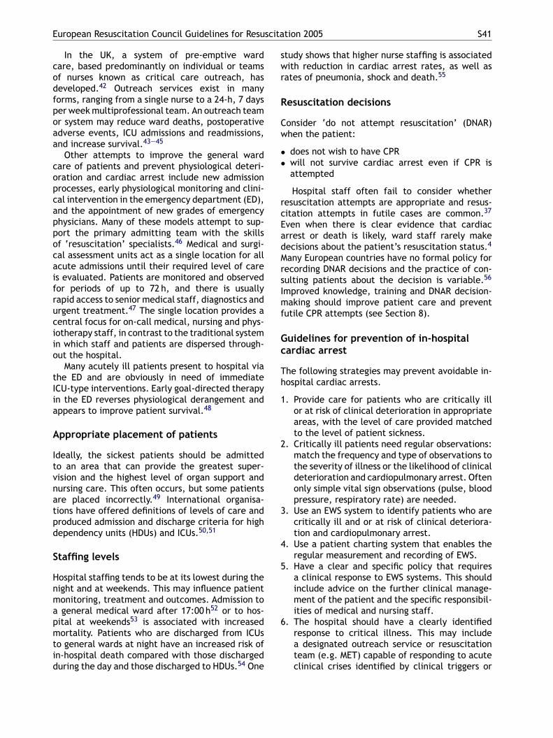
European Resuscitation Council Guidelines for Resuscitation 2005 S41
In the UK, a system of pre-emptive wardcare, based predominantly on individual or teamsof nurses known as critical care outreach, hasdeveloped.42 Outreach services exist in manyforms, ranging from a single nurse to a 24-h, 7 daysper week multiprofessional team. An outreach teamor system may reduce ward deaths, postoperativeadverse events, ICU admissions and readmissions,and increase survival.43—45
Other attempts to improve the general wardcare of patients and prevent physiological deteri-oration and cardiac arrest include new admissionprocesses, early physiological monitoring and clini-cal intervention in the emergency department (ED),and the appointment of new grades of emergencyphysicians. Many of these models attempt to sup-port the primary admitting team with the skillsof ‘resuscitation’ specialists.46 Medical and surgi-cal assessment units act as a single location for allacute admissions until their required level of careis evaluated. Patients are monitored and observedfor periods of up to 72 h, and there is usuallyrapid access to senior medical staff, diagnostics andurgent treatment.47 The single location provides acentral focus for on-call medical, nursing and phys-iio
tIia
A
Itvnatpd
S
Hnmapmtid
study shows that higher nurse staffing is associatedwith reduction in cardiac arrest rates, as well asrates of pneumonia, shock and death.55
Resuscitation decisions
Consider ‘do not attempt resuscitation’ (DNAR)when the patient:
• does not wish to have CPR• will not survive cardiac arrest even if CPR is
attempted
Hospital staff often fail to consider whetherresuscitation attempts are appropriate and resus-citation attempts in futile cases are common.37
Even when there is clear evidence that cardiacarrest or death is likely, ward staff rarely makedecisions about the patient’s resuscitation status.4
Many European countries have no formal policy forrecording DNAR decisions and the practice of con-sulting patients about the decision is variable.56
Improved knowledge, training and DNAR decision-making should improve patient care and preventfutile CPR attempts (see Section 8).
Gc
Th
1
2
3
4
5
6
otherapy staff, in contrast to the traditional systemn which staff and patients are dispersed through-ut the hospital.
Many acutely ill patients present to hospital viahe ED and are obviously in need of immediateCU-type interventions. Early goal-directed therapyn the ED reverses physiological derangement andppears to improve patient survival.48
ppropriate placement of patients
deally, the sickest patients should be admittedo an area that can provide the greatest super-ision and the highest level of organ support andursing care. This often occurs, but some patientsre placed incorrectly.49 International organisa-ions have offered definitions of levels of care androduced admission and discharge criteria for highependency units (HDUs) and ICUs.50,51
taffing levels
ospital staffing tends to be at its lowest during theight and at weekends. This may influence patientonitoring, treatment and outcomes. Admission togeneral medical ward after 17:00 h52 or to hos-
ital at weekends53 is associated with increasedortality. Patients who are discharged from ICUs
o general wards at night have an increased risk ofn-hospital death compared with those dischargeduring the day and those discharged to HDUs.54 One
uidelines for prevention of in-hospitalardiac arrest
he following strategies may prevent avoidable in-ospital cardiac arrests.
. Provide care for patients who are critically illor at risk of clinical deterioration in appropriateareas, with the level of care provided matchedto the level of patient sickness.
. Critically ill patients need regular observations:match the frequency and type of observations tothe severity of illness or the likelihood of clinicaldeterioration and cardiopulmonary arrest. Oftenonly simple vital sign observations (pulse, bloodpressure, respiratory rate) are needed.
. Use an EWS system to identify patients who arecritically ill and or at risk of clinical deteriora-tion and cardiopulmonary arrest.
. Use a patient charting system that enables theregular measurement and recording of EWS.
. Have a clear and specific policy that requiresa clinical response to EWS systems. This shouldinclude advice on the further clinical manage-ment of the patient and the specific responsibil-ities of medical and nursing staff.
. The hospital should have a clearly identifiedresponse to critical illness. This may includea designated outreach service or resuscitationteam (e.g. MET) capable of responding to acuteclinical crises identified by clinical triggers or
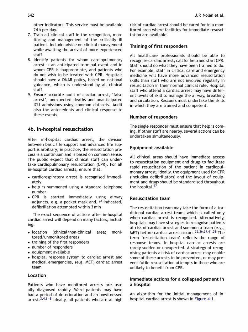
S42 J.P. Nolan et al.
other indicators. This service must be available24 h per day.
7. Train all clinical staff in the recognition, mon-itoring and management of the critically illpatient. Include advice on clinical managementwhile awaiting the arrival of more experiencedstaff.
8. Identify patients for whom cardiopulmonaryarrest is an anticipated terminal event and inwhom CPR is inappropriate, and patients whodo not wish to be treated with CPR. Hospitalsshould have a DNAR policy, based on nationalguidance, which is understood by all clinicalstaff.
9. Ensure accurate audit of cardiac arrest, ‘falsearrest’, unexpected deaths and unanticipatedICU admissions using common datasets. Auditalso the antecedents and clinical response tothese events.
4b. In-hospital resuscitation
After in-hospital cardiac arrest, the divisionbetween basic life support and advanced life sup-
risk of cardiac arrest should be cared for in a mon-itored area where facilities for immediate resusci-tation are available.
Training of first responders
All healthcare professionals should be able torecognise cardiac arrest, call for help and start CPR.Staff should do what they have been trained to do.For example, staff in critical care and emergencymedicine will have more advanced resuscitationskills than staff who are not involved regularly inresuscitation in their normal clinical role. Hospitalstaff who attend a cardiac arrest may have differ-ent levels of skill to manage the airway, breathingand circulation. Rescuers must undertake the skillsin which they are trained and competent.
Number of responders
The single responder must ensure that help is com-ing. If other staff are nearby, several actions can beundertaken simultaneously.
Equipment available
Atrm(mt
R
TdwhaMtrrnsvu
Ia
Ah
port is arbitrary; in practice, the resuscitation pro-cess is a continuum and is based on common sense.The public expect that clinical staff can under-take cardiopulmonary resuscitation (CPR). For allin-hospital cardiac arrests, ensure that:
• cardiorespiratory arrest is recognised immedi-ately
• help is summoned using a standard telephonenumber
• CPR is started immediately using airwayadjuncts, e.g. a pocket mask and, if indicated,defibrillation attempted within 3 min
The exact sequence of actions after in-hospitalcardiac arrest will depend on many factors, includ-ing:
• location (clinical/non-clinical area; moni-tored/unmonitored area)
• training of the first responders• number of responders• equipment available• hospital response system to cardiac arrest and
medical emergencies, (e.g. MET) cardiac arrestteam
Location
Patients who have monitored arrests are usu-ally diagnosed rapidly. Ward patients may havehad a period of deterioration and an unwitnessedarrest.3,4,6—8 Ideally, all patients who are at high
ll clinical areas should have immediate accesso resuscitation equipment and drugs to facilitateapid resuscitation of the patient in cardiopul-onary arrest. Ideally, the equipment used for CPR
including defibrillators) and the layout of equip-ent and drugs should be standardised throughout
he hospital.57
esuscitation team
he resuscitation team may take the form of a tra-itional cardiac arrest team, which is called onlyhen cardiac arrest is recognised. Alternatively,ospitals may have strategies to recognise patientst risk of cardiac arrest and summon a team (e.g.,ET) before cardiac arrest occurs.35,36,39,41,58 The
erm ‘resuscitation team’ reflects the range ofesponse teams. In hospital cardiac arrests arearely sudden or unexpected. A strategy of recog-ising patients at risk of cardiac arrest may enableome of these arrests to be prevented, or may pre-ent futile resuscitation attempts in those who arenlikely to benefit from CPR.
mmediate actions for a collapsed patient inhospital
n algorithm for the initial management of in-ospital cardiac arrest is shown in Figure 4.1.
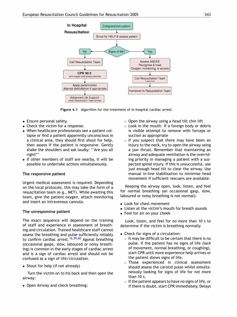
European Resuscitation Council Guidelines for Resuscitation 2005 S43
Figure 4.1 Algorithm for the treatment of in-hospital cardiac arrest.
• Ensure personal safety.• Check the victim for a response.• When healthcare professionals see a patient col-
lapse or find a patient apparently unconscious ina clinical area, they should first shout for help,then assess if the patient is responsive. Gentlyshake the shoulders and ask loudly: ‘‘Are you allright?’’
• If other members of staff are nearby, it will bepossible to undertake actions simultaneously.
The responsive patient
Urgent medical assessment is required. Dependingon the local protocols, this may take the form of aresuscitation team (e.g., MET). While awaiting thisteam, give the patient oxygen, attach monitoringand insert an intravenous cannula.
The unresponsive patient
The exact sequence will depend on the trainingof staff and experience in assessment of breath-ing and circulation. Trained healthcare staff cannotassess the breathing and pulse sufficiently reliablyt 16,59,60
(iac
•
a
•
◦ Open the airway using a head tilt chin lift◦ Look in the mouth. If a foreign body or debris
is visible attempt to remove with forceps orsuction as appropriate
◦ If you suspect that there may have been aninjury to the neck, try to open the airway usinga jaw thrust. Remember that maintaining anairway and adequate ventilation is the overrid-ing priority in managing a patient with a sus-pected spinal injury. If this is unsuccessful, usejust enough head tilt to clear the airway. Usemanual in-line stabilisation to minimise headmovement if sufficient rescuers are available.
Keeping the airway open, look, listen, and feelfor normal breathing (an occasional gasp, slow,laboured or noisy breathing is not normal):
• Look for chest movement• Listen at the victim’s mouth for breath sounds• Feel for air on your cheek
Look, listen, and feel for no more than 10 s todetermine if the victim is breathing normally
• Check for signs of a circulation:◦ It may be difficult to be certain that there is no
o confirm cardiac arrest. Agonal breathingoccasional gasps, slow, laboured or noisy breath-ng) is common in the early stages of cardiac arrestnd is a sign of cardiac arrest and should not beonfused as a sign of life/circulation.
Shout for help (if not already)
Turn the victim on to his back and then open theirway:
Open Airway and check breathing:
pulse. If the patient has no signs of life (lackof movement, normal breathing, or coughing),start CPR until more experience help arrives orthe patient shows signs of life.
◦ Those experienced in clinical assessmentshould assess the carotid pulse whilst simulta-neously looking for signs of life for not morethan 10 s.
◦ If the patient appears to have no signs of life, orif there is doubt, start CPR immediately. Delays

S44 J.P. Nolan et al.
in diagnosis of cardiac arrest and starting CPRwill adversely effect survival must be avoided.
If there is a pulse or signs of life, urgent medi-cal assessment is required. Depending on the localprotocols, this may take the form of a resusci-tation team. While awaiting this team, give thepatient oxygen, attach monitoring, and insert anintravenous cannula.
If there is no breathing, but there is a pulse (res-piratory arrest), ventilate the patient’s lungs andcheck for a circulation every 10 breaths.
Starting in-hospital CPR
• One person starts CPR as others call the resusci-tation team and collect the resuscitation equip-ment and a defibrillator. If only one memberof staff is present, this will mean leaving thepatient.
• Give 30 chest compressions followed by 2 venti-lations.
• Undertaking chest compressions properly is tir-ing; try to change the person doing chest com-pressions every 2 min.
• Recommence chest compressions immediatelyafter the defibrillation attempt. Minimise inter-ruptions to chest compressions.
• Continue resuscitation until the resuscitationteam arrives or the patient shows signs of life.Follow the voice prompts if using an AED. If usinga manual defibrillator, follow the universal algo-rithm for advanced life support (Section 4c).
• Once resuscitation is underway, and if there aresufficient staff present, prepare intravenous can-nulae and drugs likely to be used by the resusci-tation team (e.g. adrenaline).
• Identify one person to be responsible for han-dover to the resuscitation team leader. Locatethe patient’s records.
• The quality of chest compressions during in-hospital CPR is frequently sub-optimal.61,62 Theteam leader should monitor the quality of CPRand change CPR providers if the quality of CPRis poor. The person providing chest compressionsshould be changed every 2 min.
The monitored and witnessed cardiac arrest
If a patient has a monitored and witnessed cardiaca
••
•
T
TcrmTs
4
I
Hdtd
• Maintain the airway and ventilate the lungs withthe most appropriate equipment immediately tohand. A pocket mask, which may be supple-mented with an oral airway, is usually readilyavailable. Alternatively, use a laryngeal mask air-way (LMA) and self-inflating bag, or bag-mask,according to local policy. Tracheal intubationshould be attempted only by those who aretrained, competent and experienced in this skill.
• Use an inspiratory time of 1 s and give enoughvolume to produce a normal chest rise. Add sup-plemental oxygen as soon as possible.
• Once the patient’s trachea has been intubated,continue chest compressions uninterrupted(except for defibrillation or pulse checks whenindicated), at a rate of 100 min−1, and ventilatethe lungs at approximately 10 breaths min−1.Avoid hyperventilation.
• If there is no airway and ventilation equipmentavailable, give mouth-to-mouth ventilation. Ifthere are clinical reasons to avoid mouth-to-mouth contact, or you are unwilling or unableto do this, do chest compressions until help orairway equipment arrives.
• When the defibrillator arrives, apply the pad-dles to the patient and analyse the rhythm. Ifself-adhesive defibrillation pads are available,apply these without interrupting chest compres-sions. Pause briefly to assess the heart rhythm. Ifindicated, attempt either manual or automatedexternal defibrillation (AED).
rrest, act as follows.
Confirm cardiac arrest and shout for help.Consider a precordial thump if the rhythm isVF/VT and a defibrillator is not immediatelyavailable.If the initial rhythm is VF/VT and a defibrillatoris immediately available, give a shock first. Theuse of adhesive electrode pads or a ‘quick-look’paddles technique will enable rapid assessmentof heart rhythm compared with attaching ECGelectrodes.63
raining for healthcare professionals
he Immediate Life Support course trains health-are professionals in the skills required to startesuscitation, including defibrillation, and to beembers of a cardiac arrest team (see Section 9).64
he Advanced Life Support (ALS) course teaches thekills required for leading a resuscitation team.65,66
c. ALS treatment algorithm
ntroduction
eart rhythms associated with cardiac arrest areivided into two groups: shockable rhythms (ven-ricular fibrillation/pulseless ventricular tachycar-ia (VF/VT)) and non-shockable rhythms (asystole
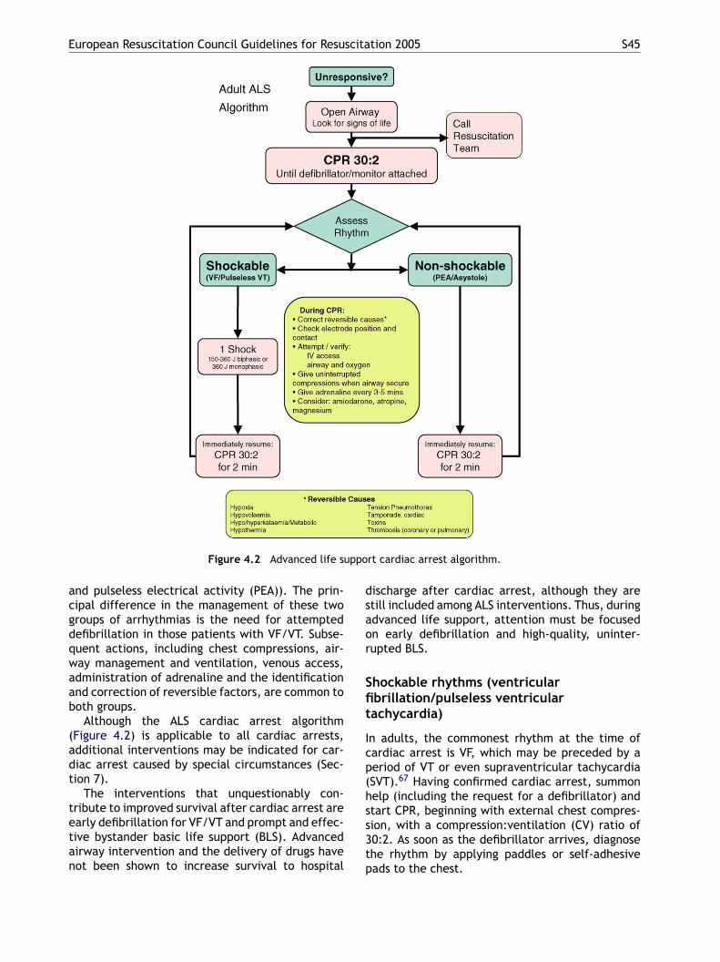
European Resuscitation Council Guidelines for Resuscitation 2005 S45
Figure 4.2 Advanced life support cardiac arrest algorithm.
and pulseless electrical activity (PEA)). The prin-cipal difference in the management of these twogroups of arrhythmias is the need for attempteddefibrillation in those patients with VF/VT. Subse-quent actions, including chest compressions, air-way management and ventilation, venous access,administration of adrenaline and the identificationand correction of reversible factors, are common toboth groups.
Although the ALS cardiac arrest algorithm(Figure 4.2) is applicable to all cardiac arrests,additional interventions may be indicated for car-diac arrest caused by special circumstances (Sec-tion 7).
The interventions that unquestionably con-tribute to improved survival after cardiac arrest areearly defibrillation for VF/VT and prompt and effec-tive bystander basic life support (BLS). Advancedairway intervention and the delivery of drugs havenot been shown to increase survival to hospital
discharge after cardiac arrest, although they arestill included among ALS interventions. Thus, duringadvanced life support, attention must be focusedon early defibrillation and high-quality, uninter-rupted BLS.
Shockable rhythms (ventricularfibrillation/pulseless ventriculartachycardia)
In adults, the commonest rhythm at the time ofcardiac arrest is VF, which may be preceded by aperiod of VT or even supraventricular tachycardia(SVT).67 Having confirmed cardiac arrest, summonhelp (including the request for a defibrillator) andstart CPR, beginning with external chest compres-sion, with a compression:ventilation (CV) ratio of30:2. As soon as the defibrillator arrives, diagnosethe rhythm by applying paddles or self-adhesivepads to the chest.
S46 J.P. Nolan et al.
If VF/VT is confirmed, charge the defibrillatorand give one shock (150—200-J biphasic or 360-J monophasic). Without reassessing the rhythm orfeeling for a pulse, resume CPR (CV ratio 30:2)immediately after the shock, starting with chestcompressions. Even if the defibrillation attempt issuccessful in restoring a perfusing rhythm, it is veryrare for a pulse to be palpable immediately afterdefibrillation,68 and the delay in trying to palpatea pulse will further compromise the myocardiumif a perfusing rhythm has not been restored.69 Ifa perfusing rhythm has been restored, giving chestcompressions does not increase the chance of VFrecurring.70 In the presence of post-shock asystole,chest compressions may usefully induce VF.70 Con-tinue CPR for 2 min, then pause briefly to check themonitor: if there is still VF/VT, give a second shock(150—360-J biphasic or 360-J monophasic). ResumeCPR immediately after the second shock.
Pause briefly after 2 min of CPR to check themonitor: if there is still VF/VT, give adrenalinefollowed immediately by a third shock (150—360-J biphasic or 360-J monophasic) and resumptionof CPR (drug-shock-CPR-rhythm check sequence).Minimise the delay between stopping chest com-
and energy substrates and increase the probabil-ity of restoring a perfusing rhythm after shockdelivery.71 Analyses of VF waveform character-istics predictive of shock success indicate thatthe shorter the time between chest compressionand shock delivery, the more likely the shockwill be successful.71,72 Reduction in the intervalfrom compression to shock delivery by even afew seconds can increase the probability of shocksuccess.73
Regardless of the arrest rhythm, give adrenaline1 mg every 3—5 min until ROSC is achieved; thiswill be once every two loops of the algorithm. Ifsigns of life return during CPR (movement, normalbreathing, or coughing), check the monitor: if anorganised rhythm is present, check for a pulse. If apulse is palpable, continue post-resuscitation careand/or treatment of peri-arrest arrhythmia. If nopulse is present, continue CPR. Providing CPR witha CV ratio of 30:2 is tiring; change the individualundertaking compressions every 2 min.
Precordial thump
Consider giving a single precordial thump when car-dsaaiiatchtilrdswri
A
DqaTetemo
pressions and delivery of the shock. The adenalinethat is given immediately before the shock will becirculated by the CPR that immediately follows theshock. After drug delivery and 2 min of CPR, anal-yse the rhythm and be prepared to deliver anothershock immediately if indicated. If VF/VT persistsafter the third shock, give an intravenous bolus ofamiodarone 300 mg. Inject the amiodarone duringthe brief rhythm analysis before delivery of thefourth shock.
When the rhythm is checked 2 min after givinga shock, if a nonshockable rhythm is present andthe rhythm is organised (complexes appear regularor narrow), try to palpate a pulse. Rhythm checksmust be brief, and pulse checks undertaken onlyif an organised rhythm is observed. If an organ-ised rhythm is seen during a 2 min period of CPR,do not interrupt chest compressions to palpate apulse unless the patient shows signs of life suggest-ing ROSC. If there is any doubt about the presenceof a pulse in the presence of an organised rhythm,resume CPR. If the patient has ROSC, begin postre-suscitation care. If the patient’s rhythm changesto asystole or PEA, see non-shockable rhythmsbelow.
During treatment of VF/VT, healthcare providersmust practice efficient coordination between CPRand shock delivery. When VF is present for morethan a few minutes, the myocardium is depletedof oxygen and metabolic substrates. A briefperiod of chest compressions will deliver oxygen
iac arrest is confirmed rapidly after a witnessed,udden collapse and a defibrillator is not immedi-tely to hand (Section 3).74 These circumstancesre most likely to occur when the patient is mon-tored. A precordial thump should be undertakenmmediately after confirmation of cardiac arrestnd only by healthcare professionals trained inhe technique. Using the ulnar edge of a tightlylenched fist, deliver a sharp impact to the loweralf of the sternum from a height of about 20 cm,hen retract the fist immediately to create anmpulse-like stimulus. A precordial thump is mostikely to be successful in converting VT to sinushythm. Successful treatment of VF by precor-ial thump is much less likely: in all the reporteduccessful cases, the precordial thump was givenithin the first 10 s of VF.75 There are very rare
eports of a precordial thump converting a perfus-ng to a non-perfusing rhythm.76
irway and ventilation
uring the treatment of persistent VF, ensure good-uality chest compressions between defibrillationttempts. Consider reversible causes (4 H’s and 4’s) and, if identified, correct them. Check thelectrode/defibrillating paddle positions and con-acts, and the adequacy of the coupling medium,.g. gel pads. Tracheal intubation provides theost reliable airway, but should be attempted
nly if the healthcare provider is properly trained
European Resuscitation Council Guidelines for Resuscitation 2005 S47
and has adequate ongoing experience with thetechnique. Personnel skilled in advanced airwaymanagement should attempt laryngoscopy with-out stopping chest compressions; a brief pause inchest compressions may be required as the tubeis passed through the vocal cords. Alternatively, toavoid any interruptions in chest compressions, theintubation attempt may be deferred until returnof spontaneous circulation. No intubation attemptshould take longer than 30 s: if intubation has notbeen achieved after this time, recommence bag-mask ventilation. After intubation, confirm cor-rect tube position and secure it adequately. Oncethe patient’s trachea has been intubated, con-tinue chest compressions, at a rate of 100 min−1,without pausing during ventilation. Ventilate thelungs at 10 breaths min−1; do not hyperventilatethe patient. A pause in the chest compressionsallows the coronary perfusion pressure to fall sub-stantially. On resuming compressions there is somedelay before the original coronary perfusion pres-sure is restored, thus chest compressions that arenot interrupted for ventilation result in a substan-tially higher mean coronary perfusion pressure.
In the absence of personnel skilled in trachealiboatvqpt
t
I
PEbadcaCPpbed
IcA
route for vascular access in children, it can also beeffective in adults.78 Intraosseous injection of drugsachieves adequate plasma concentrations in a timecomparable with injection through a central venouscatheter. The intraosseous route also enables with-drawal of marrow for venous blood gas analysis andmeasurement of electrolytes and haemoglobin con-centration.
Tracheal route. If neither intravenous norintraosseous access can be established, somedrugs can be given by the tracheal route. How-ever, unpredictable plasma concentrations areachieved when drugs are given via a tracheal tube,and the optimal tracheal dose of most drugs isunknown. During CPR, the equipotent dose ofadrenaline given via the trachea is three to tentimes higher than the intravenous dose.79,80 Someanimal studies suggest that the lower adrenalineconcentrations achieved when the drug is given viathe trachea may produce transient beta-adrenergiceffects, which will cause hypotension and lowercoronary artery perfusion pressure.81—84 If givenvia the trachea, the dose of adrenaline is 3 mgdiluted to at least 10 ml with sterile water. Dilutionwith water instead of 0.9% saline may achievebs
Aascaartoooamhooangcgc
Agiha
ntubation, acceptable alternatives are the Com-itube, laryngeal mask airway (LMA), ProSeal LMA,r Laryngeal Tube (Section 4d). Once one of theseirways has been inserted, attempt to deliver con-inuous chest compressions, uninterrupted duringentilation. If excessive gas leakage causes inade-uate ventilation of the patient’s lungs, chest com-ressions will have to be interrupted to enable ven-ilation (using a CV ratio of 30:2).
During continuous chest compressions, ventilatehe lungs at 10 breaths min−1.
ntravenous access and drugs
eripheral versus central venous drug delivery.stablish intravenous access if this has not alreadyeen achieved. Although peak drug concentrationsre higher and circulation times are shorter whenrugs are injected into a central venous catheterompared with a peripheral cannula,77 insertion ofcentral venous catheter requires interruption of
PR and is associated with several complications.eripheral venous cannulation is quicker, easier toerform and safer. Drugs injected peripherally muste followed by a flush of at least 20 ml of fluid andlevation of the extremity for 10—20 s to facilitaterug delivery to the central circulation.
ntraosseous route. If intravenous access is diffi-ult or impossible, consider the intraosseous route.lthough normally considered as an alternative
etter drug absorption.85 The solutions in prefilledyringes are acceptable for this purpose.
drenaline. Despite the widespread use ofdrenaline during resuscitation, and severaltudies involving vasopressin, there is no placebo-ontrolled study that shows that the routine use ofny vasopressor at any stage during human cardiacrrest increases survival to hospital discharge. Cur-ent evidence is insufficient to support or refutehe routine use of any particular drug or sequencef drugs. Despite the lack of human data, the usef adrenaline is still recommended, based largelyn animal data. The alpha-adrenergic actions ofdrenaline cause vasoconstriction, which increasesyocardial and cerebral perfusion pressure. The
igher coronary blood flow increases the frequencyf the VF waveform and should improve the chancef restoring a circulation when defibrillation isttempted.86—88 The optimal duration of CPR andumber of shocks that should be given beforeiving drugs is unknown. On the basis of expertonsensus, if VF/VT persists after two shocks,ive adrenaline and repeat every 3—5 min duringardiac arrest. Do not interrupt CPR to give drugs.
nti-arrhythmic drugs. There is no evidence thativing any anti-arrhythmic drug routinely dur-ng human cardiac arrest increases survival toospital discharge. In comparison with placebo89
nd lidocaine,90 the use of amiodarone in shock-
S48 J.P. Nolan et al.
refractory VF improves the short-term outcome ofsurvival to hospital admission. In these studies,the anti-arrhythmic therapy was given if VF/VTpersisted after at least three shocks; however,these were delivered using the conventional three-stacked shocks strategy. There are no data on theuse of amiodarone for shock-refractory VF/VT whensingle shocks are used. On the basis of expert con-sensus, if VF/VT persists after three shocks, give300 mg amiodarone by bolus injection. A furtherdose of 150 mg may be given for recurrent or refrac-tory VF/VT, followed by an infusion of 900 mg over24. Lidocaine 1 mg kg−1 may be used as an alterna-tive if amiodarone is not available, but do not givelidocaine if amiodarone has been given already.
Magnesium. Although the routine use of mag-nesium in cardiac arrest does not increasesurvival,91—95 give magnesium (8 mmol = 4 ml 50%magnesium sulphate or 2 g) for refractory VF ifthere is any suspicion of hypomagnesaemia (e.g.,patients on potassium-losing diuretics).
Bicarbonate. Administering sodium bicarbonateroutinely during cardiac arrest and CPR (especiallyin out-of-hospital cardiac arrests) or after return of
Non-shockable rhythms (PEA and asystole)
Pulseless electrical activity (PEA) is defined ascardiac electrical activity in the absence of anypalpable pulses. These patients often have somemechanical myocardial contractions, but these aretoo weak to produce a detectable pulse or bloodpressure. PEA is often caused by reversible condi-tions, and can be treated if those conditions areidentified and corrected (see below). Survival fol-lowing cardiac arrest with asystole or PEA is unlikelyunless a reversible cause can be found and treatedeffectively.
If the initial monitored rhythm is PEA or asys-tole, start CPR 30:2 and give adrenaline 1 mg assoon as intravascular access is achieved. If asystoleis displayed, check without stopping CPR that theleads are attached correctly. Asystole is a condi-tion that could be exacerbated or precipitated byexcessive vagal tone and, theoretically, this couldbe reversed by a vagolytic drug; therefore, despitethe lack of evidence that routine atropine for asys-tolic cardiac arrest increases survival, give atropine3 mg (the dose that will provide maximum vagalblockade) if there is asystole or the rhythm is slowP −1
ad2pEopapsa
tbi
aitafiipawti
t
spontaneous circulation is not recommended. Givesodium bicarbonate (50 mmol) if cardiac arrest isassociated with hyperkalaemia or tricyclic antide-pressant overdose; repeat the dose according to theclinical condition and result of repeated blood gasanalysis. Some experts give bicarbonate if the arte-rial pH is less than 7.1, but this is controversial.During cardiac arrest, arterial blood gas values donot reflect the acid—base state of the tissues96; thetissue pH will be lower than that in arterial blood.Mixed venous blood values give a more accurateestimate of the pH in the tissues,96 but it is rarefor a pulmonary artery catheter to be in situ at thetime of cardiac arrest. If a central venous catheteris in situ, central venous blood gas analysis will pro-vide a closer estimate of tissue acid/base state thanthat provided by arterial blood.
Persistent ventricular fibrillation
In VF persists, consider changing the position ofthe paddles (Section 3). Review all potentiallyreversible causes (see below) and treat any thatare identified.
The duration of any individual resuscitationattempt is a matter of clinical judgement, takinginto consideration the circumstances and the per-ceived prospect of a successful outcome. If it wasconsidered appropriate to start resuscitation, it isusually considered worthwhile continuing as long asthe patient remains in VF/VT.
EA (rate <60 min ). Secure the airway as soons possible, to enable chest compressions to beelivered without pausing during ventilation. Aftermin of CPR, recheck the rhythm. If no rhythm isresent (asystole), or if there is no change in theCG appearance, resume CPR immediately. If anrganised rhythm is present, attempt to palpate aulse. If no pulse is present (or if there is any doubtbout the presence of a pulse), continue CPR. If aulse is present, begin post-resuscitation care. Ifigns of life return during CPR, check the rhythmnd attempt to palpate a pulse.
Whenever a diagnosis of asystole is made, checkhe ECG carefully for the presence of P waves,ecause this may respond to cardiac pacing. Theres no benefit in attempting to pace true asystole.
If there is doubt about whether the rhythm issystole or fine VF, do not attempt defibrillation;nstead, continue chest compressions and ventila-ion. Fine VF that is difficult to distinguish fromsystole will not be shocked successfully into a per-using rhythm. Continuing good-quality CPR maymprove the amplitude and frequency of the VF andmprove the chance of successful defibrillation to aerfusing rhythm. Delivering repeated shocks in anttempt to defibrillate what is thought to be fine VFill increase myocardial injury, both directly from
he electricity and indirectly from the interruptionsn coronary blood flow.
During the treatment of asystole or PEA, ifhe rhythm changes to VF, follow the left side of
European Resuscitation Council Guidelines for Resuscitation 2005 S49
the algorithm. Otherwise, continue CPR and giveadrenaline every 3—5 min (every other loop of thealgorithm).
Potentially reversible causes
Potential causes or aggravating factors for whichspecific treatment exists must be considered duringany cardiac arrest. For ease of memory, these aredivided into two groups of four based upon theirinitial letter: either H or T. More details on many ofthese conditions are covered in Section 7.
The four Hs
Minimise the risk of hypoxia by ensuring that thepatient’s lungs are ventilated adequately with 100%oxygen. Make sure there is adequate chest riseand bilateral breath sounds. Using the techniquesdescribed in Section 4d, check carefully that thetracheal tube is not misplaced in a bronchus or theoesophagus.
Pulseless electrical activity caused by hypo-volaemia is due usually to severe haemorrhage.Tgars
adptvec
(t
T
AocDt
baatit
In the absence of a specific history, the acci-dental or deliberate ingestion of therapeutic ortoxic substances may be revealed only by labora-tory investigations (Section 7b). Where available,the appropriate antidotes should be used, but mostoften treatment is supportive.
The commonest cause of thromboembolic ormechanical circulatory obstruction is massive pul-monary embolus. If cardiac arrest is thought to becaused by pulmonary embolism, consider giving athrombolytic drug immediately (Section 4e).97
4d. Airway management and ventilation
Introduction
Patients requiring resuscitation often have anobstructed airway, usually secondary to loss of con-sciousness, but occasionally it may be the primarycause of cardiorespiratory arrest. Prompt assess-ment, with control of the airway and ventilation ofthe lungs, is essential. This will help to prevent sec-ondary hypoxic damage to the brain and other vitalorgans. Without adequate oxygenation it may beiTmid
A
C
OpataUtdtcsgtwaacgbst
his may be precipitated by trauma (Section 7i),astrointestinal bleeding or rupture of an aorticneurysm. Intravascular volume should be restoredapidly with fluid, coupled with urgent surgery totop the haemorrhage.
Hyperkalaemia, hypokalaemia, hypocalcaemia,cidaemia and other metabolic disorders areetected by biochemical tests or suggested by theatient’s medical history, e.g. renal failure (Sec-ion 7a). A 12-lead ECG may be diagnostic. Intra-enous calcium chloride is indicated in the pres-nce of hyperkalaemia, hypocalcaemia and calciumhannel-blocker overdose.
Suspect hypothermia in any drowning incidentSections 7c and d); use a low-reading thermome-er.
he four Ts
tension pneumothorax may be the primary causef PEA and may follow attempts at central venousatheter insertion. The diagnosis is made clinically.ecompress rapidly by needle thoracocentesis, andhen insert a chest drain.
Cardiac tamponade is difficult to diagnoseecause the typical signs of distended neck veinsnd hypotension are usually obscured by therrest itself. Cardiac arrest after penetrating chestrauma is highly suggestive of tamponade and is anndication for needle pericardiocentesis or resusci-ative thoracotomy (see Section 7i).
mpossible to restore a spontaneous cardiac output.hese principles may not apply to the witnessed pri-ary cardiac arrest in the vicinity of a defibrillator;
n this case, the priority is immediate attemptedefibrillation.
irway obstruction
auses of airway obstruction
bstruction of the airway may be partial or com-lete. It may occur at any level, from the nosend mouth down to the trachea (Figure 4.3). Inhe unconscious patient, the commonest site ofirway obstruction is at the level of the pharynx.ntil recently this obstruction had been attributedo posterior displacement of the tongue caused byecreased muscle tone; with the tongue ultimatelyouching the posterior pharyngeal wall. The pre-ise cause of airway obstruction in the unconscioustate has been identified by studying patients undereneral anaesthesia.98,99 These studies of anaes-hetised patients have shown that the site of air-ay obstruction is at the soft palate and epiglottisnd not the tongue. Obstruction may be causedlso by vomit or blood (regurgitation of gastricontents or trauma), or by foreign bodies. Laryn-eal obstruction may be caused by oedema fromurns, inflammation or anaphylaxis. Upper airwaytimulation may cause laryngeal spasm. Obstruc-ion of the airway below the larynx is less com-
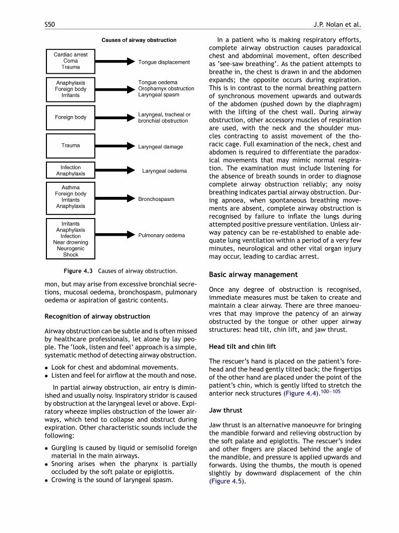
S50 J.P. Nolan et al.
Figure 4.3 Causes of airway obstruction.
mon, but may arise from excessive bronchial secre-tions, mucosal oedema, bronchospasm, pulmonaryoedema or aspiration of gastric contents.
Recognition of airway obstruction
Airway obstruction can be subtle and is often missedby healthcare professionals, let alone by lay peo-ple. The ‘look, listen and feel’ approach is a simple,systematic method of detecting airway obstruction.
• Look for chest and abdominal movements.• Listen and feel for airflow at the mouth and nose.
In partial airway obstruction, air entry is dimin-ished and usually noisy. Inspiratory stridor is causedby obstruction at the laryngeal level or above. Expi-ratory wheeze implies obstruction of the lower air-ways, which tend to collapse and obstruct duringexpiration. Other characteristic sounds include thefollowing:
• Gurgling is caused by liquid or semisolid foreignmaterial in the main airways.
• Snoring arises when the pharynx is partiallyoccluded by the soft palate or epiglottis.
• Crowing is the sound of laryngeal spasm.
In a patient who is making respiratory efforts,complete airway obstruction causes paradoxicalchest and abdominal movement, often describedas ‘see-saw breathing’. As the patient attempts tobreathe in, the chest is drawn in and the abdomenexpands; the opposite occurs during expiration.This is in contrast to the normal breathing patternof synchronous movement upwards and outwardsof the abdomen (pushed down by the diaphragm)with the lifting of the chest wall. During airwayobstruction, other accessory muscles of respirationare used, with the neck and the shoulder mus-cles contracting to assist movement of the tho-racic cage. Full examination of the neck, chest andabdomen is required to differentiate the paradox-ical movements that may mimic normal respira-tion. The examination must include listening forthe absence of breath sounds in order to diagnosecomplete airway obstruction reliably; any noisybreathing indicates partial airway obstruction. Dur-ing apnoea, when spontaneous breathing move-ments are absent, complete airway obstruction isrecognised by failure to inflate the lungs duringattempted positive pressure ventilation. Unless air-way patency can be re-established to enable ade-qmm
B
Oimvos
H
Thopa
J
Jttatfs(
uate lung ventilation within a period of a very fewinutes, neurological and other vital organ injuryay occur, leading to cardiac arrest.
asic airway management
nce any degree of obstruction is recognised,mmediate measures must be taken to create andaintain a clear airway. There are three manoeu-
res that may improve the patency of an airwaybstructed by the tongue or other upper airwaytructures: head tilt, chin lift, and jaw thrust.
ead tilt and chin lift
he rescuer’s hand is placed on the patient’s fore-ead and the head gently tilted back; the fingertipsf the other hand are placed under the point of theatient’s chin, which is gently lifted to stretch thenterior neck structures (Figure 4.4).100—105
aw thrust
aw thrust is an alternative manoeuvre for bringinghe mandible forward and relieving obstruction byhe soft palate and epiglottis. The rescuer’s indexnd other fingers are placed behind the angle ofhe mandible, and pressure is applied upwards andorwards. Using the thumbs, the mouth is openedlightly by downward displacement of the chinFigure 4.5).
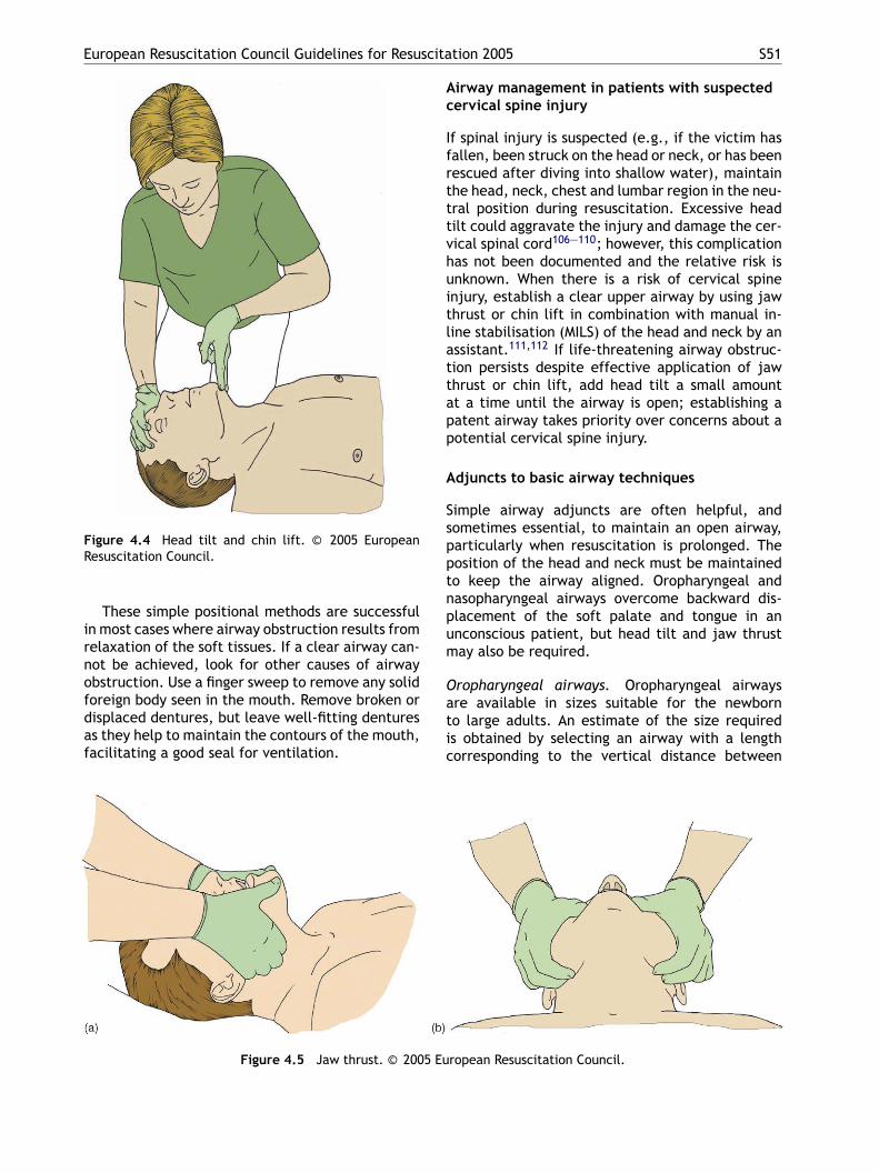
European Resuscitation Council Guidelines for Resuscitation 2005 S51
Figure 4.4 Head tilt and chin lift. © 2005 EuropeanResuscitation Council.
These simple positional methods are successfulin most cases where airway obstruction results fromrelaxation of the soft tissues. If a clear airway can-not be achieved, look for other causes of airwayobstruction. Use a finger sweep to remove any solidforeign body seen in the mouth. Remove broken ordisplaced dentures, but leave well-fitting denturesas they help to maintain the contours of the mouth,facilitating a good seal for ventilation.
Airway management in patients with suspectedcervical spine injury
If spinal injury is suspected (e.g., if the victim hasfallen, been struck on the head or neck, or has beenrescued after diving into shallow water), maintainthe head, neck, chest and lumbar region in the neu-tral position during resuscitation. Excessive headtilt could aggravate the injury and damage the cer-vical spinal cord106—110; however, this complicationhas not been documented and the relative risk isunknown. When there is a risk of cervical spineinjury, establish a clear upper airway by using jawthrust or chin lift in combination with manual in-line stabilisation (MILS) of the head and neck by anassistant.111,112 If life-threatening airway obstruc-tion persists despite effective application of jawthrust or chin lift, add head tilt a small amountat a time until the airway is open; establishing apatent airway takes priority over concerns about apotential cervical spine injury.
Adjuncts to basic airway techniques
Simple airway adjuncts are often helpful, andspptnpum
Oatic
5 Eu
Figure 4.5 Jaw thrust. © 200ometimes essential, to maintain an open airway,articularly when resuscitation is prolonged. Theosition of the head and neck must be maintainedo keep the airway aligned. Oropharyngeal andasopharyngeal airways overcome backward dis-lacement of the soft palate and tongue in annconscious patient, but head tilt and jaw thrustay also be required.
ropharyngeal airways. Oropharyngeal airwaysre available in sizes suitable for the newborno large adults. An estimate of the size requireds obtained by selecting an airway with a lengthorresponding to the vertical distance between
ropean Resuscitation Council.
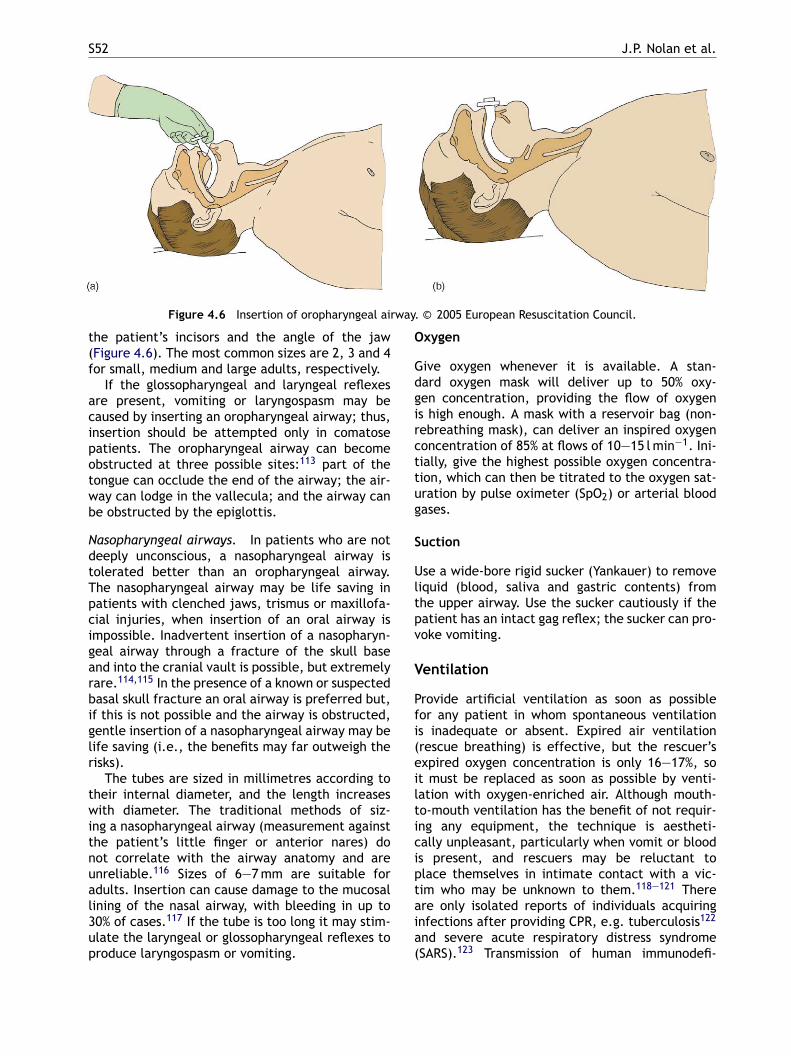
S52 J.P. Nolan et al.
Figure 4.6 Insertion of oropharyngeal airway. © 2005 European Resuscitation Council.
the patient’s incisors and the angle of the jaw(Figure 4.6). The most common sizes are 2, 3 and 4for small, medium and large adults, respectively.
If the glossopharyngeal and laryngeal reflexesare present, vomiting or laryngospasm may becaused by inserting an oropharyngeal airway; thus,insertion should be attempted only in comatosepatients. The oropharyngeal airway can becomeobstructed at three possible sites:113 part of thetongue can occlude the end of the airway; the air-way can lodge in the vallecula; and the airway canbe obstructed by the epiglottis.
Nasopharyngeal airways. In patients who are notdeeply unconscious, a nasopharyngeal airway istolerated better than an oropharyngeal airway.The nasopharyngeal airway may be life saving inpatients with clenched jaws, trismus or maxillofa-cial injuries, when insertion of an oral airway isimpossible. Inadvertent insertion of a nasopharyn-geal airway through a fracture of the skull baseand into the cranial vault is possible, but extremelyrare.114,115 In the presence of a known or suspectedbasal skull fracture an oral airway is preferred but,if this is not possible and the airway is obstructed,
Oxygen
Give oxygen whenever it is available. A stan-dard oxygen mask will deliver up to 50% oxy-gen concentration, providing the flow of oxygenis high enough. A mask with a reservoir bag (non-rebreathing mask), can deliver an inspired oxygenconcentration of 85% at flows of 10—15 l min−1. Ini-tially, give the highest possible oxygen concentra-tion, which can then be titrated to the oxygen sat-uration by pulse oximeter (SpO2) or arterial bloodgases.
Suction
Use a wide-bore rigid sucker (Yankauer) to removeliquid (blood, saliva and gastric contents) fromthe upper airway. Use the sucker cautiously if thepatient has an intact gag reflex; the sucker can pro-voke vomiting.
Ventilation
Provide artificial ventilation as soon as possiblefor any patient in whom spontaneous ventilationi(eilticiptaia(
gentle insertion of a nasopharyngeal airway may belife saving (i.e., the benefits may far outweigh therisks).
The tubes are sized in millimetres according totheir internal diameter, and the length increaseswith diameter. The traditional methods of siz-ing a nasopharyngeal airway (measurement againstthe patient’s little finger or anterior nares) donot correlate with the airway anatomy and areunreliable.116 Sizes of 6—7 mm are suitable foradults. Insertion can cause damage to the mucosallining of the nasal airway, with bleeding in up to30% of cases.117 If the tube is too long it may stim-ulate the laryngeal or glossopharyngeal reflexes toproduce laryngospasm or vomiting.
s inadequate or absent. Expired air ventilationrescue breathing) is effective, but the rescuer’sxpired oxygen concentration is only 16—17%, sot must be replaced as soon as possible by venti-ation with oxygen-enriched air. Although mouth-o-mouth ventilation has the benefit of not requir-ng any equipment, the technique is aestheti-ally unpleasant, particularly when vomit or bloods present, and rescuers may be reluctant tolace themselves in intimate contact with a vic-im who may be unknown to them.118—121 Therere only isolated reports of individuals acquiringnfections after providing CPR, e.g. tuberculosis122
nd severe acute respiratory distress syndromeSARS).123 Transmission of human immunodefi-
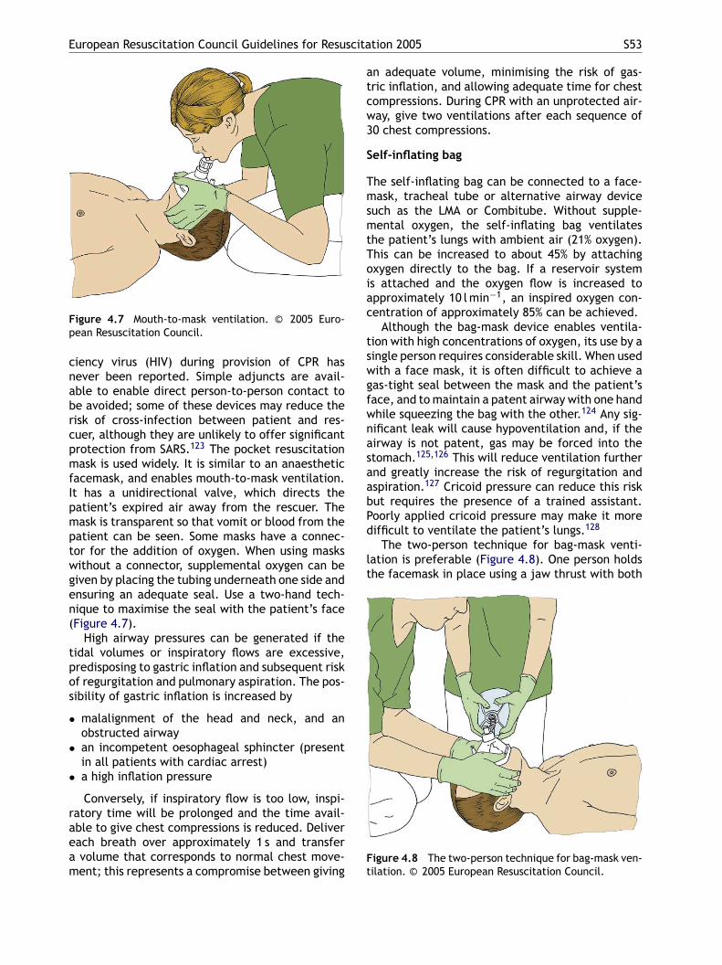
European Resuscitation Council Guidelines for Resuscitation 2005 S53
Figure 4.7 Mouth-to-mask ventilation. © 2005 Euro-pean Resuscitation Council.
ciency virus (HIV) during provision of CPR hasnever been reported. Simple adjuncts are avail-able to enable direct person-to-person contact tobe avoided; some of these devices may reduce therisk of cross-infection between patient and res-cuer, although they are unlikely to offer significantprotection from SARS.123 The pocket resuscitationmask is used widely. It is similar to an anaestheticfacemask, and enables mouth-to-mask ventilation.It has a unidirectional valve, which directs thepatient’s expired air away from the rescuer. Themask is transparent so that vomit or blood from thepatient can be seen. Some masks have a connec-tor for the addition of oxygen. When using maskswithout a connector, supplemental oxygen can begiven by placing the tubing underneath one side andensuring an adequate seal. Use a two-hand tech-nique to maximise the seal with the patient’s face(Figure 4.7).
High airway pressures can be generated if thetidal volumes or inspiratory flows are excessive,predisposing to gastric inflation and subsequent riskof regurgitation and pulmonary aspiration. The pos-sibility of gastric inflation is increased by
• malalignment of the head and neck, and an
•
•
raeam
an adequate volume, minimising the risk of gas-tric inflation, and allowing adequate time for chestcompressions. During CPR with an unprotected air-way, give two ventilations after each sequence of30 chest compressions.
Self-inflating bag
The self-inflating bag can be connected to a face-mask, tracheal tube or alternative airway devicesuch as the LMA or Combitube. Without supple-mental oxygen, the self-inflating bag ventilatesthe patient’s lungs with ambient air (21% oxygen).This can be increased to about 45% by attachingoxygen directly to the bag. If a reservoir systemis attached and the oxygen flow is increased toapproximately 10 l min−1, an inspired oxygen con-centration of approximately 85% can be achieved.
Although the bag-mask device enables ventila-tion with high concentrations of oxygen, its use by asingle person requires considerable skill. When usedwith a face mask, it is often difficult to achieve agas-tight seal between the mask and the patient’sface, and to maintain a patent airway with one handwhile squeezing the bag with the other.124 Any sig-nificant leak will cause hypoventilation and, if theairway is not patent, gas may be forced into thestomach.125,126 This will reduce ventilation furtherand greatly increase the risk of regurgitation andaspiration.127 Cricoid pressure can reduce this riskbut requires the presence of a trained assistant.Poorly applied cricoid pressure may make it moredifficult to ventilate the patient’s lungs.128
The two-person technique for bag-mask venti-lation is preferable (Figure 4.8). One person holdsthe facemask in place using a jaw thrust with both
Figure 4.8 The two-person technique for bag-mask ven-tilation. © 2005 European Resuscitation Council.
obstructed airwayan incompetent oesophageal sphincter (presentin all patients with cardiac arrest)a high inflation pressure
Conversely, if inspiratory flow is too low, inspi-atory time will be prolonged and the time avail-ble to give chest compressions is reduced. Deliverach breath over approximately 1 s and transfervolume that corresponds to normal chest move-ent; this represents a compromise between giving

S54 J.P. Nolan et al.
hands, and an assistant squeezes the bag. In thisway, a better seal can be achieved and the patient’slungs can be ventilated more effectively and safely.
Once a tracheal tube, Combitube or supraglotticairway device has been inserted, ventilate the lungsat a rate of 10 breaths min−1 and continue chestcompressions without pausing during ventilations.The seal of the LMA around the larynx is unlikelyto be good enough to prevent at least some gasleaking when inspiration coincides with chest com-pressions. Moderate gas leakage is acceptable, par-ticularly as most of this gas will pass up through thepatient’s mouth; if excessive gas leakage results ininadequate ventilation of the patient’s lungs, chestcompressions will have to be interrupted to enableventilation, using a compression—ventilation ratioof 30:2.
Automatic ventilators
Very few studies address specific aspects of ventila-tion during advanced life support. There are somedata indicating that the ventilation rates deliveredby healthcare personnel during cardiac arrest areexcessive.61,129 Automatic ventilators or resuscita-
A manikin study of simulated cardiac arrest anda study involving fire-fighters ventilating the lungsof anaesthetised patients both showed a signifi-cant decrease in gastric inflation with manually-triggered flow-limited oxygen-powered resuscita-tors and mask compared with a bag-mask.130,131
However, the effect of automatic resuscitators ongastric inflation in humans in cardiac arrest has notbeen studied, and there are no data demonstratingclear benefit over bag-valve-mask devices.
Alternative airway devices
The tracheal tube has generally been consideredthe optimal method of managing the airway dur-ing cardiac arrest. There is evidence that, withoutadequate training and experience, the incidence ofcomplications, such as unrecognised oesophagealintubation (6—14% in some studies)132—135 anddislodgement, is unacceptably high.136 Prolongedattempts at tracheal intubation are harmful; thecessation of chest compressions during this timewill compromise coronary and cerebral perfusion.Several alternative airway devices have been con-sidered for airway management during CPR. TheC(ibbrsrmnor
L
Ttaihstw
eimmitb
tors provide a constant flow of gas to the patientduring inspiration; the volume delivered is depen-dent on the inspiratory time (a longer time providesa greater tidal volume). Because pressure in the air-way rises during inspiration, these devices are oftenpressure limited to protect the lungs against baro-trauma. An automatic ventilator can be used witheither a facemask or other airway device (e.g., tra-cheal tube, LMA).
An automatic resuscitator should be set ini-tially to deliver a tidal volume of 6—7 ml kg−1 at10 breaths min−1. Some ventilators have coordi-nated markings on the controls to facilitate easyand rapid adjustment for patients of different sizes,and others are capable of sophisticated variation inrespiratory pattern. In the presence of a sponta-neous circulation, the correct setting will be deter-mined by analysis of the patient’s arterial bloodgases.
Automatic resuscitators provide many advan-tages over alternative methods of ventilation.
• In unintubated patients, the rescuer has bothhands free for mask and airway alignment.
• Cricoid pressure can be applied with one handwhile the other seals the mask on the face.
• In intubated patients they free the rescuer forother tasks.
• Once set, they provide a constant tidal volume,respiratory rate and minute ventilation; thus,they may help to avoid excessive ventilation.
ombitube, the LMA, and the Laryngeal TubeLT) are the only alternative devices to be stud-ed during CPR, but none of these studies haveeen powered adequately to enable survival toe studied as a primary endpoint; instead, mostesearchers have studied insertion and ventilationuccess rates. There are no data supporting theoutine use of any specific approach to airwayanagement during cardiac arrest. The best tech-
ique is dependent on the precise circumstancesf the cardiac arrest and the competence of theescuer.
aryngeal mask airway (LMA)
he laryngeal mask airway comprises a wide-boreube with an elliptical inflated cuff designed to sealround the laryngeal opening (Figure 4.9). It is eas-er to insert than a tracheal tube.137—143 The LMAas been studied during CPR, but none of thesetudies has compared it directly with the trachealube. During CPR, successful ventilation is achievedith the LMA in 72—98% of cases.144—150
Ventilation using the LMA is more efficient andasier than with a bag-mask.124 When an LMA can benserted without delay it is preferable to avoid bag-ask ventilation altogether. When used for inter-ittent positive pressure ventilation, provided high
nflation pressures (>20 cm H2O) are avoided, gas-ric inflation can be minimised. In comparison withag-mask ventilation, use of a self-inflating bag and
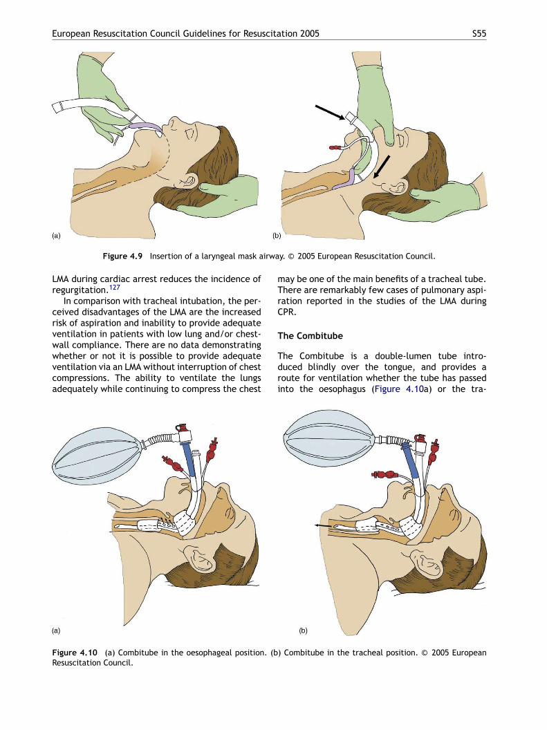
European Resuscitation Council Guidelines for Resuscitation 2005 S55
Figure 4.9 Insertion of a laryngeal mask airway. © 2005 European Resuscitation Council.
LMA during cardiac arrest reduces the incidence ofregurgitation.127
In comparison with tracheal intubation, the per-ceived disadvantages of the LMA are the increasedrisk of aspiration and inability to provide adequateventilation in patients with low lung and/or chest-wall compliance. There are no data demonstratingwhether or not it is possible to provide adequateventilation via an LMA without interruption of chestcompressions. The ability to ventilate the lungsadequately while continuing to compress the chest
may be one of the main benefits of a tracheal tube.There are remarkably few cases of pulmonary aspi-ration reported in the studies of the LMA duringCPR.
The Combitube
The Combitube is a double-lumen tube intro-duced blindly over the tongue, and provides aroute for ventilation whether the tube has passedinto the oesophagus (Figure 4.10a) or the tra-
F . (bR
igure 4.10 (a) Combitube in the oesophageal positionesuscitation Council.
) Combitube in the tracheal position. © 2005 European
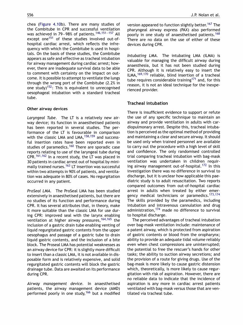
S56 J.P. Nolan et al.
chea (Figure 4.10b). There are many studies ofthe Combitube in CPR and successful ventilationwas achieved in 79—98% of patients.146,151—157 Allexcept one151 of these studies involved out-of-hospital cardiac arrest, which reflects the infre-quency with which the Combitube is used in hospi-tals. On the basis of these studies, the Combitubeappears as safe and effective as tracheal intubationfor airway management during cardiac arrest; how-ever, there are inadequate survival data to be ableto comment with certainty on the impact on out-come. It is possible to attempt to ventilate the lungsthrough the wrong port of the Combitube (2.2% inone study)152: This is equivalent to unrecognisedoesophageal intubation with a standard trachealtube.
Other airway devices
Laryngeal Tube. The LT is a relatively new air-way device; its function in anaesthetised patientshas been reported in several studies. The per-formance of the LT is favourable in comparisonwith the classic LMA and LMA,158,159 and success-ful insertion rates have been reported even in
version appeared to function slightly better.167 Thepharyngeal airway express (PAX) also performedpoorly in one study of anaesthetised patients.168
There are no data on the use of either of thesedevices during CPR.
Intubating LMA. The intubating LMA (ILMA) isvaluable for managing the difficult airway duringanaesthesia, but it has not been studied duringCPR. Although it is relatively easy to insert theILMA,169,170 reliable, blind insertion of a trachealtube requires considerable training171 and, for thisreason, it is not an ideal technique for the inexpe-rienced provider.
Tracheal intubation
There is insufficient evidence to support or refutethe use of any specific technique to maintain anairway and provide ventilation in adults with car-diopulmonary arrest. Despite this, tracheal intuba-tion is perceived as the optimal method of providingand maintaining a clear and secure airway. It shouldbe used only when trained personnel are availabletatviiddcagTiat
oaoaetttbwgnavt
studies of paramedics.160 There are sporadic casereports relating to use of the laryngeal tube duringCPR.161,162 In a recent study, the LT was placed in30 patients in cardiac arrest out of hospital by mini-mally trained nurses.163 LT insertion was successfulwithin two attempts in 90% of patients, and ventila-tion was adequate in 80% of cases. No regurgitationoccurred in any patient.
ProSeal LMA. The ProSeal LMA has been studiedextensively in anaesthetised patients, but there areno studies of its function and performance duringCPR. It has several attributes that, in theory, makeit more suitable than the classic LMA for use dur-ing CPR: improved seal with the larynx enablingventilation at higher airway pressures,164,165 theinclusion of a gastric drain tube enabling venting ofliquid regurgitated gastric contents from the upperoesophagus and passage of a gastric tube to drainliquid gastric contents, and the inclusion of a biteblock. The Proseal LMA has potential weaknesses asan airway device for CPR: it is slightly more difficultto insert than a classic LMA, it is not available in dis-posable form and is relatively expensive, and solidregurgitated gastric contents will block the gastricdrainage tube. Data are awaited on its performanceduring CPR.
Airway management device. In anaesthetisedpatients, the airway management device (AMD)performed poorly in one study,166 but a modified
o carry out the procedure with a high level of skillnd confidence. The only randomised controlledrial comparing tracheal intubation with bag-maskentilation was undertaken in children requir-ng airway management out-of-hospital.172 In thisnvestigation there was no difference in survival toischarge, but it is unclear how applicable this pae-iatric study is to adult resuscitation. Two reportsompared outcomes from out-of-hospital cardiacrrest in adults when treated by either emer-ency medical technicians or paramedics.173,174
he skills provided by the paramedics, includingntubation and intravenous cannulation and drugdministration,174 made no difference to survivalo hospital discharge.
The perceived advantages of tracheal intubationver bag-mask ventilation include: maintenance ofpatent airway, which is protected from aspirationf gastric contents or blood from the oropharynx;bility to provide an adequate tidal volume reliablyven when chest compressions are uninterrupted;he potential to free the rescuer’s hands for otherasks; the ability to suction airway secretions; andhe provision of a route for giving drugs. Use of theag-mask is more likely to cause gastric distensionhich, theoretically, is more likely to cause regur-itation with risk of aspiration. However, there areo reliable data to indicate that the incidence ofspiration is any more in cardiac arrest patientsentilated with bag-mask versus those that are ven-ilated via tracheal tube.
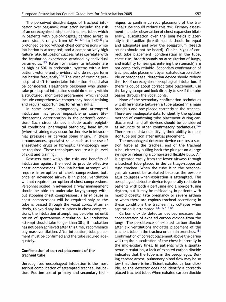
European Resuscitation Council Guidelines for Resuscitation 2005 S57
The perceived disadvantages of tracheal intu-bation over bag-mask ventilation include: the riskof an unrecognised misplaced tracheal tube, whichin patients with out-of-hospital cardiac arrest insome studies ranges from 6%132—134 to 14%135; aprolonged period without chest compressions whileintubation is attempted; and a comparatively highfailure rate. Intubation success rates correlate withthe intubation experience attained by individualparamedics.175 Rates for failure to intubate areas high as 50% in prehospital systems with a lowpatient volume and providers who do not performintubation frequently.134 The cost of training pre-hospital staff to undertake intubation should alsobe considered. Healthcare personnel who under-take prehospital intubation should do so only withina structured, monitored programme, which shouldinclude comprehensive competency-based trainingand regular opportunities to refresh skills.
In some cases, laryngoscopy and attemptedintubation may prove impossible or cause life-threatening deterioration in the patient’s condi-tion. Such circumstances include acute epiglot-tal conditions, pharyngeal pathology, head injury(where straining may occur further rise in intracra-ncabo
icrowPsocttsrahbmq
Ct
Ust
niques to confirm correct placement of the tra-cheal tube should reduce this risk. Primary assess-ment includes observation of chest expansion bilat-erally, auscultation over the lung fields bilater-ally in the axillae (breath sounds should be equaland adequate) and over the epigastrium (breathsounds should not be heard). Clinical signs of cor-rect tube placement (condensation in the tube,chest rise, breath sounds on auscultation of lungs,and inability to hear gas entering the stomach) arenot completely reliable. Secondary confirmation oftracheal tube placement by an exhaled carbon diox-ide or oesophageal detection device should reducethe risk of unrecognised oesophageal intubation. Ifthere is doubt about correct tube placement, usethe laryngoscope and look directly to see if the tubepasses through the vocal cords.
None of the secondary confirmation techniqueswill differentiate between a tube placed in a mainbronchus and one placed correctly in the trachea.There are inadequate data to identify the optimalmethod of confirming tube placement during car-diac arrest, and all devices should be consideredas adjuncts to other confirmatory techniques.176
There are no data quantifying their ability to mon-i
ttsiargaoprmota
clatCwtniilip
ial pressure) or cervical spine injury. In theseircumstances, specialist skills such as the use ofnaesthetic drugs or fibreoptic laryngoscopy maye required. These techniques require a high levelf skill and training.
Rescuers must weigh the risks and benefits ofntubation against the need to provide effectivehest compressions. The intubation attempt willequire interruption of chest compressions but,nce an advanced airway is in place, ventilationill not require interruption of chest compressions.ersonnel skilled in advanced airway managementhould be able to undertake laryngoscopy with-ut stopping chest compressions; a brief pause inhest compressions will be required only as theube is passed through the vocal cords. Alterna-ively, to avoid any interruptions in chest compres-ions, the intubation attempt may be deferred untileturn of spontaneous circulation. No intubationttempt should take longer than 30 s; if intubationas not been achieved after this time, recommenceag-mask ventilation. After intubation, tube place-ent must be confirmed and the tube secured ade-uately.
onfirmation of correct placement of theracheal tube
nrecognised oesophageal intubation is the mosterious complication of attempted tracheal intuba-ion. Routine use of primary and secondary tech-
tor tube position after initial placement.The oesophageal detector device creates a suc-
ion force at the tracheal end of the trachealube, either by pulling back the plunger on a largeyringe or releasing a compressed flexible bulb. Airs aspirated easily from the lower airways through
tracheal tube placed in the cartilage-supportedigid trachea. When the tube is in the oesopha-us, air cannot be aspirated because the oesoph-gus collapses when aspiration is attempted. Theesophageal detector device is generally reliable inatients with both a perfusing and a non-perfusinghythm, but it may be misleading in patients withorbid obesity, late pregnancy or severe asthma
r when there are copious tracheal secretions; inhese conditions the trachea may collapse whenspiration is attempted.133,177—180
Carbon dioxide detector devices measure theoncentration of exhaled carbon dioxide from theungs. The persistence of exhaled carbon dioxidefter six ventilations indicates placement of theracheal tube in the trachea or a main bronchus.181
onfirmation of correct placement above the carinaill require auscultation of the chest bilaterally in
he mid-axillary lines. In patients with a sponta-eous circulation, a lack of exhaled carbon dioxidendicates that the tube is in the oesophagus. Dur-ng cardiac arrest, pulmonary blood flow may be soow that there is insufficient exhaled carbon diox-de, so the detector does not identify a correctlylaced tracheal tube. When exhaled carbon dioxide
S58 J.P. Nolan et al.
is detected in cardiac arrest, it indicates reliablythat the tube is in the trachea or main bronchus but,when it is absent, tracheal tube placement is bestconfirmed with an oesophageal detector device. Avariety of electronic as well as simple, inexpensive,colorimetric carbon dioxide detectors are availablefor both in-hospital and out-of-hospital use.
Cricoid pressure
During bag-mask ventilation and attempted intuba-tion, cricoid pressure applied by a trained assis-tant should prevent passive regurgitation of gas-tric contents and the consequent risk of pulmonaryaspiration. If the technique is applied impreciselyor with excessive force, ventilation and intubationcan be made more difficult.128 If ventilation of thepatient’s lungs is not possible, reduce the pressureapplied to the cricoid cartilage or remove it com-pletely. If the patient vomits, release the cricoidimmediately.
Securing the tracheal tube
4e. Assisting the circulation
Drugs and fluids for cardiac arrest
This topic is divided into: drugs used during themanagement of a cardiac arrest; anti-arrhythmicdrugs used in the peri-arrest period; other drugsused in the peri-arrest period; fluids; and routesfor drug delivery. Every effort has been made toprovide accurate information on the drugs in theseguidelines, but literature from the relevant phar-maceutical companies will provide the most up-to-date data.
Drugs used during the treatment of cardiacarrest
Only a few drugs are indicated during the imme-diate management of a cardiac arrest, and thereis limited scientific evidence supporting their use.Drugs should be considered only after initial shockshave been delivered (if indicated) and chest com-pressions and ventilation have been started.
There are three groups of drugs relevant to themanagement of cardiac arrest that were revieweddsdr
V
Tsasobsftc
AAa4aicbcbdai
Accidental dislodgement of a tracheal tube canoccur at any time, but may be more likely duringresuscitation and during transport. The most effec-tive method for securing the tracheal tube has yetto be determined; use either conventional tapes orties, or purpose-made tracheal tube holders.
Cricothyroidotomy
Occasionally, it will be impossible to ventilate anapnoeic patient with a bag-mask, or to pass a tra-cheal tube or alternative airway device. This mayoccur in patients with extensive facial trauma orlaryngeal obstruction due to oedema or foreignmaterial. In these circumstances, delivery of oxy-gen through a needle or surgical cricothyroidotomymay be life-saving. A tracheostomy is contraindi-cated in an emergency, as it is time consuming,hazardous and requires considerable surgical skilland equipment.
Surgical cricothyroidotomy provides a defini-tive airway that can be used to ventilate thepatient’s lungs until semi-elective intubation or tra-cheostomy is performed. Needle cricothyroidotomyis a much more temporary procedure providingonly short-term oxygenation. It requires a wide-bore, non-kinking cannula, a high-pressure oxygensource, runs the risk of barotrauma and can be par-ticularly ineffective in patients with chest trauma.It is also prone to failure because of kinking of thecannula, and is unsuitable for patient transfer.
uring the 2005 Consensus Conference: vasopres-ors, anti-arrhythmics and other drugs. Routes ofrug delivery other than the optimal intravenousoute were also reviewed and are discussed.
asopressors
here are currently no placebo-controlled studieshowing that the routine use of any vasopressor atny stage during human cardiac arrest increasesurvival to hospital discharge. The primary goalf cardiopulmonary resuscitation is to re-establishlood flow to vital organs until the restoration ofpontaneous circulation. Despite the lack of datarom cardiac arrest in humans, vasopressors con-inue to be recommended as a means of increasingerebral and coronary perfusion during CPR.
drenaline (epinephrine) versus vasopressin.drenaline has been the primary sympathomimeticgent for the management of cardiac arrest for0 years.182 Its primary efficacy is due to itslpha-adrenergic, vasoconstrictive effects caus-ng systemic vasoconstriction, which increasesoronary and cerebral perfusion pressures. Theeta-adrenergic actions of adrenaline (inotropic,hronotropic) may increase coronary and cerebrallood flow, but concomitant increases in myocar-ial oxygen consumption, ectopic ventricularrrhythmias (particularly when the myocardiums acidotic) and transient hypoxaemia due to
European Resuscitation Council Guidelines for Resuscitation 2005 S59
pulmonary arteriovenous shunting may offset thesebenefits.
The potentially deleterious beta-effects ofadrenaline have led to exploration of alternativevasopressors. Vasopressin is a naturally occurringantidiuretic hormone. In very high doses it is apowerful vasoconstrictor that acts by stimulationof smooth muscle V1 receptors. The importance ofvasopressin in cardiac arrest was first recognised instudies of out-of-hospital cardiac arrest patients,where vasopressin levels were found to be higher insuccessfully resuscitated patients.183,184 Althoughclinical185,186 and animal187—189 studies demon-strated improved haemodynamic variables whenusing vasopressin as an alternative to adrenalineduring resuscitation from cardiac arrest, some,186
but not all, demonstrated improved survival.190,191
The first clinical use of vasopressin during car-diac arrest was reported in 1996 and appearedpromising. In a study of cardiac arrest patientsrefractory to standard therapy with adrenaline,vasopressin restored a spontaneous circulation inall eight patients, three of whom were dischargedneurologically intact.186 The following year, thesame group published a small randomised trialotvtaAttriiaiacdofi(lhwvw
tedcd
cant difference in the rate of death before hospitaldischarge.195
Participants at the 2005 Consensus Conferencedebated in depth the treatment recommendationsthat should follow from this evidence. Despite theabsence of placebo-controlled trials, adrenalinehas been the standard vasopressor in cardiac arrest.It was agreed that there is currently insufficientevidence to support or refute the use of vaso-pressin as an alternative to, or in combination with,adrenaline in any cardiac arrest rhythm. Currentpractice still supports adrenaline as the primaryvasopressor for the treatment of cardiac arrest ofall rhythms.
AdrenalineIndications
• Adrenaline is the first drug used in cardiac arrestof any aetiology: it is included in the ALS algo-rithm for use every 3—5 min of CPR.
• Adrenaline is preferred in the treatment of ana-phylaxis (Section 7g).
• Adrenaline is second-line treatment for cardio-genic shock.
vlo1A
ddr
etOacIccw
t
•
•
Bc
f out-of-hospital ventricular fibrillation, in whichhe rates of successful resuscitation and sur-ival for 24 h were significantly higher in patientsreated with vasopressin than in those treated withdrenaline.192 Following these two studies, themerican Heart Association (AHA) recommendedhat vasopressin could be used as an alternativeo adrenaline for the treatment of adult shock-efractory VF.182 The success of these small stud-es led to two large randomised studies compar-ng vasopressin with adrenaline for in-hospital193
nd out-of-hospital194 cardiac arrest. Both stud-es randomised patients to receive vasopressin ordrenaline initially, and used adrenaline as a res-ue treatment in patients refractory to the initialrug. Both studies were unable to demonstrate anverall increase in the rates of ROSC or survivalor vasopressin 40 U,193 with the dose repeatedn one study,194 when compared with adrenaline1 mg, repeated), as the initial vasopressor. In thearge out-of-hospital cardiac arrest study,194 post-oc analysis suggested that the subset of patientsith asystole had significant improvement in sur-ival to discharge, but survival neurologically intactas no different.A recent meta-analysis of five randomised
rials195 showed no statistically significant differ-nce between vasopressin and adrenaline for ROSC,eath within 24 h or death before hospital dis-harge. The subgroup analysis based on initial car-iac rhythm did not show any statistically signifi-
Dose. During cardiac arrest, the initial intra-enous dose of adrenaline is 1 mg. When intravascu-ar (intravenous or intra-osseous) access is delayedr cannot be achieved, give 2—3 mg, diluted to0 ml with sterile water, via the tracheal tube.bsorption via the tracheal route is highly variable.
There is no evidence supporting the use of higheroses of adrenaline for patients in refractory car-iac arrest. In some cases, an adrenaline infusion isequired in the post-resuscitation period.
Following return of spontaneous circulation,xcessive (≥1 mg) doses of adrenaline may induceachycardia, myocardial ischaemia, VT and VF.nce a perfusing rhythm is established, if furtherdrenaline is deemed necessary, titrate the dosearefully to achieve an appropriate blood pressure.ntravenous doses of 50—100 mcg are usually suffi-ient for most hypotensive patients. Use adrenalineautiously in patients with cardiac arrest associatedith cocaine or other sympathomimetic drugs.
Use. Adrenaline is available most commonly inwo dilutions:
1 in 10,000 (10 ml of this solution contains 1 mgof adrenaline)1 in 1000 (1 ml of this solution contains 1 mg ofadrenaline)
oth these dilutions are used routinely in Europeanountries.
S60 J.P. Nolan et al.
Various other pressor drugs (e.g.,noradrenaline)196 have been used experimen-tally as an alternative to adrenaline for thetreatment of cardiac arrest.
Anti-arrhythmics
As with vasopressors, the evidence that anti-arrhythmic drugs are of benefit in cardiac arrestis limited. No anti-arrhythmic drug given duringhuman cardiac arrest has been shown to increasesurvival to hospital discharge, although amiodaronehas been shown to increase survival to hospitaladmission.89,90 Despite the lack of human long-termoutcome data, the balance of evidence is in favourof the use anti-arrhythmic drugs for the manage-ment of arrhythmias in cardiac arrest.
Amiodarone. Amiodarone is a membrane-stabilising anti-arrhythmic drug that increases theduration of the action potential and refractoryperiod in atrial and ventricular myocardium. Atri-oventricular conduction is slowed, and a similareffect is seen with accessory pathways. Amiodaronehas a mild negative inotropic action and causesperipheral vasodilation through non-competitive
volume of 20 ml (or from a pre-filled syringe), ifVF/VT persists after the third shock. Amiodaronecan cause thrombophlebitis when injected into aperipheral vein; use a central venous catheter ifone is in situ but,if not, use a large peripheral veinand a generous flush. Details about the use of amio-darone for the treatment of other arrhythmias aregiven in Section 4f.
Clinical aspects of use. Amiodarone may para-doxically be arrhythmogenic, especially if givenconcurrently with drugs that prolong the QTinterval. However, it has a lower incidence ofpro-arrhythmic effects than other anti-arrhythmicdrugs under similar circumstances. The major acuteadverse effects from amiodarone are hypotensionand bradycardia, which can be prevented by slow-ing the rate of drug infusion, or can be treated withfluids and/or inotropic drugs. The side effects asso-ciated with prolonged oral use (abnormalities ofthyroid function, corneal microdeposits, peripheralneuropathy, and pulmonary/hepatic infiltrates) arenot relevant in the acute setting.
Lidocaine. Until the publication of the 2000 ILCORgohiuha
amassttewiofif
ntdtiLmdt
alpha-blocking effects. The hypotension thatoccurs with intravenous amiodarone is related tothe rate of delivery and is due more to the solvent(Polysorbate 80), which causes histamine release,rather than the drug itself.197 The use of anaqueous amiodarone preparation that is relativelyfree from these side effects is encouraged but isnot yet widely available 198,199.
Following three initial shocks, amiodarone inshock-refractory VF improves the short-term out-come of survival to hospital admission com-pared with placebo89 or lignocaine.90 Amio-darone also appears to improve the response todefibrillation when given to humans or animalswith VF or haemodynamically unstable ventriculartachycardia.198—202 There is no evidence to indi-cate the time at which amiodarone should be givenwhen using a single shock strategy. In the clini-cal studies to date, the amiodarone was given ifVF/VT persisted after at least three shocks. For thisreason, and in the absence of any other data, amio-darone 300 mg is recommended if VF/VT persistsafter three shocks.
Indications. Amiodarone is indicated in
• refractory VF/VT• haemodynamically stable ventricular tachycardia
(VT) and other resistant tachyarrhythmias (Sec-tion 4f)
Dose. Consider an initial intravenous dose of300 mg amiodarone, diluted in 5% dextrose to a
uidelines, lidocaine was the antiarrhythmic drugf choice. Comparative studies with amiodarone90
ave displaced it from this position, and lidocaines now recommended only when amiodarone isnavailable. Amiodarone should be available at allospital arrests and to all out-of-hospital arreststtended by ambulance crew.
Lidocaine is a membrane-stabilising anti-rrhythmic drug that acts by increasing theyocyte refractory period. It decreases ventricular
utomaticity, and its local anaesthetic actionuppresses ventricular ectopic activity. Lidocaineuppresses activity of depolarised, arrhythmogenicissues while interfering minimally with the elec-rical activity of normal tissues. Therefore, it isffective in suppressing arrhythmias associatedith depolarisation (e.g. ischaemia, digitalis toxic-
ty) but is relatively ineffective against arrhythmiasccurring in normally polarised cells (e.g., atrialbrillation/flutter). Lidocaine raises the thresholdor ventricular fibrillation.
Lidocaine toxicity causes paraesthesia, drowsi-ess, confusion and muscular twitching progressingo convulsions. It is considered generally that a safeose of lidocaine must not exceed 3 mg kg−1 overhe first hour. If there are signs of toxicity, stop thenfusion immediately; treat seizures if they occur.idocaine depresses myocardial function, but to auch lesser extent than amiodarone. The myocar-ial depression is usually transient and can bereated with intravenous fluids or vasopressors.
European Resuscitation Council Guidelines for Resuscitation 2005 S61
Indications. Lidocaine is indicated in refractoryVF/VT (when amiodarone is unavailable).
Dose. When amiodarone is unavailable, con-sider an initial dose of 100 mg (1—1.5 mg kg−1) oflidocaine for VF/pulseless VT refractory to threeshocks. Give an additional bolus of 50 mg if neces-sary. The total dose should not exceed 3 mg kg−1
during the first hour.
Clinical aspects of use. Lidocaine is metabolisedby the liver, and its half-life is prolonged if thehepatic blood flow is reduced, e.g. in the pres-ence of reduced cardiac output, liver diseaseor in the elderly. During cardiac arrest normalclearance mechanisms do not function, thus highplasma concentrations may be achieved after asingle dose. After 24 h of continuous infusion, theplasma half-life increases significantly. Reduce thedose in these circumstances, and regularly reviewthe indication for continued therapy. Lidocaine isless effective in the presence of hypokalaemiaand hypomagnesaemia, which should be correctedimmediately.
Magnesium sulphate. Magnesium is an important
Indications. Magnesium sulphate is indicated in
• shock-refractory VF in the presence of possiblehypomagnesaemia
• ventricular tachyarrhythmias in the presence ofpossible hypomagnesaemia
• torsades de pointes• digoxin toxicity
Dose. In shock-refractory VF, give an initial intra-venous dose of 2 g (4 ml (8 mmol)) of 50% magne-sium sulphate) peripherally over 1—2 min; it maybe repeated after 10—15 min. Preparations of mag-nesium sulphate solutions differ among Europeancountries.
Clinical aspects of use. Hypokalaemic patientsare often hypomagnesaemic. If ventricular tach-yarrhythmias arise, intravenous magnesium is asafe, effective treatment. The role of magnesiumin acute myocardial infarction is still in doubt. Mag-nesium is excreted by the kidneys, but side effectsassociated with hypermagnesaemia are rare, evenin renal failure. Magnesium inhibits smooth mus-cle contraction, causing vasodilation and a dose-related hypotension, which is usually transient andresponds to intravenous fluids and vasopressors.
O
TirRbnd
Apmenst
(tAvAa
••
•
constituent of many enzyme systems, especiallythose involved with ATP generation in muscle.It plays a major role in neurochemical transmis-sion, where it decreases acetylcholine release andreduces the sensitivity of the motor endplate.Magnesium also improves the contractile responseof the stunned myocardium, and limits infarctsize by a mechanism that has yet to be fullyelucidated.203 The normal plasma range of magne-sium is 0.8—1.0 mmol l−1.
Hypomagnesaemia is often associated withhypokalaemia, and may contribute to arrhythmiasand cardiac arrest. Hypomagnesaemia increasesmyocardial digoxin uptake and decreases cellularNa+/K+-ATP-ase activity. Patients with hypomagne-saemia, hypokalaemia, or both may become car-diotoxic even with therapeutic digitalis levels. Mag-nesium deficiency is not uncommon in hospitalisedpatients and frequently coexists with other elec-trolyte disturbances, particularly hypokalaemia,hypophosphataemia, hyponatraemia and hypocal-caemia.
Although the benefits of giving magnesium inknown hypomagnesaemic states are recognised, thebenefit of giving magnesium routinely during car-diac arrest is unproven. Studies in adults in and outof hospital91—95,204 have failed to demonstrate anyincrease in the rate of ROSC when magnesium isgiven routinely during CPR. There is some evidencethat magnesium may be beneficial in refractoryVF.205
ther drugs
he evidence for the benefits of other drugs, includ-ng atropine, aminophylline and calcium, givenoutinely during human cardiac arrest, is limited.ecommendations for the use of these drugs areased on our understanding of their pharmacody-amic properties and the pathophysiology of car-iac arrest.
tropine. Atropine antagonises the action of thearasympathetic neurotransmitter acetylcholine atuscarinic receptors. Therefore, it blocks the
ffect of the vagus nerve on both the sinoatrial (SA)ode and the atrioventricular (AV) node, increasinginus automaticity and facilitating AV node conduc-ion.
Side effects of atropine are dose-relatedblurred vision, dry mouth and urinary retention);hey are not relevant during a cardiac arrest.cute confusional states may occur after intra-enous injection, particularly in elderly patients.fter cardiac arrest, dilated pupils should not bettributed solely to atropine.
Atropine is indicated in:
asystolepulseless electrical activity (PEA) with a rate<60 min−1
sinus, atrial, or nodal bradycardia when thehaemodynamic condition of the patient is unsta-ble
S62 J.P. Nolan et al.
The recommended adult dose of atropine forasystole or PEA with a rate <60 min−1 is 3 mg intra-venously in a single bolus. Its use in the treatmentof bradycardia is covered in Section 4f. Severalrecent studies have failed to demonstrate any ben-efit from atropine in out-of-hospital or in-hospitalcardiac arrests174,206—210; however, asystole carriesa grave prognosis and there are anecdotal accountsof success after giving atropine. It is unlikely to beharmful in this situation.
Theophylline (aminophylline). Theophylline is aphosphodiesterase inhibitor that increases tissueconcentrations of cAMP and releases adrenalinefrom the adrenal medulla. It has chronotropic andinotropic actions. The limited studies of amino-phylline in bradyasystolic cardiac arrest have failedto demonstrate an increase in ROSC or survival tohospital discharge211—214; the same studies havenot shown that harm is caused by aminophylline.
Aminophylline is indicated in:
• asystolic cardiac arrest• peri-arrest bradycardia refractory to atropine
Theophylline is given as aminophylline, a mix-
not give calcium solutions and sodium bicarbonatesimultaneously by the same route.
Buffers. Cardiac arrest results in combined res-piratory and metabolic acidosis caused by cessa-tion of pulmonary gas exchange and the devel-opment of anaerobic cellular metabolism, respec-tively. The best treatment of acidaemia in cardiacarrest is chest compression; some additional ben-efit is gained by ventilation. If the arterial bloodpH is less than 7.1 (or base excess more negativethan −10 mmol l−1) during or following resuscita-tion from cardiac arrest, consider giving small dosesof sodium bicarbonate (50 ml of an 8.4% solution).During cardiac arrest, arterial gas values may bemisleading and bear little relationship to the tissueacid—base state96; analysis of central venous bloodmay provide a better estimation of tissue pH (seeSection 4c). Bicarbonate causes generation of car-bon dioxide, which diffuses rapidly into cells. Thishas the following effects.
• It exacerbates intracellular acidosis.• It produces a negative inotropic effect on
ischaemic myocardium.• It presents a large, osmotically active, sodium
ture of theophylline with ethylenediamine, which is20 times more soluble than theophylline alone. Therecommended adult dose is 250—500 mg (5 mg kg−1)given by slow intravenous injection.
Theophylline has a narrow therapeutic win-dow with an optimal plasma concentration of10—20 mg l−1 (55—110 mmol l−1). Above this con-centration, side effects such as arrhythmias andconvulsions may occur, especially when givenrapidly by intravenous injection.
Calcium. Calcium plays a vital role in the cellu-lar mechanisms underlying myocardial contraction.There are very few data supporting any benefi-cial action for calcium after most cases of car-diac arrest. High plasma concentrations achievedafter injection may be harmful to the ischaemicmyocardium and may impair cerebral recovery.Give calcium during resuscitation only when indi-cated specifically, i.e. in pulseless electrical activ-ity caused by
• hyperkalaemia• hypocalcaemia• overdose of calcium channel-blocking drugs
The initial dose of 10 ml 10% calcium chloride(6.8 mmol Ca2+) may be repeated if necessary.Calcium can slow the heart rate and precipitatearrhythmias. In cardiac arrest, calcium may begiven by rapid intravenous injection. In the pres-ence of a spontaneous circulation give it slowly. Do
load to an already compromised circulation andbrain.
• It produces a shift to the left in the oxygen disso-ciation curve, further inhibiting release of oxygento the tissues.
Mild acidaemia causes vasodilation and canincrease cerebral blood flow. Therefore, full cor-rection of the arterial blood pH may theoreticallyreduce cerebral blood flow at a particularly criticaltime. As the bicarbonate ion is excreted as car-bon dioxide via the lungs, ventilation needs to beincreased. For all these reasons, metabolic acidosismust be severe to justify giving sodium bicarbon-ate.
Several animal and clinical studies have exam-ined the use of buffers during cardiac arrest. Clin-ical studies using Tribonate®215 or sodium bicar-bonate as buffers have failed to demonstrate anyadvantage.216—220 Only one study has found clinicalbenefit, suggesting that EMS systems using sodiumbicarbonate earlier and more frequently had sig-nificantly higher ROSC and hospital discharge ratesand better long-term neurological outcome.221 Ani-mal studies have generally been inconclusive, butsome have shown benefit in giving sodium bicarbon-ate to treat cardiovascular toxicity (hypotension,cardiac arrhythmias) caused by tricyclic antidepres-sants and other fast sodium channel blockers (Sec-tion 7b).222 Giving sodium bicarbonate routinelyduring cardiac arrest and CPR (especially in out-
European Resuscitation Council Guidelines for Resuscitation 2005 S63
of-hospital cardiac arrests) or after return of spon-taneous circulation is not recommended. Considersodium bicarbonate for life-threatening hyper-kalaemia or cardiac arrest associated with hyper-kalaemia, severe metabolic acidosis, or tricyclicoverdose. Give 50 mmol (50 ml of an 8.4% solu-tion) of sodium bicarbonate intravenously. Repeatthe dose as necessary, but use acid/base analysis(either arterial or central venous) to guide therapy.Severe tissue damage may be caused by subcuta-neous extravasation of concentrated sodium bicar-bonate. The solution is incompatible with calciumsalts as it causes the precipitation of calcium car-bonate.
Thrombolysis during CPR. Adult cardiac arrestis usually caused by acute myocardial ischaemiafollowing coronary artery occlusion by thrombus.There are several reports on the successful useof thrombolytics during cardiac arrest, particu-larly when the arrest was caused by pulmonaryembolism. The use of thrombolytic drugs to breakdown coronary artery and pulmonary artery throm-bus has been the subject of several studies. Throm-bolytics have also been demonstrated in animalsflaeC
baisdgrtooifs
ssoSsiy
mtt
to proven or suspected acute pulmonary embolus.Thrombolysis may be considered in adult cardiacarrest on a case by case basis following initial fail-ure of standard resuscitation in patients in whoman acute thrombotic aetiology for the arrest is sus-pected. Ongoing CPR is not a contraindication tothrombolysis.
Following thrombolysis during CPR for acute pul-monary embolism, survival and good neurologicaloutcome have been reported in cases requiring inexcess of 60 min of CPR. If a thrombolytic drug isgiven in these circumstances, consider performingCPR for at least 60—90 min before termination ofresuscitation attempts.235,236
Intravenous fluids
Hypovolaemia is a potentially reversible cause ofcardiac arrest. Infuse fluids rapidly if hypovolaemiais suspected. In the initial stages of resuscitationthere are no clear advantages to using colloid, souse saline or Hartmann’s solution. Avoid dextrose,which is redistributed away from the intravascu-lar space rapidly and causes hyperglycaemia, whichmay worsen neurological outcome after cardiac
tudies to have beneficial effects on cerebral bloodow during cardiopulmonary resuscitation,223,224
nd a clinical study has reported less anoxicncephalopathy after thrombolytic therapy duringPR.225
Several studies have examined the use of throm-olytic therapy given during non-traumatic cardiacrrest refractory to standard therapy. Two stud-es have shown an increase in ROSC with non-ignificant improvements in survival to hospitalischarge,97,226 and a further study demonstratedreater ICU survival.225 A small series of caseeports has also reported survival to discharge inhree cases refractory to standard therapy with VFr PEA treated with thrombolytics227; conversely,ne large clinical trial228 failed to show any signif-cant benefit for thrombolytics in cases of undif-erentiated PEA out-of-hospital cardiac arrest unre-ponsive to initial interventions.
When given to cardiac arrest patients withuspected or proven pulmonary embolus, twotudies have demonstrated possible benefits229,230;ne found an improvement in 24-h survival.229
everal clinical studies97,226,229,231 and caseeries227,230,232—234 have not demonstrated anyncrease in bleeding complications with thrombol-sis during CPR in non-traumatic cardiac arrest.
There are insufficient clinical data to recom-end the routine use of thrombolysis during non-
raumatic cardiac arrest. Consider thrombolyticherapy when cardiac arrest is thought to be due
arrest.237—244
Whether fluids should be infused routinely duringcardiac arrest is controversial. There are no pub-lished human studies of routine fluid use comparedto no fluids during normovolaemic cardiac arrest.Four animal studies245—248 of experimental ventric-ular fibrillation neither support nor refute the useof intravenous fluids routinely. In the absence ofhypovolaemia, infusion of an excessive volume offluid is likely to be harmful. Use intravenous fluidto flush peripherally injected drugs into the centralcirculation.
Alternative routes for drug delivery
Intraosseous route
If intravenous access cannot be established,intraosseous delivery of resuscitation drugs willachieve adequate plasma concentrations. Severalstudies indicate that intraosseous access is safeand effective for fluid resuscitation, drug deliveryand laboratory evaluation.78,249—255 Traditionally,the intraosseous route is used mainly for children,but it is also effective in adults.
Drugs given via the tracheal tube
Resuscitation drugs can also be given via the tra-cheal tube, but the plasma concentrations achievedusing this route are variable and substantially
S64 J.P. Nolan et al.
lower than those achieved by the intravenous orintraosseous routes.
Doses of adrenaline 3—10 times higher than whengiven intravenously are required to achieve sim-ilar plasma concentrations.79,80 During CPR, lungperfusion is only 10—30% of the normal value,resulting in a pulmonary adrenaline depot. Whencardiac output is restored after a high dose ofendobronchial adrenaline, prolonged reabsorptionof adrenaline from the lungs into the pulmonarycirculation may occur, causing arterial hyperten-sion, malignant arrhythmias and recurrence of VF.80
Lidocaine and atropine can also be given via a tra-cheal tube, but the plasma concentrations achievedare also variable.256—258 If intravenous access isdelayed or cannot be achieved, consider obtain-ing intraosseous access. Give drugs via the trachealtube if intravascular (intravenous or intraosseous)access is delayed or cannot be achieved. There areno benefits from endobronchial injection comparedwith injection of the drug directly into the trachealtube.256 Dilution with water instead of 0.9% salinemay achieve better drug absorption and cause lessreduction in PaO2.85,259
Interposed abdominal compression (IAC-CPR)
The IAC-CPR technique involves compression ofthe abdomen during the relaxation phase ofchest compression.269,270 This enhances venousreturn during CPR271,272 and improves ROSCand short-term survival.273,274 One study showedimproved survival to hospital discharge with IAC-CPR compared with standard CPR for out-of-hospital cardiac arrest,274 but another showed nosurvival advantage.275 CPR devices include thefollowing.
Active compression-decompression CPR(ACD-CPR)
ACD-CPR is achieved with a hand-held deviceequipped with a suction cup to lift the ante-rior chest actively during decompression. Decreas-ing intrathoracic pressure during the decompres-sion phase increases venous return to the heartand increases cardiac output and subsequent coro-nary and cerebral perfusion pressures during thecompression phase.276—279Results of ACD-CPR havebeen mixed. In some clinical studies ACD-CPRiddAhdoht
csoiAb
I
Ttritc(tdifm
CPR techniques and devices
At best, standard manual CPR produces coronaryand cerebral perfusion that is just 30% of normal.260
Several CPR techniques and devices may improvehaemodynamics or short-term survival when usedby well-trained providers in selected cases. To date,no adjunct has consistently been shown to be supe-rior to conventional manual CPR. CPR techniquesinclude the following.
High-frequency chest compressions (HFCC)
High-frequency (>100 compressions min−1) manualor mechanical chest compressions improve haemo-dynamics but have not been shown to improve long-term outcome.261—265
Open-chest CPR
Open-chest CPR produces better coronary perfusioncoronary pressure than standard CPR266 and maybe indicated for patients with cardiac arrest due totrauma (see Section 7i), in the early postoperativephase after cardiothoracic surgery267,268 (see Sec-tion 7h) or when the chest or abdomen is alreadyopen (transdiaphragmatic approach), for example,in trauma surgery.
mproved haemodynamics compared with stan-ard CPR,173,277,279,280 but in another study itid not.281 In three randomised studies,280,282,283
CD-CPR improved long-term survival after out-of-ospital cardiac arrest; however, in five other ran-omised studies, ACD-CPR made no difference toutcome.284—288 The efficacy of ACD-CPR may beighly dependent on the quality and duration ofraining.289
A meta-analysis of 10 trials of out-of-hospitalardiac arrest and two of in-hospital cardiac arresthowed no early or late survival benefit to ACD-CPRver conventional CPR.290 Two post-mortem stud-es have shown more rib and sternal fractures afterCD-CPR compared with conventional CPR,291,292
ut another found no difference.293
mpedance threshold device (ITD)
he impedance threshold device (ITD) is a valvehat limits air entry into the lungs during chestecoil between chest compressions; this decreasesntrathoracic pressure and increases venous returno the heart. When used with a cuffed tra-heal tube and active compression-decompressionACD),294—296 the ITD is thought to act synergis-ically to enhance venous return during activeecompression. The ITD has also been used dur-ng conventional CPR with a tracheal tube oracemask.297 If rescuers can maintain a tight face-ask seal, the ITD may create the same negative
European Resuscitation Council Guidelines for Resuscitation 2005 S65
intrathoracic pressure as when used with a trachealtube.297
In two randomised studies of out-of-hospitalcardiac arrest, ACD-CPR plus the ITD improvedROSC and 24-h survival compared with standardCPR alone.296,298 When used during standard CPR,the ITD increased 24-h survival after PEA out-of-hospital cardiac arrest.297
Mechanical piston CPR
Mechanical piston devices depress the sternumby means of a compressed gas-powered plungermounted on a backboard. In several studies inanimals,299,300 mechanical piston CPR improvedend-tidal carbon dioxide, cardiac output, cerebralblood flow, MAP and short-term neurological out-come. Studies in humans also document improve-ment in end-tidal carbon dioxide and mean arterialpressure when using mechanical piston CPR com-pared with conventional CPR.301—303
Lund University cardiac arrest system (LUCAS)CPR
TiissAicadpcLd
ttap
L
Tciahmdo
Phased thoracic—abdominalcompression—decompression CPR (PTACD-CPR)
Phased thoracic—abdominal compression—decom-pression CPR combines the concepts of IAC-CPRand ACD-CPR. It comprises a hand-held devicethat alternates chest compression and abdominaldecompression with chest decompression andabdominal compression. One randomised study ofadults in cardiac arrest documented no improve-ment in survival from use of PTACD-CPR.311
Minimally invasive direct cardiac massage
Minimally invasive direct cardiac massage (MIDCM)is accomplished by insertion of a small plunger-likedevice through a 2—4-cm incision in the chest wall.In one clinical study the MIDCM generated improvedblood pressure over standard CPR, but the devicecaused cardiac rupture in one postoperative cardio-vascular surgical patient.312 The plunger device isno longer manufactured.
4f. Peri-arrest arrhythmias
I
AataamuThAsbnmpeaa
mw
P
Icpm
he Lund University cardiac arrest system (LUCAS)s a gas-driven sternal compression device thatncorporates a suction cup for active decompres-ion. There are no published randomised humantudies comparing LUCAS-CPR with standard CPR.
study of pigs with VF showed that LUCAS-CPRmproves haemodynamic and short-term survivalompared with standard CPR.304 The LUCAS waslso used in 20 patients, but incomplete outcomeata were reported.304 In another pig study, in com-arison with standard CPR, LUCAS-CPR increasederebral blood flow and cardiac output.305 TheUCAS enables delivery of continuous compressionsuring transport and defibrillation.
Mechanical piston CPR or LUCAS CPR may be par-icularly useful when prolonged CPR is required;his might include during transport to hospital orfter cardiac arrest following hypothermia306 oroisoning.
oad-distributing band CPR or vest CPR
he load distributing band (LDB) is a cir-umferential chest compression device compris-ng a pneumatically actuated constricting bandnd backboard. The use of LDB CPR improvesaemodynamics.307—309 A case—control study docu-ented improvement in survival to the emergencyepartment when LDB-CPR was delivered after out-f-hospital cardiac arrest.310
ntroduction
successful strategy to reduce the mortalitynd morbidity of cardiac arrest includes measureso prevent other potentially serious arrhythmias,nd optimal treatment should they occur. Cardiacrrhythmias are well recognised complications ofyocardial infarction. They may precede ventric-
lar fibrillation or follow successful defibrillation.he treatment algorithms described in this sectionave been designed to enable the non-specialistLS provider to treat the patient effectively andafely in an emergency; for this reason, they haveeen kept as simple as possible. If patients areot acutely ill there may be several other treat-ent options, including the use of drugs (oral orarenteral), that will be less familiar to the non-xpert. In this situation there will be time to seekdvice from cardiologists or other doctors with theppropriate expertise.
More comprehensive information on theanagement of arrhythmias can be found atww.escardio.org.
rinciples of treatment
n all cases, give oxygen and insert an intravenousannula while the arrhythmia is assessed. Wheneverossible, record a 12-lead ECG; this will help deter-ine the precise rhythm, either before treatment
S66 J.P. Nolan et al.
or retrospectively, if necessary with the help of anexpert. Correct any electrolyte abnormalities (e.g.,K+, Mg2+, Ca2+) (Section 7a).
The assessment and treatment of all arrhyth-mias addresses two factors: the condition of thepatient (stable versus unstable), and the nature ofthe arrhythmia.
Adverse signs
The presence or absence of adverse signs or symp-toms will dictate the appropriate treatment formost arrhythmias. The following adverse factorsindicate a patient who is unstable because of thearrhythmia.
1. Clinical evidence of low cardiac output. Thisis seen as pallor, sweating, cold and clammyextremities (increased sympathetic activity),impaired consciousness (reduced cerebral bloodflow), and hypotension (e.g., systolic blood pres-sure <90 mmHg).
2. Excessive tachycardia. Coronary blood flowoccurs predominantly during diastole. Very highheart rates (e.g., >150 min−1) reduce diastole
1. anti-arrhythmic (and other) drugs2. attempted electrical cardioversion3. cardiac pacing
All anti-arrhythmic treatments—–physical man-oeuvres, drugs, or electrical treatment—–can alsobe pro-arrhythmic, so that clinical deteriorationmay be caused by the treatment rather thanlack of effect. Furthermore, the use of multipleanti-arrhythmic drugs or high doses of a single drugcan cause myocardial depression and hypotension.This may cause a deterioration of the cardiacrhythm. Anti-arrhythmic drugs are slower in effectand less reliable than electrical cardioversion inconverting a tachycardia to sinus rhythm; thus,drugs tend to be reserved for stable patientswithout adverse signs, and electrical cardioversionis usually the preferred treatment for the unstablepatient displaying adverse signs.
Once the arrhythmia has been treated success-fully, repeat the 12-lead ECG to enable detectionof any underlying abnormalities that may requirelong-term therapy.
Bradycardia
Aocos
d(c
••••
5eotuaanhzanc
•
critically, decreasing coronary blood flow andcausing myocardial ischaemia. Broad, complextachycardias are tolerated less well by the heartthan narrow, complex tachycardias.
3. Excessive bradycardia. This is defined as aheart rate of <40 beats min−1, but rates of<60 beats min−1 may not be tolerated bypatients with poor cardiac reserve. Even a higherheart rate may be inappropriately slow for apatient with a low stroke-volume.
4. Heart failure. By reducing coronary artery bloodflow, arrhythmias compromise myocardial per-formance. In acute situations this is manifestedby pulmonary oedema (failure of the left ven-tricle) or raised jugular venous pressure, andhepatic engorgement (failure of the right ven-tricle).
5. Chest pain. The presence of chest pain impliesthat the arrhythmia (particularly a tach-yarrhythmia) is causing myocardial ischaemia.This is especially important if there is underly-ing coronary artery disease or structural heartdisease in which myocardial ischaemia is likelyto lead to further life-threatening complicationsincluding cardiac arrest.
Treatment options
Having determined the rhythm and the presence orabsence of adverse signs, there are broadly threeoptions for immediate treatment:
bradycardia is defined strictly as a heart ratef <60 beats min−1. However, it is more helpful tolassify a bradycardia as absolute (<40 beats min−1)r relative, when the heart rate is inappropriatelylow for the haemodynamic state of the patient.
The first step in the assessment of bradycar-ia is to determine if the patient is unstableFigure 4.11). The following adverse signs may indi-ate instability:
systolic blood pressure <90 mmHgheart rate <40 beats min−1
ventricular arrhythmias requiring suppressionheart failure
If adverse signs are present, give atropine,00 mcg, intravenously and, if necessary, repeatvery 3—5 min to a total of 3 mg. Doses of atropinef less than 500 mcg paradoxically may cause fur-her slowing of the heart rate.313 In healthy vol-nteers a dose of 3 mg produces the maximumchievable increase in resting heart rate.314 Usetropine cautiously in the presence of acute coro-ary ischaemia or myocardial infarction; increasedeart rate may worsen ischaemia or increase theone of infarction. If a satisfactory response ischieved with atropine, or the patient is stable,ext determine the risk of asystole, which is indi-ated by:
recent asystole
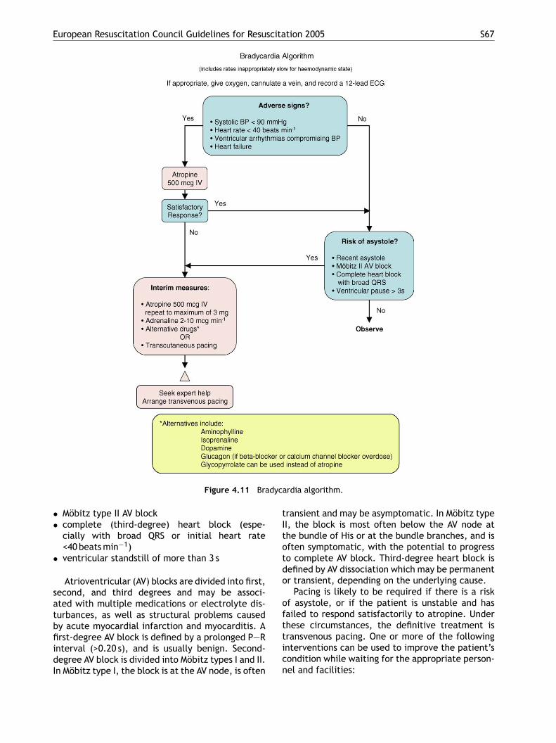
European Resuscitation Council Guidelines for Resuscitation 2005 S67
Figure 4.11 Bradycardia algorithm.
• Mobitz type II AV block• complete (third-degree) heart block (espe-
cially with broad QRS or initial heart rate<40 beats min−1)
• ventricular standstill of more than 3 s
Atrioventricular (AV) blocks are divided into first,second, and third degrees and may be associ-ated with multiple medications or electrolyte dis-turbances, as well as structural problems causedby acute myocardial infarction and myocarditis. Afirst-degree AV block is defined by a prolonged P—Rinterval (>0.20 s), and is usually benign. Second-degree AV block is divided into Mobitz types I and II.In Mobitz type I, the block is at the AV node, is often
transient and may be asymptomatic. In Mobitz typeII, the block is most often below the AV node atthe bundle of His or at the bundle branches, and isoften symptomatic, with the potential to progressto complete AV block. Third-degree heart block isdefined by AV dissociation which may be permanentor transient, depending on the underlying cause.
Pacing is likely to be required if there is a riskof asystole, or if the patient is unstable and hasfailed to respond satisfactorily to atropine. Underthese circumstances, the definitive treatment istransvenous pacing. One or more of the followinginterventions can be used to improve the patient’scondition while waiting for the appropriate person-nel and facilities:
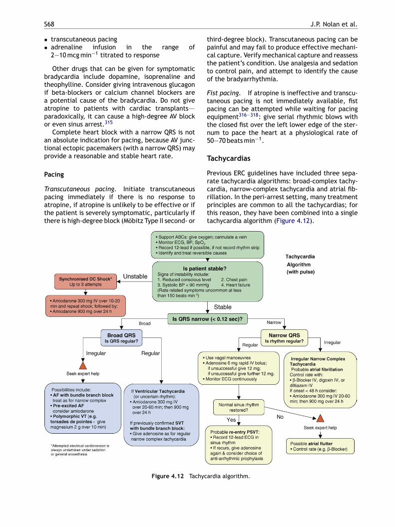
S68 J.P. Nolan et al.
• transcutaneous pacing• adrenaline infusion in the range of
2—10 mcg min−1 titrated to response
Other drugs that can be given for symptomaticbradycardia include dopamine, isoprenaline andtheophylline. Consider giving intravenous glucagonif beta-blockers or calcium channel blockers area potential cause of the bradycardia. Do not giveatropine to patients with cardiac transplants—–paradoxically, it can cause a high-degree AV blockor even sinus arrest.315
Complete heart block with a narrow QRS is notan absolute indication for pacing, because AV junc-tional ectopic pacemakers (with a narrow QRS) mayprovide a reasonable and stable heart rate.
Pacing
Transcutaneous pacing. Initiate transcutaneouspacing immediately if there is no response toatropine, if atropine is unlikely to be effective or ifthe patient is severely symptomatic, particularly ifthere is high-degree block (Mobitz Type II second- or
third-degree block). Transcutaneous pacing can bepainful and may fail to produce effective mechani-cal capture. Verify mechanical capture and reassessthe patient’s condition. Use analgesia and sedationto control pain, and attempt to identify the causeof the bradyarrhythmia.
Fist pacing. If atropine is ineffective and transcu-taneous pacing is not immediately available, fistpacing can be attempted while waiting for pacingequipment316—318: give serial rhythmic blows withthe closed fist over the left lower edge of the ster-num to pace the heart at a physiological rate of50—70 beats min−1.
Tachycardias
Previous ERC guidelines have included three sepa-rate tachycardia algorithms: broad-complex tachy-cardia, narrow-complex tachycardia and atrial fib-rillation. In the peri-arrest setting, many treatmentprinciples are common to all the tachycardias; forthis reason, they have been combined into a singletachycardia algorithm (Figure 4.12).
Figure 4.12 Tachyca
rdia algorithm.
European Resuscitation Council Guidelines for Resuscitation 2005 S69
If the patient is unstable and deteriorating,with signs and symptoms caused by the tachycar-dia (e.g., impaired conscious level, chest pain,heart failure, hypotension or other signs of shock),attempt synchronised cardioversion immediately.In patients with otherwise normal hearts, serioussigns and symptoms are uncommon if the ventricu-lar rate is <150 beats min−1. Patients with impairedcardiac function or significant comorbidity may besymptomatic and unstable at lower heart rates. Ifcardioversion fails to restore sinus rhythm and thepatient remains unstable, give amiodarone 300 mgintravenously over 10—20 min and re-attempt elec-trical cardioversion. The loading dose of amio-darone can be followed by an infusion of 900 mgover 24 h. Serial DC shocks are not appropriate forrecurrent (within hours or days) paroxysms (self-terminating episodes) of atrial fibrillation. This isrelatively common in critically ill patients whomay have ongoing precipitating factors causing thearrhythmia (e.g., metabolic disturbance, sepsis).Cardioversion does not prevent subsequent arrhyth-mias. If there are recurrent episodes, treat themwith drugs.
S
Iostripsbmiaeb
oatttbscibcpsc
Broad-complex tachycardia
In broad-complex tachycardias the QRS complexesare >0.12 s and are usually ventricular in ori-gin. Although broad-complex tachycardias may becaused by supraventricular rhythms with aberrantconduction, in the unstable patient in the peri-arrest context assume they are ventricular in origin.In the stable patient with broad-complex tachycar-dia, the next step is to determine if the rhythm isregular or irregular.
Regular broad complex tachycardia. A regularbroad-complex tachycardia is likely to be ventric-ular tachycardia or SVT with bundle branch block.Stable ventricular tachycardia can be treated withamiodarone 300 mg intravenously over 20—60 minfollowed by an infusion of 900 mg over 24 h. If thebroad-complex regular tachycardia is thought to beSVT with bundle branch block, give adenosine, usingthe strategy indicated for narrow-complex tachy-cardia (below).
Irregular broad complex tachycardia. Irregularbroad complex tachycardia is most likely tobeaowWiQAsu
mIAsdad
svhvoih(caa
ynchronised electrical cardioversion
f electrical cardioversion is used to convert atrialr ventricular tachyarrhythmias, the shock must beynchronised with the R wave of the ECG ratherhan with the T wave. By avoiding the relativeefractory period in this way, the risk of induc-ng ventricular fibrillation is minimised. Consciousatients must be anaesthetised or sedated beforeynchronised cardioversion is attempted. For aroad-complex tachycardia and AF, start with 200-Jonophasic or 120—150 J biphasic and increase in
ncrements if this fails (see Section 3). Atrial flutternd paroxysmal SVT will often convert with lowernergies: start with 100-J monophasic or 70—120-Jiphasic.
If the patient with tachycardia is stable (no seri-us signs or symptoms caused by the tachycardia)nd is not deteriorating, there is time to evaluatehe rhythm using the 12-lead ECG and determinereatment options. The ALS provider may not havehe expertise to diagnose the tachycardia precisely,ut should be capable of differentiating betweeninus tachycardia, narrow-complex SVT and broad-omplex tachycardia. If the patient is stable theres normally time to consult an expert. If the patientecomes unstable, proceed immediately to syn-hronised electrical cardioversion. Management ofatients with significant comorbid conditions andymptomatic tachycardia requires treatment of theomorbid conditions.
e AF with bundle branch block, but carefulxamination of a 12-lead ECG (if necessary byn expert) may enable confident identificationf the rhythm. Another possible cause is AFith ventricular pre-excitation (in patients witholff—Parkinson—White (WPW) syndrome). There
s more variation in the appearance and width of theRS complexes than in AF with bundle branch block.third possible cause is polymorphic VT (e.g., tor-
ade de pointes), but polymorphic VT is relativelynlikely to be present without adverse features.
Seek expert help with the assessment and treat-ent of irregular broad-complex tachyarrhythmia.
f treating AF with bundle branch block, treat as forF (see below). If pre-excited AF (or atrial flutter) isuspected, avoid adenosine, digoxin, verapamil andiltiazem. These drugs block the AV node and causerelative increase in pre-excitation. Electrical car-ioversion is usually the safest treatment option.
Treat torsades de pointes VT immediately bytopping all drugs known to prolong QT inter-al. Correct electrolyte abnormalities, especiallyypokalaemia. Give magnesium sulphate, 2 g, intra-enously over 10 min.319,320 Obtain expert help, asther treatment (e.g., overdrive pacing) may bendicated to prevent relapse once the arrhythmiaas been corrected. If adverse features developwhich is usual), arrange immediate synchronisedardioversion. If the patient becomes pulseless,ttempt defibrillation immediately (cardiac arrestlgorithm).

S70 J.P. Nolan et al.
Narrow-complex tachycardia
Regular narrow-complex tachycardias include:
• sinus tachycardia• AV nodal re-entry tachycardia (AVNRT, the com-
monest type of SVT)• AV re-entry tachycardia (AVRT (due to WPW syn-
drome))• atrial flutter with regular AV conduction (usually
2:1)
Irregular narrow-complex tachycardia is mostcommonly AF or sometimes atrial flutter with vari-able AV conduction (‘variable block’).
Regular narrow-complex tachycardiaSinus tachycardia. Sinus tachycardia is a com-
mon physiological response to a stimulus such asexercise or anxiety. In a sick patient it may be seenin response to many stimuli, such as pain, fever,anaemia, blood loss and heart failure. Treatmentis almost always directed at the underlying cause;trying to slow sinus tachycardia that has occurredin response to most of these situations will makethe situation worse.
or will slow the ventricular response enabling iden-tification of the rhythm. Most typical atrial flutterhas an atrial rate of about 300 beats min−1, so atrialflutter with 2:1 block tends to produce a tachy-cardia of about 150 beats min−1. Much faster rates(170 beats min−1 or more) are unlikely to be due toatrial flutter with 2:1 block.
Treatment of regular narrow complex tachycar-dia. If the patient is unstable with adverse fea-tures caused by the arrhythmia, attempt synchro-nised electrical cardioversion. It is reasonable togive adenosine to an unstable patient with a regu-lar narrow-complex tachycardia while preparationsare made for synchronised cardioversion; however,do not delay electrical cardioversion if the adeno-sine fails to restore sinus rhythm. In the absence ofadverse features, proceed as follows.
• Start with vagal manoeuvres. Carotid sinus mas-sage or the Valsalva manoeuvre will terminateup to a quarter of episodes of paroxysmal SVT.A Valsalva manoeuvre (forced expiration againsta closed glottis) in the supine position may bethe most effective technique. A practical wayof achieving this without protracted explana-
•
•
•
AVNRT and AVRT (paroxysmal SVT). AVNRT is thecommonest type of paroxysmal SVT, often seen inpeople without any other form of heart diseaseand is relatively uncommon in a peri-arrest setting.It causes a regular narrow-complex tachycardia,often with no clearly visible atrial activity on theECG, with heart rates usually well above the typicalrange of sinus rates at rest (60—120 beats min−1).It is usually benign, unless there is additional co-incidental structural heart disease or coronary dis-ease, but may cause symptoms that the patientfinds frightening.
AV re-entry tachycardia (AVRT) is seen in patientswith the WPW syndrome and is also usually benignunless there happens to be additional structuralheart disease. The common type of AVRT is a regu-lar narrow-complex tachycardia, also often havingno visible atrial activity on the ECG.
Atrial flutter with regular AV conduction (often2:1 block). Atrial flutter with regular AV conduc-tion (often 2:1 block) produces a regular narrow-complex tachycardia in which it may be difficultto see atrial activity and identify flutter waveswith confidence, so it may be indistinguishable ini-tially from AVNRT and AVRT. When atrial flutterwith 2:1 block or even 1:1 conduction is accompa-nied by bundle branch block, it produces a regularbroad-complex tachycardia that will usually be verydifficult to distinguish from VT; treatment of thisrhythm as if it were VT will usually be effective,
tion is to ask the patient to blow into a 20-ml syringe with enough force to push back theplunger. Avoid carotid massage if a carotid bruitis present; rupture of an atheromatous plaquecould cause cerebral embolism and stroke. In thecontext of acute ischaemia or digitalis toxicity,sudden bradycardia may trigger VF. Record anECG (preferably multi-lead) during each manoeu-vre. If the rhythm is atrial flutter, slowing of theventricular response will often occur and demon-strate flutter waves.If the arrhythmia persists and is not atrial flutter,use adenosine. Give 6 mg as a rapid intravenousbolus. Record an ECG (preferably multi-lead) dur-ing each injection. If the ventricular rate slowstransiently but the arrhythmia then persists, lookfor atrial activity such as atrial flutter or otheratrial tachycardia and treat accordingly. If thereis no response to adenosine 6 mg, give a 12-mgbolus; if there is no response, give one further12 mg-bolus.Successful termination of a tachyarrhythmia byvagal manoeuvres or adenosine indicates thatit was almost certainly AVNRT or AVRT. Monitorthe patients for further rhythm abnormalities.Treat recurrence either with further adenosine orwith a longer-acting drug with AV nodal-blockingaction (e.g., diltiazem or beta-blocker).Vagal manoeuvres or adenosine will terminatealmost all AVNRT or AVRT within seconds. Failureto terminate a regular narrow-complex tachycar-
European Resuscitation Council Guidelines for Resuscitation 2005 S71
dia with adenosine suggests an atrial tachycardiasuch as atrial flutter.
• If adenosine is contraindicated or fails to ter-minate a regular narrow-complex tachycardiawithout demonstrating that it is atrial flutter,give a calcium channel blocker (e.g., verapamil2.5—5 mg intravenously over 2 min).
Irregular narrow-complex tachycardia
An irregular narrow-complex tachycardia is mostlikely to be AF with an uncontrolled ventricularresponse or, less commonly, atrial flutter with vari-able AV block. Record a 12-lead ECG to identifythe rhythm. If the patient is unstable with adversefeatures caused by the arrhythmia, attempt syn-chronised electrical cardioversion.
If there are no adverse features, treatmentoptions include:
• rate control by drug therapy• rhythm control using drugs to encourage chemi-
cal cardioversion• rhythm control by electrical cardioversion•
plte4tastam
caottudrc
ospac
Antiarrhythmic drugs
Adenosine
Adenosine is a naturally occurring purinenucleotide. It slows transmission across theAV node but has little effect on other myocardialcells or conduction pathways. It is highly effectivefor terminating paroxysmal SVT with re-entrantcircuits that include the AV node (AVNRT). Inother narrow-complex tachycardias, adenosine willreveal the underlying atrial rhythms by slowing theventricular response. It has an extremely short half-life of 10—15 s and, therefore, is given as a rapidbolus into a fast running intravenous infusion or fol-lowed by a saline flush. The smallest dose likely tobe effective is 6 mg (which is outside some currentlicences for an initial dose) and, if unsuccessfulthis can be followed with up to two doses each of12 mg every 1—2 min. Patients should be warnedof transient unpleasant side effects, in particularnausea, flushing, and chest discomfort.327 Adeno-sine is not available in some European countries,but adenosine triphosphate (ATP) is an alternative.In a few European countries neither preparationmay be available; verapamil is probably the nextbbdvetrptcpctmd
A
Ipat
•
•
•
treatment to prevent complications (e.g., anti-coagulation)
Obtain expert help to determine the most appro-riate treatment for the individual patient. Theonger a patient remains in AF, the greater ishe likelihood of atrial clot developing. In gen-ral, patients who have been in AF for more than8 h should not be treated by cardioversion (elec-rical or chemical) until they have received fullnticoagulation or absence of atrial clot has beenhown by transoesophageal echocardiography. Ifhe aim is to control heart rate, options include
beta-blocker,321,322 digoxin, diltiazem,323,324
agnesium325,326 or combinations of these.If the duration of AF is less than 48 h and rhythm
ontrol is considered appropriate, this may bettempted using amiodarone (300 mg intravenouslyver 20—60 min followed by 900 mg over 24 h). Ibu-ilide or flecainide can also be given for rhythm con-rol, but expert advice should be obtained beforesing these drugs for this purpose. Electrical car-ioversion remains an option in this setting and willestore sinus rhythm in more patients than chemi-al cardioversion.
Seek expert help if any patient with AF is knownr found to have ventricular pre-excitation (WPWyndrome). Avoid using adenosine, diltiazem, vera-amil or digoxin to patients with pre-excited AF ortrial flutter, as these drugs block the AV node andause a relative increase in pre-excitation.
est choice. Theophylline and related compoundslock the effect of adenosine. Patients receivingipyridamole or carbamazepine, or with dener-ated (transplanted) hearts, display a markedlyxaggerated effect that may be hazardous. Inhese patients, or if injected into a central vein,educe the initial dose of adenosine to 3 mg. In theresence of WPW syndrome, blockage of conduc-ion across the AV node by adenosine may promoteonduction across an accessory pathway. In theresence of supraventricular arrhythmias this mayause a dangerously rapid ventricular response. Inhe presence of WPW syndrome, rarely, adenosineay precipitate atrial fibrillation associated with aangerously rapid ventricular response.
miodarone
ntravenous amiodarone has effects on sodium,otassium and calcium channels as well as alpha-nd beta-adrenergic blocking properties. Indica-ions for intravenous amiodarone include:
control of haemodynamically stable VT, polymor-phic VT and wide-complex tachycardia of uncer-tain originparoxysmal SVT uncontrolled by adenosine, vagalmanoeuvres or AV nodal blockadeto control rapid ventricular rate due to accessorypathway conduction in pre-excited atrial arrhyth-mias
S72 J.P. Nolan et al.
Give amiodarone, 300 mg intravenously, over10—60 min depending on the circumstances andhaemodynamic stability of the patient. This load-ing dose is followed by an infusion of 900 mg over24 h. Additional infusions of 150 mg can be repeatedas necessary for recurrent or resistant arrhyth-mias to a maximum manufacturer-recommendedtotal daily dose of 2 g (this maximum licensed dosevaries between countries). In patients known tohave severely impaired heart function, intravenousamiodarone is preferable to other anti-arrhythmicdrugs for atrial and ventricular arrhythmias. Majoradverse effects from amiodarone are hypotensionand bradycardia, which can be prevented by slow-ing the rate of drug infusion. The hypotension asso-ciated with amiodarone is caused by vasoactive sol-vents (Polysorbate 80 and benzyl alcohol). A newaqueous formulation of amiodarone does not con-tain these solvents and causes no more hypotensionthan lidocaine.198 Whenever possible, intravenousamiodarone should be given via a central venouscatheter; it causes thrombophlebitis when infusedinto a peripheral vein. In an emergency it should beinjected into a large peripheral vein.
severe LV dysfunction. For the reasons stated underadenosine (above), calcium channel blockers areconsidered harmful when given to patients withAF or atrial flutter associated with known pre-excitation (WPW) syndrome.
Beta-adrenergic blockers
Beta-blocking drugs (atenolol, metoprolol,labetalol (alpha- and beta-blocking effects),propranolol, esmolol) reduce the effects of cir-culating catecholamines and decrease heart rateand blood pressure. They also have cardiopro-tective effects for patients with acute coronarysyndromes. Beta-blockers are indicated for thefollowing tachycardias:
• narrow-complex regular tachycardias uncon-trolled by vagal manoeuvres and adenosine in thepatient with preserved ventricular function
• to control rate in AF and atrial flutter when ven-tricular function is preserved
The intravenous dose of atenolol (beta1) is5 mg given over 5 min, repeated if necessary after10 min. Metoprolol (beta1) is given in doses of2pgv
li55
dtiba
M
Mlsr
4
I
Rov
Calcium channel blockers: verapamil anddiltiazem
Verapamil and diltiazem are calcium channel block-ing drugs that slow conduction and increase refrac-toriness in the AV node. Intravenous diltiazem isnot available in some countries. These actions mayterminate re-entrant arrhythmias and control ven-tricular response rate in patients with a variety ofatrial tachycardias. Indications include:
• stable regular narrow-complex tachycardiasuncontrolled or unconverted by adenosine orvagal manoeuvres
• to control ventricular rate in patients with AF oratrial flutter and preserved ventricular functionwhen the duration of the arrhythmia is less than48 h
The initial dose of verapamil is 2.5—5 mg intra-venously given over 2 min. In the absence of a ther-apeutic response or drug-induced adverse event,give repeated doses of 5—10 mg every 15—30 minto a maximum of 20 mg. Verapamil should be givenonly to patients with narrow-complex paroxysmalSVT or arrhythmias known with certainty to be ofsupraventricular origin.
Diltiazem at a dose of 250 mcg kg−1, followed bya second dose of 350 mcg kg−1, is as effective asverapamil. Verapamil and, to a lesser extent, dil-tiazem may decrease myocardial contractility andcritically reduce cardiac output in patients with
—5 mg at 5-min intervals to a total of 15 mg. Pro-ranolol (beta1 and beta2 effects), 100 mcg kg−1, isiven slowly in three equal doses at 2—3-min inter-als.
Intravenous esmolol is a short-acting (half-ife of 2—9 min) beta1-selective beta-blocker. Its given as an intravenous loading dose of00 mcg kg−1 over 1 min, followed by an infusion of0—200 mcg kg−1 min−1.
Side effects of beta-blockade include bradycar-ias, AV conduction delays and hypotension. Con-raindications to the use of beta-adrenergic block-ng agents include second- or third-degree heartlock, hypotension, severe congestive heart failurend lung disease associated with bronchospasm.
agnesium
agnesium can be given for control of ventricu-ar rate in atrial fibrillation.326,328—330 Give magne-ium sulphate 2 g (8 mmol) over 10 min. This can beepeated once if necessary.
g. Post-resuscitation care
ntroduction
OSC is the just the first step toward the goalf complete recovery from cardiac arrest. Inter-entions in the post-resuscitation period are likely
European Resuscitation Council Guidelines for Resuscitation 2005 S73
to influence the final outcome significantly,237,331
yet there are relatively few data relating to thisphase. Of 22,105 patients admitted to intensivecare units in the UK after cardiac arrest, 9974 (45%)survived to leave intensive care and 6353 (30%)survived to hospital discharge (data from IntensiveCare National Audit and Research Centre (ICNARC),London, December 1995 to October 2004). To returnthe patient to a state of normal cerebral func-tion with no neurological deficit, a stable cardiacrhythm and normal haemodynamic function, fur-ther resuscitation tailored to each patient’s individ-ual needs is required. The post-resuscitation phasestarts at the location where ROSC is achieved but,once stabilised, the patient is transferred to themost appropriate high-care area (e.g., intensivecare unit, coronary care unit) for continued mon-itoring and treatment.
Airway and breathing
Patients who have had a brief period of car-diac arrest responding immediately to appropri-ate treatment may achieve an immediate returnof normal cerebral function. These patients donsafoswcctclnrbnttq
aodtcorgaoa
Circulation
If there is evidence of coronary occlusion, considerthe need for immediate revascularisation by throm-bolysis or percutaneous coronary intervention (seeacute coronary syndromes).
Haemodynamic instability is common after car-diac arrest and manifests as hypotension, lowcardiac index and arrhythmias.337 This post-resuscitation myocardial dysfunction (or myocar-dial stunning) is usually transient and often reverseswithin 24—48 h.338 The post-resuscitation periodis associated with marked elevations in plasmacytokine concentrations, manifesting as a sepsis-like syndrome and multiple organ dysfunction.339
Infusion of fluids may be required to increaseright heart filling pressures or, conversely, diureticsand vasodilators may be needed to treat left ven-tricular failure. In the ICU an arterial line for contin-uous blood pressure monitoring is essential, and theuse of a non-invasive or invasive (pulmonary arterycatheter) cardiac output monitor may be helpful.There are very few randomised trials evaluating therole of blood pressure on the outcome after car-diac arrest. One randomised study demonstratednpsRc2tat
tqihvt4
D
C
IbsacpUpp
ot require tracheal intubation and ventilation buthould be given oxygen via a facemask. Hypoxiand hypercarbia both increase the likelihood of aurther cardiac arrest and may contribute to sec-ndary brain injury. Consider tracheal intubation,edation and controlled ventilation in any patientith obtunded cerebral function. Ensure the tra-heal tube is positioned correctly well above thearina. Hypocarbia causes cerebral vasoconstric-ion and a decreased cerebral blood flow.332 Afterardiac arrest, hypocapnia induced by hyperventi-ation causes cerebral ischaemia.333—336 There areo data to support the targeting of a specific arte-ial PCO2 after resuscitation from cardiac arrest,ut it is reasonable to adjust ventilation to achieveormocarbia and to monitor this using the end-idal PCO2 and arterial blood gas values. Adjusthe inspired oxygen concentrations to achieve ade-uate arterial oxygen saturation.
Insert a gastric tube to decompress the stom-ch; gastric distension caused by mouth-to-mouthr bag-mask-valve ventilation will splint theiaphragm and impair ventilation. Avoid coughing;his will increase intracranial pressure and mayause transient hypoxaemia. Give adequate dosesf sedative and, if absolutely necessary, give a neu-omuscular blocking drug. Obtain a chest radio-raph to check the position of the tracheal tubend central venous lines, etc., assess for pulmonaryedema and to detect complications from CPR suchs a pneumothorax associated with rib fractures.
o difference in the neurological outcome amongatients randomised to a mean arterial blood pres-ure of >100 mmHg versus ≤100 mmHg 5 min afterOSC; however, good functional recovery was asso-iated with a higher blood pressure during the firsth after ROSC.340 In the absence of definitive data,
arget the mean arterial blood pressure to achieven adequate urine output, taking into considerationhe patient’s normal blood pressure.
Immediately after a cardiac arrest there isypically a period of hyperkalaemia. Subse-uent endogenous catecholamine release promotesntracellular transportation of potassium, causingypokalaemia. Hypokalaemia may predispose toentricular arrhythmias. Give potassium to main-ain the serum potassium concentration between.0 and 4.5 mmol l−1.
isability (optimising neurological recovery)
erebral perfusion
mmediately after ROSC there is a period of cere-ral hyperaemia.341 After 15—30 min of reperfu-ion, however, global cerebral blood flow decreasesnd there is generalised hypoperfusion. Normalerebral autoregulation is lost, leaving cerebralerfusion dependent on mean arterial pressure.nder these circumstances, hypotension will com-romise cerebral blood flow severely and will com-ound any neurological injury. Thus, after ROSC,
S74 J.P. Nolan et al.
maintain mean arterial pressure at the patient’snormal level.
Sedation
Although it has been common practice to sedateand ventilate patients for up to 24 h after ROSC,there are no data to support a defined period ofventilation, sedation and neuromuscular blockadeafter cardiac arrest. The duration of sedation andventilation may be influenced by the use of thera-peutic hypothermia (see below). There are no datato indicate whether or not the choice of sedationinfluences outcome, but short-acting drugs (e.g.,propofol, alfentanil, remifentanil) will enable ear-lier neurological assessment. There is an increasedincidence of pneumonia when sedation is prolongedbeyond 48 h after prehospital or in-hospital cardiacarrest.342
Control of seizures
Seizures and/or myoclonus occur in 5—15% ofadult patients who achieve ROSC, and in approx-imately 40% of those who remain comatose.343
showed improved outcome in adults remainingcomatose after initial resuscitation from out-of-hospital VF cardiac arrest, who were cooledwithin minutes to hours after ROSC.354,355 Thesubjects were cooled to 32—34 ◦C for 12—24 h. Onestudy documented improved metabolic endpoints(lactate and O2 extraction) when comatose adultpatients were cooled after ROSC from out-of-hospital cardiac arrest in which the initial rhythmwas PEA/asystole.356 A small study showed ben-efit after therapeutic hypothermia in comatosesurvivors of non-VF arrest.357
External and/or internal cooling techniques canbe used to initiate cooling.354—356,358—361 An infu-sion of 30 mg kg−1 of 4 ◦C-saline decreases coretemperature by 1.5 ◦C.358,359,361,362 Intravascularcooling enables more precise control of core tem-perature than external methods, but it is unknownwhether this improves outcome.360,363—365
Complications of mild therapeutic hypother-mia include increased infection, cardiovascularinstability, coagulopathy, hyperglycaemia and elec-trolyte abnormalities such as hypophosphataemiaand hypomagnesaemia.366,367
Unconscious adult patients with spontaneous cir-csslataaqiee(ott
B
Tgahwc8tbsto
Seizures increase cerebral metabolism by up tofour-fold. Prolonged seizure activity may causecerebral injury, and should be controlled with ben-zodiazepines, phenytoin, propofol or a barbiturate.Each of these drugs can cause hypotension, andthis must be treated appropriately. Seizures andmyoclonus per se are not related significantly tooutcome, but status epilepticus and, in particu-lar, status myoclonus are associated with a pooroutcome.343,344
Temperature control
Treatment of hyperpyrexia. A period of hyper-thermia (hyperpyrexia) is common in the first 48 hafter cardiac arrest.345—347 The risk of a poor neuro-logical outcome increases for each degree of bodytemperature >37 ◦C.348 Antipyretics and/or phys-ical cooling methods decrease infarct volumes inanimal models of global ischaemia.349,350 Treat anyhyperthermia occurring in the first 72 h after car-diac arrest with antipyretics or active cooling.
Therapeutic hypothermia. Mild therapeutichypothermia is thought to suppress many ofthe chemical reactions associated with reperfu-sion injury. These reactions include free-radicalproduction, excitatory amino acid release, andcalcium shifts, which can in turn lead to mito-chondrial damage and apoptosis (programmedcell death).351—353 Two randomised clinical trials
ulation after out-of-hospital VF cardiac arresthould be cooled to 32—34 ◦C. Cooling should betarted as soon as possible and continued for ateast 12—24 h.368—374 Induced hypothermia mightlso benefit unconscious adult patients with spon-aneous circulation after out-of-hospital cardiacrrest from a non-shockable rhythm, or cardiacrrest in hospital. Treat shivering by ensuring ade-uate sedation and giving neuromuscular block-ng drugs. Bolus doses of neuromuscular block-rs are usually adequate, but infusions are nec-ssary occasionally. Rewarm the patient slowly0.25—0.5 ◦C h−1) and avoid hyperthermia. Theptimum target temperature, rate of cooling, dura-ion of hypothermia and rate of rewarming have yeto be determined; further studies are essential.
lood glucose control
here is a strong association between high bloodlucose after resuscitation from cardiac arrestnd poor neurological outcome.237—244 Persistentyperglycaemia after stroke is also associatedith a worse neurological outcome.375—378 Tightontrol of blood glucose (4.4—6.1 mmol l−1 or0—110 mg dl−1) using insulin reduces hospital mor-ality in critically ill adults,379,380 but this has noteen demonstrated in post-cardiac arrest patientspecifically. The benefit is thought to result fromhe strict glycaemic control rather than the dosef insulin infused.381 One rat study has shown
European Resuscitation Council Guidelines for Resuscitation 2005 S75
that glucose plus insulin improves cerebral out-come after asphyxial cardiac arrest.382 There areno randomised controlled human trials of glucosecontrol after cardiac arrest. The optimal blood glu-cose target in critically ill patients has not beendetermined. Comatose patients are at particularrisk from unrecognised hypoglycaemia, and the riskof this complication occurring increases as the tar-get blood glucose concentration is lowered.
In common with all critically ill patients, patientsadmitted to a critical care environment after car-diac arrest should have their blood glucose moni-tored frequently and hyperglycaemia treated withan insulin infusion. The blood glucose concentrationthat triggers insulin therapy, and the target rangeof blood glucose concentrations, should be deter-mined by local policy. There is a need for studies ofglucose control after cardiac arrest.
Prognostication
Once a heart has been resuscitated to a stablerhythm and cardiac output, the organ that influ-ences an individual’s survival most significantly isthe brain. Two thirds of those dying after admissiontfaadtrs
C
Toa5horps
B
Mliafidtf
only meta-analysis to look at this topic estimatedthat to obtain 95% CI with 5% false-positive ratewould require a study population of approximately600 patients.400 No study this large has beenconducted, and these biochemical tests remainunreliable for predicting outcome in individualcases.
Electrophysiological tests
Median nerve somatosensory evoked potentials innormothermic patients, comatose for at least 72 hafter cardiac arrest, predict poor outcome with100% specificity.384 Bilateral absence of the N20component of the evoked potentials in comatosepatients with coma of hypoxic-anoxic origin is uni-formly fatal. When recorded at least 24—48 h afterROSC, the electroencephalogram (EEG), provideslimited prognostic information.401—413 A normal orgrossly abnormal EEG predicts outcome reliably, butan EEG between these extremes is unreliable forprognostication.
References
o ICU following out-of-hospital cardiac arrest dierom neurological injury.383 A quarter of those dyingfter admission to ICU following in-hospital cardiacrrest die from neurological injury. A means of pre-icting neurological outcome that can be appliedo individual patients immediately after ROSC isequired. Such a test of prognosis must have 100%pecificity.linical tests
here are no neurological signs that can predictutcome in the first hours after ROSC. By 3 daysfter the onset of coma relating to cardiac arrest,0% of patients with no chance of ultimate recoveryave died. In the remaining patients, the absencef pupil light reflexes on day 3 and an absent motoresponse to pain on day 3 are both independentlyredictive of a poor outcome (death or vegetativetate) with very high specificity.384—386
iochemical tests
easurement of serum neuron-specific eno-ase (NSE) and protein S-100b may be usefuln determining the outcome of a cardiacrrest.237,243,244,387—399 However, the 95% con-dence interval (CI) in the trials undertaken toate is wide, and in many of the studies returno consciousness (without comment on level ofunction) was considered a ‘‘good’’ outcome. The
1. Gwinnutt CL, Columb M, Harris R. Outcome after cardiacarrest in adults in UK hospitals: effect of the 1997 guide-lines. Resuscitation 2000;47:125—35.
2. Peberdy MA, Kaye W, Ornato JP, et al. Cardiopulmonaryresuscitation of adults in the hospital: a report of 14720cardiac arrests from the National Registry of Cardiopul-monary Resuscitation. Resuscitation 2003;58:297—308.
3. Hodgetts TJ, Kenward G, Vlackonikolis I, et al. Inci-dence, location and reasons for avoidable in-hospital car-diac arrest in a district general hospital. Resuscitation2002;54:115—23.
4. Kause J, Smith G, Prytherch D, Parr M, Flabouris A, Hill-man K. A comparison of antecedents to cardiac arrests,deaths and emergency intensive care admissions in Aus-tralia and New Zealand, and the United Kingdom—–theACADEMIA study. Resuscitation 2004;62:275—82.
5. Herlitz J, Bang A, Aune S, Ekstrom L, Lundstrom G, Holm-berg S. Characteristics and outcome among patients suf-fering in-hospital cardiac arrest in monitored and non-monitored areas. Resuscitation 2001;48:125—35.
6. Franklin C, Mathew J. Developing strategies to preventinhospital cardiac arrest: analyzing responses of physiciansand nurses in the hours before the event. Crit Care Med1994;22:244—7.
7. McQuillan P, Pilkington S, Allan A, et al. Confidential inquiryinto quality of care before admission to intensive care. BMJ1998;316:1853—8.
8. National Confidential Enquiry into Patient Outcome andDeath. An Acute Problem? London, National ConfidentialEnquiry into Patient Outcome and Death, 2005.
9. Cashman JN. In-hospital cardiac arrest: what happens tothe false arrests? Resuscitation 2002;53:271—6.
10. Smith GB, Poplett N. Knowledge of aspects of acute carein trainee doctors. Postgrad Med J 2002;78:335—8.
11. Meek T. New house officers’ knowledge of resuscitation,fluid balance and analgesia. Anaesthesia 2000;55:1128—9.
S76 J.P. Nolan et al.
12. Gould TH, Upton PM, Collins P. A survey of the intendedmanagement of acute postoperative pain by newly quali-fied doctors in the south west region of England in August1992. Anaesthesia 1994;49:807—10.
13. Jackson E, Warner J. How much do doctors know aboutconsent and capacity? J R Soc Med 2002;95:601—3.
14. Kruger PS, Longden PJ. A study of a hospital staff’sknowledge of pulse oximetry. Anaesth Intensive Care1997;25:38—41.
15. Wheeler DW, Remoundos DD, Whittlestone KD, et al.Doctors’ confusion over ratios and percentages in drugsolutions: the case for standard labelling. J R Soc Med2004;97:380—3.
16. Perkins GD, Stephenson B, Hulme J, Monsieurs KG. Birming-ham assessment of breathing study (BABS). Resuscitation2005;64:109—13.
17. Goldacre MJ, Lambert T, Evans J, Turner G. Preregistra-tion house officers’ views on whether their experience atmedical school prepared them well for their jobs: nationalquestionnaire survey. BMJ 2003;326:1011—2.
18. Thwaites BC, Shankar S, Niblett D, Saunders J. Can consul-tants resuscitate? J R Coll Physicians Lond 1992;26:265—7.
19. Saravanan P, Soar J. A survey of resuscitation training needsof senior anaesthetists. Resuscitation 2005;64:93—6.
20. Featherstone P, Smith GB, Linnell M, Easton S, Osgood VM.Impact of a one-day inter-professional course (ALERTTM)on attitudes and confidence in managing critically ill adultpatients. Resuscitation 2005;65:329—36.
21. Harrison GA, Jacques TC, Kilborn G, McLaws ML. The preva-lence of recordings of the signs of critical conditions and
ness of the emergency response. Resuscitation 2004;62:291—7.
34. Soar J, McKay U. A revised role for the hospital cardiacarrest team? Resuscitation 1998;38:145—9.
35. Bellomo R, Goldsmith D, Uchino S, et al. A prospectivebefore-and-after trial of a medical emergency team. MedJ Aust 2003;179:283—7.
36. Buist MD, Moore GE, Bernard SA, Waxman BP, AndersonJN, Nguyen TV. Effects of a medical emergency teamon reduction of incidence of and mortality from unex-pected cardiac arrests in hospital: preliminary study. BMJ2002;324:387—90.
37. Parr MJ, Hadfield JH, Flabouris A, Bishop G, Hillman K. TheMedical Emergency Team: 12 month analysis of reasons foractivation, immediate outcome and not-for-resuscitationorders. Resuscitation 2001;50:39—44.
38. Bellomo R, Goldsmith D, Uchino S, et al. Prospective con-trolled trial of effect of medical emergency team on post-operative morbidity and mortality rates. Crit Care Med2004;32:916—21.
39. Kenward G, Castle N, Hodgetts T, Shaikh L. Evaluation of amedical emergency team one year after implementation.Resuscitation 2004;61:257—63.
40. Jones D, Bates S, Warrillow S, et al. Circadian pattern ofactivation of the medical emergency team in a teachinghospital. Crit Care 2005;9:R303—6.
41. The MERIT study investigators. Introduction of the medi-cal emergency team (MET) system: a cluster-randomisedcontrolled trial. Lancet 2005;365:2091—7.
42. Critical care outreach 2003: progress in developing ser-
emergency responses in hospital wards—–the SOCCER study.Resuscitation 2005;65:149—57.22. Buist M, Bernard S, Nguyen TV, Moore G, Anderson J. Asso-ciation between clinically abnormal observations and sub-sequent in-hospital mortality: a prospective study. Resus-citation 2004;62:137—41.
23. Goldhill DR, Worthington L, Mulcahy A, Tarling M, SumnerA. The patient-at-risk team: identifying and managing seri-ously ill ward patients. Anaesthesia 1999;54:853—60.
24. Hodgetts TJ, Kenward G, Vlachonikolis IG, Payne S, CastleN. The identification of risk factors for cardiac arrest andformulation of activation criteria to alert a medical emer-gency team. Resuscitation 2002;54:125—31.
25. Subbe CP, Davies RG, Williams E, Rutherford P, Gemmell L.Effect of introducing the Modified Early Warning score onclinical outcomes, cardio-pulmonary arrests and intensivecare utilisation in acute medical admissions. Anaesthesia2003;58:797—802.
26. Lee A, Bishop G, Hillman KM, Daffurn K. The Medical Emer-gency Team. Anaesth Intensive Care 1995;23:183—6.
27. Cuthbertson BH. Outreach critical care—–cash for no ques-tions? Br J Anaesth 2003;90:4—6.
28. Parr M. Critical care outreach: some answers, more ques-tions. Intensive Care Med 2004;30:1261—2.
29. Goldhill DR, McNarry AF. Physiological abnormalities inearly warning scores are related to mortality in adult inpa-tients. Br J Anaesth 2004;92:882—4.
30. Subbe CP, Williams EM, Gemmell LW. Are medical emer-gency teams picking up enough patients with increasedrespiratory rate? Crit Care Med 2004;32:1983—4.
31. McBride J, Knight D, Piper J, Smith GB. Long-term effectof introducing an early warning score on respiratory ratecharting on general wards. Resuscitation 2005;65:41—4.
32. Carberry M. Implementing the modified early warning sys-tem: our experiences. Nurs Crit Care 2002;7:220—6.
33. Sandroni C, Ferro G, Santangelo S, et al. In-hospital car-diac arrest: survival depends mainly on the effective-
vices. The National Outreach Report 2003. London, Depart-ment of Health and National Health Service ModernisationAgency; 2003.
43. Ball C, Kirkby M, Williams S. Effect of the critical care out-reach team on patient survival to discharge from hospitaland readmission to critical care: non-randomised popula-tion based study. BMJ 2003;327:1014.
44. Priestley G, Watson W, Rashidian A, et al. Introducing Crit-ical Care Outreach: a ward-randomised trial of phasedintroduction in a general hospital. Intensive Care Med2004;30:1398—404.
45. Story DA, Shelton AC, Poustie SJ, Colin-Thome NJ, McNi-col PL. The effect of critical care outreach on post-operative serious adverse events. Anaesthesia 2004;59:762—6.
46. Szalados JE. Critical care teams managing floor patients:the continuing evolution of hospitals into intensive careunits? Crit Care Med 2004;32:1071—2.
47. Cooke MW, Higgins J, Kidd P. Use of emergency observa-tion and assessment wards: a systematic literature review.Emerg Med J 2003;20:138—42.
48. Rivers E, Nguyen B, Havstad S, et al. Early goal-directedtherapy in the treatment of severe sepsis and septic shock.N Engl J Med 2001;345:1368—77.
49. Leeson-Payne CG, Aitkenhead AR. A prospective study toassess the demand for a high dependency unit. Anaesthesia1995;50:383—7.
50. Guidelines for the utilisation of intensive care units. Euro-pean Society of Intensive Care Medicine. Intensive CareMed 1994;20:163—4.
51. Haupt MT, Bekes CE, Brilli RJ, et al. Guidelines on criticalcare services and personnel: recommendations based on asystem of categorization of three levels of care. Crit CareMed 2003;31:2677—83.
52. Hillson SD, Rich EC, Dowd B, Luxenberg MG. Call nightsand patients care: effects on inpatients at one teachinghospital. J Gen Intern Med 1992;7:405—10.
European Resuscitation Council Guidelines for Resuscitation 2005 S77
53. Bell CM, Redelmeier DA. Mortality among patients admittedto hospitals on weekends as compared with weekdays. NEngl J Med 2001;345:663—8.
54. Beck DH, McQuillan P, Smith GB. Waiting for the break ofdawn? The effects of discharge time, discharge TISS scoresand discharge facility on hospital mortality after intensivecare. Intensive Care Med 2002;28:1287—93.
55. Needleman J, Buerhaus P, Mattke S, Stewart M, ZelevinskyK. Nurse-staffing levels and the quality of care in hospitals.N Engl J Med 2002;346:1715—22.
56. Baskett PJ, Lim A. The varying ethical attitudes towardsresuscitation in Europe. Resuscitation 2004;62:267—73.
57. Gabbott D, Smith G, Mitchell S, et al. Cardiopulmonaryresuscitation standards for clinical practice and training inthe UK. Resuscitation 2005;64:13—9.
58. Bristow PJ, Hillman KM, Chey T, et al. Rates of in-hospitalarrests, deaths and intensive care admissions: the effect ofa medical emergency team. Med J Aust 2000;173:236—40.
59. Eberle B, Dick WF, Schneider T, Wisser G, Doetsch S,Tzanova I. Checking the carotid pulse check: diagnosticaccuracy of first responders in patients with and withouta pulse. Resuscitation 1996;33:107—16.
60. Ruppert M, Reith MW, Widmann JH, et al. Checkingfor breathing: evaluation of the diagnostic capability ofemergency medical services personnel, physicians, med-ical students, and medical laypersons. Ann Emerg Med1999;34:720—9.
61. Abella BS, Alvarado JP, Myklebust H, et al. Quality ofcardiopulmonary resuscitation during in-hospital cardiacarrest. JAMA 2005;293:305—10.
73. Eftestol T, Sunde K, Steen PA. Effects of interrupting pre-cordial compressions on the calculated probability of defib-rillation success during out-of-hospital cardiac arrest. Cir-culation 2002;105:2270—3.
74. Caldwell G, Millar G, Quinn E. Simple mechanical meth-ods for cardioversion: defence of the precordial thump andcough version. Br Med J 1985;291:627—30.
75. Kohl P, King AM, Boulin C. Antiarrhythmic effects of acutemechanical stiumulation. In: Kohl P, Sachs F, Franz MR, edi-tors. Cardiac mechano-electric feedback and arrhythmias:form pipette to patient. Philadelphia: Elsevier Saunders;2005. p. 304—14.
76. Krijne R. Rate acceleration of ventricular tachycardia aftera precordial chest thump. Am J Cardiol 1984;53:964—5.
77. Emerman CL, Pinchak AC, Hancock D, Hagen JF. Effect ofinjection site on circulation times during cardiac arrest.Crit Care Med 1988;16:1138—41.
78. Glaeser PW, Hellmich TR, Szewczuga D, Losek JD, Smith DS.Five-year experience in prehospital intraosseous infusionsin children and adults. Ann Emerg Med 1993;22:1119—24.
79. Schuttler J, Bartsch A, Ebeling BJ, et al. Endobronchialadministration of adrenaline in preclinical cardiopul-monary resuscitation. Anasth Intensivther Notfallmed1987;22:63—8.
80. Hornchen U, Schuttler J, Stoeckel H, Eichelkraut W,Hahn N. Endobronchial instillation of epinephrineduring cardiopulmonary resuscitation. Crit Care Med1987;15:1037—9.
81. Vaknin Z, Manisterski Y, Ben-Abraham R, et al. Is endotra-cheal adrenaline deleterious because of the beta adrener-
62. Abella BS, Sandbo N, Vassilatos P, et al. Chest compressionrates during cardiopulmonary resuscitation are suboptimal:a prospective study during in-hospital cardiac arrest. Cir-culation 2005;111:428—34.
63. Perkins GD, Roberts C, Gao F. Delays in defibrillation: influ-ence of different monitoring techniques. Br J Anaesth2002;89:405—8.
64. Soar J, Perkins GD, Harris S, Nolan JP. The immediate lifesupport course. Resuscitation 2003;57:21—6.
65. Nolan J. Advanced life support training. Resuscitation2001;50:9—11.
66. Perkins G, Lockey A. The advanced life support providercourse. BMJ 2002;325:S81.
67. Bayes de Luna A, Coumel P, Leclercq JF. Ambulatory suddencardiac death: mechanisms of production of fatal arrhyth-mia on the basis of data from 157 cases. Am Heart J1989;117:151—9.
68. Rea TD, Shah S, Kudenchuk PJ, Copass MK, Cobb LA. Auto-mated external defibrillators: to what extent does the algo-rithm delay CPR? Ann Emerg Med 2005;46:132—41.
69. van Alem AP, Sanou BT, Koster RW. Interruption of car-diopulmonary resuscitation with the use of the automatedexternal defibrillator in out-of-hospital cardiac arrest. AnnEmerg Med 2003;42:449—57.
70. Hess EP, White RD. Ventricular fibrillation is not pro-voked by chest compression during post-shock organizedrhythms in out-of-hospital cardiac arrest. Resuscitation2005;66:7—11.
71. Eftestol T, Wik L, Sunde K, Steen PA. Effects of cardiopul-monary resuscitation on predictors of ventricular fibrilla-tion defibrillation success during out-of-hospital cardiacarrest. Circulation 2004;110:10—5.
72. Eftestol T, Sunde K, Aase SO, Husoy JH, Steen PA. Predict-ing outcome of defibrillation by spectral characterizationand nonparametric classification of ventricular fibrillationin patients with out-of-hospital cardiac arrest. Circulation2000;102:1523—9.
gic effect? Anesth Analg 2001;92:1408—12.82. Manisterski Y, Vaknin Z, Ben-Abraham R, et al. Endotra-
cheal epinephrine: a call for larger doses. Anesth Analg2002;95:1037—41 [table of contents].
83. Efrati O, Ben-Abraham R, Barak A, et al. Endo-bronchial adrenaline: should it be reconsidered? Doseresponse and haemodynamic effect in dogs. Resuscitation2003;59:117—22.
84. Elizur A, Ben-Abraham R, Manisterski Y, et al. Trachealepinephrine or norepinephrine preceded by beta blockadein a dog model. Can beta blockade bestow any benefits?Resuscitation 2003;59:271—6.
85. Naganobu K, Hasebe Y, Uchiyama Y, Hagio M, Ogawa H. Acomparison of distilled water and normal saline as diluentsfor endobronchial administration of epinephrine in the dog.Anesth Analg 2000;91:317—21.
86. Eftestol T, Wik L, Sunde K, Steen PA. Effects of cardiopul-monary resuscitation on predictors of ventricular fibrilla-tion defibrillation success during out-of-hospital cardiacarrest. Circulation 2004;110:10—5.
87. Berg RA, Hilwig RW, Kern KB, Ewy GA. Precountershockcardiopulmonary resuscitation improves ventricular fibril-lation median frequency and myocardial readiness for suc-cessful defibrillation from prolonged ventricular fibrilla-tion: a randomized, controlled swine study. Ann Emerg Med2002;40:563—70.
88. Achleitner U, Wenzel V, Strohmenger HU, et al. The bene-ficial effect of basic life support on ventricular fibrillationmean frequency and coronary perfusion pressure. Resusci-tation 2001;51:151—8.
89. Kudenchuk PJ, Cobb LA, Copass MK, et al. Amiodarone forresuscitation after out-of-hospital cardiac arrest due toventricular fibrillation. N Engl J Med 1999;341:871—8.
90. Dorian P, Cass D, Schwartz B, Cooper R, GelaznikasR, Barr A. Amiodarone as compared with lidocaine forshock-resistant ventricular fibrillation. N Engl J Med2002;346:884—90.
S78 J.P. Nolan et al.
91. Thel MC, Armstrong AL, McNulty SE, Califf RM, O’ConnorCM. Randomised trial of magnesium in in-hospital cardiacarrest. Lancet 1997;350:1272—6.
92. Allegra J, Lavery R, Cody R, et al. Magnesium sulfate inthe treatment of refractory ventricular fibrillation in theprehospital setting. Resuscitation 2001;49:245—9.
93. Fatovich D, Prentice D, Dobb G. Magnesium in in-hospitalcardiac arrest. Lancet 1998;351:446.
94. Hassan TB, Jagger C, Barnett DB. A randomised trial toinvestigate the efficacy of magnesium sulphate for refrac-tory ventricular fibrillation. Emerg Med J 2002;19:57—62.
95. Miller B, Craddock L, Hoffenberg S, et al. Pilot study ofintravenous magnesium sulfate in refractory cardiac arrest:safety data and recommendations for future studies. Resus-citation 1995;30:3—14.
96. Weil MH, Rackow EC, Trevino R, Grundler W, Falk JL, Grif-fel MI. Difference in acid—base state between venous andarterial blood during cardiopulmonary resuscitation. N EnglJ Med 1986;315:153—6.
97. Bottiger BW, Bode C, Kern S, et al. Efficacy and safety ofthrombolytic therapy after initially unsuccessful cardiopul-monary resuscitation: a prospective clinical trial. Lancet2001;357:1583—5.
98. Boidin MP. Airway patency in the unconscious patient. Br JAnaesth 1985;57:306—10.
99. Nandi PR, Charlesworth CH, Taylor SJ, Nunn JF, Dore CJ.Effect of general anaesthesia on the pharynx. Br J Anaesth1991;66:157—62.
100. Guildner CW. Resuscitation: opening the airway. A compar-ative study of techniques for opening an airway obstructed
112. Lennarson PJ, Smith DW, Sawin PD, Todd MM, Sato Y,Traynelis VC. Cervical spinal motion during intubation: effi-cacy of stabilization maneuvers in the setting of completesegmental instability. J Neurosurg Spine 2001;94:265—70.
113. Marsh AM, Nunn JF, Taylor SJ, Charlesworth CH. Airwayobstruction associated with the use of the Guedel airway.Br J Anaesth 1991;67:517—23.
114. Schade K, Borzotta A, Michaels A. Intracranial malpositionof nasopharyngeal airway. J Trauma 2000;49:967—8.
115. Muzzi DA, Losasso TJ, Cucchiara RF. Complication from anasopharyngeal airway in a patient with a basilar skull frac-ture. Anesthesiology 1991;74:366—8.
116. Roberts K, Porter K. How do you size a nasopharyngeal air-way. Resuscitation 2003;56:19—23.
117. Stoneham MD. The nasopharyngeal airway. Assessmentof position by fibreoptic laryngoscopy. Anaesthesia1993;48:575—80.
118. Moser DK, Dracup K, Doering LV. Effect of cardiopulmonaryresuscitation training for parents of high-risk neonateson perceived anxiety, control, and burden. Heart Lung1999;28:326—33.
119. Kandakai T, King K. Perceived self-efficacy in perform-ing lifesaving skills: an assessment of the American RedCross’s Responding to Emergencies course. J Health Educ1999;30:235—41.
120. Lester CA, Donnelly PD, Assar D. Lay CPR trainees: retrain-ing, confidence and willingness to attempt resuscitation 4years after training. Resuscitation 2000;45:77—82.
121. Pane GA, Salness KA. A survey of participants in a mass CPRtraining course. Ann Emerg Med 1987;16:1112—6.
1
1
1
1
1
1
1
1
1
1
by the tongue. JACEP 1976;5:588—90.101. Safar P, Aguto-Escarraga L. Compliance in apneic anes-
thetized adults. Anesthesiology 1959;20:283—9.102. Greene DG, Elam JO, Dobkin AB, Studley CL. Cinefluoro-
graphic study of hyperextension of the neck and upperairway patency. Jama 1961;176:570—3.
103. Morikawa S, Safar P, Decarlo J. Influence of the head-jaw position upon upper airway patency. Anesthesiology1961;22:265—70.
104. Ruben HM, Elam JO, Ruben AM, Greene DG. Investigation ofupper airway problems in resuscitation. 1. Studies of pha-ryngeal X-rays and performance by laymen. Anesthesiology1961;22:271—9.
105. Elam JO, Greene DG, Schneider MA, et al. Head-tilt methodof oral resuscitation. JAMA 1960;172:812—5.
106. Aprahamian C, Thompson BM, Finger WA, Darin JC. Experi-mental cervical spine injury model: evaluation of airwaymanagement and splinting techniques. Ann Emerg Med1984;13:584—7.
107. Donaldson 3rd WF, Heil BV, Donaldson VP, Silvaggio VJ. Theeffect of airway maneuvers on the unstable C1-C2 segment.A cadaver study. Spine 1997;22:1215—8.
108. Donaldson 3rd WF, Towers JD, Doctor A, Brand A, DonaldsonVP. A methodology to evaluate motion of the unstable spineduring intubation techniques. Spine 1993;18:2020—3.
109. Hauswald M, Sklar DP, Tandberg D, Garcia JF. Cervicalspine movement during airway management: cinefluoro-scopic appraisal in human cadavers. Am J Emerg Med1991;9:535—8.
110. Brimacombe J, Keller C, Kunzel KH, Gaber O, Boehler M,Puhringer F. Cervical spine motion during airway manage-ment: a cinefluoroscopic study of the posteriorly destabi-lized third cervical vertebrae in human cadavers. AnesthAnalg 2000;91:1274—8.
111. Majernick TG, Bieniek R, Houston JB, Hughes HG. Cervicalspine movement during orotracheal intubation. Ann EmergMed 1986;15:417—20.
22. Heilman KM, Muschenheim C. Primary cutaneous tubercu-losis resulting from mouth-to-mouth respiration. N Engl JMed 1965;273:1035—6.
23. Christian MD, Loutfy M, McDonald LC, et al. Possible SARScoronavirus transmission during cardiopulmonary resusci-tation. Emerg Infect Dis 2004;10:287—93.
24. Alexander R, Hodgson P, Lomax D, Bullen C. A comparisonof the laryngeal mask airway and Guedel airway, bag andface mask for manual ventilation following formal training.Anaesthesia 1993;48:231—4.
25. Dorges V, Sauer C, Ocker H, Wenzel V, Schmucker P.Smaller tidal volumes during cardiopulmonary resuscita-tion: comparison of adult and paediatric self-inflatablebags with three different ventilatory devices. Resuscita-tion 1999;43:31—7.
26. Ocker H, Wenzel V, Schmucker P, Dorges V. Effective-ness of various airway management techniques in a benchmodel simulating a cardiac arrest patient. J Emerg Med2001;20:7—12.
27. Stone BJ, Chantler PJ, Baskett PJ. The incidence of regurgi-tation during cardiopulmonary resuscitation: a comparisonbetween the bag valve mask and laryngeal mask airway.Resuscitation 1998;38:3—6.
28. Hartsilver EL, Vanner RG. Airway obstruction with cricoidpressure. Anaesthesia 2000;55:208—11.
29. Aufderheide TP, Sigurdsson G, Pirrallo RG, et al.Hyperventilation-induced hypotension during cardiopul-monary resuscitation. Circulation 2004;109:1960—5.
30. Stallinger A, Wenzel V, Wagner-Berger H, et al. Effects ofdecreasing inspiratory flow rate during simulated basic lifesupport ventilation of a cardiac arrest patient on lung andstomach tidal volumes. Resuscitation 2002;54:167—73.
31. Noordergraaf GJ, van Dun PJ, Kramer BP, et al. Canfirst responders achieve and maintain normocapnia whensequentially ventilating with a bag-valve device and twooxygen-driven resuscitators? A controlled clinical trial in104 patients. Eur J Anaesthesiol 2004;21:367—72.
European Resuscitation Council Guidelines for Resuscitation 2005 S79
132. Jones JH, Murphy MP, Dickson RL, Somerville GG, Brizen-dine EJ. Emergency physician-verified out-of-hospital intu-bation: miss rates by paramedics. Acad Emerg Med2004;11:707—9.
133. Pelucio M, Halligan L, Dhindsa H. Out-of-hospital experi-ence with the syringe esophageal detector device. AcadEmerg Med 1997;4:563—8.
134. Sayre MR, Sakles JC, Mistler AF, Evans JL, Kramer AT, Panci-oli AM. Field trial of endotracheal intubation by basic EMTs.Ann Emerg Med 1998;31:228—33.
135. Katz SH, Falk JL. Misplaced endotracheal tubes byparamedics in an urban emergency medical services sys-tem. Ann Emerg Med 2001;37:32—7.
136. Nolan JP, Prehospital. resuscitative airway care: shouldthe gold standard be reassessed? Curr Opin Crit Care2001;7:413—21.
137. Davies PR, Tighe SQ, Greenslade GL, Evans GH. Laryngealmask airway and tracheal tube insertion by unskilled per-sonnel. Lancet 1990;336:977—9.
138. Flaishon R, Sotman A, Ben-Abraham R, Rudick V, Varssano D,Weinbroum AA. Antichemical protective gear prolongs timeto successful airway management: a randomized, crossoverstudy in humans. Anesthesiology 2004;100:260—6.
139. Ho BY, Skinner HJ, Mahajan RP. Gastro-oesophageal refluxduring day case gynaecological laparoscopy under positivepressure ventilation: laryngeal mask vs. tracheal intuba-tion. Anaesthesia 1998;53:921—4.
140. Reinhart DJ, Simmons G. Comparison of placement ofthe laryngeal mask airway with endotracheal tube byparamedics and respiratory therapists. Ann Emerg Med
1
1
1
1
1
1
1
1
1
1
1
152. Lefrancois DP, Dufour DG. Use of the esophageal trachealcombitube by basic emergency medical technicians. Resus-citation 2002;52:77—83.
153. Ochs M, Vilke GM, Chan TC, Moats T, Buchanan J. Successfulprehospital airway management by EMT-Ds using the com-bitube. Prehosp Emerg Care 2000;4:333—7.
154. Vezina D, Lessard MR, Bussieres J, Topping C, TrepanierCA. Complications associated with the use of theesophageal—tracheal Combitube. Can J Anaesth1998;45:76—80.
155. Richards CF. Piriform sinus perforation duringesophageal—tracheal Combitube placement. J EmergMed 1998;16:37—9.
156. Rumball C, Macdonald D, Barber P, Wong H, Smecher C.Endotracheal intubation and esophageal tracheal Com-bitube insertion by regular ambulance attendants: a com-parative trial. Prehosp Emerg Care 2004;8:15—22.
157. Rabitsch W, Schellongowski P, Staudinger T, et al. Compar-ison of a conventional tracheal airway with the Combitubein an urban emergency medical services system run byphysicians. Resuscitation 2003;57:27—32.
158. Cook TM, McCormick B, Asai T. Randomized compari-son of laryngeal tube with classic laryngeal mask airwayfor anaesthesia with controlled ventilation. Br J Anaesth2003;91:373—8.
159. Cook TM, McKinstry C, Hardy R, Twigg S. Randomizedcrossover comparison of the ProSeal laryngeal mask airwaywith the laryngeal tube during anaesthesia with controlledventilation. Br J Anaesth 2003;91:678—83.
160. Asai T, Kawachi S. Use of the laryngeal tube by paramedic
1
1
1
1
1
1
1
1
1
1
1
1994;24:260—3.41. Rewari W, Kaul HL. Regurgitation and aspiration during
gynaecological laparoscopy: comparison between laryngealmask airway and tracheal intubation. J Anaesth Clin Phar-macol 1999;15:67—70.
42. Pennant JH, Walker MB. Comparison of the endotra-cheal tube and laryngeal mask in airway management byparamedical personnel. Anesth Analg 1992;74:531—4.
43. Maltby JR, Beriault MT, Watson NC, Liepert DJ, Fick GH.LMA-Classic and LMA-ProSeal are effective alternatives toendotracheal intubation for gynecologic laparoscopy. CanJ Anaesth 2003;50:71—7.
44. Rumball CJ, MacDonald D, The PTL. Combitube, laryngealmask, and oral airway: a randomized prehospital compar-ative study of ventilatory device effectiveness and cost-effectiveness in 470 cases of cardiorespiratory arrest. Pre-hosp Emerg Care 1997;1:1—10.
45. Verghese C, Prior-Willeard PF, Baskett PJ. Immediate man-agement of the airway during cardiopulmonary resuscita-tion in a hospital without a resident anaesthesiologist. EurJ Emerg Med 1994;1:123—5.
46. Tanigawa K, Shigematsu A. Choice of airway devices for12,020 cases of nontraumatic cardiac arrest in Japan. Pre-hosp Emerg Care 1998;2:96—100.
47. The use of the laryngeal mask airway by nurses during car-diopulmonary resuscitation: results of a multicentre trial.Anaesthesia 1994;49:3—7.
48. Grantham H, Phillips G, Gilligan JE. The laryngeal mask inprehospital emergency care. Emerg Med 1994;6:193—7.
49. Kokkinis K. The use of the laryngeal mask airway in CPR.Resuscitation 1994;27:9—12.
50. Leach A, Alexander CA, Stone B. The laryngeal mask in car-diopulmonary resuscitation in a district general hospital: apreliminary communication. Resuscitation 1993;25:245—8.
51. Staudinger T, Brugger S, Watschinger B, et al. Emergencyintubation with the Combitube: comparison with the endo-tracheal airway. Ann Emerg Med 1993;22:1573—5.
staff. Anaesthesia 2004;59:408—9.61. Asai T, Moriyama S, Nishita Y, Kawachi S. Use of the
laryngeal tube during cardiopulmonary resuscitation byparamedical staff. Anaesthesia 2003;58:393—4.
62. Genzwuerker HV, Dhonau S, Ellinger K. Use of the laryn-geal tube for out-of-hospital resuscitation. Resuscitation2002;52:221—4.
63. Kette F, Reffo I, Giordani G, et al. The use of laryngealtube by nurses in out-of-hospital emergencies: preliminaryexperience. Resuscitation 2005;66:21—5.
64. Cook TM, Nolan JP, Verghese C, et al. Randomized crossovercomparison of the proseal with the classic laryngeal maskairway in unparalysed anaesthetized patients. Br J Anaesth2002;88:527—33.
65. Cook TM, Lee G, Nolan JP. The ProSealTM laryngeal maskairway: a review of the literature [Le masque laryngeProSealTM: un examen des publications]. Can J Anaesth2005;52:739—60.
66. Cook TM, Gupta K, Gabbott DA, Nolan JP. An evalu-ation of the airway management device. Anaesthesia2001;56:660—4.
67. Chiu CL, Wang CY. An evaluation of the modifiedairway management device. Anaesth Intensive Care2004;32:77—80.
68. Cook TM, McCormick B, Gupta K, Hersch P, Simpson T. Anevaluation of the PA(Xpress) pharyngeal airway—–a new sin-gle use airway device. Resuscitation 2003;58:139—43.
69. Burgoyne L, Cyna A. Laryngeal mask vs intubating laryngealmask: insertion and ventilation by inexperienced resusci-tators. Anaesth Intensive Care 2001;29:604—8.
70. Choyce A, Avidan MS, Shariff A, Del Aguila M, RadcliffeJJ, Chan T. A comparison of the intubating and standardlaryngeal mask airways for airway management by inexpe-rienced personnel. Anaesthesia 2001;56:357—60.
71. Baskett PJ, Parr MJ, Nolan JP. The intubating laryngealmask. Results of a multicentre trial with experience of 500cases. Anaesthesia 1998;53:1174—9.
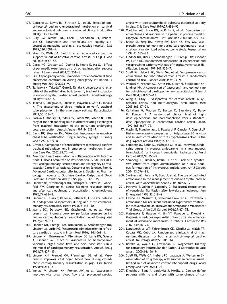
S80 J.P. Nolan et al.
172. Gausche M, Lewis RJ, Stratton SJ, et al. Effect of out-of-hospital pediatric endotracheal intubation on survivaland neurological outcome: a controlled clinical trial. JAMA2000;283:783—970.
173. Guly UM, Mitchell RG, Cook R, Steedman DJ, Robert-son CE. Paramedics and technicians are equally suc-cessful at managing cardiac arrest outside hospital. BMJ1995;310:1091—4.
174. Stiell IG, Wells GA, Field B, et al. Advanced cardiac lifesupport in out-of-hospital cardiac arrest. N Engl J Med2004;351:647—56.
175. Garza AG, Gratton MC, Coontz D, Noble E, Ma OJ. Effectof paramedic experience on orotracheal intubation successrates. J Emerg Med 2003;25:251—6.
176. Li J. Capnography alone is imperfect for endotracheal tubeplacement confirmation during emergency intubation. JEmerg Med 2001;20:223—9.
177. Tanigawa K, Takeda T, Goto E, Tanaka K. Accuracy and relia-bility of the self-inflating bulb to verify tracheal intubationin out-of-hospital cardiac arrest patients. Anesthesiology2000;93:1432—6.
178. Takeda T, Tanigawa K, Tanaka H, Hayashi Y, Goto E, TanakaK. The assessment of three methods to verify trachealtube placement in the emergency setting. Resuscitation2003;56:153—7.
179. Baraka A, Khoury PJ, Siddik SS, Salem MR, Joseph NJ. Effi-cacy of the self-inflating bulb in differentiating esophagealfrom tracheal intubation in the parturient undergoingcesarean section. Anesth Analg 1997;84:533—7.
180. Davis DP, Stephen KA, Vilke GM. Inaccuracy in endotra-
arrest with postcountershock pulseless electrical activityin pigs. Crit Care Med 1999;27:486—92.
190. Voelckel WG, Lurie KG, McKnite S, et al. Comparison ofepinephrine and vasopressin in a pediatric porcine model ofasphyxial cardiac arrest. Crit Care Med 2000;28:3777—83.
191. Babar SI, Berg RA, Hilwig RW, Kern KB, Ewy GA. Vaso-pressin versus epinephrine during cardiopulmonary resus-citation: a randomized swine outcome study. Resuscitation1999;41:185—92.
192. Lindner KH, Dirks B, Strohmenger HU, Prengel AW, LindnerIM, Lurie KG. Randomised comparison of epinephrine andvasopressin in patients with out-of-hospital ventricular fib-rillation. Lancet 1997;349:535—7.
193. Stiell IG, Hebert PC, Wells GA, et al. Vasopressin versusepinephrine for inhospital cardiac arrest: a randomisedcontrolled trial. Lancet 2001;358:105—9.
194. Wenzel V, Krismer AC, Arntz HR, Sitter H, Stadlbauer KH,Lindner KH. A comparison of vasopressin and epinephrinefor out-of-hospital cardiopulmonary resuscitation. N Engl JMed 2004;350:105—13.
195. Aung K, Htay T. Vasopressin for cardiac arrest: a sys-tematic review and meta-analysis. Arch Intern Med2005;165:17—24.
196. Callaham M, Madsen C, Barton C, Saunders C, DaleyM, Pointer J. A randomized clinical trial of high-dose epinephrine and norepinephrine versus standard-dose epinephrine in prehospital cardiac arrest. JAMA1992;268:2667—72.
197. Masini E, Planchenault J, Pezziardi F, Gautier P, Gagnol JP.Histamine-releasing properties of Polysorbate 80 in vitro
1
1
2
2
2
2
2
2
2
2
cheal tube verification using a Toomey syringe. J EmergMed 1999;17:35—8.
181. Grmec S. Comparison of three different methods to confirmtracheal tube placement in emergency intubation. Inten-sive Care Med 2002;28:701—4.
182. American Heart Association in collaboration with Interna-tional Liaison Committee on Resuscitation. Guidelines 2000for Cardiopulmonary Resuscitation and Emergency Cardio-vascular Care: International Consensus on Science. Part 6.Advanced Cardiovascular Life Support: Section 6. Pharma-cology II: Agents to Optimize Cardiac Output and BloodPressure. Circulation 2000;102(Suppl. I):I129—35.
183. Lindner KH, Strohmenger HU, Ensinger H, Hetzel WD, Ahne-feld FW, Georgieff M. Stress hormone response duringand after cardiopulmonary resuscitation. Anesthesiology1992;77:662—8.
184. Lindner KH, Haak T, Keller A, Bothner U, Lurie KG. Releaseof endogenous vasopressors during and after cardiopul-monary resuscitation. Heart 1996;75:145—50.
185. Morris DC, Dereczyk BE, Grzybowski M, et al. Vaso-pressin can increase coronary perfusion pressure duringhuman cardiopulmonary resuscitation. Acad Emerg Med1997;4:878—83.
186. Lindner KH, Prengel AW, Brinkmann A, Strohmenger HU,Lindner IM, Lurie KG. Vasopressin administration in refrac-tory cardiac arrest. Ann Intern Med 1996;124:1061—4.
187. Lindner KH, Brinkmann A, Pfenninger EG, Lurie KG, GoertzA, Lindner IM. Effect of vasopressin on hemodynamicvariables, organ blood flow, and acid—base status in apig model of cardiopulmonary resuscitation. Anesth Analg1993;77:427—35.
188. Lindner KH, Prengel AW, Pfenninger EG, et al. Vaso-pressin improves vital organ blood flow during closed-chest cardiopulmonary resuscitation in pigs. Circulation1995;91:215—21.
189. Wenzel V, Lindner KH, Prengel AW, et al. Vasopressinimproves vital organ blood flow after prolonged cardiac
and in vivo: correlation with its hypotensive action in thedog. Agents Actions 1985;16:470—7.
98. Somberg JC, Bailin SJ, Haffajee CI, et al. Intravenous lido-caine versus intravenous amiodarone (in a new aqueousformulation) for incessant ventricular tachycardia. Am JCardiol 2002;90:853—9.
99. Somberg JC, Timar S, Bailin SJ, et al. Lack of a hypoten-sive effect with rapid administration of a new aque-ous formulation of intravenous amiodarone. Am J Cardiol2004;93:576—81.
00. Skrifvars MB, Kuisma M, Boyd J, et al. The use of undilutedamiodarone in the management of out-of-hospital cardiacarrest. Acta Anaesthesiol Scand 2004;48:582—7.
01. Petrovic T, Adnet F, Lapandry C. Successful resuscitationof ventricular fibrillation after low-dose amiodarone. AnnEmerg Med 1998;32:518—9.
02. Levine JH, Massumi A, Scheinman MM, et al. Intravenousamiodarone for recurrent sustained hypotensive ventricu-lar tachyarrhythmias. Intravenous Amiodarone MulticenterTrial Group. J Am Coll Cardiol 1996;27:67—75.
03. Matsusaka T, Hasebe N, Jin YT, Kawabe J, Kikuchi K.Magnesium reduces myocardial infarct size via enhance-ment of adenosine mechanism in rabbits. Cardiovasc Res2002;54:568—75.
04. Longstreth Jr WT, Fahrenbruch CE, Olsufka M, Walsh TR,Copass MK, Cobb LA. Randomized clinical trial of mag-nesium, diazepam, or both after out-of-hospital cardiacarrest. Neurology 2002;59:506—14.
05. Baraka A, Ayoub C, Kawkabani N. Magnesium therapyfor refractory ventricular fibrillation. J Cardiothorac VascAnesth 2000;14:196—9.
06. Stiell IG, Wells GA, Hebert PC, Laupacis A, Weitzman BN.Association of drug therapy with survival in cardiac arrest:limited role of advanced cardiac life support drugs. AcadEmerg Med 1995;2:264—73.
07. Engdahl J, Bang A, Lindqvist J, Herlitz J. Can we definepatients with no and those with some chance of sur-
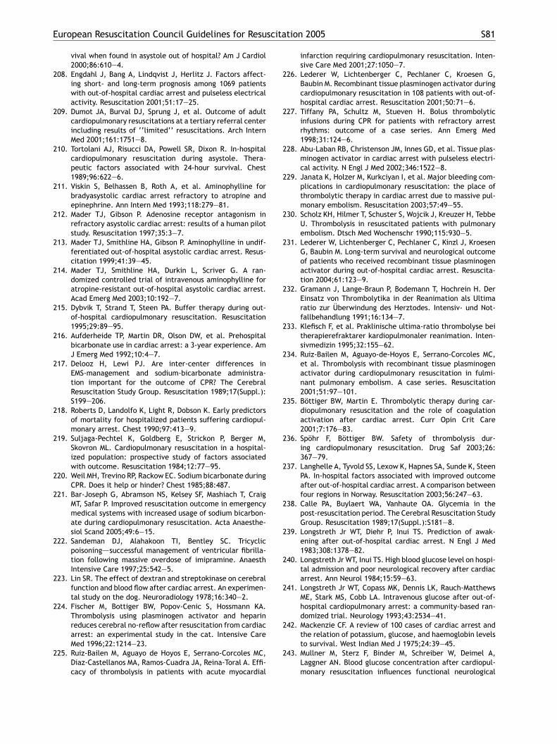
European Resuscitation Council Guidelines for Resuscitation 2005 S81
vival when found in asystole out of hospital? Am J Cardiol2000;86:610—4.
208. Engdahl J, Bang A, Lindqvist J, Herlitz J. Factors affect-ing short- and long-term prognosis among 1069 patientswith out-of-hospital cardiac arrest and pulseless electricalactivity. Resuscitation 2001;51:17—25.
209. Dumot JA, Burval DJ, Sprung J, et al. Outcome of adultcardiopulmonary resuscitations at a tertiary referral centerincluding results of ‘‘limited’’ resuscitations. Arch InternMed 2001;161:1751—8.
210. Tortolani AJ, Risucci DA, Powell SR, Dixon R. In-hospitalcardiopulmonary resuscitation during asystole. Thera-peutic factors associated with 24-hour survival. Chest1989;96:622—6.
211. Viskin S, Belhassen B, Roth A, et al. Aminophylline forbradyasystolic cardiac arrest refractory to atropine andepinephrine. Ann Intern Med 1993;118:279—81.
212. Mader TJ, Gibson P. Adenosine receptor antagonism inrefractory asystolic cardiac arrest: results of a human pilotstudy. Resuscitation 1997;35:3—7.
213. Mader TJ, Smithline HA, Gibson P. Aminophylline in undif-ferentiated out-of-hospital asystolic cardiac arrest. Resus-citation 1999;41:39—45.
214. Mader TJ, Smithline HA, Durkin L, Scriver G. A ran-domized controlled trial of intravenous aminophylline foratropine-resistant out-of-hospital asystolic cardiac arrest.Acad Emerg Med 2003;10:192—7.
215. Dybvik T, Strand T, Steen PA. Buffer therapy during out-of-hospital cardiopulmonary resuscitation. Resuscitation1995;29:89—95.
2
2
2
2
2
2
2
2
2
2
infarction requiring cardiopulmonary resuscitation. Inten-sive Care Med 2001;27:1050—7.
226. Lederer W, Lichtenberger C, Pechlaner C, Kroesen G,Baubin M. Recombinant tissue plasminogen activator duringcardiopulmonary resuscitation in 108 patients with out-of-hospital cardiac arrest. Resuscitation 2001;50:71—6.
227. Tiffany PA, Schultz M, Stueven H. Bolus thrombolyticinfusions during CPR for patients with refractory arrestrhythms: outcome of a case series. Ann Emerg Med1998;31:124—6.
228. Abu-Laban RB, Christenson JM, Innes GD, et al. Tissue plas-minogen activator in cardiac arrest with pulseless electri-cal activity. N Engl J Med 2002;346:1522—8.
229. Janata K, Holzer M, Kurkciyan I, et al. Major bleeding com-plications in cardiopulmonary resuscitation: the place ofthrombolytic therapy in cardiac arrest due to massive pul-monary embolism. Resuscitation 2003;57:49—55.
230. Scholz KH, Hilmer T, Schuster S, Wojcik J, Kreuzer H, TebbeU. Thrombolysis in resuscitated patients with pulmonaryembolism. Dtsch Med Wochenschr 1990;115:930—5.
231. Lederer W, Lichtenberger C, Pechlaner C, Kinzl J, KroesenG, Baubin M. Long-term survival and neurological outcomeof patients who received recombinant tissue plasminogenactivator during out-of-hospital cardiac arrest. Resuscita-tion 2004;61:123—9.
232. Gramann J, Lange-Braun P, Bodemann T, Hochrein H. DerEinsatz von Thrombolytika in der Reanimation als Ultimaratio zur Uberwindung des Herztodes. Intensiv- und Not-fallbehandlung 1991;16:134—7.
233. Klefisch F, et al. Praklinische ultima-ratio thrombolyse bei
2
2
2
2
2
2
2
2
2
2
16. Aufderheide TP, Martin DR, Olson DW, et al. Prehospitalbicarbonate use in cardiac arrest: a 3-year experience. AmJ Emerg Med 1992;10:4—7.
17. Delooz H, Lewi PJ. Are inter-center differences inEMS-management and sodium-bicarbonate administra-tion important for the outcome of CPR? The CerebralResuscitation Study Group. Resuscitation 1989;17(Suppl.):S199—206.
18. Roberts D, Landolfo K, Light R, Dobson K. Early predictorsof mortality for hospitalized patients suffering cardiopul-monary arrest. Chest 1990;97:413—9.
19. Suljaga-Pechtel K, Goldberg E, Strickon P, Berger M,Skovron ML. Cardiopulmonary resuscitation in a hospital-ized population: prospective study of factors associatedwith outcome. Resuscitation 1984;12:77—95.
20. Weil MH, Trevino RP, Rackow EC. Sodium bicarbonate duringCPR. Does it help or hinder? Chest 1985;88:487.
21. Bar-Joseph G, Abramson NS, Kelsey SF, Mashiach T, CraigMT, Safar P. Improved resuscitation outcome in emergencymedical systems with increased usage of sodium bicarbon-ate during cardiopulmonary resuscitation. Acta Anaesthe-siol Scand 2005;49:6—15.
22. Sandeman DJ, Alahakoon TI, Bentley SC. Tricyclicpoisoning—–successful management of ventricular fibrilla-tion following massive overdose of imipramine. AnaesthIntensive Care 1997;25:542—5.
23. Lin SR. The effect of dextran and streptokinase on cerebralfunction and blood flow after cardiac arrest. An experimen-tal study on the dog. Neuroradiology 1978;16:340—2.
24. Fischer M, Bottiger BW, Popov-Cenic S, Hossmann KA.Thrombolysis using plasminogen activator and heparinreduces cerebral no-reflow after resuscitation from cardiacarrest: an experimental study in the cat. Intensive CareMed 1996;22:1214—23.
25. Ruiz-Bailen M, Aguayo de Hoyos E, Serrano-Corcoles MC,Diaz-Castellanos MA, Ramos-Cuadra JA, Reina-Toral A. Effi-cacy of thrombolysis in patients with acute myocardial
therapierefraktarer kardiopulmonaler reanimation. Inten-sivmedizin 1995;32:155—62.
34. Ruiz-Bailen M, Aguayo-de-Hoyos E, Serrano-Corcoles MC,et al. Thrombolysis with recombinant tissue plasminogenactivator during cardiopulmonary resuscitation in fulmi-nant pulmonary embolism. A case series. Resuscitation2001;51:97—101.
35. Bottiger BW, Martin E. Thrombolytic therapy during car-diopulmonary resuscitation and the role of coagulationactivation after cardiac arrest. Curr Opin Crit Care2001;7:176—83.
36. Spohr F, Bottiger BW. Safety of thrombolysis dur-ing cardiopulmonary resuscitation. Drug Saf 2003;26:367—79.
37. Langhelle A, Tyvold SS, Lexow K, Hapnes SA, Sunde K, SteenPA. In-hospital factors associated with improved outcomeafter out-of-hospital cardiac arrest. A comparison betweenfour regions in Norway. Resuscitation 2003;56:247—63.
38. Calle PA, Buylaert WA, Vanhaute OA. Glycemia in thepost-resuscitation period. The Cerebral Resuscitation StudyGroup. Resuscitation 1989;17(Suppl.):S181—8.
39. Longstreth Jr WT, Diehr P, Inui TS. Prediction of awak-ening after out-of-hospital cardiac arrest. N Engl J Med1983;308:1378—82.
40. Longstreth Jr WT, Inui TS. High blood glucose level on hospi-tal admission and poor neurological recovery after cardiacarrest. Ann Neurol 1984;15:59—63.
41. Longstreth Jr WT, Copass MK, Dennis LK, Rauch-MatthewsME, Stark MS, Cobb LA. Intravenous glucose after out-of-hospital cardiopulmonary arrest: a community-based ran-domized trial. Neurology 1993;43:2534—41.
42. Mackenzie CF. A review of 100 cases of cardiac arrest andthe relation of potassium, glucose, and haemoglobin levelsto survival. West Indian Med J 1975;24:39—45.
43. Mullner M, Sterz F, Binder M, Schreiber W, Deimel A,Laggner AN. Blood glucose concentration after cardiopul-monary resuscitation influences functional neurological
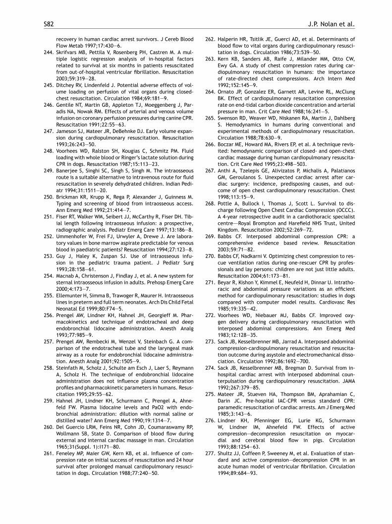
S82 J.P. Nolan et al.
recovery in human cardiac arrest survivors. J Cereb BloodFlow Metab 1997;17:430—6.
244. Skrifvars MB, Pettila V, Rosenberg PH, Castren M. A mul-tiple logistic regression analysis of in-hospital factorsrelated to survival at six months in patients resuscitatedfrom out-of-hospital ventricular fibrillation. Resuscitation2003;59:319—28.
245. Ditchey RV, Lindenfeld J. Potential adverse effects of vol-ume loading on perfusion of vital organs during closed-chest resuscitation. Circulation 1984;69:181—9.
246. Gentile NT, Martin GB, Appleton TJ, Moeggenberg J, Par-adis NA, Nowak RM. Effects of arterial and venous volumeinfusion on coronary perfusion pressures during canine CPR.Resuscitation 1991;22:55—63.
247. Jameson SJ, Mateer JR, DeBehnke DJ. Early volume expan-sion during cardiopulmonary resuscitation. Resuscitation1993;26:243—50.
248. Voorhees WD, Ralston SH, Kougias C, Schmitz PM. Fluidloading with whole blood or Ringer’s lactate solution duringCPR in dogs. Resuscitation 1987;15:113—23.
249. Banerjee S, Singhi SC, Singh S, Singh M. The intraosseousroute is a suitable alternative to intravenous route for fluidresuscitation in severely dehydrated children. Indian Pedi-atr 1994;31:1511—20.
250. Brickman KR, Krupp K, Rega P, Alexander J, Guinness M.Typing and screening of blood from intraosseous access.Ann Emerg Med 1992;21:414—7.
251. Fiser RT, Walker WM, Seibert JJ, McCarthy R, Fiser DH. Tib-ial length following intraosseous infusion: a prospective,radiographic analysis. Pediatr Emerg Care 1997;13:186—8.
262. Halperin HR, Tsitlik JE, Guerci AD, et al. Determinants ofblood flow to vital organs during cardiopulmonary resusci-tation in dogs. Circulation 1986;73:539—50.
263. Kern KB, Sanders AB, Raife J, Milander MM, Otto CW,Ewy GA. A study of chest compression rates during car-diopulmonary resuscitation in humans: the importanceof rate-directed chest compressions. Arch Intern Med1992;152:145—9.
264. Ornato JP, Gonzalez ER, Garnett AR, Levine RL, McClungBK. Effect of cardiopulmonary resuscitation compressionrate on end-tidal carbon dioxide concentration and arterialpressure in man. Crit Care Med 1988;16:241—5.
265. Swenson RD, Weaver WD, Niskanen RA, Martin J, DahlbergS. Hemodynamics in humans during conventional andexperimental methods of cardiopulmonary resuscitation.Circulation 1988;78:630—9.
266. Boczar ME, Howard MA, Rivers EP, et al. A technique revis-ited: hemodynamic comparison of closed- and open-chestcardiac massage during human cardiopulmonary resuscita-tion. Crit Care Med 1995;23:498—503.
267. Anthi A, Tzelepis GE, Alivizatos P, Michalis A, PalatianosGM, Geroulanos S. Unexpected cardiac arrest after car-diac surgery: incidence, predisposing causes, and out-come of open chest cardiopulmonary resuscitation. Chest1998;113:15—9.
268. Pottle A, Bullock I, Thomas J, Scott L. Survival to dis-charge following Open Chest Cardiac Compression (OCCC).A 4-year retrospective audit in a cardiothoracic specialistcentre—–Royal Brompton and Harefield NHS Trust, UnitedKingdom. Resuscitation 2002;52:269—72.
2
2
2
2
2
2
2
2
2
252. Ummenhofer W, Frei FJ, Urwyler A, Drewe J. Are labora-tory values in bone marrow aspirate predictable for venousblood in paediatric patients? Resuscitation 1994;27:123—8.
253. Guy J, Haley K, Zuspan SJ. Use of intraosseous infu-sion in the pediatric trauma patient. J Pediatr Surg1993;28:158—61.
254. Macnab A, Christenson J, Findlay J, et al. A new system forsternal intraosseous infusion in adults. Prehosp Emerg Care2000;4:173—7.
255. Ellemunter H, Simma B, Trawoger R, Maurer H. Intraosseouslines in preterm and full term neonates. Arch Dis Child FetalNeonatal Ed 1999;80:F74—5.
256. Prengel AW, Lindner KH, Hahnel JH, Georgieff M. Phar-macokinetics and technique of endotracheal and deependobronchial lidocaine administration. Anesth Analg1993;77:985—9.
257. Prengel AW, Rembecki M, Wenzel V, Steinbach G. A com-parison of the endotracheal tube and the laryngeal maskairway as a route for endobronchial lidocaine administra-tion. Anesth Analg 2001;92:1505—9.
258. Steinfath M, Scholz J, Schulte am Esch J, Laer S, ReymannA, Scholz H. The technique of endobronchial lidocaineadministration does not influence plasma concentrationprofiles and pharmacokinetic parameters in humans. Resus-citation 1995;29:55—62.
259. Hahnel JH, Lindner KH, Schurmann C, Prengel A, Ahne-feld FW. Plasma lidocaine levels and PaO2 with endo-bronchial administration: dilution with normal saline ordistilled water? Ann Emerg Med 1990;19:1314—7.
260. Del Guercio LRM, Feins NR, Cohn JD, Coumaraswamy RP,Wollmann SB, State D. Comparison of blood flow duringexternal and internal cardiac massage in man. Circulation1965;31(Suppl. 1):I171—80.
261. Feneley MP, Maier GW, Kern KB, et al. Influence of com-pression rate on initial success of resuscitation and 24 hoursurvival after prolonged manual cardiopulmonary resusci-tation in dogs. Circulation 1988;77:240—50.
69. Babbs CF. Interposed abdominal compression CPR: acomprehensive evidence based review. Resuscitation2003;59:71—82.
70. Babbs CF, Nadkarni V. Optimizing chest compression to res-cue ventilation ratios during one-rescuer CPR by profes-sionals and lay persons: children are not just little adults.Resuscitation 2004;61:173—81.
71. Beyar R, Kishon Y, Kimmel E, Neufeld H, Dinnar U. Intratho-racic and abdominal pressure variations as an efficientmethod for cardiopulmonary resuscitation: studies in dogscompared with computer model results. Cardiovasc Res1985;19:335—42.
72. Voorhees WD, Niebauer MJ, Babbs CF. Improved oxy-gen delivery during cardiopulmonary resuscitation withinterposed abdominal compressions. Ann Emerg Med1983;12:128—35.
73. Sack JB, Kesselbrenner MB, Jarrad A. Interposed abdominalcompression-cardiopulmonary resuscitation and resuscita-tion outcome during asystole and electromechanical disso-ciation. Circulation 1992;86:1692—700.
74. Sack JB, Kesselbrenner MB, Bregman D. Survival from in-hospital cardiac arrest with interposed abdominal coun-terpulsation during cardiopulmonary resuscitation. JAMA1992;267:379—85.
75. Mateer JR, Stueven HA, Thompson BM, Aprahamian C,Darin JC. Pre-hospital IAC-CPR versus standard CPR:paramedic resuscitation of cardiac arrests. Am J Emerg Med1985;3:143—6.
76. Lindner KH, Pfenninger EG, Lurie KG, SchurmannW, Lindner IM, Ahnefeld FW. Effects of activecompression—decompression resuscitation on myocar-dial and cerebral blood flow in pigs. Circulation1993;88:1254—63.
77. Shultz JJ, Coffeen P, Sweeney M, et al. Evaluation of stan-dard and active compression—decompression CPR in anacute human model of ventricular fibrillation. Circulation1994;89:684—93.
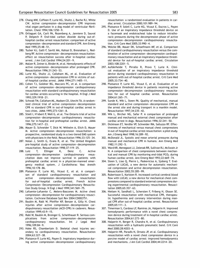
European Resuscitation Council Guidelines for Resuscitation 2005 S83
278. Chang MW, Coffeen P, Lurie KG, Shultz J, Bache RJ, WhiteCW. Active compression—decompression CPR improvesvital organ perfusion in a dog model of ventricular fibril-lation. Chest 1994;106:1250—9.
279. Orliaguet GA, Carli PA, Rozenberg A, Janniere D, SauvalP, Delpech P. End-tidal carbon dioxide during out-of-hospital cardiac arrest resuscitation: comparison of activecompression—decompression and standard CPR. Ann EmergMed 1995;25:48—51.
280. Tucker KJ, Galli F, Savitt MA, Kahsai D, Bresnahan L, Red-berg RF. Active compression—decompression resuscitation:effect on resuscitation success after in-hospital cardiacarrest. J Am Coll Cardiol 1994;24:201—9.
281. Malzer R, Zeiner A, Binder M, et al. Hemodynamic effects ofactive compression—decompression after prolonged CPR.Resuscitation 1996;31:243—53.
282. Lurie KG, Shultz JJ, Callaham ML, et al. Evaluation ofactive compression—decompression CPR in victims of out-of-hospital cardiac arrest. JAMA 1994;271:1405—11.
283. Cohen TJ, Goldner BG, Maccaro PC, et al. A comparisonof active compression—decompression cardiopulmonaryresuscitation with standard cardiopulmonary resuscitationfor cardiac arrests occurring in the hospital. N Engl J Med1993;329:1918—21.
284. Schwab TM, Callaham ML, Madsen CD, Utecht TA. A random-ized clinical trial of active compression—decompressionCPR vs standard CPR in out-of-hospital cardiac arrest intwo cities. JAMA 1995;273:1261—8.
285. Stiell I, H’ebert P, Well G, et al. Tne Ontario trial of activecompression—decompression cardiopulmonary resuscita-
2
2
2
2
2
2
2
2
2
resuscitation: a randomized evaluation in patients in car-diac arrest. Circulation 2000;101:989—94.
295. Plaisance P, Soleil C, Lurie KG, Vicaut E, Ducros L, PayenD. Use of an inspiratory impedance threshold device ona facemask and endotracheal tube to reduce intratho-racic pressures during the decompression phase of activecompression—decompression cardiopulmonary resuscita-tion. Crit Care Med 2005;33:990—4.
296. Wolcke BB, Mauer DK, Schoefmann MF, et al. Comparisonof standard cardiopulmonary resuscitation versus the com-bination of active compression—decompression cardiopul-monary resuscitation and an inspiratory impedance thresh-old device for out-of-hospital cardiac arrest. Circulation2003;108:2201—5.
297. Aufderheide T, Pirrallo R, Provo T, Lurie K. Clini-cal evaluation of an inspiratory impedance thresholddevice during standard cardiopulmonary resuscitation inpatients with out-of-hospital cardiac arrest. Crit Care Med2005;33:734—40.
298. Plaisance P, Lurie KG, Vicaut E, et al. Evaluation of animpedance threshold device in patients receiving activecompression—decompression cardiopulmonary resuscita-tion for out of hospital cardiac arrest. Resuscitation2004;61:265—71.
299. Sunde K, Wik L, Steen PA. Quality of mechanical, manualstandard and active compression—decompression CPR onthe arrest site and during transport in a manikin model.Resuscitation 1997;34:235—42.
300. Wik L, Bircher NG, Safar P. A comparison of prolongedmanual and mechanical external chest compression after
3
3
3
3
3
3
3
3
3
tion for in-hospital and prehospital cardiac arrest. JAMA1996;275:1417—23.
86. Mauer D, Schneider T, Dick W, Withelm A, Elich D, MauerM. Active compression—decompression resuscitation: aprospective, randomized study in a two-tiered EMS systemwith physicians in the field. Resuscitation 1996;33:125—34.
87. Nolan J, Smith G, Evans R, et al. The United Kingdompre-hospital study of active compression—decompressionresuscitation. Resuscitation 1998;37:119—25.
88. Luiz T, Ellinger K, Denz C. Activecompression—decompression cardiopulmonary resus-citation does not improve survival in patients withprehospital cardiac arrest in a physician-manned emer-gency medical system. J Cardiothorac Vasc Anesth1996;10:178—86.
89. Plaisance P, Lurie KG, Vicaut E, et al. A compari-son of standard cardiopulmonary resuscitation andactive compression—decompression resuscitationfor out-of-hospital cardiac arrest. French ActiveCompression—Decompression Cardiopulmonary Resuscita-tion Study Group. N Engl J Med 1999;341:569—75.
90. Lafuente-Lafuente C, Melero-Bascones M. Active chestcompression—decompression for cardiopulmonary resusci-tation. Cochrane Database Syst Rev 2004:CD002751.
91. Baubin M, Rabl W, Pfeiffer KP, Benzer A, Gilly H. Chestinjuries after active compression—decompression car-diopulmonary resuscitation (ACD-CPR) in cadavers. Resus-citation 1999;43:9—15.
92. Rabl W, Baubin M, Broinger G, Scheithauer R. Serious com-plications from active compression—decompressioncardiopulmonary resuscitation. Int J Legal Med1996;109:84—9.
93. Hoke RS, Chamberlain D. Skeletal chest injuries sec-ondary to cardiopulmonary resuscitation. Resuscitation2004;63:327—38.
94. Plaisance P, Lurie KG, Payen D. Inspiratory impedance dur-ing active compression—decompression cardiopulmonary
cardiac arrest in dogs. Resuscitation 1996;32:241—50.01. Dickinson ET, Verdile VP, Schneider RM, Salluzzo RF. Effec-
tiveness of mechanical versus manual chest compressionsin out-of-hospital cardiac arrest resuscitation: a pilot study.Am J Emerg Med 1998;16:289—92.
02. McDonald JL. Systolic and mean arterial pressures duringmanual and mechanical CPR in humans. Ann Emerg Med1982;11:292—5.
03. Ward KR, Menegazzi JJ, Zelenak RR, Sullivan RJ, McSwain JrN. A comparison of chest compressions between mechan-ical and manual CPR by monitoring end-tidal PCO2 duringhuman cardiac arrest. Ann Emerg Med 1993;22:669—74.
04. Steen S, Liao Q, Pierre L, Paskevicius A, Sjoberg T. Eval-uation of LUCAS, a new device for automatic mechani-cal compression and active decompression resuscitation.Resuscitation 2002;55:285—99.
05. Rubertsson S, Karlsten R. Increased cortical cerebral bloodflow with LUCAS; a new device for mechanical chest com-pressions compared to standard external compressions dur-ing experimental cardiopulmonary resuscitation. Resusci-tation 2005;65:357—63.
06. Nielsen N, Sandhall L, Schersten F, Friberg H, Olsson SE.Successful resuscitation with mechanical CPR, therapeu-tic hypothermia and coronary intervention during man-ual CPR after out-of-hospital cardiac arrest. Resuscitation2005;65:111—3.
07. Timerman S, Cardoso LF, Ramires JA, Halperin H. Improvedhemodynamic performance with a novel chest compres-sion device during treatment of in-hospital cardiac arrest.Resuscitation 2004;61:273—80.
08. Halperin H, Berger R, Chandra N, et al. Cardiopulmonaryresuscitation with a hydraulic-pneumatic band. Crit CareMed 2000;28:N203—6.
09. Halperin HR, Paradis N, Ornato JP, et al. Cardiopulmonaryresuscitation with a novel chest compression device in aporcine model of cardiac arrest: improved hemodynamicsand mechanisms. J Am Coll Cardiol 2004;44:2214—20.
S84 J.P. Nolan et al.
310. Casner M, Anderson D, et al. Preliminary report of theimpact of a new CPR assist device on the rate of return ofspontaneous circulation in out of hospital cardiac arrest.Prehosp Emerg Med 2005;9:61—7.
311. Arntz HR, Agrawal R, Richter H, et al. Phased chestand abdominal compression—decompression versus con-ventional cardiopulmonary resuscitation in out-of-hospitalcardiac arrest. Circulation 2001;104:768—72.
312. Rozenberg A, Incagnoli P, Delpech P, et al. Prehospital useof minimally invasive direct cardiac massage (MID-CM): apilot study. Resuscitation 2001;50:257—62.
313. Dauchot P, Gravenstein JS. Effects of atropine on the elec-trocardiogram in different age groups. Clin Pharmacol Ther1971;12:274—80.
314. Chamberlain DA, Turner P, Sneddon JM. Effects of atropineon heart-rate in healthy man. Lancet 1967;2:12—5.
315. Bernheim A, Fatio R, Kiowski W, Weilenmann D, Rickli H,Rocca HP. Atropine often results in complete atrioventric-ular block or sinus arrest after cardiac transplantation: anunpredictable and dose-independent phenomenon. Trans-plantation 2004;77:1181—5.
316. Klumbies A, Paliege R, Volkmann H. Mechanical emer-gency stimulation in asystole and extreme bradycardia. ZGesamte Inn Med 1988;43:348—52.
317. Zeh E, Rahner E. The manual extrathoracal stimulation ofthe heart. Technique and effect of the precordial thump(author’s transl). Z Kardiol 1978;67:299—304.
318. Chan L, Reid C, Taylor B. Effect of three emergency pac-ing modalities on cardiac output in cardiac arrest due toventricular asystole. Resuscitation 2002;52:117—9.
330. Kalus JS, Spencer AP, Tsikouris JP, et al. Impact of pro-phylactic i.v. magnesium on the efficacy of ibutilide forconversion of atrial fibrillation or flutter. Am J Health SystPharm 2003;60:2308—12.
331. Langhelle A, Nolan J, Herlitz J, et al. Recommended guide-lines for reviewing, reporting, and conducting researchon post-resuscitation care: the Utstein style. Resuscitation2005;66:271—83.
332. Menon DK, Coles JP, Gupta AK, et al. Diffusion lim-ited oxygen delivery following head injury. Crit Care Med2004;32:1384—90.
333. Buunk G, van der Hoeven JG, Meinders AE. Cerebrovascularreactivity in comatose patients resuscitated from a cardiacarrest. Stroke 1997;28:1569—73.
334. Buunk G, van der Hoeven JG, Meinders AE. A comparisonof near-infrared spectroscopy and jugular bulb oximetryin comatose patients resuscitated from a cardiac arrest.Anaesthesia 1998;53:13—9.
335. Roine RO, Launes J, Nikkinen P, Lindroth L, Kaste M.Regional cerebral blood flow after human cardiac arrest. Ahexamethylpropyleneamine oxime single photon emissioncomputed tomographic study. Arch Neurol 1991;48:625—9.
336. Beckstead JE, Tweed WA, Lee J, MacKeen WL. Cerebralblood flow and metabolism in man following cardiac arrest.Stroke 1978;9:569—73.
337. Laurent I, Monchi M, Chiche JD, et al. Reversible myocar-dial dysfunction in survivors of out-of-hospital cardiacarrest. J Am Coll Cardiol 2002;40:2110—6.
338. Kern KB, Hilwig RW, Rhee KH, Berg RA. Myocardial dys-function after resuscitation from cardiac arrest: an exam-
3
3
3
3
3
3
3
3
3
3
3
319. Manz M, Pfeiffer D, Jung W, Lueritz B. Intravenous treat-ment with magnesium in recurrent persistent ventriculartachycardia. New Trends Arrhythmias 1991;7:437—42.
320. Tzivoni D, Banai S, Schuger C, et al. Treatment oftorsade de pointes with magnesium sulfate. Circulation1988;77:392—7.
321. Sticherling C, Tada H, Hsu W, et al. Effects of diltiazemand esmolol on cycle length and spontaneous conver-sion of atrial fibrillation. J Cardiovasc Pharmacol Ther2002;7:81—8.
322. Shettigar UR, Toole JG, Appunn DO. Combined use ofesmolol and digoxin in the acute treatment of atrial fib-rillation or flutter. Am Heart J 1993;126:368—74.
323. Demircan C, Cikriklar HI, Engindeniz Z, et al. Comparison ofthe effectiveness of intravenous diltiazem and metoprololin the management of rapid ventricular rate in atrial fibril-lation. Emerg Med J 2005;22:411—4.
324. Wattanasuwan N, Khan IA, Mehta NJ, et al. Acute ven-tricular rate control in atrial fibrillation: IV combinationof diltiazem and digoxin vs. IV diltiazem alone. Chest2001;119:502—6.
325. Davey MJ, Teubner D. A randomized controlled trial of mag-nesium sulfate, in addition to usual care, for rate controlin atrial fibrillation. Ann Emerg Med 2005;45:347—53.
326. Chiladakis JA, Stathopoulos C, Davlouros P, Manolis AS.Intravenous magnesium sulfate versus diltiazem in parox-ysmal atrial fibrillation. Int J Cardiol 2001;79:287—91.
327. Camm AJ, Garratt CJ. Adenosine and supraventriculartachycardia. N Engl J Med 1991;325:1621—9.
328. Wang HE, O’Connor RE, Megargel RE, et al. The use ofdiltiazem for treating rapid atrial fibrillation in the out-of-hospital setting. Ann Emerg Med 2001;37:38—45.
329. Martinez-Marcos FJ, Garcia-Garmendia JL, Ortega-CarpioA, Fernandez-Gomez JM, Santos JM, Camacho C. Com-parison of intravenous flecainide, propafenone, and amio-darone for conversion of acute atrial fibrillation to sinusrhythm. Am J Cardiol 2000;86:950—3.
ple of global myocardial stunning. J Am Coll Cardiol1996;28:232—40.
39. Adrie C, Adib-Conquy M, Laurent I, et al. Successfulcardiopulmonary resuscitation after cardiac arrest as a‘‘sepsis-like’’ syndrome. Circulation 2002;106:562—8.
40. Mullner M, Sterz F, Binder M, et al. Arterial blood pres-sure after human cardiac arrest and neurological recovery.Stroke 1996;27:59—62.
41. Angelos MG, Ward KR, Hobson J, Beckley PD. Organ bloodflow following cardiac arrest in a swine low-flow cardiopul-monary bypass model. Resuscitation 1994;27:245—54.
42. Rello J, Diaz E, Roque M, Valles J. Risk factors for develop-ing pneumonia within 48 hours of intubation. Am J RespirCrit Care Med 1999;159:1742—6.
43. Krumholz A, Stern BJ, Weiss HD. Outcome from coma aftercardiopulmonary resuscitation: relation to seizures andmyoclonus. Neurology 1988;38:401—5.
44. Wijdicks EF, Parisi JE, Sharbrough FW. Prognostic value ofmyoclonus status in comatose survivors of cardiac arrest.Ann Neurol 1994;35:239—43.
45. Takino M, Okada Y. Hyperthermia following cardiopul-monary resuscitation. Intensive Care Med 1991;17:419—20.
46. Hickey RW, Kochanek PM, Ferimer H, Alexander HL, Gar-man RH, Graham SH. Induced hyperthermia exacerbatesneurologic neuronal histologic damage after asphyxial car-diac arrest in rats. Crit Care Med 2003;31:531—5.
47. Takasu A, Saitoh D, Kaneko N, Sakamoto T, Okada Y. Hyper-thermia: is it an ominous sign after cardiac arrest? Resus-citation 2001;49:273—7.
48. Zeiner A, Holzer M, Sterz F, et al. Hyperthermia after car-diac arrest is associated with an unfavorable neurologicoutcome. Arch Intern Med 2001;161:2007—12.
49. Coimbra C, Boris-Moller F, Drake M, Wieloch T. Dimin-ished neuronal damage in the rat brain by late treat-ment with the antipyretic drug dipyrone or cooling fol-lowing cerebral ischemia. Acta Neuropathol (Berl) 1996;92:447—53.
European Resuscitation Council Guidelines for Resuscitation 2005 S85
350. Coimbra C, Drake M, Boris-Moller F, Wieloch T. Long-lastingneuroprotective effect of postischemic hypothermia andtreatment with an anti-inflammatory/antipyretic drug: evi-dence for chronic encephalopathic processes followingischemia. Stroke 1996;27:1578—85.
351. Colbourne F, Sutherland G, Corbett D. Postischemichypothermia. A critical appraisal with implications for clin-ical treatment. Mol Neurobiol 1997;14:171—201.
352. Ginsberg MD, Sternau LL, Globus MY, Dietrich WD, BustoR. Therapeutic modulation of brain temperature: rele-vance to ischemic brain injury. Cerebrovasc Brain MetabRev 1992;4:189—225.
353. Safar PJ, Kochanek PM. Therapeutic hypothermia after car-diac arrest. N Engl J Med 2002;346:612—3.
354. Hypothermia After Cardiac Arrest Study Group. Mild ther-apeutic hypothermia to improve the neurologic outcomeafter cardiac arrest. N Engl J Med 2002;346:549—56.
355. Bernard SA, Gray TW, Buist MD, et al. Treatment ofcomatose survivors of out-of-hospital cardiac arrest withinduced hypothermia. N Engl J Med 2002;346:557—63.
356. Hachimi-Idrissi S, Corne L, Ebinger G, Michotte Y, HuyghensL. Mild hypothermia induced by a helmet device: a clinicalfeasibility study. Resuscitation 2001;51:275—81.
357. Bernard SA, Jones BM, Horne MK. Clinical trial of inducedhypothermia in comatose survivors of out-of-hospital car-diac arrest. Ann Emerg Med 1997;30:146—53.
358. Bernard S, Buist M, Monteiro O, Smith K. Induced hypother-mia using large volume, ice-cold intravenous fluid incomatose survivors of out-of-hospital cardiac arrest: a pre-liminary report. Resuscitation 2003;56:9—13.
3
3
3
3
3
3
3
3
3
368. Agnew DM, Koehler RC, Guerguerian AM, et al. Hypother-mia for 24 hours after asphyxic cardiac arrest inpiglets provides striatal neuroprotection that is sus-tained 10 days after rewarming. Pediatr Res 2003;54:253—62.
369. Hicks SD, DeFranco DB, Callaway CW. Hypothermiaduring reperfusion after asphyxial cardiac arrest improvesfunctional recovery and selectively alters stress-induced protein expression. J Cereb Blood Flow Metab2000;20:520—30.
370. Sterz F, Safar P, Tisherman S, Radovsky A, Kuboyama K,Oku K. Mild hypothermic cardiopulmonary resuscitationimproves outcome after prolonged cardiac arrest in dogs.Crit Care Med 1991;19:379—89.
371. Xiao F, Safar P, Radovsky A. Mild protective and resuscita-tive hypothermia for asphyxial cardiac arrest in rats. Am JEmerg Med 1998;16:17—25.
372. Katz LM, Young A, Frank JE, Wang Y, Park K. Neurotensin-induced hypothermia improves neurologic outcome afterhypoxic-ischemia. Crit Care Med 2004;32:806—10.
373. Abella BS, Zhao D, Alvarado J, Hamann K, VandenHoek TL, Becker LB. Intra-arrest cooling improves out-comes in a murine cardiac arrest model. Circulation2004;109:2786—91.
374. Nolan JP, Morley PT, Vanden Hoek TL, Hickey RW. Therapeu-tic hypothermia after cardiac arrest. An advisory statementby the Advancement Life support Task Force of the Inter-national Liaison committee on Resuscitation. Resuscitation2003;57:231—5.
375. Baird TA, Parsons MW, Phanh T, et al. Persistent post-
3
3
3
3
3
3
3
3
3
3
59. Virkkunen I, Yli-Hankala A, Silfvast T. Induction of ther-apeutic hypothermia after cardiac arrest in prehospitalpatients using ice-cold Ringer’s solution: a pilot study.Resuscitation 2004;62:299—302.
60. Al-Senani FM, Graffagnino C, Grotta JC, et al. A prospec-tive, multicenter pilot study to evaluate the feasibility andsafety of using the CoolGard System and Icy catheter fol-lowing cardiac arrest. Resuscitation 2004;62:143—50.
61. Kliegel A, Losert H, Sterz F, et al. Cold simple intravenousinfusions preceding special endovascular cooling for fasterinduction of mild hypothermia after cardiac arrest—–a fea-sibility study. Resuscitation 2005;64:347—51.
62. Kim F, Olsufka M, Carlbom D, et al. Pilot study of rapid infu-sion of 2 L of 4 degrees C normal saline for induction of mildhypothermia in hospitalized, comatose survivors of out-of-hospital cardiac arrest. Circulation 2005;112:715—9.
63. Schmutzhard E, Engelhardt K, Beer R, et al. Safety andefficacy of a novel intravascular cooling device to controlbody temperature in neurologic intensive care patients: aprospective pilot study. Crit Care Med 2002;30:2481—8.
64. Diringer MN, Reaven NL, Funk SE, Uman GC. Elevatedbody temperature independently contributes to increasedlength of stay in neurologic intensive care unit patients.Crit Care Med 2004;32:1489—95.
65. Keller E, Imhof HG, Gasser S, Terzic A, Yonekawa Y.Endovascular cooling with heat exchange catheters: a newmethod to induce and maintain hypothermia. IntensiveCare Med 2003;29:939—43.
66. Polderman KH, Peerdeman SM, Girbes AR. Hypophos-phatemia and hypomagnesemia induced by coolingin patients with severe head injury. J Neurosurg2001;94:697—705.
67. Polderman KH. Application of therapeutic hypothermiain the intensive care unit. Opportunities and pitfallsof a promising treatment modality—–Part 2. Practicalaspects and side effects. Intensive Care Med 2004;30:757—69.
stroke hyperglycemia is independently associated withinfarct expansion and worse clinical outcome. Stroke2003;34:2208—14.
76. Capes SE, Hunt D, Malmberg K, Pathak P, Gerstein HC.Stress hyperglycemia and prognosis of stroke in nondia-betic and diabetic patients: a systematic overview. Stroke2001;32:2426—32.
77. Scott JF, Robinson GM, French JM, O’Connell JE, AlbertiKG, Gray CS. Glucose potassium insulin infusions in thetreatment of acute stroke patients with mild to moderatehyperglycemia: the Glucose Insulin in Stroke Trial (GIST).Stroke 1999;30:793—9.
78. Yip PK, He YY, Hsu CY, Garg N, Marangos P, Hogan EL. Effectof plasma glucose on infarct size in focal cerebral ischemia-reperfusion. Neurology 1991;41:899—905.
79. van den Berghe G, Wouters P, Weekers F, et al. Intensiveinsulin therapy in the critically ill patients. N Engl J Med2001;345:1359—67.
80. Krinsley JS. Effect of an intensive glucose management pro-tocol on the mortality of critically ill adult patients. MayoClin Proc 2004;79:992—1000.
81. Van den Berghe G, Wouters PJ, Bouillon R, et al. Out-come benefit of intensive insulin therapy in the criticallyill: insulin dose versus glycemic control. Crit Care Med2003;31:359—66.
82. Katz LM, Wang Y, Ebmeyer U, Radovsky A, Safar P. Glu-cose plus insulin infusion improves cerebral outcome afterasphyxial cardiac arrest. Neuroreport 1998;9:3363—7.
83. Laver S, Farrow C, Turner D, Nolan J. Mode of death afteradmission to an intensive care unit following cardiac arrest.Intensive Care Med 2004;30:2126—8.
84. Zandbergen EG, de Haan RJ, Stoutenbeek CP, Koel-man JH, Hijdra A. Systematic review of early predic-tion of poor outcome in anoxic-ischaemic coma. Lancet1998;352:1808—12.
85. Booth CM, Boone RH, Tomlinson G, Detsky AS. Is this patientdead, vegetative, or severely neurologically impaired?
S86 J.P. Nolan et al.
Assessing outcome for comatose survivors of cardiac arrest.Jama 2004;291:870—9.
386. Edgren E, Hedstrand U, Kelsey S, Sutton-Tyrrell K, SafarP. Assessment of neurological prognosis in comatose sur-vivors of cardiac arrest. BRCT I Study Group. Lancet1994;343:1055—9.
387. Tiainen M, Roine RO, Pettila V, Takkunen O. Serumneuron-specific enolase and S-100B protein in car-diac arrest patients treated with hypothermia. Stroke2003;34:2881—6.
388. Fogel W, Krieger D, Veith M, et al. Serum neuron-specificenolase as early predictor of outcome after cardiac arrest.Crit Care Med 1997;25:1133—8.
389. Mussack T, Biberthaler P, Kanz KG, et al. Serum S-100Band interleukin-8 as predictive markers for comparativeneurologic outcome analysis of patients after cardiacarrest and severe traumatic brain injury. Crit Care Med2002;30:2669—74.
390. Mussack T, Biberthaler P, Kanz KG, Wiedemann E, Gippner-Steppert C, Jochum M. S-100b, sE-selectin, and sP-selectinfor evaluation of hypoxic brain damage in patients aftercardiopulmonary resuscitation: pilot study. World J Surg2001;25:539—43 [discussion 44].
391. Rosen H, Karlsson JE, Rosengren L. CSF levels of neurofil-ament is a valuable predictor of long-term outcome aftercardiac arrest. J Neurol Sci 2004;221:19—24.
392. Rosen H, Rosengren L, Herlitz J, Blomstrand C. Increasedserum levels of the S-100 protein are associated withhypoxic brain damage after cardiac arrest. Stroke1998;29:473—7.
diopulmonary resuscitation: a multimodal approach com-bining neurobiochemical and electrophysiological investi-gations may provide high prognostic certainty in patientsafter cardiac arrest. Eur Neurol 2003;49:79—84.
400. Zandbergen EG, de Haan RJ, Hijdra A. Systematic review ofprediction of poor outcome in anoxic-ischaemic coma withbiochemical markers of brain damage. Intensive Care Med2001;27:1661—7.
401. Synek VM. Validity of a revised EEG coma scale for pre-dicting survival in anoxic encephalopathy. Clin Exp Neurol1989;26:119—27.
402. Moller M, Holm B, Sindrup E, Nielsen BL. Electroencephalo-graphic prediction of anoxic brain damage after resuscita-tion from cardiac arrest in patients with acute myocardialinfarction. Acta Med Scand 1978;203:31—7.
403. Scollo-Lavizzari G, Bassetti C. Prognostic value of EEGin post-anoxic coma after cardiac arrest. Eur Neurol1987;26:161—70.
404. Bassetti C, Karbowski K. Prognostic value of electroen-cephalography in non-traumatic comas. Schweiz MedWochenschr 1990;120:1425—34.
405. Bassetti C, Bomio F, Mathis J, Hess CW. Early prognosisin coma after cardiac arrest: a prospective clinical, elec-trophysiological, and biochemical study of 60 patients. JNeurol Neurosurg Psychiatry 1996;61:610—5.
406. Rothstein TL. Recovery from near death following cerebralanoxia: a case report demonstrating superiority of mediansomatosensory evoked potentials over EEG in predictinga favorable outcome after cardiopulmonary resuscitation.Resuscitation 2004;60:335—41.
4
4
4
4
4
4
4
393. Meynaar IA, Straaten HM, van der Wetering J, et al. Serumneuron-specific enolase predicts outcome in post-anoxiccoma: a prospective cohort study. Intensive Care Med2003;29:189—95.
394. Rosen H, Sunnerhagen KS, Herlitz J, Blomstrand C, Rosen-gren L. Serum levels of the brain-derived proteins S-100and NSE predict long-term outcome after cardiac arrest.Resuscitation 2001;49:183—91.
395. Schreiber W, Herkner H, Koreny M, et al. Predictors of sur-vival in unselected patients with acute myocardial infarc-tion requiring continuous catecholamine support. Resusci-tation 2002;55:269—76.
396. Schoerkhuber W, Kittler H, Sterz F, et al. Time course ofserum neuron-specific enolase. A predictor of neurologi-cal outcome in patients resuscitated from cardiac arrest.Stroke 1999;30:1598—603.
397. Bottiger BW, Mobes S, Glatzer R, et al. Astroglial proteinS-100 is an early and sensitive marker of hypoxic braindamage and outcome after cardiac arrest in humans. Cir-culation 2001;103:2694—8.
398. Martens P, Raabe A, Johnsson P, Serum. S-100 and neuron-specific enolase for prediction of regaining consciousnessafter global cerebral ischemia. Stroke 1998;29:2363—6.
399. Zingler VC, Krumm B, Bertsch T, Fassbender K, Pohlmann-Eden B. Early prediction of neurological outcome after car-
07. Berkhoff M, Donati F, Bassetti C. Postanoxic alpha (theta)coma: a reappraisal of its prognostic significance. Clin Neu-rophysiol 2000;111:297—304.
08. Kaplan PW, Genoud D, Ho TW, Jallon P. Etiology, neurologiccorrelations, and prognosis in alpha coma. Clin Neurophys-iol 1999;110:205—13.
09. Yamashita S, Morinaga T, Ohgo S, et al. Prognostic valueof electroencephalogram (EEG) in anoxic encephalopathyafter cardiopulmonary resuscitation: relationship amonganoxic period, EEG grading and outcome. Intern Med1995;34:71—6.
10. Ajisaka H. Early electroencephalographic findings inpatients with anoxic encephalopathy after cardiopul-monary arrest and successful resusitation. J Clin Neurosci2004;11:616—8.
11. Rothstein TL, Thomas EM, Sumi SM. Predicting outcome inhypoxic-ischemic coma. A prospective clinical and elec-trophysiologic study. Electroencephalogr Clin Neurophysiol1991;79:101—7.
12. Edgren E, Hedstrand U, Nordin M, Rydin E, Ronquist G.Prediction of outcome after cardiac arrest. Crit Care Med1987;15:820—5.
13. Sorensen K, Thomassen A, Wernberg M. Prognostic signifi-cance of alpha frequency EEG rhythm in coma after cardiacarrest. J Neurol Neurosurg Psychiatry 1978;41:840—2.
Resuscitation (2005) 67S1, S87—S96
European Resuscitation Council Guidelines forResuscitation 2005Section 5. Initial management of acutecoronary syndromes
Hans-Richard Arntz, Leo Bossaert, Gerasimos S. Filippatos
I
T(Araaaaimp
pmttapaadAdmStIn
d
ntroduction
he incidence of acute myocardial infarctionAMI) is decreasing in many European countries.1
lthough in-hospital mortality from AMI has beeneduced significantly by modern reperfusion ther-py and improved secondary prophylaxis,1 the over-ll 28-day mortality is virtually unchanged becausebout two thirds of those that die do so beforerrival at hospital.2 Thus, the best chance ofmproving survival after AMI is by improving treat-ent in the early, and particularly the out-of hos-ital, phase of the disease.
The term acute coronary syndrome (ACS) encom-asses three different entities within the acuteanifestation of coronary heart disease: ST eleva-
ion myocardial infarction (STEMI), non-ST eleva-ion myocardial infarction (NSTEMI) and unstablengina pectoris (UAP) (Figure 5.1). The commonathophysiology of ACS is a ruptured or erodedtherosclerotic plaque.3 Electrocardiographic char-cteristics (absence or presence of ST elevation)ifferentiate STEMI from the other forms of ACS.
Acute coronary syndromes are the commonestcause of malignant arrhythmias leading to suddencardiac death. The therapeutic goals are to treatacute life-threatening conditions, such as ventricu-lar fibrillation (VF) or extreme bradycardias, and topreserve left ventricular function and prevent heartfailure by minimising the extent of any myocar-dial infarction. These guidelines address the firsthours after onset of symptoms. Out-of-hospitaltreatment and initial therapy in the emergencydepartment may vary according to local capabili-ties, resources and regulations. The data supportingout-of-hospital treatment are usually extrapolatedfrom studies of initial treatment early after hospitaladmission; there are only few high-quality out-of-hospital studies. Comprehensive guidelines for thediagnosis and treatment of ACS with and withoutST elevation have been published by the EuropeanSociety of Cardiology and the American College ofCardiology/American Heart Association.4,5 The cur-rent recommendations are in line with these guide-lines.
NSTEMI or UAP may present with ST segmentepression or non-specific ST segment wave abnor- Diagnostic tests in acute coronary
s
Sao
Coun
alities, or even a normal ECG. In the absence ofT elevation, an increase in the plasma concentra-ion of cardiac markers, particularly troponin T oras the most specific markers of myocardial cellecrosis, indicates NSTEMI.
0300-9572/$ — see front matter © 2005 European Resuscitationoi:10.1016/j.resuscitation.2005.10.003
yndromes
ince early treatment offers the greatest benefits,nd myocardial ischaemia is the leading precipitantf sudden cardiac death, it is essential that the
cil. All Rights Reserved. Published by Elsevier Ireland Ltd.
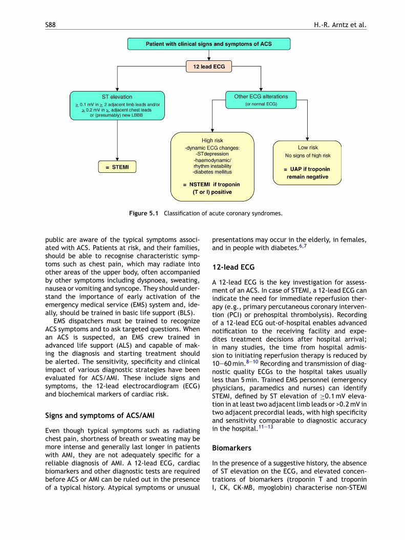
S88 H.-R. Arntz et al.
Figure 5.1 Classification of acute coronary syndromes.
public are aware of the typical symptoms associ-ated with ACS. Patients at risk, and their families,should be able to recognise characteristic symp-toms such as chest pain, which may radiate intoother areas of the upper body, often accompaniedby other symptoms including dyspnoea, sweating,nausea or vomiting and syncope. They should under-stand the importance of early activation of theemergency medical service (EMS) system and, ide-ally, should be trained in basic life support (BLS).
EMS dispatchers must be trained to recognizeACS symptoms and to ask targeted questions. Whenan ACS is suspected, an EMS crew trained inadvanced life support (ALS) and capable of mak-ing the diagnosis and starting treatment shouldbe alerted. The sensitivity, specificity and clinicalimpact of various diagnostic strategies have beenevaluated for ACS/AMI. These include signs andsymptoms, the 12-lead electrocardiogram (ECG)and biochemical markers of cardiac risk.
Signs and symptoms of ACS/AMI
Even though typical symptoms such as radiatingchest pain, shortness of breath or sweating may be
presentations may occur in the elderly, in females,and in people with diabetes.6,7
12-lead ECG
A 12-lead ECG is the key investigation for assess-ment of an ACS. In case of STEMI, a 12-lead ECG canindicate the need for immediate reperfusion ther-apy (e.g., primary percutaneous coronary interven-tion (PCI) or prehospital thrombolysis). Recordingof a 12-lead ECG out-of-hospital enables advancednotification to the receiving facility and expe-dites treatment decisions after hospital arrival;in many studies, the time from hospital admis-sion to initiating reperfusion therapy is reduced by10—60 min.8—10 Recording and transmission of diag-nostic quality ECGs to the hospital takes usuallyless than 5 min. Trained EMS personnel (emergencyphysicians, paramedics and nurses) can identifySTEMI, defined by ST elevation of ≥0.1 mV eleva-tion in at least two adjacent limb leads or >0.2 mV intwo adjacent precordial leads, with high specificityand sensitivity comparable to diagnostic accuracyin the hospital.11—13
B
IotI
more intense and generally last longer in patientswith AMI, they are not adequately specific for areliable diagnosis of AMI. A 12-lead ECG, cardiacbiomarkers and other diagnostic tests are requiredbefore ACS or AMI can be ruled out in the presenceof a typical history. Atypical symptoms or unusual
iomarkers
n the presence of a suggestive history, the absencef ST elevation on the ECG, and elevated concen-rations of biomarkers (troponin T and troponin, CK, CK-MB, myoglobin) characterise non-STEMI
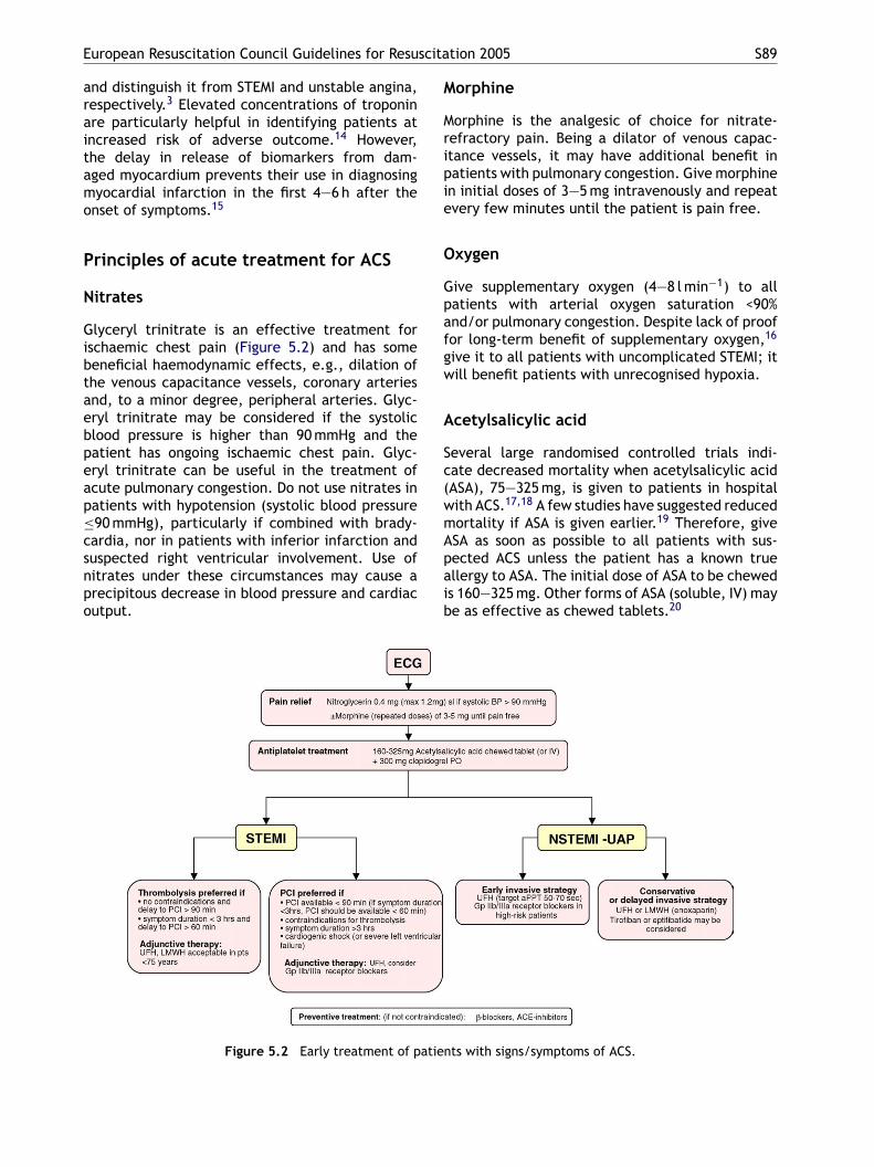
European Resuscitation Council Guidelines for Resuscitation 2005 S89
and distinguish it from STEMI and unstable angina,respectively.3 Elevated concentrations of troponinare particularly helpful in identifying patients atincreased risk of adverse outcome.14 However,the delay in release of biomarkers from dam-aged myocardium prevents their use in diagnosingmyocardial infarction in the first 4—6 h after theonset of symptoms.15
Principles of acute treatment for ACS
Nitrates
Glyceryl trinitrate is an effective treatment forischaemic chest pain (Figure 5.2) and has somebeneficial haemodynamic effects, e.g., dilation ofthe venous capacitance vessels, coronary arteriesand, to a minor degree, peripheral arteries. Glyc-eryl trinitrate may be considered if the systolicblood pressure is higher than 90 mmHg and thepatient has ongoing ischaemic chest pain. Glyc-eryl trinitrate can be useful in the treatment ofacute pulmonary congestion. Do not use nitrates inpatients with hypotension (systolic blood pressure≤csnpo
Morphine
Morphine is the analgesic of choice for nitrate-refractory pain. Being a dilator of venous capac-itance vessels, it may have additional benefit inpatients with pulmonary congestion. Give morphinein initial doses of 3—5 mg intravenously and repeatevery few minutes until the patient is pain free.
Oxygen
Give supplementary oxygen (4—8 l min−1) to allpatients with arterial oxygen saturation <90%and/or pulmonary congestion. Despite lack of prooffor long-term benefit of supplementary oxygen,16
give it to all patients with uncomplicated STEMI; itwill benefit patients with unrecognised hypoxia.
Acetylsalicylic acid
Several large randomised controlled trials indi-cate decreased mortality when acetylsalicylic acid(ASA), 75—325 mg, is given to patients in hospitalwith ACS.17,18 A few studies have suggested reducedm 19
Apaib
atie
90 mmHg), particularly if combined with brady-ardia, nor in patients with inferior infarction anduspected right ventricular involvement. Use ofitrates under these circumstances may cause arecipitous decrease in blood pressure and cardiacutput.
Figure 5.2 Early treatment of p
ortality if ASA is given earlier. Therefore, giveSA as soon as possible to all patients with sus-ected ACS unless the patient has a known truellergy to ASA. The initial dose of ASA to be cheweds 160—325 mg. Other forms of ASA (soluble, IV) maye as effective as chewed tablets.20
nts with signs/symptoms of ACS.
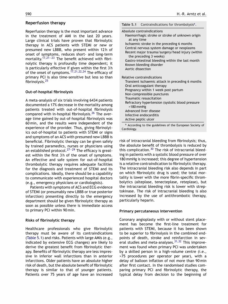
S90 H.-R. Arntz et al.
Reperfusion therapy
Reperfusion therapy is the most important advancein the treatment of AMI in the last 20 years.Large clinical trials have proven that fibrinolytictherapy in ACS patients with STEMI or new orpresumed new LBBB, who present within 12 h ofonset of symptoms, reduces short- and long-termmortality.17,21—23 The benefit achieved with fibri-nolytic therapy is profoundly time dependent; itis particularly effective if given within the first 3 hof the onset of symptoms.17,21,22,24 The efficacy ofprimary PCI is also time-sensitive but less so thanfibrinolysis.25
Out-of-hospital fibrinolysis
A meta-analysis of six trials involving 6434 patientsdocumented a 17% decrease in the mortality amongpatients treated with out-of-hospital fibrinolysiscompared with in-hospital fibrinolysis.26 The aver-age time gained by out-of-hospital fibrinolysis was60 min, and the results were independent of theexperience of the provider. Thus, giving fibrinolyt-ics out-of-hospital to patients with STEMI or signs
Table 5.1 Contraindications for thrombolysisa.
Absolute contraindicationsHaemorrhagic stroke or stroke of unknown origin
at any timeIschaemic stroke in the preceding 6 monthsCentral nervous system damage or neoplasmsRecent major trauma/surgery/head injury (within
the preceding 3 weeks)Gastro-intestinal bleeding within the last monthKnown bleeding disorderAortic dissection
Relative contraindicationsTransient ischaemic attack in preceding 6 monthsOral anticoagulant therapyPregnancy within 1 week post partumNon-compressible puncturesTraumatic resuscitationRefractory hypertension (systolic blood pressure
>180 mmHgAdvanced liver diseaseInfective endocarditisActive peptic ulcera According to the guidelines of the European Society of
Cardiology.
risk of intracranial bleeding from fibrinolysis; thus,the absolute benefit of thrombolysis is reduced bythis complication.30 The risk of intracranial bleed-ing in patients with a systolic blood pressure of over180 mmHg is increased; this degree of hypertensionis a relative contraindication to fibrinolytic therapy.The intracranial bleeding risk also depends in parton which fibrinolytic drug is used; the total mor-tality is lower with the more fibrin-specific throm-bolytics (alteplase, tenecteplase, reteplase), butthe intracranial bleeding risk is lower with strep-tokinase. The risk of intracranial bleeding is alsoincreased by the use of antithrombotic therapy,particularly heparin.
Primary percutaneous intervention
Coronary angioplasty with or without stent place-ment has become the first-line treatment forpatients with STEMI, because it has been shownto be superior to fibrinolysis in the combined end-points of death, stroke and reinfarction in sev-eral studies and meta-analyses.31,32 This improve-ment was found when primary PCI was undertakenby a skilled person in a high-volume centre (i.e.,>dapt
and symptoms of an ACS with presumed new LBBB isbeneficial. Fibrinolytic therapy can be given safelyby trained paramedics, nurses or physicians usingan established protocol.27—29 The efficacy is great-est within the first 3 h of the onset of symptoms.An effective and safe system for out-of-hospitalthrombolytic therapy requires adequate facilitiesfor the diagnosis and treatment of STEMI and itscomplications. Ideally, there should be a capabilityto communicate with experienced hospital doctors(e.g., emergency physicians or cardiologists).
Patients with symptoms of ACS and ECG evidenceof STEMI (or presumably new LBBB or true posteriorinfarction) presenting directly to the emergencydepartment should be given fibrinolytic therapy assoon as possible unless there is immediate accessto primary PCI within 90 min.
Risks of fibrinolytic therapy
Healthcare professionals who give fibrinolytictherapy must be aware of its contraindications(Table 5.1) and risks. Patients with large AMIs (e.g.,indicated by extensive ECG changes) are likely toderive the greatest benefit from fibrinolytic ther-apy. Benefits of fibrinolytic therapy are less impres-sive in inferior wall infarctions than in anteriorinfarctions. Older patients have an absolute higherrisk of death, but the absolute benefit of fibrinolytictherapy is similar to that of younger patients.Patients over 75 years of age have an increased
75 procedures per operator per year), with aelay of balloon inflation of not more than 90 minfter first contact. In the randomised studies com-aring primary PCI and fibrinolytic therapy, theypical delay from decision to the beginning of
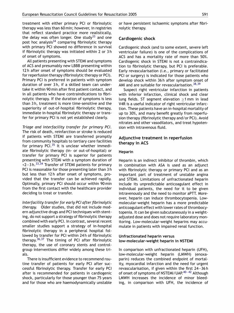
European Resuscitation Council Guidelines for Resuscitation 2005 S91
treatment with either primary PCI or fibrinolytictherapy was less than 60 min; however, in registriesthat reflect standard practice more realistically,the delay was often longer. One study33 and onepost hoc analysis34 comparing fibrinolytic therapywith primary PCI showed no difference in survivalif fibrinolytic therapy was initiated within 2 or 3 hof onset of symptoms.
All patients presenting with STEMI and symptomsof ACS and presumably new LBBB presenting within12 h after onset of symptoms should be evaluatedfor reperfusion therapy (fibrinolytic therapy or PCI).Primary PCI is preferred in patients with symptomduration of over 3 h, if a skilled team can under-take it within 90 min after first patient contact, andin all patients who have contraindications to fibri-nolytic therapy. If the duration of symptoms is lessthan 3 h, treatment is more time-sensitive and thesuperiority of out-of-hospital fibrinolytic therapy,immediate in-hospital fibrinolytic therapy or trans-fer for primary PCI is not yet established clearly.
Triage and interfacility transfer for primary PCI.The risk of death, reinfarction or stroke is reducedif patients with STEMI are transferred promptlyffatp<PbvOfd
Iteicsfilttga
tcasa
or have persistent ischaemic symptoms after fibri-nolytic therapy.
Cardiogenic shock
Cardiogenic shock (and to some extent, severe leftventricular failure) is one of the complications ofACS and has a mortality rate of more than 50%.Cardiogenic shock in STEMI is not a contraindica-tion to fibrinolytic therapy, but PCI is preferable.Early revascularisation (i.e., primary or facilitatedPCI or surgery) is indicated for those patients whodevelop shock within 36 h after symptom onset ofAMI and are suitable for revascularisation.38,39
Suspect right ventricular infarction in patientswith inferior infarction, clinical shock and clearlung fields. ST segment elevation ≥1 mm in leadV4R is a useful indicator of right ventricular infarc-tion. These patients have an in-hospital mortality ofup to 30%, and many benefit greatly from reperfu-sion therapy (fibrinolytic therapy and/or PCI). Avoidnitrates and other vasodilators, and treat hypoten-sion with intravenous fluid.
Adjunctive treatment in reperfusiont
H
Hiwiaiiiomataim
Ul
IlpiroLi
rom community hospitals to tertiary care facilitiesor primary PCI.35 It is unclear whether immedi-te fibrinolytic therapy (in- or out-of-hospital) orransfer for primary PCI is superior for patientsresenting with STEMI with a symptom duration of2—3 h.33,34 Transfer of STEMI patients for primaryCI is reasonable for those presenting later than 3 hut less than 12 h after onset of symptoms, pro-ided that the transfer can be achieved rapidly.ptimally, primary PCI should occur within 90 min
rom the first contact with the healthcare providereciding to treat or transfer.
nterfacility transfer for early PCI after fibrinolyticherapy. Older studies, that did not include mod-rn adjunctive drugs and PCI techniques with stent-ng, do not support a strategy of fibrinolytic therapyombined with early PCI. In contrast, several recentmaller studies support a strategy of in-hospitalbrinolytic therapy in a peripheral hospital fol-
owed by transfer for PCI within 24 h of fibrinolyticherapy.36,37 The timing of PCI after fibrinolyticherapy, the use of coronary stents and control-roup interventions differ widely among these tri-ls.
There is insufficient evidence to recommend rou-ine transfer of patients for early PCI after suc-essful fibrinolytic therapy. Transfer for early PCIfter is recommended for patients in cardiogenichock, particularly for those younger than 75 yearsnd for those who are haemodynamically unstable
herapy in ACS
eparin
eparin is an indirect inhibitor of thrombin, whichn combination with ASA is used as an adjunctith fibrinolytic therapy or primary PCI and as an
mportant part of treatment of unstable anginand STEMI. Limitations of unfractionated heparinnclude its unpredictable anticoagulant effect inndividual patients, the need for it to be givenntravenously and the need to monitor aPTT. More-ver, heparin can induce thrombocytopenia. Low-olecular-weight heparin has a more predictable
nticoagulant effect with lower rates of thrombocy-openia. It can be given subcutaneously in a weight-djusted dose and does not require laboratory mon-toring. Low-molecular-weight heparins may accu-ulate in patients with impaired renal function.
nfractionated heparin versusow-molecular-weight heparin in NSTEMI
n comparison with unfractionated heparin (UFH),ow-molecular-weight heparin (LMWH) (enoxa-arin) reduces the combined endpoint of mortal-ty, myocardial infarction and the need for urgentevascularisation, if given within the first 24—36 hf onset of symptoms of NSTEMI/UAP.40—42 AlthoughMWH increases the incidence of minor bleed-ng, in comparison with UFH, the incidence of
S92 H.-R. Arntz et al.
serious bleeding is not increased. Early treatmentwith LMWH (enoxaparin) is the preferred therapyfor patients with NSTEMI/UAP in addition to ASA,whenever a non-interventional strategy is planned.Consider UFH if reperfusion is planned in thefirst 24—36 h after symptom onset. Optimal targetvalue of aPPT is 50—70 s. Avoid switching betweenUFH and LMWH, because it may increase bleedingcomplications.43
Unfractionated heparin versuslow-molecular-weight heparin in STEMI
Two large randomised controlled thrombolysis stud-ies comparing LMWH with UFH demonstrated areduced frequency of ischaemic complicationswhen given to patients with STEMI within 6 h ofthe onset of symptoms.44,45 This must be balancedagainst the increase in intracranial haemorrhage inpatients over 75 years of age who receive LMWH.45
There is no evidence to support giving LMWH topatients with STEMI proceeding to an invasive strat-egy. Thus, LMWH is an acceptable alternative toUFH as an ancillary therapy for patients youngerthan 75 years without significant renal dysfunction
rent ischaemia in patients with UA/NSTEMI with-out mechanical perfusion, but showed a reduc-tion in 30-day mortality in a later meta-analysis.46
In patients with UA/NSTEMI, abciximab, given inaddition to standard therapy without mechanicalintervention, resulted in a trend towards a worseoutcome.47 Therefore, in high-risk patients, giveGp IIb/IIIa inhibitors in addition to standard ther-apy in patients for whom revascularisation ther-apy is planned. If revascularisation therapy is notplanned, tirofiban and eptifibatide can be given tohigh-risk NSTEMI/UAP patients in conjunction withASA and LMWH. Do not give abciximab if PCI is notplanned.
Gp IIb/IIIa inhibitors in STEMI. Gp IIb/IIIa receptorblockers in combination with a reduced dose of fib-rinolytics do not reduce mortality in patients withSTEMI, but increase bleeding risk in patients over75 years of age.44,48 Abciximab reduces mortalitywhen given to patients with STEMI and plannedprimary PCI, but is not beneficial in patients notproceeding to primary PCI.46 Prehospital use ofabciximab may improve the patency of the infarct-related artery with regard to PCI.49 There is nobenefit in giving tirofiban in addition to stan-ddittn
C
CviwwadraPiioodgdtfi
eA
who are treated with fibrinolytic therapy. UFH isrecommended as an ancillary therapy to fibrinolytictherapy in elderly patients and any STEMI patientfor whom revascularisation is planned. The optimaltarget value of aPPT is 50—70 s. The use of hep-arin (preferably LMWH) depends partly on whichfibrinolytic drug is used. Heparin is needed aftershorter-acting drugs because of the rebound hyper-coagulable state that occurs after a few hours,but not after streptokinase because the fibrinolyticeffect of streptokinase lasts for about 48 h.
Glycoprotein IIb/IIIa inhibitors
The platelet glycoprotein (Gp) IIb/IIIa receptor isthe final common pathway to platelet aggregation.The synthetic substances eptifibatide and tirofibanmodulate this receptor activity reversibly, whereasthe receptor antibody abciximab blocks it irre-versibly.
Gp IIb/IIIa inhibitors in NSTEMI/unstable angina.The incidences of death and recurrent ischaemiaare reduced when Gp IIb/IIIa inhibitors are addedto standard therapy including ASA and heparinin high-risk patients with UAP/NSTEMI treatedwith mechanical reperfusion.46 High-risk featuresinclude persistent pain, haemodynamic or rhythminstability, diabetes, acute or dynamic ECG changesand any elevation in cardiac troponins. Tirofibanor eptifibatide failed to reduce death or recur-
ard therapy out of hospital or in the emergencyepartment.50 Abciximab may be helpful in reduc-ng short-term mortality and short-term reinfarc-ion in patients treated with PCI without fibrinolyticherapy. Abciximab is not recommended in combi-ation with fibrinolytics in patients with STEMI.
lopidogrel
lopidogrel inhibits the platelet ADP receptor irre-ersibly, which further reduces platelet aggregationn addition to that produced by ASA. Comparedith ASA, there is no increased risk of bleedingith clopidogrel.51 If given in addition to hep-rin and ASA within 4 h of presentation, clopi-ogrel improves outcome in patients with high-isk ACS.52,53 There is a significant reduction indverse ischaemic events at 28 days after electiveCI when clopidogrel is given at least 6 h beforentervention.54 A recent trial documented a signif-cant reduction in the composite endpoint of anccluded infarct-related artery (TIMI flow grade 0r 1) on angiography or death or recurrent myocar-ial infarction before angiography, when clopido-rel (300 mg loading dose, followed by 75 mg dailyose up to 8 days in hospital) is given to patients upo 75 years of age with STEMI who are treated withbrinolytic therapy, ASA and heparin.55
Give a 300-mg oral loading dose of clopidogrelarly, as well as standard care, to patients withCS if they have an increase in serum cardiac
European Resuscitation Council Guidelines for Resuscitation 2005 S93
biomarkers and/or new ECG changes consistentwith ischaemia when a medical approach or PCI isplanned. Give clopidogrel to patients with STEMIup to 75 years of age receiving fibrinolytic therapy,ASA and heparin. Clopidogrel, 300 mg, can be giveninstead of ASA to patients with a suspected ACS whohave a true allergy to or gastrointestinal intoler-ance of ASA.
Primary and secondary preventioninterventions
Start preventive interventions, at the latest, at theinitial admission with a confirmed diagnosis of ACS.Give a beta-blocker as soon as possible unless con-traindicated or poorly tolerated. Treat all patientswith a statin (HRG co-enzyme A reductase inhibitor)unless contraindicated or poorly tolerated. Start anACE inhibitor in all patients with STEMI, all patientswith STEMI and left ventricular systolic impair-ment, and consider it in all other patients withSTEMI unless contraindicated or poorly tolerated.In patients unable to tolerate an ACE inhibitor, anangiotensin receptor blocker may be used as a sub-st
B
Sriwubam
wbutsmsit
A
AmtVA
hours after onset of symptoms.59,60 This explainswhy numerous studies have been performed withthe aim of demonstrating the prophylactic effectof anti-arrhythmic therapy. The effects of anti-arrhythmic drugs (lidocaine, magnesium, disopy-ramide, mexiletine, verapamil) given prophylacti-cally to patients with ACS have been studied.61—63
Prophylaxis with lidocaine reduces the incidenceof VF but may increase mortality.58 Routine treat-ment with magnesium in patients with AMI doesnot improve mortality.64 Arrhythmia prophylaxisusing disopyramide, mexiletine or verapamil, givenwithin the first hours of an ACS, does not improvemortality.63 In contrast, intravenous beta-blockersreduced the incidence of VF when given to patientswith ACS.56,57
Angiotensin-converting enzyme inhibitorsand angiotensin-II receptor blockers
Oral angiotensin-converting inhibitors (ACE) inhibi-tors reduce mortality when given to patients withacute myocardial infarction with or without earlyreperfusion therapy.65,66 The beneficial effects aremost pronounced in patients presenting with ante-rti1thvaApfpg42ri
S
Sdaor
R
titute in those patients with left ventricular sys-olic impairment.
eta-blockers
everal studies, undertaken mainly in the pre-eperfusion era, indicate decreased mortality andncidence of reinfarction and cardiac rupture asell as a lower incidence of VF and supraventric-lar arrhythmia in patients treated early with aeta-blocker.56,57 Intravenous beta-blockade maylso reduce mortality in patients undergoing pri-ary PCI who are not on oral beta-blockers.58
Haemodynamically stable patients presentingith an ACS should be given intravenous beta-lockers promptly, followed by regular oral therapynless contraindicated or poorly tolerated. Con-raindications to beta-blockers include hypoten-ion, bradycardia, second- or third-degree AV block,oderate to severe congestive heart failure and
evere reactive airway disease. Give a beta-blockerrrespective of the need for early revascularisationherapy.
nti-arrhythmics
part from the use of a beta-blocker as recom-ended above, there is no evidence to support
he use of anti-arrhythmic prophylaxis after ACS.F accounts for most of the early deaths fromCS; the incidence of VF is highest in the first
ior infarction, pulmonary congestion or left ven-ricular ejection fraction <40%.66 Do not give ACEnhibitors if the systolic blood pressure is less than00 mmHg at admission or if there is a known con-raindication to these drugs.66 A trend towardsigher mortality has been documented if an intra-enous ACE inhibitor is started within the first 24 hfter onset of symptoms.67 Therefore, give an oralCE inhibitor within 24 h after symptom onset inatients with AMI regardless of whether early reper-usion therapy is planned, particularly in thoseatients with anterior infarction, pulmonary con-estion or left ventricular ejection fraction below0%. Do not give intravenous ACE inhibitors within4 h of onset of symptoms. Give an angiotensineceptor blocker (ARB) to patients intolerant of ACEnhibitors.
tatins
tatins reduce the incidence of major adverse car-iovascular events when given within a few daysfter onset of ACS. Start statin therapy within 24 hf onset of symptoms of ACS. If patients are alreadyeceiving statin therapy, do not stop it.68
eferences
1. Tunstall-Pedoe H, Vanuzzo D, Hobbs M, et al. Estimationof contribution of changes in coronary care to improving
S94 H.-R. Arntz et al.
survival, event rates, and coronary heart disease mortal-ity across the WHO MONICA Project populations. Lancet2000;355:688—700.
2. Lowel H, Meisinger C, Heier M, et al. Sex specific trendsof sudden cardiac death and acute myocardial infarction:results of the population-based KORA/MONICA-Augsburg reg-ister 1985 to 1998. Dtsch Med Wochenschr 2002;127:2311—6.
3. European Society Cardiology. Myocardial infarctionredefined—–a consensus document of The Joint EuropeanSociety of Cardiology/American College of CardiologyCommittee for the redefinition of myocardial infarction. JAm Coll Cardiol 2000;36:959—69.
4. Van de Werf F, Ardissino D, Betriu A, et al. Management ofacute myocardial infarction in patients presenting with ST-segment elevation. The Task Force on the Management ofAcute Myocardial Infarction of the European Society of Car-diology. Eur Heart J 2003;24:28—66.
5. Antman EM, Anbe DT, Armstrong PW, et al. ACC/AHA guide-lines for the management of patients with ST-elevationmyocardial infarction—executive summary: a report of theAmerican College of Cardiology/American Heart Associa-tion Task Force on Practice Guidelines (Writing Commit-tee to Revise the 1999 Guidelines for the Managementof Patients With Acute Myocardial Infarction). Circulation2004;110:588—636.
6. Douglas PS, Ginsburg GS. The evaluation of chest pain inwomen. N Engl J Med 1996;334:1311—5.
7. Solomon CG, Lee TH, Cook EF, et al. Comparison of clinicalpresentation of acute myocardial infarction in patients olderthan 65 years of age to younger patients: the Multicenter
1
1
1
1
1
1
1
17. Randomised trial of intravenous streptokinase, oral aspirin,both, or neither among 17,187 cases of suspected acutemyocardial infarction: ISIS-2. ISIS-2 (Second InternationalStudy of Infarct Survival) Collaborative Group. Lancet1988;2:349—60.
18. Gurfinkel EP, Manos EJ, Mejail RI, et al. Low molecular weightheparin versus regular heparin or aspirin in the treatmentof unstable angina and silent ischemia. J Am Coll Cardiol1995;26:313—8.
19. Freimark D, Matetzky S, Leor J, et al. Timing of aspirinadministration as a determinant of survival of patients withacute myocardial infarction treated with thrombolysis. AmJ Cardiol 2002;89:381—5.
20. Husted SE, Kristensen SD, Vissinger H, Morn B, Schmidt EB,Nielsen HK. Intravenous acetylsalicylic acid—–dose-relatedeffects on platelet function and fibrinolysis in healthy males.Thromb Haemost 1992;68:226—9.
21. Indications for fibrinolytic therapy in suspected acutemyocardial infarction: collaborative overview of early mor-tality and major morbidity results from all randomised trialsof more than 1000 patients. Fibrinolytic Therapy Trialists’(FTT) Collaborative Group. Lancet 1994;343:311—22.
22. Effectiveness of intravenous thrombolytic treatment inacute myocardial infarction. Gruppo Italiano per lo Studiodella Streptochinasi nell’Infarto Miocardico (GISSI). Lancet1986;1:397—402.
23. The GUSTO investigators. An international randomized trialcomparing four thrombolytic strategies for acute myocardialinfarction. N Engl J Med 1993;329:673—82.
24. Boersma E, Maas AC, Deckers JW, Simoons ML. Early throm-
2
2
2
2
2
3
3
3
3
Chest Pain Study experience. Am J Cardiol 1989;63:772—6.8. Kereiakes DJ, Gibler WB, Martin LH, Pieper KS, Anderson LC.
Relative importance of emergency medical system transportand the prehospital electrocardiogram on reducing hospitaltime delay to therapy for acute myocardial infarction: a pre-liminary report from the Cincinnati Heart Project. Am HeartJ 1992;123(Pt 1):835—40.
9. Canto JG, Rogers WJ, Bowlby LJ, French WJ, Pearce DJ,Weaver WD. The prehospital electrocardiogram in acutemyocardial infarction: is its full potential being realized?National Registry of Myocardial Infarction 2 Investigators. JAm Coll Cardiol 1997;29:498—505.
0. Aufderheide TP, Hendley GE, Thakur RK, et al. The diagnos-tic impact of prehospital 12-lead electrocardiography. AnnEmerg Med 1990;19:1280—7.
1. Foster DB, Dufendach JH, Barkdoll CM, Mitchell BK. Prehos-pital recognition of AMI using independent nurse/paramedic12-lead ECG evaluation: impact on in-hospital times tothrombolysis in a rural community hospital. Am J Emerg Med1994;12:25—31.
2. Millar-Craig MW, Joy AV, Adamowicz M, Furber R, Thomas B.Reduction in treatment delay by paramedic ECG diagnosisof myocardial infarction with direct CCU admission. Heart1997;78:456—61.
3. Brinfield K. Identification of ST elevation AMI on prehospital12 lead ECG; accuracy of unaided paramedic interpretation.J Emerg Med 1998;16:22S.
4. Antman EM, Tanasijevic MJ, Thompson B, et al. Cardiac-specific troponin I levels to predict the risk of mortalityin patients with acute coronary syndromes. N Engl J Med1996;335:1342—9.
5. Schuchert A, Hamm C, Scholz J, Klimmeck S, Goldmann B,Meinertz T. Prehospital testing for troponin T in patientswith suspected acute myocardial infarction. Am Heart J1999;138:45—8.
6. Rawles JM, Kenmure AC. Controlled trial of oxygen in uncom-plicated myocardial infarction. BMJ 1976;1:1121—3.
bolytic treatment in acute myocardial infarction: reappraisalof the golden hour. Lancet 1996;348:771—5.
5. De Luca G, van’t Hof AW, de Boer MJ, et al. Time-to-treatment significantly affects the extent of ST-segment res-olution and myocardial blush in patients with acute myocar-dial infarction treated by primary angioplasty. Eur Heart J2004;25:1009—13.
6. Morrison LJ, Verbeek PR, McDonald AC, Sawadsky BV, CookDJ. Mortality and prehospital thrombolysis for acute myocar-dial infarction: a meta-analysis. JAMA 2000;283:2686—92.
7. Welsh RC, Goldstein P, Adgey J, et al. Variations in pre-hospital fibrinolysis process of care: insights from the Assess-ment of the Safety and Efficacy of a New Thrombolytic 3 Plusinternational acute myocardial infarction pre-hospital caresurvey. Eur J Emerg Med 2004;11:134—40.
8. Weaver W, Cerqueira M, Hallstrom A, et al. Prehospital-initiated vs hospital-initiated thrombolytic therapy: theMyocardial Infacrtion Triage and Intervention Trial (MITI).JAMA 1993;270:1203—10.
9. European Myocardial Infarction Project Group (EMIP). Pre-hospital thrombolytic therapy in patients with suspectedacute myocardial infarction. The European MyocardialInfarction Project Group. N Engl J Med 1993;329:383—9.
0. White HD. Debate: should the elderly receive thrombolytictherapy, or primary angioplasty? Current Control Trials Car-diovasc Med 2000;1:150—4.
1. Weaver WD, Simes RJ, Betriu A, et al. Comparison of primarycoronary angioplasty and intravenous thrombolytic therapyfor acute myocardial infarction: a quantitative review. JAMA1997;278:2093—8.
2. Keeley EC, Boura JA, Grines CL. Primary angioplasty ver-sus intravenous thrombolytic therapy for acute myocardialinfarction: a quantitative review of 23 randomised trials.Lancet 2003;361:13—20.
3. Widimsky P, Budesinsky T, Vorac D, et al. Long distancetransport for primary angioplasty vs immediate thrombol-ysis in acute myocardial infarction. Final results of the ran-
European Resuscitation Council Guidelines for Resuscitation 2005 S95
domized national multicentre trial—PRAGUE-2. Eur Heart J2003;24:94—104.
34. Steg PG, Bonnefoy E, Chabaud S, et al. Impact of time totreatment on mortality after prehospital fibrinolysis or pri-mary angioplasty: data from the CAPTIM randomized clinicaltrial. Circulation 2003;108:2851—6.
35. Dalby M, Bouzamondo A, Lechat P, Montalescot G. Trans-fer for primary angioplasty versus immediate thrombolysisin acute myocardial infarction: a meta-analysis. Circulation2003;108:1809—14.
36. Scheller B, Hennen B, Hammer B, et al. Beneficial effects ofimmediate stenting after thrombolysis in acute myocardialinfarction. J Am Coll Cardiol 2003;42:634—41.
37. Fernandez-Aviles F, Alonso JJ, Castro-Beiras A, et al. Rou-tine invasive strategy within 24 h of thrombolysis versusischaemia-guided conservative approach for acute myocar-dial infarction with ST-segment elevation (GRACIA-1): a ran-domised controlled trial. Lancet 2004;364:1045—53.
38. Hochman JS, Sleeper LA, Webb JG, et al. Early revascular-ization in acute myocardial infarction complicated by car-diogenic shock. SHOCK Investigators. Should we emergentlyrevascularize occluded coronaries for cardiogenic shock. NEngl J Med 1999;341:625—34.
39. Hochman JS, Sleeper LA, White HD, et al. One-year sur-vival following early revascularization for cardiogenic shock.JAMA 2001;285:190—2.
40. Antman EM, McCabe CH, Gurfinkel EP, et al. Enoxaparinprevents death and cardiac ischemic events in unstableangina/non-Q-wave myocardial infarction. Results of thethrombolysis in myocardial infarction (TIMI) 11B trial. Cir-
4
4
4
4
4
4
4
48. Topol EJ. Reperfusion therapy for acute myocardial infarc-tion with fibrinolytic therapy or combination reduced fibri-nolytic therapy and platelet glycoprotein IIb/IIIa inhibition:the GUSTO V randomised trial. Lancet 2001;357:1905—14.
49. Montalescot G, Borentain M, Payot L, Collet JP, Thomas D.Early vs late administration of glycoprotein IIb/IIIa inhibitorsin primary percutaneous coronary intervention of acute ST-segment elevation myocardial infarction: a meta-analysis.JAMA 2004;292:362—6.
50. van’t Hof AW, Ernst N, de Boer MJ, et al. Facilitation ofprimary coronary angioplasty by early start of a glycopro-tein 2b/3a inhibitor: results of the ongoing tirofiban inmyocardial infarction evaluation (On-TIME) trial. Eur HeartJ 2004;25:837—46.
51. A randomised, blinded, trial of clopidogrel versus aspirin inpatients at risk of ischaemic events (CAPRIE). CAPRIE Steer-ing Committee. Lancet 1996;348:1329—39.
52. Yusuf S, Zhao F, Mehta SR, Chrolavicius S, Tognoni G, Fox KK.Effects of clopidogrel in addition to aspirin in patients withacute coronary syndromes without ST-segment elevation. NEngl J Med 2001;345:494—502.
53. Mehta SR, Yusuf S, Peters RJ, et al. Effects of pretreatmentwith clopidogrel and aspirin followed by long-term therapyin patients undergoing percutaneous coronary intervention:the PCI-CURE study. Lancet 2001;358:527—33.
54. Steinhubl SR, Berger PB, Mann IIIrd JT, et al. Early and sus-tained dual oral antiplatelet therapy following percutaneouscoronary intervention: a randomized controlled trial. JAMA2002;288:2411—20.
55. Sabatine MS, Cannon CP, Gibson CM, et al. Addition of
5
5
5
5
6
6
6
6
6
culation 1999;100:1593—601.1. Cohen M, Demers C, Gurfinkel EP, et al. A comparison of low-
molecular-weight heparin with unfractionated heparin forunstable coronary artery disease. Efficacy and Safety of Sub-cutaneous Enoxaparin in Non-Q-Wave Coronary Events StudyGroup. N Engl J Med 1997;337:447—52.
2. Petersen JL, Mahaffey KW, Hasselblad V, et al. Efficacyand bleeding complications among patients randomized toenoxaparin or unfractionated heparin for antithrombin ther-apy in non-ST-Segment elevation acute coronary syndromes:a systematic overview. JAMA 2004;292:89—96.
3. Ferguson JJ, Califf RM, Antman EM, et al. Enoxaparin vsunfractionated heparin in high-risk patients with non-ST-segment elevation acute coronary syndromes managed withan intended early invasive strategy: primary results of theSYNERGY randomized trial. JAMA 2004;292:45—54.
4. Van de Werf FJ, Armstrong PW, Granger C, Wallentin L. Effi-cacy and safety of tenecteplase in combination with enoxa-parin, abciximab, or unfractionated heparin: the ASSENT-3 randomised trial in acute myocardial infarction. Lancet2001;358:605—13.
5. Wallentin L, Goldstein P, Armstrong PW, et al. Efficacyand safety of tenecteplase in combination with the low-molecular-weight heparin enoxaparin or unfractionated hep-arin in the prehospital setting: the Assessment of the Safetyand Efficacy of a New Thrombolytic Regimen (ASSENT)-3PLUS randomized trial in acute myocardial infarction. Cir-culation 2003;108:135—42.
6. Boersma E, Harrington RA, Moliterno DJ, et al. Platelet gly-coprotein IIb/IIIa inhibitors in acute coronary syndromes:a meta-analysis of all major randomised clinical trials.Lancet 2002;359:189—98 [erratum appears in Lancet 2002Jun 15;359(9323):2120].
7. Simoons ML. Effect of glycoprotein IIb/IIIa receptor blockerabciximab on outcome in patients with acute coronary syn-dromes without early coronary revascularisation: the GUSTOIV-ACS randomised trial. Lancet 2001;357:1915—24.
clopidogrel to aspirin and fibrinolytic therapy for myocar-dial infarction with ST-segment elevation. N Engl J Med2005;352:1179—89.
6. The MIAMI Trial Research Group. Metoprolol in acute myocar-dial infarction (MIAMI): a randomised placebo-controlledinternational trial. Eur Heart J 1985;6:199—226.
7. Randomised trial of intravenous atenolol among 16 027cases of suspected acute myocardial infarction: ISIS-1. FirstInternational Study of Infarct Survival Collaborative Group.Lancet 1986;2:57—66.
8. Halkin A, Grines CL, Cox DA, et al. Impact of intra-venous beta-blockade before primary angioplasty on sur-vival in patients undergoing mechanical reperfusion ther-apy for acute myocardial infarction. J Am Coll Cardiol2004;43:1780—7.
9. Campbell RW, Murray A, Julian DG. Ventricular arrhythmiasin first 12 h of acute myocardial infarction: natural historystudy. Br Heart J 1981;46:351—7.
0. O’Doherty M, Tayler DI, Quinn E, Vincent R, Chamberlain DA.Five hundred patients with myocardial infarction monitoredwithin one hour of symptoms. BMJ 1983;286:1405—8.
1. Teo KK, Yusuf S, Furberg CD. Effects of prophylactic antiar-rhythmic drug therapy in acute myocardial infarction. Anoverview of results from randomized controlled trials. JAMA1993;270:1589—95.
2. Sadowski ZP, Alexander JH, Skrabucha B, et al. Multicen-ter randomized trial and a systematic overview of lidocainein acute myocardial infarction. Am Heart J 1999;137:792—8.
3. McAlister FA, Teo KK. Antiarrhythmic therapies for theprevention of sudden cardiac death. Drugs 1997;54:235—52.
4. ISIS-4: a randomised factorial trial assessing early oral capto-pril, oral mononitrate, and intravenous magnesium sulphatein 58,050 patients with suspected acute myocardial infarc-tion. ISIS-4 (Fourth International Study of Infarct Survival)Collaborative Group. Lancet 1995;345:669—85.

S96 H.-R. Arntz et al.
65. Teo KK, Yusuf S, Pfeffer M, et al. Effects of long-term treat-ment with angiotensin-converting-enzyme inhibitors in thepresence or absence of aspirin: a systematic review. Lancet2002;360:1037—43.
66. ACE Inhibitor MI Collaborative Group. Indications forACE inhibitors in the early treatment of acute myocar-dial infarction: systematic overview of individual datafrom 100,000 patients in randomized trials. ACE InhibitorMyocardial Infarction Collaborative Group. Circulation1998;97:2202—12.
67. Swedberg K, Held P, Kjekshus J, Rasmussen K, Ryden L,Wedel H. Effects of the early administration of enalaprilon mortality in patients with acute myocardial infarction.Results of the Cooperative New Scandinavian Enalapril Sur-vival Study II (CONSENSUS II). N Engl J Med 1992;327:678—84.
68. Heeschen C, Hamm CW, Laufs U, Snapinn S, Bohm M,White HD. Withdrawal of statins increases event ratesin patients with acute coronary syndromes. Circulation2002;105:1446—52.
Resuscitation (2005) 67S1, S97—S133
European Resuscitation Council Guidelines forResuscitation 2005Section 6. Paediatric life support
Dominique Biarent, Robert Bingham, Sam Richmond, Ian Maconochie,Jonathan Wyllie, Sheila Simpson, Antonio Rodriguez Nunez,David Zideman
I
T
Tg1tbwcbwlETroRbcett
d
ntroduction
he process
he European Resuscitation Council (ERC) issueduidelines for paediatric life support (PLS) in 1994,998 and 2000.1—4 The last edition was based onhe International Consensus on Science publishedy the American Heart Association in collaborationith the International Liaison Committee on Resus-itation (ILCOR), undertaking a series of evidence-ased evaluations of the science of resuscitationhich culminated in the publication of the Guide-
ines 2000 for Cardiopulmonary Resuscitation andmergency Cardiovascular Care in August 2000.5,6
his process was repeated in 2004/2005, and theesulting Consensus on Science and Treatment Rec-mmendations were published simultaneously inesuscitation, Circulation and Pediatrics in Novem-er 2005.7,8 The PLS Working Party of the ERC hasonsidered this document and the supporting sci-ntific literature, and has recommended changes
Guidelines changes
The approach to changes has been to alter theguidelines in response to convincing new scientificevidence and, where possible, to simplify them inorder to assist teaching and retention. As before,there remains a paucity of good-quality evidence onpaediatric resuscitation specifically and some con-clusions have had to be drawn from animal workand extrapolated adult data.
The current guidelines have a strong focus onsimplification based on the knowledge that manychildren receive no resuscitation at all because res-cuers fear doing harm. This fear is fuelled by theknowledge that resuscitation guidelines for chil-dren are different. Consequently, a major area ofstudy was the feasibility of applying the same guid-ance for all adults and children. Bystander resusci-tation improves outcome significantly,9,10 and thereis good evidence from paediatric animal modelsthat even doing chest compressions or expired airventilation alone may be better than doing noth-
o the ERC PLS Guidelines. These are presented inhis paper.
ing at all.11 It follows that outcomes could beimproved if bystanders, who would otherwise donothing, were encouraged to begin resuscitation,es
Coun
0300-9572/$ — see front matter © 2005 European Resuscitationoi:10.1016/j.resuscitation.2005.10.010ven if they do not follow an algorithm targetedpecifically at children. There are, however, dis-
cil. All Rights Reserved. Published by Elsevier Ireland Ltd.
S98 D. Biarent et al.
tinct differences between the predominantly adultarrest of cardiac origin and asphyxial arrest, whichis most common in children,12 so a separate paedi-atric algorithm is justified for those with a duty torespond to paediatric emergencies (usually health-care professionals), who are also in a position toreceive enhanced training.
Compression:ventilation ratios
The ILCOR treatment recommendation was thatthe compression:ventilation ratio should be basedon whether one or more than one rescuers werepresent. ILCOR recommends that lay rescuers,who usually learn only single rescuer techniques,should be taught to use a ratio of 30 compres-sions to 2 ventilations, which is the same as theadult guidelines and enables anyone trained inBLS techniques to resuscitate children with mini-mal additional information. Two or more rescuerswith a duty to respond should learn a differentratio (15:2), as this has been validated by animaland manikin studies.13—17 This latter group, whowould normally be healthcare professionals, shouldreceive enhanced training targeted specifically at
ondary cardiac arrest. The onset of puberty, whichis the physiological end of childhood, is the mostlogical landmark for the upper age limit for useof paediatric guidance. This has the advantage ofbeing simple to determine, in contrast to an agelimit in years, as age may be unknown at the start ofresuscitation. Clearly, it is inappropriate and unnec-essary to establish the onset of puberty formally; ifrescuers believe the victim to be a child they shoulduse the paediatric guidelines. If a misjudgement ismade and the victim turns out to be a young adult,little harm will accrue, as studies of aetiology haveshown that the paediatric pattern of arrest contin-ues into early adulthood.19 An infant is a child under1 year of age; a child is between 1 year and puberty.It is necessary to differentiate between infants andolder children, as there are some important differ-ences between these two groups.
Chest compression technique
The modification to age definitions enables a sim-plification of the advice on chest compression.Advice for determining the landmarks for infantcompression is now the same as for older chil-dourgfttqtp
A
ClAtcrusryacpa
pa
the resuscitation of children. Although there areno data to support the superiority of any partic-ular ratio in children, ratios of between 5:1 and15:2 have been studied in manikins, and animaland mathematical models, and there is increasingevidence that the 5:1 ratio delivers an inadequatenumber of compressions.14,18 There is certainly nojustification for having two separate ratios for chil-dren aged greater or less than 8 years, so a singleratio of 15:2 for multiple rescuers with a duty torespond is a logical simplification.
It would certainly negate any benefit of simplic-ity if lay rescuers were taught a different ratio foruse if there were two of them, but those with a dutyto respond can use the 30:2 ratio if they are alone,particularly if they are not achieving an adequatenumber of compressions because of difficulty in thetransition between ventilation and compression.
Age definitions
The adoption of single compression:ventilationratios for children of all ages, together with thechange in advice on the lower age limit for theuse of automated external defibrillators (AEDs),renders the previous guideline division betweenchildren above and below 8 years of age unneces-sary. The differences between adult and paediatricresuscitation are based largely on differing aeti-ology, as primary cardiac arrest is more commonin adults whereas children usually suffer from sec-
ren, as there is evidence that the previous rec-mmendation could result in compression over thepper abdomen.20 Infant compression techniqueemains the same: two-finger compression for sin-le rescuers and two-thumb, encircling techniqueor two or more rescuers,21—25 but for older childrenhere is no division between the one- or two-handechnique.26 The emphasis is on achieving an ade-uate depth of compression with minimal interrup-ions, using one or two hands according to rescuerreference.
utomated external defibrillators
ase reports published since International Guide-ines 2000 have reported safe and successful use ofEDs in children less than 8 years of age.27,28 Fur-hermore, recent studies have shown that AEDs areapable of identifying arrhythmias in children accu-ately and that, in particular, they are extremelynlikely to advise a shock inappropriately.29,30 Con-equently, advice on the use of AEDs has beenevised to include all children aged greater than 1ear.31 Nevertheless, if there is any possibility thatn AED may need to be used in children, the pur-haser should check that the performance of thearticular model has been tested against paediatricrrhythmias.
Many manufacturers now supply purpose-madeaediatric pads or programmes, which typicallyttenuate the output of the machine to 50—75 J.32
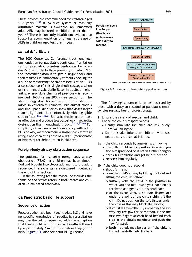
European Resuscitation Council Guidelines for Resuscitation 2005 S99
These devices are recommended for children aged1—8 years.33,34 If no such system or manuallyadjustable machine is available, an unmodifiedadult AED may be used in children older than 1year.35 There is currently insufficient evidence tosupport a recommendation for or against the use ofAEDs in children aged less than 1 year.
Manual defibrillators
The 2005 Consensus Conference treatment rec-ommendation for paediatric ventricular fibrillation(VF) or paediatric pulseless ventricular tachycar-dia (VT) is to defibrillate promptly. In adult ALS,the recommendation is to give a single shock andthen resume CPR immediately without checking fora pulse or reassessing the rhythm (see Section 3). Asa consequence of this single-shock strategy, whenusing a monophasic defibrillator in adults a higherinitial energy dose than used previously is recom-mended (360 J versus 200 J) (see Section 3). Theideal energy dose for safe and effective defibril-lation in children is unknown, but animal modelsand small paediatric series show that doses largerthan 4 J kg−1 defibrillate effectively with negligiblesadsBuo
F
Tofist
fd
6
S
Rnmtbh
Figure 6.1 Paediatric basic life support algorithm.
The following sequence is to be observed bythose with a duty to respond to paediatric emer-gencies (usually health professionals).
1. Ensure the safety of rescuer and child.2. Check the child’s responsiveness.
• Gently stimulate the child and ask loudly:‘‘Are you all right?’’
• Do not shake infants or children with sus-pected cervical spinal injuries.
3a If the child responds by answering or moving• leave the child in the position in which you
find him (provided he is not in further danger)• check his condition and get help if needed• reassess him regularly
3b If the child does not respond• shout for help;• open the child’s airway by tilting the head and
lifting the chin, as follows:o initially with the child in the position in
which you find him, place your hand on hisforehead and gently tilt his head back;
o at the same time, with your fingertip(s)under the point of the child’s chin, lift the
ide effects.27,34,36,37 Biphasic shocks are at leasts effective and produce less post-shock myocardialysfunction than monophasic shocks.33,34,37—40 Forimplicity of sequence and consistency with adultLS and ALS, we recommend a single-shock strategysing a non-escalating dose of 4 J kg−1 (monophasicr biphasic) for defibrillation in children.
oreign-body airway obstruction sequence
he guidance for managing foreign-body airwaybstruction (FBAO) in children has been simpli-ed and brought into closer alignment to the adultequence. These changes are discussed in detail athe end of this section.
In the following text the masculine includes theeminine and ‘child’ refers to both infants and chil-ren unless noted otherwise.
a Paediatric basic life support
equence of action
escuers who have been taught adult BLS and haveo specific knowledge of paediatric resuscitationay use the adult sequence, with the exception
hat they should perform 5 initial breaths followedy approximately 1 min of CPR before they go forelp (Figure 6.1; also see adult BLS guideline).
chin. Do not push on the soft tissues underthe chin as this may block the airway;
o if you still have difficulty in opening the air-way, try the jaw thrust method. Place thefirst two fingers of each hand behind eachside of the child’s mandible and push thejaw forward;
o both methods may be easier if the child isturned carefully onto his back.
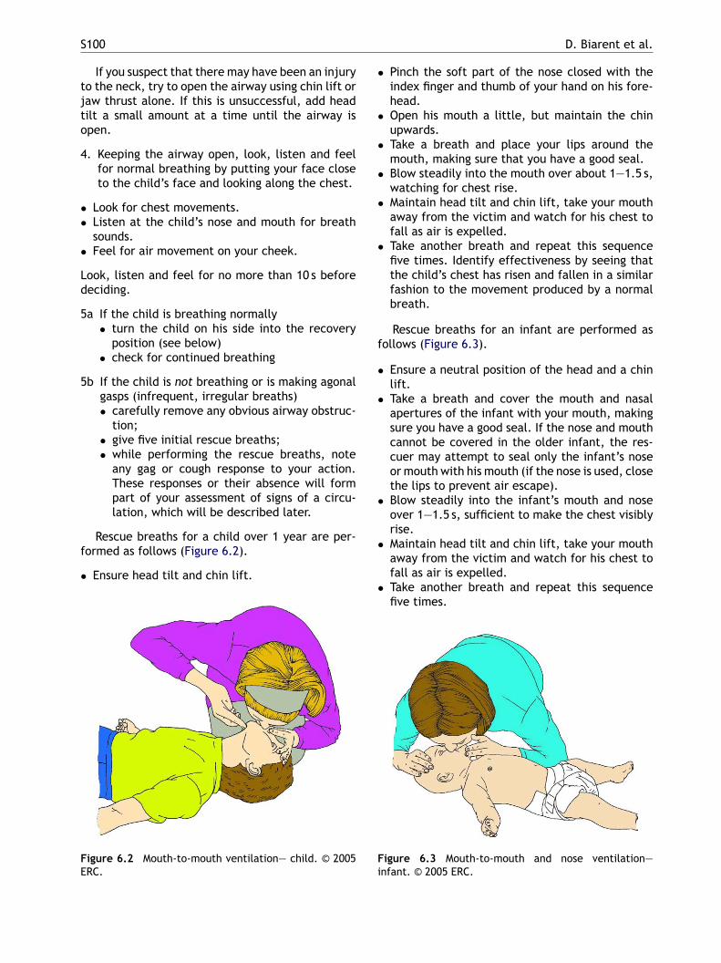
S100 D. Biarent et al.
If you suspect that there may have been an injuryto the neck, try to open the airway using chin lift orjaw thrust alone. If this is unsuccessful, add headtilt a small amount at a time until the airway isopen.
4. Keeping the airway open, look, listen and feelfor normal breathing by putting your face closeto the child’s face and looking along the chest.
• Look for chest movements.• Listen at the child’s nose and mouth for breath
sounds.• Feel for air movement on your cheek.
Look, listen and feel for no more than 10 s beforedeciding.
5a If the child is breathing normally• turn the child on his side into the recovery
position (see below)• check for continued breathing
5b If the child is not breathing or is making agonalgasps (infrequent, irregular breaths)• carefully remove any obvious airway obstruc-
• Pinch the soft part of the nose closed with theindex finger and thumb of your hand on his fore-head.
• Open his mouth a little, but maintain the chinupwards.
• Take a breath and place your lips around themouth, making sure that you have a good seal.
• Blow steadily into the mouth over about 1—1.5 s,watching for chest rise.
• Maintain head tilt and chin lift, take your mouthaway from the victim and watch for his chest tofall as air is expelled.
• Take another breath and repeat this sequencefive times. Identify effectiveness by seeing thatthe child’s chest has risen and fallen in a similarfashion to the movement produced by a normalbreath.
Rescue breaths for an infant are performed asfollows (Figure 6.3).
• Ensure a neutral position of the head and a chinlift.
• Take a breath and cover the mouth and nasalapertures of the infant with your mouth, making
tion;• give five initial rescue breaths;• while performing the rescue breaths, note
any gag or cough response to your action.These responses or their absence will formpart of your assessment of signs of a circu-lation, which will be described later.
Rescue breaths for a child over 1 year are per-formed as follows (Figure 6.2).
• Ensure head tilt and chin lift.
Figure 6.2 Mouth-to-mouth ventilation— child. © 2005ERC.
sure you have a good seal. If the nose and mouthcannot be covered in the older infant, the res-cuer may attempt to seal only the infant’s noseor mouth with his mouth (if the nose is used, closethe lips to prevent air escape).
• Blow steadily into the infant’s mouth and noseover 1—1.5 s, sufficient to make the chest visiblyrise.
• Maintain head tilt and chin lift, take your mouthaway from the victim and watch for his chest tofall as air is expelled.
• Take another breath and repeat this sequencefive times.
Figure 6.3 Mouth-to-mouth and nose ventilation—infant. © 2005 ERC.
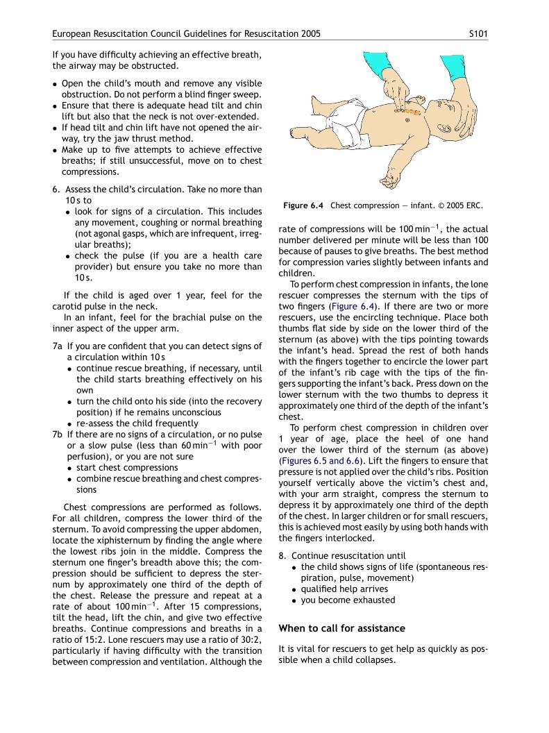
European Resuscitation Council Guidelines for Resuscitation 2005 S101
If you have difficulty achieving an effective breath,the airway may be obstructed.
• Open the child’s mouth and remove any visibleobstruction. Do not perform a blind finger sweep.
• Ensure that there is adequate head tilt and chinlift but also that the neck is not over-extended.
• If head tilt and chin lift have not opened the air-way, try the jaw thrust method.
• Make up to five attempts to achieve effectivebreaths; if still unsuccessful, move on to chestcompressions.
6. Assess the child’s circulation. Take no more than10 s to• look for signs of a circulation. This includes
any movement, coughing or normal breathing(not agonal gasps, which are infrequent, irreg-ular breaths);
• check the pulse (if you are a health careprovider) but ensure you take no more than10 s.
If the child is aged over 1 year, feel for thecarotid pulse in the neck.
In an infant, feel for the brachial pulse on thei
7
Fsltspntrtbrpb
Figure 6.4 Chest compression — infant. © 2005 ERC.
rate of compressions will be 100 min−1, the actualnumber delivered per minute will be less than 100because of pauses to give breaths. The best methodfor compression varies slightly between infants andchildren.
To perform chest compression in infants, the lonerescuer compresses the sternum with the tips oftwo fingers (Figure 6.4). If there are two or morerescuers, use the encircling technique. Place boththumbs flat side by side on the lower third of thesternum (as above) with the tips pointing towardsthe infant’s head. Spread the rest of both handswith the fingers together to encircle the lower partof the infant’s rib cage with the tips of the fin-gers supporting the infant’s back. Press down on thelower sternum with the two thumbs to depress itapproximately one third of the depth of the infant’schest.
To perform chest compression in children over1 year of age, place the heel of one handover the lower third of the sternum (as above)(Figures 6.5 and 6.6). Lift the fingers to ensure thatpressure is not applied over the child’s ribs. Positionyourself vertically above the victim’s chest and,with your arm straight, compress the sternum todott
8
W
Is
nner aspect of the upper arm.
7a If you are confident that you can detect signs ofa circulation within 10 s• continue rescue breathing, if necessary, until
the child starts breathing effectively on hisown
• turn the child onto his side (into the recoveryposition) if he remains unconscious
• re-assess the child frequentlyb If there are no signs of a circulation, or no pulse
or a slow pulse (less than 60 min−1 with poorperfusion), or you are not sure• start chest compressions• combine rescue breathing and chest compres-
sions
Chest compressions are performed as follows.or all children, compress the lower third of theternum. To avoid compressing the upper abdomen,ocate the xiphisternum by finding the angle wherehe lowest ribs join in the middle. Compress theternum one finger’s breadth above this; the com-ression should be sufficient to depress the ster-um by approximately one third of the depth ofhe chest. Release the pressure and repeat at aate of about 100 min−1. After 15 compressions,ilt the head, lift the chin, and give two effectivereaths. Continue compressions and breaths in aatio of 15:2. Lone rescuers may use a ratio of 30:2,articularly if having difficulty with the transitionetween compression and ventilation. Although the
epress it by approximately one third of the depthf the chest. In larger children or for small rescuers,his is achieved most easily by using both hands withhe fingers interlocked.
. Continue resuscitation until• the child shows signs of life (spontaneous res-
piration, pulse, movement)• qualified help arrives• you become exhausted
hen to call for assistance
t is vital for rescuers to get help as quickly as pos-ible when a child collapses.

S102 D. Biarent et al.
• When more than one rescuer is available, onestarts resuscitation while another rescuer goesfor assistance.
• If only one rescuer is present, undertake resus-citation for about 1 min before going for assis-tance. To minimise interruption in CPR, it maybe possible to carry an infant or small child whilesummoning help.
• The only exception to performing 1 min of CPRbefore going for help is in the case of a child witha witnessed, sudden collapse when the rescuer isalone. In this case cardiac arrest is likely to bearrhythmogenic in origin and the child will needdefibrillation. Seek help immediately if there isno one to go for you.
Recovery position
An unconscious child whose airway is clear, and whois breathing spontaneously, should be turned on hisside into the recovery position. There are several
recovery positions; each has its advocates. Thereare important principles to be followed.
• Place the child in as near true lateral positionas possible, with his mouth dependent to enablefree drainage of fluid.
• The position should be stable. In an infant thismay require the support of a small pillow ora rolled-up blanket placed behind the back tomaintain the position.
• Avoid any pressure on the chest that impairsbreathing.
• It should be possible to turn the child onto his sideand to return him back easily and safely, takinginto consideration the possibility of cervical spineinjury.
• Ensure the airway can be observed and accessedeasily.
• The adult recovery position is suitable for use inchildren.
Foreign-body airway obstruction (FBAO)
No new evidence on this subject was presented dur-
Figure 6.5 Chest compression with one hand — child.© 2005 ERC.
Figure 6.6 Chest compression with two hands — child.© 2005 ERC.
ing the 2005 Consensus Conference. Back blows,chest thrusts and abdominal thrusts all increaseintrathoracic pressure and can expel foreign bod-ies from the airway. In half of the episodes,more than one technique is needed to relieve theobstruction.41 There are no data to indicate whichmeasure should be used first or in which order theyshould be applied. If one is unsuccessful, try theothers in rotation until the object is cleared.
The International Guidelines 2000 algorithm isdifficult to teach and knowledge retention poor.The FBAO algorithm for children has been simpli-fied and aligned with the adult version (Figure 6.7).This should improve skill retention and encouragepeople, who might otherwise have been reluctant,to perform FBAO manoeuvres on children.
Figure 6.7 Paediatric foreign body airway obstructionalgorithm.
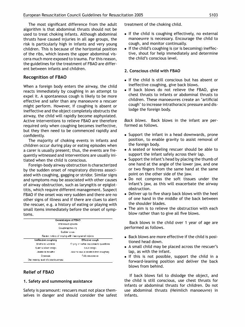
European Resuscitation Council Guidelines for Resuscitation 2005 S103
The most significant difference from the adultalgorithm is that abdominal thrusts should not beused to treat choking infants. Although abdominalthrusts have caused injuries in all age groups, therisk is particularly high in infants and very youngchildren. This is because of the horizontal positionof the ribs, which leaves the upper abdominal vis-cera much more exposed to trauma. For this reason,the guidelines for the treatment of FBAO are differ-ent between infants and children.
Recognition of FBAO
When a foreign body enters the airway, the childreacts immediately by coughing in an attempt toexpel it. A spontaneous cough is likely to be moreeffective and safer than any manoeuvre a rescuermight perform. However, if coughing is absent orineffective and the object completely obstructs theairway, the child will rapidly become asphyxiated.Active interventions to relieve FBAO are thereforerequired only when coughing becomes ineffective,but they then need to be commenced rapidly andconfidently.
The majority of choking events in infants andchildren occur during play or eating episodes when
treatment of the choking child.
• If the child is coughing effectively, no externalmanoeuvre is necessary. Encourage the child tocough, and monitor continually.
• If the child’s coughing is (or is becoming) ineffec-tive, shout for help immediately and determinethe child’s conscious level.
2. Conscious child with FBAO
• If the child is still conscious but has absent orineffective coughing, give back blows.
• If back blows do not relieve the FBAO, givechest thrusts to infants or abdominal thrusts tochildren. These manoeuvres create an ‘artificialcough’ to increase intrathoracic pressure and dis-lodge the foreign body.
Back blows. Back blows in the infant are per-formed as follows.
• Support the infant in a head downwards, proneposition, to enable gravity to assist removal ofthe foreign body.
a carer is usually present; thus, the events are fre-quently witnessed and interventions are usually ini-tiated when the child is conscious.
Foreign-body airway obstruction is characterizedby the sudden onset of respiratory distress associ-ated with coughing, gagging or stridor. Similar signsand symptoms may be associated with other causesof airway obstruction, such as laryngitis or epiglot-titis, which require different management. SuspectFBAO if the onset was very sudden and there are noother signs of illness and if there are clues to alertthe rescuer, e.g. a history of eating or playing withsmall items immediately before the onset of symp-toms.
Relief of FBAO
1. Safety and summoning assistance
Safety is paramount: rescuers must not place them-selves in danger and should consider the safest
• A seated or kneeling rescuer should be able tosupport the infant safely across their lap.
• Support the infant’s head by placing the thumb ofone hand at the angle of the lower jaw, and oneor two fingers from the same hand at the samepoint on the other side of the jaw.
• Do not compress the soft tissues under theinfant’s jaw, as this will exacerbate the airwayobstruction.
• Deliver up to five sharp back blows with the heelof one hand in the middle of the back betweenthe shoulder blades.
• The aim is to relieve the obstruction with eachblow rather than to give all five blows.
Back blows in the child over 1 year of age areperformed as follows.
• Back blows are more effective if the child is posi-tioned head down.
• A small child may be placed across the rescuer’slap, as with the infant.
• If this is not possible, support the child in aforward-leaning position and deliver the backblows from behind.
If back blows fail to dislodge the object, andthe child is still conscious, use chest thrusts forinfants or abdominal thrusts for children. Do notuse abdominal thrusts (Heimlich manoeuvre) ininfants.
S104 D. Biarent et al.
Chest thrusts for infants.
• Turn the infant into a head-downwards supineposition. This is achieved safely by placing thefree arm along the infant’s back and encirclingthe occiput with the hand.
• Support the infant down your arm, which isplaced down (or across) your thigh.
• Identify the landmark for chest compressions(lower sternum approximately a finger’s breadthabove the xiphisternum).
• Give five chest thrusts; these are similar to chestcompressions but sharper and delivered at aslower rate.
Abdominal thrusts for children over 1 year.
• Stand or kneel behind the child; place your armsunder the child’s arms and encircle his torso.
• Clench your fist and place it between the umbili-cus and xiphisternum.
• Grasp this hand with the other hand and pullsharply inwards and upwards.
• Repeat up to five times.• Ensure that pressure is not applied to the xiphoid
object more deeply into the pharynx and causeinjury.
• Open the airway using a head tilt and/or chinlift and attempt five rescue breaths. Assess theeffectiveness of each breath; if a breath does notmake the chest rise, reposition the head beforemaking the next attempt.
• Attempt five rescue breaths and, if there isno response (moving, coughing, spontaneousbreaths), proceed to chest compressions withoutfurther assessment of the circulation.
• Follow the sequence for single-rescuer CPR (step7b above) for approximately 1 min before sum-moning the EMS (if this has not already been doneby someone else).
• When the airway is opened for attempted deliv-ery of rescue breaths, look to see if the foreignbody can be seen in the mouth.
• If an object is seen, attempt to remove it with asingle finger sweep.
• If it appears the obstruction has been relieved,open and check the airway as above; deliver res-cue breaths if the child is not breathing.
• If the child regains consciousness and exhibitsspontaneous effective breathing, place him in a
process or the lower rib cage; this might causeabdominal trauma.
Following the chest or abdominal thrusts,reassess the child. If the object has not beenexpelled and the victim is still conscious, continuethe sequence of back blows and chest (for infant) orabdominal (for children) thrusts. Call out, or send,for help if it is still not available. Do not leave thechild at this stage.
If the object is expelled successfully, assessthe child’s clinical condition. It is possible thatpart of the object may remain in the respira-tory tract and cause complications. If there isany doubt, seek medical assistance. Abdominalthrusts may cause internal injuries, and all vic-tims so treated should be examined by a medicalpractitioner.42
3. Unconscious child with FBAO
If the child with FBAO is, or becomes, uncon-scious, place him on a firm, flat surface. Callout, or send, for help if it is still not available.Do not leave the child at this stage; proceed asfollows.
• Open the mouth and look for any obvious object.If an object is seen, make an attempt to remove itwith a single finger sweep. Do not attempt blindor repeated finger sweeps; these can impact the
safe position lying on his side and monitor breath-ing and conscious level while awaiting the arrivalof the EMS.
6b Paediatric advanced life support
Prevention of cardiopulmonary arrest
In children, secondary cardiopulmonary arrests,caused by either circulatory or respiratory fail-ure, are more frequent than primary arrestscaused by arrhythmias.9,12,43—46 So-called ‘asphyx-ial arrests’ or respiratory arrests are also morecommon in young adulthood (e.g., trauma, drown-ing, poisoning).47,48 The outcome from cardiopul-monary arrests in children is poor; identification ofthe antecedent stages of cardiac or respiratory fail-ure is a priority, as effective early intervention maybe life saving.
The order of assessment and intervention for anyseriously ill or injured child follows the ABC princi-ples.
• A indicates airway (Ac for airway and cervicalspine stabilisation for the injured child).
• B indicates breathing.• C indicates circulation.
Interventions are made at each step of theassessment as abnormalities are identified; the next
European Resuscitation Council Guidelines for Resuscitation 2005 S105
step of the assessment is not started until thepreceding abnormality has been managed and cor-rected if possible.
Diagnosing respiratory failure: assessmentof A and B
The first steps in the assessment of the seriously illor injured child are the management of the airwayand breathing. Abnormalities in airway patency andbreathing lead to respiratory failure. Signs of res-piratory failure are
• respiratory rate outside the normal range for thechild’s age—–either too fast or too slow
• initially increasing work of breathing whichmay progress to inadequate/decreased workof breathing, additional noises such as stridor,wheeze or grunting, or the loss of breath sounds
• cyanosis (without/with supplemental oxygen)
There may be associated signs in other organ sys-tems affected by inadequate ventilation and oxy-genation; these are detectable in the C steps ofassessment, such as
•
•
Do
Smgiitro
•
••
•••
•
• level of consciousness may decrease because ofpoor cerebral perfusion
Diagnosing cardiopulmonary arrest
Signs of cardiopulmonary arrest include
• unresponsiveness• apnoea or gasping respiratory pattern• absent circulation• pallor or deep cyanosis
In the absence of ‘signs of life’, search for acentral pulse or cardiac sounds (by direct chest aus-cultation) for a maximum of 10 s, before startingCPR. If there is any doubt, start CPR.50—53
Management of respiratory andcirculatory failure
A and B
Open the airway and ensure adequate ventilationand oxygenation.
increasing tachycardia progressing to bradycar-dia (this latter sign being an ominous indicator ofthe loss of compensatory mechanisms)alteration in the level of consciousness
iagnosing circulatory failure: assessmentf C
hock is characterised by a mismatch betweenetabolic tissue demand and delivery of oxy-
en and nutrients by the circulation.49 Physiolog-cal compensatory mechanisms lead to changesn the heart rate, in the systemic vascular resis-ance (which commonly increases as an adaptiveesponse) and in tissue and organ perfusion. Signsf circulatory failure are
increased heart rate (bradycardia is an ominoussign, heralding physiological decompensation)decreased systemic blood pressuredecreased peripheral perfusion (prolonged capil-lary refill time, decreased skin temperature, paleor mottled skin)weak or absent peripheral pulsesdecreased or increased preloaddecreased urine output and metabolic acidosis
Other systems may be affected, for example
respiratory rate may be increased initially, be-coming bradypnoeic with decompensated shock
• Deliver high-flow oxygen.• Achieving adequate ventilation and oxygenation
may include the use of airway adjuncts, bag-maskventilation (BMV), use of a laryngeal mask airway(LMA), securing a definitive airway by trachealintubation and positive pressure ventilation.
• In rare, extreme circumstances, a surgical airwaymay be required.
C
Establish cardiac monitoring.
• Secure vascular access to the circulation. Thismay be via peripheral or central intravenous (IV)or by intraosseous (IO) cannulation.
• Give a fluid bolus and/or inotropes as required.
Assess and re-assess the child continuously, eachtime commencing at Airway before Breathing,thereafter moving onto the Circulation
Airway
Open the airway using basic life support tech-niques. Oropharyngeal and nasopharyngeal airwaysadjuncts can help maintain the airway. Use theoropharyngeal airway only in the unconscious child,in whom there is no gag reflex. Use the appro-priate size, to avoid pushing the tongue back-ward and obstructing the epiglottis, or directlycompressing the glottic area. The soft palate in
S106 D. Biarent et al.
the child can be damaged by insertion of theoropharygneal airway; avoid this by inserting theoropharygneal airway under direct vision and pass-ing it over a tongue depressor or laryngoscope.The nasopharyngeal airway is tolerated better inthe conscious child (who has an effective gagreflex), but should not be used if there is a basalskull fracture or a coagulopathy. These simpleairway adjuncts do not protect the airway fromaspiration of secretions, blood or stomach con-tents.
Laryngeal mask airway
The LMA is an acceptable initial airway device forproviders experienced in its use. It may be particu-larly helpful in airway obstruction caused by upperairway abnormalities. The LMA does not, however,protect the airway from aspiration of secretions,blood or stomach contents, and therefore closeobservation is required. LMA use is associated witha higher incidence of complications in small chil-dren compared with adults.54
Tracheal intubation
tor must be experienced and familiar with rapid-sequence induction drugs.
Tracheal tube sizes. The tracheal tube internaldiameters (ID) for different ages are
• for neonates, 2.5—3.5 mm according to the for-mula (gestational age in weeks 10)
• for infants, 4 or 4.5 mm• for children older than 1 year, according to the
formula [(age in years/4) + 4]
Tracheal tube size estimation according thelength of the child’s body as measured by resusci-tation tapes is more accurate than using the aboveformulae.67
Cuffed versus uncuffed tracheal tubes. In the pre-hospital setting, an uncuffed tracheal tube may bepreferable when using sizes of up to 5.5 mm ID (i.e.,for children up to 8 years). In hospital, a cuffed tra-cheal tube may be useful in certain circumstances,e.g. in cases of poor lung compliance, high air-way resistance or large glottic air leak.68—70 Thecorrectly sized cuffed tracheal tube is as safe asan uncuffed tube for infants and children (not forneonates), provided attention is paid to its place-mcttc
CDfcgomb
•
•
•
••
•
•
•
Tracheal intubation is the most secure and effectiveway to establish and maintain the airway, preventgastric distension, protect the lungs against pul-monary aspiration, enable optimal control of theairway pressure and provide positive end expiratorypressure (PEEP). The oral route is preferable duringresuscitation. Oral intubation is usually quicker andis associated with fewer complications than nasalplacement. The judicious use of anaesthetics, seda-tives and neuromuscular blocking drugs is indicatedin the conscious child to avoid multiple intubationattempts or intubation failure.55—65 The anatomyof a child’s airway differs significantly from that ofan adult; hence, intubation of a child requires spe-cial training and experience. Check that trachealtube placement is correct by clinical examinationand end-tidal capnography. The tracheal tube mustbe secured, and monitoring of the vital signs isessential.66
It is also essential to plan an alternative airwaymanagement technique in case the trachea cannotbe intubated.
Rapid sequence induction and intubation. Thechild who is in cardiopulmonary arrest and deepcoma does not require sedation or analgesia to beintubated; otherwise, intubation must be precededby oxygenation, rapid sedation, analgesia and theuse of neuromuscular blocking drugs to minimiseintubation complications and failure.63 The intuba-
ent, size and cuff inflation pressure; excessiveuff pressure can lead to ischaemic necrosis ofhe surrounding laryngeal tissue and stenosis. Main-ain the cuff inflation pressure below 20 cmH2O andheck it regularly.71
onfirmation of correct tracheal tube placement.isplaced, misplaced or obstructed tubes occurrequently in the intubated child and are asso-iated with increased risk of death.72,73 No sin-le technique is 100% reliable for distinguishingesophageal from tracheal intubation.74—76 Assess-ent of the correct tracheal tube position is madey
observation of the tube passing beyond the vocalcordsobservation of symmetrical chest wall movementduring positive pressure ventilationobservation of mist in the tube during the expi-ratory phase of ventilationabsence of gastric distensionequal air entry heard on bilateral auscultation ofboth axillae and apices of the chestabsence of air entry into the stomach on auscul-tationdetection of end-tidal CO2 if the child has a per-fusing rhythm (this may be seen with effectiveCPR)improvement or stabilisation of SpO2 to theexpected range
European Resuscitation Council Guidelines for Resuscitation 2005 S107
• improvement of heart rate towards the age-expected value (or remaining within the normalrange)
If the child is in cardiopulmonary arrest andexhaled CO2 is not detected, or if there is anydoubt, confirm tracheal tube position by directlaryngoscopy. After correct placement and confir-mation, secure the tracheal tube and reassess itsposition. Maintain the child’s head in neutral posi-tion; flexion of the head drives the tube further intothe trachea whereas extension may pull it out of theairway.77 Confirm the position of the tracheal tubeat mid trachea by plain chest radiograph; the tra-cheal tube tip should be at the level of the 2nd or3rd thoracic vertebra.
DOPES is a useful acronym for the causes of sud-den deterioration in an intubated child
• D: displacement of the tracheal tube• O: obstruction of the tracheal tube• P: pneumothorax• E: equipment failure (source of gas, BMV, venti-
lator, etc.)• S: stomach (gastric distension may alter dia-
phragm mechanics)
B
O
Udgg
udIa
V
HvpHsaTwvr
b1c
tion is adequate during chest compressions. Whencirculation is restored, or if the child still has a per-fusing rhythm, ventilate at 12—20 breaths min−1 toachieve a normal pCO2. Hyperventilation is harm-ful.
Bag-mask ventilation. BMV is effective and safefor a child requiring assisted ventilation for a shortperiod, i.e. in the prehospital setting or in an emer-gency department.73,90—92 Assess the effectivenessof BMV by observing adequate chest rise, monitor-ing heart rate, auscultating for breath sounds andmeasuring peripheral oxygen saturation (SpO2). Anyhealthcare provider dealing with children must beable to deliver BMV effectively.
Prolonged ventilation. If prolonged ventilation isrequired, the benefits of a secured airway prob-ably outweigh the potential risks associated withtracheal intubation.
Monitoring of breathing and ventilation
End tidal CO2. Monitoring end-tidal CO2 with acolorimetric detector or capnometer confirms tra-cheal tube placement in the child weighing moretsccioaoebam
Obdmatm
Plpoccficm
reathing
xygenation
se oxygen at the highest concentration (i.e., 100%)uring resuscitation. Once circulation is restored,ive sufficient oxygen to maintain peripheral oxy-en saturation at or above 95%.78,79
Studies in neonates suggest some advantages tosing room air during resuscitation, but the evi-ence as yet is inconclusive (see Section 6c).80—83
n the older child, there is no evidence for any suchdvantages, so use 100% oxygen for resuscitation.
entilation
ealthcare providers commonly provide excessiveentilation to victims of cardiopulmonary or res-iratory arrest, and this may be detrimental.yperventilation causes increased thoracic pres-ure, decreased cerebral and coronary perfusion,nd poorer survival rates in animals and adults.84—89
he ideal tidal volume should achieve modest chestall rise. Use a ratio of 15 chest compressions to 2entilations (a lone rescuer may use 30:2); the cor-ect compression rate is 100 min−1.
Once the airway is protected by tracheal intu-ation, continue positive pressure ventilation at2—20 breaths min−1 without interrupting chestompressions. Take care to ensure that lung infla-
han 2 kg, and may be used in pre- and in-hospitalettings, as well as during any transportation of thehild.93—97 A colour change or the presence of aapnographic waveform indicates that the tube isn the tracheobronchial tree, both in the presencef a perfusing rhythm and during cardiopulmonaryrrest. Capnography does not rule out intubationf the right mainstem bronchus. The absence ofxhaled CO2 during cardiopulmonary arrest may note caused by tube misplacement, since a low orbsent end-tidal CO2 may reflect low or absent pul-onary blood flow.98—101
esophageal detector devices. The self-inflatingulb or aspirating syringe (oesophageal detectorevice, ODD) may be used for the secondary confir-ation of tracheal tube placement in children withperfusing rhythm.102,103 There are no studies on
he use of ODD in children who are in cardiopul-onary arrest.
ulse oximetry. Clinical evaluation of the oxygenevel is unreliable; therefore monitor the child’seripheral oxygen saturation continuously by pulseximetry. Pulse oximetry can be unreliable underertain conditions, e.g. if the child is in shock, inardiopulmonary arrest or has poor peripheral per-usion. Although pulse oximetry is relatively simple,t is a poor guide to tracheal tube displacement;apnography detects tracheal tube dislodgementore rapidly than pulse oximetry.104
S108 D. Biarent et al.
Circulation
Vascular access
Vascular access is essential to give drugs and fluidsand obtain blood samples. Venous access can be dif-ficult to establish during resuscitation of an infantor child.105 Limit the maximum number of attemptsto obtain IV access to three; thereafter, insert an IOneedle.106
Intraosseous access. IO access is a rapid, safe,and effective route to give drugs, fluids and bloodproducts.107—113 The onset of action and timeto achieve adequate plasma drug concentrationsare similar to those provided by central venousaccess.114,115 Bone marrow samples can be used tocross-match for blood type or group,116 for chem-ical analysis,117,118 and for blood gas measure-ment (the values are comparable to central venousblood gases).117,119,120 Flush each drug with a bolusof normal saline to ensure dispersal beyond themarrow cavity and to achieve faster distributionto the central circulation. Inject large boluses offluid using manual pressure. Intraosseous access canbe maintained until definitive IV access has been
Fluids and drugs
Volume expansion is indicated when a child showssigns of shock in the absence of volume overload.135
If systemic perfusion is inadequate, give a bolusof 20 ml kg−1 of an isotonic crystalloid, even if thesystemic blood pressure is normal. Following everybolus, re-assess the child’s clinical state using ABC,to decide whether a further bolus or other treat-ment is required.
There are insufficient data to make recommen-dations about the use of hypertonic saline for shockassociated with head injuries or hypovolaemia.136
There are also insufficient data to recommenddelayed fluid resuscitation in the hypotensive childwith blunt trauma.137 Avoid dextrose-containingsolutions unless there is hypoglycaemia.138—141
However, hypoglycaemia must actively be soughtand avoided, particularly in the small child orinfant.
Adenosine
Adenosine is an endogenous nucleotide whichcauses a brief atrioventricular (AV) block andiAmsiman
A
ApIamaicmcqcoatthnsm
established.
Intravenous access. Peripheral IV access pro-vides plasma concentrations of drugs and clinicalresponses equivalent to central or IO access.121—125
Central lines provide more secure long-termaccess121,122,124,125 but offer no advantages duringresuscitation, compared with IO or peripheral IVaccess.
Tracheal tube access
IV and IO access are better than the tracheal routefor giving drugs.126 Lipid-soluble drugs, such aslidocaine, atropine, adrenaline and naloxone areabsorbed via the lower airway.127—131 Optimal tra-cheal tube drug doses are unknown because of thegreat variability of alveolar drug absorption, butthe following dosages have been recommended asguidance
• adrenaline, 100 mcg kg−1
• lidocaine, 2—3 mg kg−1
• atropine, 30 mcg kg−1
The optimal dose of naloxone is not known.Dilute the drug in 5 ml of normal saline and fol-
low administration with five ventilations.132—134 Donot give non-lipid soluble medications (e.g., glu-cose, bicarbonate, calcium) via the tracheal tubebecause they will damage the airway mucosa.
mpairs accessory bundle re-entry at the level of theV node. Adenosine is recommended for the treat-ent of supraventricular tachycardia (SVT).142 It is
afe to use, as it has a short half-life (10 s); givet intravenously via upper limb or central veins, toinimise the time taken to reach the heart. Give
denosine rapidly, followed by a flush of 3—5 ml oformal saline.143
drenaline (epinephrine)
drenaline is an endogenous catecholamine withotent alpha, beta-1 and beta-1 adrenergic actions.t is the essential medication in cardiopulmonaryrrest, and is placed prominently in the treat-ent algorithms for non-shockable and shock-
ble rhythms. Adrenaline induces vasoconstriction,ncreases diastolic pressure and thereby improvesoronary artery perfusion pressure, enhancesyocardial contractility, stimulates spontaneous
ontractions and increases the amplitude and fre-uency of VF, so increasing the likelihood of suc-essful defibrillation. The recommended IV/IO dosef adrenaline in children is 10 mcg kg−1. The dose ofdrenaline given via the tracheal tube is ten timeshis (100 mcg kg−1).127,144—146 If needed, give fur-her doses of adrenaline every 3—5 min. The use ofigher doses of adrenaline via the IV or IO route isot recommended routinely, as it does not improveurvival or neurological outcome after cardiopul-onary arrest.147—150
European Resuscitation Council Guidelines for Resuscitation 2005 S109
Once spontaneous circulation is restored, a con-tinuous infusion of adrenaline may be required. Itshaemodynamic effects are dose related; there isalso considerable variability between children inresponse, therefore, titrate the infusion dose tothe desired effect. High infusion rates may causeexcessive vasoconstriction, compromising extrem-ity, mesenteric, and renal blood flow. High-doseadrenaline may cause severe hypertension andtachyarrhythmias.151
To avoid tissue damage it is essential to giveadrenaline through a secure intravascular line (IV orIO). Adrenaline and other catecholamines are inac-tivated by alkaline solutions and should never bemixed with sodium bicarbonate.152
Amiodarone
Amiodarone is a non-competitive inhibitor of adren-ergic receptors; it depresses conduction in myocar-dial tissue and therefore slows AV conduction andprolongs the QT interval and the refractory period.Except when given for the treatment of refrac-tory VF/pulseless VT, amiodarone must be injectedslowly (over 10—20 min) with systemic blood pres-srwip
A
Abam
C
Ctna
G
Nhaaogonh
and hypoglycaemia following return of spontaneouscirculation (ROSC).
Magnesium
There is no evidence for giving magnesium rou-tinely during cardiopulmonary arrest.165 Magne-sium treatment is indicated in the child with doc-umented hypomagnesaemia or with torsades depointes VF, regardless of the cause.166
Sodium bicarbonate
Giving sodium bicarbonate routinely during car-diopulmonary arrest and CPR or after ROSC isnot recommended.167,168 After effective ventila-tion and chest compressions have been achievedand adrenaline given, sodium bicarbonate may beconsidered for the child who has had a prolongedcardiopulmonary arrest and severe metabolic aci-dosis. Sodium bicarbonate may also be consid-ered in the case of haemodynamic instabilityand co-existing hyperkalaemia, or in the man-agement of tricyclic overdose. Excessive quan-tities of sodium bicarbonate may impair tissueonc
L
Ldtd
P
PlSipbvs
V
VaswTaT
ure and ECG monitoring to avoid fast-infusion-elated hypotension. This side effect is less commonith the aqueous solution.153 Other rare but signif-
cant adverse effects are bradycardia and polymor-hic VT.154
tropine
tropine accelerates sinus and atrial pacemakersy blocking the parasympathetic response. It maylso increase AV conduction. Small doses (<100 mcg)ay cause paradoxical bradycardia.155
alcium
alcium is essential for myocardial con-raction156,157 but routine use of calcium doesot improve the outcome from cardiopulmonaryrrest.158—160
lucose
eonatal, child and adult data show that bothyperglycaemia and hypoglycaemia are associ-ted with poor outcome after cardiopulmonaryrrest,161—163 but it is uncertain if this is causativer merely an association.164 Check blood or plasmalucose concentration and monitor closely in any illr injured child, including after cardiac arrest. Doot give glucose-containing fluids during CPR unlessypoglycaemia is present. Avoid hyperglycaemia
xygen delivery, produce hypokalaemia, hyper-atraemia and hyperosmolality and inactivateatecholamines.
idocaine
idocaine is less effective than amiodarone forefibrillation-resistant VF/VT in adults,169 andherefore is not the first-line treatment inefibrillation-resistant VF/VT in children.
rocainamide
rocainamide slows intra-atrial conduction and pro-ongs the QRS and QT intervals; it can be used inVT170,171 or VT172 resistant to other medications,n the haemodynamically stable child. However,aediatric data are sparse and procainamide shoulde used cautiously.173,174 Procainamide is a potentasodilator and can cause hypotension; infuse itlowly with careful monitoring.170,175,176
asopressin
asopressin is an endogenous hormone that actst specific receptors, mediating systemic vasocon-triction (via V1 receptor) and the reabsorption ofater in the renal tubule (by the V2 receptor).177
he use of vasopressin for the treatment of cardiacrrest in adults is discussed in detail in Section 4e.here is currently insufficient evidence to support
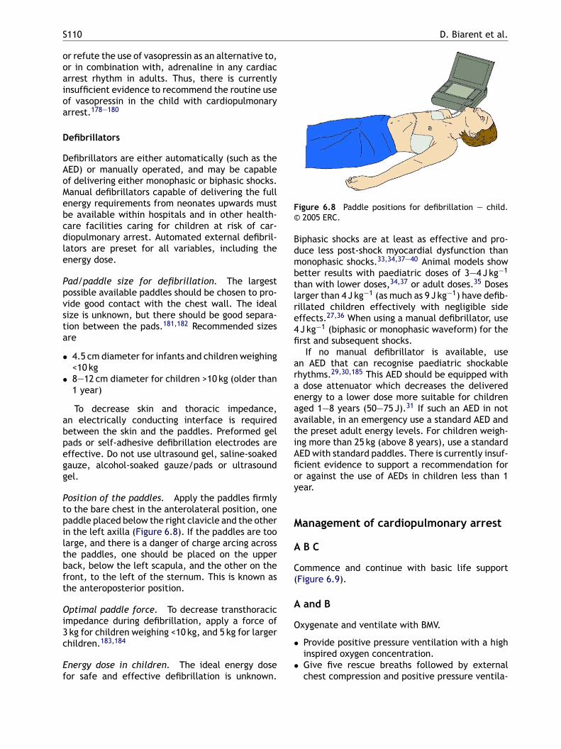
S110 D. Biarent et al.
or refute the use of vasopressin as an alternative to,or in combination with, adrenaline in any cardiacarrest rhythm in adults. Thus, there is currentlyinsufficient evidence to recommend the routine useof vasopressin in the child with cardiopulmonaryarrest.178—180
Defibrillators
Defibrillators are either automatically (such as theAED) or manually operated, and may be capableof delivering either monophasic or biphasic shocks.Manual defibrillators capable of delivering the fullenergy requirements from neonates upwards mustbe available within hospitals and in other health-care facilities caring for children at risk of car-diopulmonary arrest. Automated external defibril-lators are preset for all variables, including theenergy dose.
Pad/paddle size for defibrillation. The largestpossible available paddles should be chosen to pro-vide good contact with the chest wall. The idealsize is unknown, but there should be good separa-tion between the pads.181,182 Recommended sizes
Figure 6.8 Paddle positions for defibrillation — child.© 2005 ERC.
Biphasic shocks are at least as effective and pro-duce less post-shock myocardial dysfunction thanmonophasic shocks.33,34,37—40 Animal models showbetter results with paediatric doses of 3—4 J kg−1
than with lower doses,34,37 or adult doses.35 Doseslarger than 4 J kg−1 (as much as 9 J kg−1) have defib-rillated children effectively with negligible sideeffects.27,36 When using a manual defibrillator, use4 J kg−1 (biphasic or monophasic waveform) for thefirst and subsequent shocks.
If no manual defibrillator is available, usean AED that can recognise paediatric shockablerhythms.29,30,185 This AED should be equipped witha dose attenuator which decreases the deliveredenergy to a lower dose more suitable for childrenaged 1—8 years (50—75 J).31 If such an AED in notavailable, in an emergency use a standard AED andthe preset adult energy levels. For children weigh-ing more than 25 kg (above 8 years), use a standardAED with standard paddles. There is currently insuf-ficient evidence to support a recommendation foror against the use of AEDs in children less than 1year.
M
A
C(
A
O
•
•
are
• 4.5 cm diameter for infants and children weighing<10 kg
• 8—12 cm diameter for children >10 kg (older than1 year)
To decrease skin and thoracic impedance,an electrically conducting interface is requiredbetween the skin and the paddles. Preformed gelpads or self-adhesive defibrillation electrodes areeffective. Do not use ultrasound gel, saline-soakedgauze, alcohol-soaked gauze/pads or ultrasoundgel.
Position of the paddles. Apply the paddles firmlyto the bare chest in the anterolateral position, onepaddle placed below the right clavicle and the otherin the left axilla (Figure 6.8). If the paddles are toolarge, and there is a danger of charge arcing acrossthe paddles, one should be placed on the upperback, below the left scapula, and the other on thefront, to the left of the sternum. This is known asthe anteroposterior position.
Optimal paddle force. To decrease transthoracicimpedance during defibrillation, apply a force of3 kg for children weighing <10 kg, and 5 kg for largerchildren.183,184
Energy dose in children. The ideal energy dosefor safe and effective defibrillation is unknown.
anagement of cardiopulmonary arrest
B C
ommence and continue with basic life supportFigure 6.9).
and B
xygenate and ventilate with BMV.
Provide positive pressure ventilation with a highinspired oxygen concentration.Give five rescue breaths followed by externalchest compression and positive pressure ventila-
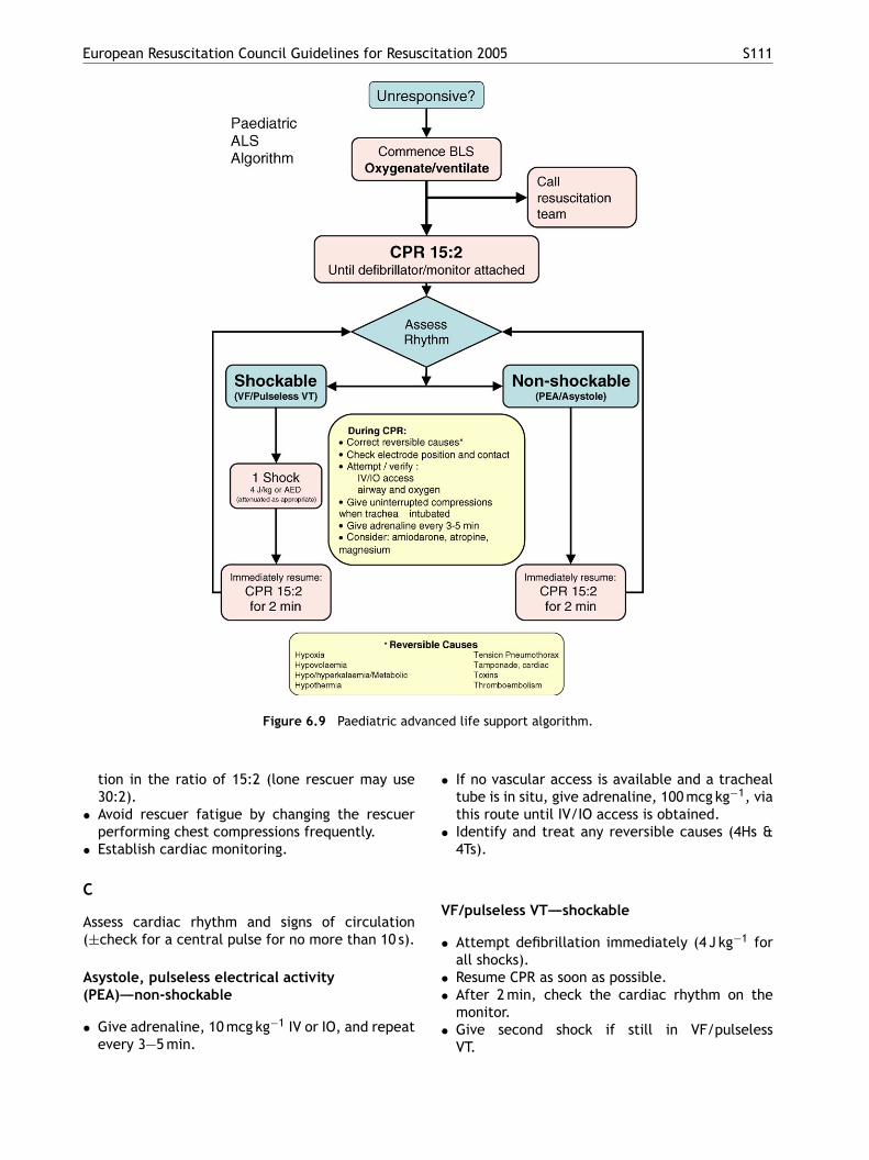
European Resuscitation Council Guidelines for Resuscitation 2005 S111
Figure 6.9 Paediatric advanced life support algorithm.
tion in the ratio of 15:2 (lone rescuer may use30:2).
• Avoid rescuer fatigue by changing the rescuerperforming chest compressions frequently.
• Establish cardiac monitoring.
C
Assess cardiac rhythm and signs of circulation(±check for a central pulse for no more than 10 s).
Asystole, pulseless electrical activity(PEA)—–non-shockable
• Give adrenaline, 10 mcg kg−1 IV or IO, and repeatevery 3—5 min.
• If no vascular access is available and a trachealtube is in situ, give adrenaline, 100 mcg kg−1, viathis route until IV/IO access is obtained.
• Identify and treat any reversible causes (4Hs &4Ts).
VF/pulseless VT—–shockable
• Attempt defibrillation immediately (4 J kg−1 forall shocks).
• Resume CPR as soon as possible.• After 2 min, check the cardiac rhythm on the
monitor.• Give second shock if still in VF/pulseless
VT.
S112 D. Biarent et al.
• Immediately resume CPR for 2 min and checkmonitor; if no change, give adrenaline followedimmediately by a 3rd shock.
• CPR for 2 min.• Give amiodarone if still in VF/pulseless VT fol-
lowed immediately by a 4th shock.• Give adrenaline every 3—5 min during CPR.• If the child remains in VF/pulseless VT, continue
to alternate shocks with 2 min of CPR.• If signs of life become evident, check the monitor
for an organised rhythm; if this is present, checkfor a central pulse.
• Identify and treat any reversible causes (4Hs &4Ts).
• If defibrillation was successful but VF/pulselessVT recurs, resume CPR, give amiodarone anddefibrillate again at the dose that was effectivepreviously. Start a continuous infusion of amio-darone.
Reversible causes of cardiac arrest (4 Hsand 4 Ts)
• Hypoxia• Hypovolaemia
tion between a shockable and a non-shockable car-diac rhythm. Invasive monitoring of systemic bloodpressure may help to improve effectiveness of chestcompression,186 but must not delay the provision ofbasic or advanced resuscitation.
Shockable rhythms comprise pulseless VT andVF. These rhythms are more likely in the childwho presents with sudden collapse. Non-shockablerhythms comprise PEA, bradycardia (<60 beatsmin−1 with no signs of circulation) and asystole. PEAand bradycardia often have wide QRS complexes.
Non-shockable rhythms
Most cardiopulmonary arrests in children and ado-lescents are of respiratory origin.19,44,187—189 Aperiod of immediate CPR is therefore mandatoryin this age group, before searching for an AEDor manual defibrillator, as their immediate avail-ability will not improve the outcome of a res-piratory arrest.11,13 Bystander CPR is associatedwith a better neurological outcome in adults andchildren.9,10,190 The most common ECG patterns ininfants, children and adolescents with cardiopul-monary arrest are asystole and PEA. PEA is charac-ticica
S
ViVmmtatamstdrii
D
ArA
• Hyper/hypokalaemia• Hypothermia• Tension pneumothorax• Tamponade (coronary or pulmonary)• Toxic/therapeutic disturbances• Thrombosis (coronary or pulmonary)
Sequence of events in cardiopulmonaryarrest
• When a child becomes unresponsive, with-out signs of life (no breathing, cough or anydetectable movement), start CPR immediately.
• Provide BMV with 100% oxygen.• Commence monitoring. Send for a manual or
automatic external defibrillator (AED) to identifyand treat shockable rhythms as quickly as possi-ble.
In the less common circumstance of a witnessedsudden collapse, early activation of emergency ser-vices and getting an AED may be more appropriate;start CPR as soon as possible.
Rescuers must perform CPR with minimal inter-ruption until attempted defibrillation.
Cardiac monitoring
Position the cardiac monitor leads or defibrillationpaddles as soon as possible, to enable differentia-
erised by organised, wide complex electrical activ-ty, usually at a slow rate, and absent pulses. PEAommonly follows a period of hypoxia or myocardialschaemia, but occasionally can have a reversibleause (i.e., one of the 4 H’s and 4 T’S) that led tosudden impairment of cardiac output.
hockable rhythms
F occurs in 3.8—19% of cardiopulmonary arrestsn children9,45,188,189; the incidence of VF/pulselessT increases with age.185,191 The primary deter-inant of survival from VF/pulseless VT cardiopul-onary arrest is the time to defibrillation. Prehospi-
al defibrillation within the first 3 min of witnesseddult VF arrest results in >50% survival. However,he success of defibrillation decreases dramaticallys the time to defibrillation increases; for everyinute delay in defibrillation (without any CPR),
urvival decreases by 7—10%. Survival after morehan 12 min of VF in adult victims is <5%.192 Car-iopulmonary resuscitation provided before defib-illation for response intervals longer than 5 minmproved outcome in some studies,193,194 but notn others.195
rugs in shockable rhythms
drenaline is given every 3—5 min by the IV or IOoute in preference to the tracheal tube route.miodarone is indicated in defibrillation-resistant
European Resuscitation Council Guidelines for Resuscitation 2005 S113
VF/pulseless VT. Experimental and clinical experi-ence with amiodarone in children is scarce; evi-dence from adult studies169,196,197 demonstratesincreased survival to hospital admission, but notto hospital discharge. One paediatric case seriesdemonstrates the effectiveness of amiodarone forlife-threatening ventricular arrhythmias.198 There-fore, IV amiodarone has a role in the treatment ofdefibrillation refractory or recurrent VF/pulselessVT in children.
Arrhythmias
Unstable arrhythmias
Check the central pulse of any child with anarrhythmia; if the pulse is absent, proceed totreating the child as being in cardiopulmonaryarrest. If the child has a central pulse, evaluatehis haemodynamic status. Whenever the haemody-namic status is compromised, the first steps are asfollows.
• Open the airway.••
•
••
•
B
Bsdtpf
btgvo
Aooii
Tachycardia
Narrow complex tachycardia. If supraventricu-lar tachycardia (SVT) is the likely rhythm, vagalmanoeuvres (Valsalva or diving reflex) may be usedin haemodynamically stable children. The manoeu-vres can be used in unstable children if they donot delay chemical or electrical cardioversion.200 Ifthe child is haemodynamically unstable, omit vagalmanoeuvres and attempt electrical cardioversionimmediately. Adenosine is usually effective in con-verting SVT into sinus rhythm. Adenosine is givenby rapid IV injection as closely as practical to theheart (see above), followed immediately by a bolusof normal saline.
Electrical cardioversion (synchronised with Rwave) is indicated in the haemodynamically com-promised child, in whom vascular access is notavailable, or in whom adenosine has failed to con-vert the rhythm. The first energy dose for electricalcardioversion of SVT is 0.5—1 J kg−1 and the seconddose is 2 J kg−1. If unsuccessful, give amiodaroneor procainamide under guidance from a paediatriccardiologist or intensivist before the third attempt.
Amiodarone has been shown to be effectiveistcdicta
WQsHudwlcdcmssdt
S
Cm
Assist ventilation and give oxygen.Attach ECG monitor or defibrillator and assess thecardiac rhythm.Evaluate if the rhythm is slow or fast for thechild’s age.Evaluate if the rhythm is regular or irregular.Measure QRS complex (narrow complexes, <0.08 sduration; large complexes, >0.08 s).The treatment options are dependent on thechild’s haemodynamic stability.
radycardia
radycardia is caused commonly by hypoxia, acido-is and severe hypotension; it may progress to car-iopulmonary arrest. Give 100% oxygen, and posi-ive pressure ventilation if required, to any childresenting with bradyarrhythmia and circulatoryailure.
If a poorly perfused child has a heart rate <60eats min−1, and does not respond rapidly to ven-ilation with oxygen, start chest compressions andive adrenaline. If the bradycardia is caused byagal stimulation, provide ventilation with 100%xygen and give atropine, before giving adrenaline.
A cardiac pacemaker is useful only in cases ofV block or sinus node dysfunction unresponsive toxygenation, ventilation, chest compressions andther medications; the pacemaker is not effectiven asystole or arrhythmias caused by hypoxia orschaemia.199
n the treatment of SVT in several paediatrictudies.198,201—207 However, since most studies ofhe use of amiodarone in narrow-complex tachy-ardias have been for junctional ectopic tachycar-ia in postoperative children, the applicability ofts use in all cases of SVT may be limited. If thehild is haemodynamically stable, early consulta-ion with an expert is recommended before givingmiodarone.
ide complex tachycardia. In children, wide-RS-complex tachycardia is more likely to beupraventricular than ventricular in origin.208
owever, wide-QRS-complex tachycardia, althoughncommon, must be considered to be VT in haemo-ynamically unstable children until proven other-ise. VT occurs most often in the child with under-
ying heart disease (e.g., after cardiac surgery,ardiomyopathy, myocarditis, electrolyte disor-ers, prolonged QT interval, central intracardiacatheter). Synchronised cardioversion is the treat-ent of choice for unstable VT with a pulse. Con-
ider antiarrhythmic therapy if a second cardiover-ion dose is unsuccessful or if VT recurs. Amio-arone has been shown to be safe and effective inreating paediatric arrhythmias.198,202,203,209
table arrhythmias
ontact an expert before initiating therapy, whileaintaining the child’s ABC. Depending on the
S114 D. Biarent et al.
child’s clinical history, presentation and ECG diag-nosis, a child with stable, wide-QRS-complex tachy-cardia may be treated for SVT and be given vagalmanoeuvres or adenosine. Otherwise, consideramiodarone as a treatment option; similarly, con-sider amiodarone if the diagnosis of VT is confirmedby ECG. Procainamide may also be considered instable SVT refractory to vagal manoeuvres andadenosine210—212 as well as in stable VT.172,213,214
Do not give procainamide with amiodarone.
Post-arrest management
Myocardial dysfunction is common after cardiopul-monary resuscitation.215,216 Vasoactive drugs mayimprove the child’s post-arrest haemodynamic val-ues, but the drugs must be titrated according to theclinical condition. They must be given continuouslythrough an IV line.
Temperature control and management
Hypothermia is common in the child following car-217
Fever is common following cardiopulmonaryresuscitation; it is associated with a poor neuro-logical outcome,230—232 the risk of which increaseswith each degree of body temperature greaterthan 37 ◦C.230 There are limited experimentaldata suggesting that the treatment of fever withantipyretics and/or physical cooling reduces neu-ronal damage.233,234 Antipyretics and accepteddrugs to treat fever are safe; therefore, use themto treat fever aggressively.
Prognosis of cardiopulmonary arrest
There are no simple guidelines to determine whenresuscitative efforts become futile. After 20 minof resuscitation, the team leader of the resus-citation team should consider whether or notto stop.187,235—239 The relevant considerations inthe decision to continue the resuscitation includethe cause of arrest,45,240 pre-existing conditions,whether the arrest was witnessed, the durationof untreated cardiopulmonary arrest (‘‘no flow’’),the effectiveness and duration of CPR (‘‘lowflow’’), the promptness of extracorporeal life sup-pad
P
TdrtptttrttsiPhsc
F
Asp
diopulmonary resuscitation. Central hypother-mia (32—34 ◦C) may be beneficial, whereas fevermay be detrimental to the injured brain of sur-vivors. Although there are no paediatric studies,mild hypothermia has an acceptable safety pro-file in adults218,219 and neonates;220—224 it couldincrease the number of neurologically intact sur-vivors.
A child who regains a spontaneous circulationbut remains comatose after cardiopulmonary arrestmay benefit from being cooled to a core tem-perature of 32—34 ◦C for 12—24 h. The success-fully resuscitated child with hypothermia and ROSCshould not be actively rewarmed unless the coretemperature is below 32 ◦C. Following a periodof mild hypothermia, rewarm the child slowly at0.25—0.5 ◦C h−1.
There are several methods to induce, moni-tor and maintain body temperature in children.External and/or internal cooling techniques canbe used to initiate cooling.225—227 Shivering canbe prevented by deep sedation and neuromuscularblockade. Complications can occur and include anincreased risk of infection, cardiovascular instabil-ity, coagulopathy, hyperglycaemia and electrolyteabnormalities.228,229
The optimum target temperature, rate of cool-ing, duration of hypothermia and rate of re-warming after deliberate cooling have yet to bedetermined; currently, no specific protocol for chil-dren can be recommended.
ort for a reversible disease process241—243 andssociated special circumstances (e.g, icy waterrowning,9,244 exposure to toxic drugs).
arental presence
he majority of parents would like to be presenturing resuscitation and when any procedure is car-ied out on their child.245—255. Parents witnessingheir child’s resuscitation can see that everythingossible has been attempted.256—260 Furthermore,hey may have the opportunity to say goodbye toheir child; allowing parents to be at the side ofheir child has been shown to help them gain aealistic view of the attempted resuscitation andhe child’s death.261 Families who were present atheir child’s death showed less anxiety and depres-ion, better adjustment and had an improved griev-ng process when assessed several months later.260
arental presence in the resuscitation room mayelp healthcare providers maintain their profes-ional behaviour while also helping them to see thehild as a human being and a family member.261
amily presence guidelines
dedicated member of the resuscitation teamhould be present with the parents to explain therocess in an empathetic manner, ensuring that
European Resuscitation Council Guidelines for Resuscitation 2005 S115
the parents do not interfere with or distract theresuscitation. If the presence of the parents isimpeding the progress of the resuscitation, theyshould be sensitively asked to leave. When appro-priate, physical contact with the child should beallowed and, wherever possible, the parents shouldbe allowed to be with their dying child at the finalmoment.256,261—264
The leader of the resuscitation team, not theparents, will decide when to stop the resuscita-tion; this should be expressed with sensitivity andunderstanding. After the event the team should bedebriefed, to enable any concerns to be expressedand for the team to reflect on their clinical practicein a supportive environment.
6c Resuscitation of babies at birth
Introduction
The following guidelines for resuscitation at birthhave been developed during the process that cul-minated in the 2005 International Consensus Con-ference on Emergency Cardiovascular Care (ECC)awetto
wths
P
RbitcO1atrattti
(0.2%) appeared to need resuscitation at delivery.Of these, 90% responded to mask inflation alone,whereas the remaining 10% appeared not to respondto mask inflation and therefore were intubated atbirth.
Resuscitation or specialist help at birth is morelikely to be needed by babies with intrapartum evi-dence of significant fetal compromise, babies deliv-ering before 35 weeks’ gestation, babies deliveringvaginally by the breech and multiple pregnancies.Although it is often possible to predict the needfor resuscitation before a baby is born, this is notalways the case. Therefore, personnel trained innewborn life support should be easily available atevery delivery and, should there be any need forresuscitation, the care of the baby should be theirsole responsibility. One person experienced in tra-cheal intubation of the newborn should also beeasily available for normal low-risk deliveries and,ideally, in attendance for deliveries associated witha high risk for neonatal resuscitation. Local guide-lines indicating who should attend deliveries shouldbe developed based on current practice and clinicalaudit.
An organised programme educating in the stan-dni
P
Tatdstbbftabmmn
E
RIattdp
nd Cardiopulmonary Resuscitation (CPR) Scienceith Treatment Recommendations.265 They are anxtension of the guidelines already published byhe ERC,2 and take into account recommenda-ions made by other national266 and internationalrganisations.267
The guidelines that follow do not define the onlyay that resuscitation at birth should be achieved;
hey merely represent a widely accepted view ofow resuscitation at birth can be carried out bothafely and effectively.
reparation
elatively few babies need any resuscitation atirth. Of those that do need help, the overwhelm-ng majority will require only assisted lung aera-ion. A small minority may need a brief period ofhest compressions in addition to lung aeration.f 100,000 babies born in Sweden in 1 year, only0 per 1000 (1%) babies weighing 2.5 kg or moreppeared to need resuscitation at delivery.268 Ofhose babies receiving resuscitation, 8 per 1000esponded to mask inflation and only 2 per 1000ppeared to need intubation.268 The same studyried to assess the unexpected need for resuscita-ion at birth, and found that for low-risk babies, i.e.hose born after 32 weeks’ gestation and follow-ng an apparently normal labour, about 2 per 1000
ards and skills required for resuscitation of theewborn is therefore essential for any institutionn which deliveries occur.
lanned home deliveries
he recommendations for those who should attendplanned home delivery vary from country to coun-
ry, but the decision to undergo a planned homeelivery, once agreed by the medical and midwiferytaff, should not compromise the standard of ini-ial resuscitation at birth. There will inevitablye some limitations to resuscitation of a newbornaby in the home because of the distance fromurther assistance, and this must be made clearo the mother at the time plans for home deliveryre made. Ideally, two trained professionals shoulde present at all home deliveries;269 one of theseust be fully trained and experienced in providingask ventilation and chest compressions in the
ewborn.
quipment and environment
esuscitation at birth is often a predictable event.t is therefore simpler to prepare the environmentnd the equipment before delivery of the babyhan is the case in adult resuscitation. Resuscita-ion should ideally take place in a warm, well-lit,raught-free area with a flat resuscitation surfacelaced below a radiant heater and other resusci-
S116 D. Biarent et al.
tation equipment immediately available. All equip-ment must be checked daily.
When a birth takes place in a non-designateddelivery area, the recommended minimum set ofequipment includes a device for safe, assisted lungaeration of an appropriate size for the newborn,warm dry towels and blankets, a clean (sterile)instrument for cutting the umbilical cord and cleangloves for the attendant. It may also be helpful tohave a suction device with a suitably sized suctioncatheter and a tongue depressor (or laryngoscope),to enable the oropharynx to be examined.
Temperature control
Naked, wet, newborn babies cannot maintain theirbody temperature in a room that feels comfortablywarm for adults. Compromised babies are partic-ularly vulnerable.270 Exposure of the newborn tocold stress will lower arterial oxygen tension271 andincrease metabolic acidosis.272 Prevent heat loss by
• protecting the baby from draughts• keeping the delivery room warm
Respiratory activity
Check whether the baby is breathing. If so, eval-uate the rate, depth and symmetry of respiration,together with any abnormal breathing pattern suchas gasping or grunting.
Heart rate
This is best evaluated by listening to the apex beatwith a stethoscope. Feeling the pulse in the baseof the umbilical cord is often effective but can bemisleading; cord pulsation is only reliable if foundto be more than 100 beats min−1.276
Colour
A healthy baby is born blue but becomes pink within30 s of the onset of effective breathing. Observewhether the baby is centrally pink, cyanosed orpale. Peripheral cyanosis is common and does not,by itself, indicate hypoxaemia.
• drying the term baby immediately after delivery.Cover the head and body of the baby, apart fromthe face, with a warm towel to prevent furtherheat loss. Alternatively, place the baby skin toskin with the mother and cover both with a towel
• placing the baby on a warm surface under a pre-heated radiant warmer if resuscitation is needed
In very preterm babies (especially below 28weeks’ gestation), drying and wrapping may not besufficiently effective. A more effective method ofkeeping these babies warm is to cover the head andbody of the baby (apart from the face) with plas-tic wrapping, without drying the baby beforehand,and then to place the baby so covered under radiantheat.
Initial assessment
The Apgar scoring system was not designed to iden-tify prospectively babies needing resuscitation.273
Several studies have also suggested that it ishighly subjective.274 However, components of thescore, namely respiratory rate, heart rate andcolour, if assessed rapidly, can identify babies need-ing resuscitation.275 Furthermore, repeated assess-ment of these components can indicate whether thebaby is responding or whether further efforts areneeded.
Tone
A very floppy baby is likely to be unconscious and islikely to need respiratory support.
Tactile stimulation
Drying the baby usually produces enough stimula-tion to induce effective respiration. Avoid morevigorous methods of stimulation. If the baby failsto establish spontaneous and effective respirationsfollowing a brief period of stimulation, further sup-port will be required.
Classification according to initial assessment
On the basis of the initial assessment, the babiescan usually be divided into four groups.
Group 1: vigorous breathing or cryinggood tonerapidly becoming pinkheart rate higher than 100 beats min−1
This baby requires no intervention other thandrying, wrapping in a warm towel and, whereappropriate, handing to the mother. The baby willremain warm through skin-to-skin contact withmother under a cover, and may be put to the breastat this stage.
European Resuscitation Council Guidelines for Resuscitation 2005 S117
Group 2: breathing inadequately or apnoeicremaining centrally bluenormal or reduced toneheart rate less than 100 beats min−1
This baby may respond to tactile stimulationand/or facial oxygen, but may need mask inflation.
Group 3: breathing inadequately or apnoeicblue or palefloppyheart rate less than 100 beats min−1
This baby may improve with mask inflation butmay also require chest compressions.
Group 4: breathing inadequately or apnoeicpalefloppyno detectable heart rate
This baby will require immediate airway control,lung inflation and ventilation. Once this has beensuccessfully accomplished, the baby may also needchest compressions and perhaps drugs.
There remains a very rare group of babies who,though breathing adequately and with a good heartrate, remain blue. This group includes a range of
bradycardia.277 The presence of thick meconium ina non-vigorous baby is the only indication for con-sidering immediate suction. If suction is required,it is best done under direct vision. Connect a 12—14FG suction catheter, or a Yankauer sucker, to a suc-tion source not exceeding −100 mmHg.
Breathing
There is currently insufficient evidence to specifythe concentration of oxygen to be used when start-ing resuscitation. After initial steps at birth, if res-piratory efforts are absent or inadequate, lung aer-ation is the priority (Figure 6.12). The primary mea-sure of adequate initial lung inflation is a promptimprovement in heart rate; assess chest wall move-ment if the heart rate does not improve.
For the first few breaths maintain the initialinflation pressure for 2—3 s. This will help lungexpansion. Most babies needing resuscitation atbirth will respond with a rapid increase in heartrate within 30 s of lung inflation. If the heart rateincreases but the baby is not breathing adequately,continue ventilation at a rate of about 30 breathsmin−1, allowing approximately 1 s for each infla-ti
brImvttahatieterdl
e
C
Cefrv
possible diagnoses such as diaphragmatic hernia,surfactant deficiency, congenital pneumonia, pneu-mothorax or cyanotic congenital heart disease.
Newborn life support
Commence newborn life support (Figure 6.10) ifassessment demonstrates that the baby has failedto establish adequate regular normal breathing, orhas a heart rate of less than 100 beats min−1. Open-ing the airway and aerating the lungs is usuallyall that is necessary. Furthermore, more complexinterventions will be futile unless these two firststeps have been successfully completed.
Airway
The baby should be on his or her back with thehead in a neutral position (Figure 6.11). A 2-cmthickness of the blanket or towel placed underthe baby’s shoulder may be helpful in maintainingproper head position. In floppy babies, applicationof jaw thrust or the use of an appropriately sizedoropharyngeal airway may be helpful in opening theairway.
Suction is needed only if there is particulate mat-ter or blood obstructing the airway. Aggressive pha-ryngeal suction can delay the onset of spontaneousbreathing and cause laryngeal spasm and vagal
ion, until there is adequate spontaneous breath-ng.
Adequate passive ventilation is usually indicatedy either a rapidly increasing heart rate or a heartate that is maintained faster than 100 beats min−1.f the baby does not respond in this way, theost likely reason is inadequate airway control or
entilation. Look for passive chest movement inime with inflation efforts; if these are present,hen lung aeration has been achieved. If thesere absent, then airway control and lung aerationave not been confirmed. Without adequate lungeration chest compressions will be ineffective;herefore, confirm lung aeration before progress-ng to circulatory support. Some practitioners willnsure lung aeration by tracheal intubation, buthis requires training and experience to be achievedffectively. If this skill is not available and the heartate is decreasing, re-evaluate airway position andeliver aeration breaths while summoning a col-eague with intubation skills.
Continue ventilatory support until the baby hasstablished normal regular breathing.
irculatory support
irculatory support with chest compressions isffective only if the lungs have first been success-ully inflated. Give chest compressions if the heartate is less than 60 beats min−1 despite adequateentilation. The optimal technique is to place the
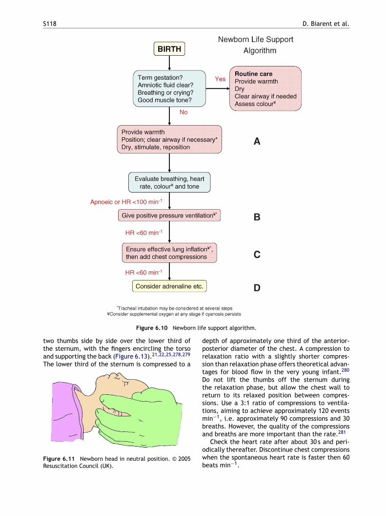
S118 D. Biarent et al.
Figure 6.10 Newborn life support algorithm.
two thumbs side by side over the lower third ofthe sternum, with the fingers encircling the torsoand supporting the back (Figure 6.13).21,22,25,278,279
The lower third of the sternum is compressed to a
Figure 6.11 Newborn head in neutral position. © 2005Resuscitation Council (UK).
depth of approximately one third of the anterior-posterior diameter of the chest. A compression torelaxation ratio with a slightly shorter compres-sion than relaxation phase offers theoretical advan-tages for blood flow in the very young infant.280
Do not lift the thumbs off the sternum duringthe relaxation phase, but allow the chest wall toreturn to its relaxed position between compres-sions. Use a 3:1 ratio of compressions to ventila-tions, aiming to achieve approximately 120 eventsmin−1, i.e. approximately 90 compressions and 30breaths. However, the quality of the compressionsand breaths are more important than the rate.281
Check the heart rate after about 30 s and peri-odically thereafter. Discontinue chest compressionswhen the spontaneous heart rate is faster then 60beats min−1.
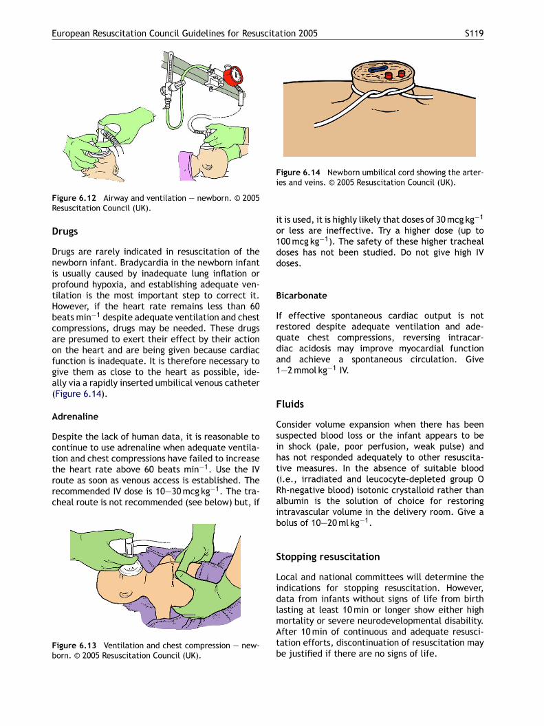
European Resuscitation Council Guidelines for Resuscitation 2005 S119
Figure 6.12 Airway and ventilation — newborn. © 2005Resuscitation Council (UK).
Drugs
Drugs are rarely indicated in resuscitation of thenewborn infant. Bradycardia in the newborn infantis usually caused by inadequate lung inflation orprofound hypoxia, and establishing adequate ven-tilation is the most important step to correct it.However, if the heart rate remains less than 60beats min−1 despite adequate ventilation and chestcompressions, drugs may be needed. These drugsare presumed to exert their effect by their actionon the heart and are being given because cardiacfunction is inadequate. It is therefore necessary togive them as close to the heart as possible, ide-ally via a rapidly inserted umbilical venous catheter(Figure 6.14).
Adrenaline
Despite the lack of human data, it is reasonable tocontinue to use adrenaline when adequate ventila-tion and chest compressions have failed to increasethe heart rate above 60 beats min−1. Use the IVroute as soon as venous access is established. Therecommended IV dose is 10—30 mcg kg−1. The tra-c
Fb
Figure 6.14 Newborn umbilical cord showing the arter-ies and veins. © 2005 Resuscitation Council (UK).
it is used, it is highly likely that doses of 30 mcg kg−1
or less are ineffective. Try a higher dose (up to100 mcg kg−1). The safety of these higher trachealdoses has not been studied. Do not give high IVdoses.
Bicarbonate
If effective spontaneous cardiac output is notrestored despite adequate ventilation and ade-quate chest compressions, reversing intracar-diac acidosis may improve myocardial functionand achieve a spontaneous circulation. Give1—2 mmol kg−1 IV.
Fluids
Consider volume expansion when there has beensuspected blood loss or the infant appears to bein shock (pale, poor perfusion, weak pulse) andhas not responded adequately to other resuscita-tive measures. In the absence of suitable blood(i.e., irradiated and leucocyte-depleted group ORaib
S
LidlmAtb
heal route is not recommended (see below) but, if
igure 6.13 Ventilation and chest compression — new-orn. © 2005 Resuscitation Council (UK).
h-negative blood) isotonic crystalloid rather thanlbumin is the solution of choice for restoringntravascular volume in the delivery room. Give aolus of 10—20 ml kg−1.
topping resuscitation
ocal and national committees will determine thendications for stopping resuscitation. However,ata from infants without signs of life from birthasting at least 10 min or longer show either highortality or severe neurodevelopmental disability.fter 10 min of continuous and adequate resusci-ation efforts, discontinuation of resuscitation maye justified if there are no signs of life.
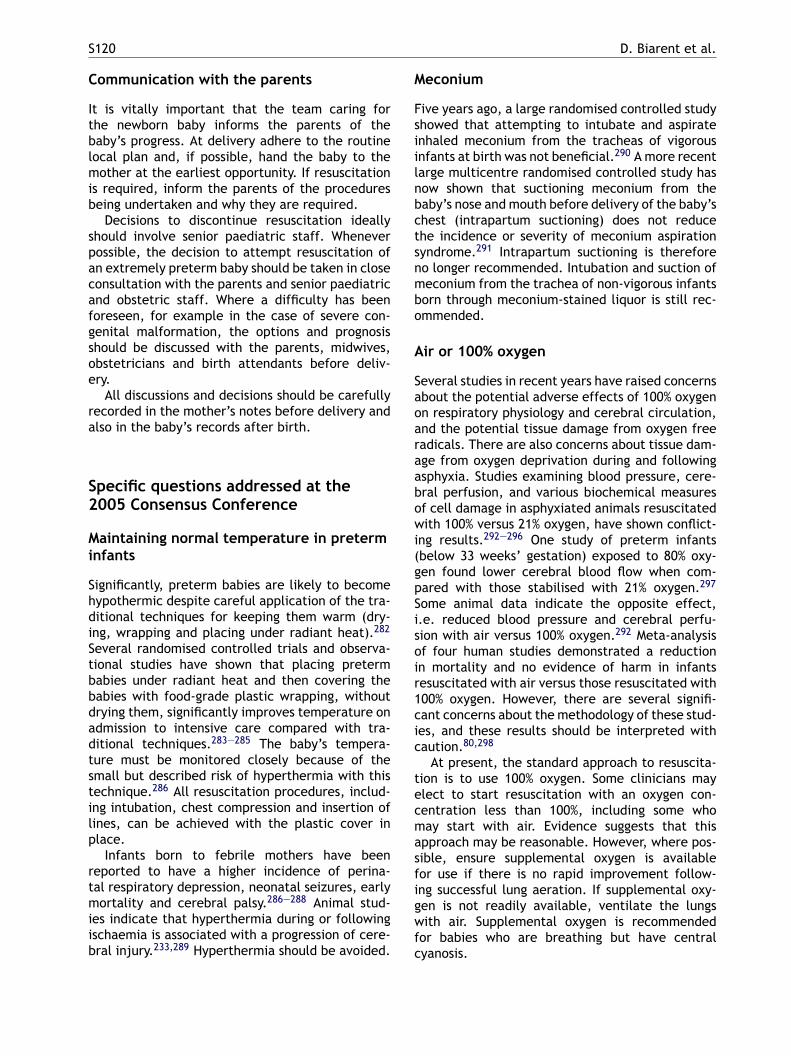
S120 D. Biarent et al.
Communication with the parents
It is vitally important that the team caring forthe newborn baby informs the parents of thebaby’s progress. At delivery adhere to the routinelocal plan and, if possible, hand the baby to themother at the earliest opportunity. If resuscitationis required, inform the parents of the proceduresbeing undertaken and why they are required.
Decisions to discontinue resuscitation ideallyshould involve senior paediatric staff. Wheneverpossible, the decision to attempt resuscitation ofan extremely preterm baby should be taken in closeconsultation with the parents and senior paediatricand obstetric staff. Where a difficulty has beenforeseen, for example in the case of severe con-genital malformation, the options and prognosisshould be discussed with the parents, midwives,obstetricians and birth attendants before deliv-ery.
All discussions and decisions should be carefullyrecorded in the mother’s notes before delivery andalso in the baby’s records after birth.
Meconium
Five years ago, a large randomised controlled studyshowed that attempting to intubate and aspirateinhaled meconium from the tracheas of vigorousinfants at birth was not beneficial.290 A more recentlarge multicentre randomised controlled study hasnow shown that suctioning meconium from thebaby’s nose and mouth before delivery of the baby’schest (intrapartum suctioning) does not reducethe incidence or severity of meconium aspirationsyndrome.291 Intrapartum suctioning is thereforeno longer recommended. Intubation and suction ofmeconium from the trachea of non-vigorous infantsborn through meconium-stained liquor is still rec-ommended.
Air or 100% oxygen
Several studies in recent years have raised concernsabout the potential adverse effects of 100% oxygenon respiratory physiology and cerebral circulation,and the potential tissue damage from oxygen freeradicals. There are also concerns about tissue dam-age from oxygen deprivation during and followingabowi(gpSisoir1cic
tecmasfigwfc
Specific questions addressed at the2005 Consensus Conference
Maintaining normal temperature in preterminfants
Significantly, preterm babies are likely to becomehypothermic despite careful application of the tra-ditional techniques for keeping them warm (dry-ing, wrapping and placing under radiant heat).282
Several randomised controlled trials and observa-tional studies have shown that placing pretermbabies under radiant heat and then covering thebabies with food-grade plastic wrapping, withoutdrying them, significantly improves temperature onadmission to intensive care compared with tra-ditional techniques.283—285 The baby’s tempera-ture must be monitored closely because of thesmall but described risk of hyperthermia with thistechnique.286 All resuscitation procedures, includ-ing intubation, chest compression and insertion oflines, can be achieved with the plastic cover inplace.
Infants born to febrile mothers have beenreported to have a higher incidence of perina-tal respiratory depression, neonatal seizures, earlymortality and cerebral palsy.286—288 Animal stud-ies indicate that hyperthermia during or followingischaemia is associated with a progression of cere-bral injury.233,289 Hyperthermia should be avoided.
sphyxia. Studies examining blood pressure, cere-ral perfusion, and various biochemical measuresf cell damage in asphyxiated animals resuscitatedith 100% versus 21% oxygen, have shown conflict-
ng results.292—296 One study of preterm infantsbelow 33 weeks’ gestation) exposed to 80% oxy-en found lower cerebral blood flow when com-ared with those stabilised with 21% oxygen.297
ome animal data indicate the opposite effect,.e. reduced blood pressure and cerebral perfu-ion with air versus 100% oxygen.292 Meta-analysisf four human studies demonstrated a reductionn mortality and no evidence of harm in infantsesuscitated with air versus those resuscitated with00% oxygen. However, there are several signifi-ant concerns about the methodology of these stud-es, and these results should be interpreted withaution.80,298
At present, the standard approach to resuscita-ion is to use 100% oxygen. Some clinicians maylect to start resuscitation with an oxygen con-entration less than 100%, including some whoay start with air. Evidence suggests that this
pproach may be reasonable. However, where pos-ible, ensure supplemental oxygen is availableor use if there is no rapid improvement follow-ng successful lung aeration. If supplemental oxy-en is not readily available, ventilate the lungsith air. Supplemental oxygen is recommended
or babies who are breathing but have centralyanosis.
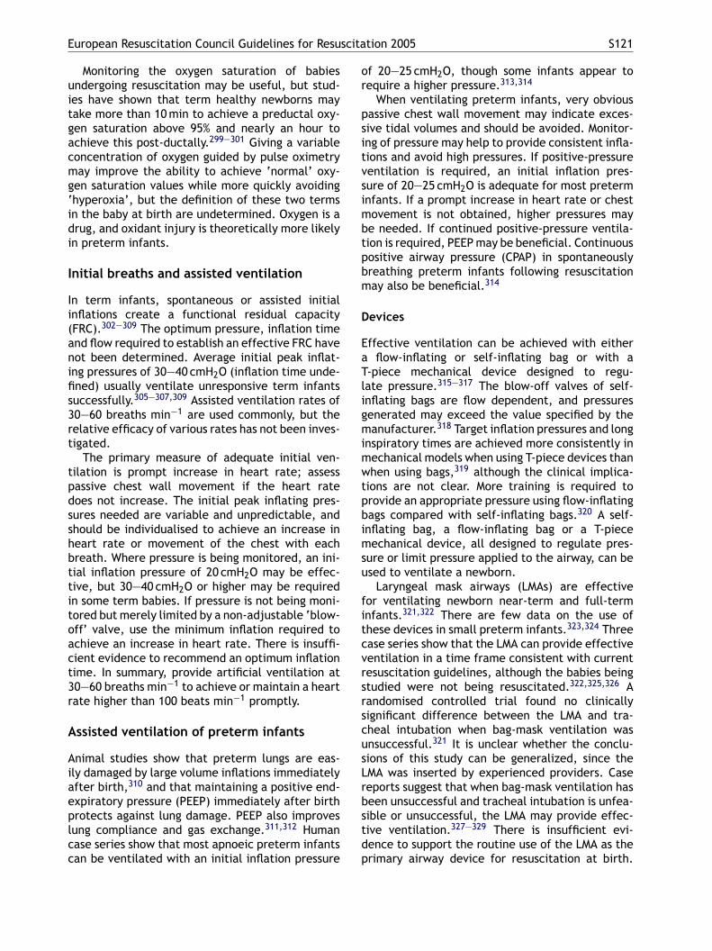
European Resuscitation Council Guidelines for Resuscitation 2005 S121
Monitoring the oxygen saturation of babiesundergoing resuscitation may be useful, but stud-ies have shown that term healthy newborns maytake more than 10 min to achieve a preductal oxy-gen saturation above 95% and nearly an hour toachieve this post-ductally.299—301 Giving a variableconcentration of oxygen guided by pulse oximetrymay improve the ability to achieve ‘normal’ oxy-gen saturation values while more quickly avoiding‘hyperoxia’, but the definition of these two termsin the baby at birth are undetermined. Oxygen is adrug, and oxidant injury is theoretically more likelyin preterm infants.
Initial breaths and assisted ventilation
In term infants, spontaneous or assisted initialinflations create a functional residual capacity(FRC).302—309 The optimum pressure, inflation timeand flow required to establish an effective FRC havenot been determined. Average initial peak inflat-ing pressures of 30—40 cmH2O (inflation time unde-fined) usually ventilate unresponsive term infantssuccessfully.305—307,309 Assisted ventilation rates of30—60 breaths min−1 are used commonly, but thert
tpdsshbttitoact3r
A
Aiaeplcc
of 20—25 cmH2O, though some infants appear torequire a higher pressure.313,314
When ventilating preterm infants, very obviouspassive chest wall movement may indicate exces-sive tidal volumes and should be avoided. Monitor-ing of pressure may help to provide consistent infla-tions and avoid high pressures. If positive-pressureventilation is required, an initial inflation pres-sure of 20—25 cmH2O is adequate for most preterminfants. If a prompt increase in heart rate or chestmovement is not obtained, higher pressures maybe needed. If continued positive-pressure ventila-tion is required, PEEP may be beneficial. Continuouspositive airway pressure (CPAP) in spontaneouslybreathing preterm infants following resuscitationmay also be beneficial.314
Devices
Effective ventilation can be achieved with eithera flow-inflating or self-inflating bag or with aT-piece mechanical device designed to regu-late pressure.315—317 The blow-off valves of self-inflating bags are flow dependent, and pressuresgenerated may exceed the value specified by them 318
imwtpbimsu
fitcvrsrscusLrbstdp
elative efficacy of various rates has not been inves-igated.
The primary measure of adequate initial ven-ilation is prompt increase in heart rate; assessassive chest wall movement if the heart rateoes not increase. The initial peak inflating pres-ures needed are variable and unpredictable, andhould be individualised to achieve an increase ineart rate or movement of the chest with eachreath. Where pressure is being monitored, an ini-ial inflation pressure of 20 cmH2O may be effec-ive, but 30—40 cmH2O or higher may be requiredn some term babies. If pressure is not being moni-ored but merely limited by a non-adjustable ‘blow-ff’ valve, use the minimum inflation required tochieve an increase in heart rate. There is insuffi-ient evidence to recommend an optimum inflationime. In summary, provide artificial ventilation at0—60 breaths min−1 to achieve or maintain a heartate higher than 100 beats min−1 promptly.
ssisted ventilation of preterm infants
nimal studies show that preterm lungs are eas-ly damaged by large volume inflations immediatelyfter birth,310 and that maintaining a positive end-xpiratory pressure (PEEP) immediately after birthrotects against lung damage. PEEP also improvesung compliance and gas exchange.311,312 Humanase series show that most apnoeic preterm infantsan be ventilated with an initial inflation pressure
anufacturer. Target inflation pressures and longnspiratory times are achieved more consistently inechanical models when using T-piece devices thanhen using bags,319 although the clinical implica-
ions are not clear. More training is required torovide an appropriate pressure using flow-inflatingags compared with self-inflating bags.320 A self-nflating bag, a flow-inflating bag or a T-pieceechanical device, all designed to regulate pres-
ure or limit pressure applied to the airway, can besed to ventilate a newborn.
Laryngeal mask airways (LMAs) are effectiveor ventilating newborn near-term and full-termnfants.321,322 There are few data on the use ofhese devices in small preterm infants.323,324 Threease series show that the LMA can provide effectiveentilation in a time frame consistent with currentesuscitation guidelines, although the babies beingtudied were not being resuscitated.322,325,326 Aandomised controlled trial found no clinicallyignificant difference between the LMA and tra-heal intubation when bag-mask ventilation wasnsuccessful.321 It is unclear whether the conclu-ions of this study can be generalized, since theMA was inserted by experienced providers. Caseeports suggest that when bag-mask ventilation haseen unsuccessful and tracheal intubation is unfea-ible or unsuccessful, the LMA may provide effec-ive ventilation.327—329 There is insufficient evi-ence to support the routine use of the LMA as therimary airway device for resuscitation at birth.
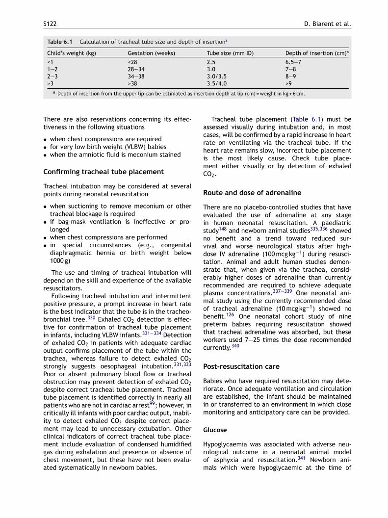
S122 D. Biarent et al.
Table 6.1 Calculation of tracheal tube size and depth of insertiona
Child’s weight (kg) Gestation (weeks) Tube size (mm ID) Depth of insertion (cm)a
<1 <28 2.5 6.5—71—2 28—34 3.0 7—82—3 34—38 3.0/3.5 8—9>3 >38 3.5/4.0 >9
a Depth of insertion from the upper lip can be estimated as insertion depth at lip (cm) = weight in kg + 6 cm.
There are also reservations concerning its effec-tiveness in the following situations
• when chest compressions are required• for very low birth weight (VLBW) babies• when the amniotic fluid is meconium stained
Confirming tracheal tube placement
Tracheal intubation may be considered at severalpoints during neonatal resuscitation
• when suctioning to remove meconium or othertracheal blockage is required
• if bag-mask ventilation is ineffective or pro-longed
• when chest compressions are performed• in special circumstances (e.g., congenital
diaphragmatic hernia or birth weight below1000 g)
The use and timing of tracheal intubation willdepend on the skill and experience of the availableresuscitators.
Following tracheal intubation and intermittentpositive pressure, a prompt increase in heart rateis the best indicator that the tube is in the tracheo-
Tracheal tube placement (Table 6.1) must beassessed visually during intubation and, in mostcases, will be confirmed by a rapid increase in heartrate on ventilating via the tracheal tube. If theheart rate remains slow, incorrect tube placementis the most likely cause. Check tube place-ment either visually or by detection of exhaledCO2.
Route and dose of adrenaline
There are no placebo-controlled studies that haveevaluated the use of adrenaline at any stagein human neonatal resuscitation. A paediatricstudy148 and newborn animal studies335,336 showedno benefit and a trend toward reduced sur-vival and worse neurological status after high-dose IV adrenaline (100 mcg kg−1) during resusci-tation. Animal and adult human studies demon-strate that, when given via the trachea, consid-erably higher doses of adrenaline than currentlyrecommended are required to achieve adequateplasma concentrations.337—339 One neonatal ani-mal study using the currently recommended doseof tracheal adrenaline (10 mcg kg−1) showed nobptwc
P
Braim
G
Hrom
bronchial tree.330 Exhaled CO2 detection is effec-tive for confirmation of tracheal tube placementin infants, including VLBW infants.331—334 Detectionof exhaled CO2 in patients with adequate cardiacoutput confirms placement of the tube within thetrachea, whereas failure to detect exhaled CO2strongly suggests oesophageal intubation.331,333
Poor or absent pulmonary blood flow or trachealobstruction may prevent detection of exhaled CO2despite correct tracheal tube placement. Trachealtube placement is identified correctly in nearly allpatients who are not in cardiac arrest99; however, incritically ill infants with poor cardiac output, inabil-ity to detect exhaled CO2 despite correct place-ment may lead to unnecessary extubation. Otherclinical indicators of correct tracheal tube place-ment include evaluation of condensed humidifiedgas during exhalation and presence or absence ofchest movement, but these have not been evalu-ated systematically in newborn babies.
enefit.126 One neonatal cohort study of ninereterm babies requiring resuscitation showedhat tracheal adrenaline was absorbed, but theseorkers used 7—25 times the dose recommendedurrently.340
ost-resuscitation care
abies who have required resuscitation may dete-iorate. Once adequate ventilation and circulationre established, the infant should be maintainedn or transferred to an environment in which closeonitoring and anticipatory care can be provided.
lucose
ypoglycaemia was associated with adverse neu-ological outcome in a neonatal animal modelf asphyxia and resuscitation.341 Newborn ani-als which were hypoglycaemic at the time of
European Resuscitation Council Guidelines for Resuscitation 2005 S123
an anoxic or hypoxic-ischaemic insult had largerareas of cerebral infarction and/or decreased sur-vival compared with controls.342,343 One clinicalstudy demonstrated an association between hypo-glycaemia and poor neurological outcome followingperinatal asphyxia.344 No clinical neonatal studieshave investigated the relationship between hyper-glycaemia and neurological outcome, although inadults hyperglycaemia is associated with a worseoutcome.345 The range of blood glucose concentra-tion that is associated with the least brain injuryfollowing asphyxia and resuscitation cannot bedefined on available evidence. Infants who requiresignificant resuscitation should be monitored andtreated to maintain blood glucose within the nor-mal range.
Induced hypothermia
In a multicentre trial involving newborns withsuspected asphyxia (indicated by need for resus-citation at birth, metabolic acidosis and earlyencephalopathy), selective head cooling (34.5 ◦C)was associated with a non-significant reduction inthe number of survivors with severe disability at1gbIptssamvtphcsch
tbwna
W
MiSa
continue life support in severely compromisedbabies.350 There is considerable variability amongproviders about the benefits and disadvantages ofaggressive therapies in such babies.351,352
Withholding resuscitation
It is possible to identify conditions associated withhigh mortality and poor outcome, where withhold-ing resuscitation may be considered reasonable,particularly when there has been the opportunityfor discussion with parents.282,353 A consistent andcoordinated approach to individual cases by theobstetric and neonatal teams and the parents is animportant goal. Withholding resuscitation and dis-continuation of life-sustaining treatment during orfollowing resuscitation are considered by many tobe ethically equivalent, and clinicians should notbe hesitant to withdraw support when the possi-bility of functional survival is highly unlikely. Thefollowing guidelines must be interpreted accordingto current regional outcomes.
• Where gestation, birth weight and/or congeni-tal anomalies are associated with almost certain
•
•
W
Dbedaro
8 months, but a significant benefit in the sub-roup with moderate encephalopathy as judgedy amplitude-integrated electroencephalogram.220
nfants with severe electroencephalographic sup-ression and seizures did not benefit fromreatment.346 A second, small, controlled pilottudy in asphyxiated infants with early inducedystemic hypothermia resulted in fewer deathsnd disabilities at 12 months. Modest hypother-ia is associated with bradycardia and ele-
ated blood pressure that do not usually requirereatment, but a rapid increase in body tem-erature may cause hypotension.347 Profoundypothermia (core temperature below 33 ◦C) mayause arrhythmia, bleeding, thrombosis and sep-is, but studies so far have not reported theseomplications in infants treated with modestypothermia.220,348
There are insufficient data to recommend rou-ine use of modest systemic or selective cere-ral hypothermia following resuscitation of infantsith suspected asphyxia. Further clinical trials areeeded to determine which infants benefit mostnd which method of cooling is most effective.
ithholding or discontinuing resuscitation
ortality and morbidity for newborns varies accord-ng to region and to availability of resources.349
ocial science studies indicate that parents desirelarger role in decisions to resuscitate and to
early death, and unacceptably high morbidity islikely among the rare survivors, resuscitation isnot indicated. Examples from the published liter-ature include extreme prematurity (gestationalage <23 weeks and/or birthweight <400 g), andanomalies such as anencephaly and confirmed tri-somy 13 or 18.Resuscitation is nearly always indicated in con-ditions associated with a high survival rate andacceptable morbidity. This will generally includebabies with gestational age of 25 weeks orabove (unless there is evidence of fetal compro-mise such as intrauterine infection or hypoxia-ischaemia) and those with most congenital mal-formations.In conditions associated with uncertain progno-sis, where there is borderline survival and a rel-atively high rate of morbidity, and where theanticipated burden to the child is high, parentaldesires regarding resuscitation should be sup-ported.
ithdrawing resuscitation efforts
ata from infants without signs of life fromirth, lasting at least 10 min or longer, showither high mortality or severe neurodevelopmentalisability.354,355 After 10 min of uninterrupted anddequate resuscitation efforts, discontinuation ofesuscitation may be justified if there are no signsf life.
S124 D. Biarent et al.
References
1. Zideman D, Bingham R, Beattie T, et al. Guidelines forpaediatric life support: a Statement by the Paediatric LifeSupport Working Party of the European Resuscitation Coun-cil. Resuscitation 1994;27:91—105.
2. European Resuscitation Council. Paediatric life support:(including the recommendations for resuscitation of babiesat birth). Resuscitation 1998;37:95—6.
3. Phillips B, Zideman D, Garcia-Castrillo L, Felix M, Shwarz-Schwierin U. European Resuscitation Council Guidelines2000 for Basic Paediatric Life Support. A statement fromthe Paediatric Life Support Working Group and approvedby the Executive Committee of the European ResuscitationCouncil. Resuscitation 2001;48:223—9.
4. Phillips B, Zideman D, Garcia-Castrillo L, Felix M, Shwarz-Schwierin V. European Resuscitation Council Guidelines2000 for Advanced Paediatric Life Support. A statementfrom Paediatric Life Support Working Group and approvedby the Executive Committee of the European ResuscitationCouncil. Resuscitation 2001;48:231—4.
5. American Heart Association in collaboration with Interna-tional Liaison Committee on Resuscitation. Guidelines forCardiopulmonary Resuscitation and Emergency Cardiovas-cular Care—–an international consensus on science. Resus-citation 2000;46:3—430.
6. American Heart Association in collaboration with Interna-tional Liaison Committee on Resuscitation. Guidelines 2000for Cardiopulmonary Resuscitation and Emergency Cardio-
a physiological and mathematical analysis. Resuscitation2002;54:147—57.
17. Babbs CF, Nadkarni V. Optimizing chest compression to res-cue ventilation ratios during one-rescuer CPR by profes-sionals and lay persons: children are not just little adults.Resuscitation 2004;61:173—81.
18. Whyte SD, Wyllie JP. Paediatric basic life support: a prac-tical assessment. Resuscitation 1999;41:153—217.
19. Safranek DJ, Eisenberg MS, Larsen MP. The epidemiol-ogy of cardiac arrest in young adults. Ann Emerg Med1992;21:1102—6.
20. Clements F, McGowan J. Finger position for chest com-pressions in cardiac arrest in infants. Resuscitation2000;44:43—6.
21. Houri PK, Frank LR, Menegazzi JJ, Taylor R. A randomized,controlled trial of two-thumb vs two-finger chest compres-sion in a swine infant model of cardiac arrest. PrehospEmerg Care 1997;1:65—7.
22. David R. Closed chest cardiac massage in the newborninfant. Pediatrics 1988;81:552—4.
23. Dorfsman ML, Menegazzi JJ, Wadas RJ, Auble TE. Two-thumb vs two-finger chest compression in an infant modelof prolonged cardiopulmonary resuscitation. Acad EmergMed 2000;7:1077—82.
24. Whitelaw CC, Slywka B, Goldsmith LJ. Comparison of a two-finger versus two-thumb method for chest compressions byhealthcare providers in an infant mechanical model. Resus-citation 2000;43:213—6.
25. Menegazzi JJ, Auble TE, Nicklas KA, Hosack GM, Rack L,Goode JS. Two-thumb versus two-finger chest compression
vascular Care: International Consensus on Science. Circu-lation 2000;102(Suppl. I):I-1—I-370.
7. International Liaison Committee on Resuscitation. 2005International Consensus on Cardiopulmonary Resuscitationand Emergency Cardiovascular Care Science with Treat-ment Recommendations. Resuscitation 2005;67:157—341.
8. International Liaison Committee on Resuscitation. 2005International Consensus on Cardiopulmonary Resuscitationand Emergency Cardiovascular Care Science with Treat-ment Recommendations. Circulation, in press.
9. Kuisma M, Suominen P, Korpela R. Paediatric out-of-hospitalcardiac arrests: epidemiology and outcome. Resuscitation1995;30:141—50.
10. Kyriacou DN, Arcinue EL, Peek C, Kraus JF. Effect of imme-diate resuscitation on children with submersion injury.Pediatrics 1994;94:137—42.
11. Berg RA, Hilwig RW, Kern KB, Ewy GA. Bystander’’chest compressions and assisted ventilation independentlyimprove outcome from piglet asphyxial pulseless ‘‘cardiacarrest’’. Circulation 2000;101:1743—8.
12. Young KD, Seidel JS. Pediatric cardiopulmonary resus-citation: a collective review. Ann Emerg Med 1999;33:195—205.
13. Berg RA, Hilwig RW, Kern KB, Babar I, Ewy GA. Simu-lated mouth-to-mouth ventilation and chest compressions(bystander cardiopulmonary resuscitation) improves out-come in a swine model of prehospital pediatric asphyxialcardiac arrest. Crit Care Med 1999;27:1893—9.
14. Dorph E, Wik L, Steen PA. Effectiveness of ventilation-compression ratios 1:5 and 2:15 in simulated single res-cuer paediatric resuscitation. Resuscitation 2002;54:259—64.
15. Turner I, Turner S, Armstrong V. Does the compression toventilation ratio affect the quality of CPR: a simulationstudy. Resuscitation 2002;52:55—62.
16. Babbs CF, Kern KB. Optimum compression to ventila-tion ratios in CPR under realistic, practical conditions:
during CRP in a swine infant model of cardiac arrest. AnnEmerg Med 1993;22:240—3.
26. Stevenson AG, McGowan J, Evans AL, Graham CA. CPR forchildren: one hand or two? Resuscitation 2005;64:205—8.
27. Gurnett CA, Atkins DL. Successful use of a biphasic wave-form automated external defibrillator in a high-risk child.Am J Cardiol 2000;86:1051—3.
28. Konig B, Benger J, Goldsworthy L. Automatic externaldefibrillation in a 6 year old. Arch Dis Child 2005;90:310—1.
29. Atkinson E, Mikysa B, Conway JA, et al. Specificity and sen-sitivity of automated external defibrillator rhythm analysisin infants and children. Ann Emerg Med 2003;42:185—96.
30. Cecchin F, Jorgenson DB, Berul CI, et al. Is arrhythmiadetection by automatic external defibrillator accurate forchildren? Sensitivity and specificity of an automatic exter-nal defibrillator algorithm in 696 pediatric arrhythmias.Circulation 2001;103:2483—8.
31. Samson R, Berg R, Bingham R. Pediatric Advanced Life Sup-port Task Force ILCoR. Use of automated external defibril-lators for children: an update. An advisory statement fromthe Pediatric Advanced Life Support Task Force of the Inter-national Liaison Committee on Resuscitation. Resuscitation2003;57:237—43.
32. Jorgenson D, Morgan C, Snyder D, et al. Energy attenuatorfor pediatric application of an automated external defib-rillator. Crit Care Med 2002;30:S145—7.
33. Tang W, Weil MH, Jorgenson D, et al. Fixed-energy bipha-sic waveform defibrillation in a pediatric model of cardiacarrest and resuscitation. Crit Care Med 2002;30:2736—41.
34. Berg RA, Chapman FW, Berg MD, et al. Attenuated adultbiphasic shocks compared with weight-based monophasicshocks in a swine model of prolonged pediatric ventricularfibrillation. Resuscitation 2004;61:189—97.
35. Berg RA, Samson RA, Berg MD, et al. Better outcome afterpediatric defibrillation dosage than adult dosage in a swinemodel of pediatric ventricular fibrillation. J Am Coll Cardiol2005;45:786—9.
European Resuscitation Council Guidelines for Resuscitation 2005 S125
36. Rossano JQ, Schiff L, Kenney MA, Atkins DL. Survivalis not correlated with defibrillation dosing in pedi-atric out-of-hospital ventricular fibrillation. Circulation2003;108:320—1.
37. Clark CB, Zhang Y, Davies LR, Karlsson G, Kerber RE. Pedi-atric transthoracic defibrillation: biphasic versus monopha-sic waveforms in an experimental model. Resuscitation2001;51:159—63.
38. Schneider T, Martens PR, Paschen H, et al. Multicenter,randomized, controlled trial of 150-J biphasic shocks com-pared with 200- to 360-J monophasic shocks in the resusci-tation of out-of-hospital cardiac arrest victims. OptimizedResponse to Cardiac Arrest (ORCA) Investigators. Circula-tion 2000;102:1780—7.
39. Faddy SC, Powell J, Craig JC. Biphasic and monophasicshocks for transthoracic defibrillation: a meta analysis ofrandomised controlled trials. Resuscitation 2003;58:9—16.
40. van Alem AP, Chapman FW, Lank P, Hart AA, KosterRW. A prospective, randomised and blinded compari-son of first shock success of monophasic and biphasicwaveforms in out-of-hospital cardiac arrest. Resuscitation2003;58:17—24.
41. Redding JS. The choking controversy: critique of evidenceon the Heimlich maneuver. Crit Care Med 1979;7:475—9.
42. International Liaison Committee on Resuscitation. Part 2.Adult Basic Life Support. 2005 International Consensuson Cardiopulmonary Resuscitation and Emergency Cardio-vascular Care Science with Treatment Recommendations.Resuscitation 2005;67:187—200.
43. Sirbaugh PE, Pepe PE, Shook JE, et al. A prospective,
53. Frederick K, Bixby E, Orzel MN, Stewart-Brown S, WillettK. Will changing the emphasis from ‘pulseless’ to ‘no signsof circulation’ improve the recall scores for effective lifesupport skills in children? Resuscitation 2002;55:255—61.
54. Park C, Bahk JH, Ahn WS, Do SH, Lee KH. The laryn-geal mask airway in infants and children. Can J Anaesth2001;48:413—7.
55. Hedges JR, Dronen SC, Feero S, Hawkins S, Syverud SA,Shultz B. Succinylcholine-assisted intubations in prehospi-tal care. Ann Emerg Med 1988;17:469—72.
56. Murphy-Macabobby M, Marshall WJ, Schneider C, Dries D.Neuromuscular blockade in aeromedical airway manage-ment. Ann Emerg Med 1992;21:664—8.
57. Sayre M, Weisgerber I. The use of neuromuscular block-ing agents by air medical services. J Air Med Transp1992;11:7—11.
58. Rose W, Anderson L, Edmond S. Analysis of intubations.Before and after establishment of a rapid sequence intuba-tion protocol for air medical use. Air Med J 1994;13:475—8.
59. Sing RF, Reilly PM, Rotondo MF, Lynch MJ, McCans JP,Schwab CW. Out-of-hospital rapid-sequence induction forintubation of the pediatric patient. Acad Emerg Med1996;3:41—5.
60. Ma OJ, Atchley RB, Hatley T, Green M, Young J, Brady W.Intubation success rates improve for an air medical pro-gram after implementing the use of neuromuscular block-ing agents. Am J Emerg Med 1998;16:125—7.
61. Tayal V, Riggs R, Marx J, Tomaszewski C, Schneider R. Rapid-sequence intubation at an emergency medicine residency:success rate and adverse events during a two-year period.
population-based study of the demographics, epidemi-ology, management, and outcome of out-of-hospitalpediatric cardiopulmonary arrest. Ann Emerg Med1999;33:174—84.
44. Hickey RW, Cohen DM, Strausbaugh S, Dietrich AM. Pedi-atric patients requiring CPR in the prehospital setting. AnnEmerg Med 1995;25:495—501.
45. Reis AG, Nadkarni V, Perondi MB, Grisi S, Berg RA. Aprospective investigation into the epidemiology of in-hospital pediatric cardiopulmonary resuscitation usingthe international Utstein reporting style. Pediatrics2002;109:200—9.
46. Young KD, Gausche-Hill M, McClung CD, Lewis RJ. Aprospective, population-based study of the epidemiologyand outcome of out-of-hospital pediatric cardiopulmonaryarrest. Pediatrics 2004;114:157—64.
47. Richman PB, Nashed AH. The etiology of cardiac arrest inchildren and young adults: special considerations for EDmanagement. Am J Emerg Med 1999;17:264—70.
48. Engdahl J, Bang A, Karlson BW, Lindqvist J, Herlitz J. Char-acteristics and outcome among patients suffering from outof hospital cardiac arrest of non-cardiac aetiology. Resus-citation 2003;57:33—41.
49. Carcillo JA. Pediatric septic shock and multiple organ fail-ure. Crit Care Clin 2003;19:413—40, viii.
50. Eberle B, Dick WF, Schneider T, Wisser G, Doetsch S,Tzanova I. Checking the carotid pulse check: diagnosticaccuracy of first responders in patients with and withouta pulse. Resuscitation 1996;33:107—16.
51. Moule P. Checking the carotid pulse: diagnostic accuracyin students of the healthcare professions. Resuscitation2000;44:195—201.
52. Lapostolle F, Le Toumelin P, Agostinucci JM, Catineau J,Adnet F. Basic cardiac life support providers checking thecarotid pulse: performance, degree of conviction, andinfluencing factors. Acad Emerg Med 2004;11:878—80.
Acad Emerg Med 1999;6:31—7.62. Wang HE, Sweeney TA, O’Connor RE, Rubinstein H.
Failed prehospital intubations: an analysis of emergencydepartment courses and outcomes. Prehosp Emerg Care2001;5:134—41.
63. Sagarin MJ, Chiang V, Sakles JC, et al. Rapid sequenceintubation for pediatric emergency airway management.Pediatr Emerg Care 2002;18:417—43.
64. Kaye K, Frascone RJ, Held T. Prehospital rapid-sequenceintubation: a pilot training program. Prehosp Emerg Care2003;7:235—40.
65. Wang HE, Kupas DF, Paris PM, Bates RR, Costantino JP, YealyDM. Multivariate predictors of failed prehospital endotra-cheal intubation. Acad Emerg Med 2003;10:717—24.
66. Pepe P, Zachariah B, Chandra N. Invasive airway techniquein resuscitation. Ann Emerg Med 1991;22:393—403.
67. Luten RC, Wears RL, Broselow J, et al. Length-based endo-tracheal tube and emergency equipment in pediatrics. AnnEmerg Med 1992;21:900—4.
68. Deakers TW, Reynolds G, Stretton M, Newth CJ. Cuffedendotracheal tubes in pediatric intensive care. J Pediatr1994;125:57—62.
69. Khine HH, Corddry DH, Kettrick RG, et al. Compar-ison of cuffed and uncuffed endotracheal tubes inyoung children during general anesthesia. Anesthesiology1997;86:627—31.
70. Newth CJ, Rachman B, Patel N, Hammer J. The use ofcuffed versus uncuffed endotracheal tubes in pediatricintensive care. J Pediatr 2004;144:333—7.
71. Mhanna MJ, Zamel YB, Tichy CM, Super DM. The ‘‘air leak’’test around the endotracheal tube, as a predictor of pos-textubation stridor, is age dependent in children. Crit CareMed 2002;30:2639—43.
72. Katz SH, Falk JL. Misplaced endotracheal tubes byparamedics in an urban emergency medical services sys-tem. Ann Emerg Med 2001;37:32—7.
S126 D. Biarent et al.
73. Gausche M, Lewis RJ, Stratton SJ, et al. Effect of out-of-hospital pediatric endotracheal intubation on survivaland neurological outcome: a controlled clinical trial. JAMA2000;283:783—90.
74. Kelly JJ, Eynon CA, Kaplan JL, de Garavilla L, Dalsey WC.Use of tube condensation as an indicator of endotrachealtube placement. Ann Emerg Med 1998;31:575—8.
75. Andersen KH, Hald A. Assessing the position of the tra-cheal tube: the reliability of different methods. Anaesthe-sia 1989;44:984—5.
76. Andersen KH, Schultz-Lebahn T. Oesophageal intubationcan be undetected by auscultation of the chest. ActaAnaesthesiol Scand 1994;38:580—2.
77. Hartrey R, Kestin IG. Movement of oral and nasal trachealtubes as a result of changes in head and neck position.Anaesthesia 1995;50:682—7.
78. Van de Louw A, Cracco C, Cerf C, et al. Accuracy of pulseoximetry in the intensive care unit. Intensive Care Med2001;27:1606—13.
79. Seguin P, Le Rouzo A, Tanguy M, Guillou YM, Feuillu A,Malledant Y. Evidence for the need of bedside accuracyof pulse oximetry in an intensive care unit. Crit Care Med2000;28:703—6.
80. Tan A, Schulze A, O’Donnell CP, Davis PG. Air versus oxygenfor resuscitation of infants at birth. Cochrane Database SystRev 2004:CD002273.
81. Ramji S, Rasaily R, Mishra PK, et al. Resuscitation of asphyx-iated newborns with room air or 100% oxygen at birth: amulticentric clinical trial. Indian Pediatr 2003;40:510—7.
82. Vento M, Asensi M, Sastre J, Garcia-Sala F, Pallardo FV,
tube placement in infants and children. Ann Emerg Med1992;21:142—5.
94. Bhende MS, Thompson AE, Orr RA. Utility of an end-tidalcarbon dioxide detector during stabilization and trans-port of critically ill children. Pediatrics 1992;89(pt 1):1042—4.
95. Bhende MS, LaCovey DC. End-tidal carbon dioxide mon-itoring in the prehospital setting. Prehosp Emerg Care2001;5:208—13.
96. Ornato JP, Shipley JB, Racht EM, et al. Multicenter study ofa portable, hand-size, colorimetric end-tidal carbon diox-ide detection device. Ann Emerg Med 1992;21:518—23.
97. Gonzalez del Rey JA, Poirier MP, Digiulio GA. Evaluationof an ambu-bag valve with a self-contained, colorimetricend-tidal CO2 system in the detection of airway mishaps:an animal trial. Pediatr Emerg Care 2000;16:121—3.
98. Bhende MS, Thompson AE. Evaluation of an end-tidal CO2detector during pediatric cardiopulmonary resuscitation.Pediatrics 1995;95:395—9.
99. Bhende MS, Karasic DG, Karasic RB. End-tidal carbon diox-ide changes during cardiopulmonary resuscitation afterexperimental asphyxial cardiac arrest. Am J Emerg Med1996;14:349—50.
100. DeBehnke DJ, Hilander SJ, Dobler DW, Wickman LL, SwartGL. The hemodynamic and arterial blood gas response toasphyxiation: a canine model of pulseless electrical activ-ity. Resuscitation 1995;30:169—75.
101. Ornato JP, Garnett AR, Glauser FL. Relationship betweencardiac output and the end-tidal carbon dioxide tension.Ann Emerg Med 1990;19:1104—6.
1
1
1
1
1
1
1
1
1
1
1
Vina J. Resuscitation with room air instead of 100% oxygenprevents oxidative stress in moderately asphyxiated termneonates. Pediatrics 2001;107:642—7.
83. Saugstad OD. Resuscitation of newborn infants with roomair or oxygen. Semin Neonatol 2001;6:233—9.
84. Aufderheide TP, Lurie KG. Death by hyperventilation: acommon and life-threatening problem during cardiopul-monary resuscitation. Crit Care Med 2004;32:S345—51.
85. Aufderheide TP, Sigurdsson G, Pirrallo RG, et al.Hyperventilation-induced hypotension during cardiopul-monary resuscitation. Circulation 2004;109:1960—5.
86. Wik L, Kramer-Johansen J, Myklebust H, et al. Quality ofcardiopulmonary resuscitation during out-of-hospital car-diac arrest. JAMA 2005;293:299—304.
87. Abella BS, Alvarado JP, Myklebust H, et al. Quality ofcardiopulmonary resuscitation during in-hospital cardiacarrest. AMA 2005;293:305—10.
88. Abella BS, Sandbo N, Vassilatos P, et al. Chest compressionrates during cardiopulmonary resuscitation are suboptimal:a prospective study during in-hospital cardiac arrest. Cir-culation 2005;111:428—34.
89. Borke WB, Munkeby BH, Morkrid L, Thaulow E, SaugstadOD. Resuscitation with 100% O(2) does not protect themyocardium in hypoxic newborn piglets. Arch Dis ChildFetal Neonatal Ed 2004;89:F156—60.
90. Stockinger ZT, McSwain Jr NE. Prehospital endotrachealintubation for trauma does not improve survival over bag-valve-mask ventilation. J Trauma 2004;56:531—6.
91. Pitetti R, Glustein JZ, Bhende MS. Prehospital care and out-come of pediatric out-of-hospital cardiac arrest. PrehospEmerg Care 2002;6:283—90.
92. Cooper A, DiScala C, Foltin G, Tunik M, Markenson D,Welborn C. Prehospital endotracheal intubation for severehead injury in children: a reappraisal. Semin Pediatr Surg2001;10:3—6.
93. Bhende MS, Thompson AE, Cook DR, Saville AL. Validity of adisposable end-tidal CO2 detector in verifying endotracheal
02. Sharieff GQ, Rodarte A, Wilton N, Silva PD, Bleyle D. Theself-inflating bulb as an esophageal detector device in chil-dren weighing more than twenty kilograms: a comparisonof two techniques. Ann Emerg Med 2003;41:623—9.
03. Sharieff GQ, Rodarte A, Wilton N, Bleyle D. The self-inflating bulb as an airway adjunct: is it reliable in chil-dren weighing less than 20 kilograms? Acad Emerg Med2003;10:303—8.
04. Poirier MP, Gonzalez Del-Rey JA, McAneney CM, DiGiulio GA.Utility of monitoring capnography, pulse oximetry, and vitalsigns in the detection of airway mishaps: a hyperoxemicanimal model. Am J Emerg Med 1998;16:350—2.
05. Lillis KA, Jaffe DM. Prehospital intravenous access in chil-dren. Ann Emerg Med 1992;21:1430—4.
06. Kanter RK, Zimmerman JJ, Strauss RH, Stoeckel KA. Pedi-atric emergency intravenous access. Evaluation of a proto-col. Am J Dis Child 1986;140:132—4.
07. Banerjee S, Singhi SC, Singh S, Singh M. The intraosseousroute is a suitable alternative to intravenous route for fluidresuscitation in severely dehydrated children. Indian Pedi-atr 1994;31:1511—20.
08. Glaeser PW, Hellmich TR, Szewczuga D, Losek JD, Smith DS.Five-year experience in prehospital intraosseous infusionsin children and adults. Ann Emerg Med 1993;22:1119—24.
09. Guy J, Haley K, Zuspan SJ. Use of intraosseous infu-sion in the pediatric trauma patient. J Pediatr Surg1993;28:158—61.
10. Orlowski JP, Julius CJ, Petras RE, Porembka DT, Gal-lagher JM. The safety of intraosseous infusions: risks offat and bone marrow emboli to the lungs. Ann Emerg Med1989;18:1062—7.
11. Orlowski JP, Porembka DT, Gallagher JM, Lockrem JD, Van-Lente F. Comparison study of intraosseous, central intra-venous, and peripheral intravenous infusions of emergencydrugs. Am J Dis Child 1990;144:112—7.
12. Abe KK, Blum GT, Yamamoto LG. Intraosseous is fasterand easier than umbilical venous catheterization in new-
European Resuscitation Council Guidelines for Resuscitation 2005 S127
born emergency vascular access models. Am J Emerg Med2000;18:126—9.
113. Ellemunter H, Simma B, Trawoger R, Maurer H. Intraosseouslines in preterm and full term neonates. Arch Dis Child FetalNeonatal Ed 1999;80:F74—5.
114. Cameron JL, Fontanarosa PB, Passalaqua AM. A compar-ative study of peripheral to central circulation deliverytimes between intraosseous and intravenous injection usinga radionuclide technique in normovolemic and hypovolemiccanines. J Emerg Med 1989;7:123—7.
115. Warren DW, Kissoon N, Sommerauer JF, Rieder MJ. Compari-son of fluid infusion rates among peripheral intravenous andhumerus, femur, malleolus, and tibial intraosseous sitesin normovolemic and hypovolemic piglets. Ann Emerg Med1993;22:183—6.
116. Brickman KR, Krupp K, Rega P, Alexander J, Guinness M.Typing and screening of blood from intraosseous access.Ann Emerg Med 1992;21:414—7.
117. Johnson L, Kissoon N, Fiallos M, Abdelmoneim T, MurphyS. Use of intraosseous blood to assess blood chemistriesand hemoglobin during cardiopulmonary resuscitation withdrug infusions. Crit Care Med 1999;27:1147—52.
118. Ummenhofer W, Frei FJ, Urwyler A, Drewe J. Are labora-tory values in bone marrow aspirate predictable for venousblood in paediatric patients? Resuscitation 1994;27:123—8.
119. Kissoon N, Idris A, Wenzel V, Murphy S, Rush W. Intraosseousand central venous blood acid—base relationship dur-ing cardiopulmonary resuscitation. Pediatr Emerg Care1997;13:250—3.
1
1
1
1
1
1
1
1
1
1
130. Crespo SG, Schoffstall JM, Fuhs LR, Spivey WH. Compari-son of two doses of endotracheal epinephrine in a cardiacarrest model. Ann Emerg Med 1991;20:230—4.
131. Lee PL, Chung YT, Lee BY, Yeh CY, Lin SY, Chao CC. Theoptimal dose of atropine via the endotracheal route. MaZui Xue Za Zhi 1989;27:35—8.
132. Hahnel JH, Lindner KH, Schurmann C, Prengel A, AhnefeldFW. Plasma lidocaine levels and PaO2 with endobronchialadministration: dilution with normal saline or distilledwater? Ann Emerg Med 1990;19:1314—7.
133. Jasani MS, Nadkarni VM, Finkelstein MS, Mandell GA,Salzman SK, Norman ME. Effects of different techniquesof endotracheal epinephrine administration in pediatricporcine hypoxic-hypercarbic cardiopulmonary arrest. CritCare Med 1994;22:1174—80.
134. Steinfath M, Scholz J, Schulte am Esch J, Laer S, ReymannA, Scholz H. The technique of endobronchial lidocaineadministration does not influence plasma concentrationprofiles and pharmacokinetic parameters in humans. Resus-citation 1995;29:55—62.
135. Carcillo JA, Fields AI. Clinical practice parameters forhemodynamic support of pediatric and neonatal patientsin septic shock. Crit Care Med 2002;30:1365—78.
136. Simma B, Burger R, Falk M, Sacher P, Fanconi S. A prospec-tive, randomized, and controlled study of fluid man-agement in children with severe head injury: lactatedRinger’s solution versus hypertonic saline. Crit Care Med1998;26:1265—70.
137. Rocha e Silva M. Hypertonic saline resuscitation. Medicina(B Aries) 1998; 58:393-402.
1
1
1
1
1
1
1
1
1
1
20. Abdelmoneim T, Kissoon N, Johnson L, Fiallos M, Mur-phy S. Acid-base status of blood from intraosseous andmixed venous sites during prolonged cardiopulmonaryresuscitation and drug infusions. Crit Care Med 1999;27:1923—8.
21. Venkataraman ST, Orr RA, Thompson AE. Percutaneous infr-aclavicular subclavian vein catheterization in critically illinfants and children. J Pediatr 1988;113:480—5.
22. Fleisher G, Caputo G, Baskin M. Comparison of externaljugular and peripheral venous administration of sodiumbicarbonate in puppies. Crit Care Med 1989;17:251—4.
23. Hedges JR, Barsan WB, Doan LA, et al. Central versusperipheral intravenous routes in cardiopulmonary resusci-tation. Am J Emerg Med 1984;2:385—90.
24. Neufeld JD, Marx JA, Moore EE, Light AI. Comparison ofintraosseous, central, and peripheral routes of crystalloidinfusion for resuscitation of hemorrhagic shock in a swinemodel. J Trauma 1993;34:422—8.
25. Stenzel JP, Green TP, Fuhrman BP, Carlson PE, Marches-sault RP. Percutaneous femoral venous catheteriza-tions: a prospective study of complications. J Pediatr1989;114:411—5.
26. Kleinman ME, Oh W, Stonestreet BS. Comparison of intra-venous and endotracheal epinephrine during cardiopul-monary resuscitation in newborn piglets. Crit Care Med1999;27:2748—54.
27. Efrati O, Ben-Abraham R, Barak A, et al. Endo-bronchial adrenaline: should it be reconsidered? Doseresponse and haemodynamic effect in dogs. Resuscitation2003;59:117—22.
28. Howard RF, Bingham RM. Endotracheal compared withintravenous administration of atropine. Arch Dis Child1990;65:449—50.
29. Prengel AW, Lindner KH, Hahnel J, Ahnefeld FW. Endotra-cheal and endobronchial lidocaine administration: effectson plasma lidocaine concentration and blood gases. CritCare Med 1991;19:911—5.
38. Katz LM, Wang Y, Ebmeyer U, Radovsky A, Safar P. Glu-cose plus insulin infusion improves cerebral outcome afterasphyxial cardiac arrest. Neuroreport 1998;9:3363—7.
39. Longstreth Jr WT, Copass MK, Dennis LK, Rauch-MatthewsME, Stark MS, Cobb LA. Intravenous glucose after out-of-hospital cardiopulmonary arrest: a community-based ran-domized trial. Neurology 1993;43:2534—41.
40. Chang YS, Park WS, Ko SY, et al. Effects of fastingand insulin-induced hypoglycemia on brain cell membranefunction and energy metabolism during hypoxia-ischemiain newborn piglets. Brain Res 1999;844:135—42.
41. Cherian L, Goodman JC, Robertson CS. Hyperglycemiaincreases brain injury caused by secondary ischemiaafter cortical impact injury in rats. Crit Care Med1997;25:1378—83.
42. Paul T, Bertram H, Bokenkamp R, Hausdorf G. Supraven-tricular tachycardia in infants, children and adolescents:diagnosis, and pharmacological and interventional therapy.Paediatr Drugs 2000;2:171—81.
43. Losek JD, Endom E, Dietrich A, Stewart G, Zempsky W,Smith K. Adenosine and pediatric supraventricular tachy-cardia in the emergency department: multicenter studyand review. Ann Emerg Med 1999;33:185—91.
44. Roberts JR, Greenburg MI, Knaub M, Baskin SI. Compari-son of the pharmacological effects of epinephrine admin-istered by the intravenous and endotracheal routes. JACEP1978;7:260—4.
45. Zaritsky A. Pediatric resuscitation pharmacology. Membersof the Medications in Pediatric Resuscitation Panel. AnnEmerg Med 1993;22(pt 2):445—55.
46. Manisterski Y, Vaknin Z, Ben-Abraham R, et al. Endotra-cheal epinephrine: a call for larger doses. Anesth Analg2002;95:1037—41 [table of contents].
47. Patterson MD, Boenning DA, Klein BL, et al. The use ofhigh-dose epinephrine for patients with out-of-hospitalcardiopulmonary arrest refractory to prehospital interven-tions. Pediatr Emerg Care 2005;21:227—37.
S128 D. Biarent et al.
148. Perondi MB, Reis AG, Paiva EF, Nadkarni VM, Berg RA. Acomparison of high-dose and standard-dose epinephrinein children with cardiac arrest. N Engl J Med 2004;350:1722—30.
149. Carpenter TC, Stenmark KR. High-dose epinephrineis not superior to standard-dose epinephrine in pedi-atric in-hospital cardiopulmonary arrest. Pediatrics1997;99:403—8.
150. Dieckmann R, Vardis R. High-dose epinephrine in pedi-atric out-of-hospital cardiopulmonary arrest. Pediatrics1995;95:901—13.
151. Berg RA, Otto CW, Kern KB, et al. High-dose epinephrineresults in greater early mortality after resuscitation fromprolonged cardiac arrest in pigs: a prospective, randomizedstudy. Crit Care Med 1994;22:282—90.
152. Rubertsson S, Wiklund L. Hemodynamic effects ofepinephrine in combination with different alkaline buffersduring experimental, open-chest, cardiopulmonary resus-citation. Crit Care Med 1993;21:1051—7.
153. Somberg JC, Timar S, Bailin SJ, et al. Lack of a hypoten-sive effect with rapid administration of a new aque-ous formulation of intravenous amiodarone. Am J Cardiol2004;93:576—81.
154. Yap S-C, Hoomtje T, Sreeram N. Polymorphic ventricu-lar tachycardia after use of intravenous amiodarone forpostoperative junctional ectopic tachycardia. Int J Cardiol2000;76:245—7.
155. Dauchot P, Gravenstein JS. Effects of atropine on the elec-trocardiogram in different age groups. Clin Pharmacol Ther1971;12:274—80.
resuscitation-effect on immediate outcome. Resuscitation2004;60:219—23.
168. Bar-Joseph G, Abramson NS, Kelsey SF, Mashiach T, CraigMT, Safar P. Improved resuscitation outcome in emergencymedical systems with increased usage of sodium bicarbon-ate during cardiopulmonary resuscitation. Acta Anaesthe-siol Scand 2005;49:6—15.
169. Dorian P, Cass D, Schwartz B, Cooper R, GelaznikasR, Barr A. Amiodarone as compared with lidocaine forshock-resistant ventricular fibrillation. N Engl J Med2002;346:884—90.
170. Walsh EP, Saul JP, Sholler GF, et al. Evaluation of a stagedtreatment protocol for rapid automatic junctional tachy-cardia after operation for congenital heart disease. J AmColl Cardiol 1997;29:1046—53.
171. Wang L. Congenital long QT syndrome: 50 years of electro-physiological research from cell to bedside. Acta Cardio-logica 2003;58:133—8.
172. Singh BN, Kehoe R, Woosley RL, Scheinman M, QuartB, Sotalol Multicenter Study Group. Multicenter trial ofsotalol compared with procainamide in the suppression ofinducible ventricular tachycardia: a double-blind, random-ized parallel evaluation. Am Heart J 1995;129:87—97.
173. Luedtke SA, Kuhn RJ, McCaffrey FM. Pharmacologic man-agement of supraventricular tachycardias in children, part2: atrial flutter, atrial fibrillation, and junctional and atrialectopic tachycardia. Ann Pharmacother 1997;31:1347—59.
174. Luedtke SA, Kuhn RJ, McCaffrey FM. Pharmacologic man-agement of supraventricular tachycardias in children. Part1: Wolff-Parkinson-White and atrioventricular nodal reen-
1
1
1
1
1
1
1
1
1
1
1
156. Stulz PM, Scheidegger D, Drop LJ, Lowenstein E, Laver MB.Ventricular pump performance during hypocalcemia: clin-ical and experimental studies. J Thorac Cardiovasc Surg1979;78:185—94.
157. van Walraven C, Stiell IG, Wells GA, Hebert PC, Van-demheen K, The OTAC Study Group. Do advanced cardiaclife support drugs increase resuscitation rates from in-hospital cardiac arrest? Ann Emerg Med 1998;32:544—53.
158. Paraskos JA. Cardiovascular pharmacology III: atropine,calcium, calcium blockers, and (beta)-blockers. Circulation1986;74:IV-86—9.
159. Stueven HA, Thompson B, Aprahamian C, Tonsfeldt DJ,Kastenson EH. The effectiveness of calcium chloride inrefractory electromechanical dissociation. Ann Emerg Med1985;14:626—9.
160. Stueven HA, Thompson B, Aprahamian C, Tonsfeldt DJ, Kas-tenson EH. Lack of effectiveness of calcium chloride inrefractory asystole. Ann Emerg Med 1985;14:630—2.
161. Srinivasan V, Spinella PC, Drott HR, Roth CL, Helfaer MA,Nadkarni V. Association of timing, duration, and intensityof hyperglycemia with intensive care unit mortality in crit-ically ill children. Pediatr Crit Care Med 2004;5:329—36.
162. Krinsley JS. Effect of an intensive glucose management pro-tocol on the mortality of critically ill adult patients. MayoClin Proc 2004;79:992—1000.
163. Losek JD. Hypoglycemia and the ABC’S (sugar) of pediatricresuscitation. Ann Emerg Med 2000;35:43—6.
164. Finney SJ, Zekveld C, Elia A, Evans TW. Glucose control andmortality in critically ill patients. Jama 2003;290:2041—7.
165. Allegra J, Lavery R, Cody R, et al. Magnesium sulfate inthe treatment of refractory ventricular fibrillation in theprehospital setting. Resuscitation 2001;49:245—9.
166. Tzivoni D, Banai S, Schuger C, et al. Treatment oftorsade de pointes with magnesium sulfate. Circulation1988;77:392—7.
167. Lokesh L, Kumar P, Murki S, Narang A. A random-ized controlled trial of sodium bicarbonate in neonatal
try. Ann Pharmacother 1997;31:1227—43.75. Mandapati R, Byrum CJ, Kavey RE, et al. Procainamide for
rate control of postsurgical junctional tachycardia. PediatrCardiol 2000;21:123—8.
76. Wang JN, Wu JM, Tsai YC, Lin CS. Ectopic atrial tachycardiain children. J Formos Med Assoc 2000;99:766—70.
77. Holmes CL, Landry DW, Granton JT. Science review: Vaso-pressin and the cardiovascular system part 1—–receptorphysiology. Crit Care 2003;7:427—34.
78. Voelckel WG, Lurie KG, McKnite S, et al. Effects ofepinephrine and vasopressin in a piglet model of prolongedventricular fibrillation and cardiopulmonary resuscitation.Crit Care Med 2002;30:957—62.
79. Voelckel WG, Lurie KG, McKnite S, et al. Comparison ofepinephrine and vasopressin in a pediatric porcine modelof asphyxial cardiac arrest. Crit Care Med 2000;28:3777—83.
80. Mann K, Berg RA, Nadkarni V. Beneficial effects of vaso-pressin in prolonged pediatric cardiac arrest: a case series.Resuscitation 2002;52:149—56.
81. Atkins DL, Kerber RE. Pediatric defibrillation: current flowis improved by using ‘‘adult’’ electrode paddles. Pediatrics1994;94:90—3.
82. Atkins DL, Sirna S, Kieso R, Charbonnier F, Kerber RE. Pedi-atric defibrillation: importance of paddle size in deter-mining transthoracic impedance. Pediatrics 1988;82:914—8.
83. Bennetts SH, Deakin CD, Petley GW, Clewlow F. Is optimalpaddle force applied during paediatric external defibrilla-tion? Resuscitation 2004;60:29—32.
84. Deakin C, Bennetts S, Petley G, Clewlow F. What is the opti-mal paddle force for paediatric defibrillation? Resuscitation2002;55:59.
85. Atkins DL, Hartley LL, York DK. Accurate recognition andeffective treatment of ventricular fibrillation by auto-mated external defibrillators in adolescents. Pediatrics1998;101(pt 1):393—7.
European Resuscitation Council Guidelines for Resuscitation 2005 S129
186. Pierpont GL, Kruse JA, Nelson DH. Intra-arterial monitoringduring cardiopulmonary resuscitation. Cathet CardiovascDiagn 1985;11:513—20.
187. Zaritsky A, Nadkarni V, Getson P, Kuehl K. CPR in children.Ann Emerg Med 1987;16:1107—11.
188. Mogayzel C, Quan L, Graves JR, Tiedeman D, FahrenbruchC, Herndon P. Out-of-hospital ventricular fibrillation in chil-dren and adolescents: causes and outcomes. Ann EmergMed 1995;25:484—91.
189. Herlitz J, Engdahl J, Svensson L, Young M, Angquist KA,Holmberg S. Characteristics and outcome among childrensuffering from out of hospital cardiac arrest in Sweden.Resuscitation 2005;64:37—40.
190. Berg RA. Role of mouth-to-mouth rescue breathing inbystander cardiopulmonary resuscitation for asphyxial car-diac arrest. Crit Care Med 2000;28(Suppl.):N193—5.
191. Appleton GO, Cummins RO, Larson MP, Graves JR. CPRand the single rescuer: at what age should you ‘‘callfirst’’ rather than ‘‘call fast’’? Ann Emerg Med 1995;25:492—4.
192. Larsen MP, Eisenberg MS, Cummins RO, Hallstrom AP.Predicting survival from out-of-hospital cardiac arrest: agraphic model. Ann Emerg Med 1993;22:1652—8.
193. Cobb LA, Fahrenbruch CE, Walsh TR, et al. Influenceof cardiopulmonary resuscitation prior to defibrillation inpatients with out-of-hospital ventricular fibrillation. JAMA1999;281:1182—8.
194. Wik L, Hansen TB, Fylling F, et al. Delaying defibrillation togive basic cardiopulmonary resuscitation to patients without-of-hospital ventricular fibrillation: a randomized trial.
1
1
1
1
1
2
2
2
2
2
of congenital heart defects. Eur J Cardio-Thoracic Surg2002;21:255—9.
205. Figa FH, Gow RM, Hamilton RM, Freedom RM. Clinical effi-cacy and safety of intravenous Amiodarone in infants andchildren. Am J Cardiol 1994;74:573—7.
206. Hoffman TM, Bush DM, Wernovsky G, et al. Postopera-tive junctional ectopic tachycardia in children: incidence,risk factors, and treatment. Ann Thorac Surg 2002;74:1607—11.
207. Soult JA, Munoz M, Lopez JD, Romero A, Santos J, TovaruelaA. Efficacy and safety of intravenous amiodarone for short-term treatment of paroxysmal supraventricular tachycar-dia in children. Pediatr Cardiol 1995;16:16—9.
208. Benson Jr D, Smith W, Dunnigan A, Sterba R, Gallagher J.Mechanisms of regular wide QRS tachycardia in infants andchildren. Am J Cardiol 1982;49:1778—88.
209. Drago F, Mazza A, Guccione P, Mafrici A, Di Liso G, RagoneseP. Amiodarone used alone or in combination with propra-nolol: a very effective therapy for tachyarrhythmias ininfants and children. Pediatr Cardiol 1998;19:445—9.
210. Benson DJ, Dunnigan A, Green T, Benditt D, Schneider S.Periodic procainamide for paroxysmal tachycardia. Circu-lation 1985;72:147—52.
211. Komatsu C, Ishinaga T, Tateishi O, Tokuhisa Y, YoshimuraS. Effects of four antiarrhythmic drugs on the inductionand termination of paroxysmal supraventricular tachycar-dia. Jpn Circ J 1986;50:961—72.
212. Mandel WJ, Laks MM, Obayashi K, Hayakawa H, DaleyW. The Wolff-Parkinson-White syndrome: pharmacologiceffects of procaine amide. Am Heart J 1975;90:744—54.
2
2
2
2
2
2
2
2
2
2
2
JAMA 2003;289:1389—95.95. Jacobs IG, Finn JC, Oxer HF, Jelinek GA. CPR before defibril-
lation in out-of-hospital cardiac arrest: a randomized trial.Emerg Med Australas 2005;17:39—45.
96. Somberg JC, Bailin SJ, Haffajee CI, et al. Intravenous lido-caine versus intravenous amiodarone (in a new aqueousformulation) for incessant ventricular tachycardia. Am JCardiol 2002;90:853—9.
97. Kudenchuk PJ, Cobb LA, Copass MK, et al. Amiodaronefor resuscitation after out-of-hospital cardiac arrest dueto ventricular fibrillation. N Engl J Med 1999;341:871—8.
98. Perry JC, Fenrich AL, Hulse JE, Triedman JK, Friedman RA,Lamberti JJ. Pediatric use of intravenous amiodarone: effi-cacy and safety in critically ill patients from a multicenterprotocol. J Am Coll Cardiol 1996;27:1246—50.
99. Cummins RO, Graves JR, Larsen MP, et al. Out-of-hospitaltranscutaneous pacing by emergency medical techniciansin patients with asystolic cardiac arrest. N Engl J Med1993;328:1377—82.
00. Sreeram N, Wren C. Supraventricular tachycardia ininfants: response to initial treatment. Arch Dis Child1990;65:127—9.
01. Bianconi L, Castro AMD, Dinelli M, et al. Comparison ofintravenously administered dofetilide versus amiodaronein the acute termination of atrial fibrillation and flut-ter. A multicentre, randomized, double-blind, placebo-controlled study. Eur Heart J 2000;21:1265—73.
02. Burri S, Hug MI, Bauersfeld U. Efficacy and safety of intra-venous amiodarone for incessant tachycardias in infants.Eur J Pediatr 2003;162:880—4.
03. Celiker A, Ceviz N, Ozme S. Effectiveness and safety ofintravenous amiodarone in drug-resistant tachyarrhythmiasof children. Acta Paediatrica Japonica 1998;40:567—72.
04. Dodge-Khatami A, Miller O, Anderson R, Gil-Jaurena J,Goldman A, de Leval M. Impact of junctional ectopictachycardia on postoperative morbidity following repair
13. Meldon SW, Brady WJ, Berger S, Mannenbach M. Pediatricventricular tachycardia: a review with three illustrativecases. Pediatr Emerg Care 1994;10:294—300.
14. Shih JY, Gillette PC, Kugler JD, et al. The electrophysiologiceffects of procainamide in the immature heart. PediatrPharmacol (New York) 1982;2:65—73.
15. Hildebrand CA, Hartmann AG, Arcinue EL, Gomez RJ, BingRJ. Cardiac performance in pediatric near-drowning. CritCare Med 1988;16:331—5.
16. Checchia PA, Sehra R, Moynihan J, Daher N, Tang W, WeilMH. Myocardial injury in children following resuscitationafter cardiac arrest. Resuscitation 2003;57:131—7.
17. Hickey RW, Ferimer H, Alexander HL, et al. Delayed,spontaneous hypothermia reduces neuronal damageafter asphyxial cardiac arrest in rats. Crit Care Med2000;28:3511—6.
18. Hypothermia After Cardiac Arrest Study Aroup. Mild ther-apeutic hypothermia to improve the neurologic outcomeafter cardiac arrest. N Engl J Med 2002;346:549—56.
19. Bernard SA, Gray TW, Buist MD, et al. Treatment ofcomatose survivors of out-of-hospital cardiac arrest withinduced hypothermia. N Engl J Med 2002;346:557—63.
20. Gluckman PD, Wyatt JS, Azzopardi D, et al. Selectivehead cooling with mild systemic hypothermia after neona-tal encephalopathy: multicentre randomised trial. Lancet2005;365:663—70.
21. Battin MR, Penrice J, Gunn TR, Gunn AJ. Treatment ofterm infants with head cooling and mild systemic hypother-mia (35.0 degrees C and 34.5 degrees C) after perinatalasphyxia. Pediatrics 2003;111:244—51.
22. Compagnoni G, Pogliani L, Lista G, Castoldi F, FontanaP, Mosca F. Hypothermia reduces neurological damagein asphyxiated newborn infants. Biol Neonate 2002;82:222—7.
23. Gunn AJ, Gluckman PD, Gunn TR. Selective head coolingin newborn infants after perinatal asphyxia: a safety study.Pediatrics 1998;102:885—92.
S130 D. Biarent et al.
224. Debillon T, Daoud P, Durand P, et al. Whole-body coolingafter perinatal asphyxia: a pilot study in term neonates.Dev Med Child Neurol 2003;45:17—23.
225. Hachimi-Idrissi S, Corne L, Ebinger G, Michotte Y, HuyghensL. Mild hypothermia induced by a helmet device: a clinicalfeasibility study. Resuscitation 2001;51:275—81.
226. Bernard S, Buist M, Monteiro O, Smith K. Induced hypother-mia using large volume, ice-cold intravenous fluid incomatose survivors of out-of-hospital cardiac arrest: a pre-liminary report. Resuscitation 2003;56:9—13.
227. Kliegel A, Losert H, Sterz F, et al. Cold simple intravenousinfusions preceding special endovascular cooling for fasterinduction of mild hypothermia after cardiac arrest—–a fea-sibility study. Resuscitation 2005;64:347—51.
228. Polderman KH, Peerdeman SM, Girbes AR. Hypophos-phatemia and hypomagnesemia induced by coolingin patients with severe head injury. J Neurosurg2001;94:697—705.
229. Polderman KH. Application of therapeutic hypothermia inthe intensive care unit. Opportunities and pitfalls of apromising treatment modality—–Part 2. Practical aspectsand side effects. Intensive Care Med 2004;30:757—69.
230. Zeiner A, Holzer M, Sterz F, et al. Hyperthermia after car-diac arrest is associated with an unfavorable neurologicoutcome. Arch Intern Med 2001;161:2007—12.
231. Takino M, Okada Y. Hyperthermia following cardiopul-monary resuscitation. Intensive Care Med 1991;17:419—20.
232. Takasu A, Saitoh D, Kaneko N, Sakamoto T, Okada Y. Hyper-thermia: is it an ominous sign after cardiac arrest? Resus-citation 2001;49:273—7.
243. Parra DA, Totapally BR, Zahn E, et al. Outcome of cardiopul-monary resuscitation in a pediatric cardiac intensive careunit. Crit Care Med 2000;28:3296—300.
244. Idris AH, Berg RA, Bierens J, et al. Recommended guidelinesfor uniform reporting of data from drowning: the ‘‘Utsteinstyle’’. Resuscitation 2003;59:45—57.
245. Bauchner H, Waring C, Vinci R. Parental presence duringprocedures in an emergency room: results from 50 obser-vations. Pediatrics 1991;87:544—8.
246. Bauchner H, Vinci R, Waring C. Pediatric procedures: doparents want to watch? Pediatrics 1989;84:907—9 [com-ment].
247. Bauchner H, Zuckerman B. Cocaine, sudden infantdeath syndrome, and home monitoring. J Pediatr1990;117:904—6.
248. Haimi-Cohen Y, Amir J, Harel L, Straussberg R, VarsanoY. Parental presence during lumbar puncture: anxietyand attitude toward the procedure. Clin Pediatr (Phila)1996;35:2—4.
249. Sacchetti A, Lichenstein R, Carraccio CA, Harris RH. Familymember presence during pediatric emergency departmentprocedures. Pediatr Emerg Care 1996;12:268—71.
250. Boie ET, Moore GP, Brummett C, Nelson DR. Do parentswant to be present during invasive procedures performedon their children in the emergency department? A surveyof 400 parents. Ann Emerg Med 1999;34:70—4.
251. Taylor N, Bonilla L, Silver P, Sagy M. Pediatric procedure: doparents want to be present? Crit Care Med 1996;24:A131.
252. Powers KS, Rubenstein JS. Family presence during invasiveprocedures in the pediatric intensive care unit: a prospec-
2
2
2
2
2
2
2
2
2
2
2
233. Coimbra C, Boris-Moller F, Drake M, Wieloch T. Diminishedneuronal damage in the rat brain by late treatment withthe antipyretic drug dipyrone or cooling following cerebralischemia. Acta Neuropathol (Berl) 1996;92:447—53.
234. Coimbra C, Drake M, Boris-Moller F, Wieloch T. Long-lastingneuroprotective effect of postischemic hypothermia andtreatment with an anti-inflammatory/antipyretic drug: evi-dence for chronic encephalopathic processes followingischemia. Stroke 1996;27:1578—85.
235. Gillis J, Dickson D, Rieder M, Steward D, Edmonds J.Results of inpatient pediatric resuscitation. Crit Care Med1986;14:469—71.
236. Schindler MB, Bohn D, Cox PN, et al. Outcome of out-of-hospital cardiac or respiratory arrest in children. N Engl JMed 1996;335:1473—9.
237. Suominen P, Korpela R, Kuisma M, Silfvast T, Olkkola KT.Paediatric cardiac arrest and resuscitation provided byphysician-staffed emergency care units. Acta AnaesthesiolScand 1997;41:260—5.
238. Lopez-Herce J, Garcia C, Rodriguez-Nunez A, et al. Long-term outcome of paediatric cardiorespiratory arrest inSpain. Resuscitation 2005;64:79—85.
239. Lopez-Herce J, Garcia C, Dominguez P, et al. Character-istics and outcome of cardiorespiratory arrest in children.Resuscitation 2004;63:311—20.
240. Hazinski MF, Chahine AA, Holcomb III GW, Morris Jr JA. Out-come of cardiovascular collapse in pediatric blunt trauma.Ann Emerg Med 1994;23:1229—35.
241. Morris MC, Wernovsky G, Nadkarni VM. Survival outcomesafter extracorporeal cardiopulmonary resuscitation insti-tuted during active chest compressions following refractoryin-hospital pediatric cardiac arrest. Pediatr Crit Care Med2004;5:440—6.
242. Duncan BW, Ibrahim AE, Hraska V, et al. Use of rapid-deployment extracorporeal membrane oxygenation for theresuscitation of pediatric patients with heart disease aftercardiac arrest. J Thorac Cardiovasc Surg 1998;116:305—11.
tive study. Arch Pediatr Adolesc Med 1999;153:955—8.53. Cameron JA, Bond MJ, Pointer SC. Reducing the anx-
iety of children undergoing surgery: parental presenceduring anaesthetic induction. J Paediatr Child Health1996;32:51—6.
54. Merritt KA, Sargent JR, Osborn LM. Attitudes regardingparental presence during medical procedures. Am J DisChild 1990;144:270—1.
55. Wolfram RW, Turner ED. Effects of parental presence duringchildren’s venipuncture. Acad Emerg Med 1996;3:58—64.
56. Jarvis AS. Parental presence during resuscitation: attitudesof staff on a paediatric intensive care unit. Intensive CritCare Nurs 1998;14:3—7.
57. Meyers TA, Eichhorn DJ, Guzzetta CE. Do families want tobe present during CPR? A retrospective survey. J Emerg Nurs1998;24:400—5.
58. Doyle CJ, Post H, Burney RE, Maino J, Keefe M, Rhee KJ.Family participation during resuscitation: an option. AnnEmerg Med 1987;16:673—5.
59. Hanson C, Strawser D. Family presence during car-diopulmonary resuscitation: Foote Hospital emergencydepartment’s nine-year perspective. J Emerg Nurs1992;18:104—6.
60. Robinson SM, Mackenzie-Ross S, Campbell Hewson GL, Egle-ston CV, Prevost AT. Psychological effect of witnessed resus-citation on bereaved relatives. Lancet 1998;352:614—7.
61. Meyers TA, Eichhorn DJ, Guzzetta CE, et al. Family pres-ence during invasive procedures and resuscitation. Am JNurs 2000;100:32—42 [quiz 3].
62. Beckman AW, Sloan BK, Moore GP, et al. Should parentsbe present during emergency department procedures onchildren, and who should make that decision? A survey ofemergency physician and nurse attitudes. Acad Emerg Med2002;9:154—8.
63. Eppich WJ, Arnold LD. Family member presence inthe pediatric emergency department. Curr Opin Pediatr2003;15:294—8.
European Resuscitation Council Guidelines for Resuscitation 2005 S131
264. Eichhorn DJ, Meyers TA, Mitchell TG, Guzzetta CE. Openingthe doors: family presence during resuscitation. J Cardio-vasc Nurs 1996;10:59—70.
265. International Liaison Committee on Resuscitation. Part7. Neonatal Life Support. 2005 International Consensuson Cardiopulmonary Resuscitation and Emergency Cardio-vascular Care Science with Treatment Recommendations.Resuscitation 2005;67:293—303.
266. Resuscitation Council (UK). Resuscitation at birth. Newbornlife support course provider manual. London, ResuscitationCouncil (UK); 2001.
267. Niermeyer S, Kattwinkel J, Van Reempts P, et al. Interna-tional Guidelines for Neonatal Resuscitation: an excerptfrom the Guidelines 2000 for Cardiopulmonary Resus-citation and Emergency Cardiovascular Care: Interna-tional Consensus on Science. Contributors and Review-ers for the Neonatal Resuscitation Guidelines. PediatricsE2000;106:29.
268. Palme-Kilander C. Methods of resuscitation in low-Apgar-score newborn infants—–a national survey. Acta Paediatr1992;81:739—44.
269. British Paediatric Association Working Party. NeonatalResuscitation. London: British Paediatric Association; 1993.
270. Dahm LS, James LS. Newborn temperature and calculatedheat loss in the delivery room. Pediatrics 1972;49:504—13.
271. Stephenson J, Du J, Tk O. The effect if cooling on blood gastensions in newborn infants. J Pediatr 1970;76:848—52.
272. Gandy GM, Adamsons Jr K, Cunningham N, Silverman WA,James LS. Thermal environment and acid—base homeosta-sis in human infants during the first few hours of life. J Clin
2
2
2
2
2
2
2
2
2
2
2
2
2
2
controlled trial of polyethylene occlusive skin wrapping invery preterm infants. J Pediatr 2004;145:750—3.
287. Lieberman E, Eichenwald E, Mathur G, Richardson D,Heffner L, Cohen A. Intrapartum fever and unexplainedseizures in term infants. Pediatrics 2000;106:983—8.
288. Grether JK, Nelson KB. Maternal infection and cere-bral palsy in infants of normal birth weight. JAMA1997;278:207—11.
289. Dietrich WD, Alonso O, Halley M, Busto R. Delayed posttrau-matic brain hyperthermia worsens outcome after fluid per-cussion brain injury: a light and electron microscopic studyin rats. Neurosurgery 1996;38:533—41 [discussion 41].
290. Wiswell TE, Gannon CM, Jacob J, et al. Delivery roommanagement of the apparently vigorous meconium-stainedneonate: results of the multicenter, international collabo-rative trial. Pediatrics 2000;105:1—7.
291. Vain NE, Szyld EG, Prudent LM, Wiswell TE, Aguilar AM,Vivas NI. Oropharyngeal and nasopharyngeal suctioning ofmeconium-stained neonates before delivery of their shoul-ders: multicentre, randomised controlled trial. Lancet2004;364:597—602.
292. Solas AB, Kutzsche S, Vinje M, Saugstad OD. Cerebralhypoxemia-ischemia and reoxygenation with 21% or 100%oxygen in newborn piglets: effects on extracellular levelsof excitatory amino acids and microcirculation. Pediatr CritCare Med 2001;2:340—5.
293. Solas AB, Kalous P, Saugstad OD. Reoxygenationwith 100 or 21% oxygen after cerebral hypoxemia-ischemia-hypercapnia in newborn piglets. Biol Neonate2004;85:105—11.
2
2
2
2
2
2
3
3
3
3
Invest 1964;43:751—8.73. Apgar V. A proposal for a new method of evaluation of the
newborn infant. Curr Res Anesth Analg 1953:32.74. Anonymous. Is the Apgar score outmoded? Lancet
1989;i:591—2.75. Chamberlain G, Banks J. Assessment of the Apgar score.
Lancet 1974;2:1225—8.76. Owen CJ, Wyllie JP. Determination of heart rate in the baby
at birth. Resuscitation 2004;60:213—7.77. Cordero Jr L, Hon EH. Neonatal bradycardia following
nasopharyngeal stimulation. J Pediatr 1971;78:441—7.78. Thaler MM, Stobie GH. An improved technique of external
caridac compression in infants and young children. N EnglJ Med 1963;269:606—10.
79. Todres ID, Rogers MC. Methods of external cardiac massagein the newborn infant. J Pediatr 1975;86:781—2.
80. Dean JM, Koehler RC, Schleien CL, et al. Improved bloodflow during prolonged cardiopulmonary resuscitation with30% duty cycle in infant pigs. Circulation 1991;84:896—904.
81. Whyte SD, Sinha AK, Wyllie JP. Neonatal resuscitation: apractical assessment. Resuscitation 1999;40:21—5.
82. Costeloe K, Hennessy E, Gibson AT, Marlow N, WilkinsonAR. The EPICure study: outcomes to discharge from hospi-tal for infants born at the threshold of viability. Pediatrics2000;106:659—71.
83. Vohra S, Frent G, Campbell V, Abbott M, Whyte R. Effect ofpolyethylene occlusive skin wrapping on heat loss in verylow birth weight infants at delivery: a randomized trial. JPediatr 1999;134:547—51.
84. Lenclen R, Mazraani M, Jugie M, et al. Use of a polyethy-lene bag: a way to improve the thermal environment ofthe premature newborn at the delivery room. Arch Pediatr2002;9:238—44.
85. Bjorklund LJ, Hellstrom-Westas L. Reducing heat loss atbirth in very preterm infants. J Pediatr 2000;137:739—40.
86. Vohra S, Roberts RS, Zhang B, Janes M, Schmidt B. Heatloss prevention (HeLP) in the delivery room: a randomized
94. Solas AB, Munkeby BH, Saugstad OD. Comparison ofshort- and long-duration oxygen treatment after cerebralasphyxia in newborn piglets. Pediatr Res 2004;56:125—31.
95. Huang CC, Yonetani M, Lajevardi N, Delivoria-PapadopoulosM, Wilson DF, Pastuszko A. Comparison of postasphyxialresuscitation with 100% and 21% oxygen on cortical oxy-gen pressure and striatal dopamine metabolism in newbornpiglets. J Neurochem 1995;64:292—8.
96. Kutzsche S, Ilves P, Kirkeby OJ, Saugstad OD. Hydrogen per-oxide production in leukocytes during cerebral hypoxia andreoxygenation with 100% or 21% oxygen in newborn piglets.Pediatr Res 2001;49:834—42.
97. Lundstrom KE, Pryds O, Greisen G. Oxygen at birth and pro-longed cerebral vasoconstriction in preterm infants. ArchDis Child Fetal Neonatal Ed 1995;73:F81—6.
98. Davis PG, Tan A, O’Donnell CP, Schulze A. Resuscitationof newborn infants with 100% oxygen or air: a systematicreview and meta-analysis. Lancet 2004;364:1329—33.
99. Harris AP, Sendak MJ, Donham RT. Changes in arterialoxygen saturation immediately after birth in the humanneonate. J Pediatr 1986;109:117—9.
00. Reddy VK, Holzman IR, Wedgwood JF. Pulse oximetry sat-urations in the first 6 hours of life in normal term infants.Clin Pediatr (Phila) 1999;38:87—92.
01. Toth B, Becker A, Seelbach-Gobel B. Oxygen saturation inhealthy newborn infants immediately after birth measuredby pulse oximetry. Arch Gynecol Obstet 2002;266:105—7.
02. Karlberg P, Koch G. Respiratory studies in newborn infants.III. Development of mechanics of breathing during thefirst week of life. A longitudinal study Acta Paediatr1962;(Suppl. 135):121—9.
03. Vyas H, Milner AD, Hopkins IE. Intrathoracic pressure andvolume changes during the spontaneous onset of respira-tion in babies born by cesarean section and by vaginaldelivery. J Pediatr 1981;99:787—91.
S132 D. Biarent et al.
304. Mortola JP, Fisher JT, Smith JB, Fox GS, Weeks S, WillisD. Onset of respiration in infants delivered by cesareansection. J Appl Physiol 1982;52:716—24.
305. Hull D. Lung expansion and ventilation during resuscitationof asphyxiated newborn infants. J Pediatr 1969;75:47—58.
306. Upton CJ, Milner AD. Endotracheal resuscitation ofneonates using a rebreathing bag. Arch Dis Child1991;66:39—42.
307. Vyas H, Milner AD, Hopkin IE, Boon AW. Physiologicresponses to prolonged and slow-rise inflation in the resus-citation of the asphyxiated newborn infant. J Pediatr1981;99:635—9.
308. Vyas H, Field D, Milner AD, Hopkin IE. Determinants of thefirst inspiratory volume and functional residual capacity atbirth. Pediatr Pulmonol 1986;2:189—93.
309. Boon AW, Milner AD, Hopkin IE. Lung expansion, tidalexchange, and formation of the functional residual capac-ity during resuscitation of asphyxiated neonates. J Pediatr1979;95:1031—6.
310. Ingimarsson J, Bjorklund LJ, Curstedt T, et al. Incompleteprotection by prophylactic surfactant against the adverseeffects of large lung inflations at birth in immature lambs.Intensive Care Med 2004;30:1446—53.
311. Nilsson R, Grossmann G, Robertson B. Bronchiolar epitheliallesions induced in the premature rabbit neonate by shortperiods of artificial ventilation. Acta Pathol Microbiol Scand[A] 1980;88:359—67.
312. Probyn ME, Hooper SB, Dargaville PA, et al. Positiveend expiratory pressure during resuscitation of prema-ture lambs rapidly improves blood gases without adversely
dysplasia undergoing cryotherapy for retinopathy of thepremature. Anesthesiology 1995;83:422—4.
325. Paterson SJ, Byrne PJ, Molesky MG, Seal RF, Finucane BT.Neonatal resuscitation using the laryngeal mask airway.Anesthesiology 1994;80:1248—53.
326. Trevisanuto D, Ferrarese P, Zanardo V, Chiandetti L. Laryn-geal mask airway in neonatal resuscitation: a survey ofcurrent practice and perceived role by anaesthesiologistsand paediatricians. Resuscitation 2004;60:291—6.
327. Hansen TG, Joensen H, Henneberg SW, Hole P. Laryn-geal mask airway guided tracheal intubation in a neonatewith the Pierre Robin syndrome. Acta Anaesthesiol Scand1995;39:129—31.
328. Osses H, Poblete M, Asenjo F. Laryngeal mask for diffi-cult intubation in children. Paediatr Anaesth 1999;9:399—401.
329. Stocks RM, Egerman R, Thompson JW, Peery M. Airwaymanagement of the severely retrognathic child: use of thelaryngeal mask airway. Ear Nose Throat J 2002;81:223—6.
330. Palme-Kilander C, Tunell R, Chiwei Y. Pulmonary gasexchange immediately after birth in spontaneously breath-ing infants. Arch Dis Child 1993;68:6—10.
331. Aziz HF, Martin JB, Moore JJ. The pediatric disposable end-tidal carbon dioxide detector role in endotracheal intuba-tion in newborns. J Perinatol 1999;19:110—3.
332. Bhende MS, LaCovey D. A note of caution about the continu-ous use of colorimetric end-tidal CO2 detectors in children.Pediatrics 1995;95:800—1.
333. Repetto JE, Donohue P-CP, Baker SF, Kelly L, Nogee LM.Use of capnography in the delivery room for assessment of
3
3
3
3
3
3
3
3
3
3
affecting arterial pressure. Pediatr Res 2004;56:198—204.
313. Hird MF, Greenough A, Gamsu HR. Inflating pressures foreffective resuscitation of preterm infants. Early Hum Dev1991;26:69—72.
314. Lindner W, Vossbeck S, Hummler H, Pohlandt F. Deliv-ery room management of extremely low birth weightinfants: spontaneous breathing or intubation? Pediatrics1999;103:961—7.
315. Allwood AC, Madar RJ, Baumer JH, Readdy L, Wright D.Changes in resuscitation practice at birth. Arch Dis ChildFetal Neonatal Ed 2003;88:F375—9.
316. Cole AF, Rolbin SH, Hew EM, Pynn S. An improved ventila-tor system for delivery-room management of the newborn.Anesthesiology 1979;51:356—8.
317. Hoskyns EW, Milner AD, Hopkin IE. A simple method of facemask resuscitation at birth. Arch Dis Child 1987;62:376—8.
318. Ganga-Zandzou PS, Diependaele JF, Storme L, et al. Is Ambuventilation of newborn infants a simple question of finger-touch? Arch Pediatr 1996;3:1270—2.
319. Finer NN, Rich W, Craft A, Henderson C. Comparison ofmethods of bag and mask ventilation for neonatal resusci-tation. Resuscitation 2001;49:299—305.
320. Kanter RK. Evaluation of mask-bag ventilation in resuscita-tion of infants. Am J Dis Child 1987;141:761—3.
321. Esmail N, Saleh M, et al. Laryngeal mask airway ver-sus endotracheal intubation for Apgar score improve-ment in neonatal resuscitation. Egypt J Anesthesiol2002;18:115—21.
322. Gandini D, Brimacombe JR. Neonatal resuscitation withthe laryngeal mask airway in normal and low birth weightinfants. Anesth Analg 1999;89:642—3.
323. Brimacombe J, Gandini D. Airway rescue and drug deliveryin an 800 g neonate with the laryngeal mask airway. Paedi-atr Anaesth 1999;9:178.
324. Lonnqvist PA. Successful use of laryngeal mask airway inlow-weight expremature infants with bronchopulmonary
endotracheal tube placement. J Perinatol 2001;21:284—7.34. Roberts WA, Maniscalco WM, Cohen AR, Litman RS,
Chhibber A. The use of capnography for recognition ofesophageal intubation in the neonatal intensive care unit.Pediatr Pulmonol 1995;19:262—8.
35. Berg RA, Otto CW, Kern KB, et al. A randomized,blinded trial of high-dose epinephrine versus standard-doseepinephrine in a swine model of pediatric asphyxial cardiacarrest. Crit Care Med 1996;24:1695—700.
36. Burchfield DJ, Preziosi MP, Lucas VW, Fan J. Effects ofgraded doses of epinephrine during asphxia-induced brady-cardia in newborn lambs. Resuscitation 1993;25:235—44.
37. Ralston SH, Voorhees WD, Babbs CF. Intrapulmonaryepinephrine during prolonged cardiopulmonary resuscita-tion: improved regional blood flow and resuscitation indogs. Ann Emerg Med 1984;13:79—86.
38. Ralston SH, Tacker WA, Showen L, Carter A, Babbs CF.Endotracheal versus intravenous epinephrine during elec-tromechanical dissociation with CPR in dogs. Ann EmergMed 1985;14:1044—8.
39. Redding JS, Asuncion JS, Pearson JW. Effective routes ofdrug administration during cardiac arrest. Anesth Analg1967;46:253—8.
40. Schwab KO, von Stockhausen HB. Plasma catecholaminesafter endotracheal administration of adrenaline duringpostnatal resuscitation. Arch Dis Child Fetal Neonatal Ed1994;70:F213—7.
41. Brambrink AM, Ichord RN, Martin LJ, Koehler RC, Trayst-man RJ. Poor outcome after hypoxia-ischemia in newbornsis associated with physiological abnormalities during earlyrecovery. Possible relevance to secondary brain injury afterhead trauma in infants. Exp Toxicol Pathol 1999;51:151—62.
42. Vannucci RC, Vannucci SJ. Cerebral carbohydratemetabolism during hypoglycemia and anoxia in newbornrats. Ann Neurol 1978;4:73—9.
43. Yager JY, Heitjan DF, Towfighi J, Vannucci RC. Effectof insulin-induced and fasting hypoglycemia on peri-
European Resuscitation Council Guidelines for Resuscitation 2005 S133
natal hypoxic-ischemic brain damage. Pediatr Res1992;31:138—42.
344. Salhab WA, Wyckoff MH, Laptook AR, Perlman JM. Initialhypoglycemia and neonatal brain injury in term infants withsevere fetal acidemia. Pediatrics 2004;114:361—6.
345. Kent TA, Soukup VM, Fabian RH. Heterogeneity affectingoutcome from acute stroke therapy: making reperfusionworse. Stroke 2001;32:2318—27.
346. Eicher DJ, Wagner CL, Katikaneni LP, et al. Moderatehypothermia in neonatal encephalopathy: efficacy out-comes. Pediatr Neurol 2005;32:11—7.
347. Thoresen M, Whitelaw A. Cardiovascular changes dur-ing mild therapeutic hypothermia and rewarming ininfants with hypoxic-ischemic encephalopathy. Pediatrics2000;106:92—9.
348. Shankaran S, Laptook A, Wright LL, et al. Whole-bodyhypothermia for neonatal encephalopathy: animal obser-vations as a basis for a randomized, controlled pilot studyin term infants. Pediatrics 2002;110:377—85.
349. De Leeuw R, Cuttini M, Nadai M, et al. Treatment choicesfor extremely preterm infants: an international perspec-tive. J Pediatr 2000;137:608—16.
350. Lee SK, Penner PL, Cox M. Comparison of the attitudesof health care professionals and parents toward activetreatment of very low birth weight infants. Pediatrics1991;88:110—4.
351. Kopelman LM, Irons TG, Kopelman AE. Neonatologistsjudge the ‘‘Baby Doe’’ regulations. N Engl J Med1988;318:677—83.
352. Sanders MR, Donohue PK, Oberdorf MA, Rosenkrantz TS,Allen MC. Perceptions of the limit of viability: neonatolo-gists’ attitudes toward extremely preterm infants. J Peri-natol 1995;15:494—502.
353. Draper ES, Manktelow B, Field DJ, James D. Tables forpredicting survival for preterm births are updated. BMJ2003;327:872.
354. Jain L, Ferre C, Vidyasagar D, Nath S, Sheftel D. Car-diopulmonary resuscitation of apparently stillborn infants:survival and long-term outcome. J Pediatr 1991;118:778—82.
355. Haddad B, Mercer BM, Livingston JC, Talati A, Sibai BM.Outcome after successful resuscitation of babies born withapgar scores of 0 at both 1 and 5 minutes. Am J ObstetGynecol 2000;182:1210—4.
Resuscitation (2005) 67S1, S135—S170
European Resuscitation Council Guidelines forResuscitation 2005Section 7. Cardiac arrest in special circumstances
Jasmeet Soar, Charles D. Deakin, Jerry P. Nolan, Gamal Abbas,Annette Alfonzo, Anthony J. Handley, David Lockey,Gavin D. Perkins, Karl Thies
7d
O
Eatwkstsa
cTsdv
maoTt2
d
a. Life-threatening electrolyteisorders
verview
lectrolyte abnormalities can cause cardiacrrhythmias or cardiopulmonary arrest. Life-hreatening arrhythmias are associated commonlyith potassium disorders, particularly hyper-alaemia, and less commonly with disorders oferum calcium and magnesium. In some casesherapy for life-threatening electrolyte disordershould start before the laboratory results becomevailable.
The electrolyte values for definitions have beenhosen as a guide to clinical decision-making.he precise values that trigger treatment deci-ions will depend on the patient’s clinical con-ition and the rate of change of the electrolytealues.
There is little or no evidence base for the treat-
Prevention of electrolyte disorders
• Treat life-threatening electrolyte abnormalitiesbefore cardiac arrest occurs.
• After initial treatment, remove any precipitatingfactors (e.g., drugs) and monitor electrolyte lev-els to prevent recurrence of the abnormality.
• Monitor renal function in patients at risk of elec-trolyte disorders.
• In haemodialysis patients, review the dialy-sis prescription regularly to avoid inappropriateelectrolyte shifts during treatment.
Potassium disorders
Potassium homeostasis
Extracellular potassium concentration is regulatedtightly between 3.5—5.0 mmol l−1. A large con-centration gradient normally exists between theintracellular and extracellular fluid compartments.This potassium gradient across the cell membranes
ent of electrolyte abnormalities during cardiacrrest. Guidance during cardiac arrest is basedn the strategies used in the non-arrest patient.here are no major changes in the treatment of
contributes to the excitability of nerve and mus-cle cells, including the myocardium. Evaluationof serum potassium must take into consideration
Co
hese disorders since the International Guidelines000.1
0300-9572/$ — see front matter © 2005 European Resuscitationoi:10.1016/j.resuscitation.2005.10.004
the effects of changes in serum pH. When serumpH decreases, serum potassium increases because
uncil. All Rights Reserved. Published by Elsevier Ireland Ltd.
S136 J. Soar et al.
potassium shifts from the cellular to the vascularspace. When serum pH increases, serum potassiumdecreases because potassium shifts intracellularly.We therefore anticipate the effects of pH changeson serum potassium during the therapy for hyper-kalaemia or hypokalaemia.
Hyperkalaemia
This is the most common electrolyte disorder asso-ciated with cardiopulmonary arrest. It is usuallycaused by increased potassium release from thecells or impaired excretion by the kidneys.
Definition. There is no universal definition,although we have defined hyperkalaemia as a serumpotassium concentration higher than 5.5 mmol l−1;in practice, hyperkalaemia is a continuum. Asthe potassium concentration increases above thisvalue, the risk of adverse events increases and theneed for urgent treatment increases. Severe hyper-kalaemia has been defined as a serum potassiumconcentration higher than 6.5 mmol l−1.
Causes. There are several potential causes of
• first-degree heart block (prolonged PR interval)>0.2 s;
• flattened or absent P waves;• tall, peaked (tented) T waves, larger than R wave
in more than one lead;• ST segment depression;• S and T waves merging;• widened QRS >0.12 s;• ventricular tachycardia (VT);• bradycardia;• cardiac arrest, i.e., pulseless electrical activity
(PEA), ventricular fibrillation (VF), asystole.
Treatment of hyperkalaemia. The five key stepsin treating hyperkalaemia are:
1. cardiac protection by antagonising the effects ofhyperkalaemia;
2. shifting potassium into cells;3. removing potassium from the body;4. monitoring serum potassium for rebound hyper-
kalaemia;5. prevention of recurrence of hyperkalaemia.
When hyperkalaemia is strongly suspected, e.g.,in the presence of ECG changes, start life-saving
hyperkalaemia, including renal failure, drugs(angiotensin converting enzyme inhibitors (ACEI),angiotensin II receptor blockers (ARB), potassium-sparing diuretics, non-steroidal anti-inflammatorydrugs (NSAIDs), beta-blockers, trimethoprim, tis-sue breakdown (rhabdomyolysis, tumour lysis,haemolysis), metabolic acidosis, endocrine disor-ders (Addison’s disease), hyperkalaemic periodicparalysis, or diet, which may be the sole causein patients with established renal failure. Abnor-mal erythrocytes or thrombocytosis may cause aspuriously high potassium concentration. The riskof hyperkalaemia is even greater when there isa combination of factors, such as the concomi-tant use of ACEI and NSAIDs or potassium-sparingdiuretics.
Recognition of hyperkalaemia. Exclude hyper-kalaemia in patients with an arrhythmia or car-diac arrest.2 Patients may present with weaknessprogressing to flaccid paralysis, paraesthesia ordepressed deep tendon reflexes. The first indica-tor of hyperkalaemia may also be the presence ofECG abnormalities, arrhythmias, cardiopulmonaryarrest or sudden death. The effect of hyper-kalaemia on the ECG depends on the absolute serumpotassium as well as the rate of increase. Mostpatients will have ECG abnormalities at a serumpotassium concentration higher than 6.7 mmol l−1.3
The ECG manifestations of hyperkalaemia are usu-ally progressive and include:
treatment even before laboratory results are avail-able. The management of hyperkalaemia is the sub-ject of a recent Cochrane review.4
Patient not in cardiac arrest. If the patientis not in cardiac arrest, assess fluid status;if hypovolaemic, give fluid to enhance urinarypotassium excretion. The values for classificationare an approximate guide. For mild elevation(5.5—6 mmol l−1), remove potassium from the bodywith:
• potassium exchange resins, i.e., calcium reso-nium 15—30 g or sodium polystyrene sulfonate(Kayexalate®) 15—30 g in 50—100 ml of 20% sor-bitol, given either orally or by retention enema(onset in 1—3 h, maximal effect at 6 h);
• diuretics, i.e., furosemide 1 mg kg−1 IV slowly(onset with the diuresis);
• dialysis; haemodialysis is more efficient thanperitoneal dialysis at removing potassium (imme-diate onset, 25—30 mmol potassium h−1 removedwith haemodialysis).
For moderate elevation (6—6.5 mmol l−1) with-out ECG changes, shift potassium into cells with:
• dextrose/insulin: 10 units short-acting insulinand 50 g glucose IV over 15—30 min (onset in15—30 min, maximal effect at 30—60 min; mon-itor blood glucose). Use in addition to removalstrategies above.
European Resuscitation Council Guidelines for Resuscitation 2005 S137
For severe elevation (≥6.5 mmol l−1) withoutECG changes, shift potassium into cells with:
• salbutamol, 5 mg nebulised. Several doses maybe required (onset in 15—30 min);
• sodium bicarbonate, 50 mmol IV over 5 min ifmetabolic acidosis present (onset in 15—30 min).Bicarbonate alone is less effective than glucoseplus insulin or nebulised salbutamol; it is bestused in conjunction with these medications;5,6
• use multiple shifting agents in addition toremoval strategies above.
For severe elevation (≥6.5 mmol l−1) with toxicECG changes, protect the heart first with:
• calcium chloride, i.e., 10 ml 10% calcium chlorideIV over 2—5 min to antagonise the toxic effects ofhyperkalaemia at the myocardial cell membrane.This protects the heart by reducing the risk ofVF, but does not lower serum potassium (onset in1—3 min). Use in addition to potassium removaland shifting strategies stated above.
Patient in cardiac arrest. If the patient is incardiac arrest, there are no modifications to BLSiAaht
fiu
•
•
•
•
mfititnkuu
breakdown. Dialysis is also indicated when hyper-kalaemia is resistant to medical management.Serum potassium frequently rebounds after ini-tial treatment. In unstable patients, continuousveno-venous haemofiltration (CVVH) is less likelyto compromise cardiac output than intermittenthaemodialysis.
Hypokalaemia
Hypokalaemia is common in hospital patients.7
Hypokalaemia increases the incidence of arrhyth-mias, particularly in patients with pre-existingheart disease and in those treated with digoxin.
Definition. Hypokalaemia is defined as a serumpotassium <3.5 mmol l−1. Severe hypokalaemia isdefined as a K+ < 2.5 mmol l−1 and may be associ-ated with symptoms.
Causes. Causes of hypokalaemia include gas-trointestinal loss (diarrhoea), drugs (diuretics,laxatives, steroids), renal losses (renal tubulardisorders, diabetes insipidus, dialysis), endocrinedisorders (Cushing’s syndrome, hyperaldostero-nism), metabolic alkalosis, magnesium depletionaf
Rlcosl
tac(p
••••
•ThEspIi1
n the presence of electrolyte abnormalities. ForLS, follow the universal algorithm. The generalpproach to treatment depends on the degree ofyperkalaemia, rate of rise of serum potassium andhe patient’s clinical condition.
In cardiopulmonary arrest, protect the heartrst, then apply shifting and removal strategiessing:
calcium chloride: 10 ml of 10% calcium chlorideIV by rapid bolus injection to antagonise the toxiceffects of hyperkalaemia at the myocardial cellmembrane;sodium bicarbonate: 50 mmol IV by rapid injec-tion (if severe acidosis or renal failure);dextrose/insulin: 10 units short-acting insulin and50 g glucose IV by rapid injection;haemodialysis: consider this for cardiac arrestinduced by hyperkalaemia, which is resistant tomedical treatment.
Indications for dialysis. Haemodialysis is theost effective method of removal of potassium
rom the body. The principal mechanism of actions the diffusion of potassium ions across theransmembrane potassium ion gradient. The typ-cal decline in serum potassium is 1 mmol l−1 inhe first 60 min, followed by 1 mmol l−1 over theext 2 h. Consider haemodialysis early for hyper-alaemia associated with established renal fail-re, oliguric acute renal failure (<400 ml day−1
rine output) or when there is marked tissue
nd poor dietary intake. Treatment strategies usedor hyperkalaemia may also induce hypokalaemia.
ecognition of hypokalaemia. Exclude hypoka-aemia in every patient with an arrhythmia orardiac arrest. In dialysis patients, hypokalaemiaccurs commonly at the end of a haemodialysisession or during treatment with continuous ambu-atory peritoneal dialysis (CAPD).
As serum potassium concentration decreases,he nerves and muscles are predominantlyffected, causing fatigue, weakness, legramps and constipation. In severe casesK+ < 2.5 mmol l−1), rhabdomyolysis, ascendingaralysis and respiratory difficulties may occur.
ECG features of hypokalaemia comprise:
U waves;T-wave flattening;ST segment changes;arrhythmias, especially if patient is takingdigoxin;cardiopulmonary arrest (PEA, VF, asystole).
reatment. Treatment depends on the severity ofypokalaemia and the presence of symptoms andCG abnormalities. Gradual replacement of potas-ium is preferable but in emergency intravenousotassium is required. The maximum recommendedV dose of potassium is 20 mmol h−1, but more rapidnfusion, e.g., 2 mmol min−1 for 10 min followed by0 mmol over 5—10 min is indicated for unstable
S138 J. Soar et al.
arrhythmias when cardiac arrest is imminent. Con-tinuous ECG monitoring is essential during IV infu-sion, and the dose should be titrated after repeatedsampling of serum potassium levels.
Many patients who are potassium deficient arealso deficient in magnesium. Magnesium is impor-tant for potassium uptake and for the mainte-nance of intracellular potassium levels, partic-ularly in the myocardium. Repletion of magne-sium stores will facilitate more rapid correction ofhypokalaemia and is recommended in severe casesof hypokalaemia.8
Calcium and magnesium disorders
The recognition and management of calcium andmagnesium disorders is summarised in Table 7.1.
Summary
Electrolyte abnormalities are among the most com-mon causes of cardiac arrhythmias. Of all theelectrolyte abnormalities, hyperkalaemia is mostrapidly fatal. A high degree of clinical suspicion andimmediate treatment of the underlying electrolyte
arrest secondary to a decreased conscious level isa common cause of death. Alcohol excess is oftenassociated with self-poisoning.
• After opening and clearing the airway, check forbreathing and a pulse. Avoid mouth-to-mouthresuscitation in the presence of toxins, suchas cyanide, hydrogen sulphide, corrosives andorganophosphates. Ventilate the patient’s lungsusing a pocket- or bag-mask and the highestpossible concentration of oxygen. Be careful inparaquat poisoning as pulmonary injury may beexacerbated by high concentrations of oxygen.14
• There is a high incidence of pulmonary aspira-tion of gastric contents after poisoning. Intubateunconscious patients who cannot protect theirairway early, using a rapid-sequence inductionwith cricoid pressure to decrease the risk of aspi-ration (see section 4d). This must be undertakenby persons trained in the technique.
• In the event of cardiac arrest, provide standardbasic and advanced life support.
• With the exception of torsades de pointes(see below), cardioversion is indicated for life-threatening tachyarrhythmias (see section 4f).
• Drug-induced hypotension is common after self-
•
•
•
S
Tpiot
tog
abnormalities can prevent many patients from pro-gressing to cardiac arrest.
7b. Poisoning
General considerations
Poisoning is an infrequent cause of cardiac arrest,but remains a leading cause in victims younger than40 years.9—12 Most research on this topic consistsprimarily of small case series, animal studies andcase reports.
Self-poisoning with therapeutic or recreationaldrugs is the main reason for hospital admission.Drug toxicity can also be caused by inappropriatedosing and drug interactions. Accidental poisoningis commonest in children. Homicidal poisoning isuncommon. Industrial accidents, warfare or ter-rorism may cause extensive chemical or radiationexposure. Decontamination and safe managementfor mass casualty incidents is not part of theseguidelines.
Resuscitation
Treatment of the self-poisoning (‘overdose’)patient is based on an ABCDE approach to pre-vent cardiorespiratory arrest whilst awaiting drugelimination.13 Airway obstruction and respiratory
poisoning. This usually responds to fluid therapy,but occasionally inotropic support is required.Once resuscitation is under way, try to identifythe poison(s). Relatives, friends and ambulancecrews can usually provide useful information.Examination of the patient may reveal diagnos-tic clues, such as odours, needle puncture marks,pinpoint pupils, tablet residues, signs of corro-sion in the mouth or blisters associated with pro-longed coma.Measure the patient’s temperature; hypo- orhyperthermia may occur after drug overdose (seesections 7d and 7e).Consult regional or national poisons centresfor information on treatment of the poi-soned patient.15,16 The World Health Orga-nization lists poison centres on its website:http://www.who.int/ipcs/poisons/centre/en/.
pecific therapeutic measures
here are few specific therapeutic measures foroisons that are useful immediately. The emphasiss on intensive supportive therapy, with correctionf hypoxia, hypotension and acid/base and elec-rolyte disorders.
Therapeutic measures include limiting absorp-ion of ingested poisons, enhancing elimination,r the use of specific antidotes. For up-to-dateuidance in severe or uncommon poisonings, seek
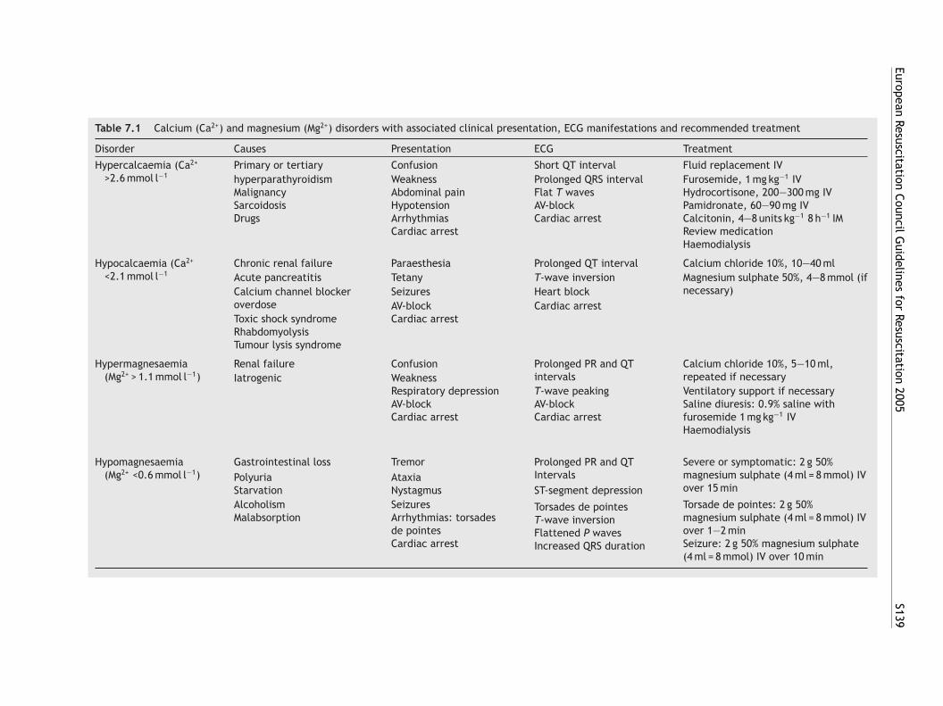
EuropeanResuscitation
CouncilGuidelines
forResuscitation
2005S139
Table 7.1 Calcium (Ca2+) and magnesium (Mg2+) disorders with associated clinical presentation, ECG manifestations and recommended treatment
Disorder Causes Presentation ECG Treatment
Hypercalcaemia (Ca2+
>2.6 mmol l−1Primary or tertiary Confusion Short QT interval Fluid replacement IVhyperparathyroidism Weakness Prolonged QRS interval Furosemide, 1 mg kg−1 IVMalignancy Abdominal pain Flat T waves Hydrocortisone, 200—300 mg IVSarcoidosis Hypotension AV-block Pamidronate, 60—90 mg IVDrugs Arrhythmias Cardiac arrest Calcitonin, 4—8 units kg−1 8 h−1 IM
Cardiac arrest Review medicationHaemodialysis
Hypocalcaemia (Ca2+
<2.1 mmol l−1Chronic renal failure Paraesthesia Prolonged QT interval Calcium chloride 10%, 10—40 mlAcute pancreatitis Tetany T-wave inversion Magnesium sulphate 50%, 4—8 mmol (if
necessary)Calcium channel blockeroverdose
Seizures Heart blockAV-block Cardiac arrest
Toxic shock syndromeRhabdomyolysis
Cardiac arrest
Tumour lysis syndrome
Hypermagnesaemia(Mg2+ > 1.1 mmol l−1)
Renal failure Confusion Prolonged PR and QTintervals
Calcium chloride 10%, 5—10 ml,repeated if necessaryIatrogenic Weakness
Respiratory depressionAV-blockCardiac arrest
T-wave peakingAV-blockCardiac arrest
Ventilatory support if necessarySaline diuresis: 0.9% saline withfurosemide 1 mg kg−1 IVHaemodialysis
Hypomagnesaemia(Mg2+ <0.6 mmol l−1)
Gastrointestinal loss Tremor Prolonged PR and QTIntervals
Severe or symptomatic: 2 g 50%magnesium sulphate (4 ml = 8 mmol) IVover 15 min
Polyuria AtaxiaStarvation Nystagmus ST-segment depressionAlcoholismMalabsorption
SeizuresArrhythmias: torsadesde pointesCardiac arrest
Torsade de pointes: 2 g 50%magnesium sulphate (4 ml = 8 mmol) IVover 1—2 minSeizure: 2 g 50% magnesium sulphate(4 ml = 8 mmol) IV over 10 min
Torsades de pointesT-wave inversionFlattened P wavesIncreased QRS duration
S140 J. Soar et al.
advice from a poisons centre.
• Activated charcoal is known to adsorb certaindrugs. Its value decreases over time after inges-tion. There is no evidence that ingestion of char-coal improves clinical outcome. According to evi-dence from volunteer studies, consider giving asingle dose of activated charcoal to patients whohave ingested a potentially toxic amount of poi-son (known to be adsorbed by activated charcoal)up to 1 h previously.17 Give it only to patientswith an intact or protected airway. Multiple dosesof activated charcoal can be beneficial in life-threatening poisoning with carbemazepine, dap-sone, phenobarbital, quinine and theophylline.
• Gastric lavage followed by activated charcoaltherapy is useful only within 1 h of ingesting thepoison.17 Generally this should be carried outafter tracheal intubation. Delayed gastric lavagehas very little effect on drug absorption and maypropel drugs further along the gastrointestinaltract.18 Do not give ipecacuanha syrup to inducevomiting; there is little evidence of benefit.19
• There is little evidence for the use of laxatives,e.g., lactulose or magnesium citrate, to enhance
20
or dicobalt edetate for cyanides; digoxin-specificFab antibodies for digoxin; flumazenil for ben-zodiazepines; and naloxone for opioids. Reversalof benzodiazepine intoxication with flumazenilis associated with significant toxicity in patientswith benzodiazepine dependence or co-ingestionof proconvulsant medications, such as tricyclicantidepressants.24 The routine use of flumazenilin the comatose patient with an overdose is notrecommended.
Specific antidotes
These guidelines will address only some causes ofcardiorespiratory arrest due to poisoning.
Opioid poisoning
Opioid poisoning commonly causes respiratorydepression followed by respiratory insufficiency orrespiratory arrest. The respiratory effects of opi-oids are reversed rapidly by the opiate antago-nist naloxone. In severe respiratory depression, theevidence shows fewer adverse events when air-way opening, oxygen administration and ventila-toeidcaInetIonoronda
ocarp
iwua
drug elimination from the gut.• Whole-bowel irrigation by enteral administra-
tion of a polyethylene glycol solution can reducedrug absorption by cleansing the gastrointesti-nal tract. It can be useful in cases of potentiallytoxic ingestion of sustained release or enteric-coated drugs, oral iron poisoning and the removalof ingested packets of illicit drugs.21
• Urine alkalinisation (pH 7.5) by giving IV sodiumbicarbonate can be useful in moderate-to-severesalicylate poisoning in patients who do not needhaemodialysis.22 Urine alkalinisation can also beuseful in tricyclic overdose (see below).
• Haemodialysis or haemoperfusion can be use-ful for elimination of specific life-threateningtoxins. Haemodialysis removes drugs or metabo-lites that are water soluble, have a low vol-ume of distribution and low plasma proteinbinding.23 It may be considered for poison-ing with methanol, ethylene glycol, salicylatesand lithium. Haemoperfusion involves passingblood through an absorptive-containing cartridge(usually charcoal). This technique removes sub-stances that have a high degree of plasma pro-tein binding. Charcoal haemoperfusion may beindicated for intoxications with carbamazepine,phenobarbital, phenytoin and theophylline.
• Specific antidotes (see below) which may beeffective include: N-acetylcysteine for parac-etamol; high-dose atropine for organophosphateinsecticides; sodium nitrite, sodium thiosulphate
ion are carried out before giving naloxone in casesf opioid-induced respiratory depression;25—30 how-ver, the use of naloxone can prevent the need forntubation. The preferred route for giving naloxoneepends on the skills of the rescuer: IV, intramus-ular (IM), subcutaneous (SC), endotracheal (ET)nd intranasal (IN) routes can be used. The non-V routes can be quicker because time is saved inot having to establish IV access, which can bextremely difficult in an IV drug abuser. The ini-ial doses of naloxone are 400 mcg IV,27 800 mcgM, 800 mcg SC,27 2 mg IN31 or 1—2 mg ET. Largepioid overdoses may require titration to a totalaloxone dose of 6—10 mg. The duration of actionf naloxone is approximately 45—70 min, but respi-atory depression can persist for 4—5 h after opioidverdose. Thus, the clinical effects of naloxone mayot last as long as those of a significant opioid over-ose. Titrate the dose until the victim is breathingdequately and has protective airway reflexes.
Acute withdrawal from opioids produces a statef sympathetic excess and may cause compli-ations, such as pulmonary oedema, ventricularrrhythmia and severe agitation. Use naloxoneeversal of opiate intoxication with caution inatients suspected of opioid dependence.
There is no good evidence that naloxonemproves outcome once cardiac arrest associatedith opioid toxicity has occurred. Cardiac arrest issually secondary to a respiratory arrest and associ-ted with severe brain hypoxia. Prognosis is poor.26
European Resuscitation Council Guidelines for Resuscitation 2005 S141
Giving naloxone is unlikely to be harmful. Once car-diac arrest has occurred, follow the standard resus-citation protocols.
Tricyclic antidepressants
Self-poisoning with tricyclic antidepressants is com-mon and can cause hypotension, seizures andarrhythmias. Anticholinergic effects include mydri-asis, fever, dry skin, delirium, tachycardia, ileusand urinary retention. Most life-threatening prob-lems occur within the first 6 h after ingestion. Awidening QRS complex indicates a greater risk ofarrhythmias. There is evidence to support the use ofsodium bicarbonate to treat arrhythmias induced bytricyclic antidepressants and/or hypotension.32—47
The exact threshold for starting treatment basedon QRS duration has not been established. No studyhas investigated the optimal target arterial or uri-nary pH with bicarbonate therapy, but an arterialpH of 7.45—7.55 has been commonly accepted andseems reasonable. Hypertonic saline may also beeffective in treating cardiac toxicity.48
Cocaine toxicity
SctmtccSddmbat
D
Sdbcosa(aabvd
tively with digoxin-specific antibody fragments.53
Antibody-specific therapy may also be effective inpoisoning from plants as well as Chinese herbalmedications containing digitalis glycosides.53—55
Vasopressors, inotropes, calcium, glucagon,phosphodiesterase inhibitors and insulin-glucosemay all be useful in beta-blocker and calcium chan-nel blocker overdose.56—58 Transcutaneous pacingmay be effective for severe bradycardia caused bypoisoning and overdose (see section 3).
Further treatment and prognosis
A long period of coma in a single position can causepressure sores and rhabdomyolysis. Measure elec-trolytes (particularly potassium), blood glucose andarterial blood gases. Monitor temperature becausethermoregulation is impaired. Both hypothermiaand hyperthermia (hyperpyrexia) can occur afterthe overdose of some drugs. Retain samples of bloodand urine for analysis.
Be prepared to continue resuscitation for a pro-longed period, particularly in young patients asthe poison may be metabolised or excreted duringextended life support measures.
i
•
••
7
O
DEshvlaiassar
E
Tm
ympathetic overstimulation associated withocaine toxicity may cause agitation, symptomaticachycardia, hypertensive crisis, hyperthermia andyocardial ischaemia with angina. Glyceryl trini-
rate and phentolamine reverse cocaine-inducedoronary vasoconstriction, labetalol has no signifi-ant effect, and propranolol makes it worse.49—52
mall doses of IV benzodiazepines (midazolam,iazepam, lorazepam) are effective first-linerugs. Use nitrates only as second-line therapy foryocardial ischaemia. Labetalol (alpha- and beta-locker) is useful for the treatment of tachycardiand hypertensive emergencies due to cocaineoxicity.
rug-induced severe bradycardia
evere bradycardia from poisoning or drug over-ose may be refractory to standard ALS protocolsecause of prolonged receptor binding or directellular toxicity. Atropine may be life saving inrganophosphate, carbamate or nerve agent poi-oning. Give atropine for bradycardia caused bycetylcholinesterase-inhibiting substances. Large2—4 mg) and repeated doses may be required tochieve a clinical effect. Isoprenaline may be usefult high doses in refractory bradycardia induced byeta-antagonist receptor blockade. Heart block andentricular arrhythmias associated with digoxin origitalis glycoside poisoning may be treated effec-
Alternative approaches which may be effectiven severely poisoned patients include:
higher doses of medication than in standard pro-tocols;non-standard drug therapies;prolonged CPR.
c. Drowning
verview
rowning is a common cause of accidental death inurope. The most important and detrimental con-equence of drowning is hypoxia. The duration ofypoxia is the critical factor in determining theictim’s outcome. Therefore, oxygenation, venti-ation and perfusion should be restored as rapidlys possible. Immediate resuscitation at the scenes essential for survival and neurological recoveryfter drowning. This will require bystander provi-ion of CPR plus immediate activation of the EMSystem. Victims who have spontaneous circulationnd breathing when they reach hospital usuallyecover with good outcomes.
pidemiology
he World Health Organization (WHO) esti-ates that, worldwide, drowning accounts for
S142 J. Soar et al.
approximately 450,000 deaths each year. A further1.3 million disability-adjusted life-years are losteach year as a result of premature death or disabil-ity from drowning;59 97% of deaths from drowningoccur in low- and middle-income countries.59 In2002, there were 427 deaths from drowning in theUnited Kingdom (Royal Society for the Preventionof Accidents 2002) and 4073 in the United States(National Center for Injury Prevention 2002),yielding an annual incidence of drowning of 0.8and 1.45 per 100,000 population, respectively.Death from drowning is more common in youngmales and is the leading cause of accidental deathin Europe in this group.59 Alcohol consumption is acontributory factor in up to 70% of drownings.60
The guidelines in this chapter focus on the treat-ment of the individual drowning victim rather thanthe management of mass casualty aquatic inci-dents.
Definitions, classifications and reporting
Over 30 different terms have been used to describethe process and outcome from submersion- andimmersion-related incidents.61 To improve clarity
tim, reaching with a rescue aid (e.g., stick or cloth-ing), or throwing a rope or buoyant rescue aid maybe effective if the victim is close to dry land. Alter-natively, use a boat or other water vehicle to assistwith the rescue. Avoid entry into the water when-ever possible. If entry into the water is essential,take a buoyant rescue aid or flotation device.
Remove all the drowning victims from the waterby the fastest and safest means available and resus-citate as quickly as possible. The incidence ofcervical spine injury in drowning victims is low(approximately 0.5%).63 Spinal immobilisation canbe difficult to perform in the water and can delayremoval from the water and adequate resuscita-tion of the victim. Poorly applied cervical collarscan also cause airway obstruction in unconsciouspatients.64 Despite potential spinal injury, victimswho are pulseless and apnoeic should be removedfrom water as quickly as possible (even if a backsupport device is not available), while attempt-ing to limit neck flexion and extension. Cervicalspine immobilisation is not indicated unless signs ofsevere injury are apparent or the history is consis-tent with the possibility of severe injury.65 Thesecircumstances include a history of diving, water-sctrc
R
TdPptaswttmwdamnw
imitt
and to help comparability of future scientific andepidemiological reports, the International LiaisonCommittee on Resuscitation (ILCOR) has proposednew definitions related to drowning.62 Drowningitself is defined as a process resulting in primary res-piratory impairment from submersion/immersion ina liquid medium. Implicit in this definition is thata liquid/air interface is present at the entranceof the victim’s airway, preventing the victim frombreathing air. The victim may live or die after thisprocess, but whatever the outcome, he or she hasbeen involved in a drowning incident. Immersionmeans to be covered in water or other fluid. Fordrowning to occur, usually at least the face andairway must be immersed. Submersion implies thatthe entire body, including the airway, is under thewater or other fluid.
ILCOR recommends that the following terms,previously used, should no longer be used: dry andwet drowning, active and passive drowning, silentdrowning, secondary drowning and drowned versusnear-drowned.62
Basic life support
Aquatic rescue and recovery from the water
Always be aware of personal safety and minimisethe danger to yourself and the victim at all times.Whenever possible, attempt to save the drowningvictim without entry into water. Talking to the vic-
lide use, signs of trauma or signs of alcohol intoxi-ation. Whenever possible, remove the victim fromhe water in a horizontal position to minimise theisks of post-immersion hypotension and cardiovas-ular collapse.66
escue breathing
he first and most important treatment for therowning victim is alleviation of hypoxaemia.rompt initiation of rescue breathing or positiveressure ventilation increases the survival.67,68 Inhe apnoeic victim, start rescue breathing as soons the victim’s airway is opened and the rescuer’safety ensured. This can sometimes be achievedhen the victim is still in shallow water. It is likely
o be difficult to pinch the victim’s nose, so mouth-o-nose ventilation may be used as an alternative toouth-to-mouth ventilation. If the victim is in deepater, start in-water rescue breathing if trained too so, ideally with the support of a buoyant rescueid,69 although in-water, unsupported resuscitationay also be possible.70 Untrained rescuers should
ot attempt to perform any form of resuscitationith a victim in deep water.If there is no spontaneous breathing after open-
ng the airway, give rescue breaths for approxi-ately 1 min.69 If the victim does not start breath-
ng spontaneously, further management depends onhe distance from land. If the victim can be broughto land in <5 min of rescue time, continue rescue
European Resuscitation Council Guidelines for Resuscitation 2005 S143
breaths while towing. If more than an estimated5 min from land, give further rescue breaths over1 min, then bring the victim to land as quickly aspossible without further attempts at ventilation.69
There is no need to clear the airway of aspi-rated water. The majority of drowning victims aspi-rate only a modest amount of water, and this isabsorbed rapidly into the central circulation. Anattempt to remove water from the air passagesby any means other than suction is unnecessaryand dangerous. Abdominal thrusts cause regurgi-tation of gastric contents and subsequent aspira-tion. They have been associated with other life-threatening injuries and should not be performedunless there are clear signs of foreign-body airwayobstruction.71
Chest compression
As soon as the victim is removed from water,check for breathing. A healthcare professional whois trained in pulse checking may also check forpulse, but this may be even more difficult to findin a drowning victim, particularly if cold. If thevictim is not breathing, start chest compressionsiw
D
Iattst≤aa
R
RlpsttItdsngr
Advanced life support
Airway and breathing
Give high-flow oxygen during the initial assess-ment of the spontaneously breathing drowningvictim. Consider non-invasive ventilation or contin-uous positive airway pressure if the victim fails torespond to treatment with high-flow oxygen.76 Usepulse oximetry and arterial blood gas analysis totitrate the concentration of inspired oxygen andto provide an indicator of the adequacy of ven-tilation. Consider early intubation and controlledventilation for victims who fail to respond to theseinitial measures or who have a reduced level ofconsciousness. Take care to ensure optimal preoxy-genation before intubation. Use a rapid-sequenceinduction with cricoid pressure to reduce the highrisk of aspiration.77 Protect the airway of the victimin cardiopulmonary arrest early in the resuscita-tion attempt, ideally with a tracheal tube. Reducedpulmonary compliance requiring high inflation pres-sures may limit the use of adjuncts, such as thelaryngeal mask airway. Initiate ventilation with ahigh-inspired oxygen concentration as soon as pos-st
C
Fsatbml
bstwom
D
Mefioiiift
mmediately. Chest compression is ineffective inater.72,73
efibrillation
f the victim is unresponsive and not breathing andn AED is available, attach it to the victim andurn it on. Before attaching the AED pads, dryhe victim’s chest to enable adherence. Deliverhocks according to the AED prompts. If the vic-im is hypothermic with a core body temperature30 ◦C (86 ◦F), limit defibrillation to a total of threettempts until the core body temperature risesbove 30 ◦C (86 ◦F).74
egurgitation during resuscitation
egurgitation of stomach contents is common fol-owing resuscitation from drowning and will com-licate efforts to maintain a patent airway. In onetudy, regurgitation occurred in two-thirds of vic-ims who received rescue breathing and 86% of vic-ims who required compression and ventilation.75
f regurgitation occurs, turn the victim’s mouth tohe side and remove the regurgitated material usingirected suction if possible. If spinal cord injury isuspected, log-roll the victim, keeping the head,eck and torso aligned, before aspirating the regur-itated material. Log-rolling will require severalescuers.
ible, to treat the severe hypoxaemia that is likelyo be present.
irculation and defibrillation
ollow standard advanced life support protocols. Ifevere hypothermia is present (core body temper-ture ≤30 ◦C or 86 ◦F), limit defibrillation attemptso three, and withhold IV drugs until the coreody temperature increases above these levels. Ifoderate hypothermia is present, give IV drugs at
onger than standard intervals (see section 7d).During prolonged immersion, victims may
ecome hypovolaemic from the hydrostatic pres-ure of water on the body. Give IV fluid to correcthe hypovolaemia but avoid excessive volumes,hich may cause pulmonary oedema. After returnf spontaneous circulation, use haemodynamiconitoring to guide fluid resuscitation.
iscontinuing resuscitation efforts
aking a decision to discontinue resuscitationfforts on a victim of drowning is notoriously dif-cult. No single factor can accurately predict goodr poor survival with 100% certainty. Decisions maden the field frequently prove later to have beenncorrect.78 Continue resuscitation unless theres clear evidence that resuscitation attempts areutile (e.g., massive traumatic injuries, rigor mor-is, putrefaction etc.), or timely evacuation to
S144 J. Soar et al.
a medical facility is not possible. Neurologicallyintact survival has been reported in several victimssubmerged for greater than 60 min.79,80
Post-resuscitation care
Salt versus fresh water
Much attention has been focused in the past on dif-ferences between salt- and fresh-water drowning.Extensive data from animal studies and human caseseries have shown that, irrespective of the tonicityof the inhaled fluid, the predominant pathophys-iological process is hypoxaemia, driven by surfac-tant wash-out and dysfunction, alveolar collapse,atelectasis and intrapulmonary shunting. Small dif-ferences in electrolyte disturbance are rarely of anyclinical relevance and do not usually require treat-ment.
Lung injury
Victims of drowning are at high risk of develop-ing acute respiratory distress syndrome (ARDS) forupto 72 h after submersion. Protective ventilationstrategies improve survival in patients with ARDS.81
In contrast, there is evidence of benefit frominduced hypothermia for comatose victims resus-citated from prehospital cardiac arrests.83 To date,there is no convincing evidence to guide therapyin this patient group. A pragmatic approach mightbe to consider instituting active rewarming until acore temperature of 32—34 ◦C is achieved, and thenactively to avoid hyperthermia (>37 ◦C) during thesubsequent period of intensive care (InternationalLife Saving Federation, 2003).
Other supportive care
Attempts have been made to improve neurologi-cal outcome following drowning with the use ofbarbiturates, intracranial pressure (ICP) monitoringand steroids. None of these interventions has beenshown to alter the outcome. In fact, signs of highICP serve as a symptom of significant neurologicalhypoxic injury, and no evidence that attempts toalter the ICP will affect the outcome.65
7d. Hypothermia
D
Htm(ptcWifmipetp
tIhntsb
D
Bp
The propensity towards alveolar collapse mayrequire the use of PEEP or other alveolar recruit-ment manoeuvres to reverse severe hypoxaemia.82
Extracorporeal membrane oxygenation and nitricoxide administration have been used in some cen-tres for refractory hypoxaemia in drowning victimsbut the efficacy of these treatments is unproven.65
Pneumonia is common after drowning. Prophy-lactic antibiotics have not been shown to be ofbenefit, although they may be considered aftersubmersion in grossly contaminated water such assewage. Give broad-spectrum antibiotics if signs ofinfection develop subsequently.65
Hypothermia
Victims of submersion may develop primary orsecondary hypothermia. If the submersion occursin icy water (<5 ◦C or 41 ◦F), hypothermia maydevelop rapidly and provide some protectionagainst hypoxia. Such effects, however, have typ-ically been reported after submersion of childrenin icy water.59 Hypothermia may also develop asa secondary complication of the submersion, andsubsequent heat loss through evaporation dur-ing attempted resuscitation. In these victims thehypothermia is not protective (see section 7d).
Several small clinical studies in patients withaccidental hypothermia have shown that survivalmay be improved by either passive or active warm-ing out of hospital or in the emergency room.65
efinition
ypothermia exists when the body core tempera-ure is below 35 ◦C and is classified arbitrarily asild (35—32 ◦C), moderate (32—30 ◦C) or severe
less than 30 ◦C). Hypothermia can occur in peo-le with normal thermoregulation who are exposedo cold environments, particularly wet or windyonditions, or following immersion in cold water.hen thermoregulation is impaired, for example,
n the elderly and very young, hypothermia mayollow a mild cold insult. The risk of hypother-ia is also increased by drug or alcohol ingestion,
llness, injury or neglect. Hypothermia may be sus-ected from the clinical history or a brief externalxamination of a collapsed patient. A low-readinghermometer is needed to measure the core tem-erature and confirm diagnosis.
In some cases, hypothermia may exert a protec-ive effect on the brain after cardiac arrest.84,85
ntact neurological recovery may be possible afterypothermic cardiac arrest, although those withon-asphyxial arrest have a better prognosis thanhose with asphyxial hypothermic arrest.86—88 Life-aving procedures should not be withheld on theasis of clinical presentation alone.87
ecision to resuscitate
eware of pronouncing death in a hypothermicatient, as cold alone may produce a very slow,
European Resuscitation Council Guidelines for Resuscitation 2005 S145
small-volume, irregular pulse and an unrecordableblood pressure. Hypothermia protects the brain andvital organs, and associated arrhythmias are poten-tially reversible either before or during rewarming.At 18 ◦C the brain can tolerate periods of circula-tory arrest for 10 times longer than at 37 ◦C. Dilatedpupils can be caused by a variety of insults and mustnot be taken as a sign of death.
On discovering a hypothermic cardiac arrestvictim in cold environment, it is not always easyto distinguish between primary and secondaryhypothermia. Cardiac arrest could be caused pri-marily by hypothermia, or hypothermia could fol-low a normothermic cardiac arrest (e.g., cardiacarrest caused by myocardial ischaemia in a personin cold environment).
Do not confirm death until the patient has beenrewarmed or until attempts to raise the core tem-perature have failed; prolonged resuscitation maybe necessary. In the prehospital setting, resusci-tation should be withheld only if the patient hasobvious lethal injuries or if the body is completelyfrozen making resuscitation attempts impossible.89
In the hospital setting, use clinical judgment todetermine when to stop resuscitating a hypother-
Use the same ventilation and chest compressionrates as for a normothermic patient. Hypothermiacan cause stiffness of the chest wall, making venti-lation and chest compression difficult.
The hypothermic heart may be unresponsiveto cardioactive drugs, attempted electrical pac-ing and attempted defibrillation. Drug metabolismis slowed, leading to potentially toxic plasmaconcentrations of any drug given repeatedly.90
The evidence for the efficacy of drugs in severehypothermia is limited and based mainly on animalstudies. Adrenaline may be effective in increas-ing coronary perfusion pressure, but not survival,in severe hypothermic cardiac arrest.93,94 The effi-cacy of amiodarone is also reduced.95 For thesereasons, withhold adrenaline and other drugs untilthe patient has been warmed to a temperaturegreater than 30 ◦C. Once 30 ◦C has been reached,the intervals between doses should be doubled. Asthe patient’s temperature returns towards normal,the standard drug protocols should be used.
Remember to rule out other primary causes ofcardiorespiratory arrest using the four Hs and fourTs approach (e.g., drug overdose, hypothyroidism,trauma).
A
Ab(fip
trmanbpa
fiieVtuAi
R
Gf
mic arrest victim.
Resuscitation
All the principles of prevention, basic and advancedlife support apply to the hypothermic patient. Donot delay the urgent procedures, such as intu-bation and insertion of vascular catheters. Intu-bation can provoke VF in a patient with severehypothermia.87,90
• Clear the airway and, if there is no spontaneousrespiratory effort, ventilate the patient’s lungswith high concentrations of oxygen. If possible,use warmed (40—46 ◦C) and humidified oxygen.Consider careful tracheal intubation when indi-cated according to the ALS algorithm.
• Palpate a major artery and, if available, look atthe ECG for up to 1 min and look for signs of lifebefore concluding that there is no cardiac out-put. If a Doppler ultrasound probe is available,use it to establish whether there is peripheralblood flow. If the victim is pulseless, start chestcompressions immediately. If there is any doubtabout whether a pulse is present, start CPR.
• Once resuscitation is under way, confirmhypothermia with a low-reading thermometer.The method of temperature measurementshould be the same throughout resuscitation andrewarming. Use oesophageal, bladder, rectal ortympanic temperature measurements.91,92
rrhythmias
s the body core temperature decreases, sinusradycardia tends to give way to atrial fibrillationAF) followed by ventricular fibrillation (VF) andnally asystole.96 Follow the standard treatmentrotocols.
Severely hypothermic victims (core tempera-ure <30 ◦C) in cardiac arrest in hospital must beapidly rewarmed using internal methods. Arrhyth-ias other than VF tend to revert spontaneously
s the core temperature increases, and usually doot require immediate treatment. Bradycardia maye physiological in severe hypothermia, and cardiacacing is not indicated unless bradycardia persistsfter rewarming.
The temperature at which defibrillation shouldrst be attempted and how often it should be tried
n the severely hypothermic patient has not beenstablished. AEDs may be used on these patients. IfF is detected, give a shock; if VF/VT persists afterhree shocks, delay further defibrillation attemptsntil the core temperature is above 30 ◦C.97,98 If anED is used, follow the AED prompts while rewarm-
ng the patient.
ewarming
eneral measures for all casualties include removalrom the cold environment, prevention of further
S146 J. Soar et al.
heat loss and rapid transfer to the hospital. Removecold or wet clothing as soon as possible. Cover thedry casualties with blankets and keep them out ofthe wind.
Rewarming may be passive external, activeexternal, or active internal. Passive warming isachieved with blankets and a warm room, and issuitable for conscious victims with mild hypother-mia. In severe hypothermia or cardiac arrest,active warming is required, but this must notdelay transport to a hospital where more advancedrewarming techniques are available. Several tech-niques have been described, although there are noclinical trials of outcome to determine the bestrewarming method. Studies show that forced airrewarming and warm IV fluids are effective inpatients with severe hypothermia and a perfus-ing rhythm.99,100 Other warming techniques includethe use of warm humidified gases, gastric, peri-toneal, pleural or bladder lavage with warm fluids(at 40 ◦C), and extracorporeal blood warming withpartial bypass.87,90,101—103
In the patient with cardiac arrest and hypother-mia, cardiopulmonary bypass is the preferredmethod of active internal rewarming because it
tions, or secondary to endogenous heat produc-tion.
Environment-related hyperthermia occurs whereheat, usually in the form of radiant energy, isabsorbed by the body at a rate faster than that canbe lost by thermoregulatory mechanisms. Hyper-thermia occurs along a continuum of heat-relatedconditions, starting with heat stress, progressingto heat exhaustion, to heat stroke (HS) and finallymultiorgan dysfunction and cardiac arrest in someinstances.108
Malignant hyperthermia (MH) is a rare disorder ofskeletal muscle calcium homeostasis characterisedby muscle contracture and life-threatening hyper-metabolic crisis following exposure of geneticallypredisposed individuals to halogenated anaesthet-ics and depolarising muscle relaxants.109,110
The key features and treatment of heat stressand heat exhaustion are included in Table 7.2.
Heat stroke (HS)
HS is a systemic inflammatory response with acore temperature above 40.6 ◦C, accompanied bymental state change and varying levels of organ
also provides circulation, oxygenation and ventila-tion, while the core body temperature is increasedgradually.104,105 Survivors in one case series had anaverage of 65 min of conventional CPR before car-diopulmonary bypass.105 Unfortunately, facilitiesfor cardiopulmonary bypass are not always avail-able and a combination of methods may have to beused.
During rewarming, patients will require largevolumes of fluids as their vascular space expandswith vasodilation. Warm all the IV fluids. Use con-tinuous haemodynamic monitoring and, if possi-ble, treat the patient in a critical care unit. Avoidhyperthermia during and after the warming period.Although there are no formal studies, once ROSChas been achieved use standard strategies for post-resuscitation care, including mild hypothermia ifappropriate (section 4g). There is no evidencefor the routine use of steroids, barbiturates orantibiotics.106,107
7e. Hyperthermia
Definition
Hyperthermia occurs when the body’s abilityto thermoregulate fails, and core temperatureexceeds the one that is normally maintainedby homeostatic mechanisms. Hyperthermia maybe exogenous, caused by environmental condi-
dysfunction. There are two forms of HS: clas-sic non-exertion heat stroke (CHS) occuring dur-ing high environmental temperatures and ofteneffecting the elderly during heat waves;111 exer-tion heat stroke (EHS) occuring during strenu-ous physical exercise in high environmental tem-peratures and/or high humidity usually effectinghealthy young adults.112 Mortality from HS rangesbetween 10 and 50%.113
Predisposing factors
The elderly are at an increased risk for heat-relatedillness because of underlying illness, medicationuse, declining thermoregulatory mechanisms andlimited social support. There are several risk fac-tors: lack of acclimatisation, dehydration, obe-sity, alcohol, cardiovascular disease, skin condi-tions (psoriasis, eczema, scleroderma, burn, cys-tic fibrosis), hyperthyroidism, phaeochromocytomaand drugs (anticholinergics, diamorphine, cocaine,amphetamine, phenothiazines, sympathomimetics,calcium channel blockers, beta-blockers).
Clinical presentation
Heat stroke can resemble septic shock and may becaused by similar mechanisms.114 Features include:
• core temperature 40.6 ◦C or more;• hot, dry skin (sweating is present in about 50% of
cases of exertional heat stroke);
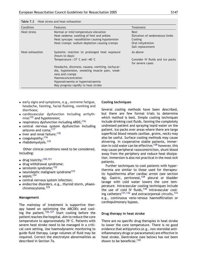
European Resuscitation Council Guidelines for Resuscitation 2005 S147
Table 7.2 Heat stress and heat exhaustion
Condition Features Treatment
Heat stress Normal or mild temperature elevation RestHeat oedema: swelling of feet and ankles Elevation of oedematous limbsHeat syncope: vasodilation causing hypotension CoolingHeat cramps: sodium depletion causing cramps Oral rehydration
Salt replacement
Heat exhaustion Systemic reaction to prolonged heat exposure(hours to days)
As above
Temperature >37 ◦C and <40 ◦C Consider IV fluids and ice packsfor severe cases
Headache, dizziness, nausea, vomiting, tachycar-dia, hypotension, sweating muscle pain, weak-ness and crampsHaemoconcentrationHyponatraemia or hypernatraemiaMay progress rapidly to heat stroke
• early signs and symptoms, e.g., extreme fatigue,headache, fainting, facial flushing, vomiting anddiarrhoea;
• cardiovascular dysfunction including arrhyth-mias115 and hypotension;
• respiratory dysfunction including ARDS;116
• central nervous system dysfunction includingseizures and coma;117
• liver and renal failure;118
• coagulopathy;116
• rhabdomyolysis.119
Other clinical conditions need to be considered,including:
• drug toxicity;120,121
• drug withdrawal syndrome;• serotonin syndrome;122
• neuroleptic malignant syndrome123
• sepsis;124
• central nervous system infection;• endocrine disorders, e.g., thyroid storm, phaeo-
chromocytoma.125
Management
Taiptscgrd
Cooling techniques
Several cooling methods have been described,but there are few formal trials to determinewhich method is best. Simple cooling techniquesinclude drinking cool fluids, fanning the completelyundressed patient and spraying tepid water on thepatient. Ice packs over areas where there are largesuperficial blood vessels (axillae, groins, neck) mayalso be useful. Surface cooling methods may causeshivering. In cooperative stable patients, immer-sion in cold water can be effective;128 however, thismay cause peripheral vasoconstriction, shunt bloodaway from the periphery and reduce heat dissipa-tion. Immersion is also not practical in the most sickpatients.
Further techniques to cool patients with hyper-thermia are similar to those used for therapeu-tic hypothermia after cardiac arrest (see section4g). Gastric, peritoneal,129 pleural or bladderlavage with cold water lowers the core tem-perature. Intravascular cooling techniques includethe use of cold IV fluids,130 intravascular cool-ing catheters131,132 and extracorporeal circuits,133
e.g., continuous veno-venous haemofiltration or
he mainstay of treatment is supportive ther-py based on optimising the ABCDEs and cool-ng the patient.126,127 Start cooling before theatient reaches the hospital. Aim to reduce the coreemperature to approximately 39 ◦C. Patients withevere heat stroke need to be managed in a criti-al care setting. Use haemodynamic monitoring touide fluid therapy. Large volumes of fluid may beequired. Correct the electrolyte abnormalities asescribed in Section 7a.
cardiopulmonary bypass.
Drug therapy in heat stroke
There are no specific drug therapies in heat stroketo lower the core temperature. There is no goodevidence that antipyretics (e.g., non-steroidal anti-inflammatory drugs or paracetamol) are effective inheat stroke. Dantrolene (see below) has not beenshown to be beneficial.134
S148 J. Soar et al.
Malignant hyperthermia (MH)
MH is a life-threatening genetic sensitivity of skele-tal muscles to volatile anaesthetics and depolaris-ing neuromuscular blocking drugs, occurring duringor after anaesthesia. Stop triggering agents imme-diately; give oxygen, correct acidosis and elec-trolyte abnormalities. Start active cooling and givedantrolene.135
Modifications to cardiopulmonaryresuscitation and post-resuscitation care
There are no specific studies on cardiac arrest inhyperthermia. If cardiac arrest occurs, follow stan-dard procedures for basic and advanced life sup-port and cool the patient. There are no data onthe effects of hyperthermia on defibrillation thresh-old; therefore, attempt defibrillation according tocurrent guidelines, while continuing to cool thepatient. Animal studies suggest that the progno-sis is poor compared with normothermic cardiacarrest.136,137 The risk of unfavourable neurologicaloutcome increases for each degree of body tem-perature >37 ◦C.138 Provide post-resuscitation care
(e.g., beta-adrenergic agonists, aminophylline)or electrolyte abnormalities;
• dynamic hyperinflation, i.e., autopositive end-expiratory pressure (auto-PEEP), can occur inmechanically ventilated asthmatics. Auto-PEEPis caused by air trapping and ‘breath stack-ing’ (breathed air entering and being unable toescape). Gradual build-up of pressure occurs andreduces blood flow and blood pressure;
• tension pneumothorax (often bilateral).
The four Hs and four Ts approach to reversiblecauses helps identify these causes in cardiac arrest.
Diagnosis
Wheezing is a common physical finding, but sever-ity does not correlate with the degree of airwayobstruction. The absence of wheezing may indi-cate critical airway obstruction, whereas increasedwheezing may indicate a positive response to bron-chodilator therapy. SaO2 may not reflect progres-sive alveolar hypoventilation, particularly if oxygenis being given. The SaO2 may initially decrease dur-ing the therapy because beta-agonists cause bothbronchodilation and vasodilation and may increase
according to the normal guidelines.
7f. Asthma
Introduction
Approximately 300 million people of all agesand ethnic backgrounds suffer from asthmaworldwide.139 Asthma still causes many deaths inyoung adults, mostly among those with chronicsevere asthma, adverse psychosocial circumstancesand poor medical management. National and inter-national guidance for the management of asthmaalready exists.139,140 The following guidelines focuson the treatment of patients with near-fatal asthmaand cardiac arrest.
Causes of cardiac arrest
Cardiac arrest in the asthmatic person is often aterminal event after a period of hypoxaemia; occa-sionally, it may be sudden. Cardiac arrest in asth-matics has been linked to:
• severe bronchospasm and mucous plugging lead-ing to asphyxia (this condition causes the vastmajority of asthma-related deaths);
• cardiac arrhythmias caused by hypoxia, which isthe common cause of asthma-related arrhythmia.Arrhythmias can be caused by stimulant drugs
intrapulmonary shunting initially.Other causes of wheezing include: pulmonary
oedema, chronic obstructive pulmonary disease(COPD), pneumonia, anaphylaxis,141 pneumonia,foreign bodies, pulmonary embolism, bronchiecta-sis and subglottic mass.142
The severity of an asthma attack is defined inTable 7.3.
Key interventions to prevent arrest
The patient with severe asthma requires aggres-sive medical management to prevent deteriora-tion. Base assessment and treatment on an ABCDEapproach. Experienced clinicians should treat thesehigh-risk patients in a critical care area. The spe-cific drugs and the treatment sequence will varyaccording to local practice.
Oxygen
Use a concentration of inspired oxygen that willachieve an SaO2 ≥92%. High-flow oxygen by maskis sometimes necessary. Consider rapid-sequenceinduction and tracheal intubation if, despite effortsto optimise drug therapy, the patient has:
• decreased conscious level, coma;• profuse sweating;• reduced muscle tone (clinical signs of hypercar-
bia);
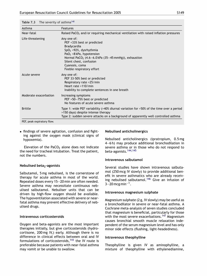
European Resuscitation Council Guidelines for Resuscitation 2005 S149
Table 7.3 The severity of asthma140
Asthma Features
Near-fatal Raised PaCO2 and/or requiring mechanical ventilation with raised inflation pressures
Life-threatening Any one of:PEF <33% best or predictedBradycardiaSpO2 <92%, dysrhythmiaPaO2 <8 kPa, hypotensionNormal PaCO2 (4.6—6.0 kPa (35—45 mmHg)), exhaustionSilent chest, confusionCyanosis, comaFeeble respiratory effort
Acute severe Any one of:PEF 33-50% best or predictedRespiratory rate >25/minHeart rate >110/minInability to complete sentences in one breath
Moderate exacerbation Increasing symptomsPEF >50—75% best or predictedNo features of acute severe asthma
Brittle Type 1: wide PEF variability (>40% diurnal variation for >50% of the time over a period>150 days) despite intense therapyType 2: sudden severe attacks on a background of apparently well controlled asthma
PEF, peak expiratory flow.
• findings of severe agitation, confusion and fight-ing against the oxygen mask (clinical signs ofhypoxemia).
Elevation of the PaCO2 alone does not indicatethe need for tracheal intubation. Treat the patient,not the numbers.
Nebulised beta2-agonists
Salbutamol, 5 mg nebulised, is the cornerstone oftherapy for acute asthma in most of the world.Repeated doses every 15—20 min are often needed.Severe asthma may necessitate continuous neb-ulised salbutamol. Nebuliser units that can bedriven by high-flow oxygen should be available.The hypoventilation associated with severe or near-fatal asthma may prevent effective delivery of neb-ulised drugs.
Intravenous corticosteroids
Oxygen and beta-agonists are the most importanttherapies initially, but give corticosteroids (hydro-cortisone, 200 mg IV,) early. Although there is no
Nebulised anticholinergics
Nebulised anticholinergics (ipratropium, 0.5 mg4—6 h) may produce additional bronchodilation insevere asthma or in those who do not respond tobeta-agonists.144,145
Intravenous salbutamol
Several studies have shown intravenous salbuta-mol (250 mcg IV slowly) to provide additional ben-efit in severe asthmatics who are already receiv-ing nebulised salbutamol.146 Give an infusion of3—20 mcg min−1.
Intravenous magnesium sulphate
Magnesium sulphate (2 g, IV slowly) may be useful asa bronchodilator in severe or near-fatal asthma. ACochrane meta-analysis of seven studies concludedthat magnesium is beneficial, particularly for thosewith the most severe exacerbations.147 Magnesiumcauses bronchial smooth muscle relaxation inde-pendent of the serum magnesium level and has onlyminor side effects (flushing, light-headedness).
I
Tm
difference in clinical effects between oral and IVformulations of corticosteroids,143 the IV route ispreferable because patients with near-fatal asthmamay vomit or be unable to swallow.
ntravenous theophylline
heophylline is given IV as aminophylline, aixture of theophylline with ethylenediamine,
S150 J. Soar et al.
which is 20 times more soluble than theophyllinealone. Aminophylline should only be consideredin severe or near-fatal asthma. A loading doseof 5 mg kg−1 is given over 20—30 min (unless onmaintenance therapy), followed by an infusion of500—700 mcg kg−1 h−1. Addition of this drug to highdoses of beta-agonists increases side effects morethan it increases bronchodilation. Check levels toavoid toxicity.
Subcutaneous or intramuscular adrenaline andterbutaline
Adrenaline and terbutaline are adrenergic agentsthat may be given subcutaneously to patientswith acute severe asthma. The dose of subcuta-neous adrenaline is 300 mcg up to a total of threedoses at 20-min intervals. Adrenaline may cause anincrease in heart rate, myocardial irritability andincreased oxygen demand; however, its use (evenin patients over 35 years old) is well tolerated.148
Terbutaline is given in a dose of 250 mcg subcu-taneously, which can be repeated in 30—60 min.These drugs are more commonly given to childrenwith acute asthma and, although most studies have
Non-invasive ventilation
Non-invasive ventilation decreases the intubationrate and mortality in COPD;154 however, its rolein patients with severe acute asthma is uncertain.Although promising, a recent Cochrane review sug-gests that more studies are needed.155
Management of cardiac arrest
Basic life support
Give basic life support according to the standardguidelines. Ventilation will be difficult because ofincreased airway resistance; try to prevent gastricinflation.
Advanced life support
Modifications to standard ALS guidelines. Con-sider the need for intubation early. The peakairway pressures recorded during the ventila-tion of patients with severe asthma (mean67.8 ± 11.1 cmH20 in 12 patients) are significantlyhigher than the normal lower oesophageal sphinc-t 156
ataits
(fcttdflmbmwu
rsdttnipus
shown them to be equally effective,149 one studyconcluded that terbutaline was superior.150 Thesealternative routes may need to be considered whenIV access is impossible.
Intravenous fluids
Severe or near-fatal asthma is associated withdehydration and hypovolaemia, and this will fur-ther compromise the circulation in patients withdynamic hyperinflation of the lungs. If there is evi-dence of hypovolaemia or dehydration, give IV flu-ids.
Heliox
Heliox is a mixture of helium and oxygen (usually80:20 or 70:30). A recent meta-analysis of fourclinical trials did not support the use of helioxin the initial treatment of patients with acuteasthma.151
Ketamine
Ketamine is a parenteral dissociative anaestheticwith bronchodilatory properties. One case seriessuggested substantial effectiveness,152 but the sin-gle randomised trial published to date showedno benefit to ketamine compared with standardcare.153
er pressure (approximately 20 cmH20). There issignificant risk of gastric inflation and hypoven-
ilation of the lungs when attempting to ventilatesevere asthmatic without a tracheal tube. Dur-
ng cardiac arrest this risk is even higher becausehe lower oesophageal sphincter pressure is sub-tantially less than normal.157
The new recommended respiratory rate10 breaths min−1) and tidal volume requiredor a normal chest rise during CPR should notause dynamic hyperinflation of the lungs (gasrapping). Tidal volume depends on the inspiratoryime and inspiratory flow, while lung emptyingepends on the expiratory time and expiratoryow. In mechanically ventilated severe asth-atics, increasing the expiratory time (achievedy reducing the respiratory rate) provides onlyoderate gains in terms of reduced gas trappinghen a minute volume of less than 10 l min−1 issed.156
There is limited evidence from the caseeports of unexpected ROSC in patients withuspected gas trapping when the tracheal tube isisconnected.158—161 If dynamic hyperinflation ofhe lungs is suspected during CPR, compression ofhe chest wall and/or a period of apnoea (discon-ection of tracheal tube) may relieve gas trappingf dynamic hyperinflation occurs. Although thisrocedure is supported by limited evidence, it isnlikely to be harmful in an otherwise desperateituation.
European Resuscitation Council Guidelines for Resuscitation 2005 S151
Dynamic hyperinflation increases transthoracicimpedance.162 Consider the higher shock energiesfor defibrillation if initial defibrillation attemptsfail.
There is no good evidence for the use of open-chest cardiac compressions in patients with asthma-associated cardiac arrest. Working through the fourHs and four Ts will identify potentially reversiblecourses of asthma related cardiac arrest. Tensionpneumothorax can be difficult to diagnose in car-diac arrest; it may be indicated by unilateral expan-sion of the chest wall, shifting of the trachea andsubcutaneous emphysema. Release air from thepleural space with needle decompression. Insert alarge-gauge cannula in the second intercostal spacein the mid clavicular line, being careful to avoiddirect puncture of the lung. If air is emitted, inserta chest tube. Always consider bilateral pneumoth-oraces in asthma-related cardiac arrest.
Post-resuscitation care
The following should be added to usual manage-ment after ROSC:
•
•
•
•
7
I
Acotgrc
Anaphylaxis is a severe life-threatening, gen-eralised or systemic hypersensitivity reaction.Investigations will show whether the reactionis allergic (immunoglobulin E (IgE) or non IgEmediated) or non-allergic anaphylaxis. The termanaphylactoid reaction is no longer used. An ana-phylactic reaction is generally defined as a severe,systemic allergic reaction characterized by multi-system involvement, including the airway, vascularsystem, gastrointestinal tract and skin. Severecases may cause complete airway obstructionsecondary to laryngeal oedema, bronchospasm,hypotension, cardiovascular collapse and death.Other symptoms include rhinitis, conjunctivitis,abdominal pain, vomiting, diarrhoea and a senseof impending doom. There is also usually a colourchange; the patient may appear either flushed orpale. Anaphylactic reactions vary in severity, andprogress may be rapid, slow or (unusually) biphasic.Rarely, manifestations may be delayed (this mayoccur with latex allergy), or persist for more than24 h.
Pathophysiology
Optimise the medical management of bron-chospasm.Use permissive hypercapnia; it may not be pos-sible to achieve normal oxygenation and venti-lation in a patient with severe bronchospasm.Efforts to achieve normal arterial blood gas val-ues may worsen lung injury. Mild hypoventilationreduces the risk of barotraumas, and hypercap-noea is typically well-tolerated.163 Target lowerarterial blood oxygen saturations (e.g., 90%).Provide sedation (neuromuscular paralysis ifneeded) and controlled ventilation. Despite theabsence of formal studies, ketamine and inhala-tional anaesthetics have bronchodilator proper-ties that may be useful in the asthmatic patientwho is difficult to ventilate.Involve a senior critical care doctor early.
g. Anaphylaxis
ntroduction
naphylaxis is a rare, but potentially reversible,ause of cardiac arrest. Although the managementf cardiac arrest secondary to anaphylaxis followshe general principles described elsewhere in theseuidelines, the pathophysiological processes occur-ing during anaphylaxis may require additional spe-ific therapy.
Initial exposure to an allergen may trigger animmune response that sensitises the body to sub-sequent exposure. This sensitisation results inantigen-specific IgE bound to the cell membrane ofbasophils and mast cells. On repeat exposure, theantigen is bound by the IgE, triggering release ofa series of inflammatory mediators including his-tamines, leukotrienes, prostaglandins, thrombox-anes and bradykinins. These mediators act system-ically to cause increased mucous membrane secre-tion, increased capillary permeability and markedlyreduced vascular smooth muscle tone. This causesthe clinical symptoms of angioedema and airwayswelling, bronchospasm, hypotension and cardio-vascular collapse.
Anaphylaxis is caused by a hypersensitivity reac-tion in which histamine, serotonin and othervasoactive substances are released from basophilsand mast cells in response to an IgE-mediated reac-tion. Antigen-specific immunoglobulins are pro-duced after initial exposure to an allergen. Sub-sequent re-exposure to this allergen provokes ananaphylactic reaction, although many anaphylacticreactions occur without known previous exposure.
Aetiology
Although anaphylaxis is relatively common, pro-gression to a severe life-threatening reaction israre. Any antigen capable of activating IgE can the-oretically be a trigger for anaphylaxis. The com-
S152 J. Soar et al.
monest causes of life-threatening reactions aredrugs, stinging insects and food. In as many as 5%of the cases, the antigen triggering the anaphylaxiscannot be identified.
Drugs
Neuromuscular blocking drugs (particularly suxam-ethonium) and antibiotics are the most commontriggers for drug-induced anaphylaxis.164 Aspirin,non-steroidal anti-inflammatory drugs and IV con-trast agents are also common causes of life-threatening anaphylaxis.
Latex
Latex, or natural rubber, is a significant trigger ofanaphylaxis among hospitalised patients because offrequent instrumentation and operations in whichlatex products are used. Avoidance is the onlyeffective therapy, and the availability of latex-freeclinic and hospital environments, including patientand operating rooms, is now a priority.165 Life-threatening anaphylactic reactions to latex are very
166,167
Signs and symptoms
Anaphylaxis should be considered when two ormore body systems are affected (cutaneous, res-piratory, cardiovascular, neurological or gastroin-testinal), with or without cardiovascular or air-way involvement. Symptoms may be particularlysevere in patients with asthma, those taking beta-adrenoceptor blockers and during neuraxial anaes-thesia: states associated with reduced endogenouscatecholamine response. The speed of the onset ofsigns and symptoms is related to the likely severityof the ensuing anaphylaxis.
Early signs and symptoms include urticaria, rhini-tis, conjunctivitis, abdominal pain, vomiting anddiarrhoea. Flushing is common but pallor may alsooccur. Marked upper airway (laryngeal) oedemaand bronchospasm may develop, causing stridorand wheezing (or high airway pressures in venti-lated patients). In asthmatics, this may be par-ticularly severe and difficult to treat. Cardiovas-cular collapse is the most common peri-arrestmanifestation. Vasodilation causes relative hypo-volaemia, exacerbated by true volume loss asincreased capillary permeability results in extrava-sfff
D
Tacftoarapoec
•
•
rare with a decade-long registry of anaphy-lactic deaths in England not registering any latex-associated deaths.168,169
Stinging insects
The prevalence of IgE-mediated systemic reac-tions to insect stings is 2.8% in temperateclimates, although higher in countries, such asAustralia where exposure to insect stings is morecommon.170 The stinging insects belong to theHymenoptera order and include hornets, wasps,honeybees and fire ants. Most stings cause localreactions with pain and swelling at the site butprogress to anaphylaxis in susceptible persons.Fatal anaphylaxis occurs in people who are re-stungafter a previous sting has induced IgE antibodies.Fatal reactions occur within 10—15 min, with car-diovascular collapse being the commonest cause ofdeath.168,169,171
Foods
Life-threatening allergic reactions to food areincreasing. Peanuts, seafood (in particular prawnsand shellfish) and wheat are the foods asso-ciated most frequently with life-threateninganaphylaxis.172 Bronchospasm, angioedema, air-way obstruction and asphyxia comprise the mostcommon fatal mechanism.168,169,171
ation of intravascular fluid. Additional cardiac dys-unction may follow from underlying disease orrom the development of myocardial ischaemiarom adrenaline administration.168,169,171
ifferential diagnosis
he lack of any consistent clinical manifestationnd a wide range of possible presentations mayause diagnostic difficulty. In each case, take asull a history and examination as possible. The his-ory of previous allergic reactions, as well as thatf the recent incident is important. Pay particularttention to the condition of the skin, the pulseate, the blood pressure and the upper airways,nd auscultate the chest. Measure and record theeak flow where possible. Consider the diagnosisf other conditions only after anaphylaxis has beenxcluded; failure to identify and treat anaphylaxisan be fatal:173,174
ACE inhibitors may cause angioedema withmarked swelling of the upper airway. This reac-tion may occur at any time and is not related to aninitial exposure to the drug. The best treatmentfor this form of angioedema is unclear, but earlyrecognition and appropriate airway managementare critical.175
Hereditary angioedema is familial and indistin-guishable from the early angioedema of anaphy-laxis or drug-related angioedema. An important
European Resuscitation Council Guidelines for Resuscitation 2005 S153
distinguishing feature is the absence of urticariawith hereditary angioedema. This is treated withC1 esterase inhibitor, either as a specific concen-trate or contained within fresh frozen plasma.
• Severe asthma may present with bronchospasmand stridor, which are also common features ofsevere anaphylaxis. However, asthma attacks donot usually present with urticaria or angioedema.
• Rarely, panic attacks may be associated withfunctional stridor as a result of forced adductionof the vocal cords. As with asthma, there is nourticaria, angioedema, hypoxia or hypotension.
• Vasovagal reactions cause sudden collapse andextreme bradycardia that may be mistaken forabsence of a pulse. Recovery is usually rela-tively rapid, and is not associated with urticaria,angioedema or bronchospasm.
Considerations in relation to treatment
Wide variations in aetiology, severity and organinvolvement preclude standardised treatment rec-ommendations. The lack of clinical trials necessi-tates guidelines based on consensus opinion.
Adrenaline is generally agreed to be the mostitvpml
armatmctdaf
mrOp
G
ARoeh
tion can develop rapidly due to soft tissue swelling.Consider early tracheal intubation; delay may makeintubation extremely difficult.
Oxygen
Give high-flow oxygen (10—15 l min−1).
Adrenaline
Give adrenaline intramuscularly to all patients withclinical signs of shock, airway swelling or definitebreathing difficulty; adrenaline will be absorbedrapidly. Inspiratory stridor, wheeze, cyanosis, pro-nounced tachycardia and decreased capillary fill-ing indicate a severe reaction. For adults, givean IM dose of adrenaline, 0.5 ml of 1:1000 solu-tion (500 mcg). If the patient’s condition fails toimprove, repeat the dose after about 5 min. In somecases several doses may be needed, particularly ifimprovement is transient. The IM route is prefer-able to SC administration because absorption ismore rapid in shock.177,178
IV adrenaline (in a dilution of at least 1:10,000;never 1:1000) is hazardous and must be reservedfafdttbgoa
A
G1H
H
GspihCagtar
mportant drug for any severe anaphylactic reac-ion. As an alpha-agonist, it reverses peripheralasodilation and reduces oedema. Its beta-agonistroperties dilate the airways, increase the force ofyocardial contraction and suppress histamine and
eukotriene release.Adrenaline is most effective when given early
fter the onset of the reaction, but it is not withoutisk, particularly when given IV. When given intra-uscularly, adrenaline is very safe. Adverse effects
re extremely rare, and the only patient reportedo have had a myocardial infarction after intra-uscular injection had numerous risk factors for
oronary disease. Sometimes there has been uncer-ainty as to whether complications (e.g., myocar-ial ischaemia) have been due to the effects of thellergen itself or to adrenaline given as treatmentor it.168,176
Rarely, adrenaline may fail to reverse the clinicalanifestations of anaphylaxis, particularly in late
eactions or in patients treated with beta-blockers.ther measures then assume greater importance,articularly volume replacement.
eneral resuscitation measures
ll victims should recline in a position of comfort.emove the likely allergen (i.e., stop drug infusionr blood transfusion). Lying flat, with or without leglevation, may be helpful for hypotension but notelpful for breathing difficulties. Airway obstruc-
or patients with profound shock that is immedi-tely life threatening and for special indications,or example during anaesthesia. A further 10-foldilution to 1:100,000 adrenaline enables finer titra-ion of the dose and increases its safety by reducinghe risk of unwanted adverse effects. This shoulde carried out with a minimum of electrocardio-raphic monitoring. Doctors experienced in the usef IV adrenaline may prefer to use the IV route inny patient with signs of severe anaphylaxis.
ntihistamine
ive an H1-antihistamine (e.g., chlorphenamine0—20 mg) by slow IV injection. Consider also an2-blocker, e.g., ranitidine, 50 mg IV.179
ydrocortisone
ive hydrocortisone by slow IV injection afterevere attacks to help avert late sequelae. This isarticularly important for asthmatics (who are at anncreased risk of severe or fatal anaphylaxis) if theyave been treated with corticosteroids previously.orticosteroids are considered as slow-acting drugsnd may take up to 4—6 h to have an effect, even ifiven IV. However, they may help in the emergencyreatment of an acute attack, and they also haverole in preventing or shortening the protracted
eactions.
S154 J. Soar et al.
Inhaled bronchodilators
An inhaled beta2 agonist, such as salbutamol(5 mg, repeated if necessary), may help reversethe refractory bronchospasm. Inhaled ipratropium(500 mcg, repeated as necessary) may be partic-ularly useful for the treatment of bronchospasmin patients on beta-blockers. Some cases of near-fatal asthma may really be anaphylaxis, resultingin mistaken overtreatment with conventional bron-chodilators rather than more specific treatmentwith adrenaline.141
Intravenous fluids
If severe hypotension does not respond rapidly todrug treatment, give fluid; a rapid infusion of 1—2 lmay be required. Further fluid is likely to be nec-essary.
Potential therapies
Vasopressin. There are case reports thatvasopressin may benefit severely hypotensivepatients.180,181
Antihistamines
Give an antihistamine IV if antihistamine has notalready been given before the arrest.179
Steroids
Steroids given during a cardiac arrest will have littleimmediate effect but, if ROSC is restored, they maybe effective in the post-resuscitation period.
Prolonged CPR
Patients with anaphylaxis are often young, withhealthy hearts and cardiovascular systems. Effec-tive CPR may maintain sufficient oxygen deliveryuntil the catastrophic effects of the anaphylacticreaction resolve.
Airway obstruction
Airway obstruction may occur rapidly in severe ana-phylaxis, particularly in patients with angioedema.
Atropine. Case reports also suggest that, whenrelative or severe bradycardia is present, there maybe a role for atropine.174
Glucagon. For patients unresponsive toadrenaline, especially those receiving beta-blockers, glucagon may be effective. This agentis short-acting (1—2 mg every 5 min IM, or IV).Nausea, vomiting and hyperglycaemia are commonside effects.
Envenomation
Rarely, insect envenomation by bees, but notwasps, leaves a venom sac. Immediately scrapeaway any insect parts at the site of the sting.182
Squeezing may increase envenomation.
Cardiac arrest
In addition to the ALS drugs, consider the followingtherapies.
Rapid fluid resuscitation
Near-fatal anaphylaxis produces profound vasodila-tion and a relative hypovolaemia. Massive volumereplacement is essential. Use at least two large-bore cannulae with pressure bags to give large vol-umes (as much as 4—8 l IV fluid may be necessary inthe immediate resuscitation period).
Warning signs are lingual and labial swelling,hoarseness and oropharyngeal swelling. Considerearly, elective intubation. As airway obstructionprogresses, both LMAs and Combitubes are likelyto be difficult to insert. Tracheal intubation andcricothyroidotomy will also become increasinglydifficult. Attempts at tracheal intubation may exac-erbate laryngeal oedema. Early involvement of asenior anaesthetist is mandatory when managingthese patients.
Observation
Warn patients with even moderate attacks of thepossibility of an early recurrence of symptoms and,in some circumstances, keep them under observa-tion for 8—24 h. This caution is particularly applica-ble to:
• severe reactions with slow onset due to idio-pathic anaphylaxis;
• reactions in severe asthmatics or with a severeasthmatic component;
• reactions with the possibility of continuingabsorption of allergen;
• patients with a previous history of biphasicreactions.179,183—187
A patient who remains symptom-free for 4 h aftertreatment may be discharged.188
European Resuscitation Council Guidelines for Resuscitation 2005 S155
Investigations and further management
Measurement of mast cell tryptase may help withretrospective diagnosis of anaphylaxis.189,190 Takethree 10-ml clotted blood samples:
• immediately after the reaction has been treated;• about 1 h after reaction;• about 6 h and up to 24 h after reaction.
It is important to identify the allergen after suc-cessful resuscitation from anaphylaxis, to preventrecurrence. Refer the patient to a specialist clinic.Patients at very high risk of anaphylaxis may carrytheir own adrenaline syringe for self-administrationand wear a ‘MedicAlert’ type bracelet. Report reac-tions to drugs to the appropriate monitoring agency.
7h. Cardiac arrest following cardiacsurgery
Cardiac arrest following major cardiac surgery(both on and off bypass) is relatively common in theimmediate postoperative phase, with a reported
191
Diagnosis
An immediate decision on the likely cause of car-diac arrest must be made to enable rapid interven-tion and successful resuscitation. Auscultation ofthe chest, examination of the ECG and chest radio-graph, transoesophageal/transthoracic echocardio-graphy and measurement of blood loss from chestdrains will aid in identifying the cause of the arrest.Actively seek and exclude reversible causes of car-diac arrest: the four Hs and four Ts. Myocardialischaemia often causes myocardial irritability andprogressive hypotension before an arrest. A tensionpneumothorax and cardiac tamponade will causeprogressive hypotension and an increasing centralvenous pressure. Increasing airway pressures andpoor air entry in the affected lung will differenti-ate between the two conditions. Lack of drainage ofblood from the chest drains does not exclude haem-orrhage or tamponade, because drains may blockwith clot.
Treatment
Treatment of cardiac arrest following cardiac
incidence of 0.7% in the first 24 h and 1.4%within the first 8 days.192 Cardiac arrest is usu-ally caused by specific pathology that is reversibleif treated promptly and appropriately, and there-fore, has a relatively high survival rate. Car-diac arrest is usually preceded by physiologicaldeterioration,193 although it may occur suddenlyin stable patients.191 Continuous monitoring on theintensive care unit (ICU) enables immediate inter-vention at the time of arrest. Survival to hospi-tal discharge of patients suffering from cardiacarrest during the first 24 h after adult cardiacsurgery is reported as 54%192—79%191,194 and 41%in children.193Aetiology
Perioperative myocardial infarction is the common-est cause of sudden cardiac arrest and is often sec-ondary to graft occlusion.191,192
The main causes of cardiac arrest in the initialpostoperative period include:
• myocardial ischaemia;• tension pneumothorax;• haemorrhage causing hypovolaemic shock;• cardiac tamponade;• disconnection of pacing system in pacing-
dependent patient;• electrolyte disturbances (particularly hypo/
hyperkalaemia).
surgery follows the same principles of BLS andALS that have already been described in theseguidelines. Seek assistance from experienced clin-icians without delay. Exclude immediately cor-rectable causes, such as pacing-lead disconnectionand tension pneumothorax. Extreme bradycardia orasystole may respond to pacing via internal pacing-wires (if present) connected to an external pace-maker. Ensure correction of hypo/hyperkalaemiaand hypomagnesaemia. Rapid restoration of an ade-quate blood volume is important, ensuring thathaemoglobin levels are maintained no lower than8.0 g dl−1. Be careful when giving IV adrenaline, asthe resulting hypertension may cause catastrophicfailure of anastomoses.
External chest compressions
External chest compressions may be necessary butmay cause sternal subluxation, fractured ribs anddamage to grafts. Continuous observation of theinvasive blood pressure will enable the force ofcompression to be optimised. Effective externalchest compressions should take precedence overthe concerns of damage to grafts.
Internal cardiac massage
Mechanical factors (e.g., haemorrhage, tampon-ade, graft occlusion) account for a substantialproportion of causes of sudden cardiac arrest
S156 J. Soar et al.
occurring in haemodynamically stable patients dur-ing the immediate postoperative period.191 Correc-tion of this pathology may require chest reopen-ing and therefore internal cardiac massage. Upto 10% of patients may need chest reopening fol-lowing cardiac surgery.195 Overall survival to dis-charge the following internal cardiac massage is17%196—25%.195 Cardiac arrest on the ICU, arrestwithin 24 h of surgery, and reopening within 10 minof arrest are independent predictors of survival.195
The high incidence of potentially correctablemechanical causes of arrest, in conjunction withthe high survival rate achieved by open CPR, sup-ports an early approach to open-chest CPR in thesepatients.191,197 Reopen the patient’s chest imme-diately if there is no output with external chestcompressions or if there is a shockable rhythmrefractory to cardioversion. Management of asys-tole usually requires prompt chest opening. Open-ing of the chest is relatively straightforward and,if indicated, should be undertaken within 10 min ofcardiac arrest. Consider training the non-surgicalmedical staff to open the wound and remove ster-nal wires, while a surgeon is summoned. Make surethat a chest opening kit is immediately available on
Internal defibrillation
Internal defibrillation using paddles applied directlyacross the ventricles requires considerably lessenergy than that used for external defibrillation.Biphasic shocks are substantially more effectivethan the monophasic shocks for direct defibrilla-tion. For biphasic shocks, starting at 5 J createsthe optimum conditions for lowest threshold andcumulative energy, whereas 10 or 20 J offers opti-mum conditions for more rapid defibrillation andfewer shocks.200 Monophasic shocks require approx-imately double these energy levels.200
7i. Traumatic cardiorespiratory arrest
Introduction
Cardiac arrest secondary to traumatic injury hasa very high mortality, with an overall survival ofjust 2.2% (range, 0—3.7%) (Table 7.4).201—207 Inthose who survive, neurological disability is com-mon, being absent in only 0.8% of those sufferingfrom traumatic cardiorespiratory arrest (TCRA).
Da
Tpad
C
CctvmcadamRcfr
T
Acg
the ICU. The invasive blood pressure will guide theeffectiveness of internal cardiac massage Removethe blood clot carefully, either manually or by suc-tioning, to avoid damaging the grafts. Early iden-tification and treatment of underlying pathology ischallenging under these circumstances and requiresan experienced surgeon.
Reinstitution of emergency cardiopulmonarybypass
The need for emergency cardiopulmonary bypass(CPB) may occur in approximately 0.8% patients,occurring at a mean of 7 h postoperatively,198 andis usually indicated to correct surgical bleeding orgraft occlusion and rest an exhausted myocardium.Emergency institution of CPB should be availableon all units undertaking cardiac surgery. Survival todischarge the rates of 32%,195 42%198 and 56.3%199
have been reported when CPB is reinstituted on theICU. Survival rates decline rapidly when this pro-cedure is undertaken more than 24 h after surgeryand when performed on the ward rather than theICU. Emergency CPB should probably be restrictedto patients who arrest within 72 h of surgery, assurgically remediable problems are unlikely afterthis time.195 Ensuring adequate re-anticoagulationbefore commencing CPB, or the use of a heparin-bonded CPB circuit, is important. The need for afurther period of aortic cross-clamping does notpreclude a favourable outcome.198
iagnosis of traumatic cardiorespiratoryrrest
he diagnosis of TCRA is made clinically: the traumaatient is unresponsive, apnoeic and pulseless. Bothsystole and organised cardiac activity without car-iac output are regarded as TCRA.
ommotio cordis
ommotio cordis is actual or near cardiac arrestaused by a blunt impact to the chest wall overhe heart.208—211 A blow on the chest during theulnerable phase of the cardiac cycle may causealignant arrhythmias (usually VF). Syncope after
hest wall impact may be caused by non-sustainedrrhythmic events. Commotio cordis occurs mostlyuring sports (most commonly baseball) and recre-tional activities, and victims are usually youngales (mean age 14 years). The Commotio Cordis
egistry in Minneapolis is accruing 5—15 cases ofommotio cordis each year. The overall survival raterom commotio cordis is 15%, but reaches 25% ifesuscitation is started within 3 min.211
rauma secondary to medical events
cardiorespiratory arrest caused by a medi-al pathology (e.g., cardiac arrhythmia, hypo-lycaemia, seizure) may precipitate a secondary
European Resuscitation Council Guidelines for Resuscitation 2005 S157
traumatic event (e.g., fall, road traffic accident,etc.). Traumatic injuries may not be the primarycause of a cardiorespiratory arrest.
Mechanism of injury
Blunt trauma
Of 1242 patients with cardiac arrest after blunttrauma, 19 (1.5%) survived, but only 2 (0.16%) hada good neurological outcome (Table 7.4).
Penetrating trauma
Of 839 patients with cardiac arrest after pene-trating injury, there were 16 (1.9%) survivors, ofwhom 12 (1.4%) had a good neurological outcome(Table 7.4).
Signs of life and initial ECG activity
There are no reliable predictors of survival forTCRA. One study reported that the presence ofreactive pupils and sinus rhythm correlated signif-icantly with survival.217 In a study of penetratingtsuarPtAtr
•
•
rtr
T
SCiC1t
but treatment on scene should focus on good qual-ity BLS and ALS and exclusion of reversible causes.Look for and treat any medical condition that mayhave precipitated the trauma event. Undertakeonly the essential lifesaving interventions on sceneand, if the patient has signs of life, transfer rapidlyto the nearest appropriate hospital. Consider on-scene thoracostomy for appropriate patients.227,228
Do not delay for unproven interventions, such asspinal immobilisation.229
Resuscitative thoracotomy
Prehospital. Resuscitative thoracotomy has beenreported as futile if out-of-hospital time hasexceeded 30 min;225 others consider thoracotomyto be futile in patients with blunt trauma requiringmore than 5 min of prehospital CPR and in patientswith penetrating trauma requiring more than 15 minof CPR.226 With these time limits in mind, one UKservice recommends that, if surgical interventioncannot be accomplished within 10 min after lossof pulse in patients with penetrating chest injury,on-scene thoracotomy should be considered.227
Following this approach, of 39 patients who
rauma, pupil reactivity, respiratory activity andinus rhythm were correlated with survival but werenreliable.207 Three studies reported no survivorsmong patients presenting with asystole or agonalhythms.202,207,218 Another reported no survivors inEA after blunt trauma.219 Based on these studies,he American College of Surgeons and the Nationalssociation of EMS Physicians produced prehospi-al guidelines on withholding resuscitation.220 Theyecommend withholding resuscitation in:blunt trauma victims presenting apnoeic andpulseless, and without organised ECG activity;penetrating trauma victims found apnoeic andpulseless after rapid assessment for signs of life,such as pupillary reflexes, spontaneous move-ment or organised ECG activity.
A recent retrospective study questions theseecommendations: in a series of 184 TCRA vic-ims, several survivors met the criteria for non-esuscitation.221
reatment
urvival from TCRA is correlated with duration ofPR and prehospital time.205,222—226 Prolonged CPR
s associated with a poor outcome; the maximumPR time associated with a favourable outcome is6 min.205,222—224 The level of prehospital interven-ion will depend on the skills of local EMS providers,
underwent thoracotomy at scene, 4 patients sur-vived and 3 of these made a good neurologicalrecovery.
Hospital. A relatively simple technique forresuscitative thoracotomy has been describedrecently.228,230 The American College of Surgeonshas published practice guidelines for emergencydepartment thoracotomy (EDT) based on a meta-analysis of 42 outcome studies including 7035EDTs.231 The overall survival rate was 7.8%, andof 226 survivors (5%), only 34 (15%) exhibited aneurological deficit. The investigators concludedthe following:
• After blunt trauma, EDT should be limited tothose with vital signs on arrival and a witnessedcardiac arrest (estimated survival rate 1.6%).
• Emergency department thoracotomy is bestapplied to patients with penetrating cardiacinjuries, who arrive at the trauma centre aftershort on-scene and transport times, with wit-nessed signs of life or ECG activity (estimatedsurvival rate 31%).
• Emergency department thoracotomy should beundertaken in penetrating non-cardiac thoracicinjuries even though survival rates are low.
• Emergency department thoracotomy should beundertaken in patients with exsanguinatingabdominal vascular injury even though survivalrates are low. This procedure should be used as
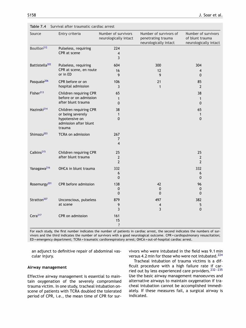
S158 J. Soar et al.
Table 7.4 Survival after traumatic cardiac arrest
Source Entry criteria Number of survivorsneurologically intact
Number of survivors ofpenetrating traumaneurologically intact
Number of survivorsof blunt traumaneurologically intact
Bouillon212 Pulseless, requiringCPR at scene
22443
Battistella202 Pulseless, requiringCPR at scene, en routeor in ED
604 300 30416 12 49 9 0
Pasquale206 CPR before or onhospital admission
106 21 853 1 2
Fisher213 Children requiring CPRbefore or on admissionafter blunt trauma
65 381 10 0
Hazinski214 Children requiring CPRor being severelyhypotensive onadmission after blunttrauma
38 651 10 0
Shimazu203 TCRA on admission 26774
Calkins215 Children requiring CPRafter blunt trauma
25 252 22 2
Yanagawa216 OHCA in blunt trauma 332 3326 60 0
Rosemurgy201 CPR before admission 138 42 960 0 00 0 0
Stratton207 Unconscious, pulselessat scene
879 497 3829 4 53 3 0
Cera217 CPR on admission 16115
?
For each study, the first number indicates the number of patients in cardiac arrest, the second indicates the numbers of sur-vivors and the third indicates the number of survivors with a good neurological outcome. CPR = cardiopulmonary resuscitation;ED = emergency department; TCRA = traumatic cardiorespiratory arrest; OHCA = out-of-hospital cardiac arrest.
an adjunct to definitive repair of abdominal vas-cular injury.
Airway management
Effective airway management is essential to main-tain oxygenation of the severely compromisedtrauma victim. In one study, tracheal intubation on-scene of patients with TCRA doubled the toleratedperiod of CPR, i.e., the mean time of CPR for sur-
vivors who were intubated in the field was 9.1 minversus 4.2 min for those who were not intubated.224
Tracheal intubation of trauma victims is a dif-ficult procedure with a high failure rate if car-ried out by less experienced care providers.232—235
Use the basic airway management manoeuvres andalternative airways to maintain oxygenation if tra-cheal intubation cannot be accomplished immedi-ately. If these measures fail, a surgical airway isindicated.
European Resuscitation Council Guidelines for Resuscitation 2005 S159
Ventilation
In low cardiac output states positive pressure ven-tilation causes further circulatory depression, oreven cardiac arrest, by impeding venous return tothe heart.236 Monitor ventilation with capnome-try and adjust to achieve normocapnia. This mayenable slow respiratory rates and low tidal volumes,and the corresponding decrease in transpulmonarypressure may increase venous return and cardiacoutput.
Chest decompression
Effective decompression of a tension pneumothoraxcan be achieved quickly by lateral thoracostomy,which is likely to be more effective than needlethoracostomy and quicker than inserting a chesttube.237
Effectiveness of chest compressions in TCRA
In hypovolaemic cardiac arrest or cardiac tam-ponade, chest compressions are unlikely to be aseffective as in cardiac arrest from other causes;238
nonetheless, return of spontaneous circulation withAcpo
H
Eptlhg
P
Itnottt
F
Fonad
servative approach to IV fluid infusion, with per-missive hypotension until surgical haemostasis isachieved.241,242 In the UK, the National Institute forClinical Excellence (NICE) has published guidelineson prehospital fluid replacement in trauma.243 Therecommendations include giving 250 ml boluses ofcrystalloid solution until a radial pulse is achieved,and not delaying rapid transport of trauma victimsfor fluid infusion in the field. Prehospital fluid ther-apy may have a role in prolonged entrapments, butthere is no reliable evidence for this.244,245
Ultrasound
Ultrasound is a valuable tool in the evaluation of thecompromised trauma victim. Haemoperitoneum,haemo- or pneumothorax and cardiac tamponadecan be diagnosed reliably in minutes even in theprehospital phase.246 Diagnostic peritoneal lavageand needle pericardiocentesis have virtually disap-peared from clinical practice since the introductionof sonography in trauma care. Prehospital ultra-sound is now available, although its benefits are yetto be proven.
V
Tim
7p
O
Mtdwpnror
pmissrs
LS in patients with TCRA is well described. Chestompressions are still the standard of care inatients with cardiac arrest, irrespective of aeti-logy.
aemorrhage control
arly haemorrhage control is vital. Handle theatient gently at all times, to prevent clot disrup-ion. Apply external compression and pelvic andimb splints when appropriate. Delays in surgicalaemostasis are disastrous for patients with exsan-uinating trauma.
ericardiocentesis
n patients with suspected trauma-related cardiacamponade, needle pericardiocentesis is probablyot a useful procedure.239 There is no evidencef benefit in the literature. It may increase sceneime, cause myocardial injury and delay effectiveherapeutic measures, such as emergency thoraco-omy.
luids and blood transfusion on scene
luid resuscitation of trauma victims before haem-rrhage is controlled is controversial, and there iso clear consensus on when it should be startednd what fluids should be given.240 Limited evi-ence and general consensus support a more con-
asopressors
he possible role of vasopressors (e.g., vasopressin)n trauma resuscitation is unclear and is basedainly on case reports.247
j. Cardiac arrest associated withregnancy
verview
ortality related to pregnancy in developed coun-ries is rare, occurring in an estimated 1:30,000eliveries.248 The fetus must always be consideredhen an adverse cardiovascular event occurs in aregnant woman. Resuscitation guidelines for preg-ancy are based largely on case series and scientificationale. Most reports address the causes in devel-ped countries, whereas the majority of pregnancy-elated deaths occur in the developing world.
Significant physiological changes occur duringregnancy, e.g., cardiac output, blood volume,inute ventilation and oxygen consumption all
ncrease. Furthermore, the gravid uterus may causeignificant compression of iliac and abdominal ves-els when the mother is in the supine position,esulting in reduced cardiac output and hypoten-ion.
S160 J. Soar et al.
Causes
There are many causes of cardiac arrest in pregnantwomen. A review of nearly 2 million pregnancies inthe UK248 showed that maternal death was associ-ated with:
• pre-existing cardiac disease;• thromboembolism;• suicide;• hypertensive disorders of pregnancy;• sepsis;• ectopic pregnancy;• haemorrhage;• amniotic fluid embolism;
Pregnant women can also suffer the same causesof cardiac arrest as women of the same agegroup.
Key interventions to prevent cardiac arrest
In an emergency, use an ABCDE approach. Manycardiovascular problems associated with pregnancyare caused by caval compression. Treat a distressedor compromised pregnant patient as follows:
of the diaphragm and abdominal contents causedby the gravid uterus. Attempt defibrillation usingstandard energy doses.252 There is no evidencethat shocks from a direct current defibrillator haveadverse effects on the fetal heart. Left lateral tiltand large breasts will make it difficult to place anapical defibrillator paddle. Adhesive defibrillatorpads are preferable to paddles in pregnancy.
Modifications to advanced life support
There is a greater potential for gastro-oesophagealsphincter insufficiency and risk of pulmonary aspi-ration of gastric contents. Early tracheal intubationwith correctly applied cricoid pressure decreasesthis risk. Tracheal intubation will make ventilationof the lungs easier in the presence of increasedintra-abdominal pressure.
A tracheal tube 0.5—1 mm internal diameter (ID)smaller than that used for a non-pregnant womanof similar size may be necessary because of mater-nal airway narrowing from oedema and swelling.253
Tracheal intubation may be more difficult in thepregnant patient.254 Expert help, a failed intuba-tion drill and the use of alternative airway devicesm
R
Rrdacattbbeoi
H
Laeposjbtif
• Place the patient in the left lateral position ormanually and gently displace the uterus to theleft.
• Give 100% oxygen.• Give a fluid bolus.• Immediately re-evaluate the need for any drugs
being given.• Seek expert help early.
Modifications to BLS guidelines for cardiacarrest
After 20 weeks’ gestation, the pregnant woman’suterus can press down against the inferior vena cavaand the aorta, impeding venous return and cardiacoutput. Uterine obstruction of venous return cancause pre-arrest hypotension or shock and, in thecritically ill patient, may precipitate arrest.249,250
After cardiac arrest, the compromise in venousreturn and cardiac output by the gravid uteruslimit the effectiveness of chest compressions. Non-cardiac arrest data show that the gravid uterus canbe shifted away from the cava in most cases byplacing the patient in 15 degrees of left lateraldecubitus position.251 Tilt may be accomplished bymanual or mechanical means. There is no evidenceto guide the hand position for optimum chest com-pressions in the pregnant patient. A hand positionhigher than the normal position for chest compres-sion may be needed to adjust for the elevation
ay be needed (see section 4d).255
eversible causes
escuers should attempt to identify common andeversible causes of cardiac arrest in pregnancyuring resuscitation attempts. The 4 Hs and 4 Tspproach helps to identify all the common causes ofardiac arrest in pregnancy. Pregnant patients aret risk of all the other causes of cardiac arrest forheir age group (e.g., anaphylaxis, drug overdose,rauma). Consider the use of abdominal ultrasoundy a skilled operator to detect pregnancy and possi-le causes during cardiac arrest in pregnancy; how-ver, do not delay other treatments. Specific causesf cardiac arrest in pregnancy include the follow-ng:
aemorrhage
ife-threatening haemorrhage can occur bothntenatally and postnatally. Associations includectopic pregnancy, placental abruption, placentaraevia and uterine rupture.248 A massive haem-rrhage protocol must be available in all units andhould be updated and rehearsed regularly in con-unction with the blood bank. Women at high risk ofleeding should be delivered in centres with facili-ies for blood transfusion, intensive care and othernterventions, and plans should be made in advanceor their management. Treatment is based on an
European Resuscitation Council Guidelines for Resuscitation 2005 S161
ABCDE approach. The key step is to stop the bleed-ing. Consider the following:
• fluid resuscitation including use of rapid transfu-sion system and cell salvage;256
• correction of coagulopathy. There may be a rolefor recombinant Factor VIIa;257
• oxytocin and prostaglandins to correct uterineatony;258
• uterine compression sutures;259
• radiological embolisation;260
• hysterectomy;• aortic cross-clamping in catastrophic haemo-
rrhage.261
Drugs
Iatrogenic overdose is possible in eclamptic womenreceiving magnesium sulphate, particularly if thewoman becomes oliguric. Give calcium to treatmagnesium toxicity (see life-threatening elec-trolyte abnormalities).
Central neural blockade for analgesia or anaes-thesia may cause problems due to sympatheticblockade (hypotension, bradycardia) or local anaes-thetic toxicity.262
C
PcttfkcdPsir
P
Espptop
L
Stw
Amniotic fluid embolism
Amniotic fluid embolism may present with breath-lessness, cyanosis, arrhythmias, hypotension andhaemorrhage associated with disseminated intra-vascular coagulopathy.272 Presentation is variableand may be similar to anaphylaxis. Treatment issupportive, as there is no specific therapy. Suc-cessful use of cardiopulmonary bypass for womensuffering life-threatening amniotic fluid embolismduring labour and delivery is reported.273
If immediate resuscitation attempts fail
Consider the need for an emergency hysterotomyor Caesarean section as soon as a pregnant womangoes into cardiac arrest. In some circumstancesimmediate resuscitation attempts will restore aperfusing rhythm; in early pregnancy this mayenable the pregnancy to proceed to term. Wheninitial resuscitation attempts fail, delivery ofthe fetus may improve the chances of successfulresuscitation of the mother and fetus.274—276 Thebest survival rate for infants over 24—25 weeks’
ardiovascular disease
ulmonary hypertension causes most deaths fromongenital heart disease. Peripartum cardiomyopa-hy, myocardial infarction and aneurysm or dissec-ion of the aorta or its branches cause most deathsrom acquired cardiac disease.263,264 Patients withnown cardiac disease need to be managed in a spe-ialist unit. Pregnant women with coronary arteryisease may suffer an acute coronary syndrome.ercutaneous coronary intervention is the reperfu-ion strategy of choice for ST-elevation myocardialnfarction in pregnancy because fibrinolytics areelatively contraindicated.265
re-eclampsia and eclampsia
clampsia is defined as the development of convul-ions and/or unexplained coma during pregnancy orostpartum in patients with signs and symptoms ofre-eclampsia.266,267 Magnesium sulphate is effec-ive in preventing approximately half of the casesf eclampsia developing in labour or immediatelyostpartum in women with pre-eclampsia.
ife-threatening pulmonary embolism
uccessful use of fibrinolytics for massive, life-hreatening pulmonary embolism in pregnantomen has been reported.268—271
gestation occurs when delivery of the infant isachieved within 5 min after the mother’s cardiacarrest.274,277—279 This requires that the providercommence the hysterotomy at about 4 min aftercardiac arrest. Delivery will relieve caval compres-sion and improve chances of maternal resuscita-tion. The Caesarean delivery also enables accessto the infant so that newborn resuscitation canbegin.
Decision-making for emergency hysterotomy
Consider gestational age. The gravid uterusreaches a size that will begin to compromise aorto-caval blood flow at approximately 20 weeks’ ges-tation; however, fetal viability begins at approx-imately 24—25 weeks. Portable ultrasounds areavailable in some emergency departments and mayaid in determination of gestational age (in expe-rienced hands) and positioning, provided their usedoes not delay the decision to perform emergencyhysterotomy.280
• At gestational age <20 weeks, urgent Caesareandelivery need not be considered, because agravid uterus of this size is unlikely to signifi-cantly compromise maternal cardiac output.
• At gestational age approximately 20—23 weeks,initiate emergency hysterotomy to enable suc-cessful resuscitation of the mother, not survival
S162 J. Soar et al.
of the delivered infant, which is unlikely at thisgestational age.
• At gestational age approximately ≥24—25 weeks,initiate emergency hysterotomy to save the lifeof both the mother and the infant.
Planning for emergencies. Advanced life supportin pregnancy requires coordination of maternalresuscitation, Caesarean delivery of the fetus andnewborn resuscitation within 5 min. To achieve this,units likely to deal with cardiac arrest in pregnancyshould:
• have plans and equipment for resuscitation ofboth the pregnant woman and newborn in place;
• ensure early involvement of obstetric and neona-tal teams;
• ensure regular training in obstetric emergencies.
7k. Electrocution
Introduction
Electrical injury is a relatively infrequent butpotentially devastating multisystem injury with
• Current may precipitate ventricular fibrillation(VF) if it traverses the myocardium duringthe vulnerable period (analogous to an R-on-T phenomenon).283 Electrical current may alsocause myocardial ischaemia because of coronaryartery spasm. Asystole may be primary, or sec-ondary to asphyxia following respiratory arrest.
Current that traverses the myocardium is morelikely to be fatal. A transthoracic (hand-to-hand)pathway is more likely to be fatal than a vertical(hand-to-foot) or straddle (foot-to-foot) pathway.There may be extensive tissue destruction along thecurrent pathway.
Lightning strike
Lightning strikes deliver as much as 300 kilo-volts over a few ms. Most of the current froma lightning strike passes over the surface of thebody in a process called ‘external flashover’. Bothindustrial shocks and lightning strikes cause deepburns at the point of contact. For industry thepoints of contact are usually on the upper limbs,hmmotcoageriat(sCatrcan
D
Tapa
high morbidity and mortality, causing 0.54 deathsper 100,000 people each year. Most electricalinjuries in adults occur in the workplace and areassociated generally with high voltage, whereaschildren are at risk primarily at home, where thevoltage is lower (220 V in Europe, Australia and Asia;110 V in the USA and Canada).281 Electrocution fromlightning strikes is rare, but worldwide it causes1000 deaths each year.282
Electric shock injuries are caused by the directeffects of current on cell membranes and vascularsmooth muscle. The thermal energy associated withhigh-voltage electrocution will also cause burns.Factors influencing the severity of electrical injuryinclude whether the current is alternating (AC) ordirect (DC), voltage, magnitude of energy deliv-ered, resistance to current flow, pathway of currentthrough the patient, and the area and duration ofcontact. Skin resistance is decreased by moisture,which increases the likelihood of injury. Electriccurrent follows the path of least resistance; con-ductive neurovascular bundles within limbs are par-ticularly prone to damage.
Contact with AC may cause tetanic contractionof skeletal muscle, which may prevent release fromthe source of electricity. Myocardial or respiratoryfailure may cause immediate death.
• Respiratory arrest may be caused by paralysis ofthe central respiratory control system or the res-piratory muscles.
ands and wrists, whereas for lightning they areostly on the head, neck and shoulders. Injuryay also occur indirectly through ground current
r current ‘splashing’ from a tree or other objecthat is hit by lightning.284 Explosive force mayause blunt trauma.285 The pattern and severityf injury from a lightning strike varies consider-bly, even among affected individuals from a sin-le group.286—288 As with industrial and domesticlectric shock, death is caused by cardiac287—291 orespiratory arrest.284,292 In those who survive thenitial shock, extensive catecholamine release orutonomic stimulation may occur, causing hyper-ension, tachycardia, non-specific ECG changesincluding prolongation of the QT interval and tran-ient T-wave inversion), and myocardial necrosis.reatine kinase may be released from myocardialnd skeletal muscle. Lightning can also cause cen-ral and peripheral nerve damage; brain haemor-hage and oedema, and peripheral nerve injury areommon. Mortality from lightning injuries is as highs 30%, with up to 70% of survivors sustaining sig-ificant morbidity.293—295
iagnosis
he circumstances surrounding the incident are notlways known. Unconscious patients with linear orunctuate burns or feathering should be treated asvictims of lightning strike.284
European Resuscitation Council Guidelines for Resuscitation 2005 S163
Rescue
Ensure that any power source is switched off anddo not approach the casualty until it is safe.High voltage (above domestic mains) electricitycan arc and conduct through the ground for upto a few metres around the casualty. It is safeto approach and handle casualties after lightningstrike, although it would be wise to move to a saferenvironment, particularly if lightning has been seenwithin 30 min.284
Resuscitation
Start standard basic and advanced life support with-out delay.
• Airway management may be difficult if there areelectrical burns around the face and neck. Earlytracheal intubation is needed in these cases, asextensive soft-tissue oedema may develop caus-ing airway obstruction. Head and spine traumacan occur after electrocution. Immobilise thespine until evaluation can be performed.
• Muscular paralysis, especially after high voltage,may persist for several hours;294 ventilatory sup-
•
•
•
•
•
•
•
dapr
in respiratory or cardiac arrest. Victims with res-piratory arrest may require only ventilation toavoid secondary hypoxic cardiac arrest. Resusci-tative attempts may have higher success ratesin lightning victims than in patients with car-diac arrest from other causes, and efforts maybe effective even when the interval before theresuscitative attempt is prolonged.292 Dilated ornon-reactive pupils should never be used as a prog-nostic sign, particularly in patients suffering a light-ning strike.284
There are conflicting reports on the vulnerabilityof the fetus to electric shock. The clinical spec-trum of electrical injury ranges from a transientunpleasant sensation for the mother with no effecton her fetus, to fetal death either immediately ora few days later. Several factors, such as the mag-nitude of the current and the duration of contact,are thought to affect outcome.299
Further treatment and prognosis
Immediate resuscitation in young victims of cardiacarrest due to electrocution can result in survival.Successful resuscitation has been reported afterpths
••••
dtdTara
R
port is required during this period.VF is the commonest initial arrhythmia after high-voltage AC shock; treat with prompt attempteddefibrillation. Asystole is more common after DCshock; use standard protocols for this and otherarrhythmias.Remove smouldering clothing and shoes to pre-vent further thermal injury.Vigorous fluid therapy is required if there issignificant tissue destruction. Maintain a goodurine output to enhance the excretion of myo-globin, potassium and other products of tissuedamage.291
Consider early surgical intervention in patientswith severe thermal injuries.Maintain spinal immobilisation if there is a likeli-hood of head or neck trauma.296,297
Conduct a thorough secondary survey to excludetraumatic injuries caused by tetanic muscularcontraction or by the person being thrown.297,298
Electrocution can cause severe, deep soft-tissueinjury with relatively minor skin wounds, becausecurrent tends to follow neurovascular bundles;look carefully for features of compartment syn-drome, which will necessitate fasciotomy.
Patients struck by lightning are most likely toie if they suffer immediate cardiac or respiratoryrrest and are not treated rapidly. When multi-le victims are struck simultaneously by lightning,escuers should give highest priority to patients
rolonged life support. All those who survive elec-rical injury should be monitored in hospital if theyave a history of cardiorespiratory problems or haveuffered:
loss of consciousness;cardiac arrest;electrocardiographic abnormalities;soft-tissue damage and burns.
Severe burns (thermal or electrical), myocar-ial necrosis, the extent of central nervous sys-em injury, and secondary multisystem organ failureetermine the morbidity and long-term prognosis.here is no specific therapy for electrical injury,nd the management is symptomatic. Preventionemains the best way to minimise the prevalencend severity of electrical injury.
eferences
1. Guidelines 2000 for Cardiopulmonary Resuscitation andEmergency Cardiovascular Care: International Consensuson Science. Part 8: Advanced challenges in resuscitation.Section 1. Life-threatening electrolyte abnormalities. Cir-culation 2000;102:I-122—217.
2. Niemann JT, Cairns CB. Hyperkalemia and ionized hypocal-cemia during cardiac arrest and resuscitation: possible cul-prits for postcountershock arrhythmias? Ann Emerg Med1999;34:1—7.
3. Ahmed J, Weisberg LS. Hyperkalemia in dialysis patients.Semin Dial 2001;14:348—56.
S164 J. Soar et al.
4. Mahoney B, Smith W, Lo D, Tsoi K, Tonelli M, Clase C. Emer-gency interventions for hyperkalaemia. Cochrane DatabaseSyst Rev 2005. CD003235.
5. Ngugi NN, McLigeyo SO, Kayima JK. Treatment ofhyperkalaemia by altering the transcellular gradient inpatients with renal failure: effect of various therapeuticapproaches. East Afr Med J 1997;74:503—9.
6. Allon M, Shanklin N. Effect of bicarbonate administrationon plasma potassium in dialysis patients: interactions withinsulin and albuterol. Am J Kidney Dis 1996;28:508—14.
7. Rastegar A, Soleimani M. Hypokalaemia and hyper-kalaemia. Postgrad Med J 2001;77:759—64.
8. Cohn JN, Kowey PR, Whelton PK, Prisant LM. New guide-lines for potassium replacement in clinical practice: a con-temporary review by the National Council on Potassium inClinical Practice. Arch Intern Med 2000;160:2429—36.
9. Litovitz TL, Felberg L, White S, Klein-Schwartz W. 1995annual report of the American Association of Poison ControlCenters Toxic Exposure Surveillance System. Am J EmergMed 1996;14:487—537.
10. McCaig LF, Burt CW. Poisoning-related visits to emergencydepartments in the United States, 1993—1996. J ToxicolClin Toxicol 1999;37:817—26.
11. Fingerhut LA, Cox CS. Poisoning mortality, 1985—1995. Pub-lic Health Rep 1998;113:218—33.
12. Watson WA, Litovitz TL, Klein-Schwartz W, et al. 2003annual report of the American Association of Poison ControlCenters Toxic Exposure Surveillance System. Am J EmergMed 2004;22:335—404.
13. Zimmerman JL. Poisonings and overdoses in the intensive
27. Wanger K, Brough L, Macmillan I, Goulding J, MacPhail I,Christenson JM. Intravenous vs. subcutaneous naloxone forout-of-hospital management of presumed opioid overdose.Acad Emerg Med 1998;5:293—9.
28. Hasan RA, Benko AS, Nolan BM, Campe J, Duff J, ZureikatGY. Cardiorespiratory effects of naloxone in children. AnnPharmacother 2003;37:1587—92.
29. Sporer KA. Acute heroin overdose. Ann Intern Med1999;130:584—90.
30. Schneir AB, Vadeboncoeur TF, Offerman SR, et al. Massiveoxycontin ingestion refractory to naloxone therapy. AnnEmerg Med 2002;40:425—8.
31. Kelly AM, Kerr D, Dietze P, Patrick I, Walker T, Koutsogian-nis Z. Randomised trial of intranasal versus intramuscularnaloxone in prehospital treatment for suspected opioidoverdose. Med J Aust 2005;182:24—7.
32. Brown TC. Sodium bicarbonate treatment for tricyclicantidepressant arrhythmias in children. Med J Aust1976;2:380—2.
33. Hoffman JR, Votey SR, Bayer M, Silver L. Effect of hyper-tonic sodium bicarbonate in the treatment of moderate-to-severe cyclic antidepressant overdose. Am J Emerg Med1993;11:336—41.
34. Knudsen K, Abrahamsson J. Epinephrine and sodium bicar-bonate independently and additively increase survivalin experimental amitriptyline poisoning. Crit Care Med1997;25:669—74.
35. Nattel S, Mittleman M. Treatment of ventricular tach-yarrhythmias resulting from amitriptyline toxicity in dogs.J Pharmacol Exp Ther 1984;231:430—5.
care unit: general and specific management issues. CritCare Med 2003;31:2794—801.
14. Suntres ZE. Role of antioxidants in paraquat toxicity. Toxi-cology 2002;180:65—77.
15. Facility assessment guidelines for regional toxicology treat-ment centers. American Academy of Clinical Toxicology. JToxicol Clin Toxicol 1993;31:211—7.
16. Poison information and treatment systems. American Col-lege of Emergency Physicians. Ann Emerg Med 1996;28:384.
17. Chyka PA, Seger D, Krenzelok EP, Vale JA. Positionpaper: single-dose activated charcoal. Clin Toxicol (Phila)2005;43:61—87.
18. Vale JA, Kulig K. Position paper: gastric lavage. J ToxicolClin Toxicol 2004;42:933—43.
19. Krenzelok EP. Ipecac syrup-induced emesis.. no evidenceof benefit. Clin Toxicol (Phila) 2005;43:11—2.
20. Position paper: cathartics. J Toxicol Clin Toxicol2004;42:243—53.
21. Position paper: whole bowel irrigation. J Toxicol Clin Toxi-col 2004;42:843—54.
22. Proudfoot AT, Krenzelok EP, Vale JA. Position Paper on urinealkalinization. J Toxicol Clin Toxicol 2004;42:1—26.
23. Golper TA, Bennett WM. Drug removal by continuousarteriovenous haemofiltration. A review of the evidencein poisoned patients. Med Toxicol Adverse Drug Exp1988;3:341—9.
24. Pitetti RD, Singh S, Pierce MC. Safe and efficacious use ofprocedural sedation and analgesia by nonanesthesiologistsin a pediatric emergency department. Arch Pediatr AdolescMed 2003;157:1090—6.
25. Osterwalder JJ. Naloxone — for intoxications with intra-venous heroin and heroin mixtures — harmless or haz-ardous? A prospective clinical study. J Toxicol Clin Toxicol1996;34:409—16.
26. Sporer KA, Firestone J, Isaacs SM. Out-of-hospital treat-ment of opioid overdoses in an urban setting. Acad EmergMed 1996;3:660—7.
36. Nattel S, Keable H, Sasyniuk BI. Experimental amitriptylineintoxication: electrophysiologic manifestations and man-agement. J Cardiovasc Pharmacol 1984;6:83—9.
37. Pentel P, Benowitz N. Efficacy and mechanism of action ofsodium bicarbonate in the treatment of desipramine toxi-city in rats. J Pharmacol Exp Ther 1984;230:12—9.
38. Brown TC, Barker GA, Dunlop ME, Loughnan PM. Theuse of sodium bicarbonate in the treatment of tri-cyclic antidepressant-induced arrhythmias. Anaesth Inten-sive Care 1973;1:203—10.
39. Brown TC. Tricyclic antidepressant overdosage: experimen-tal studies on the management of circulatory complica-tions. Clin Toxicol 1976;9:255—72.
40. Hedges JR, Baker PB, Tasset JJ, Otten EJ, Dalsey WC,Syverud SA. Bicarbonate therapy for the cardiovascular tox-icity of amitriptyline in an animal model. J Emerg Med1985;3:253—60.
41. Sasyniuk BI, Jhamandas V, Valois M. Experimentalamitriptyline intoxication: treatment of cardiac toxicitywith sodium bicarbonate. Ann Emerg Med 1986;15:1052—9.
42. Stone CK, Kraemer CM, Carroll R, Low R. Does a sodium-free buffer affect QRS width in experimental amitriptylineoverdose? Ann Emerg Med 1995;26:58—64.
43. McCabe JL, Cobaugh DJ, Menegazzi JJ, Fata J. Exper-imental tricyclic antidepressant toxicity: a randomized,controlled comparison of hypertonic saline solution,sodium bicarbonate, and hyperventilation. Ann Emerg Med1998;32:329—33.
44. Sasyniuk BI, Jhamandas V. Mechanism of reversal oftoxic effects of amitriptyline on cardiac Purkinjefibers by sodium bicarbonate. J Pharmacol Exp Ther1984;231:387—94.
45. Sasyniuk BI, Jhamandas V. Frequency-dependent effects ofamitriptyline on Vmax in canine Purkinje fibers and its alter-ation by alkalosis. Proc West Pharmacol Soc 1986;29:73—5.
46. Bou-Abboud E, Nattel S. Molecular mechanisms of thereversal of imipramine-induced sodium channel blockade
European Resuscitation Council Guidelines for Resuscitation 2005 S165
by alkalinization in human cardiac myocytes. CardiovascRes 1998;38:395—404.
47. Levitt MA, Sullivan Jr JB, Owens SM, Burnham L, FinleyPR. Amitriptyline plasma protein binding: effect of plasmapH and relevance to clinical overdose. Am J Emerg Med1986;4:121—5.
48. McKinney PE, Rasmussen R. Reversal of severe tricyclicantidepressant-induced cardiotoxicity with intravenoushypertonic saline solution. Ann Emerg Med 2003;42:20—4.
49. Brogan WCI, Lange RA, Kim AS, Moliterno DJ, Hillis LD.Alleviation of cocaine-induced coronary vasoconstrictionby nitroglycerin. J Am Coll Cardiol 1991;18:581—6.
50. Lange RA, Cigarroa RG, Yancy Jr CW, et al. Cocaine-induced coronary-artery vasoconstriction. N Engl J Med1989;321:1557—62.
51. Lange RA, Cigarroa RG, Flores ED, et al. Potentiationof cocaine-induced coronary vasoconstriction by beta-adrenergic blockade. Ann Intern Med 1990;112:897—903.
52. Boehrer JD, Moliterno DJ, Willard JE, Hillis LD, Lange RA.Influence of labetalol on cocaine-induced coronary vaso-constriction in humans. Am J Med 1993;94:608—10.
53. Bosse GM, Pope TM. Recurrent digoxin overdose and treat-ment with digoxin-specific Fab antibody fragments. JEmerg Med 1994;12:179—85.
54. Eddleston M, Rajapakse S, Rajakanthan, et al. Anti-digoxinFab fragments in cardiotoxicity induced by ingestion ofyellow oleander: a randomised controlled trial. Lancet2000;355:967—72.
55. Dasgupta A, Szelei-Stevens KA. Neutralization of freedigoxin-like immunoreactive components of oriental
67. Goh SH, Low BY. Drowning and near-drowning—–somelessons learnt. Ann Acad Med Singapore 1999;28:183—8.
68. Quan L, Wentz KR, Gore EJ, Copass MK. Outcome andpredictors of outcome in pediatric submersion victimsreceiving prehospital care in King County. Wash Pediatr1990;86:586—93.
69. Szpilman D, Soares M. In-water resuscitation—–is it worth-while? Resuscitation 2004;63:25—31.
70. Perkins GD. In-water resuscitation: a pilot evaluation.Resuscitation 2005;65:321—4.
71. Rosen P, Stoto M, Harley J. The use of the Heimlich maneu-ver in near-drowning: Institute of Medicine report. J EmergMed 1995;13:397—405.
72. March NF, Matthews RC. New techniques in external car-diac compressions. Aquatic cardiopulmonary resuscitation.JAMA 1980;244:1229—32.
73. March NF, Matthews RC. Feasibility study of CPR in thewater. Undersea Biomed Res 1980;7:141—8.
74. Thomas R, Cahill CJ. Successful defibrillation in profoundhypothermia (core body temperature 25.6 degrees C).Resuscitation 2000;47:317—20.
75. Manolios N, Mackie I. Drowning and near-drowning on Aus-tralian beaches patrolled by life-savers: a 10-year study,1973—1983. Med J Aust 1988;148:165—7, 70—71.
76. Modell JH, Calderwood HW, Ruiz BC, Downs JB, Chap-man Jr R. Effects of ventilatory patterns on arterial oxy-genation after near-drowning in sea water. Anesthesiology1974;40:376—84.
77. Golden FS, Tipton MJ, Scott RC. Immersion, near-drowningand drowning. Br J Anaesth 1997;79:214—25.
medicines Dan Shen and Lu-Shen-Wan by the Fab frag-ment of antidigoxin antibody (Digibind). Am J Clin Pathol2004;121:276—81.
56. Bailey B. Glucagon in beta-blocker and calcium channelblocker overdoses: a systematic review. J Toxicol Clin Tox-icol 2003;41:595—602.
57. Dewitt CR, Waksman JC. Pharmacology, pathophysiologyand management of calcium channel blocker and beta-blocker toxicity. Toxicol Rev 2004;23:223—38.
58. Wax PM, Erdman AR, Chyka PA, et al. beta-blocker inges-tion: an evidence-based consensus guideline for out-of-hospital management. Clin Toxicol (Phila) 2005;43:131—46.
59. Peden MM, McGee K. The epidemiology of drowning world-wide. Inj Control Saf Promot 2003;10:195—9.
60. Driscoll TR, Harrison JA, Steenkamp M. Review of the roleof alcohol in drowning associated with recreational aquaticactivity. Inj Prev 2004;10:107—13.
61. Papa L, Hoelle R, Idris A. Systematic review of definitionsfor drowning incidents. Resuscitation 2005;65:255—64.
62. Idris AH, Berg RA, Bierens J, et al. Recommended guidelinesfor uniform reporting of data from drowning: The ‘‘Utsteinstyle’’. Resuscitation 2003;59:45—57.
63. Watson RS, Cummings P, Quan L, Bratton S, Weiss NS. Cer-vical spine injuries among submersion victims. J Trauma2001;51:658—62.
64. Dodd FM, Simon E, McKeown D, Patrick MR. The effect of acervical collar on the tidal volume of anaesthetised adultpatients. Anaesthesia 1995;50:961—3.
65. International Liaison Committee on Resuscitation 2005.International Consensus on Cardiopulmonary Resuscitationand Emergency Cardiovascular Care Science with Treat-ment Recommendations. Resuscitation 2005;67:157—341.
66. Golden FS, Hervey GR, Tipton MJ. Circum-rescue col-lapse: collapse, sometimes fatal, associated with rescueof immersion victims. J R Nav Med Serv 1991;77:139—49.
78. Wyatt JP, Tomlinson GS, Busuttil A. Resuscitation ofdrowning victims in south-east Scotland. Resuscitation1999;41:101—4.
79. Schmidt U, Fritz KW, Kasperczyk W, Tscherne H. Successfulresuscitation of a child with severe hypothermia after car-diac arrest of 88 min. Prehosp Disaster Med 1995;10:60—2.
80. Bolte RG, Black PG, Bowers RS, Thorne JK, Corneli HM. Theuse of extracorporeal rewarming in a child submerged for66 min. JAMA 1988;260:377—9.
81. The acute respiratory distress syndrome network. Ventila-tion with lower tidal volumes as compared with traditionaltidal volumes for acute lung injury and the acute respira-tory distress syndrome. N Engl J Med 2000;342:1301—8.
82. Moran I, Zavala E, Fernandez R, Blanch L, Mancebo J.Recruitment manoeuvres in acute lung injury/acuterespiratory distress syndrome. Eur Respir J Suppl2003;42:37s—42s.
83. Nolan JP, Morley PT, Vanden Hoek TL, Hickey RW. Therapeu-tic hypothermia after cardiac arrest. An advisory statementby the Advancement Life support Task Force of the Inter-national Liaison committee on Resuscitation. Resuscitation2003;57:231—5.
84. Holzer M, Behringer W, Schorkhuber W, et al. Mildhypothermia and outcome after CPR. Hypothermia for Car-diac Arrest (HACA) Study Group. Acta Anaesthesiol ScandSuppl 1997;111:55—8.
85. Sterz F, Safar P, Tisherman S, Radovsky A, Kuboyama K,Oku K. Mild hypothermic cardiopulmonary resuscitationimproves outcome after prolonged cardiac arrest in dogs.Crit Care Med 1991;19:379—89.
86. Farstad M, Andersen KS, Koller ME, Grong K, Segadal L,Husby P. Rewarming from accidental hypothermia by extra-corporeal circulation. A retrospective study. Eur J Cardio-thorac Surg 2001;20:58—64.
87. Schneider SM. Hypothermia: from recognition to rewarm-ing. Emerg Med Rep 1992;13:1—20.
S166 J. Soar et al.
88. Gilbert M, Busund R, Skagseth A, Nilsen PA, Solbø JP. Resus-citation from accidental hypothermia of 13.7 ◦C with circu-latory arrest. Lancet 2000;355:375—6.
89. Danzl DF, Pozos RS, Auerbach PS, et al. Multicen-ter hypothermia survey. Ann Emerg Med 1987;16:1042—55.
90. Reuler JB. Hypothermia: pathophysiology, clinical settings,and management. Ann Intern Med 1978;89:519—27.
91. Lefrant JY, Muller L, de La Coussaye JE, et al. Temperaturemeasurement in intensive care patients: comparison of uri-nary bladder, oesophageal, rectal, axillary, and inguinalmethods versus pulmonary artery core method. IntensiveCare Med 2003;29:414—8.
92. Robinson J, Charlton J, Seal R, Spady D, Joffres MR.Oesophageal, rectal, axillary, tympanic and pulmonaryartery temperatures during cardiac surgery. Can J Anaesth1998;45:317—23.
93. Krismer AC, Lindner KH, Kornberger R, et al. Cardiopul-monary resuscitation during severe hypothermia in pigs:does epinephrine or vasopressin increase coronary perfu-sion pressure? Anesth Analg 2000;90:69—73.
94. Kornberger E, Lindner KH, Mayr VD, et al. Effects ofepinephrine in a pig model of hypothermic cardiac arrestand closed-chest cardiopulmonary resuscitation combinedwith active rewarming. Resuscitation 2001;50:301—8.
95. Stoner J, Martin G, O’Mara K, Ehlers J, Tomlanovich M.Amiodarone and bretylium in the treatment of hypother-mic ventricular fibrillation in a canine model. Acad EmergMed 2003;10:187—91.
96. Mattu A, Brady WJ, Perron AD. Electrocardiographic
107. Safar P. Cerebral resuscitation after cardiac arrest:research initiatives and future directions [published cor-rection appears in Ann Emerg Med. 1993;22:759]. AnnEmerg Med 1993;22(pt 2):324—49.
108. Bouchama A, Knochel JP. Heat stroke. N Engl J Med2002;346:1978—88.
109. Wappler F. Malignant hyperthermia. Eur J Anaesthesiol2001;18:632—52.
110. Ali SZ, Taguchi A, Rosenberg H. Malignant hyperther-mia. Best Pract Res Clin Anaesthesiol 2003;17:519—33.
111. Bouchama A. The 2003 European heat wave. Intensive CareMed 2004;30:1—3.
112. Coris EE, Ramirez AM, Van Durme DJ. Heat illness in ath-letes: the dangerous combination of heat, humidity andexercise. Sports Med 2004;34:9—16.
113. Grogan H, Hopkins PM. Heat stroke: implications for criticalcare and anaesthesia. Br J Anaesth 2002;88:700—7.
114. Bouchama A, De Vol EB. Acid-base alterations in heat-stroke. Intensive Care Med 2001;27:680—5.
115. Akhtar MJ, al-Nozha M, al-Harthi S, Nouh MS. Electrocardio-graphic abnormalities in patients with heat stroke. Chest1993;104:411—4.
116. el-Kassimi FA, Al-Mashhadani S, Abdullah AK, Akhtar J.Adult respiratory distress syndrome and disseminatedintravascular coagulation complicating heat stroke. Chest1986;90:571—4.
117. Waruiru C, Appleton R. Febrile seizures: an update. ArchDis Child 2004;89:751—6.
118. Berger J, Hart J, Millis M, Baker AL. Fulminant hepatic fail-
1
1
1
1
1
1
1
1
1
1
1
1
manifestations of hypothermia. Am J Emerg Med2002;20:314—26.
97. Southwick FS, DalglishS P.H. Jr. Recovery after prolongedasystolic cardiac arrest in profound hypothermia: a casereport and literature review. JAMA 1980;243:1250—3.
98. Ujhelyi MR, Sims JJ, Dubin SA, Vender J, Miller AW.Defibrillation energy requirements and electrical hetero-geneity during total body hypothermia. Crit Care Med2001;29:1006—11.
99. Kornberger E, Schwarz B, Lindner KH, Mair P. Forcedair surface rewarming in patients with severe accidentalhypothermia. Resuscitation 1999;41:105—11.
100. Roggla M, Frossard M, Wagner A, Holzer M, Bur A,Roggla G. Severe accidental hypothermia with or with-out hemodynamic instability: rewarming without theuse of extracorporeal circulation. Wien Klin Wochenschr2002;114:315—20.
101. Weinberg AD, Hamlet MP, Paturas JL, White RD, McAninchGW. Cold weather emergencies: principles of patient man-agement. Branford, CN: American Medical Publishing Co.;1990.
102. Zell SC, Kurtz KJ. Severe exposure hypothermia: a resusci-tation protocol. Ann Emerg Med 1985;14:339—45.
103. Althaus U, Aeberhard P, Schupbach P, Nachbur BH, Muh-lemann W. Management of profound accidental hypother-mia with cardiorespiratory arrest. Ann Surg 1982;195:492—5.
104. Walpoth BH, Walpoth-Aslan BN, Mattle HP, et al. Outcomeof survivors of accidental deep hypothermia and circulatoryarrest treated with extracorporeal blood warming. N EnglJ Med 1997;337:1500—5.
105. Silfvast T, Pettila V. Outcome from severe accidentalhypothermia in Southern Finland—–a 10-year review. Resus-citation 2003;59:285—90.
106. Moss J. Accidental severe hypothermia. Surg GynecolObstet 1986;162:501—13.
ure from heat stroke requiring liver transplantation. J ClinGastroenterol 2000;30:429—31.
19. Huerta-Alardin AL, Varon J, Marik PE. Bench-to-bedsidereview: Rhabdomyolysis—–an overview for clinicians. CritCare 2005;9:158—69.
20. Wolff ED, Driessen OMJ. Theophylline intoxication in achild. Ned Tijdschr Geneeskd 1977;121:896—901.
21. Sidor K, Mikolajczyk W, Horwath-Stolarczyk A. Acute poi-soning in children hospitalized at the Medical UniversityHospital No 3 in Warsaw, between 1996 and 2000. PediatrPolska 2002;77:509—16.
22. Boyer EW, Shannon M. The serotonin syndrome. N Engl JMed 2005;352:1112—20.
23. Bhanushali MJ, Tuite PJ. The evaluation and managementof patients with neuroleptic malignant syndrome. NeurolClin 2004;22:389—411.
24. Abraham E, Matthay MA, Dinarello CA, et al. Consensusconference definitions for sepsis, septic shock, acute lunginjury, and acute respiratory distress syndrome: time for are-evaluation. Crit Care Med 2000;28:232—5.
25. Savage MW, Mah PM, Weetman AP, Newell-Price J.Endocrine emergencies. Postgrad Med J 2004;80:506—15.
26. Hadad E, Weinbroum AA, Ben-Abraham R. Drug-inducedhyperthermia and muscle rigidity: a practical approach.Eur J Emerg Med 2003;10:149—54.
27. Halloran LL, Bernard DW. Management of drug-inducedhyperthermia. Curr Opin Pediatr 2004;16:211—5.
28. Armstrong LE, Crago AE, Adams R, Roberts WO, Maresh CM.Whole-body cooling of hyperthermic runners: comparisonof two field therapies. Am J Emerg Med 1996;14:355—8.
29. Horowitz BZ. The golden hour in heat stroke: use of icedperitoneal lavage. Am J Emerg Med 1989;7:616—9.
30. Bernard S, Buist M, Monteiro O, Smith K. Induced hypother-mia using large volume, ice-cold intravenous fluid incomatose survivors of out-of-hospital cardiac arrest: a pre-liminary report. Resuscitation 2003;56:9—13.
European Resuscitation Council Guidelines for Resuscitation 2005 S167
131. Schmutzhard E, Engelhardt K, Beer R, et al. Safetyand efficacy of a novel intravascular cooling device tocontrol body temperature in neurologic intensive carepatients: a prospective pilot study. Crit Care Med 2002;30:2481—8.
132. Al-Senani FM, Graffagnino C, Grotta JC, et al. A prospec-tive, multicenter pilot study to evaluate the feasibilityand safety of using the CoolGard system and icy catheterfollowing cardiac arrest. Resuscitation 2004;62:143—50.
133. Behringer W, Safar P, Wu X, et al. Veno-venous extracorpo-real blood shunt cooling to induce mild hypothermia in dogexperiments and review of cooling methods. Resuscitation2002;54:89—98.
134. Hadad E, Cohen-Sivan Y, Heled Y, Epstein Y. Clinical review:treatment of heat stroke—–should dantrolene be consid-ered? Crit Care 2005;9:86—91.
135. Krause T, Gerbershagen MU, Fiege M, Weisshorn R, WapplerF. Dantrolene—–a review of its pharmacology, therapeuticuse and new developments. Anaesthesia 2004;59:364—73.
136. Eshel G, Safar P, Sassano J, Stezoski W. Hyperthermia-induced cardiac arrest in dogs and monkeys. Resuscitation1990;20:129—43.
137. Eshel G, Safar P, Radovsky A, Stezoski SW. Hyperthermia-induced cardiac arrest in monkeys: limited efficacy of stan-dard CPR. Aviat Space Environ Med 1997;68:415—20.
138. Zeiner A, Holzer M, Sterz F, et al. Hyperthermia after car-diac arrest is associated with an unfavorable neurologicoutcome. Arch Intern Med 2001;161:2007—12.
139. Masoli M, Fabian D, Holt S, Beasley R. The global burden
1
1
1
1
1
1
1
1
1
1
1
151. Rodrigo GJ, Rodrigo C, Pollack CV, Rowe B. Use of helium-oxygen mixtures in the treatment of acute asthma: a sys-tematic review. Chest 2003;123:891—6.
152. Petrillo TM, Fortenberry JD, Linzer JF, Simon HK. Emer-gency department use of ketamine in pediatric status asth-maticus. J Asthma 2001;38:657—64.
153. Howton JC, Rose J, Duffy S, Zoltanski T, Levitt MA. Random-ized, double-blind, placebo-controlled trial of intravenousketamine in acute asthma. Ann Emerg Med 1996;27:170—5.
154. Antonelli M, Pennisi MA, Montini L. Clinical review: nonin-vasive ventilation in the clinical setting—–experience fromthe past 10 years. Crit Care 2005;9:98—103.
155. Ram FS, Wellington S, Rowe BH, Wedzicha JA. Non-invasivepositive pressure ventilation for treatment of respira-tory failure due to severe acute exacerbations of asthma.Cochrane Database Syst Rev 2005:CD004360.
156. Leatherman JW, McArthur C, Shapiro RS. Effect of pro-longation of expiratory time on dynamic hyperinflation inmechanically ventilated patients with severe asthma. CritCare Med 2004;32:1542—5.
157. Bowman FP, Menegazzi JJ, Check BD, Duckett TM. Loweresophageal sphincter pressure during prolonged cardiacarrest and resuscitation. Ann Emerg Med 1995;26:216—9.
158. Lapinsky SE, Leung RS. Auto-PEEP and electromechanicaldissociation. N Engl J Med 1996;335:674.
159. Rogers PL, Schlichtig R, Miro A, Pinsky M. Auto-PEEP duringCPR. An ‘‘occult’’ cause of electromechanical dissociation?Chest 1991;99:492—3.
160. Rosengarten PL, Tuxen DV, Dziukas L, Scheinkestel C, Mer-
1
1
1
1
1
1
1
1
1
1
of asthma: executive summary of the GINA DisseminationCommittee report. Allergy 2004;59:469—78.
40. BTS/SIGN. British Thoracic Society, Scottish IntercollegiateGuidelines Network (SIGN). British guideline on the man-agement of asthma. Thorax 2003;58(Suppl. I):i1—94.
41. Rainbow J, Browne GJ. Fatal asthma or anaphylaxis? EmergMed J 2002;19:415—7.
42. Kokturk N, Demir N, Kervan F, Dinc E, Koybasioglu A, Turk-tas H. A subglottic mass mimicking near-fatal asthma: achallenge of diagnosis. J Emerg Med 2004;26:57—60.
43. Ratto D, Alfaro C, Sipsey J, Glovsky MM, Sharma OP. Areintravenous corticosteroids required in status asthmaticus?JAMA 1988;260:527—9.
44. Aaron SD. The use of ipratropium bromide for the manage-ment of acute asthma exacerbation in adults and children:a systematic review. J Asthma 2001;38:521—30.
45. Rodrigo G, Rodrigo C, Burschtin O. A meta-analysis ofthe effects of ipratropium bromide in adults with acuteasthma. Am J Med 1999;107:363—70.
46. Munro A, Jacobs M. Best evidence topic reports. Is intra-venous aminophylline better than intravenous salbutamolin the treatment of moderate to severe asthma? Emerg MedJ 2004;21:78—80.
47. Rowe BH, Bretzlaff JA, Bourdon C, Bota GW, Camargo JrCA. Magnesium sulfate for treating exacerbations of acuteasthma in the emergency department. Cochrane DatabaseSyst Rev 2000:CD001490.
48. Cydulka R, Davison R, Grammer L, Parker M, Mathews IVJ. The use of epinephrine in the treatment of older adultasthmatics. Ann Emerg Med 1988;17:322—6.
49. Victoria MS, Battista CJ, Nangia BS. Comparison of sub-cutaneous terbutaline with epinephrine in the treat-ment of asthma in children. J Allergy Clin Immunol1977;59:128—35.
50. Victoria MS, Battista CJ, Nangia BS. Comparison betweenepinephrine and terbutaline injections in the acute man-agement of asthma. J Asthma 1989;26:287—90.
rett K, Bowes G. Circulatory arrest induced by inter-mittent positive pressure ventilation in a patient withsevere asthma. Anaesth Intensive Care 1991;19:118—21.
61. Sprung J, Hunter K, Barnas GM, Bourke DL. Abdominaldistention is not always a sign of esophageal intuba-tion: cardiac arrest due to ‘‘auto-PEEP’’. Anesth Analg1994;78:801—4.
62. Deakin CD, McLaren RM, Petley GW, Clewlow F, Dalrymple-Hay MJ. Effects of positive end-expiratory pressure ontransthoracic impedance—–implications for defibrillation.Resuscitation 1998;37:9—12.
63. Mazzeo AT, Spada A, Pratico C, Lucanto T, Santamaria LB.Hypercapnia: what is the limit in paediatric patients? Acase of near-fatal asthma successfully treated by multi-pharmacological approach. Paediatr Anaesth 2004;14:596—603.
64. Mertes PM, Laxenaire MC, Alla F. Anaphylactic and anaphy-lactoid reactions occurring during anesthesia in France in1999—2000. Anesthesiology 2003;99:536—45.
65. Yunginger JW. Latex allergy in the workplace: anoverview of where we are. Ann Allergy Asthma Immunol1999;83:630—3.
66. Dreyfus DH, Fraser B, Randolph CC. Anaphylaxis to latexin patients without identified risk factors for latex allergy.Conn Med 2004;68:217—22.
67. Ownby DR. A history of latex allergy. J Allergy Clin Immunol2002;110:S27—32.
68. Pumphrey RS. Lessons for management of anaphy-laxis from a study of fatal reactions. Clin Exp Allergy2000;30:1144—50.
69. Pumphrey RS. Fatal anaphylaxis in the UK, 1992—2001.Novartis Found Symp 2004;257:116—28, discussion 28—32,57—60, 276—285.
70. Incorvaia C, Senna G, Mauro M, et al. Prevalence of allergicreactions to Hymenoptera stings in northern Italy. AllergImmunol (Paris) 2004;36:372—4.
S168 J. Soar et al.
171. Pumphrey RS, Roberts IS. Postmortem findings after fatalanaphylactic reactions. J Clin Pathol 2000;53:273—6.
172. Mullins RJ. Anaphylaxis: risk factors for recurrence. ClinExp Allergy 2003;33:1033—40.
173. Brown AF. Anaphylaxis: quintessence, quarrels, and quan-daries. Emerg Med J 2001;18:328.
174. Brown AF. Anaphylaxis gets the adrenaline going. EmergMed J 2004;21:128—9.
175. Ishoo E, Shah UK, Grillone GA, Stram JR, FuleihanNS. Predicting airway risk in angioedema: staging sys-tem based on presentation. Otolaryngol Head Neck Surg1999;121:263—8.
176. Pumphrey R. Anaphylaxis: can we tell who is at riskof a fatal reaction? Curr Opin Allergy Clin Immunol2004;4:285—90.
177. Simons FE, Gu X, Simons KJ. Epinephrine absorption inadults: intramuscular versus subcutaneous injection. JAllergy Clin Immunol 2001;108:871—3.
178. Simons FER, Chan ES, Gu X, Simons KJ. Epinephrine forthe out-of-hospital (first-aid) treatment of anaphylaxis ininfants: is the ampule/syringe/needle method practical? JAllergy Clin Immunol 2001;108:1040—4.
179. Winbery SL, Lieberman PL. Histamine and antihistaminesin anaphylaxis. Clin Allergy Immunol 2002;17:287—317.
180. Kill C, Wranze E, Wulf H. Successful treatment of severeanaphylactic shock with vasopressin. Two case reports. IntArch Allergy Immunol 2004;134:260—1.
181. Williams SR, Denault AY, Pellerin M, Martineau R. Vaso-pressin for treatment of shock following aprotinin admin-istration. Can J Anaesth 2004;51:169—72.
193. Rhodes JF, Blaufox AD, Seiden HS, et al. Cardiac arrestin infants after congenital heart surgery. Circulation1999;100:II194—9.
194. Dimopoulou I, Anthi A, Michalis A, Tzelepis GE. Func-tional status and quality of life in long-term survivorsof cardiac arrest after cardiac surgery. Crit Care Med2001;29:1408—11.
195. Mackay JH, Powell SJ, Osgathorp J, Rozario CJ. Six-yearprospective audit of chest reopening after cardiac arrest.Eur J Cardiothorac Surg 2002;22:421—5.
196. Pottle A, Bullock I, Thomas J, Scott L. Survival to dischargefollowing open chest cardiac compression (OCCC). A 4-yearretrospective audit in a cardiothoracic specialist centre—–Royal Brompton and Harefield NHS Trust, United Kingdom.Resuscitation 2002;52:269—72.
197. Raman J, Saldanha RF, Branch JM, et al. Open cardiac com-pression in the postoperative cardiac intensive care unit.Anaesth Intensive Care 1989;17:129—35.
198. Birdi I, Chaudhuri N, Lenthall K, Reddy S, Nashef SA. Emer-gency reinstitution of cardiopulmonary bypass followingcardiac surgery: outcome justifies the cost. Eur J Cardio-thorac Surg 2000;17:743—6.
199. Rousou JA, Engelman RM, Flack III JE, Deaton DW, Owen SG.Emergency cardiopulmonary bypass in the cardiac surgicalunit can be a lifesaving measure in postoperative cardiacarrest. Circulation 1994;90:II280—4.
200. Schwarz B, Bowdle TA, Jett GK, et al. Biphasic shocks com-pared with monophasic damped sine wave shocks for directventricular defibrillation during open heart surgery. Anes-thesiology 2003;98:1063—9.
2
2
2
2
2
2
2
2
2
2
2
2
182. Visscher PK, Vetter RS, Camazine S. Removing bee stings.Lancet 1996;348:301—2.
183. Yocum MW, Butterfield JH, Klein JS, Volcheck GW,Schroeder DR, Silverstein MD. Epidemiology of anaphylaxisin Olmsted County: a population-based study. J Allergy ClinImmunol 1999;104(pt 1):452—6.
184. Ellis AK, Day JH. Diagnosis and management of anaphylaxis.CMAJ 2003;169:307—11.
185. Smith PL, Kagey-Sobotka A, Bleecker ER, et al. Physio-logic manifestations of human anaphylaxis. J Clin Invest1980;66:1072—80.
186. Stark BJ, Sullivan TJ. Biphasic and protracted anaphylaxis.J Allergy Clin Immunol 1986;78:76—83.
187. Brazil E, MacNamara AF. Not so immediate’’ hypersensitiv-ity: the danger of biphasic anaphylactic reactions. J AccidEmerg Med 1998;15:252—3.
188. Brady Jr WJ, Luber S, Carter CT, Guertler A, Lind-beck G. Multiphasic anaphylaxis: an uncommon eventin the emergency department. Acad Emerg Med 1997;4:193—7.
189. Joint Working Party of the Association of Anaesthetists ofGreat Britain and Ireland and the British Society for Allergyand Clinical Immunology. Suspected anaphylactic reactionsassociated with anaesthesia. 3rd ed. London: The Associa-tion of Anaesthetists of Great Britain and Ireland and BritishSociety for Allergy and Clinical Immunology; 2003.
190. Payne V, Kam PC. Mast cell tryptase: a review ofits physiology and clinical significance. Anaesthesia2004;59:695—703.
191. Anthi A, Tzelepis GE, Alivizatos P, Michalis A, PalatianosGM, Geroulanos S. Unexpected cardiac arrest after car-diac surgery: incidence, predisposing causes, and out-come of open chest cardiopulmonary resuscitation. Chest1998;113:15—9.
192. Wahba A, Gotz W, Birnbaum DE. Outcome of cardiopul-monary resuscitation following open heart surgery. ScandCardiovasc J 1997;31:147—9.
01. Rosemurgy AS, Norris PA, Olson SM, Hurst JM, Albrink MH.Prehospital traumatic cardiac arrest: the cost of futility. JTrauma 1993;35:468—73.
02. Battistella FD, Nugent W, Owings JT, Anderson JT.Field triage of the pulseless trauma patient. Arch Surg1999;134:742—5.
03. Shimazu S, Shatney CH. Outcomes of trauma patientswith no vital signs on hospital admission. J Trauma1983;23:213—6.
04. Stockinger ZT, McSwain Jr NE. Additional evidence in sup-port of withholding or terminating cardiopulmonary resus-citation for trauma patients in the field. J Am Coll Surg2004;198:227—31.
05. Fulton RL, Voigt WJ, Hilakos AS. Confusion surroundingthe treatment of traumatic cardiac arrest. J Am Coll Surg1995;181:209—14.
06. Pasquale MD, Rhodes M, Cipolle MD, Hanley T, Wasser T.Defining ‘‘dead on arrival’’: impact on a level I trauma cen-ter. J Trauma 1996;41:726—30.
07. Stratton SJ, Brickett K, Crammer T. Prehospital pulseless,unconscious penetrating trauma victims: field assessmentsassociated with survival. J Trauma 1998;45:96—100.
08. Maron BJ, Gohman TE, Kyle SB, Estes III NA, Link MS.Clinical profile and spectrum of commotio cordis. JAMA2002;287:1142—6.
09. Maron BJ, Estes III NA, Link MS. Task Force 11: commotiocordis. J Am Coll Cardiol 2005;45:1371—3.
10. Nesbitt AD, Cooper PJ, Kohl P. Rediscovering commotiocordis. Lancet 2001;357:1195—7.
11. Link MS, Estes M, Maron BJ. Sudden death caused bychest wall trauma (commotio cordis). In: Kohl P, Sachs F,Franz MR, editors. Cardiac mechano-electric feedback andarrhythmias: from pipette to patient. Philadelphia: Else-vier Saunders; 2005. p. 270—6.
12. Bouillon B, Walther T, Kramer M, Neugebauer E. Trauma andcirculatory arrest: 224 preclinical resuscitations in Colognein 1990 [in German]. Anaesthesist 1994;43:786—90.
European Resuscitation Council Guidelines for Resuscitation 2005 S169
213. Fisher B, Worthen M. Cardiac arrest induced by blunttrauma in children. Pediatr Emerg Care 1999;15:274—6.
214. Hazinski MF, Chahine AA, Holcomb III GW, Morris Jr JA. Out-come of cardiovascular collapse in pediatric blunt trauma.Ann Emerg Med 1994;23:1229—35.
215. Calkins CM, Bensard DD, Partrick DA, Karrer FM. A criticalanalysis of outcome for children sustaining cardiac arrestafter blunt trauma. J Pediatr Surg 2002;37:180—4.
216. Yanagawa Y, Saitoh D, Takasu A, Kaneko N, Sakamoto T,Okada Y. Experience of treatment for blunt traumaticout-of-hospital cardiopulmonary arrest patients over 24years: head injury vs. non-head injury. No Shinkei Geka2004;32:231—5.
217. Cera SM, Mostafa G, Sing RF, Sarafin JL, Matthews BD, Heni-ford BT. Physiologic predictors of survival in post-traumaticarrest. Am Surg 2003;69:140—4.
218. Esposito TJ, Jurkovich GJ, Rice CL, Maier RV, Copass MK,Ashbaugh DG. Reappraisal of emergency room thoracotomyin a changing environment. J Trauma 1991;31:881—5, dis-cussion 5—7.
219. Martin SK, Shatney CH, Sherck JP, et al. Blunt traumapatients with prehospital pulseless electrical activity(PEA): poor ending assured. J Trauma 2002;53:876—80, dis-cussion 80—81.
220. Domeier RM, McSwain Jr NE, Hopson LR, et al. Guidelinesfor withholding or termination of resuscitation in prehos-pital traumatic cardiopulmonary arrest. J Am Coll Surg2003;196:475—81.
221. Pickens JJ, Copass MK, Bulger EM. Trauma patients
2
2
2
2
2
2
2
2
2
2
2
bation: miss rates by paramedics. Acad Emerg Med2004;11:707—9.
233. Jemmett ME, Kendal KM, Fourre MW, Burton JH. Unrec-ognized misplacement of endotracheal tubes in a mixedurban to rural emergency medical services setting. AcadEmerg Med 2003;10:961—5.
234. Katz SH, Falk JL. Misplaced endotracheal tubes byparamedics in an urban emergency medical services sys-tem. Ann Emerg Med 2001;37:32—7.
235. Deakin CD, Peters R, Tomlinson P, Cassidy M. Securing theprehospital airway: a comparison of laryngeal mask inser-tion and endotracheal intubation by UK paramedics. EmergMed J 2005;22:64—7.
236. Pepe PE, Roppolo LP, Fowler RL. The detrimental effectsof ventilation during low-blood-flow states. Curr Opin CritCare 2005;11:212—8.
237. Deakin CD, Davies G, Wilson A. Simple thoracostomy avoidschest drain insertion in prehospital trauma. J Trauma1995;39:373—4.
238. Luna GK, Pavlin EG, Kirkman T, Copass MK, Rice CL. Hemo-dynamic effects of external cardiac massage in traumashock. J Trauma 1989;29:1430—3.
239. Gao JM, Gao YH, Wei GB, et al. Penetrating cardiacwounds: principles for surgical management. World J Surg2004;28:1025—9.
240. Kwan I, Bunn F, Roberts I. Timing and volume of fluid admin-istration for patients with bleeding. Cochrane DatabaseSyst Rev 2003. CD002245.
241. Pepe PE, Mosesso VN, Falk JL. Prehospital fluid resuscita-tion of the patient with major trauma. Prehosp Emerg Care
2
2
2
2
2
2
2
2
2
2
2
receiving CPR: predictors of survival. J Trauma 2005;58:951—8.
22. Gervin AS, Fischer RP. The importance of prompt transportof salvage of patients with penetrating heart wounds. JTrauma 1982;22:443—8.
23. Branney SW, Moore EE, Feldhaus KM, Wolfe RE. Criticalanalysis of two decades of experience with postinjuryemergency department thoracotomy in a regional traumacenter. J Trauma 1998;45:87—94, discussion -5.
24. Durham III LA, Richardson RJ, Wall Jr MJ, Pepe PE, MattoxKL. Emergency center thoracotomy: impact of prehospitalresuscitation. J Trauma 1992;32:775—9.
25. Frezza EE, Mezghebe H. Is 30 min the golden period to per-form emergency room thoratomy (ERT) in penetrating chestinjuries? J Cardiovasc Surg 1999;40:147—51.
26. Powell DW, Moore EE, Cothren CC, et al. Is emergencydepartment resuscitative thoracotomy futile care for thecritically injured patient requiring prehospital cardiopul-monary resuscitation? J Am Coll Surg 2004;199:211—5.
27. Coats TJ, Keogh S, Clark H, Neal M. Prehospital resus-citative thoracotomy for cardiac arrest after pene-trating trauma: rationale and case series. J Trauma2001;50:670—3.
28. Wise D, Davies G, Coats T, Lockey D, Hyde J, Good A.Emergency thoracotomy: ‘‘how to do it’’. Emerg Med J2005;22:22—4.
29. Kwan I, Bunn F, Roberts I. Spinal immobilisation for traumapatients. Cochrane Database Syst Rev 2001. CD002803.
30. Voiglio EJ, Coats TJ, Baudoin YP, Davies GD, Wil-son AW. Resuscitative transverse thoracotomy. Ann Chir2003;128:728—33.
31. Practice management guidelines for emergency depart-ment thoracotomy. Working Group, Ad Hoc Subcommitteeon Outcomes, American College of Surgeons-Committee onTrauma. J Am Coll Surg 2001;193:303—9.
32. Jones JH, Murphy MP, Dickson RL, Somerville GG, Brizen-dine EJ. Emergency physician-verified out-of-hospital intu-
2002;6:81—91.42. Bickell WH, Wall Jr MJ, Pepe PE, et al. Immediate versus
delayed fluid resuscitation for hypotensive patients withpenetrating torso injuries. N Engl J Med 1994;331:1105—9.
43. National Institute for Clinical Excellence. Prehospital ini-tiation of fluid replacement therapy for trauma. London:National Institute for Clinical Excellence; 2004.
44. Sumida MP, Quinn K, Lewis PL, et al. Prehospital bloodtransfusion versus crystalloid alone in the air medical trans-port of trauma patients. Air Med J 2000;19:140—3.
45. Barkana Y, Stein M, Maor R, Lynn M, Eldad A. Prehospi-tal blood transfusion in prolonged evacuation. J Trauma1999;46:176—80.
46. Walcher F, Kortum S, Kirschning T, Weihgold N, Marzi I. Opti-mized management of polytraumatized patients by prehos-pital ultrasound. Unfallchirurg 2002;105:986—94.
47. Krismer AC, Wenzel V, Voelckel WG, et al. Employing vaso-pressin as an adjunct vasopressor in uncontrolled traumatichemorrhagic shock. Three cases and a brief analysis of theliterature. Anaesthesist 2005;54:220—4.
48. Department of Health, Welsh Office, Scottish OfficeDepartment of Health, Department of Health and SocialServices, Northern Ireland. Why mothers die. Report onconfidential enquiries into maternal deaths in the UnitedKingdom, 2000—2002. London: The Stationery Office;2004.
49. Page-Rodriguez A, Gonzalez-Sanchez JA. Perimortemcesarean section of twin pregnancy: case report and reviewof the literature. Acad Emerg Med 1999;6:1072—4.
50. Cardosi RJ, Porter KB. Cesarean delivery of twins dur-ing maternal cardiopulmonary arrest. Obstet Gynecol1998;92:695—7.
51. Kinsella SM. Lateral tilt for pregnant women: why 15degrees? Anaesthesia 2003;58:835—6.
52. Nanson J, Elcock D, Williams M, Deakin CD. Do physiologicalchanges in pregnancy change defibrillation energy require-ments? Br J Anaesth 2001;87:237—9.
S170 J. Soar et al.
253. Johnson MD, Luppi CJ, Over DC. Cardiopulmonary resus-citation. In: Gambling DR, Douglas MJ, editors. Obstetricanesthesia and uncommon disorders. Philadelphia: W.B.Saunders; 1998. p. 51—74.
254. Rahman K, Jenkins JG. Failed tracheal intubation in obstet-rics: no more frequent but still managed badly. Anaesthesia2005;60:168—71.
255. Henderson JJ, Popat MT, Latto IP, Pearce AC. Difficultairway society guidelines for management of the unan-ticipated difficult intubation. Anaesthesia 2004;59:675—94.
256. Catling S, Joels L. Cell salvage in obstetrics: the time hascome. Br J Obstet Gynaecol 2005;112:131—2.
257. Ahonen J, Jokela R. Recombinant factor VIIa for life-threatening post-partum haemorrhage. Br J Anaesth2005;94:592—5.
258. Bouwmeester FW, Bolte AC, van Geijn HP. Pharmacologicaland surgical therapy for primary postpartum hemorrhage.Curr Pharm Des 2005;11:759—73.
259. El-Hamamy E, CBL. A worldwide review of the uses of theuterine compression suture techniques as alternative tohysterectomy in the management of severe post-partumhaemorrhage. J Obstet Gynaecol 2005;25:143—9.
260. Hong TM, Tseng HS, Lee RC, Wang JH, Chang CY. Uterineartery embolization: an effective treatment for intractableobstetric haemorrhage. Clin Radiol 2004;59:96—101.
261. Yu S, Pennisi JA, Moukhtar M, Friedman EA. Placentalabruption in association with advanced abdominal preg-nancy. A case report. J Reprod Med 1995;40:731—5.
275. Guidelines 2000 for Cardiopulmonary Resuscitation andEmergency Cardiovascular Care: International Consensuson Science. Part 8: advanced challenges in resuscitation.Section 3. Special challenges in ECC. 3F: cardiac arrestassociated with pregnancy. Resuscitation 2000;46:293—5.
276. Cummins RO, Hazinski MF, Zelop CM. Chapter 4, Part 6:cardiac arrest associated with pregnancy. In: Cummins R,Hazinski M, Field J, editors. ACLS—–the reference text-book. Dallas: American Heart Association; 2003, p. 143—158.
277. Oates S, Williams GL, Rees GA. Cardiopulmonary resuscita-tion in late pregnancy. BMJ 1988;297:404—5.
278. Strong THJ, Lowe RA. Perimortem cesarean section. Am JEmerg Med 1989;7:489—94.
279. Boyd R, Teece S. Towards evidence based emergencymedicine: best BETs from the Manchester Royal Infir-mary. Perimortem caesarean section. Emerg Med J2002;19:324—5.
280. Moore C, Promes SB. Ultrasound in pregnancy. Emerg MedClin N Am 2004;22:697—722.
281. Budnick LD. Bathtub-related electrocutions in the UnitedStates, 1979 to 1982. JAMA 1984;252:918—20.
282. Lightning-associated deaths—–United States, 1980—1995.MMWR Morb Mortal Wkly Rep 1998;47:391—4.
283. Geddes LA, Bourland JD, Ford G. The mechanism under-lying sudden death from electric shock. Med Instrum1986;20:303—15.
284. Zafren K, Durrer B, Herry JP, Brugger H. Lightning injuries:prevention and on-site treatment in mountains and remoteareas. Official guidelines of the International Commission
2
2
2
2
2
2
2
2
2
2
2
2
2
2
2
262. Wlody D. Complications of regional anesthesia in obstet-rics. Clin Obstet Gynecol 2003;46:667—78.
263. Ray P, Murphy GJ, Shutt LE. Recognition and managementof maternal cardiac disease in pregnancy. Br J Anaesth2004;93:428—39.
264. Abbas AE, Lester SJ, Connolly H. Pregnancy and the cardio-vascular system. Int J Cardiol 2005;98:179—89.
265. Doan-Wiggins L. Resuscitation of the pregnant patient suf-fering sudden death. In: Paradis NA, Halperin HR, NowakRM, editors. Cardiac arrest: the science and practiceof resuscitation medicine. Baltimore: Williams & Wilkins;1997. p. 812—9.
266. Sibai B, Dekker G, Kupferminc M. Pre-eclampsia. Lancet2005;365:785—99.
267. Sibai BM. Diagnosis, prevention, and management ofeclampsia. Obstet Gynecol 2005;105:402—10.
268. Dapprich M, Boessenecker W. Fibrinolysis with alteplasein a pregnant woman with stroke. Cerebrovasc Dis2002;13:290.
269. Turrentine MA, Braems G, Ramirez MM. Use of thrombolyt-ics for the treatment of thromboembolic disease duringpregnancy. Obstet Gynecol Surv 1995;50:534—41.
270. Thabut G, Thabut D, Myers RP, et al. Thrombolytic therapyof pulmonary embolism: a meta-analysis. J Am Coll Cardiol2002;40:1660—7.
271. Patel RK, Fasan O, Arya R. Thrombolysis in pregnancy.Thromb Haemost 2003;90:1216—7.
272. Tuffnell DJ. Amniotic fluid embolism. Curr Opin ObstetGynecol 2003;15:119—22.
273. Stanten RD, Iverson LI, Daugharty TM, Lovett SM, TerryC, Blumenstock E. Amniotic fluid embolism causing catas-trophic pulmonary vasoconstriction: diagnosis by trans-esophageal echocardiogram and treatment by cardiopul-monary bypass. Obstet Gynecol 2003;102:496—8.
274. Katz VL, Dotters DJ, Droegemueller W. Perimortemcesarean delivery. Obstet Gynecol 1986;68:571—6.
for Mountain Emergency Medicine and the Medical Com-mission of the International Mountaineering and Climb-ing Federation (ICAR and UIAA MEDCOM). Resuscitation2005;65:369—72.
85. Cherington M. Lightning injuries. Ann Emerg Med1995;25:517—9.
86. Fahmy FS, Brinsden MD, Smith J, Frame JD. Lightning: themultisystem group injuries. J Trauma 1999;46:937—40.
87. Patten BM. Lightning and electrical injuries. Neurol Clin1992;10:1047—58.
88. Browne BJ, Gaasch WR. Electrical injuries and lightning.Emerg Med Clin North Am 1992;10:211—29.
89. Kleiner JP, Wilkin JH. Cardiac effects of lightning stroke.JAMA 1978;240:2757—9.
90. Lichtenberg R, Dries D, Ward K, Marshall W, Scanlon P. Car-diovascular effects of lightning strikes. J Am Coll Cardiol1993;21:531—6.
91. Cooper MA. Emergent care of lightning and electricalinjuries. Semin Neurol 1995;15:268—78.
92. Milzman DP, Moskowitz L, Hardel M. Lightning strikes at amass gathering. S Med J 1999;92:708—10.
93. Cooper MA. Lightning injuries: prognostic signs for death.Ann Emerg Med 1980;9:134—8.
94. Kleinschmidt-DeMasters BK. Neuropathology of lightningstrike injuries. Semin Neurol 1995;15:323—8.
95. Stewart CE. When lightning strikes. Emerg Med Serv2000;29:57—67, quiz 103.
96. Duclos PJ, Sanderson LM. An epidemiological descriptionof lightning-related deaths in the United States. Int J Epi-demiol 1990;19:673—9.
97. Epperly TD, Stewart JR. The physical effects of lightninginjury. J Fam Pract 1989;29:267—72.
98. Whitcomb D, Martinez JA, Daberkow D. Lightning injuries.S Med J 2002;95:1331—4.
99. Goldman RD, Einarson A, Koren G. Electric shock duringpregnancy. Can Fam Physician 2003;49:297—8.

Resuscitation (2005) 67S1, S171—S180
European Resuscitation Council Guidelines forResuscitation 2005Section 8. The ethics of resuscitation andend-of-life decisions
Peter J.F. Baskett, Petter A. Steen, Leo Bossaert
I
Sehottrpamd
tmtdvdSboitsm
d
ntroduction
uccessful resuscitation attempts have broughtxtended, useful and precious life to many, andappiness and relief to their relatives and lovednes. And yet, there are occasions when resuscita-ion attempts have merely prolonged suffering andhe process of dying. In few cases resuscitation hasesulted in the ultimate tragedy—–the patient in aersistent vegetative state. Resuscitation attemptsre unsuccessful in 70—95% of cases and death ulti-ately is inevitable. All would wish to die withignity.
Several ethical decisions are required to ensurehat the decisions to attempt or withhold cardiopul-onary resuscitation (CPR) are appropriate, and
hat patients and their loved ones are treated withignity. These decisions may be influenced by indi-idual, international and local cultural, legal, tra-itional, religious, social and economic factors.1—10
ometimes the decisions can be made in advance,
This section of the guidelines deals with ethicalaspects and decisions, including
• advance directives, sometimes known as livingwills;
• when not to start resuscitation attempts;• when to stop resuscitation attempts;• decision making by non-physicians;• when to withdraw treatment from those in a per-
sistent vegetative state following resuscitation;• decisions about family members or loved ones
who wish to be present during resuscitation;• decisions about research and training on the
recently dead;• the breaking of bad news to relatives and loved
ones;• staff support.
Principles
ut often they have to be made in a matter of sec-nds at the time of the emergency. Therefore, its important that healthcare providers understandhe principles involved before they are put in a
The four key principles are beneficence, non-maleficence, justice and autonomy.11
Beneficence implies that healthcare providersmust provide benefit while balancing benefit and
Co
ituation where a resuscitation decision must beade.
0300-9572/$ — see front matter © 2005 European Resuscitationoi:10.1016/j.resuscitation.2005.10.005
risks. Commonly this will involve attempting resus-citation, but on occasion it will mean withholding
uncil. All Rights Reserved. Published by Elsevier Ireland Ltd.
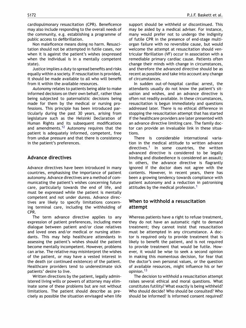
S172 P.J.F. Baskett et al.
cardiopulmonary resuscitation (CPR). Beneficencemay also include responding to the overall needs ofthe community, e.g. establishing a programme ofpublic access to defibrillation.
Non maleficence means doing no harm. Resusci-tation should not be attempted in futile cases, norwhen it is against the patient’s wishes (expressedwhen the individual is in a mentally competentstate).
Justice implies a duty to spread benefits and risksequally within a society. If resuscitation is provided,it should be made available to all who will benefitfrom it within the available resources.
Autonomy relates to patients being able to makeinformed decisions on their own behalf, rather thanbeing subjected to paternalistic decisions beingmade for them by the medical or nursing pro-fessions. This principle has been introduced par-ticularly during the past 30 years, arising fromlegislature such as the Helsinki Declaration ofHuman Rights and its subsequent modificationsand amendments.12 Autonomy requires that thepatient is adequately informed, competent, freefrom undue pressure and that there is consistencyin the patient’s preferences.
support should be withheld or discontinued. Thismay be aided by a medical adviser. For instance,many would prefer not to undergo the indignityof futile CPR in the presence of end-stage multi-organ failure with no reversible cause, but wouldwelcome the attempt at resuscitation should ven-tricular fibrillation (VF) occur in association with aremediable primary cardiac cause. Patients oftenchange their minds with change in circumstances,and therefore the advanced directive should be asrecent as possible and take into account any changeof circumstances.
In sudden out-of-hospital cardiac arrest, theattendants usually do not know the patient’s sit-uation and wishes, and an advance directive isoften not readily available. In these circumstances,resuscitation is begun immediately and questionsaddressed later. There is no ethical difference instopping the resuscitation attempt that has startedif the healthcare providers are later presented withan advance directive limiting care. The family doc-tor can provide an invaluable link in these situa-tions.
There is considerable international varia-tion in the medical attitude to written advanced 1
abiicbpa
Wa
Wttmtlteitoo
rcWs
Advance directives
Advance directives have been introduced in manycountries, emphasising the importance of patientautonomy. Advance directives are a method of com-municating the patient’s wishes concerning futurecare, particularly towards the end of life, andmust be expressed while the patient is mentallycompetent and not under duress. Advance direc-tives are likely to specify limitations concern-ing terminal care, including the withholding ofCPR.
The term advance directive applies to anyexpression of patient preferences, including meredialogue between patient and/or close relativesand loved ones and/or medical or nursing atten-dants. This may help healthcare attendants inassessing the patient’s wishes should the patientbecome mentally incompetent. However, problemscan arise. The relative may misinterpret the wishesof the patient, or may have a vested interest inthe death (or continued existence) of the patient.Healthcare providers tend to underestimate sickpatients’ desire to live.
Written directions by the patient, legally admin-istered living wills or powers of attorney may elim-inate some of these problems but are not withoutlimitations. The patient should describe as pre-cisely as possible the situation envisaged when life
irectives. In some countries, the writtendvanced directive is considered to be legallyinding and disobedience is considered an assault;n others, the advance directive is flagrantlygnored if the doctor does not agree with theontents. However, in recent years, there haseen a growing tendency towards compliance withatient autonomy and a reduction in patronisingttitudes by the medical profession.1
hen to withhold a resuscitationttempt
hereas patients have a right to refuse treatment,hey do not have an automatic right to demandreatment; they cannot insist that resuscitationust be attempted in any circumstance. A doc-
or is required only to provide treatment that isikely to benefit the patient, and is not requiredo provide treatment that would be futile. How-ver, it would be wise to seek a second opinionn making this momentous decision, for fear thathe doctor’s own personal values, or the questionf available resources, might influence his or herpinion.13
The decision to withhold a resuscitation attemptaises several ethical and moral questions. Whatonstitutes futility? What exactly is being withheld?ho should decide? Who should be consulted? Who
hould be informed? Is informed consent required?
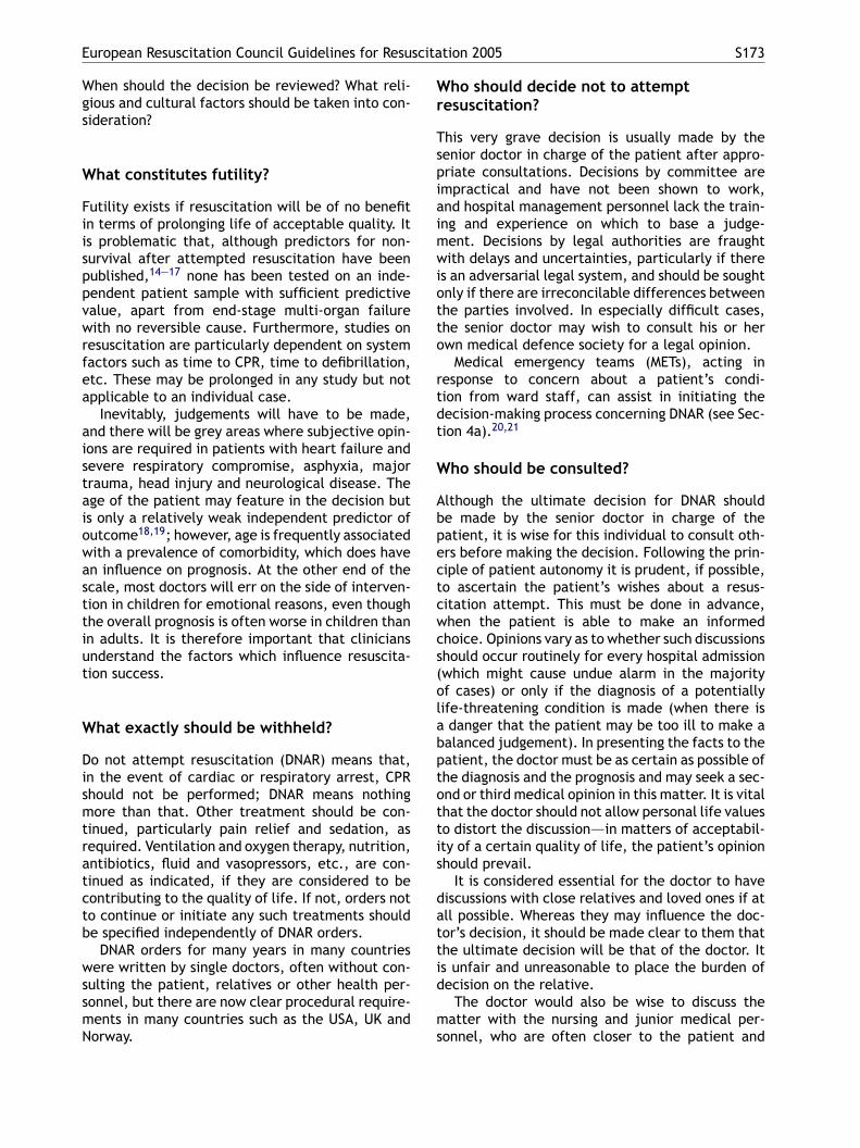
European Resuscitation Council Guidelines for Resuscitation 2005 S173
When should the decision be reviewed? What reli-gious and cultural factors should be taken into con-sideration?
What constitutes futility?
Futility exists if resuscitation will be of no benefitin terms of prolonging life of acceptable quality. Itis problematic that, although predictors for non-survival after attempted resuscitation have beenpublished,14—17 none has been tested on an inde-pendent patient sample with sufficient predictivevalue, apart from end-stage multi-organ failurewith no reversible cause. Furthermore, studies onresuscitation are particularly dependent on systemfactors such as time to CPR, time to defibrillation,etc. These may be prolonged in any study but notapplicable to an individual case.
Inevitably, judgements will have to be made,and there will be grey areas where subjective opin-ions are required in patients with heart failure andsevere respiratory compromise, asphyxia, majortrauma, head injury and neurological disease. Theage of the patient may feature in the decision but
Who should decide not to attemptresuscitation?
This very grave decision is usually made by thesenior doctor in charge of the patient after appro-priate consultations. Decisions by committee areimpractical and have not been shown to work,and hospital management personnel lack the train-ing and experience on which to base a judge-ment. Decisions by legal authorities are fraughtwith delays and uncertainties, particularly if thereis an adversarial legal system, and should be soughtonly if there are irreconcilable differences betweenthe parties involved. In especially difficult cases,the senior doctor may wish to consult his or herown medical defence society for a legal opinion.
Medical emergency teams (METs), acting inresponse to concern about a patient’s condi-tion from ward staff, can assist in initiating thedecision-making process concerning DNAR (see Sec-tion 4a).20,21
Who should be consulted?
Although the ultimate decision for DNAR should
is only a relatively weak independent predictor ofoutcome18,19; however, age is frequently associatedwith a prevalence of comorbidity, which does havean influence on prognosis. At the other end of thescale, most doctors will err on the side of interven-tion in children for emotional reasons, even thoughthe overall prognosis is often worse in children thanin adults. It is therefore important that cliniciansunderstand the factors which influence resuscita-tion success.What exactly should be withheld?
Do not attempt resuscitation (DNAR) means that,in the event of cardiac or respiratory arrest, CPRshould not be performed; DNAR means nothingmore than that. Other treatment should be con-tinued, particularly pain relief and sedation, asrequired. Ventilation and oxygen therapy, nutrition,antibiotics, fluid and vasopressors, etc., are con-tinued as indicated, if they are considered to becontributing to the quality of life. If not, orders notto continue or initiate any such treatments shouldbe specified independently of DNAR orders.
DNAR orders for many years in many countrieswere written by single doctors, often without con-sulting the patient, relatives or other health per-sonnel, but there are now clear procedural require-ments in many countries such as the USA, UK andNorway.
be made by the senior doctor in charge of thepatient, it is wise for this individual to consult oth-ers before making the decision. Following the prin-ciple of patient autonomy it is prudent, if possible,to ascertain the patient’s wishes about a resus-citation attempt. This must be done in advance,when the patient is able to make an informedchoice. Opinions vary as to whether such discussionsshould occur routinely for every hospital admission(which might cause undue alarm in the majorityof cases) or only if the diagnosis of a potentiallylife-threatening condition is made (when there isa danger that the patient may be too ill to make abalanced judgement). In presenting the facts to thepatient, the doctor must be as certain as possible ofthe diagnosis and the prognosis and may seek a sec-ond or third medical opinion in this matter. It is vitalthat the doctor should not allow personal life valuesto distort the discussion—–in matters of acceptabil-ity of a certain quality of life, the patient’s opinionshould prevail.
It is considered essential for the doctor to havediscussions with close relatives and loved ones if atall possible. Whereas they may influence the doc-tor’s decision, it should be made clear to them thatthe ultimate decision will be that of the doctor. Itis unfair and unreasonable to place the burden ofdecision on the relative.
The doctor would also be wise to discuss thematter with the nursing and junior medical per-sonnel, who are often closer to the patient and
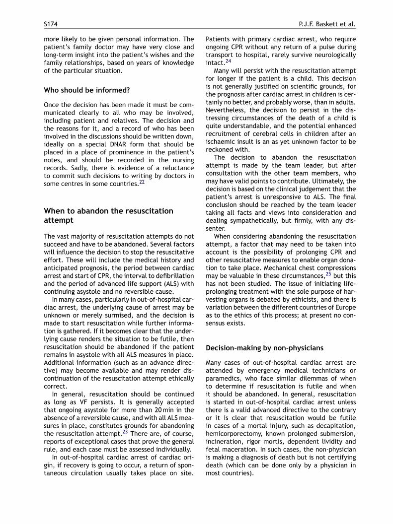
S174 P.J.F. Baskett et al.
more likely to be given personal information. Thepatient’s family doctor may have very close andlong-term insight into the patient’s wishes and thefamily relationships, based on years of knowledgeof the particular situation.
Who should be informed?
Once the decision has been made it must be com-municated clearly to all who may be involved,including patient and relatives. The decision andthe reasons for it, and a record of who has beeninvolved in the discussions should be written down,ideally on a special DNAR form that should beplaced in a place of prominence in the patient’snotes, and should be recorded in the nursingrecords. Sadly, there is evidence of a reluctanceto commit such decisions to writing by doctors insome centres in some countries.22
When to abandon the resuscitationattempt
Patients with primary cardiac arrest, who requireongoing CPR without any return of a pulse duringtransport to hospital, rarely survive neurologicallyintact.24
Many will persist with the resuscitation attemptfor longer if the patient is a child. This decisionis not generally justified on scientific grounds, forthe prognosis after cardiac arrest in children is cer-tainly no better, and probably worse, than in adults.Nevertheless, the decision to persist in the dis-tressing circumstances of the death of a child isquite understandable, and the potential enhancedrecruitment of cerebral cells in children after anischaemic insult is an as yet unknown factor to bereckoned with.
The decision to abandon the resuscitationattempt is made by the team leader, but afterconsultation with the other team members, whomay have valid points to contribute. Ultimately, thedecision is based on the clinical judgement that thepatient’s arrest is unresponsive to ALS. The finalconclusion should be reached by the team leadertaking all facts and views into consideration anddealing sympathetically, but firmly, with any dis-senter.
aaotmhpvvas
D
Maptiitoihifidm
The vast majority of resuscitation attempts do notsucceed and have to be abandoned. Several factorswill influence the decision to stop the resuscitativeeffort. These will include the medical history andanticipated prognosis, the period between cardiacarrest and start of CPR, the interval to defibrillationand the period of advanced life support (ALS) withcontinuing asystole and no reversible cause.
In many cases, particularly in out-of-hospital car-diac arrest, the underlying cause of arrest may beunknown or merely surmised, and the decision ismade to start resuscitation while further informa-tion is gathered. If it becomes clear that the under-lying cause renders the situation to be futile, thenresuscitation should be abandoned if the patientremains in asystole with all ALS measures in place.Additional information (such as an advance direc-tive) may become available and may render dis-continuation of the resuscitation attempt ethicallycorrect.
In general, resuscitation should be continuedas long as VF persists. It is generally acceptedthat ongoing asystole for more than 20 min in theabsence of a reversible cause, and with all ALS mea-sures in place, constitutes grounds for abandoningthe resuscitation attempt.23 There are, of course,reports of exceptional cases that prove the generalrule, and each case must be assessed individually.
In out-of-hospital cardiac arrest of cardiac ori-gin, if recovery is going to occur, a return of spon-taneous circulation usually takes place on site.
When considering abandoning the resuscitationttempt, a factor that may need to be taken intoccount is the possibility of prolonging CPR andther resuscitative measures to enable organ dona-ion to take place. Mechanical chest compressionsay be valuable in these circumstances,25 but this
as not been studied. The issue of initiating life-rolonging treatment with the sole purpose of har-esting organs is debated by ethicists, and there isariation between the different countries of Europes to the ethics of this process; at present no con-ensus exists.
ecision-making by non-physicians
any cases of out-of-hospital cardiac arrest arettended by emergency medical technicians oraramedics, who face similar dilemmas of wheno determine if resuscitation is futile and whent should be abandoned. In general, resuscitations started in out-of-hospital cardiac arrest unlesshere is a valid advanced directive to the contraryr it is clear that resuscitation would be futilen cases of a mortal injury, such as decapitation,emicorporectomy, known prolonged submersion,ncineration, rigor mortis, dependent lividity andetal maceration. In such cases, the non-physicians making a diagnosis of death but is not certifyingeath (which can be done only by a physician inost countries).
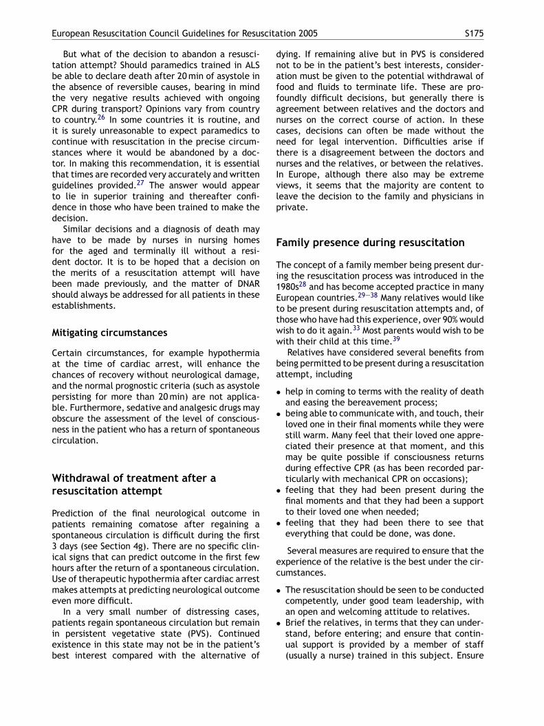
European Resuscitation Council Guidelines for Resuscitation 2005 S175
But what of the decision to abandon a resusci-tation attempt? Should paramedics trained in ALSbe able to declare death after 20 min of asystole inthe absence of reversible causes, bearing in mindthe very negative results achieved with ongoingCPR during transport? Opinions vary from countryto country.26 In some countries it is routine, andit is surely unreasonable to expect paramedics tocontinue with resuscitation in the precise circum-stances where it would be abandoned by a doc-tor. In making this recommendation, it is essentialthat times are recorded very accurately and writtenguidelines provided.27 The answer would appearto lie in superior training and thereafter confi-dence in those who have been trained to make thedecision.
Similar decisions and a diagnosis of death mayhave to be made by nurses in nursing homesfor the aged and terminally ill without a resi-dent doctor. It is to be hoped that a decision onthe merits of a resuscitation attempt will havebeen made previously, and the matter of DNARshould always be addressed for all patients in theseestablishments.
M
Cacapbonc
Wr
Pps3ihUme
pieb
dying. If remaining alive but in PVS is considerednot to be in the patient’s best interests, consider-ation must be given to the potential withdrawal offood and fluids to terminate life. These are pro-foundly difficult decisions, but generally there isagreement between relatives and the doctors andnurses on the correct course of action. In thesecases, decisions can often be made without theneed for legal intervention. Difficulties arise ifthere is a disagreement between the doctors andnurses and the relatives, or between the relatives.In Europe, although there also may be extremeviews, it seems that the majority are content toleave the decision to the family and physicians inprivate.
Family presence during resuscitation
The concept of a family member being present dur-ing the resuscitation process was introduced in the1980s28 and has become accepted practice in manyEuropean countries.29—38 Many relatives would liketo be present during resuscitation attempts and, ofthose who have had this experience, over 90% wouldww
ba
•
•
•
•
ec
•
•
itigating circumstances
ertain circumstances, for example hypothermiat the time of cardiac arrest, will enhance thehances of recovery without neurological damage,nd the normal prognostic criteria (such as asystoleersisting for more than 20 min) are not applica-le. Furthermore, sedative and analgesic drugs maybscure the assessment of the level of conscious-ess in the patient who has a return of spontaneousirculation.
ithdrawal of treatment after aesuscitation attempt
rediction of the final neurological outcome inatients remaining comatose after regaining apontaneous circulation is difficult during the firstdays (see Section 4g). There are no specific clin-
cal signs that can predict outcome in the first fewours after the return of a spontaneous circulation.se of therapeutic hypothermia after cardiac arrestakes attempts at predicting neurological outcome
ven more difficult.In a very small number of distressing cases,
atients regain spontaneous circulation but remainn persistent vegetative state (PVS). Continuedxistence in this state may not be in the patient’sest interest compared with the alternative of
ish to do it again.33 Most parents would wish to beith their child at this time.39
Relatives have considered several benefits fromeing permitted to be present during a resuscitationttempt, including
help in coming to terms with the reality of deathand easing the bereavement process;being able to communicate with, and touch, theirloved one in their final moments while they werestill warm. Many feel that their loved one appre-ciated their presence at that moment, and thismay be quite possible if consciousness returnsduring effective CPR (as has been recorded par-ticularly with mechanical CPR on occasions);feeling that they had been present during thefinal moments and that they had been a supportto their loved one when needed;feeling that they had been there to see thateverything that could be done, was done.
Several measures are required to ensure that thexperience of the relative is the best under the cir-umstances.
The resuscitation should be seen to be conductedcompetently, under good team leadership, withan open and welcoming attitude to relatives.Brief the relatives, in terms that they can under-stand, before entering; and ensure that contin-ual support is provided by a member of staff(usually a nurse) trained in this subject. Ensure
S176 P.J.F. Baskett et al.
that relatives understand that the choice to bepresent is entirely theirs, and do not provokefeelings of guilt, whatever their decision.
• Make the relatives aware of the proceduresthey are likely to see (e.g., tracheal intuba-tion, insertion of central venous catheters) andthe patient’s response (e.g., convulsive move-ments after defibrillation). Emphasise the impor-tance of not interfering with any procedures andexplain clearly the dangers of doing so.
• In the majority, of cases it will be necessary toexplain that the patient has not responded to theresuscitation attempt and that the attempt hasto be abandoned. This decision should be madeby the team leader, involving the members of theteam. Explain to the relatives that there may bea brief interval while equipment is removed, andthat then they will be able to return to be withtheir loved one at their leisure, alone or sup-ported, as they wish. Certain tubes and cannulaemay have to be left in place for medicolegal rea-sons.
• Finally, there should be an opportunity for therelative to reflect, ask questions about the causeand the process, and be given advice about the
Training and research on the recentlydead
Another matter that has raised considerable debateis the ethics, and in some cases the legality,of undertaking training and/or research on therecently dead.
Training
The management of resuscitation can be taughtusing scenarios with manikins and modern simula-tors, but training in certain skills required duringresuscitation is notoriously difficult. External chestcompressions and, to an extent, expired air venti-lation and insertion of oropharyngeal and nasopha-ryngeal airways can be taught using manikins; butdespite technological advances in manikins and sim-ulators, many other skills that are needed on aregular basis during resuscitation can be acquiredsatisfactorily only through practice on humans,dead or alive. These other skills include, for exam-ple, central and peripheral venous access, arterialpuncture and cannulation, venous cut-down, bag-mrasmsdceeeadsssult
cawpcritlac
procedure for registering the death and the sup-port services available.
In the event of an out-of-hospital arrest, therelatives may already be present, and possibly per-forming basic life support (BLS). Offer them theoption to stay; they may appreciate the opportunityto help and travel in the ambulance to hospital. Ifdeath is pronounced at the scene, offer the rela-tives the help and support of their family doctor orcommunity nurse and bereavement councillor.
For resuscitation staff, both in and out hospital,it is worth offering training in the matter of rela-tives being present.40
With increasing experience of family presenceduring resuscitation attempts, it is clear that prob-lems rarely, if ever, arise. In the majority ofinstances, relatives come in and stay for just afew minutes and then leave, satisfied that theyhave taken the opportunity to be there to sup-port their loved one and say goodbye as theywould have wished. Ten years ago most staffwould not have countenanced the presence ofrelatives during resuscitation, but a recent sur-vey has shown an increasingly open attitude andappreciation of the autonomy of both patient andrelatives.1 Perhaps this is related to a generallymore permissive and less autocratic attitude. Inter-national cultural and social variations still exist,and must be understood and appreciated withsensitivity.
ask ventilation, tracheal intubation, cricothy-oidotomy, needle thoracostomy, chest drainagend open-chest cardiac massage. Some of thesekills may be practised during routine clinical work,ostly involving anaesthesia, and to a lesser degree
urgery; but others such as cricothyroidotomy, nee-le thoracostomy and open chest cardiac massageannot, and are needed only in a life-threateningmergency when it is difficult to justify a teachingxercise. In modern day practice, with practition-rs being called increasingly to account and patientutonomy prevailing, it is becoming more and moreifficult to obtain permission for student practice ofkills in the living. Gone are the days when admis-ion to a ‘teaching hospital’ implied automatic con-ent for students to practise procedures on patientsnder supervision as they wished. And yet the pub-ic expect, and are entitled to, competent practi-ioners for generation after generation.
So the question arises as to whether it is ethi-ally and morally appropriate to undertake trainingnd practice on the living or the dead. There is aide diversity of opinion on this matter.41 Many,articularly those in the Islamic nations, find theoncept of any skills training and practice on theecently dead completely abhorrent because of annnate respect for the dead body. Others will accepthe practice of non-invasive procedures that do noteave a mark, such as tracheal intubation; and somere open and frank enough to accept that any pro-edure may be learned on the dead body with the
European Resuscitation Council Guidelines for Resuscitation 2005 S177
justification that the learning of skills is paramountfor the well-being of future patients.
One option is to request informed consent forthe procedure from the relative of the deceased.However, only some will obtain permission,1,40 andmany find this very difficult to do in the harrowingcircumstances of breaking bad news simultaneouslyto the recently bereaved. As a result, frequentlyonly non-invasive procedures are practised, on thebasis that what is not seen will not cause distress.The days of undertaking any procedure without con-sent are rapidly coming to an end, and perhaps itis now becoming increasingly necessary to mount apublicity campaign to exhort the living to give per-mission for training on their dead body through anadvance directive, in much the same way as per-mission for transplant of organs may be given. Itmay be that an ‘opt-out’ rather than an ‘opt-in’arrangement may be adopted, but this will requirechanges in the law in most countries. It is advisedthat healthcare professionals learn local and hos-pital policies regarding this issue and follow theestablished policy.
Research
Research on the recently dead is likely toencounter similar restrictions unless previous per-mission is granted as part of an advance directiveby the patient, or permission can be given imme-diately by the relative who is next of kin. Legalownership of the recently dead is established onlyin a few countries, but in many countries it is atleast tacitly agreed that the body ‘belongs’ to therelatives (unless there are suspicious circumstancesor the cause of death is unknown), and permissionfor any research must be granted by the next of kinunless there is an advance directive giving consent.Obtaining consent from relatives in the stressfulcircumstances of immediate bereavement is unen-viable and potentially damaging to the relationshipbetween doctor and relative.
Research can still be carried out during post-mortem examination, for instance to study thetraumatic damage resulting from the use of spe-cific methods of chest compression, but all bodyparts must be returned to the patient unless spe-cific permission is obtained from relatives to dootherwise.
Bc
Batiphmidl
Cw
IdTmiitmtaitit
There are important ethical issues relating toundertaking randomized clinical trials for patientsin cardiac arrest who cannot give informed con-sent to participate in research studies. Progress inimproving the dismal rates of successful resuscita-tion will only come through the advancement ofscience through clinical studies. The utilitarian con-cept in ethics looks to the greatest good for thegreatest number of people. This must be balancedwith respect for patient autonomy, according towhich patients should not be enrolled in researchstudies without their informed consent. Over thepast decade, legal directives have been introducedinto the USA and the European Union42,43 that placesignificant barriers to research on patients dur-ing resuscitation without informed consent fromthe patient or immediate relative.44 There aredata showing that such regulations deter researchprogress in resuscitation.45 It is indeed possible thatthese directives may in themselves conflict withthe basic human right to good medical treatmentas set down in the Helsinki Agreement.12 Researchin resuscitation emanating from the USA has fallendramatically in the last decade,46 and it appearsvery likely that the European Union will followsuit as the rules bite there.47 The US authoritieshave, to a very limited extent, sought to introducemethods of exemption,42 but these are still asso-ciated with problems and almost insurmountabledifficulties.45
reaking bad news and bereavementounselling
reaking news of the death of a patient to a rel-tive is an unenviable task. It is a moment thathe relative will remember for ever, so it is verymportant to do it as correctly and sensitively asossible. It also places a considerable stress on theealthcare provider who has this difficult duty. Bothay need support in the ensuing hours and days. It
s salutatory that the breaking of bad news is sel-om taught in medical school or at postgraduateevel.1
ontacting the family in the case of deathithout the relatives being present
f the relatives are not present when the patienties, they must be contacted as soon as possible.he caller may not be known to the relative andust take great care to ensure that his or her
dentity is made quite clear to the relative and,n turn, the caller must make sure of the rela-ionship of the call recipient to the deceased. Inany cases it is not stated on the telephone that
he patient has actually died, unless the distancend travel time are prolonged (e.g., the relatives in another country). Many find that it is bettero say that the patient is seriously and criticallyll or injured and that the relatives should comeo hospital immediately, so that a full explanation
S178 P.J.F. Baskett et al.
can be given face to face. It is wise to request thatrelatives to ask a friend to drive them to hospital,and to state that nothing will be gained by drivingat speed. When the relatives arrive they should begreeted right away by a competent and knowledge-able member of staff, and the situation explainedimmediately. Delays in being told the facts areagonising.
Who should break the bad news to therelative?
Gone are the days when it was acceptable for thepatronising senior doctor to delegate the breakingof bad news to a junior assistant. Nowadays, it isgenerally agreed that it is the duty of the seniordoctor or the team leader to talk to the relatives.Nevertheless, it is wise to be accompanied by anexperienced nurse who may be a great comfort forthe patient (and indeed the doctor).
Where and how should bad news be given?
The environment where bad news is given is vitally
• relief (‘‘I am so glad his suffering is over,’’ or‘‘He went suddenly—–that is what he would havewished’’);
• anger with the patient (‘‘I told him to stop smok-ing,’’ or ‘‘He was too fat to play squash,’’ or‘‘Look at the mess he has left me in’’);
• self-guilt (‘‘If only I had not argued with him thismorning before he left for work,’’ or ‘‘Why did Inot tell the doctor he got chest pain?’’);
• anger with the medical system (‘‘Why did theambulance take so long?’’ or ‘‘The doctor wasfar too young and did not know what he/she wasdoing’’);
• uncontrollable wailing and crying and anguish;• complete expressionless catatonia.
It may be useful to reassure the family that theydid everything correctly, such as calling for help andgetting to the hospital but, in the vast majority ofcases, healthcare providers are unable to restartthe heart.
Some time may elapse before conversation canresume and, at this stage, ask relatives if they haveany questions about the medical condition and thetreatment given. It is wise to be completely openand honest about this, but always say ‘‘He did not
important. There should be a room set aside forrelatives of the seriously ill that is tastefully andcomfortably furnished, with free access to a tele-phone, television and fresh flowers daily (whichmay be provided by the florist who runs the flowershop that is in most hospitals in Europe).
There are some basic principles to be followedwhen breaking bad news, that should be adhered toif grave errors are to be avoided and the relative isnot to be discomforted. It is essential to know thefacts of the case and to make quite sure to whomwho you are talking. Body language is vital; alwayssit at the same level as the patient and relative;do not stand up when they are sitting down. Makesure you are cleanly dressed; wearing blood-stainedclothing is not good. Do not give the impressionthat you are busy and in a hurry. Give the newsthey are anxious to hear immediately, using thewords ‘‘dead’’ or ‘‘has died’’, ‘‘I am very sorry tohave to tell you that your father/husband/son hasdied’’. Do not leave any room for doubt by usingsuch phrases as ‘‘passed on’’ or left us’’ or ‘‘goneup above’’.
Discussing the medical details comprehensivelyat this stage is not helpful; wait until they are askedfor. Touching may be appropriate, such as holdinghands or placing an arm on the shoulder, but peopleand customs vary and the doctor needs to be awareof these. Do not be ashamed if you shed a tear your-self. Allow time for the news to be assimilated bythe relative. Reactions may vary, including
suffer’’.In the majority of cases the relative will wish
to see the body. It is important that the body andbedclothes are clean and all tubes and cannulaeare removed, unless these are needed for post-mortem examination. The image of the body willleave an impression on the relative that will last forever. A post-mortem examination may be required,and this should requested with tact and sensitiv-ity, explaining that the procedure will be carriedout by a professional pathologist and will help todetermine the precise cause of death.
Children
Breaking bad news to children may be perceived topresent a special problem, but experience seemsto indicate that it is better to be quite open andhonest with them, so helping to dispel the night-marish fantasies that children may concoct aboutdeath. It is helpful to contact the school, so thatthe teachers and fellow pupils can be prepared toreceive the child back into the school environmentwith support and sensitivity.
Closure
In many cases this will be the relative’s firstexperience of death, and help should be offeredwith the bewildering administration of the official
European Resuscitation Council Guidelines for Resuscitation 2005 S179
registration of death, funeral arrangements andsocioeconomic support by the hospital or commu-nity social worker. Depending on religious beliefs,the hospital padre or priest may have a vital role toplay. Whenever possible, family physicians shouldbe informed immediately by telephone or e-mailwith the essential details of the case, so that theycan give full support to the relatives. A follow-up telephone call to the relative a day or twolater from a member of the hospital staff whowas involved, offering to be of help and to answerany questions that the relative may have forgottenabout at the time, is always appreciated.
Staff debrief
Although many members of staff seem, and oftenare, little affected by death in the course of theirwork, this should not be assumed. Their senseof accomplishment and job satisfaction may beaffected adversely, and there may be feelings ofguilt, inadequacy and failure. This may be particu-larly apparent in, but not restricted to, very juniormembers of staff. A team debrief of the eventusing positive and constructive critique techniques
References
1. Baskett PJ, Lim A. The varying ethical attitudes towardsresuscitation in Europe. Resuscitation 2004;62:267—73.
2. Sprung CL, Cohen SL, Sjokvist P, et al. End-of-life practicesin European intensive care units: the Ethicus Study. JAMA2003;290:790—7.
3. Richter J, Eisemann MR, Bauer B, Kreibeck H, Astrom S.Decision-making in the treatment of elderly people: a cross-cultural comparison between Swedish and German physi-cians and nurses. Scand J Caring Sci 2002;16:149—56.
4. da Costa DE, Ghazal H, Al Khusaiby S. Do not resuscitateorders and ethical decisions in a neonatal intensive careunit in a Muslim community. Arch Dis Child Fetal NeonatalEd 2002;86:F115—9.
5. Ho NK. Decision-making: initiation and withdrawing life sup-port in the asphyxiated infants in developing countries. Sin-gapore Med J 2001;42:402—5.
6. Richter J, Eisemann M, Zgonnikova E. Doctors’ authori-tarianism in end-of-life treatment decisions. A compari-son between Russia, Sweden and Germany. J Med Ethics2001;27:186—91.
7. Cuttini M, Nadai M, Kaminski M, et al. End-of-life decisions inneonatal intensive care: physicians’ self-reported practicesin seven European countries. EURONIC Study Group. Lancet2000;355:2112—8.
8. Konishi E. Nurses’ attitudes towards developing a do notresuscitate policy in Japan. Nurs Ethics 1998;5:218—27.
9. Muller JH, Desmond B. Ethical dilemmas in a cross-cultural
1
1
1
1
1
1
1
1
1
1
2
should be conducted and personal bereavementcounselling offered to those with a particular need.How this is done will vary with the individual andwill range from an informal chat in the pub or cafe(which seems to deal effectively with many cases)to professional counselling. It should be explainedthat distress after a death at work may be a normalreaction to an abnormal situation.
Conclusions
Resuscitation has given many a new lease of life,to the delight of themselves and their relatives,but has the potential to bring misery to a few. Thischapter addresses how that misery can be reducedby not attempting resuscitation in inappropriatecircumstances or in cases with a valid advanceddirective, and when to discontinue the resuscita-tion attempt in cases of futility or PVS.
Ethical issues such as training and research onthe recently dead, and the presence of family mem-bers during the resuscitation attempt, place fur-ther burdens on the medical profession but must bedealt with sympathetically, and with an apprecia-tion of growing patient autonomy and human rightsthroughout the world.
Finally, the breaking of bad news is one of themost difficult tasks to be faced by the medical andnursing professions. It requires time, training, com-passion and understanding.
context. A Chinese example. West J Med 1992;157:323—7.0. Edgren E. The ethics of resuscitation; differences between
Europe and the USA-Europe should not adopt Americanguidelines without debate. Resuscitation 1992;23:85—9.
1. Beauchamp TL, Childress J, editors. Principles of biomedicalethics. 3rd ed. Oxford: Oxford University Press; 1994.
2. Declaration of Helsinki. Ethical principles for medicalresearch involving human subjects adopted by the 18th WMAGeneral Assembly, Helsinki, Finland, June 1964 and amendedat the 29th, 35th, 41st, 48th, and 52nd WMA Assemblies.Geneva, 1964.
3. Aasland OG, Forde R, Steen PA. Medical end-of-life decisionsin Norway. Resuscitation 2003;57:312—3.
4. Danciu SC, Klein L, Hosseini MM, Ibrahim L, Coyle BW, KehoeRF. A predictive model for survival after in-hospital car-diopulmonary arrest. Resuscitation 2004;62:35—42.
5. Dautzenberg PL, Broekman TC, Hooyer C, Schonwetter RS,Duursma SA. Review: patient-related predictors of car-diopulmonary resuscitation of hospitalized patients. AgeAgeing 1993;22:464—75.
6. Haukoos JS, Lewis RJ, Niemann JT. Prediction rules for esti-mating neurologic outcome following out-of-hospital cardiacarrest. Resuscitation 2004;63:145—55.
7. Herlitz J, Engdahl J, Svensson L, Young M, Angquist KA, Holm-berg S. Can we define patients with no chance of survivalafter out-of-hospital cardiac arrest? Heart 2004;90:1114—8.
8. Herlitz J, Engdahl J, Svensson L, Angquist KA, Young M,Holmberg S. Factors associated with an increased chanceof survival among patients suffering from an out-of-hospitalcardiac arrest in a national perspective in Sweden. Am HeartJ 2005;149:61—6.
9. Ebell MH. Prearrest predictors of survival following in-hospital cardiopulmonary resuscitation: a meta-analysis. JFam Pract 1992;34:551—8.
0. Hillman K, Parr M, Flabouris A, Bishop G, Stewart A. Redefin-ing in-hospital resuscitation: the concept of the medicalemergency team. Resuscitation 2001;48:105—10.
S180 P.J.F. Baskett et al.
21. The MERIT study investigators. Introduction of the medicalemergency team (MET) system: a cluster-randomised con-trolled trial. Lancet 2005;365:2091—7.
22. Sovik O, Naess AC. Incidence and content of written guide-lines for ‘‘do not resuscitate’’ orders. Survey at six dif-ferent somatic hospitals in Oslo. Tidsskr Nor Laegeforen1997;117:4206—9.
23. Bonnin MJ, Pepe PE, Kimball KT, Clark Jr PS. Distinct cri-teria for termination of resuscitation in the out-of-hospitalsetting. JAMA 1993;270:1457—62.
24. Kellermann AL, Hackman BB, Somes G. Predicting the out-come of unsuccessful prehospital advanced cardiac life sup-port. JAMA 1993;270:1433—6.
25. Steen S, Liao Q, Pierre L, Paskevicius A, Sjoberg T. Evaluationof LUCAS, a new device for automatic mechanical compres-sion and active decompression resuscitation. Resuscitation2002;55:285—99.
26. Naess A, Steen E, Steen P. Ethics in treatment deci-sions during out-of-hospital resuscitation. Resuscitation1997;35:245—56.
27. Joint Royal Colleges Ambulance Liaison Committee. Newslet-ter 1996 and 2001. Royal College of Physicians: London.
28. Doyle CJ, Post H, Burney RE, Maino J, Keefe M, Rhee KJ. Fam-ily participation during resuscitation: an option. Ann EmergMed 1987;16:673—5.
29. Adams S, Whitlock M, Higgs R, Bloomfield P, Baskett PJ.Should relatives be allowed to watch resuscitation? BMJ1994;308:1687—92.
30. Hanson C, Strawser D. Family presence during cardiopul-monary resuscitation: Foote Hospital emergency depart-
3
3
3
3
3
36. Robinson SM, Mackenzie-Ross S, Campbell Hewson GL, Egle-ston CV, Prevost AT. Psychological effect of witnessedresuscitation on bereaved relatives. Lancet 1998;352:614—7[comment].
37. Baskett PJF. The ethics of resuscitation. In: Colquhoun MC,Handley AJ, Evans TR, editors. The ABC of resuscitation. 5thed. London: BMJ Publishing Group; 2004.
38. Azoulay E, Sprung CL. Family-physician interactions in theintensive care unit. Crit Care Med 2004;32:2323—8.
39. Bouchner H, Vinci R, Waring C. Pediatric procedures: do par-ents want to watch? Pediatrics 1989;84:907—9.
40. Resuscitation Council (UK) Project Team. Should relativeswitness resuscitation? London, UK: Resuscitation Council;1996.
41. Morag RM, DeSouza S, Steen PA, et al. Performing procedureson the newly deceased for teaching purposes: what if wewere to ask? Arch Intern Med 2005;165:92—6.
42. US Department of Health and Human Services. Protection ofhuman subjects: informed consent and waiver of informedconsent requirements in certain emergency circumstances.In: 61 Federal Register 51528 (1996) codified at CFR #50.24and #46.408; 1996.
43. Fontaine N, Rosengren B. Directive/20/EC of the EuropeanParliament and Council of 4th April 2001 on the approxima-tion of the laws, regulations and administrative provisionsof the Member States relating to the implementation ofgood clinical practice in the conduct of trials on medicalproducts for human use. Off J Eur Commun 2001;121:34—44.
44. Lemaire F, Bion J, Blanco J, et al. The European Union
4
4
4
ment’s nine-year perspective. J Emerg Nurs 1992;18:104—6.1. Cooke MW. I desperately needed to see my son. BMJ
1991;302:1023.2. Gregory CM. I should have been with Lisa as she died. Accid
Emerg Nurs 1995;3:136—8.3. Boie ET, Moore GP, Brummett C, Nelson DR. Do parents want
to be present during invasive procedures performed on theirchildren in the emergency department? A survey of 400 par-ents. Ann Emerg Med 1999;34:70—4.
4. Boudreaux ED, Francis JL, Loyacano T. Family presence dur-ing invasive procedures and resuscitations in the emergencydepartment: a critical review and suggestions for futureresearch. Ann Emerg Med 2002;40:193—205.
5. Martin J. Rethinking traditional thoughts. J Emerg Nurs1991;17:67—8.
Directive on Clinical Research: present status of imple-mentation in EU member states’ legislations with regardto the incompetent patient. Intensive Care Med 2005;31:476—9.
5. Nichol G, Huszti E, Rokosh J, Dumbrell A, McGowan J, BeckerL. Impact of informed consent requirements on cardiacarrest research in the United States: exception from consentor from research? Resuscitation 2004;62:3—23.
6. Mosesso Jr VN, Brown LH, Greene HL, et al. Conductingresearch using the emergency exception from informed con-sent: the public access defibrillation (PAD) trial experience.Resuscitation 2004;61:29—36.
7. Sterz F, Singer EA, Bottiger B, et al. A serious threat toevidence based resuscitation within the European Union.Resuscitation 2002;53:237—8.
Resuscitation (2005) 67S1, S181—S189
European Resuscitation Council Guidelines forResuscitation 2005Section 9. Principles of training in resuscitation
Peter J. F. Baskett, Jerry P. Nolan, Anthony Handley, Jasmeet Soar,Dominique Biarent, Sam Richmond
I
Tiaevr6wb
O
Twttbpac
M
Tc
d
ntroduction
here are a variety of methods used for train-ng in resuscitation. None are perfect and, in thebsence of frequent practice, retention of knowl-dge and skills is suboptimal. The optimal inter-al for retraining has not been established, butepeated refresher training at intervals of less thanmonths seems to be needed for most individualsho are not undertaking resuscitation on a regularasis.1—12
bjectives
he objective of training is to equip the learnerith the ability to be able to undertake resuscita-
ion in a real clinical situation at the level at whichhey would be expected to perform, be they be layystander, first responder in the community or hos-ital, a healthcare professional working in an acuterea, or a member of the medical emergency or
established European Resuscitation Council (ERC)course with small-group (four to eight mem-bers) participation using interactive discussion andhands-on practice for skills and clinical scenar-ios for problem-solving and team leadership.13
The ratio of instructors to candidates shouldrange from 1:3 to 1:6, depending on the type ofcourse.
Core knowledge should be acquired by candi-dates before the course by study of the coursemanual or an interactive CD designed for the pur-pose. The course should aim to produce an improve-ment in competence in the learner, and thereshould be a test of core knowledge and an ongoingassessment of practical skills and scenario manage-ment. Sophisticated manikins, simulators and vir-tual reality techniques may be incorporated intothe scenario-based training.14
For basic life support (BLS) by lay people or firstresponders, home-based learning using a video orinteractive CD with a simple manikin may offera valuable alternative to traditional instructor-
ardiac arrest response team. based courses.15—19 This method minimises candi-date disruption and instructor time and finances.However, the role of the instructor should not beuaoa
Coun
ethods
raining should follow the principles of adult edu-ation and learning. Generally this will mean an
0300-9572/$ — see front matter © 2005 European Resuscitationoi:10.1016/j.resuscitation.2005.10.006
nderestimated and, in addition to explaining situ-tions that were unforeseen on the original videor CD, the instructor can act as a role modelnd provide invaluable enthusiasm and motivation.
cil. All Rights Reserved. Published by Elsevier Ireland Ltd.
S182 P.J.F. Baskett et al.
Group participation has also been demonstrated toenhance the overall learning process.
Ethos
The course should be taught by trained instruc-tors who have undertaken the relevant specific ERCcourse in teaching and assessment. Teaching shouldbe conducted by encouragement with construc-tive feedback on performance rather than humil-iation. First names are encouraged among bothfaculty and candidates to reduce apprehension,and the mentor/mentee system is used to enhancefeedback and support for the candidate. Stress isinevitable,20 particularly during assessment, butthe aim of the instructors is to enable the candi-dates to do their best.
Language
Initially, the ERC courses were taught in English byan international faculty.13 As local instructors havebeen trained, and manuals and course materialshave been translated into different languages, the
GIC, include an educator who has undertaken spe-cific training in medical educational practice andthe principles of adult learning. Details of theseinstructor courses are given below. There are noformal tests for candidates during the course, butassessment is done by the faculty and feedback isgiven as appropriate.
Instructor candidate stage
Following successful completion of an instructorcourse, the individual is designated as an instruc-tor candidate (IC), normally taught on two separatecourses under supervision, and is given constructivefeedback on performance. After experience of twocourses, the IC normally progresses to full instruc-tor status, but occasionally the faculty decides thata further course is required or, rarely, that the can-didate is not suitable to progress to be an instructor.An appeal can be lodged with the relevant Inter-national Course Committee, which makes the finaldecision.
Course director status
StptImachewcr
I
IctccvlLlttH(ir
courses, particularly the provider courses, are nowtaught increasingly in the candidates’ native lan-guage.
Instructors
A tried and tested method has evolved for identify-ing and training instructors.
Identification of instructor potentials
Instructors will be individuals who, in the opinion ofthe faculty, have demonstrated good competencein the subjects at a provider course and, impor-tantly, have shown qualities of leadership and clin-ical credibility and skills that involve being articu-late, supportive and motivated. These individualswill be invited to take part in an instructor coursecalled the Generic Instructor Course (GIC) in thecase of Advanced Life Support (ALS) and EuropeanPaediatric Life Support (EPLS) courses, or BasicLife Support (BLS)/Automated External Defibrilla-tion (AED) Instructor Course in the BLS and AEDcourses. An instructor course for Immediate LifeSupport (ILS) is under development.
The instructor courses
These are conducted for instructor potentials (IPs)by experienced instructors and, in the case of the
elected individuals may progress to the status ofhe course director. They will be selected by theireers and approved by the relevant committee ofhe National Resuscitation Council or the relevantnternational Course Committee. Course directorsust be relatively senior individuals with consider-
ble clinical credibility, good judgement and impec-able powers of assessment and fairness. They willave embraced the educational principles inher-nt in the instructor course. Normally, individualsill have had experience of teaching on at least sixourses and will have been appointed course codi-ector on at least one occasion.
nterchange of instructors
nterchange between instructors of different dis-iplines is possible. For instance, an ALS instruc-or may proceed directly to be an IC on an EPLSourse, provided that he or she has passed the EPLSourse and has been identified as an IP and viceersa. There is no need to repeat the GIC. Simi-arly, current instructors in the Advanced Traumaife Support (ATLS) Course of the American Col-ege of Surgeons, having been identified as an IP inhe relevant provider course, may proceed directlyo being an IC in ALS or EPLS. Current Americaneart Association Advanced Cardiac Life Support
ACLS) or Paediatric Advanced Life Support (PALS)nstructors may proceed directly to IC status in theelevant course.
European Resuscitation Council Guidelines for Resuscitation 2005 S183
Code of conduct
All instructors must adhere to the code of conductfor the instructors, which is set out in Appendix A.
The Basic Life Support (BLS) andAutomated External Defibrillator (AED)courses
BLS and AED courses are appropriate for a widerange of providers. These may include clinical andnon-clinical healthcare professionals (particularlythose who are less likely to be faced with havingto manage a cardiac arrest), general practitioners,dentists, medical students, first-aid workers, life-guards, those with a duty of care for others (suchas school teachers and care workers), and membersof first-responder schemes, as well as members ofthe general public.
Provider course format
The aim of these provider courses is to enable eachcandidate to gain competency in BLS or the useobmcI
isnimdbppat
wAs
I
Mpqtibt
ignated as IP. The aim is to be as inclusive as possibleregarding the course attendance, the over-ridingcriterion being that all candidates should have thepotential and knowledge to teach the subject.
The BLS/AED instructor course follows the prin-ciples of the GIC, with an emphasis on teachinglay people. Following successful completion of thecourse, each candidate becomes an IC and teachestwo BLS or AED courses before becoming a fullinstructor.
Introducing courses into a country
Many ERC BLS and AED provider courses are run by,or under the control of, the National ResuscitationCouncil. The normal procedure for introducing ERCBLS/AED courses into a country is that ERC interna-tional instructors visit that country to run a 2-daycombined BLS provider, AED provider and BLS/AEDinstructor course. If there are local instructors(e.g., those who have passed an ERC course suc-cessfully, or who are ERC ALS instructors), theyteach on the course in a 1:1 ratio of international tolocal instructor, with the course director (an inter-national instructor) as an additional person who can
f AED. Details of appropriate competencies haveeen published by the ERC BLS Working Group anday be found on http://www.erc.edu. BLS and AED
ourses are developed and managed by the ERCnternational BLS Course Committee (ICC).
Each BLS or AED provider course lasts approx-mately half a day and consists of skill demon-trations and hands-on practice, with a minimumumber of lectures. The recommended ratio ofnstructors to candidates is 1:6, with at least oneanikin and one AED for each group of six candi-ates. Formal assessment is not usually undertaken,ut each candidate receives individual feedback onerformance. Those who need a certificate of com-etency for professional or personal use may bessessed continuously during the course or defini-ively at the end.
BLS provider and AED provider manuals, togetherith certificates, may be purchased from the ERC.pproved alternative manuals, translated if neces-ary into the local language, may also be used.
nstructor course
any of the candidates attending a BLS or AEDrovider course are lay people, and some subse-uently want to become instructors themselves. Forhis reason, the ERC has developed a 1-day BLS/AEDnstructor course. Candidates for this course muste healthcare professionals, or lay people who holdhe ERC BLS or AED provider certificate and are des-
support local instructors. After a successful coursethe local instructors become full ERC instructors,and the outstanding local instructors are selectedto become instructor trainers. Subsequent coursesare normally held in the language of the countryconcerned, and training materials are translatedinto that language. The candidates who are on thecombined course qualify, hopefully, as ERC BLS/AEDICs. They then need to teach on one or two providercourses, under the supervision of full instructors,before becoming full instructors themselves.
The Immediate Life Support (ILS) course
The ILS course is for the majority of healthcareprofessionals who attend cardiac arrests rarelybut have the potential to be first respondersor cardiac-arrest team members.21 The courseteaches the healthcare professionals the skills thatare most likely to result in successful resusci-tation while awaiting the arrival of the resusci-tation team.22 Importantly, the ILS course alsoincludes a section on preventing cardiac arrest,and complements other short courses that focus onmanaging sick patients in the first 24 h of critical ill-ness when critical care expertise is not immediatelyavailable.23—25 There is a large group of potentialcandidates including nurses, nursing students, doc-tors, medical students, dentists, physiotherapists,radiographers and cardiac technicians.
S184 P.J.F. Baskett et al.
Current ALS instructors and ICs can teach andassess on ILS courses. There is also a pilot projectunderway to develop specific ILS instructors. Theremust be at least 1 instructor for every 6 can-didates, with a maximum of 30 candidates on acourse.
Course format
The ILS course is delivered over 1 day and compriseslectures, hands-on skills teaching and cardiac-arrest scenario teaching (CASTeach) using manikins.The programme includes a number of options thatallow instructors to tailor the course to their can-didate group.
Course content
The course covers those skills that are most likelyto result in successful resuscitation: causes and pre-vention of cardiac arrest, starting CPR, basic airwayskills and defibrillation (AED or manual). There areoptions to include the teaching of the laryngealmask airway and drug treatments during cardiac
rillation. With a supportive approach, the major-ity of candidates achieve the course learning out-comes.
Equipment
The ILS course is designed to be straightforwardto run. Most courses are conducted in hospitalswith small groups of candidates (average 12 can-didates). The course requires lecture facilities anda skills teaching area for each group of six candi-dates. There needs to be at least one ALS manikinfor every six candidates. The course should be suit-able for local needs. Course centres should tryas far as possible to train candidates to use theequipment (e.g., defibrillator type) that is availablelocally.
Course report and results sheet
A course report and the results sheet are compiledby the course director and filed with the NationalResuscitation Council and the ERC.
T
TattTamwtcwdmeida
C
T(setifn
arrest. Once all the skills have been covered, thereis a cardiac arrest demonstration by the instruc-tors that outlines the first-responder role to thecandidates. This is followed by the CASTeach sta-tion where candidates practise. ILS candidates arenot usually expected to undertake the role of theteam leader. Candidates should be able to starta resuscitation attempt and continue until moreexperienced help arrives. When appropriate, theinstructor takes over as a resuscitation team leader.This is not always necessary, as in some scenariosresuscitation may be successful before more expe-rienced help arrives. Set scenarios are used thatare adapted to the workplace and the clinical roleof the candidate.
Assessment
Candidate’s performances are assessed contin-uously and they must show their competencethroughout the ILS course. There are no formaltesting stations, removing the threat associatedwith spot testing at the end of the course. Candi-dates are sent the assessment forms with the pre-course materials. The forms indicate clearly howtheir performance will be measured against a pre-determined criteria. Assessment on the ILS courseenables the candidate to see what is expected andframe learning around achievement of these out-comes. The following practical skills are assessed onthe ILS course: airway management, BLS and defib-
he Advanced Life Support (ALS) course
he target candidates for this course are doctorsnd senior nurses working in emergency areas ofhe hospital and those who may be members ofhe medical emergency or cardiac arrest teams.26
he course is also suitable for senior paramedicsnd certain hospital technicians. The ILS course isore suitable for first-responder nurses, doctorsho rarely encounter cardiac arrest in their prac-
ice, and emergency medical technicians. Up to 32andidates can be accommodated on the course,ith a ratio of at least 1 instructor for every 3 can-idates. Up to a maximum of 50% of the instructorsay be ICs. Groups for teaching should not exceed
ight candidates and should be six ideally. Eachnstructor acts as a mentor for a small group of can-idates. The course normally lasts for two to twond a half days.
ourse format
he course format has very few formal lecturesfour), and teaching concentrates on hands-onkills, clinically based scenarios in small groups withmphasis on the team leader approach and interac-ive group discussions. Mentor/mentee sessions arencluded, to allow candidates to give and receiveeedback. Faculty meetings are held at the begin-ing of the course and at the end of each day of the
European Resuscitation Council Guidelines for Resuscitation 2005 S185
course. Social occasions, such as course and facultydinners, add greatly to the course interaction andenjoyment.
Course content
The course content is based on the currentERC guidelines for resuscitation. Candidates areexpected to have studied the ALS course manualcarefully before the course.
The course aims to train candidates to highlightthe causes of cardiac arrest, identify sick patients indanger of deterioration and manage cardiac arrestand the immediate periarrest problems encoun-tered in and around the first hour or so of the event.It is not a course in advanced intensive care or car-diology. Competence in BLS is expected before thecandidate enrols for the course.
Emphasis is placed on the techniques of safedefibrillation and ECG interpretation, the manage-ment of the airway and ventilation, the manage-ment of periarrest rhythms, simple acid/base bal-ance and special circumstances relating to car-diac arrest. Post-resuscitation care, ethical aspectsrelated to resuscitation and care of the bereaved
Course report and results sheet
A course report and the results sheet are compiledby the course director and filed with the nationalresuscitation council and the ERC.
The European Paediatric Life Support(EPLS) course
The EPLS course is designed for healthcare workerswho are involved in the resuscitation of a newborn,an infant or a child whether in or out of hospitalThe course aims at providing caregivers with theknowledge and skills for the management of thecritically ill child during the first hour of illnessand to prevent progression of diseases to cardiacarrest.
Competence in basic paediatric life support is aprerequisite, although a 90-min refresher course onBLS and relief of foreign-body airway obstructionis included. The EPLS course is suitable for doc-tors, nurses, emergency medical technicians andparamedics, etc., who have a duty to respondto sick newborns, infants and children in theirppp
admeralUIcist
C
T(iTtmcmaia
are included in the course.
Assessment and testing
Each candidate is assessed individually andreviewed at the end of each day by the facultyat their meeting. Feedback is given as required.There is a testing scenario towards the end ofthe course, and an on going assessment of themanagement of the sick patient and the needto be able to defibrillate effectively and safely.There is a multiple-choice question paper takenat the end of the course to test core knowledge.Candidates are required to achieve 75% to pass thistest.
Course venue and equipment
The course requires four practical rooms, a lectureroom, a faculty room and facilities for lunches andrefreshments. At least two digital projectors andcomputers and up to four flip charts are needed.The practical rooms each should have an adult ALSmanikin with ECG simulator and a defibrillator. Fouradult airway manikins are required, together withthe equipment for simple airway care and ventila-tion, tracheal intubation and placing a supraglot-tic airway, such as the laryngeal mask. Intravenouscannulae, syringes, infusion fluids and simulateddrugs make up the list.
ractice.27,28 EPLS is not a course in neonatal oraediatric intensive care aimed at the advancedroviders.
The course can accommodate 24 candidates withratio of at least 1 instructor for every 4 candi-
ates. In exceptional circumstances, 28 candidatesay be accepted with extra instructors. Experi-
nce in paediatrics is necessary to keep scenariosealistic and to answer candidates’ questions, sominimum of 50% of the faculty must have regu-
ar experience in neonatal or paediatric practice.p to a maximum of 50% of the instructors may be
Cs. Groups for teaching should not exceed eightandidates and ideally should be five or six; twonstructors act as mentors for a group of five toeven candidates. The course normally lasts for twoo two and a half days.
ourse format
he new course format has fewer formal lecturesthree). Teaching of knowledge and skills is givenn small groups using clinically based scenarios.he emphasis is on assessment and treatment ofhe sick child, team working and leadership. For-al mentor/mentee sessions are included, to allow
andidates to give and receive feedback. Facultyeetings are held at the beginning of the course
nd at the end of each day of the course. Feedbacks also given to ICs after each series of workshopsnd after their lectures.
S186 P.J.F. Baskett et al.
Course content
The course content follows the current ERC guide-lines for neonatal and paediatric resuscitation. Thecourse candidates are expected to have studied themanual before attending the course. In the futurethey also may receive a CD or a DVD for hometraining in BLS.15 A precourse MCQ is sent with themanual to candidates 4—6 weeks before the course.It is collected at the beginning and feedback is givenduring the course.
The EPLS is aimed at training the candidatesto understand the causes and mechanisms of car-diorespiratory arrest in neonates and children, torecognise and treat the critically ill neonate, infantor child and to manage cardiac arrest if it occurs.Skills taught include airway management, bag-maskventilation, log roll and cervical collar placement,oxygen delivery, an introduction to intubation andvascular access, safe defibrillation, cardioversionand AED use.
Each candidate is assessed individually andreviewed by the faculty. Feedback is given asrequired. A BLS assessment follows the BLSrefresher course, and a second scenario-based test
upon to start resuscitation at birth the backgroundknowledge and skills to approach the managementof the newborn infant during the first 10—20 minin a competent manner. The course is suitable formidwives, nurses and doctors and, like most suchcourses, works best with candidates from a mixtureof specialties.
The course is usually conducted over 1 day andruns best with 24 candidates, though up to 32 maybe permitted. There should be one instructor forevery three candidates in addition to the coursedirector.
Course format
The NLS manual is sent to each of the candidates 4weeks before the course. Each candidate receivesa multiple-choice questionnaire, with the manualand is asked to complete this and bring it to thecourse. There are two 30-min and two 15-min lec-tures. The candidates are then divided into fourgroups and pass through three workstations beforelunch. The afternoon is taken up by a demonstra-tion scenario, followed by 2 h of scenario teach-ing in small groups and finally a theoretical andppas
C
TsfraawiaRs
C
Atr
T
To
at the end of the course emphasises the assess-ment of the sick child and the core skills. There is amultiple-choice question paper taken at the end ofthe course to test the core knowledge. Candidatesare required to achieve 75% to pass this test.
Course venue and equipment
The course requires four practical rooms, a lectureroom, a faculty room and facilities for lunches andrefreshments. At least one digital projector andcomputer and up to four flip charts are needed.Paediatric manikins (infant and child for basic andadvanced techniques) and adjuncts must be avail-able in each classroom. One defibrillator, one AEDand one rhythms simulator device must also beavailable.
Course report and results sheet
A course report and the results sheet are compiledby the course director and filed with the nationalresuscitation council and the ERC.
The Newborn Life Support (NLS) course
This course is designed for health workers likely tobe present at the birth of a baby in the course oftheir job. It aims to give those who may be called
ractical assessment by an MCQ and an individualractical airway test. The course concentrates onirway management but also covers chest compres-ion, umbilical venous access and drugs.
ourse venue and equipment
he venue requires a lecture room, four good-ized practical rooms, a faculty room and facilitiesor lunch and refreshments. A digital projector isequired in the lecture theatre and a flip chart or
black/white board in each practical room. Ide-lly, one of the practical rooms should have hand-ashing facilities. At least four infant BLS and four
nfant ALS manikins (ideally six of each) should bevailable, as well as other airway adjuncts. Fouresuscitaires, ideally complete with gas cylinders,hould also be available.
ourse report and results sheet
course report and results sheet are compiled byhe course director and lodged with the nationalesuscitation council and the ERC.
he Generic Instructor Course (GIC)
his course is for candidates who have been rec-mmended as IP, emanating from the ALS or EPLS
European Resuscitation Council Guidelines for Resuscitation 2005 S187
provider courses. In some, the MIMMS course isundertaken under the auspices of the ALSG, andIPs from that course may take the GIC to qualifyas ICs for teaching that course. There should be amaximum of 24 candidates, with a ratio of at least1 instructor to 3 candidates. Instructors must allbe fully experienced ERC instructors, not ICs. A keyperson is the educator. Groups should not exceed sixcandidates. The emphasis of the course is on devel-oping instruction skills. Core knowledge of the orig-inal provider course is assumed. The course lasts fortwo to two and a half days.
Course format
The course format is largely interactive. The edu-cator plays a key role and leads many of the discus-sions and feedback. There is one formal lecture oneffective teaching and adult learning, conductedby the educator. This lecture is interspersed withgroup activities. The remainder of the course isconducted in small group discussions and skill- andscenario-based hands-on sessions.
Mentor/mentee sessions are included, and thereis a faculty meeting at the beginning of the coursea
C
Tai(abTdtfesp
afEthCi
tfnat
closed discussions and the role and qualities of theinstructor.
Assessment
Each candidate has ongoing assessment by the fac-ulty throughout the course. Candidates’ perfor-mances and attitudes are discussed at the dailyfaculty meetings and feedback is given as required.Successful candidates may proceed to the status ofIC.
Course venue and equipment
This is as for the original provider course. If thecandidates come from mixed backgrounds, then avariety of equipment is required.
Course report and results sheet
A course report is compiled by the course directorand the educator. This and the results sheet arefiled with the national resuscitation council and theERC.
T
TtGuqdpesc
easc
C
Tgog
C
Tm
nd at the end of each day.
ourse content
he course concentrates on teaching techniquesnd skills. Candidates are expected to have stud-ed the GIC manual carefully before the coursereference manual). The theoretical background ofdult learning and effective teaching is coveredy the educator at the beginning of the course.he features of PowerPoint and the flip chart areemonstrated, and candidates have an opportunityo present a 5-min lecture and are given personaleedback on their performance. The principle ofquipment familiarisation, followed by a demon-tration by the faculty with subsequent candidateractice, is followed in all aspects of the course.
The teaching of skills is based on the four-stagepproach. Scenario-based sessions use scenariosrom the candidate’s original provider course.mphasis is placed on the role of the instructorhroughout this teaching day, and each candidateas the opportunity to adopt the instructor role.onstructive feedback is a key element of the
nstructor role.During the second day, the emphasis moves
o assessment and, after demonstrations by theaculty, all candidates are offered the opportu-ity to act in the instructor assessor role for thessessment of skills and scenario leadership. Fur-her sessions include the conduct of open and
he Educator Master Class (EMC)
his course, normally held annually, is designed forhose aspiring to become medical educators for theIC. Suitable candidates are selected by the fac-lty, and generally must have a background andualification in medical education or must haveemonstrated a special commitment to educationalractice over a number of years. They should havexperience of a provider course and a GIC, andhould have studied the background reading for theourse.
The instructors for the course are experiencedducators. A maximum of 18 candidates can beccommodated with 6 instructors. The groupshould comprise a maximum of six candidates. Theourse lasts just under 2 days.
ourse format
he course consists mainly of closed discussionroups for the whole course, led by one or twof the instructors, together with break-out smallroup discussions and problem solving.
ourse content
he course covers the theoretical framework foredical educators, assessment and quality control,
S188 P.J.F. Baskett et al.
teaching methodologies, critical appraisal, the roleof the mentor, multiprofessional education strate-gies and continued development of the medicaleducator.
Assessment
Each candidate has ongoing assessment by the fac-ulty throughout the course. Individual progress isdiscussed at a faculty meeting at the end of eachday, and candidates are given the feedback asappropriate. Successful candidates may proceed tothe status of educator candidate (EC), where theywill be supervised and assessed by an experiencededucator and course director until it is decidedwhether or not they will be suitable educators towork on their own.
Course venue and equipment
The course venue requires a lecture room and threebreak-out rooms. A digital projector and three flipcharts are needed; no manikins are required.
• cooperate with other instructors, educators andadministrators (the faculty) and recognise andrespect their individual contributions
• avoid any abuse of their position and maintainconfidentiality about candidates’ results and per-formance.
References
1. Makker R, Gray-Siracusa K, Evers M. Evaluation of advancedcardiac life support in a community teaching hospital by useof actual cardiac arrests. Heart Lung 1995;24:116—20.
2. Anthonypillai F. Retention of advanced cardiopulmonaryresuscitation knowledge by intensive care trained nurses.Intensive Crit Care Nurs 1992;8:180—4.
3. Azcona LA, Gutierrez GE, Fernandez CJ, Natera OM, Ruiz-Speare O, Ali J. Attrition of advanced trauma life support(ATLS) skills among ATLS instructors and providers in Mexico.J Am Coll Surg 2002;195:372—7.
4. Birnbaum ML, Robinson NE, Kuska BM, Stone HL, FrybackDG, Rose JH. Effect of advanced cardiac life-support train-ing in rural, community hospitals. Crit Care Med 1994;22:741—9.
5. Hammond F, Saba M, Simes T, Cross R. Advanced life support:retention of registered nurses’ knowledge 18 months afterinitial training. Aust Crit Care 2000;13:99—104.
6. Kaye W, Mancini ME, Rallis SF. Advanced cardiac life support
1
1
1
1
1
1
1
1
Course report and results
The course director compiles a course report afterconsultation with the faculty. This, and the resultssheet, are conveyed to the educator’s nationalresuscitation council and the ERC.
Appendix A. European ResuscitationCouncil Code of Conduct
The Code of Conduct applies to all who instruct, orotherwise assist, at courses held under the auspicesof the ERC.
It is essential that these individuals
• fully understand that accreditation, and contin-uing accreditation, of the individual instructor orassistant is dependent on observing this code aswell as completing the necessary requirementsfor re-certification
• ensure that courses approved by the ERC are runin accordance with the ethos and regulations cur-rently in force using the manuals, slides and othermaterials to ensure that consistent standards ofattitude, knowledge and skills are achieved
• behave at all times while participating in coursesor social events related to the courses, which arerun under the auspices of the ERC, in a respon-sible manner and observe and other applicableprofessional codes of conduct
refresher course using standardized objective-based megacode testing. Crit Care Med 1987;15:55—60.
7. Kaye W, Wynne G, Marteau T, et al. An advanced resuscita-tion training course for preregistration house officers. J RColl Physicians Lond 1990;24:51—4.
8. O’Steen DS, Kee CC, Minick MP. The retention of advancedcardiac life support knowledge among registered nurses. JNurs Staff Dev 1996;12:66—72.
9. Schwid HA, O’Donnell D. Anesthesiologists’ manage-ment of simulated critical incidents. Anesthesiology1992;76:495—501.
0. Young R, King L. An evaluation of knowledge and skill reten-tion following an in-house advanced life support course. NursCrit Care 2000;5:7—14.
1. Stross JK. Maintaining competency in advanced cardiac lifesupport skills. JAMA 1983;249:3339—41.
2. Su E, Schmidt TA, Mann NC, Zechnich AD. A randomized con-trolled trial to assess decay in acquired knowledge amongparamedics completing a pediatric resuscitation course.Acad Emerg Med 2000;7:779—86.
3. Baskett P. Progress of the advanced life support courses inEurope and beyond. Resuscitation 2004;62:311—3.
4. Chamberlain DA, Hazinski MF. Education in resuscitation.Resuscitation 2003;59:11—43.
5. Braslow A, Brennan RT, Newman MM, Bircher NG, BatchellerAM, Kaye W. CPR training without an instructor: develop-ment and evaluation of a video self-instructional systemfor effective performance of cardiopulmonary resuscitation.Resuscitation 1997;34:207—20.
6. Todd KH, Braslow A, Brennan RT, et al. Randomized, con-trolled trial of video self-instruction versus traditional CPRtraining. Ann Emerg Med 1998;31:364—9.
7. Todd KH, Heron SL, Thompson M, Dennis R, O’Connor J,Kellermann AL. Simple CPR: a randomized, controlled trial ofvideo self-instructional cardiopulmonary resuscitation train-ing in an African American church congregation. Ann EmergMed 1999;34:730—7.
European Resuscitation Council Guidelines for Resuscitation 2005 S189
18. Batcheller AM, Brennan RT, Braslow A, Urrutia A, Kaye W.Cardiopulmonary resuscitation performance of subjects overforty is better following half-hour video self-instruction com-pared to traditional four-hour classroom training. Resuscita-tion 2000;43:101—10.
19. Lynch B, Einspruch E, Nichol G, Becker L, Aufderheide T, IdrisA. Effectiveness of a 30-minute CPR self-instruction programfor lay responders: a controlled randomized study. Resusci-tation 2005;67:31—43.
20. Sandroni C, Fenici P, Cavallaro F, Bocci MG, ScapigliatiA, Antonelli M. Haemodynamic effects of mentalstress during cardiac arrest simulation testing onadvanced life support courses. Resuscitation 2005;66:39—44.
21. Soar J, Perkins GD, Harris S, Nolan JP. The immediate lifesupport course. Resuscitation 2003;57:21—6.
22. Soar J, McKay U. A revised role for the hospital cardiac arrestteam. Resuscitation 1998;38:145—9.
23. Smith GB, Osgood VM, Crane S. ALERT—–a multiprofessionaltraining course in the care of the acutely ill adult patient.Resuscitation 2002;52:281—6.
24. Smith GB, Poplett N. Impact of attending a 1-day multi-professional course (ALERT) on the knowledge of acute carein trainee doctors. Resuscitation 2004;61:117—22.
25. Featherstone P, Smith GB, Linnell M, Easton S, Osgood VM.Impact of a one-day inter-professional course (ALERTTM)on attitudes and confidence in managing critically ill adultpatients. Resuscitation 2005;65:329—36.
26. Nolan J. Advanced life support training. Resuscitation2001;50:9—11.
27. Buss PW, McCabe M, Evans RJ, Davies A, Jenkins H. A surveyof basic resuscitation knowledge among resident paediatri-cians. Arch Dis Child 1993;68:75—8.
28. Carapiet D, Fraser J, Wade A, Buss PW, Bingham R. Changesin paediatric resuscitation knowledge among doctors. ArchDis Child 2001;84:412—4.