Embed Size (px)
Citation preview

Journal of Biotech Research [ISSN: 1944-3285] 2012; 4:26-43
26
Preeclampsia: Systemic Endothelial Damage Leading to Increased Activation of The Blood Coagulation Cascade
Ambreen A. Alladin and Melinda Harrison* Cabrini College, Science Department, 610 King of Prussia Road, Radnor, PA 19087, USA.
Preeclampsia is a severe pregnancy complication affecting between five and seven percent of pregnant woman worldwide. Typically, this condition presents after twenty weeks of pregnancy and as late as six weeks after the birth of a child. This disease is characterized in pregnant women without previous hypertension by a diagnosis of elevated blood pressure in excess of 140 mmHg systolic over 90 mmHg diastolic and a finding of protein in the urine in excess of 300 mg over a 24 hour period. Preeclampsia carries with it a significant risk of injury or death to both the mother and child. Common complications include stroke, placental abruption, seizures, low fetal birth weight and hemorrhage. Once preeclampsia is present in a pregnancy, there is no definitive cure other than to deliver the child. Despite a great deal of research and statistical analysis, the origin of preeclampsia remains elusive. It is posited that systemic endothelial damage causes the blood coagulation cascade to be increasingly activated and results in an excess of minute thromboses in the placenta mass and renal system. It is the dysfunction caused in the placenta and kidneys that ultimately manifests into the hallmarks of a preeclamptic pregnancy, namely hypertension and proteinuria. Further, as the link between preeclampsia and cardiovascular disease is becoming stronger it seems likely that the dysfunctions that cause preeclampsia will reassert themselves as cardiac outcomes later in the mother’s life. The intention of this paper is to examine the major avenues of research that have been put forth as moving towards an answer to the nature of this disease and assess what the factors or combinations of factors results in preeclamptic complications. * Corresponding author: Dr. Melinda Harrison; Email: [email protected].
Introduction Preeclampsia is a serious pregnancy complication that affects between five to seven percent of pregnancies around the world [1]. It is generally diagnosed by a finding of hypertension and proteinuria occurring after 20 weeks of pregnancy and can be seen as late as six weeks after delivery of the child [2]. The origins of preeclampsia have been under investigation for many years though the definitive cause of the disease remains a mystery. The goal of this review is to synthesize what is known about preeclampsia and propose a viable hypothesis as to the true origin of the disease. Pregnancy by its very nature is an altered state of the body’s ordinary workings. In a normal
pregnancy, coagulation is already elevated and there is a marked reduction of about 50% in the velocity of venous blood flow in the legs by as early as 25 weeks of pregnancy [3]. This reduced blood flow persists until about six weeks after childbirth [3]. The hypertension and proteinuria seen in preeclampsia are outward signals indicating many internal alterations to the systems of the body. When examined closely, the effects of preeclampsia on the body are far reaching and warrant further analysis. Preeclampsia is often thought of as a disorder with two components, an abnormal placental implantation coupled with endothelial dysfunction complicated by other maternal factors [4, 5, 6]. The reality is much more complex. There are changes seen in the renal and vascular systems overall. Due to endothelial dysfunction, the factors that allow blood vessel
Journal of Biotech Research [ISSN: 1944-3285] 2012; 4:26-43
27
development behave in an altered manner resulting in vasoconstrictors and procoagulants being released into the blood vessels of the placenta [5]. Endothelial lesions are seen in the kidneys along with scores of microscopic thromboses which result in reduced renal function [6]. Even more telling is the reduced kidney function that is present in nearly half of the cases of preeclampsia before proteinuria even manifests [6]. Confounding factors such as preexisting disease states also have an effect. Diseases like diabetes, autoimmune disorders and preexisting hypertension amplify preeclampsia risks [3]. Genetic blood clotting mutations can also increase the risk for developing preeclampsia and can lead to thromboembolism [3]. Most recently, preeclampsia and related pregnancy induced hypertensive disorders have been categorized as a risk factor for future cardiovascular disease while at the same time the number of pregnancy related deaths due to cardiovascular dysfunction has been rising [7, 8]. What is the common element in all of these symptoms? Blood. Pregnancy is a hyper-coagulable state by nature. It is proposed that continuous endothelial damage in the mother triggers an increased activation of the blood coagulation cascade. This increased coagulation activity results in elevated concentrations of fibrin circulating through the bloodstream. Coupled with the other factors known to contribute to the risk profile of pregnant women who develop preeclampsia, it could be that preeclampsia is a way to glimpse the future of other circulatory and cardiovascular complications.
Background Preeclampsia is a severe pregnancy complica-tion that can result in many problems, including death to the mother and/or the fetus. It is generally diagnosed by a finding of elevated
blood pressure and proteinuria (protein in the urine). In 2003, preeclampsia was the third most common cause of death associated with pregnancy, following embolism and hemorrhage [9]. As of December 2010, seven major causes of mortality related to pregnancy have been identified in the United States, each contributing between ten and thirteen percent of the pregnancy related fatalities per year [7]. The causes of death related to pregnancy are infection, hemorrhage, thrombotic pulmonary embolism, disorders related to hypertension, cardiovascular events, cardiomyopathy and non-cardiovascular issues [7]. Of note, the number of maternal fatalities due to hemorrhage and complications from hypertension declined from earlier years, yet the number of deaths related to cardiovascular factors increased [7]. Additionally, the risk profile of death from pregnancy in African-American women is seen to be three or four times higher than the risk of preeclampsia seen in other races [7]. Diagnosis of Preeclampsia In most cases, a medical diagnosis of preeclampsia is made when the mother is found to be hypertensive and have proteinuria with or without associated edema [1, 2]. This illness presents between the twentieth week of pregnancy to up to six weeks after giving birth [2]. Preeclampsia is seen in five to seven percent of first time pregnancies in the United States, but only in just over two percent of deliveries in the United States [2]. Generally, for a diagnosis of preeclamptic hypertension, elevated blood pressure is not seen prior to pregnancy [1]. High blood pressure is determined by taking two readings of 140 mmHg or higher systolic over a diastolic reading above 90 mmHg in a pregnant woman between four and six hours apart [1, 2]. If prior to pregnancy a woman had high blood pressure, the measure of hypertension is an increase of systolic blood pressure by thirty points or diastolic blood pressure by fifteen points [2].

Journal of Biotech Research [ISSN: 1944-3285] 2012; 4:26-43
28
Proteinuria is established when a 24 hour urine sample has the presence of 300 or more milligrams of protein [1]. Preeclampsia will be categorized as severe if higher than 5,000 mg of protein in urine a day is seen or if while at rest blood pressure is over 160/110 mmHg [2]. Additionally, it is fairly clear that existence of a placenta is part of the puzzle to determining the cause of preeclampsia as the disease has been seen in women with no fetus who develop a placenta in abnormal pregnancy [10]. Left untreated, preeclampsia can develop into eclampsia. Diagnostically, this is the addition of seizures due to the higher blood pressure and protein found in severely preeclamptic patients [2]. Symptoms of Preeclampsia The Preeclampsia Foundation (2010) has developed an extensive list of potential symptoms that may indicate preeclamptic symptoms in a pregnancy. These symptoms are summarized in Table 1. In some cases, preeclampsia is asymptomatic and only discovered upon examination [11]. In other cases, where high blood pressure and proteinuria are observed, other warning signs may also be present [11]. Often, symptoms of preeclampsia mirror expected symptoms in pregnancy. As a result, preeclampsia is not always discovered early. Pregnant women need to be vigilant in monitoring their symptoms and ensuring that their health care providers are fully apprised of any symptoms. Swelling due to fluid retention may occur, but when this edema is localized in the face or hands or severe enough that pitting occurs it may be a sign of preeclampsia [11]. Similarly, rapid weight gain should also be monitored. Women may also experience nausea or vomiting (after the first trimester), abdominal, shoulder or back pain, severe headaches, changes in vision and a racing pulse coupled with anxiety [11]. Risk Factors for Development of Preeclampsia
Numerous risk factors have been identified as contributors to the potential of developing preeclampsia. Controllable risk factors include weight, activity level and diet. There are also uncontrollable risk factors which include other complicating disease states such as diabetes, insulin resistance, renal disease, polycystic ovary syndrome, heart disease, vascular disease, previous preeclamptic pregnancies, multiple pregnancies, autoimmune diseases, antiphospholipid syndrome, genetic thrombophilia factors or incident; age of the mother as very young or advanced and family history [1, 2, 12]. The common thread seen while investigating preeclampsia and related pregnancy disease states has been how blood is traveling through the body. While examining the potential for adverse pregnancy outcomes in light of the hypercoaguable state of pregnancy it is seen that some conditions may cause an even greater risk of coagulation while others cause a higher bleeding risk. Whether looking at elevated blood pressure, genetic thrombophilia factors, cardiovascular risk profiles or low platelet counts in HELLP syndrome, the commonality is a measure of how well the blood in the body is moving and what is in it. This can be an assessment of blood pressure as well as a measure of how quickly the blood clots. An understanding of the blood coagulation cascade is needed in order to correlate the causes of symptoms of preeclampsia with the current investigations into the root causes of the disease. Blood Coagulation Cascade There are two major components of the blood coagulation cascade, the intrinsic and extrinsic pathways. They both feed into the common pathway which ultimately is the mechanism by which blood clots are formed [13]. The major distinction between the two pathways is that the intrinsic pathway starts inside the bloodstream while the external pathway starts in the blood vessel walls [13]. The associated clotting factors, numbered I-XIII are responsible

Journal of Biotech Research [ISSN: 1944-3285] 2012; 4:26-43
29
Table 1. Warning signs and symptoms of preeclampsia as adapted from materials from The Preeclampsia Foundation (www.preeclampsia.org).
WARNING SIGNS SYMPTOMS Hypertension (High Blood Pressure)
Blood pressure of 140/90 mmHg or greater when readings are taken twice six hours apart. Special vigilance is warranted if the diastolic rises more than 15 degrees or the systolic by more than 30 degrees
Proteinuria (Protein in the Urine)
This is when the proteins that are typically filtered by the kidneys end up in your urine. Excreting more than 300 mg of protein in 24 hours could be an indication of preeclampsia
Swelling (Edema) Significant if found in the face and hands and if it is pitting
Sudden Weight Gain In excess of two pounds in a week
Nausea or Vomiting If developing suddenly and late in the pregnancy after morning sickness has passed
Headaches Severe non-subsiding migraine like headaches
Changes in Vision Blurry or reduced vision, light sensitivity, spots or light flashes. Requires attention as it may indicate swelling in the brain
Racing Pulse/Rapid Heartbeat Newly presenting may indicate elevated blood pressure
Mental Confusion/Heightened State of Anxiety
Newly presenting may indicate elevated blood pressure
Stomach/Shoulder Pain Under the ribs on the right side or shoulder pain near the neck. May indicate liver dysfunction or onset of HELLP syndrome
Lower Back Pain Though common in pregnancy, in concert with other symptoms may indicate a serious condition
Hyperreflexia Very strong reflexes
for different interactions within the cascade [13]. These different factors are triggered as one form of a clotting factor is activated and in turn activates the next clotting factor, resulting in a cascading reaction [13, 14]. A graphic of the blood coagulation cascade is shown in Figure 1. The extrinsic pathway is set in motion when a blood vessel injury occurs [13]. As a result of the injury, tissue factor (Factor III) is released [13]. Tissue factor combines with calcium and Factor VII which causes Factor X to be activated [13]. Factor X activation signals the beginning of the common pathway [13]. The intrinsic pathway starts when Factor XII is exposed to a blood vessel injury [13]. Platelet factor speeds up the process by helping the clotting cascade in the intrinsic pathway progress [13]. The various clotting factors are triggered and activated in a series of “zymogen activations” where one clotting factor is
activated and in turn activates the next factor in line [13, 14]. The final common pathway is triggered when either the extrinsic or intrinsic pathway signals Factor X [13]. Once Factor X is active, prothrombin (Factor II) can be catalyzed into thrombin, which in turn will convert Factor I (fibrinogen) into fibrin [13]. Cross-linked fibrin is the main component of blood clots [13]. Thrombin formation is also key to a positive feedback loop that allows clotting to continue to stem blood loss from injury [13, 14]. There are many cofactors that are needed to keep the coagulation cascade functioning appropriately. Two worth particular mention are calcium and vitamin K [13]. Calcium is needed by each part of the clotting cascade to function properly and sufficient vitamin K is required for the cascade to proceed normally [13]. A lack of either of these cofactors can

Journal of Biotech Research [ISSN: 1944-3285] 2012; 4:26-43
30
Figure 1. Blood coagulation cascade.
cause the breakdown of the clotting cascade [13]. One participant in the clotting system are platelets which function to: (1) discharge factors that control the clotting process; (2) temporarily bunch to form a plug to slow blood loss due to damage in blood vessel walls; and (3) contract the platelet filaments after creation of a blood clot to reduce the size of the damage to the blood vessel walls [13]. Thrombophilia and Pregnancy Complications Thrombosis in pregnancy can be a serious complication that leads to increased risk of death. Citing the work of Rudolf Virchow, Segers et al., (2007) notes three main causes of
the formation of thrombin acknowledged as Virchow’s triad. These are alterations in the internal composition of blood; modifications in the walls of blood vessels and a change in blood flow [15]. Over the years, these classifications have been further developed and now include subcategories for acquired versus inherited causes of thrombin creation [15]. Acquired thrombotic factors include the controllable risk factors of diet, smoking, air travel and use of birth control pills as well as some less controllable factors such as diabetes, high blood pressure, surgery, autoimmune and cardiovascular diseases [15]. Inherited causes of thrombophilia include deficiencies of protein C, S or antithrombin, Factor V mutations, Factor VIII and Factor II dysfunctions [15].
Journal of Biotech Research [ISSN: 1944-3285] 2012; 4:26-43
31
Interestingly, the use of birth control pills if one has a Factor V Leiden mutation carries with it a five times greater risk of a thrombotic event than not using an oral contraceptive [15]. This is noteworthy as Factor V Leiden mutations are also one of the main contributors to preeclampsia risks in pregnancy, when a woman is clearly not going to be taking oral contraceptives. The two most common genetic factors contributing to thrombophilia in preeclampsia patients are Factor II and Factor V Leiden mutations [16]. The Factor II mutation is also known as the prothrombin 20210 or prothrombin G20210A mutation [17]. Prothrombin is a blood protein generated in the final common pathway of the blood coagulation cascade and is needed in order to allow the blood to make fibrin [17]. One who has a prothrombin mutation has a tendency towards having their blood clot more due to an excess amount of prothrombin protein [17]. Every person has two inherited prothrombin genes, one each from the father and mother, thus it is possible to have a heterozygous or homozygous prothrombin mutation [17]. The heterozygous mutation is found in approximately two percent of the white Caucasian population of the United States and is much more uncommon in other races [17]. The more rare homozygous mutation is found in one out of every 10,000 people [17]. In general, the risk of developing a clot for the first time is one in a thousand [17]. In a person with a heterozygous prothrombin mutation, this risk increases by two to three times and when a person has a homozygous prothrombin mutation the risk increases again, but the amount of risk elevation is not fully known [17]. Factor V Leiden (FVL) is also an inherited blood clotting dysfunction. FVL is known as the most common form of inherited thrombophilia and is generally found in people of European descent with one in every five thousand people having a homozygous mutation and between three and eight percent having a heterozygous mutation [18]. The F5 gene is responsible for making the
coagulation V protein, which is part of the blood coagulation cascade [18]. In people without a FVL mutation, activated protein C inactivates coagulation factor V and causes the clotting process to slow so clots do not become too big [18]. When a FVL mutation is present, activated protein C is not able to inhibit the clotting process resulting in the coagulation cascade staying active longer than necessary and the potential development of larger blood clots [18]. Presence of a heterozygous mutation raises the risk of generating a blood clot to between three and eight in a thousand while a homozygous mutation could raise the risk of generating a clot to up to eighty in a thousand [18]. Pregnancy enhances the thrombophilic potential of Factor II, Factor V Leiden and other genetic coagulation mutations [16]. In a normal pregnancy, coagulation is ordinarily elevated, but with thrombophilia disorders the risk for thromboembolism increases [3]. Marik et al., (2008) assert that documented thrombosis in pregnant women is attributable to either inherited or acquired thrombophilia in no less than half of the cases seen. This risk is further amplified if more than one inherited or acquired thrombophilic factor is present [18]. According to another article, if one compared the risk of venous thrombosis of a pregnant woman with another woman the same age who was not pregnant, the risk of thrombosis is five times greater [19]. Further, women with previous thrombotic events are over three times more likely to have another thrombosis [19]. In the study described in the Sharma et al (2008) article, the authors reviewed the data surrounding 6,987 women who delivered babies at Ballarat Health Services in Victoria, Australia between March 1999 and June 2006. Only eight women had a thrombotic event [19]. Of these eight patients, five of them had a previous thrombotic event [19]. After further testing, it was demonstrated that two of the women had genetic sources of thrombophilia, one with a
Journal of Biotech Research [ISSN: 1944-3285] 2012; 4:26-43
32
Factor II mutation and the other with a Factor V Leiden mutation [19]. After discovery of a thrombotic event, all eight women were put on anticoagulation therapy and just one patient developed preeclampsia [19]. In addition to studies connecting preeclampsia to thrombophilia, studies have also been undertaken to connect coagulation issues to loss of fetus during pregnancy. In one study undertaken in Milan between January 1995 and December 1998, the profiles of sixty-seven women who lost a fetus late in pregnancy, but
did not have previous fetal complications, were examined [20]. For purposes of this study, a loss late in pregnancy was defined as a loss of pregnancy after twenty or more weeks [20]. The premise of this study was to show that in women who lost their pregnancy in the second or third trimester often did so because of problems with the ability of the placenta to properly sustain the fetus and that this inability stemmed from thrombotic problems [20]. Of the women who experienced fetal loss, sixteen percent had either a Factor II or Factor V mutation [20]. In contrast, only six percent of the control group had a genetic thrombophillic mutation [20]. Within the study population, this demonstrated a higher incidence of pregnancy loss was associated with thrombophilic markers [20]. Examinations of the placentas from sixty-two of the sixty-seven women who lost their fetuses were conducted [20]. Three-quarters of the placentas showed abnormal activity related to vascular dysfunction, thrombosis or infarction [20]. The study concluded that thrombosis in the placenta could very likely be the reason for the fetal death and that for women who had a Factor II or Factor V genetic mutation, the likelihood of loss of pregnancy in the second or third trimester was three times higher than among women who did not have a mutation [20]. Subsequent studies have theorized that Factor V Leiden or other inherited causes of thrombotic events may impact the risk of
thrombosis by between three and forty one times [21]. These genetic mutations also are seen more often in severe cases of preeclampsia [21]. A study was undertaken in Italy between March 2000 and July 2003 to examine the relationship of thrombosis and preeclampsia [23]. Researchers evaluated a total of 1,616 white women of similar age. Half developed preeclampsia and half had pregnancies without issue [23]. The standard for preeclampsia in this study was having elevated blood pressure readings of over 140/90 mmHg with more than 300 mg of protein in the urine a day in a woman over twenty weeks pregnant [23]. Preeclampsia was considered severe if the blood pressure reading was higher than 160/110 mmHg and in excess of 500 grams of protein was seen in the urine in a day [23]. In addition, women with preexisting complications that could negatively affect the study were excluded [23]. This study was unique in that as an advanced care facility, the percentage of women demonstrating preeclamptic symptoms was much higher than would normally be found [23]. Of the 808 women who were diagnosed with preeclampsia, just over half presented with a severe form of the disease and just under half with less severe cases [23]. The women were tested for a variety of thrombotic defects, including Factor II and Factor V Leiden [23]. Only Factor V Leiden and Factor II will be discussed here as they seem to be the most commonly referenced thrombotic factors cited when discussing preeclampsia in the literature examined. Of the 406 patients who presented with severe preeclampsia, just over half had a thrombotic defect [23]. Of these women, nearly 17% had a Factor V Leiden defect and almost 11% showed a Factor II defect [23]. These two factors accounted for 54% of the total thrombotic defects tested for in severe preeclamptic cases [23]. In contrast, for the control group of 406 patients, just over 17% showed any defect [23].
Journal of Biotech Research [ISSN: 1944-3285] 2012; 4:26-43
33
Of these subjects, Factor II and Factor V Leiden deficiencies accounted for only 33% of the total thrombotic markers [23]. Of the women who presented with less severe cases of preeclampsia, only sixty-seven women had any of the thrombotic factors tested for [23]. Of these preeclamptic patients, only 4.4% had either a Factor V Leiden or Factor II dysfunction [23]. The study was able to conclude that a significant association between thrombotic defects and severe preeclampsia exists within a white population [23]. The data was able to show this trend because the cases of mild and severe preeclampsia were not evaluated together statistically [23]. This separation is crucial as it shows that indeed there is a significant correlation between thrombotic defects and severe preeclampsia [23].
Current Investigations
To date, the research on preeclampsia has focused on genetic factors, epidemiology, abnormal implantation of the placenta and on how to treat those women who have been found to be preeclamptic [24]. There are a few major areas of current focus in preeclampsia research, namely: (1) poor placental develop-ment and endothelial dysfunction; (2) preexisting disease states; and (3) blood coagulation disorders. Poor Placental Development and Endothelial Dysfunction It is well-settled that having a placenta is central to the development of preeclampsia [4, 10]. In an article on the latest advances in preeclampsia, Christopher Redman posited that the trophoblast cells found in the placenta, particularly the syncytiotrophoblast cells in direct contact with the mother’s blood, are involved with preeclampsia [4].
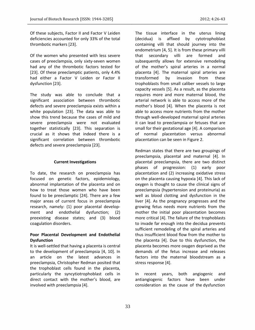
The tissue interface in the uterus lining (decidua) is affixed by cytotrophoblast containing villi that should journey into the endometrium [4, 5]. It is from these primary villi that secondary villi are formed and subsequently allows for extensive remodeling of the mother’s spiral arteries in a normal placenta [4]. The maternal spiral arteries are transformed by invasion from these trophoblasts from small caliber vessels to large capacity vessels [5]. As a result, as the placenta requires more and more maternal blood, the arterial network is able to access more of the mother’s blood [4]. When the placenta is not able to access more nutrients from the mother through well-developed maternal spiral arteries it can lead to preeclampsia or fetuses that are small for their gestational age [4]. A comparison of normal placentation versus abnormal placentation can be seen in Figure 2. Redman states that there are two groupings of preeclampsia, placental and maternal [4]. In placental preeclampsia, there are two distinct phases of progression: (1) early poor placentation and (2) increasing oxidative stress on the placenta causing hypoxia [4]. This lack of oxygen is thought to cause the clinical signs of preeclampsia (hypertension and proteinuria) as well as blood clotting and dysfunction in the liver [4]. As the pregnancy progresses and the growing fetus needs more nutrients from the mother the initial poor placentation becomes more critical [4]. The failure of the trophoblasts to invade far enough into the decidua prevents sufficient remodeling of the spiral arteries and thus insufficient blood flow from the mother to the placenta [4]. Due to this dysfunction, the placenta becomes more oxygen deprived as the demands of the fetus increase and releases factors into the maternal bloodstream as a stress response [4]. In recent years, both angiogenic and antiangiogenic factors have been under consideration as the cause of the dysfunction
Journal of Biotech Research [ISSN: 1944-3285] 2012; 4:26-43
34
Figure 2. Normal versus preeclampsia placental differentiation resulting in a reduction in blood flow in preeclamptic pregnancies. In normal pregnancies, the placenta becomes richly branched and blood flow increases as the fetus develops. Conversely, in a preeclamptic pregnancy, the cytotrophoblasts fail to sufficiently invade the spiral arteries. The blood flow to the placenta is compromised as there are fewer arteries able to deliver much needed nutrients to the fetus.
seen. A theory was developed that if angiogenic factors are being released but there is endothelial dysfunction, the needed blood vessels will not develop properly or conversely that factors that inhibit blood vessel development are being released [4, 24]. One factor that has garnered a good deal of attention is the vascular endothelial growth factor (VEGF-1), also called soluble fms-like tyrosine kinase 1 (sFlt-1) [4]. This factor binds
VEGF and placental growth factor (PIGF) limiting the ability of the placenta to thrive by acting in an anti-angiogenic manner, ultimately causing hypertension and proteinuria [4]. In normal pregnancies, sFlt-1 levels are low until the end of the second trimester while PIGF remains high to enable more blood vessels to develop in the placenta and then sFlt-1 increases to balance this growth as the pregnancy continues [25]. In preeclamptic

Journal of Biotech Research [ISSN: 1944-3285] 2012; 4:26-43
35
pregnancies, an increase in sFlt-1 has been seen along with a decrease in circulating VEGF and PIGF, often weeks before the symptoms of preeclampsia manifest [4, 5, 25]. Even more importantly, the decreased ability of VEGF and PIGF to bind to endothelial cell receptors results in the release of procoagulant proteins and vasoconstricting factors [5]. The second category of preeclampsia that Redman suggests is that of maternal preeclampsia [4]. It is thought that in these cases preexisting conditions in the mother, such as diabetes or hypertension, cause dysfunction even when the placental implantation has been normal [4]. In this sense, preeclampsia that is maternal in origin can be predictive of cardiovascular problems later in life [4]. Another feature of this complex disease is the potential for renal complications. Glomerular filtration rate (“GFR”) is a quantifiable way of measuring the overall functioning of the kidneys [13]. When the rate of blood flow is changed, it changes the blood flow to the kidneys as well. As a result, metabolic waste is not filtered out of the kidneys quickly enough and can lead to proteinuria due to injury to the glomeruli from low oxygen levels [13]. Glomerular capillary endotheliosis, where lesions are seen on the glomeruli of the kidneys is also possible. The glomeruli are inflated and the capillary lumen is occluded due to endothelial cell engorgement which increases as preeclampsia progresses [5, 6]. Upon examination by immunofluorescence, fibrin can be seen in the glomeruli [6]. Women who have other disease states that cause endothelial dysfunction are seen to have more microscopic thromboses present in the kidneys than found in preeclamptic patients without other complicating factors and typically have a more severe variant of the disease [6]. Up to half of patients with pregnancy induced hypertension (PIH) are also found to have mild glomerular endotheliosis but without the associated
proteinuria indicating that PIH may in fact be a less severe form of preeclampsia [5]. This decreased renal capacity also can explain some of the other outward symptoms of preeclampsia. Sudden weight gain or edema, while not necessary for a diagnosis of preeclampsia, generally is present [5]. The decreased GFR causes salt to be retained longer in the system and in concert with the capillary dysfunction and increased permeability of the endothelial tissue may lead to the edema seen [5]. Additionally the increase in uric acid may also contribute to the vascular damage seen in the kidneys [5]. The second major area of current research into the origin of preeclampsia is the effect of preexisting disease states on the potential to develop preeclampsia. The current research focuses less on preexisting hypertension as a risk factor for developing preeclampsia, rather, the focus is on the effects of microvascular diseases such as diabetes. Preexisting Diseases Disease states that exist prior to pregnancy may affect the potential of a mother being diagnosed with preeclampsia. Diabetes, autoimmune diseases and cardiovascular disease are all risk factors for development of preeclampsia as shown in Table 2. Preexisting diabetes, in particular diabetic neuropathy, has been linked to gestational hypertension and preeclampsia [27]. The Jensen study focused on microalbuminuria as a marker to predict preeclampsia in pregnant women with Type I diabetes [27]. Microalbuminuria is a less severe measure of proteinuria caused by compromised filtering of proteins by the kidneys and as such may be an early detector of preeclampsia [28]. Prior to this study, no link between development of preeclampsia and microalbumin levels in pregnancy had been

Journal of Biotech Research [ISSN: 1944-3285] 2012; 4:26-43
36
Table 2. Risk factors that may lead to the development of preeclampsia as adapted from materials from The Preeclampsia Foundation (www.preeclampsia.org); The Mayo Clinic (http://www.mayoclinic.com/health/preeclampsia/DS00583); [1, 2, 4, 11].
Risk Factors for Development of Preeclampsia
First Pregnancy
Previous Preeclamptic Pregnancy
Maternal Age under 18
Advanced Maternal Age, over 35
History of Hypertension Prior to Pregnancy
Diabetes (either prior to or during pregnancy)
Multiple Pregnancy
Obesity (BMI > 30)
Autoimmune Diseases (i.e., Lupus)
Polycystic Ovary Disease
Kidney Disease/Renal Insufficiency
Cardiovascular Disease
Family History
Race
Blood disorders – previous clotting disorders, genetic clotting factors,
blood vessel anomalies & thrombophilia
shown [27]. This study focused on clinical data collected between 1993 and 1999 on 1,215 Danish women who had Type I diabetes but did not have diabetic neuropathy or hypertension prior to pregnancy [27]. For purposes of this study, hypertension was characterized as blood pressure higher than 140/90 mmHg [27]. Microalbuminuria early in pregnancy was defined as urinary albumin output of 30 to 300 milligrams in the 24 hours before conception or in the first trimester of pregnancy [27]. The benchmarks for diagnosis of preeclampia were: (1) at least twenty weeks of pregnancy, (2) blood pressure in excess of 140/90 mmHg and (3) proteinuria shown as 2+ on a dipstick [27]. Microalbuminuria was found in early pregnancy in eighty-four of the women used for statistical comparisons [27]. Of these women, thirteen percent were seen to be hypertensive in the second trimester [27]. Additionally, a staggering 41 percent developed preeclampsia during their pregnancy [27]. Only twelve percent of the
women who failed to show microalbuminuria developed preeclampsia; of this twelve percent, only 1.5% of those women were hypertensive in the second trimester [27]. Additionally, an increase in A1C (indicating poor glycemic control) levels in the third trimester of one percent or more appeared to increase the probability that the mother would develop preeclampsia [27]. The study concluded that a strong association between the manifestation of preeclampsia and the presence of microalbuminuria early in pregnancy in concert with reduced glycemic control was present in women with Type I diabetes [27]. The risk of preeclampsia was seen to swell by four times in women who presented with microalbuminuria versus those that did not [27]. Blood Coagulation Disorders Genetic thrombotic factors impacting the potential incidence of preeclampsia have also
Journal of Biotech Research [ISSN: 1944-3285] 2012; 4:26-43
37
been investigated. Thromboses are often seen in the placenta of women who have had a preeclamptic pregnancy or other pregnancy related complications such as placental abruption or IUGR [29]. A good deal of research has been focused on the prothrombin (Factor II) gene mutation and the Factor V Leiden (FVL) clotting factor. One study conducted on the relationship between FVL and Factor II thrombotic factors and placental abnormalities was done using a keyword search in MEDLINE and EMBASE over a total of fifty seven years and culling down the results to ten relevant studies [30]. The results of these studies were broken into different sections: (1) the loss of pregnancy when the mother has either a FVL or a Factor II mutation; (2) the relationship between presence of a thrombotic factor and preeclampsia; (3) the relationship between having either a FVL or Factor II mutation and low infant birth weights (both slightly underweight and severely underweight); (4) the relationship between separation of the placenta from the uterus and either of these two thrombotic mutations; and (5) an analysis of the presence of a thrombotic mutation and any pregnancy complication involving the placenta [30]. The authors were unable to generate sufficient statistical power to determine if Factor II mutations were related to pregnancy loss, low birth weight or preeclampsia [30]. Similarly, insufficient power existed to allow for a relationship to be established between placental abruption and thrombotic factors [30]. While the authors claim to have had enough statistical power to show if there was a nexus between the presence of a FVL mutation and preeclampsia, no relationship was able to be demonstrated [30]. Numerous studies have attempted to show a correlation between mutations in coagulation factors and pregnancy complications including preeclampsia but most of these studies have been retrospective in nature, not prospective
cohort studies [29]. There was one prospective cohort study reported by Said et al. which involved women who had inherited thrombophilic factors but had previously not had a thrombotic episode [29]. Five factors were analyzed in this study, including FVL and Factor II mutations [29]. Of note, some of these factors can have an even greater effect towards thrombophilia when combined together [29]. Venous blood samples were taken at the conclusion of pregnancy and analyzed for thrombotic markers [29]. Severe and mild preeclampsia were among the outcomes measured [29]. The results of 1,707 women were evaluated and 97 cases of preeclampsia were found, of which 32 were considered severe [29]. The authors attempted to correlate genetic thrombotic factors to specific pregnancy complications including preeclampsia, but the study lacked sufficient power to show a significant relationship between any singular genetic thrombotic factor and preeclampsia [29]. A larger study with stronger sample controls would need to be undertaken in order to properly analyze individual genetic clotting factors and their individual adverse outcomes. One of the adverse pregnancy outcomes often cited is placental insufficiency, and many studies have tried to associate genetic thrombotic factors with this insufficiency [31]. Past results have been conflicting and generally based on small sample sizes such as those shown in the Martinelli, and Mello studies presented earlier [20, 23, 31]. Dudding et al. designed a large scale study to investigate the correlation between maternal and fetal FVL and prothrombin (PT) clotting factors to the risk of fetal growth restriction (FGR) and preeclampsia [31]. Statistical power is often cited as a limiter in these studies, so the authors here also pulled data from other cohort studies to fold into a meta-analysis [31]. Of the 14,273 pregnancies analyzed, 4,477 had
Journal of Biotech Research [ISSN: 1944-3285] 2012; 4:26-43
38
at least a FVL or PT clotting factor and enough data to allow assessment of preeclampsia [31]. When analyzed on the basis of this cohort only, the study was able to show diabetes, chronic hypertension and higher BMI were independent predictive risk factors for preeclampsia, but with insufficient power could not show an association between maternal FVL/PT and preeclampsia [31]. The findings were pooled with six other cohort studies to discover if the inability to find a connection between FVL and PT with preeclampsia was as a result of insufficient power or an actual finding of no association between the factors and this disease [31]. The pooled analysis showed an elevated risk of preeclampsia that was found to be statistically significant with an odds ratio of 1.49 (95% CI 1.13-1.96), especially in relation to severe preeclampsia but only as to FVL and preeclampsia [31]. The data was still insufficient to conclude a relationship between PT and preeclampsia in mothers [31]. There was evidence to support a finding that having FVL elevates one’s risk of developing preeclampsia but that the level of risk involved for pregnant women is also dependent upon other risk factors that may be present [31]. A study reported by Sharma et al., 2008, examined the incidence of venous thromboembolism (VTE) in pregnancy. In developing countries, hemorrhage is the number one cause of maternal death, but in the United States, Australia and the United Kingdom thromboembolism is acknowledged as the leading cause of maternal death [3, 19]. VTE risk is seen to be elevated by five times during pregnancy due to the elevated coagulable state of pregnancy [19]. While this hypercoagulation likely protects women from hemorrhage related complications during childbirth it can cause other serious complications [19]. The Sharma study aimed to show how often thromboembolism manifested in pregnancy and what factors might indicate a propensity to thromboembolism [19]. The analyzed
participants were 6,987 largely Caucasian women who delivered their babies at Ballarat Health Services over a seven year period between 1999 and 2006 [19]. After close analysis, it was seen that 87.5% of the women who developed thromboses during their pregnancies were found to have a positive family history, thrombophilia screen or VTE [19]. It was found by Marik et al. that at least half of the cases of VTE during pregnancy involve a preexisting thrombophilic factor [3]. Preeclampsia as a Predictor of Future Cardiovascular Complications for Mother and Child In the Helsinki Birth Cohort Study that was presented by Kajantie et al., (2009) a relationship between maternal preeclampsia and subsequent cardiovascular disease (CVD) in offspring was investigated. The starting point for this study was the premise that preeclamptic mothers typically have higher cardiovascular risk profiles due to increased blood pressure, cholesterol and insulin levels and an examination of the cardiovascular impact on the offspring was warranted [32]. The authors theorized that the offspring would also have a higher incidence of cardiovascular complications later in life [32]. The Helsinki study involved the analysis of 13,345 single births between 1934 and 1944 in two maternity hospitals in Helsinki. In 6,410 cases, the blood pressure and protein test results of the mother taken after 20 weeks of pregnancy were available for analysis [32]. Four classifications were created based on the found maternal characteristics: (1) severe pre-eclampsia (160/110 mmHg and proteinuria), (2) preeclampsia (pressure greater than 140/90 mmHg yet not rising to the level of severe preeclampsia and proteinuria), (3) gestational hypertension (pressure greater than 140/90 mmHg and lacking proteinuria) and (4) normal hypertension (pressure under 140/90 mmHg with no proteinuria present) [32].
Journal of Biotech Research [ISSN: 1944-3285] 2012; 4:26-43
39
The mothers who experienced any form of preeclampsia or gestational diabetes all had a higher BMI late in pregnancy than the mothers with normal blood pressure readings [32]. In fact, the percentage of first pregnancies, BMI late in pregnancy and body weight of the child at birth all increased as the severity of the pregnancy issue increased from gestational diabetes through to severe preeclampsia [32]. The cardiovascular outcomes analyzed were coronary heart disease, stroke (both hemorrhagic and thrombotic) and hypertension [32]. Just over 35% of the offspring of the original group of 6,410 had an adverse cardiovascular event [32]. Further, when correlating the percentage of offspring who developed cardiovascular outcomes to those mothers whose pregnancies were complicated by either preeclampsia or gestational hypertension, the results were even more startling [32]. Nearly half of the offspring born in preeclamptic pregnancies developed an adverse cardiovascular outcome [32]. The study results could not account for family history of cardiovascular disease, and as such, the data has a limited range of usefulness [32]. With that limitation in mind, the authors were able to show that offspring of mothers who had preeclampsia did have an elevated risk of hemorrhagic or thrombotic stroke and concluded that preeclampsia is more closely related to blood vessel dysfunction in the brain than coronary heart disease [32]. New cardiovascular disease (CVD) prevention guidance for women was promulgated by the American Heart Association (AHA) in February 2011 and the published findings were released on March 22, 2011. Of note, the AHA concluded that preeclampsia may be an indicator of cardiovascular injury to come and has placed a history of preeclampsia, gestational diabetes or hypertension incident to pregnancy as a major risk factor for CVD in women [8]. The AHA cites a finding in a recent meta-analysis that shows a double the risk in the five to fifteen years
following a preeclamptic pregnancy for ischemic heart disease, stroke and venous thromboembolic events [8]. In fact, the AHA goes as far as to say preeclampsia is pregnancy is roughly the equivalent of a “failed stress test” and is very likely a preview to forthcoming vascular or metabolic disease and endothelial dysfunction [8]. It is of critical importance for the origin of preeclampsia to be determined as the incidence of the disease continues to rise and the reach of preeclampsia stays with the mother and child far past the incident of a complicated pregnancy.
Discussion The Link Between the Blood Cascade and Preeclampsia After examining the major areas of current preeclampsia research and the major adverse pregnancy outcomes afflicting mothers today, a common thread emerged. The unifying element among the major sources of pregnancy complications is a change in the blood flow and blood vessels of the mother. An abnormal placental implantation in pregnancy results in a substandard flow of nutrients and blood to the fetus as the structure of the blood vessels simply cannot be sufficiently modified to allow proper blood flow. Once the capacity of the placenta has become over taxed, a lack of oxygen begins to allow angiogenic and anti-angiogenic factors to increase. In preeclamptic pregnancies, an increase in sFlt-1 has been seen along with a decrease in circulating VEGF and PIGF, often weeks before the warning signs of preeclampsia manifest [4, 5, 25]. The mechanisms that allow additional blood vessels to develop are blocked with a resultant decrease in the ability to bind endothelial cell receptors and even more critically, procoagulant and vasoconstricting factors are also released [5].
Journal of Biotech Research [ISSN: 1944-3285] 2012; 4:26-43
40
As a result, blood vessels already strained by improper development are subject to even more compression. This results in an increasing state of hypertension as the pregnancy progresses. The placenta begins to experience a lack of oxygen and is not as able to rid itself of its waste products. This is when the coagulation cascade comes into play. The microscopic damage to the blood vessels of the placenta sparks activity in the coagulation cascade. Fibrin is created as the cascade is activated and platelets rush to plug the tears in the blood vessels of the placenta. As the maternal spiral arteries have not developed to allow a sufficiently wide flow of blood to the placenta another increase in pressure occurs. Tiny thromboses typically seen in the placental mass of preeclamptic women begin to form [29]. Due to repeated activation of the coagulation cascade because of the constant endothelial damage being experienced as the placenta attempts to provide for the fetus, renal complications begin to manifest as well. Hypertension has already taken hold at this point in the pregnancy and the rate of blood flow has been altered as well resulting in a reduced GFR as the blood flow to the kidneys is reduced. In preeclampsia, drops in GFR of up to forty percent are seen despite renal plasma flow being at levels of a typical range for non-pregnant women [5]. This drop in GFR represents compromised renal function and metabolic waste is unable to leave the kidneys as quickly as needed, and oxygen levels are low [13]. Protein that is unable to be processed by the kidneys will spill into the urine as characteristic microalbuminuria which will progress to proteinuria due to hypoxic injury to the glomeruli of the kidneys which will now appear inflated [5, 6]. The capillaries are occluded because of this cell swelling and endothelial cell damage continues to occur [5, 6]. The blood coagulation cascade is still being activated and the excess produced fibrin is seen as thrombotic microangiopathies (TMA) in the glomeruli of the
kidneys that collect as a result of the ongoing vascular injury experienced [6]. As preeclampsia progresses and becomes more severe, the volume found in the endothelial cells continues to increase [6]. While preeclamptic women are seen to have these microscopic thromboses in their kidneys, women who have confounding disease states are in an even more critical situation [6]. For a woman who has an underlying disease state that causes endothelial dysfunction, (such as uncontrolled diabetes), the volume of microscopic thromboses present in the kidneys is seen to be even higher than preeclamptic women with no other complicating issues [6]. The decreased renal capacity will often also cause edema, another warning sign of preeclampsia [5]. Addressing the other conditions that have been under investigation as the source of preeclampsia, it seems that these factors escalate the time to preeclampsia as well as the severity of the disease. Genetic thrombotic factors certainly do impact the occurrence of preeclampsia, but are likely a risk factor for development of the disease or for a more severe variation of the disease, but perhaps not the origin of the disease in chief. Similarly, diabetes with its renal implications may really just be exacerbating renal dysfunction that is due to changes that occur in the blood flow of the placenta and kidneys. Now that a preeclamptic pregnancy is tantamount to a failed stress test in terms of cardiovascular outcomes, it seems likely that the endothelial damage and activation of the blood cascade done during preeclampsia is really a preview of forthcoming cardiovascular disease. This hypothesis would also account for many of the other adverse maternal outcomes seen in pregnancy such as pregnancy-induced hypertension, HELLP syndrome and VTE. The relevant question in predicting preeclampsia and other events may then be the entire cardiovascular profile of the mother and any

Journal of Biotech Research [ISSN: 1944-3285] 2012; 4:26-43
41
particular genetic dispositions to hypercoagulability. The need for additional studies is evident. However the mechanism of developing a large enough trial base will take time and resources. Unfortunately, the number of maternal deaths due to preeclampsia is not large enough to garner the funding needed to accurately investigate a theory of increased activation of the coagulation cascade due to repeated vascular damage being the origin of preeclampsia. In order to be successful in pinpointing the origin of preeclampsia, detailed verified family histories and blood work would have to be done. In addition, the study population would need to be expanded beyond women of European origin and be much larger than ever prospectively imagined before. It may be possible to generate useful results with a smaller study population if the population was comprised of African American women only as the risk of preeclampsia in African American women is between three and four times higher than in other races [7]. A study tailored to African Americans may shed light on underlying factors that could lead to significant results with fewer participants needed. Also, a study of this scale would likely have to follow a cohort for in excess of thirty years to yield results that could address not only the incidence of preeclampsia but the subsequent cardiovascular profile of the mother. In the meantime, how do women who are seeking to become pregnant avoid the complication of preeclampsia? Or in the alternative, how can preeclampsia be managed? If the pregnancy is planned, the opportunity exists for many avenues where the potential mother can improve her risk profile and reduce her risk of actually developing preeclampsia. First, achieving an ideal BMI prior to conception will increase overall health and address a risk factor in developing preeclampsia. Next, evaluate if other disease
states are present. This evaluation will ensure that potential mothers are in strict control of active symptoms of any other diseases and will lower the risk for development of preeclampsia. For diabetics maintaining a lower A1C and tight glycemic control would minimize the complications of diabetes that could overlap with symptoms experienced in preeclampsia, particularly those due to renal complications. For women that have a familial history of genetic clotting factors or have had prior thrombotic episodes, anticoagulation therapy may be warranted beginning prior to conception until six weeks after birth. Management of preeclampsia is another consideration. If one develops preeclampsia, there are not many options for treatment. Unfortunately, the only way to stop preeclampsia is by delivering the baby [33]. However, depending on how far into the pregnancy a woman is when she develops preeclampsia, delivery may not be an option that allows for fetal survival [33]. In milder cases, bed rest may be advocated to allow the baby more time to mature before attempting delivery [33]. The health and age of both the mother and child are considered [34]. Typically, kidney function and blood pressure are closely monitored and tests are done to assess blood clotting ability of the mother [34]. In some cases, medications to decrease blood pressure may be used and magnesium sulfate may be employed to prevent an imminent stroke [34]. Due to the elevated risk of stroke, placental abruption and seizures, there is also potential for the mother to be unable to hold the child to term, but these are situations that would have to be determined by a health care professional. Prevention of the disease is a far better option when possible. Now that a link between future cardiovascular disease and preeclamptic events has been established, it is likely that more attention will be given to maternal adverse events in general.
Journal of Biotech Research [ISSN: 1944-3285] 2012; 4:26-43
42
With more research and investigation, perhaps the origin of preeclampsia can be found and more can be done to prevent this disease.
Acknowledgments
The authors would like to acknowledge William Smalley, Dr. Kimberly Boyd, Dr. Joseph Smith, and Dr. David Dunbar for their support and guidance while researching and writing this paper.
Reference
1. Sibai B, Dekker G, Kupferminc M. 2005. Pre-eclampsia. Lancet. 365: 785-799.
2. Semenovakaya Z and Erogul M. 2010. Pregnancy, Preeclampsia. eMedicine – Medical Reference. http://emedicine.medscape.com/article/796690-overview
3. Marik PE and Plante LA. 2008. Venous Thromboembolic Disease and Pregnancy. The New England Journal of Medicine. 359: 2025-2033.
4. Redman CW and Sargent IL. 2005. Latest Advances in Understanding Preeclampsia. Science. 308(5728): 1592-1594.
5. Karumanchi SA, Maynard SE, Stillman IE, Epstein FH, Sukhatme VP. 2005. Preeclampsia: A Renal perspective. Kidney International. 67: 2101-2113.
6. Stillman IE and Karumanchi SA. 2007. The Glomerular Injury of Pre-eclampsia. Journal of the American Society of Nephrology. 18(8):2281-2284.
7. Berg CJ, Callaghan WM, Syverson C, Henderson Z. 2010. Pregnancy-Related Mortality in the United States, 1998 to 2005. Obstetrics & Gynecology. 116(6): 1302-1309.
8. Mosca L, Benjamin EJ, Berra K, Bezanson JL, Dolor RJ, Lloyd-Jones DM, Newby LK, Pina IL, Roger VL, Shaw LJ, Zhao D, Beckie TM, Bushnell C, D’Armiento J, Kris-Etherton PM, Fang J, Ganiats TG, Gomes AS, Gracia CR, Haan CK, Jackson EA, Judelson DR, Kelepouris E, Lavie CJ, Moore A, Nussmeier NA, Ofili E, Oparil S, Ouyang P, Pinn VW, Sherif K, Smith Jr SC, Sopko G, Chandra-Strobos N, Urbina EM, Vaccarino V, Wenger NK. 2011. Effectiveness-Based Guidelines for the Prevention of Cardiovascular Disease in Women 2011 Update: A Guideline from the American Heart Association. Circulation. 123(11): 1243-1262
9. Centers for Disease Control and Prevention – Division of Reproductive Health, National Center for Chronic Disease Prevention and Health Promotion. 2003. Pregnancy-Related Mortality Surveillance – United States, 1991-1999. (Surveillance Summary No. 52(SS02); 1-8). Washington, DC: Chang J, Elam-Evans LD, Herndon J, Flowers L, Seed KA, Syverson CJ. http://www.cdc.gov/mmwr/preview/ mmwrhtml/ss5202a1.htm
10. Hoegh AM, Borup R, Nielsen FC, Sorensen S, Hviid TVF. 2010. Gene Expression Profiling of Placentas Affected by Pre-Eclampsia. Journal of Biomedicine and Biotechnology. 2010 (787545) doi: 10.1155/2010/787545.
11. Preeclampsia Foundation. 2010. Signs and Symptoms. http://www.preeclampsia.org/signs-and-symptoms
12. Duckitt K and Harrington D. 2005. Risk Factors for pre-eclampsia at antenatal booking: systematic review of controlled studies. British Medical Journal. 330(7491) doi:10.1136/bmj.38380.674340.EO
13. Martini FH. 2006. Fundamentals of Anatomy & Physiology (7th ed.) San Francisco, CA: Pearson Education as Benjamin Cummings
14. Berg JM, Tymoczko JL, Stryer L. 2007. Biochemistry (6th ed.) New York, NY: W.H. Freeman.
15. Segers K, Dahlback B, Nicolaes GA. 2007. Coagulation factor V and thrombophilia: Background and mechanisms. Journal of Thrombosis and Haemostasis. 98: 530-542.
16. Omar SZ, Qvist R, Khaing SL, Muniandy S, Bhalla S. 2008. Thrombophilic mutations in pre-eclampsia and pregnancy-induced hypertension. Journal of Obstetrics and Gynecology Research. 34(2): 174-178.
17. Varga EA, Moll S. 2004. Prothrombin 20210 Mutation (Factor II Mutation). Circulation. 110, e15-e18. doi: 10.1161/01.CIR. 0000135582.53444.87
18. Genetic Home Reference – U.S. National Library of Medicine (2010, August). Factor V Leiden thrombophilia. http://ghr.nlm.nih.gov/condition/factor-v-leiden-thrombophilia
19. Sharma S and Monga D. 2008. Venous thromboembolism during pregnancy and the post-partum period: Incidence and risk factors in a large Victorian health service. Australian and New Zealand Journal of Obstetrics and Gynaecology. 48: 44-49.
20. Martinelli I, Taioli E, Cetin I, Marinoni A, Gerosa S, Villa MV, Bozzo M, Mannucci PM. 2000. Mutations in Coagulation Factors in Women with Unexplained Late Fetal Loss. The New England Journal of Medicine. 343(14): 1015-1018.
21. De Stefano V, Rossi E, Za T. 2005. Inherited thrombophilia and obstetric complications. Haematologica Reports. 1(10): 18-21.
22. Mehrabian E and Rezaeli A. 2009. Early Measurement of Thrombus Precursor Protein (TpP) in the Third Trimester as a Predictor of Preeclampsia. Journal of Cell and Tissue Research. 9(2): 1855-1858.
23. Mello G, Parretti E, Marozio L, Pizzi C, Lojacono A, Frusca T, Facchinetti F, Benedetto C. 2005. Thrombophilia Is Significantly Associated With Severe Preclampsia, Results of a Large-Scale, Case-Controlled Study. Hypertension. 46: 1270-1274.
24. Mutter WP and Karumanchi SA. 2008. Molecular Mechanisms of Preeclampsia. Microvascular Research. 75(1):1-8.
25. Lyall F and Belfort MA. 2007. Pre-eclampsia: etiology and clinical practice. New York, NY: Cambridge University Press
26. Huppertz B. 2008. Placental Origins of Preeclampsia: Challenging the Current Hypothesis. Hypertension. 51: 970-975.
27. Jensen DM, Damm P, Ovesen P, Molsted-Pedersen L, Beck-Nielsen H, Westergaard JG, Moeller M, Mathiesen ER. 2010. Microalbuminuria, Preeclampsia, and Preterm Delivery in Pregnant Women With Type 1 Diabetes. Diabetes Care. 33(1): 90-94.
28. The Nephrology Channel (2010). Proteinuria. http://www.nephrologychannel.com.
29. Said JM, Higgins JR, Moses EK, Walker SP. 2009. Inherited Thrombophilia Polymorphisms and Pregnancy Outcomes in Nulliparous Women. Obstetrics & Gynecology. 115(1): 5-13.
30. Rodger MA, Betancourt MT, Clark P, Lindquist PG, Dizon-Townson D, Said J, Seligsohn U, Carrier M, Salomon O, Greer IA. 2010. The Association of Factor V Leiden and Prothrombin Gene Mutation and Placenta-Mediated Pregnancy Complications: A Systematic Review and Meta-Analysis of
Journal of Biotech Research [ISSN: 1944-3285] 2012; 4:26-43
43
Prospective Cohort Studies. PLoS Medicine. 7(6). doi:10.1371/journal.pmed.1000290
31. Dudding T, Heron J, Thakkinstian A, Nurk E, Golding J, Pembrey M, Ring SM, Attia J, Scott RJ. 2008. Factor V Leiden is associated with pre-eclampsia but not with fetal growth restriction: a genetic association study and meta-analysis. Journal of Thrombosis and Hemostasis. 6: 1868-1875.
32. Kajantie E, Eriksson JG, Osmond C, Thornburg K, Barker DJP. 2009. Pre-Eclampsia Is Associated With Increased Risk of Stroke in Adult Offspring: The Helsinki Birth Cohort Study. Stroke. 40: 1176-1180.
33. Mayo Clinic. 2009. Preeclampsia. http://www.mayoclinic.com/health/preeclampsia/DS00583
34. Preeclampsia Foundation (2011). FAQS. http://www.preeclampsia.org/health-information/faq#one