Embed Size (px)
Citation preview

Preeclampsia, PRES and Intracranial Hemorrhage Shannon Clark, MD Asst. Professor Div. MFM UTMB-Galveston
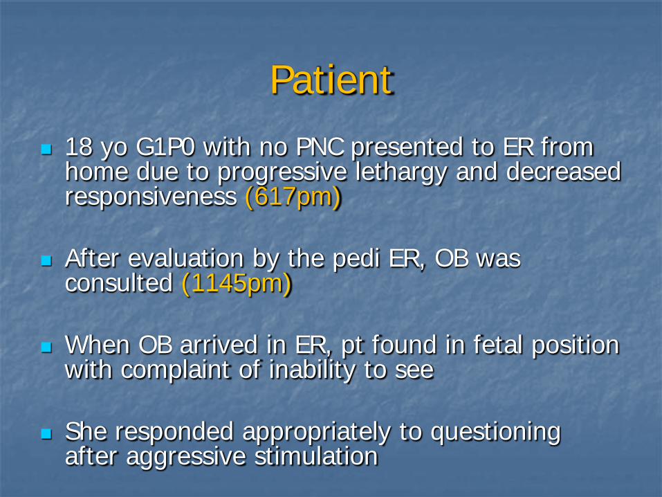
Patient 18 yo G1P0 with no PNC presented to ER from
home due to progressive lethargy and decreased responsiveness (617pm)
After evaluation by the pedi ER, OB was consulted (1145pm)
When OB arrived in ER, pt found in fetal position with complaint of inability to see
She responded appropriately to questioning after aggressive stimulation

OB assessment On PE, she appeared to be 38-39 weeks GA, but
she and parents denied the pregnancy
She reported no complaints other than vision loss, history of 2 day headache, decreased responsiveness, N/V
Bedside sono revealed fetal FL of 38+2 wks, oligo and fetal hydranencephaly vs holoproscencephaly
BP in ER was 142-159/86-92

OB assessment Labs (1006pm 7/4): H/H 11.1/32.7, pl 304, cr
0.94, beta 43754, UDS neg, UA 300mg/dL protein
She was immediately transferred to LD with working diagnosis of preeclampsia/PRES (arrived 1205am 7/5)
On LD, 4gm bolus MgSO4 given immediately, labs sent
BP 143/81, cervix 1/80/hi

OB assessment
NST with repetitive late decels and then bradycardia
Section called by MFM faculty
In OR for stat section (1232am)
Viable female infant delivered at 1246am, Apgars 5/9, thick meconium

Anesthesia preoperative assessment
Informed that patient would be coming up to the floor for possible C/S with working diagnosis of severe preeclampsia and PRES
Room was prepared for both general anesthetic and neuraxial blockade

18 yo G1P0 female
No prenatal care
Somnolent
Answering questions appropriately
Acutely Blind
Nausea/Vomiting
No neuroimaging available

Differential Diagnosis

Intraoperative Events The patient was positioned in the sitting position and
prepped and draped in a sterile fashion.
During this time, the patient was following commands and conversant.
On barbotage for spinal anesthesia the CSF was noted to contain frank blood
Uncomplicated delivery occurred 15 minutes after entering the operating room and the total case time was approximately 30 minutes
Patient immediately taken to CT for neuro-imaging


07/06/07 07/17/07

Neurosurgery events
Neurosurgery consulted at 130am
At bedside in ICU within 5 minutes-BP 200/100
Decision made for ventriculostomy after examination and imaging of patient Intraparenchymal hemorrhage 3.8 x 2.2cm at level of left caudate
nucleus with intraventricular extension and 5mm left-to-right midline shift
On exam, patient obtunded, left gaze preference with
hemineglect, localization to painful stimuli on left arm
A-line, intubation and ventriculostomy placement by 220am
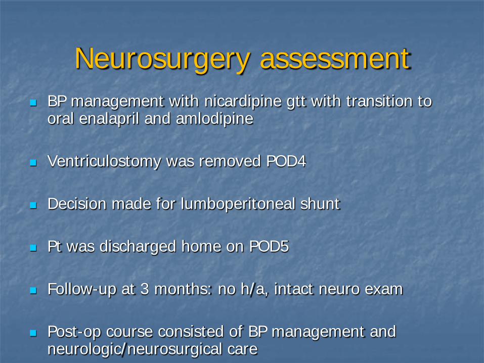
Neurosurgery assessment BP management with nicardipine gtt with transition to
oral enalapril and amlodipine
Ventriculostomy was removed POD4
Decision made for lumboperitoneal shunt
Pt was discharged home on POD5
Follow-up at 3 months: no h/a, intact neuro exam
Post-op course consisted of BP management and neurologic/neurosurgical care

Follow-up baby
Initial diagnosis was schizencephaly
VP shunt placed
Final diagnosis of porencephalic cyst/ in utero vascular infarct
Baby with mom and grandparents
Expect neurodevelopmental delay

Posterior reversible encephalopathy syndrome (PRES)
A clinical neuroradiologic syndrome of heterogenous etiologies that are grouped together because of similar findings on neuroimaging studies
Incidence is unknown

PRES First described in a 1996 case series (15
patients) (Hinchey et al., 1996)
Headache
Confusion or decreased consciousness
Visual changes (visual neglect or cortical blindness)
Seizures
Assoc posterior cerebral white matter edema

Pathophysiology
Pathogenesis is unclear, but appears related to disordered cerebral autogregulation and endothelial dysfunction
Often related to an acute increase in arterial blood pressure
Clinically indistinguishable from hypertensive encephalopathy
Given vast array of etiologies, several other pathways are possible

Pathophysiology
Likely due to vasogenic edema secondary to an acute increase in arterial blood pressure, which overwhelms the autoregulatory capacity of the cerebral vasculature, causing arteriolar vasodilation and endothelial dysfunction, leading to extravasation of fluid (i.e preeclampsia) (Thackeray and Tielborg, 2007)
Or an acute and significant episode of hypertension that causes cerebral vasoconstriction with subsequent ischemia and edema (Thackeray and Tielborg, 2007)

Normal autoregulation
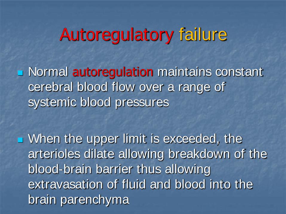
Autoregulatory failure
Normal autoregulation maintains constant cerebral blood flow over a range of systemic blood pressures
When the upper limit is exceeded, the arterioles dilate allowing breakdown of the blood-brain barrier thus allowing extravasation of fluid and blood into the brain parenchyma

Autoregulatory failure
In chronic hypertension, the range is “reset” allowing severely elevated BPs
Less severe elevations can cause PRES in the acute setting (ie preeclampsia)

Endothelial dysfunction Implicated in cases associated with
preeclampsia or cytotoxic therapies
Capillary leakage leads to blood-brain barrier disruption, which leads to vasogenic edema
In preeclampsia, markers of endothelial dysfunction (elevated LDH, abnormal RBC morphology) may precede the clinical syndrome

Associated conditions Hypertensive encephalopathy
Preeclampsia/eclampsia
Immunosuppresive therapy
Others: renal failure, chemotherapeutic
drugs, hypercalcemia (Stott et al, 2005; Kastrup et al., 2002)

Clinical Headache
Constant, nonlocalized, moderate to severe, not relieved with analgesics
Altered consciousness
Mild somnolence to confusion and agitation. May progress to stupor or coma
Visual disturbances
Hemianopia, auras, hallucinations, cortical blindness
Seizures May be the presenting manifestation Generalized tonic-clonic

Clinical Differential diagnosis
Stroke (thrombotic, embolic, hemorrhagic)
Venous thrombosis
Toxic or metabolic encephalopathy
Demyelinating disorders
Vasculitis
Encephalitis

Radiologic Neuroimaging is essential for the diagnosis
Bilateral white matter edema and hypodensities in the
posterior cerebral hemispheres (white and gray matter)
Lesions are usually parieto-occipital (98.7%), but brainstem (18.4%) and cerebellar (34.2%), thalamus (30.3%), basal ganglia (11.8%) and temporal (68.4%) and frontal lobe (78.9%) involvement can occur (Gasco et al., 2008)
Findings usually not confined to a singular vascular
territory
With prompt treatment, resolution of the findings is expected, but not always observed

Radiologic
CT or MRI
Can have similar findings to stroke

Diagnosis
Clinical symptoms and radiologic evidence support the diagnosis
Should be recognized promptly so treatment can be initiated
In our patient population with preeclampsia/eclampsia, this typically means delivery and treatment of blood pressures

Treatment
Acute-onset, severe systolic (≥ 160) or sever diastolic (≥ 110) hypertension lasting > 15 minutes is considered a hypertensive emergency
This can develop in pregnant patients with chronic HTN or in those with no history (ACOG CO 514, 2011)

Treatment
Severe hypertension can cause CNS injury
The degree of systolic hypertension may be the most important predictor of cerebral injury and infarction (ACOG CO 514, 2011)
Treatment goal is not normal BP, but a range of 140-160/90-100 Prevent repeated, prolonged exposure to severe
systolic hypertension and loss of cerebral vasculature autoregulation (ACOG CO 514, 2011)

First-line treatment (ACOG CO 514, 2011)
IV labetalol and hydralazine
Risks: Labetalol-neonatal bradycardia; avoid use in women
with asthma or heart failure
Hydralazine-maternal hypotension
If no IV access give 200mg labetalol po and
repeat in 30 minutes if needed

First-line treatment (ACOG CO 514, 2011)
Labetalol: 20mg IV over 2 minutes
Repeat with 40mg in 10 minutes if needed
Repeat with 80mg in 10 minutes if needed
Wait 10 minutes
Hydralazine:
10mg IV over 10 minutes
Switch agents
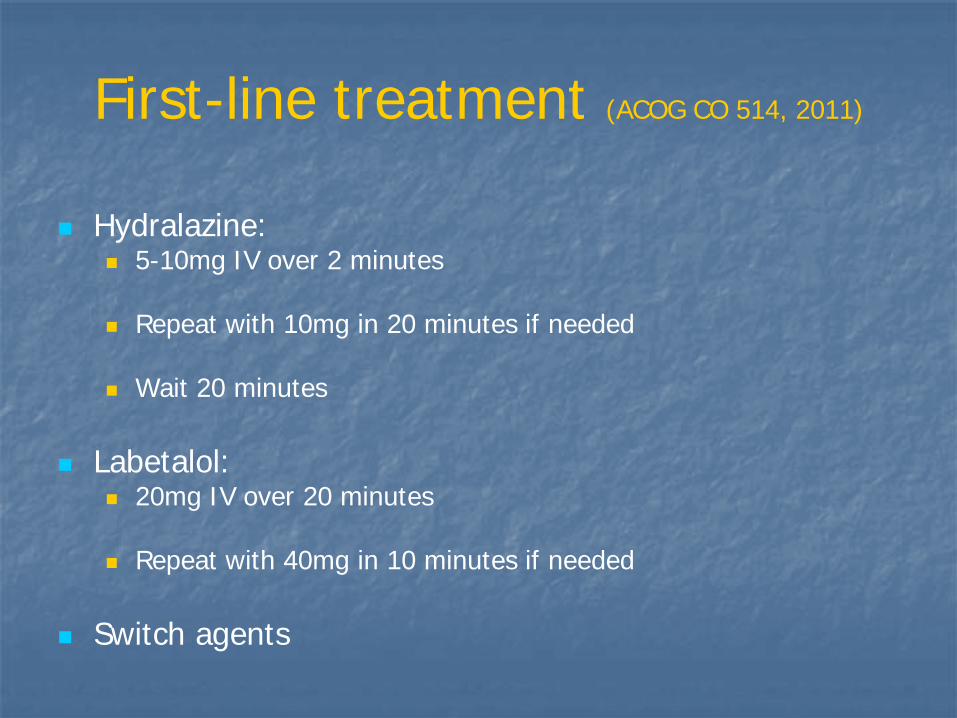
First-line treatment (ACOG CO 514, 2011)
Hydralazine: 5-10mg IV over 2 minutes
Repeat with 10mg in 20 minutes if needed
Wait 20 minutes
Labetalol:
20mg IV over 20 minutes
Repeat with 40mg in 10 minutes if needed
Switch agents

Second-line treatment (ACOG CO 514, 2011)
If labetalol and hydralazine fail, consider labetalol or nicardipine by infusion pump

Prognosis
Most cases of PRES are reversible in days to weeks with removal of the inciting factor and treatment of the blood pressure
Secondary cerebral infarction or hemorrhage
Death and permanent neurologic impairment are possible

Intracranial hemorrhage
Preeclampsia/eclampsia as causative factors of PRES is well established Presence of hemorrhagic events with PRES is
reported once
Changes in the blood brain barrier as a consequence of cerebral autoregulatory dysfunction could be responsible for the initial encephalopathy (PRES), and this disruption could create a predisposition of the microcirculation to a hemorrhagic event (Gasco et al., 2008)

Patient outcome
The favorable outcome of our patient was due to rapid delivery, blood pressure control and immediate ventriculostomy placement one the intraventricular hemorrhage was identified

Obstetricians must be vigilant and investigate suspicious acute CNS complaints with radiologic studies in antepartum/intrapartum/ postpartum patients, especially with hypertension


![Non-aneurysmal subarachnoid hemorrhage in a case of ... · associated with aneurysmal subarchnoid and intracranial hemorrhage [5]. In our case this was a primary subarchnoid hemorrhage](https://img.pdfslide.us/doc/110x75/5ed55eea6933f508e973f10c/non-aneurysmal-subarachnoid-hemorrhage-in-a-case-of-associated-with-aneurysmal.jpg)