Embed Size (px)
Citation preview
Dr Ajithkumar V R et al JMSCR Volume 07 Issue 11 November 2019 Page 82
JMSCR Vol||07||Issue||11||Page 82-93||November 2019
Predictive Value of Radiation Induced Apoptosis in Response to
Radiotherapy in Cancer Cervix Patients Attending Department of Radio
Therapy, Govt. Medical College Thrissur, A Prospective Study
Authors
Dr Ajithkumar V R1, Dr R Mahadevan
2, Dr Seena K B
3, Dr Jayakumar K L
4,
Dr Jewel Joseph5, Dr Prema K R
6, Dr Shehna A Khader
7
1Assistant Professor, Radiation Oncology,
2Professor, Radiation Oncology
3Associate Professor, Obstetrics and Gynaecology,
4Professor, Radiation Oncology
5Senior Resident, Radiation Oncology,
6Associate Professor, Radiation Oncology
7Associate Professor, Radiation Oncology
*Corresponding Author
Dr Seena K B
Department of Radiation Oncology, Government Medical College, Thrissur, India
Abstract
Aim: To correlate the apoptotic index with respect to radiation therapy in patients with carcinoma cervix
attending Govt. Medical college, Thrissur.
Materials and Methods: Patients both early stage and locally advanced cervical cancer has been
included in the study. Between December 2013 and September 2014. 26 patients were included in the
study. Pre and post Radiotherapy biopsy of patients were taken and they were examined for Nuclear
Pleomorphism, Hyper chromasia, Apotosis, Mitosis, Keratinisation, and Necrosis.
IHC was done using Bcl 2, Bax, and Ki - 67.
Results: Apoptotic index and markers favouring apoptosis can positively predict disease outcome. Patients
with good apoptosis faired well in the study. But this has to be analysed in large cohort studies.
Bax is a predictive marker for apoptosis.
Introduction
Worldwide cervical cancer is the second most
common cancer to affect women, in developing
countries including India it is the second most
common malignancy among females.
Incidence varies worldwide with the highest rates
found in Latin America and the lowest among
Jewish women in Israel.
The use of cervical screening has greatly reduced
the incidence of invasive cervical cancer in the
western countries, but it continues to be a major
cause of cancer mortality in the rest of the world
because majority of patients have locally
advanced disease at presentation.
Patients with early stage disease (IB non bulky
and stage IIA) can expect cure with either radical
hysterectomy or radical radiotherapy.
The standard treatment for stage IIB to IV A
cervical cancer has been radical radiotherapy
alone, (external pelvic radiation combined with
intracavitary brachy therapy). 5 year survival for
patients with locally advanced (stage III/IV A)
http://jmscr.igmpublication.org/home/
ISSN (e)-2347-176x ISSN (p) 2455-0450
DOI: https://dx.doi.org/10.18535/jmscr/v7i11.15

Dr Ajithkumar V R et al JMSCR Volume 07 Issue 11 November 2019 Page 83
JMSCR Vol||07||Issue||11||Page 82-93||November 2019
disease is approximately 25% (10-45%) when
treated with radiotherapy alone. Although
treatment failures outside the radiation field may
occur, the more common cause of treatment
failure is the inability of primary radiotherapy
alone to completely eradicate all pelvic disease.
The radiotherapy failure rate for patients with
stage IIB disease is approximately 20-50% and for
patients with stage IIIB disease the failure rate
ranges from 50% to as high as 75%.
Attempts at improving local control by increasing
radiation dose is limited by the maximum
tolerated dose of radiation to surrounding organs,
beyond which morbidity becomes unacceptable.
Complications are the upper limit of acceptability
with doses currently used. As a result many
strategies have been investigated to try to improve
outcome in cervical cancer. These include hyper
fractionated treatment schedules, modification of
radiation treatment volumes, hyperbaric oxygen,
hyperthermia, hypoxic cell sensitizers and neutron
therapy, unfortunately, these modalities have not
demonstrated improved therapeutic response or
local control.
For at least 25 years, clinicians have been
searching for ways of combining chemotherapy
with radiation to improve local control in cervical
cancer. Most of the early prospective randomized
trials involved the use of neoadjuvant
chemotherapy followed by radiotherapy. Despite
encouraging tumor response to various
combination chemotherapy regimens, there is no
improvement in local control or survival.
Study Group
Patients both early stage and locally advanced
cervical cancer has been included in the study.
Between December 2013 and September 2014. 26
patients were included in the study. Written
informed consent was obtained from of all
patients stage IB/IIA lesions that measure 3 cm or
less can be managed with definitive surgery or
radiation.
Routine investigations and metastatic work up
was done. This included complete biochemistry,
abdominal CT scan if indicated and a biopsy.
Initial biopsy was done in all patients. Patients
were given radiotherapy 45 gy in 23 fractions,
after 5 fractions on the first Saturday, a repeat
biopsy is also performed. Biopsies were done in
the first week itself, according to patient
convenience. The biopsy material was examined
for apoptosis and tumour cell proliferation rate.
Residual disease at the time of ICR was also
evaluated and studied.
A total 26 patients with stage IIB or III (FIGO)
were evaluated in the study and pre-treatment
results correlated to radiotherapy outcome. A
punch biopsy was taken from all patients, fixed in
buffered formalin and processed for paraffin-was
embedding. Sections 5|am thick were cut from the
paraffin-embedded tissue and one section was
stained by routine haematoxylin eosin staining for
histopathological evaluation. Duplicate serial
sections were used for immunocytochemistry. All
patients in the study received radical radiotherapy
(45Gy in 23 fractions for external-beam
radiotherapy and 8 Gy to point A in 2 sitting or 7
Gy to point A in 3 sittings by ICR (HDR).
Subjecting to Repeat Biopsy
A repeat biopsy with the patient’s consent was
needed. The patient was explained in detail on the
procedure and the benefits of understanding
radiation response. The significance was
explained and a consent obtained.
The study patients are to be followed for 5 years
or even more to assess final disease outcome.
BAX, BCL 2, Ki67 Technique for
Determination of Apoptosis
Estimation of apoptosis in tissue was determined
using the BAX, BCL 2, Ki67 assay, employing an
In Situ Cell Death Detection Kit. Briefly, sections
were dewaxed in xylene hydrated with decreasing
concentrations of ethanol and washed in distilled
water for 10 minutes. The nuclei in tissue sections
were stripped from proteins with 20 g/ml of
proteinase K diluted in sterile buffer (10mm Tris
HCI/pH 7.4-8.0) for 15 min at 37°C and
endogenous peroxidase was subsequently
Dr Ajithkumar V R et al JMSCR Volume 07 Issue 11 November 2019 Page 84
JMSCR Vol||07||Issue||11||Page 82-93||November 2019
quenched with 0.3% H2O2 in methanol for 30
minutes. The sections were covered with 50 l of a
label mix containing the modified nucleotide and
enzyme in a humidified chamber for 60 minutes at
room temperature. After appropriate washing in
PBS, the samples were first analysed under a
fluorescence microscope. For further signal
conversion analysis, 50JJ.I of converter
peroxidase POD reagent was added for 30 minutes
at room temperature. The substrate reaction was
developed using diaminobenzidine (DAB) and
then counterstained with haematoxylin. To
confirm the reaction specificity of the BAX, BCL
2, Ki67 procedure, a negative control was also
run, omitting Tdt from the reaction mixture. As a
positive control, sections of involuting rat breast
tissue were used.
Assessment of Apoptotic Cells
A cell was considered apoptotic only when
unequivocal nuclear labeling was observed in
areas of the tumor free from inflammation. Cells
exhibiting necrotic nuclear karyorrhexis as well as
those in necrotic foci were excluded. Moreover,
all results were compared with the morphological
analysis. Since the enzymatic reaction may also
label diffuse areas of necrosis, only those labeled
cells that showed additional characteristics of
apoptosis, i.e. isolated localization within an intact
cell complex without an inflammatory reaction
was regarded as positive. Grading of the BAX,
BCL 2, Ki67 reaction was done as explained
earlier. Briefly, to evaluate differences in the
various rates of BAX, BCL 2, Ki67 reactivity in
each sample, 1,000 cells were counted at random
under high power, and an apoptotic index (Al)
was expressed as shown below.
A total of 1,000 cells were evaluated in all
sections. Expression of PCNA was considered
significant when characteristic nuclear
immunoreactivity was seen in more than 10% of
the cells. In addition, an expression index was
used to evaluate PCNA expression as described
earlier. This was done by classifying the protein
expression into four categories based on the
number of cells with positive expression. Thus,
class 1 expression included those sample with less
than 10% expression, Class 2 included samples
showing 31% to 50% expression and class 4
included all cases with positive expression in
more than 50% of cells.
Immunocytochemical Localization of K i67
Immunocytochemical analysis was carried out as
described by us earlier. Briefly, sections were
dewaxed in xylene and hydrated through graded
alcohols to deionised water. Endogenous
peroxidase was blocked by a 25-min incubation in
3% H2O2 in methanol. The sections were rinsed
with distilled water and then incubated with 0.3%
bovine serum albumin to reduce non-specific
antibody binding. Sections were incubated
overnight at 4°C with monoclonal antibodies.
Sections were then incubated with biotinylated
anti- (mouse Ig) at a dilution of 1:200 and
peroxidase-conjugated streptavidin at 1:500 for 30
min each at room temperature. Washing was
carried out in phosphate-buffered saline after each
step and the peroxidase reaction was developed by
application of daminobenzidine solution. The
reaction was allowed to develop for 20 min after
which it was stopped by washing in distilled
water. The sections were then lightly
counterstained with Mayer's haematoxylin,
dehydrated in ascending grades of alcohol, fixed
in xylene and mounted in Distrene Dibutyl
phthalate Xylene. Grading of the
immunoreactivity of growth factors was done as
previously explained by us. Briefly, samples with
less than 10% positive cells were considered
negative. Samples with 11 %-30% were
considered as showing mild expression. 30-50%
moderate expression and those with over 51%
positive cells as showing intense expression.
Inclusion Criteria
1) Women with biopsy proven carcinoma of
cervix or clinically carcinoma of cervix.
Dr Ajithkumar V R et al JMSCR Volume 07 Issue 11 November 2019 Page 85
JMSCR Vol||07||Issue||11||Page 82-93||November 2019
2) Patient should have a anatomy favouring
cervical biopsy
3) WHO performance status 0-1
4) Age < 80 years.
5) Adequate haematologic function (WBC >
4000/mm3, platelet count >1,00,000/mm3
and haemoglobin >9gm%).
6) Normal renal hepatic function
7) Willingness for informed consent
document
Exclusion Criteria
1. Poor performance status
2. Bleeding disorders
3. Poor pelvic anatomy
4. Previous history of pelvic irradiation or
systemic chemotherapy.
Diagnostic Work Up
1. Patients underwent a diagnostic work up
which included:
2. Complete blood count
3. Renal and liver function tests
4. Chest X-ray
5. Ultrasound scan or CT scan of abdomen
and pelvis
6. Cystoscopy
Tumour size was defined as the maximum
diamefer of the tumour measured by ultrasound or
CT scan of the abdomen and pelvis or by clinical
examination.
Treatment
Concurrent chemoradiation with Inj. Cisplatin40
mg/m2 weekly was given for all patients.
External radiotherapy was delivered by four-field
box technique using Cobalt -60. The pelvic field
extended from the upper border of L-5 to lower
border of the obturator foramen or 3 cm below the
lowest extent of vaginal involvement (whichever
was lower), and laterally 1.5 to 2 cm beyond
lateral margin of the pelvic brim. For the lateral
fields, anterior limit was the anterior border of
pubic symphysis and the posterior limit was the
space between S-2 and S-3. Midline shield was is
not used in either arms. Dose of external
radiotherapy was 45Gy/23 Fr/4 weeks.
With the goal of keeping total duration of
treatment less than 8 weeks, intracavitary
brachytherapy was performed within 2 weeks
(preferably less than one week) after the
completion of pelvic radiation.
Evaluation of Response
During treatment initial assessment of response
was done at the time of 1st intracavitary
brachytherapy. Clinical assessment was done by
pelvic examination and response scored as:
1. No residual disease
2. Maximum dimension of residual disease
1 cm
3. Maximum dimension residual disease 1
cm
Follow-Up
On completion of treatment patients were
followed on November 1, 2014. Clinical
assessment was done and investigations were
carried out only even indicated.
The closing date of the study was November 1,
2014; All the patients without regular follow up
till closing date, patients who were presented last
to follow up and their current states was obtained
either by reply paid post cards or by telephone
enquiry.
Statistical Analysis
The Kaplan -Meier (K-M) method was used to
calculate the overall survival (OS) and disease
free survival (DFS). The association between
variables was assessed using chi-square test. All
statistical analysis was performed using SPSS
software. As the patients sample size was 26 and
the evaluable patients were 13, WS Signed Rank
Test was used.
Observation and Results
During the period from December 2013 to
September 2014 an altogether of 26 patients were
accured into the study. Of which good biopsy
specimens were obtained for only 13 patients.
Usual H&E stains are done for all patients. 6
patients slides had good processing features and
immunohistochemistry was done in those patients.

Dr Ajithkumar V R et al JMSCR Volume 07 Issue 11 November 2019 Page 86
JMSCR Vol||07||Issue||11||Page 82-93||November 2019
There were problems during preparation of wax
blocks and slides.
The first patient had 11 months of follow up. The
last patient had four months of follow up. One
patient died due to progressive disease after
radical treatment.
Dr Ajithkumar V R et al JMSCR Volume 07 Issue 11 November 2019 Page 87
JMSCR Vol||07||Issue||11||Page 82-93||November 2019
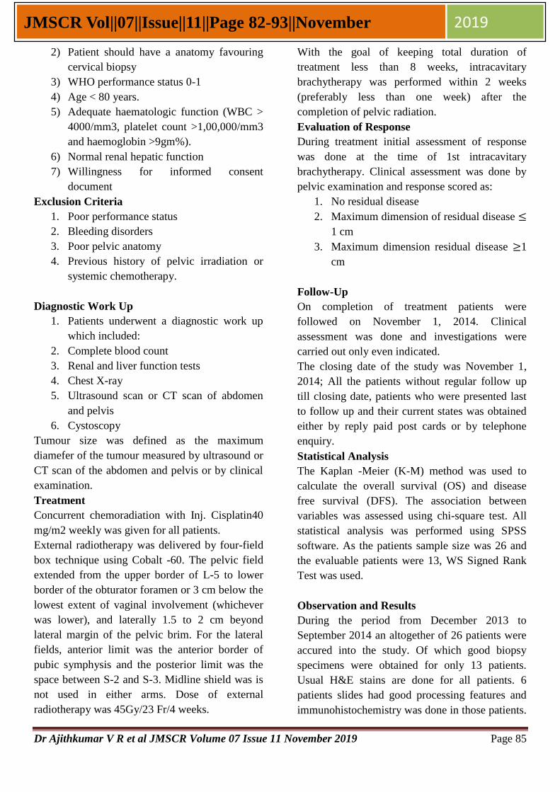
Figure - 1
Stage1 2
Stage2 15
Stage3 7
Stage4 2
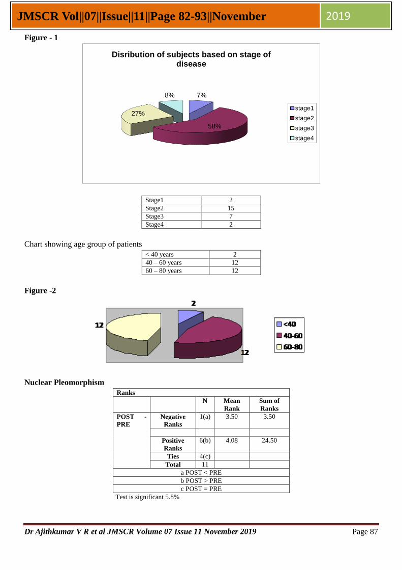
Chart showing age group of patients
< 40 years 2
40 – 60 years 12
60 – 80 years 12
Figure -2
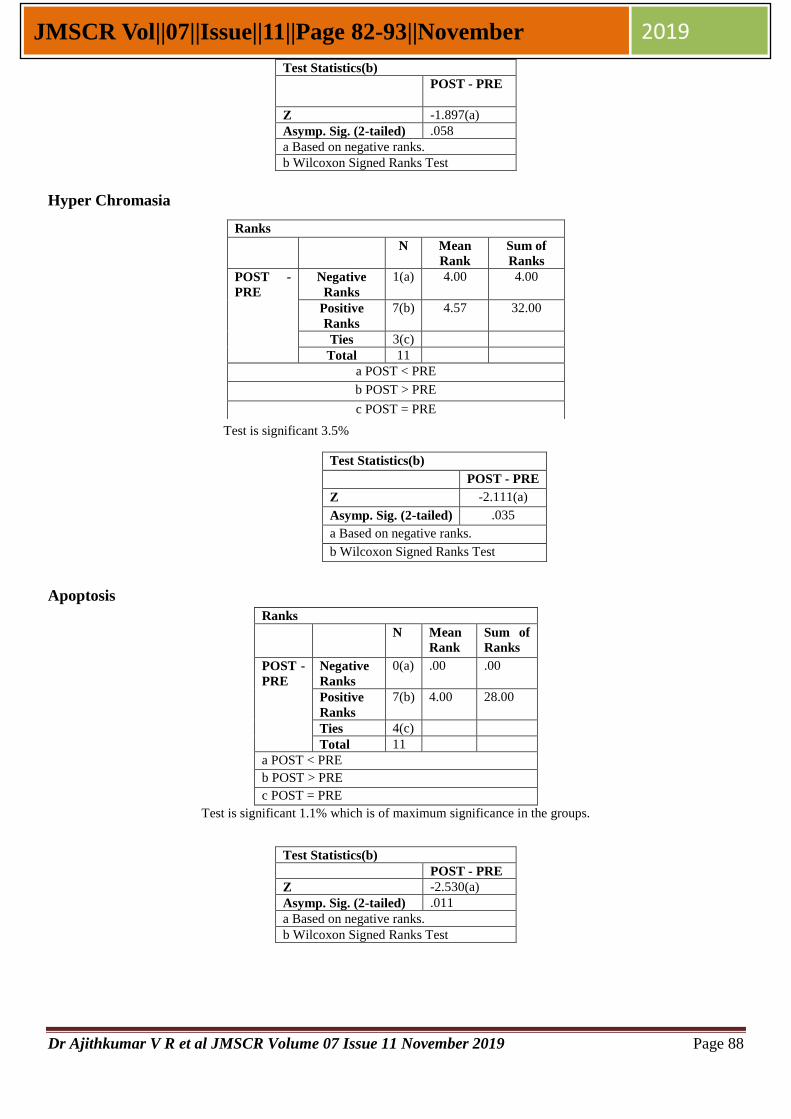
Nuclear Pleomorphism
Ranks
N Mean
Rank
Sum of
Ranks
POST -
PRE
Negative
Ranks
1(a) 3.50 3.50
Positive
Ranks
6(b) 4.08 24.50
Ties 4(c)
Total 11
a POST < PRE
b POST > PRE
c POST = PRE
Test is significant 5.8%
7%
58%
27%
8%
Disribution of subjects based on stage of disease
stage1
stage2
stage3
stage4
Dr Ajithkumar V R et al JMSCR Volume 07 Issue 11 November 2019 Page 88
JMSCR Vol||07||Issue||11||Page 82-93||November 2019
Hyper Chromasia
Test is significant 3.5%
Apoptosis
Ranks
N Mean
Rank
Sum of
Ranks
POST -
PRE
Negative
Ranks
0(a) .00 .00
Positive
Ranks
7(b) 4.00 28.00
Ties 4(c)
Total 11
a POST < PRE
b POST > PRE
c POST = PRE
Test is significant 1.1% which is of maximum significance in the groups.
Test Statistics(b)
POST - PRE
Z -1.897(a)
Asymp. Sig. (2-tailed) .058
a Based on negative ranks.
b Wilcoxon Signed Ranks Test
Ranks
N Mean
Rank
Sum of
Ranks
POST -
PRE
Negative
Ranks
1(a) 4.00 4.00
Positive
Ranks
7(b) 4.57 32.00
Ties 3(c)
Total 11
a POST < PRE
b POST > PRE
c POST = PRE
Test Statistics(b)
POST - PRE
Z -2.111(a)
Asymp. Sig. (2-tailed) .035
a Based on negative ranks.
b Wilcoxon Signed Ranks Test
Test Statistics(b)
POST - PRE
Z -2.530(a)
Asymp. Sig. (2-tailed) .011
a Based on negative ranks.
b Wilcoxon Signed Ranks Test
Dr Ajithkumar V R et al JMSCR Volume 07 Issue 11 November 2019 Page 89
JMSCR Vol||07||Issue||11||Page 82-93||November 2019
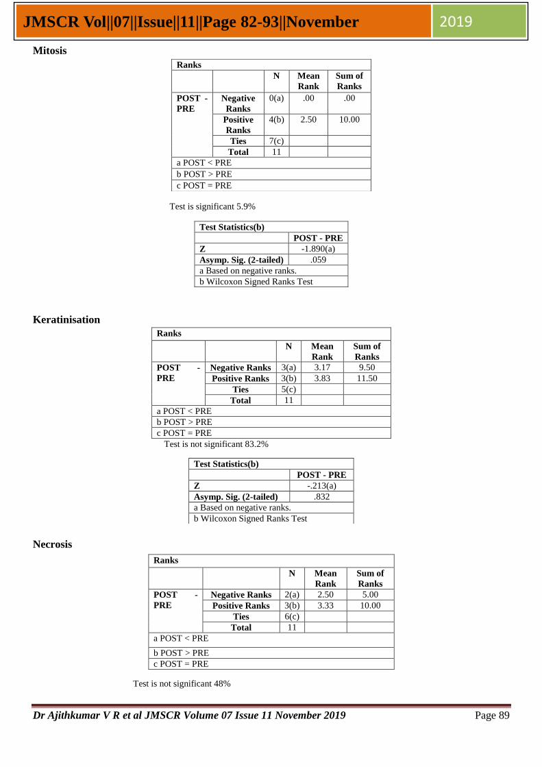
Mitosis
Test is significant 5.9%
Keratinisation
Ranks
N Mean
Rank
Sum of
Ranks
POST -
PRE
Negative Ranks 3(a) 3.17 9.50
Positive Ranks 3(b) 3.83 11.50
Ties 5(c)
Total 11
a POST < PRE
b POST > PRE
c POST = PRE
Test is not significant 83.2%
Necrosis
Test is not significant 48%
Ranks
N Mean
Rank
Sum of
Ranks
POST -
PRE
Negative
Ranks
0(a) .00 .00
Positive
Ranks
4(b) 2.50 10.00
Ties 7(c)
Total 11
a POST < PRE
b POST > PRE
c POST = PRE
Test Statistics(b)
POST - PRE
Z -1.890(a)
Asymp. Sig. (2-tailed) .059
a Based on negative ranks.
b Wilcoxon Signed Ranks Test
Test Statistics(b)
POST - PRE
Z -.213(a)
Asymp. Sig. (2-tailed) .832
a Based on negative ranks.
b Wilcoxon Signed Ranks Test
Ranks
N Mean
Rank
Sum of
Ranks
POST -
PRE
Negative Ranks 2(a) 2.50 5.00
Positive Ranks 3(b) 3.33 10.00
Ties 6(c)
Total 11
a POST < PRE
b POST > PRE
c POST = PRE

Dr Ajithkumar V R et al JMSCR Volume 07 Issue 11 November 2019 Page 90
JMSCR Vol||07||Issue||11||Page 82-93||November 2019
Bar charts on Immunohistochemistry
Figure -3
Bcl - 2
Pre Post
Patient1 3 2
Patient2 3 3
Patient3 2 3
Patient4 3 2
Patient5 3 3
Patient6 3 2
Test is not significant 31.7%
Figure -4
0
0.5
1
1.5
2
2.5
3
3.5
patient1 patient2 patient3 patient4 patient5 patient6
Sco
re
Bcl 2
pre
post
0
0.5
1
1.5
2
2.5
3
3.5
patient1 patient2 patient3 patient4 patient5 patient6
Sco
re
Bax
pre
post
Test Statistics(b)
POST - PRE
Z -.707(a)
Asymp. Sig. (2-tailed) .480
a Based on negative ranks.
b Wilcoxon Signed Ranks Test

Dr Ajithkumar V R et al JMSCR Volume 07 Issue 11 November 2019 Page 91
JMSCR Vol||07||Issue||11||Page 82-93||November 2019
Bax
Pre Post
Patient1 3 2
Patient2 3 2
Patient3 2 1
Patient4 3 2
Patient5 2 1
Patient6 2 3
Test is somewhat significant 10.2%
Figure -5
Ki - 67
Pre Post
Patient1 3 3
Patient2 3 1
Patient3 2 3
Patient4 3 2
Patient5 3 1
Patient6 3 2
Test is not significant 12.9%
Summary of the Tests
No. Parameter Positive
percentage
Z value Statistical
significance
1. Nucelar pleomorphism 4.08 1.897 5.8%
2. Hyper cromatia 4.87 2.111 3.5%
3. Apoptosis 4 2.53 1.1%
4. Mitosis 2.5 1.89 5.9
5. Keratinisation 3.83 0.2 83.2
6. Necrosis 3.33 7.07 48
No. Parameter Positive percentage Z value Statistical significance
1. Bcl2 2.5 1 31.7%
2. Bax 3.5 1.633 10.2%
3. Ki67 2 1.518 12.9
0
0.5
1
1.5
2
2.5
3
3.5
patient1 patient2 patient3 patient4 patient5 patient6
Sco
re
K1- 67
pre
post

Dr Ajithkumar V R et al JMSCR Volume 07 Issue 11 November 2019 Page 92
JMSCR Vol||07||Issue||11||Page 82-93||November 2019
Kaplan Meier Chart
Survival Function
TIME
1.21.0.8.6.4.2
Cu
m S
urv
iva
l
1.2
1.0
.8
.6
.4
.2
0.0
-.2
Survival Function
Censored
The survival curve described above is not an ideal
curve because the follow up period is only 11
months. Ideally for cancer cervix the survival
curve will be a straight line.
Discussion
Bcl2 down regulates apoptosis whereas Bax
promotes apoptosis. Ki67 is a proliferation
marker.
In this study the Bax expression in PRT specimen
and post radio therapy specimen has minimal
statistical significance. This is because Bax
favours apoptosis.
Bcl2 level in post radio therapy specimen has a
strong correlation with apoptosis (post
radiotherapy) 2.4%, pre radiotherapy necrosis
2.4%, post radiotherapy Keratinisation 2.6%, post
radiotherapy necrosis (2.6%). Bcl2 always down
regulates apoptosis. The results are correlating
with Wootempoom et al study, in which necrosis
in turn indicates low apoptosis and this will
decrease tumour control. Keratinisation means
well differentiation and this makes less radio
sensitive.
Ki67 in the post radio therapy specimen has
correlation with apoptosis because in the initial
phases of radiotherapy there is tumour
proliferation more and it leads of more of
apoptosis. This is because of cellular re
population. Second biopsy was done after 5
fractions. This is also reflected in the
Keratinisation (8.4%) and necrosis 2.3%. All of
them are due to repopulation of cervical
carcinoma cells. When a correlation between age
and stage was done there was no significant
correlation. Out of the 26 patients 15 patients were
of stage II. This is because increase of health
education and literacy in the state. Patients are
using papsmear and the newer diagnostic test for
the diagnosis.
Carcinoma of the uterine cervix is an attractive
model system for studying the clinical
applicability of laboratory based predictive assays
on tumour response to radiotherapy. It is a disease
that in many centres including ours is managed
primarily by radical radiation therapy. The clinical
correlations emerging for tumour and normal
tissue radio sensitivity and patient response to
treatment is critical issue in radiation biology. If
achievable such findings would bring about
greater individualization of patient treatment by
radio therapy.
We present a hypothesis that would explain the
paradox of high apoptotic values that predict
metastatic phenotype and poor survival. Part of
the hypothesis is addressed previously in the
statement that cancer is a multi step disease and
progression through the various phases results
from accumulation of genetic aberrations as cells
repeatedly divide and duplicate the DNA
(replication errors). Thus progression will be
directly related to the number of multiplication
cycles a tumour has had since its inception.
Furthermore tumours with higher apoptotic values
due to higher rate of cell loss require a greater
number of tumour cell multiplications to attain a
particular size or volume compared with low
apoptotic values. Furthermore clinical detection
requires the tumour to attain a critical mass and
tumours with high apoptotic values wood attain a
critical mass after a greater number of tumour cell
duplications than tumours with low apoptosis
values. Hence tumours with high apoptotic values
are likely to have acquired greater number of
genetic aberrations and this in turn would relate to

Dr Ajithkumar V R et al JMSCR Volume 07 Issue 11 November 2019 Page 93
JMSCR Vol||07||Issue||11||Page 82-93||November 2019
subclinical metastases and therapy related
resistance.
This prospective study has limitation because
sample size is only 26 and the follow up period is
only 11 months.
Conclusion
Apoptotic index and markers favouring apoptosis
can positively predict disease outcome. Patients
with good apoptosis faired well in the study. But
this has to be analysed in large cohort studies.
Bax is a predictive marker for apoptosis.
Bibliography
1. Pisani. P, Parkin D M, Bray F, Ferlay J.
Estimates of the worldwide mortality from
25 Cancers in 1990. Int. J Cancer 1999;
83: 18-29.
2. Janerich D T, Hadjimichael O, Schwaritz
P E et al. The screening histories of
women with invasive cervical cancer,
Connecticut. Am. J Public Health 1995;
85: 791-794.
3. Perez C, Breaux S, Madoc-Jones H, et al .
Radiation therapy alone in the treatment of
carcinoma of the cervix: Analysis of
tumour recurrence. Cancer. 1983; 51:
1393-1402.
4. Stehman F, Bundy B N, DiSaia P J et al.
Carcinoma of cervix treated with
irradiation therapy 1: A multivariate
analysis of prognostic variables in the
Gynaecologic Oncology Group . Cancer,
67:2776-2785.
5. Jampolis S, Andras J, Fletcher G: Analysis
of sites and causes of failure of irradiation
in invasive squamous cell carcinoma of the
intact uterine cervix. Radiology 1975; 115:
681-685.
6. Perez C. Radiation therapy in the
management of cancer of cervix: Part II
Oncology, 1993; 7: 61-76.
7. Coia L, Won M, Lanciano R ,et al. The
patterns of care outcome study for cancer
of the uterine cervix: Results of the second
National Practice Survey. Cancer 1990; 6:
2451-2456.
8. Thomas G, Dembo A, Ackerman I, et al. A
phase III study of concurrent 5-flurouracil
and/or partially hyperfractionated radiation
in advanced cancer of the cervix. 21st
Annual meeting of the society of
Gynaecologic Oncologists, Phoenix, AZ
,1997.


![[18F]ML-10 Imaging for Assessment of Apoptosis Response of ...downloads.hindawi.com/journals/cmmi/2018/9365174.pdf · Research Article [18F]ML-10 Imaging for Assessment of Apoptosis](https://img.pdfslide.us/doc/110x75/5f33b3e2a46dd76ebf44786c/18fml-10-imaging-for-assessment-of-apoptosis-response-of-research-article.jpg)


![Exosome-mediated apoptosis pathway during WSSV infection ... · Apoptosis is one type of cellular immune response 76 that plays an essential role in host antiviral immunity [25],](https://img.pdfslide.us/doc/110x75/5f09a1ac7e708231d427c313/exosome-mediated-apoptosis-pathway-during-wssv-infection-apoptosis-is-one-type.jpg)