Embed Size (px)
Citation preview

© 2016 Dental Press Journal of Orthodontics Dental Press J Orthod. 2016 Nov-Dec;21(6):115-25115
special article
Prediction of rapid maxillary expansion by assessing the
maturation of the midpalatal suture on cone beam CT
Fernanda Angelieri1, Lorenzo Franchi2, Lucia H. S. Cevidanes3, Bruno Bueno-Silva4, James A. McNamara Jr.5
1 Assistant Professor, Guarulhos University, Guarulhos, Brazil; and Visiting Scholar, Department of Orthodontics and Pediatric Dentistry, School of Dentistry, The University of Michigan, Ann Arbor, MI.
2 Research Associate, Department of Surgery and Translational Medicine, The University of Florence, Florence, Italy; and Thomas M. Graber Visiting Scholar, Department of Orthodontics and Pediatric Dentistry, School of Dentistry, The University of Michigan, Ann Arbor, MI.
3 Assistant Professor, Department of Orthodontics and Pediatric Dentistry, School of Dentistry The University of Michigan, Ann Arbor, MI.
4 Instructor Professor, Guarulhos University, Guarulhos, Brazil.5 Thomas M. and Doris Graber Endowed Professor Emeritus (Active), Department of Orthodontics and Pediatric Dentistry, School of Dentistry. Professor Emeritus of Cell and Development Biology, School of Medicine; and Research Professor Emeritus, Center of Human Growth and Development, The University of Michigan, Ann Arbor, MI.
Rapid maxillary expansion (RME) primarily involves the mechanical opening of the midpalatal suture of the maxillary and palatine bones. The fusion of the midpalatal suture determines the failure of RME, a common event in late adolescents and young adults. Recently, the assessment of the maturation of midpalatal suture as viewed using cone beam computed tomog-raphy (CBCT) has been introduced. Five maturational stages of the midpalatal suture have been presented: Stage A = straight high-density sutural line, with no or little interdigitation; Stage B = scalloped appearance of the high-density sutural line; Stage C = two parallel, scalloped, high-density lines that lie close to each other, separated in some areas by small low-density spaces; Stage D = fusion of the palatine bone where no evidence of a suture is present; and Stage E = complete fusion that extends also anteriorly in the maxilla. At Stage C, less skeletal response would be expected than at Stages A and B, as there are many bony bridges along the suture. For patients at Stages D and E, surgically assisted RME would be necessary, as the fusion of the midpalatal suture already has occurred either partially or totally. This diagnostic method can be used to estimate the prognosis of the RME, mainly for late adolescents and young adults for whom this procedure is unpredictable clinically.
Keywords: Suture. Tomography. Orthopedics. Rapid maxillary expansion.
DOI: http://dx.doi.org/10.1590/2177-6709.21.6.115-125.sar
How to cite this article: Angelieri F, Franchi L, Cevidanes LHS, Bueno-Sil-va B, McNamara Jr. JA. Prediction of rapid maxillary expansion by assessing the maturation of the midpalatal suture on cone beam CT. Dental Press J Orthod. 2016 Nov-Dec;21(6):115-25. DOI: http://dx.doi.org/10.1590/2177-6709.21.6.115-125.sar
Submitted: September 06, 2016. Revised and accepted: October 10, 2016.
» Patients displayed in this article previously approved the use of their facial and intraoral photographs.» The authors report no commercial, proprietary or financial interest in the prod-ucts or companies described in this article.
Contact address: Fernanda AngelieriAv. Santa Rosa, 233 – Jd. Santa Rosa, Porto Feliz/SP – CEP: 18.540-000E-mail: [email protected]
A expansão rápida da maxila (ERM) essencialmente consiste na abertura mecânica da sutura palatina mediana tanto nos ossos maxilares quanto nos ossos palatinos. A fusão da sutura palatina mediana determina o insucesso da ERM, um evento comum na adolescência tardia e fase adulta jovem. Recentemente, propôs-se a avaliação da maturação da sutura palatina mediana em tomo-grafias computadorizadas de feixe cônico (TCFC), sendo apresentados cinco estágios maturacionais: Estágio A = linha sutural de alta densidade retilínea, sem ou com suave interdigitação; Estágio B = linha sutural de alta densidade, com aspecto tortuoso; Estágio C = duas linhas de alta densidade, paralelas e curvilíneas, que se aproximam em algumas regiões e, em outras, são separadas por espaços de baixa densidade; Estágio D = a fusão ocorreu no osso palatino, onde não há evidência de sutura; e Estágio E = fusão completa, que se estende anteriormente na maxila. No Estágio C, esperam-se menores efeitos esqueléticos da ERM, comparado aos Estágios A e B, visto que há muitas pontes ósseas ao longo da sutura. Para pacientes nos Estágios D e E, a expansão rápida da maxila assistida cirurgicamente (ERMAC) seria necessária, já que a fusão da sutura palatina mediana ocorreu parcial ou totalmen-te. Esse método de diagnóstico pode ser utilizado para estimar o prognóstico da ERM, principalmente na adolescência tardia e fase adulta jovem, período em que esse procedimento ainda apresenta-se imprevisível clinicamente.
Palavras-chave: Sutura. Tomografia. Ortopedia. Expansão rápida da maxila.

© 2016 Dental Press Journal of Orthodontics Dental Press J Orthod. 2016 Nov-Dec;21(6):115-25116
Prediction of rapid maxillary expansion by assessing the maturation of the midpalatal suture on cone beam CTspecial article
INTRODUCTIONRapid maxillary expansion (RME) is a routine pro-
cedure performed in orthodontic practice that is used to widen maxilla in order to correct posterior crossbite and maxillary crowding.1-4 For Class III malocclusion, the RME has been employed in combination with an orthopedic facial mask to produce both skeletal and dentoalveolar effects.5
In 1860, Angell1 introduced the concept that the maxilla could be expanded by means of opening the midpalatal suture. However, only after the landmark study conducted by Haas,3,4 100 years later, this therapy became routine in Orthodontics. Clinically, RME has been indicated for growing patients, as the failure of this therapy is relatively common in adults because of the fusion of the sutures. Serious pain, mucosal ulceration or necrosis, and accentuated buccal tipping and gingival recession around the posterior teeth6-10 have been ob-served after RME failure (Fig 1).
The typical clinical criterion for making the choice between conventional RME and surgically assisted rap-id maxillary expansion (SARME) is the chronological age of the patient. However, there is no consensus in the literature about the age for indication of SARME. SARME has been recommended for patients older than 14,11 16,12 20,13 or 25 years of age.14 A difference between genders was reported in one study15 in which SARME was indicated for females older than 20 years and in males older than 25 years.
In the same way, the start and gradual fusion of the midpalatal suture presents great variability according to
the age and gender of the patient. During the matura-tion process, the interdigitation of the midpalatal su-ture increases,16,17 and the fusion begins in the posterior area, progressing from palatine bone anteriorly to the maxilla.16,18 Persson and Thilander18 have verified fu-sion of the midpalatal suture in the posterior palate of a 15-year-old female and a 21-year-old male. On the other hand, no fusion of the midpalatal suture has been observed in patients of ages 27 and 32 years,18 54 years,16 and even 71 years.19
These histological results do not match with clini-cal experience, however, which is known to be very difficult to obtain success using conventional RME in individuals older than 25 years of age.20 Some au-thors, still, have demonstrated success using conven-tional RME in adults.15,21-23 Hence, the fusion of the midpalatal suture apparently is not related directly to chronological age, particularly in late adolescents and young adults.16,18-20,24 For these patients, an in-dividualized clinical assessment of the maturation of the midpalatal suture is recommended before RME, in order to make the choice between conventional RME or SARME.
The individual assessment of the midpalatal suture prior to RME on occlusal radiographs has been intro-duced by Revelo and Fishman.25 Nevertheless, some years later, Wehrbein and Yildizhan20 demonstrated histologically that occlusal radiographs are unreliable for the diagnosis of the fusion of the midpalatal suture because of the superimposition of the vomer and the structures of the external nose in the midpalatal area.
Figure 1 - Side-effects after the RME failure: accentuated buccal inclination of the maxillary posterior teeth and necrosis of the palate.

© 2016 Dental Press Journal of Orthodontics Dental Press J Orthod. 2016 Nov-Dec;21(6):115-25117
Angelieri F, Franchi L, Cevidanes LHS, Bueno-Silva B, McNamara Jr. JA special article
Because of the absence of clinical parameters for predicting RME success in late adolescents and young adults, Angelieri et al26 have introduced an individual assessment of the maturation of the midpalatal suture using cone-beam computed tomography (CBCT) im-ages of the suture. The present article presents this clas-sification method for the assessment of the midpalatal suture in an individual patient, besides discussing the clinical implications for using this approach prior to ini-tiating RME in older adolescent and adult patients.
Classification of midpalatal suture maturation on CBCTs
For the evaluation of midpalatal suture maturation on CBCTs, several types of commercially-available softwares may be used, allowing visualization of the im-ages in axial, sagittal, and coronal views. Also it is im-portant that this software facilitate the easy adjustment of the head orientation of the patient in the CBCT im-age. Usually, the software used by our group in these types of investigations is Invivo5 (Anatomage, San Jose, CA, USA).
Firstly, the head orientation should be oriented in nat-ural head position in all three planes of space. The cursor (the position indicator) of the image analysis software is positioned at the midsagittal plane of the patient in both coronal and axial views (Fig 2). In the sagittal view, the patient’s head is adjusted so that the anteroposterior long axis of the palate is horizontal. The vertical and horizon-tal cursors should be positioned in the center of palate in axial, coronal, and sagittal views.
The most central axial cross-sectional slice is used for assessment of the midpalatal suture maturation. For selecting this slice in the sagittal plane (on the mid-
sagittal cross-sectional slice), the palate should be posi-tioned horizontally, parallel to the software’s horizontal orange line. After placing this horizontal line along the palate, the most central cross-sectional slice in the supe-rior-inferior dimension (i.e., from the nasal to the oral surface) is utilized for classification of the maturational stage of the midpalatal suture (Fig 3).
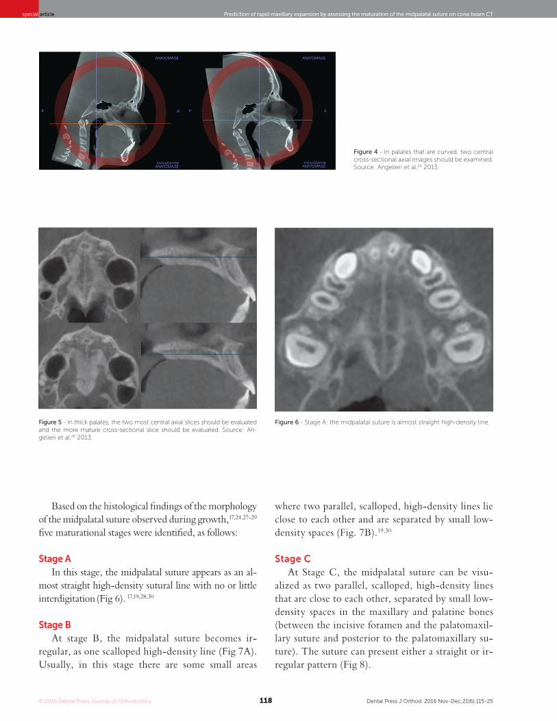
However, some individuals present a curved palatal contour, and for them, the palate should be analyzed in two separate central cross-sectional axial slices, one from posterior and another from anterior region of the midpalatal suture, separately (Fig 4). Furthermore, for subjects who presented with a thicker palate, the pal-ate should be evaluated in the two most central axial slices (Fig 5). The more matured central cross-sectional axial slice should be considered.
Figure 2 - Orientation of head position in the axial (A), sagittal (B) and coronal planes (C). Source: Angelieri et al,26 2013.
Figure 3 - Selection of the most central cross-sectional slice in the superior-inferior dimension, to assess the midpalatal suture maturation.
B CA
© 2016 Dental Press Journal of Orthodontics Dental Press J Orthod. 2016 Nov-Dec;21(6):115-25118
Prediction of rapid maxillary expansion by assessing the maturation of the midpalatal suture on cone beam CTspecial article
where two parallel, scalloped, high-density lines lie close to each other and are separated by small low-density spaces (Fig. 7B).19,30
Stage CAt Stage C, the midpalatal suture can be visu-
alized as two parallel, scalloped, high-density lines that are close to each other, separated by small low-density spaces in the maxillary and palatine bones (between the incisive foramen and the palatomaxil-lary suture and posterior to the palatomaxillary su-ture). The suture can present either a straight or ir-regular pattern (Fig 8).
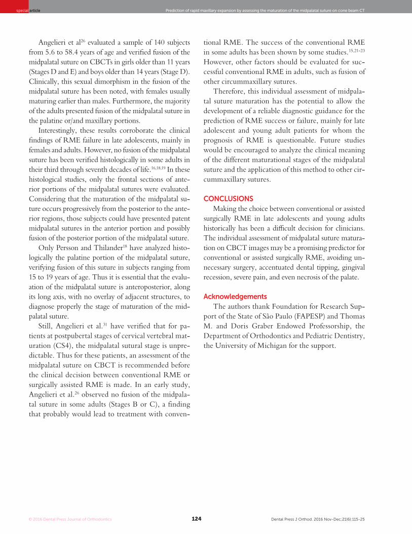
Based on the histological findings of the morphology of the midpalatal suture observed during growth,17,24,27-29 five maturational stages were identified, as follows:
Stage AIn this stage, the midpalatal suture appears as an al-
most straight high-density sutural line with no or little interdigitation (Fig 6). 17,19,28,30
Stage BAt stage B, the midpalatal suture becomes ir-
regular, as one scalloped high-density line (Fig 7A). Usually, in this stage there are some small areas
Figure 4 - In palates that are curved, two central cross-sectional axial images should be examined. Source: Angelieri et al,26 2013.
Figure 5 - In thick palates, the two most central axial slices should be evaluated and the more mature cross-sectional slice should be evaluated. Source: An-gelieri et al,26 2013.
Figure 6 - Stage A: the midpalatal suture is almost straight high-density line.

© 2016 Dental Press Journal of Orthodontics Dental Press J Orthod. 2016 Nov-Dec;21(6):115-25119
Angelieri F, Franchi L, Cevidanes LHS, Bueno-Silva B, McNamara Jr. JA special article
Stage DIn this stage, the fusion of the midpalatal suture
has occurred in the palatine bone, so the midpalatal suture cannot be visualized in the palatine bone, as usually the fusion happens from posterior to anterior portion16,18 (Fig 9). It is important to stress that the parasutural bone density is increased (high-density bone) compared to the density of the maxillary para-sutural bone. In the maxillary portion, the midpalatal suture still appears as two high-density lines separat-ed by small low-density spaces.
Stage EAt stage E, the midpalatal suture cannot be visualized
in at least a portion of the maxilla,28,29 once at least one partial fusion of this suture has happened in the maxilla (Fig 10). The parasutural bone density is increased, with the same level as in other regions of the palate.19
All maturational stages of the midpalatal suture are rep-resented in the schematic drawing depicted in Figure 11.
Figure 7 - Stage B appears as a scalloped high-density line (A); or in some areas, two parallel, scalloped high-density lines close to each other and separated by small low-density spaces – arrows in B.
Figure 8 - Stage C is characterized as two parallel, scalloped high-density lines close to each other and separated by small low-density spaces in either a straight or an irregular pattern.
A B

© 2016 Dental Press Journal of Orthodontics Dental Press J Orthod. 2016 Nov-Dec;21(6):115-25120
Prediction of rapid maxillary expansion by assessing the maturation of the midpalatal suture on cone beam CTspecial article
Figure 9 - Stage D: in the palatine bone, the midpalatal suture cannot be visual-ized and the parasutural bone density is increased.
Figure 10 - At Stage E, the midpalatal suture is not visible in at least a portion of maxilla.
Figure 11 - Schematic drawing (by Chris Jung) of the maturational stages of the midpalatal suture. Source: Angelieri et al,26 2013.
Clinical implications of midpalatal sutural maturation on CBCTs
The presence of posterior crossbite or atresia of the maxilla in late adolescents or young adults has been a challenge for orthodontists. The clinical choice between conventional RME or SARME implies possible unnec-essary surgical procedures — demanding costs and risks for patients — or side-eff ects of conventional RME fail-ure as severe pain, mucosal ulceration or necrosis, ac-centuated buccal tipping and gingival recession in the posterior teeth.6-10 There are no clinical parameters for this diffi cult decision; histological and micro-CT stud-ies have demonstrated that chronological age and gender are not a reliable parameter for the fusion of the mid-palatal suture16,18,19,20,24 (Figs 12 and 13).
CBCT imaging facilitates three dimensional visual-ization of the oral and maxillofacial structures, allow-ing the evaluation of the midpalatal suture maturation26 without the overlay of the vomer and other external structures of nose on the midpalatal region, as occurs on occlusal radiographs.20
It is interesting that the fi ve maturational stages identifi ed on CBCTs corroborate with the histological fi ndings of midpalatal suture maturation. In a landmark study, Melsen17 observed that in the juvenile period (usually up to 10 years of age), the midpalatal suture is broad and Y-shaped in frontal sections.28,29 From 10 to 13 years of age, this suture appears with a squamous path, becoming wavier with increased interdigitation at ages 13 to 14 years. These descriptions match Stages A and B, respectively, with the increase of the interdigita-tion characterizing the more matured stage.
The fusion of the midpalatal suture has been described in several histological studies. The fusion process of the midpalatal suture begins with bone spicules from suture margins along with “islands” (i.e., masses of acellular tis-sue and inconsistently-calcifi ed tissue) in the middle of the sutural gap.18,19,24,28 These spicules are present in many places along the suture, and they increase with matura-tion.18,27 The spicules appear as many scalloped areas that are close to each other and yet are separated in some zones by connective tissue.16,20 This description is compatible with Stage C, in which many bony bridges can be visual-ized along the suture, leading to more resistance for con-ventional RME. Probably, RME performed in patients at Stages A and B would have less resistance forces and more skeletal eff ects than when performed during Stage C.
Stage EStage DStage CStage BStage A

© 2016 Dental Press Journal of Orthodontics Dental Press J Orthod. 2016 Nov-Dec;21(6):115-25121
Angelieri F, Franchi L, Cevidanes LHS, Bueno-Silva B, McNamara Jr. JA special article
Figure 12 - 16-year-old boy treated with Haas expander. There was the failure of RME. Source: Angelieri et al,34 2015.

© 2016 Dental Press Journal of Orthodontics Dental Press J Orthod. 2016 Nov-Dec;21(6):115-25122
Prediction of rapid maxillary expansion by assessing the maturation of the midpalatal suture on cone beam CTspecial article
Figure 13 - Successful RME in a 16-year-old girl treated with Hyrax-expander.
Angelieri et al.31 demonstrated that the midpalatal suture maturation is related to skeletal growth, since a high correlation coefficient was observed between the cervical vertebra maturation and maturational stages of the midpalatal suture. According to the results, the pre-pubertal stages (cervical stages CS1 and CS2) are reli-able indicators for stages A and B of midpalatal suture maturation. In the pubertal stage (CS3), probably the patient will present the midpalatal suture at stage C. Considering the presence of many bony bridges along the midpalatal suture at stage C, these findings corrobo-rate the results of Baccetti et al,32 who observed more favorable skeletal changes from RME in prepubertal pa-tients compared to postpubertal patients.
Furthermore, Krukemeyer33 evaluated the correla-tion among response to RME, maturational stages of the midpalatal suture, and the stage of cervical verte-bral maturation (CVM). The maturational stages of the midpalatal suture and CVM stages were correlated in-versely with sutural expansion, i.e. the less mature the patient, the greater was sutural expansion, with more skeletal than dentoalveolar effects of RME.
On the other hand, it is important to stress that, in spite of increased sutural resistance to conven-tional RME at Stage C, the widening of maxilla orthopedically with no surgical interventional still is possible (Fig. 14). This procedure should be initi-ated immediately, due to the start of fusion of the palatine portion of the midpalatal suture might be-ing imminent.34
With the maturation of midpalatal suture, there is an increase in interdigitation.16,17 As mentioned previously, sutural fusion happens earlier in the pos-terior region and subsequently progresses toward the anterior,16,18 with resorption of cortical bone in the sutural ends and the subsequent formation of cancel-lous bone.28,29 When patients are at Stage D, it is pos-sible to visualize the interincisal diastema promoted by RME, even though no widening of the palate will have occurred posteriorly. The fusion of the palatine (Stage D) or/and maxillary portions (Stage E) of the midpalatal suture hampers the expansive forces of con-ventional RME; these patients are treated more effec-tively by surgically-assisted RME.34

© 2016 Dental Press Journal of Orthodontics Dental Press J Orthod. 2016 Nov-Dec;21(6):115-25123
Angelieri F, Franchi L, Cevidanes LHS, Bueno-Silva B, McNamara Jr. JA special article
Figure 14 - A 15-year-old boy patient at Stage C. Conventional RME still was possible. Source: Angelieri et al,34 2015.
© 2016 Dental Press Journal of Orthodontics Dental Press J Orthod. 2016 Nov-Dec;21(6):115-25124
Prediction of rapid maxillary expansion by assessing the maturation of the midpalatal suture on cone beam CTspecial article
Angelieri et al26 evaluated a sample of 140 subjects from 5.6 to 58.4 years of age and verified fusion of the midpalatal suture on CBCTs in girls older than 11 years (Stages D and E) and boys older than 14 years (Stage D). Clinically, this sexual dimorphism in the fusion of the midpalatal suture has been noted, with females usually maturing earlier than males. Furthermore, the majority of the adults presented fusion of the midpalatal suture in the palatine or/and maxillary portions.
Interestingly, these results corroborate the clinical findings of RME failure in late adolescents, mainly in females and adults. However, no fusion of the midpalatal suture has been verified histologically in some adults in their third through seventh decades of life.16,18,19 In these histological studies, only the frontal sections of ante-rior portions of the midpalatal sutures were evaluated. Considering that the maturation of the midpalatal su-ture occurs progressively from the posterior to the ante-rior regions, those subjects could have presented patent midpalatal sutures in the anterior portion and possibly fusion of the posterior portion of the midpalatal suture.
Only Persson and Thilander18 have analyzed histo-logically the palatine portion of the midpalatal suture, verifying fusion of this suture in subjects ranging from 15 to 19 years of age. Thus it is essential that the evalu-ation of the midpalatal suture is anteroposterior, along its long axis, with no overlay of adjacent structures, to diagnose properly the stage of maturation of the mid-palatal suture.
Still, Angelieri et al.31 have verified that for pa-tients at postpubertal stages of cervical vertebral mat-uration (CS4), the midpalatal sutural stage is unpre-dictable. Thus for these patients, an assessment of the midpalatal suture on CBCT is recommended before the clinical decision between conventional RME or surgically assisted RME is made. In an early study, Angelieri et al.26 observed no fusion of the midpala-tal suture in some adults (Stages B or C), a finding that probably would lead to treatment with conven-
tional RME. The success of the conventional RME in some adults has been shown by some studies.15,21-23 However, other factors should be evaluated for suc-cessful conventional RME in adults, such as fusion of other circummaxillary sutures.
Therefore, this individual assessment of midpala-tal suture maturation has the potential to allow the development of a reliable diagnostic guidance for the prediction of RME success or failure, mainly for late adolescent and young adult patients for whom the prognosis of RME is questionable. Future studies would be encouraged to analyze the clinical meaning of the different maturational stages of the midpalatal suture and the application of this method to other cir-cummaxillary sutures.
CONCLUSIONSMaking the choice between conventional or assisted
surgically RME in late adolescents and young adults historically has been a difficult decision for clinicians. The individual assessment of midpalatal suture matura-tion on CBCT images may be a promising predictor for conventional or assisted surgically RME, avoiding un-necessary surgery, accentuated dental tipping, gingival recession, severe pain, and even necrosis of the palate.
AcknowledgementsThe authors thank Foundation for Research Sup-
port of the State of São Paulo (FAPESP) and Thomas M. and Doris Graber Endowed Professorship, the Department of Orthodontics and Pediatric Dentistry, the University of Michigan for the support.
© 2016 Dental Press Journal of Orthodontics Dental Press J Orthod. 2016 Nov-Dec;21(6):115-25125
Angelieri F, Franchi L, Cevidanes LHS, Bueno-Silva B, McNamara Jr. JA special article
1. Angell EC. Treatment of irregularities of the permanent or adult teeth. Dent
Cosmos. 1860;1(9):541-4, [it continue in Dent Cosmos 1860;1(11):599-600].
2. Bishara SE, Staley RN. Maxillary expansion: clinical implications. Am J Orthod
Dentofacial Orthop. 1987 Jan;91(1):3-14.
3. Haas AJ. Rapid expansion of the maxillary dental arch and nasal cavity by
opening the mid-palatal suture. Angle Orthod. 1961;31(2):73-90.
4. Haas AJ. Palatal expansion: just the beginning of dentofacial orthopedics.
Am J Orthod. 1970;57(3):219-55.
5. Silva Filho OG, Magro AC, Capelozza Filho L. Early treatment of the Class III
malocclusion with rapid maxillary expansion and maxillary protraction. Am J
Orthod Dentofacial Orthop. 1998 Feb;113(2):196-203.
6. Bell WH, Epker BN. Surgical-orthodontic expansion of the maxilla. Am J
Orthod. 1976;70(5):517-28.
7. Betts NJ, Vanarsdall RL, Barber HD, Higgins-Barber K, Fonseca RJ. Diagnosis
and treatment of transverse maxillary deficiency. Int J Adult Orthodon
Orthognath Surg. 1995;10(2):75-96.
8. Garib DG, Henriques JF, Janson G, Freitas MR, Coelho RA. Rapid maxillary
expansion--tooth tissue-borne versus tooth-borne expanders: a computed
tomography evaluation of dentoskeletal effects. Angle Orthod. 2005
July;75(4):548-57.
9. Kiliç N, Kiki A, Oktay H. A comparison of dentoalveolar inclination treated by
two palatal expanders. Eur J Orthod. 2008 Feb;30(1):67-72.
10. Rungcharassaeng K, Caruso JM, Kan JY, Kim J, Taylor G. Factors affecting
buccal bone changes of maxillary posterior teeth after rapid maxillary
expansion. Am J Orthod Dentofacial Orthop. 2007 Oct;132(4):428.e1-8.
11. Mommaerts MY. Transpalatal distraction as a method of maxillary expansion.
Br J Oral Maxillofac Surg. 1999 Aug;37(4):268-72.
12. Epker BN, Wolford LM. Transverse maxillary deficiency dentofacial
deformities: integrated orthodontic and surgical correction. St Louis: Mosby;
1980.
13. Mossaz CF, Byloff FK, Richter M. Unilateral and bilateral corticotomies
for correction of maxillary transverse discrepancies. Eur J Orthod. 1992
Apr;14(2):110-6.
14. Timms DJ, Vero D. The relationship of rapid maxillary expansion to surgery
with special reference to midpalatal synostosis. Br J Oral Surg. 1981
Sept;19(3):180-96.
15. Alpern MC, Yurosko JJ. Rapid palatal expansion in adults with and without
surgery. Angle Orthod 1987 july;57:245-63.
16. Knaup B, Yildizhan F, Wehrbein H. Age-related changes in the midpalatal
suture. A histomorphometric study. J Orofac Orthop. 2004 Nov;65(6):467-74.
17. Melsen B. Palatal growth studied on human autopsy material. A histologic
microradiographic study. Am J Orthod. 1975 July;68(1):42-54.
18. Persson M, Thilander B. Palatal suture closure in man from 15 to 35 years of
age. Am J Orthod. 1977 July;72(1):42-52.
19. Korbmacher H, Schilling A, Püschel K, Amling M, Kahl-Nieke B. Age-
dependent three-dimensional microcomputed tomography analysis of the
human midpalatal suture. J Orofac Orthop. 2007 Sept;68(5):364-76.
20. Wehrbein H, Yildizhan F. The mid-palatal suture in young adults.
A radiological-histological investigation. Eur J Orthod. 2001 Apr;23(2):105-14.
21. Capelozza Filho L, Cardoso Neto J, da Silva Filho OG, Ursi WJ. Non-
surgically assisted rapid maxillary expansion in adults. Int J Adult Orthodon
Orthognath Surg. 1996;11(1):57-66; discussion 67-70.
22. Handelman CS, Wang L, BeGole EA, Haas AJ. Nonsurgical rapid maxillary
expansion in adults: report on 47 cases using the Haas expander. Angle
Orthod. 1997;67(4):291-305; discussion 306-8.
23. Handelmann CS, Wang L, BeGole EA, Haas AJ. Nonsurgical rapid maxillary
expansion in adults: report on 47 cases using the Haas expander. Angle
Orthod. 2000 Apr;70(2):129-44.
24. Persson M, Magnusson BC, Thilander B. Sutural closure in rabbit and man:
a morphological and histochemical study. J Anat. 1978;125(2):313-21.
25. Revelo B, Fishman LS. Maturational evaluation of ossification of the
midpalatal suture. Am J Orthod Dentofacial Orthop. 1994;105(3):288-92.
26. Angelieri F, Cevidanes LHS, Franchi L, Gonçalves JR, Benavides E,
McNamara JA Jr. Midpalatal suture maturation: classification method for
individual assessment prior to rapid maxillary expansion. Am J Orthod
Dentofacial Orthop. 2013;144(5):759-69.
27. Melsen B. A histological study of the influence of sutural morphology and
skeletal maturation on rapid palatal expansion in children. Trans Eur Orthod
Soc. 1972:499-507.
28. Cohen Jr MM. Sutural biology and the correlates of craniosynostosis. Am J
Med Genet. 1993 Oct 1;47(5):581-616.
29. Sun Z, Lee E, Herring SW. Cranial sutures and bones: growth and fusion
in relation to masticatory strain. Anat Rec A Discov Mol Cell Evol Biol.
2004;276(2):1-22.
30. Hahn W, Fricke-Zech S, Fialka-Fricke J, Dullin C, Zapf A, Gruber R, et al.
Imaging of the midpalatal suture in a porcine model: flat-panel volume
computed tomography compared with multislice computed tomography.
Oral Surg Oral Med Oral Pathol Oral Radiol Endod. 2009 Sept;108(3):443-9.
31. Angelieri F, Franchi L, Cevidanes LH, McNamara JA Jr. Diagnostic
performance of skeletal maturity for the assessment of midpalatal suture
maturation. Am J Orthod Dentofacial Orthop. 2015 Dec;148(6):1010-6.
32. Baccetti T, Franchi L, Cameron CG, McNamara JA Jr. Treatment timing for
rapid maxillary expansion. Angle Orthod. 2001 Oct;71(5):343-50.
33. Krukemeyer AM. The effects of sutural and cervical vertebral maturation
on dentoalveolar versus skeletal responses to rapid maxillary expansion
[master´s thesis]. Ann Arbor: The University of Michigan; 2013.
34. Angelieri F, Cevidanes LH, Franchi L, McNamara Jr JA. Evaluation of
facial suture maturation on CBCTs: a predictor of maxillary orthopedic
treatment response. In: Kapila S, Nervina J, Hatch N. Expedited
Orthodontics: Improving the efficiency of orthodontic treatment through
novel technologies. Ann Arbor: University of Michigan; 2015. p. 257-80.
Craniofacial Growth Series; 51.
REFERENCES