Embed Size (px)
Citation preview

Prediction of allosteric sites and mediating interactions through bond-to-bond
propensities
B.R.C. Amor,1, 2 M.T. Schaub,3 S.N. Yaliraki,1, 2 and M. Barahona3, 2
1Department of Chemistry, Imperial College London, London SW7 2AZ, United Kingdom2Institute of Chemical Biology, Imperial College London, London SW7 2AZ, United Kingdom3Department of Mathematics, Imperial College London, London SW7 2AZ, United Kingdom∗
Allosteric regulation is central to many biochemical processes. Allosteric sites provide a targetto fine-tune protein activity, yet we lack computational methods to predict them. Here, we presentan efficient graph-theoretical approach for identifying allosteric sites and the mediating interactionsthat connect them to the active site. Using an atomistic graph with edges weighted by covalentand non-covalent bond energies, we obtain a bond-to-bond propensity that quantifies the effectof instantaneous bond fluctuations propagating through the protein. We use this propensity todetect the sites and communication pathways most strongly linked to the active site, assessingtheir significance through quantile regression and comparison against a reference set of 100 genericproteins. We exemplify our method in detail with three well-studied allosteric proteins: caspase-1,CheY, and h-Ras, correctly predicting the location of the allosteric site and identifying key allostericinteractions. Consistent prediction of allosteric sites is then attained in a further set of 17 proteinsknown to exhibit allostery. Because our propensity measure runs in almost linear time, it offers ascalable approach to high-throughput searches for candidate allosteric sites.
I. INTRODUCTION
Allostery is a key molecular mechanism underpinningcontrol and modulation in a variety of cellular pro-cesses [1, 2]. Allosteric effects are those induced on themain functional site of a biomolecule by the binding ofan effector at a distant site, e.g., the binding of a co-factor modulating the catalytic rate of an enzyme [3].Despite the importance of such processes, there is still alack of understanding as to how the interactions at theallosteric site propagate across the protein and affect theactive site. In this paper, we present a graph-theoreticapproach that uses atomistic structural data to identifyallosteric sites in proteins, as well as bonds and residuesinvolved in this propagation. By defining an edge-to-edgetransfer function, which can be understood as a Green’sfunction in the edge space of the protein graph, we com-pute a bond-to-bond propensity that captures the effectinduced on any bond of the molecule by the propaga-tion of perturbations stemming from bonds at the activesite. This propensity can be computed efficiently to pre-dict allosteric sites and key bonds which are prominentlyinvolved in mediating the allosteric propagation.The growing realisation that all proteins exhibit innate
dynamic behaviour [4, 5] and the discovery of allostericeffects in single domain proteins [6] have reaffirmed theubiquitousness of this form of regulation; potentially, anyprotein could be allosteric [7]. This fact opens up impor-tant experimental directions: drugs targeted at allostericsites could offer improved specificity and control com-pared to traditional drugs that bind at the active site [3].Efficient methods able to identify putative allosteric sitesare therefore of great current interest [8]. To date, com-
∗ present address: Universit catholique de Louvain, Belgium
putational approaches to finding allosteric sites have in-volved statistical coupling analysis [9], molecular dynam-ics [10–12], machine learning [13], and normal mode anal-ysis [14]. For a comprehensive review see Ref. [15].
Classic thermodynamic models of allostery (such as theMonod-Wyman-Changeux (MWC) [16] and Koshland-Nemethy-Filmer (KNF) models [17]) were formulated toexplain cooperativity in multimeric proteins in terms ofconformational transitions in a protein landscape [11, 18].Although such models reproduce broad experimental fea-tures (e.g. the sigmoidal binding curves), they offer littleinsight into the molecular mechanisms driving and defin-ing the underlying conformational transitions. Attemptsto identify the specific residues involved in allosteric tran-sitions have led to the idea of allosteric pathways, whichaim to describe the routes through which an excitationpropagates through the protein [9, 19, 20]. Indeed, recentexperimental [21, 22] and computational [23–26] work hasshown that energy flow in globular proteins is anisotropic.Some of these studies have connected this anisotropy tothe allosteric properties of the protein [22, 26]. Ourwork builds on this line of research and aims at find-ing allosteric sites by using graph-theoretical techniquesto quantify efficiently the propagation of perturbationsthrough a protein structure described in atomistic de-tail. In [22] the authors find that internal energy flow inalbumin is anisotropic, and that this flow is altered bybinding of an allosteric ligand. Here, we also find thatthe propagation of perturbations internally is anisotropic.However, we use the term ‘allosteric’ in a more specificway, to describe locations distant from the active sitewhere a perturbation can have a functional effect on theactive site. The identification of such distant sites andthe pathways connecting them to the active-site, has be-come an area of considerable interest [12, 27, 28].
The connection between the behaviour of a diffusionprocess (e.g., a random walk) on a network and the vi-
peer-reviewed) is the author/funder. All rights reserved. No reuse allowed without permission. The copyright holder for this preprint (which was not. http://dx.doi.org/10.1101/056275doi: bioRxiv preprint first posted online May. 31, 2016;

2
brational dynamics of that network is well establishedin the biophysical literature [29, 30]. Previous network-based methods for protein structure analysis have madeuse of shortest path calculations [31], community detec-tion algorithms [32], and random walks on networks [33].However, such methods almost universally used coarse-
grained protein descriptions at the level of residues,i.e., they are based on residue-residue interaction net-works (RRINs) [34] that neglect atomistic detail. Al-though methods that use molecular dynamics simula-tions to derive edge weights for RRINs from the cross-correlations of residue fluctuations have yielded interest-ing results [35, 36], such approaches are computation-ally costly. Furthermore, Ribeiro and Ortiz have recentlyshown that RRINs are critically dependent on the chosencut-off distance, and that using energy-weighted networksthat include the covalent interactions of the backbone iscrucial for correctly identifying signal propagation path-ways [37, 38]. Our findings below show that efficientmethodologies which can exploit the physico-chemical de-tail of atomistic, energy-weighted protein networks canlead to enhanced identification of allosteric sites and rel-evant individual mediating interactions in a number ofimportant cases.
Our analysis starts by building an atomistic graphmodel of the protein: nodes are atoms and (weighted)edges represent individual bonds, with weights given byenergies from interatomic potentials. The graph includesboth covalent bonds and weak, non-covalent bonds (hy-drogen bonds, salt bridges, hydrophobic tethers and elec-trostatic interactions). Details of the construction of thegraph are given in Section IVE and in Refs. [39, 40].The resulting all-atom graph is analysed using the edge-to-edge transfer matrix M , which is akin to a discreteGreen’s function in the edge space of the graph and hasbeen recently introduced in Ref. [41] to study non-localedge coupling in graphs. In this paper, we derive a new,alternative interpretation of the matrix M and show thatit provides a means to extracting the level of influencethat the fluctuations of an edge have on any other edgeof the graph (for detailed mathematical derivations, seeMaterials and Methods, Section IVA1 and SI ). We usethis notion to calculate the propensity of each bond, Πb,i.e., a measure of how strongly bond b is coupled to the ac-tive site through the atomistic graph. Because allostericeffects are reflected on induced changes in weak bonds,yet mediated through the whole protein network, ourbond-to-bond formalism provides a natural way of un-covering how the long-range correlations between bondscontribute to allosteric signalling. Crucially, recent algo-rithmic developments [42, 43] allow these computationsto be carried out in almost linear time (in the numberof edges). Therefore, in contrast to most other computa-tional approaches, our method is easily scalable to largesystems with tens of thousands of atoms.
To establish if a bond has a high propensity Πb, andto detect important bonds (and residues), we use quan-tile regression to compare each bond to the ensemble
of bonds within the protein at a similar geometric dis-tance from the active-site (described in Materials andMethods, Section IVB). Quantile regression (QR) [44]is a robust statistical technique previously employed inmedicine [45], ecology [46] and econometrics [47]. We ad-ditionally confirm our findings by computing the statis-tical significance of the bond propensity against a refer-ence set of 100 representative proteins randomly drawnfrom the Structural Classification of Proteins (SCOP)database (see Section IID). This reference set provides uswith a pre-computed structural bootstrap against whichany protein can be tested to detect statistically signifi-cant bonds, further reducing the computational cost ofour method.In Sections IIA—IIC, we showcase our procedure
through the detailed analysis of three important allostericproteins: caspase-1, CheY, and h-Ras. In each case,given structural data and the location of the known ac-tive site, we correctly predict the location of the al-losteric site and uncover communication pathways be-tween both sites. Each of the three examples serves tohighlight particular aspects of the method. In the caseof caspase-1, comparison of our results with those ob-tained using coarse-grained residue-residue interactionsnetworks (RRINs) shows that incorporating atomisticphysico-chemical detail can indeed be necessary for thereliable identification of the allosteric site. In the caseof CheY, we illustrate how further information can begained by incorporating dynamic data from ensembles ofNMR structures: the variance of the propensity acrossthe NMR ensemble reveals residues involved in allostericsignalling which cannot be identified from the static X-ray crystal structure alone. In the case of h-Ras, ourmethod shows that signal propagation between the ac-tive and allosteric sites is crucially dependent on theinteraction between the protein and specific structuralwater molecules. Having demonstrated the insight intoallosteric mechanisms offered by our method, we thenevaluate it against a test set with a further 17 allostericproteins (see Section II E). We find that the bond-to-bond propensity is a good predictor of a site’s allostericpropensity, suggesting it could be used to guide efforts instructure-based discovery of drugs as allosteric effectors.
II. RESULTS
A. Identification of the allosteric site and
functional residues in caspase-1
Our first example is caspase-1, an allosteric protein ofgreat importance in apoptotic processes [40]. Caspase-1is a tetramer composed of two asymmetric dimers, eachcontaining one active site. Using the PDB atomic struc-ture (PDB: 2HBQ), we constructed an atomistic, energy-weighted graph representation of the protein based on in-teraction potentials, as described in Section IVE [39, 40].In order to quantify how strongly each bond is cou-
peer-reviewed) is the author/funder. All rights reserved. No reuse allowed without permission. The copyright holder for this preprint (which was not. http://dx.doi.org/10.1101/056275doi: bioRxiv preprint first posted online May. 31, 2016;

3
FIG. 1. Bond-to-bond propensities identify the allosteric site and atomistic pathway in caspase-1. (a) Thepropensities of all residues ΠR are plotted against their distance from the active site. The lines correspond to the quantileregression estimates for the p-th quantiles Qp, with p = 0.1, 0.2, . . . , 0.8, 0.9. The dashed red line indicates the Q0.90 cut-off usedfor identifying important residues. (b) The quantile scores pR for each residue are mapped onto the surface of caspase-1. Theactive-site ligand is shown in green. The allosteric binding site is identified as a hot-spot of high propensity. When a coarse-grained residue-residue interaction network with cut-off of 6A is used (right), the allosteric binding site is not identified. (c) Thepropensities of bonds Πb are plotted against their distance from the active site with the Q0.99 quantile indicated by the dashedline. (d) High quantile score bonds (pb ≥ 0.99) are shown on the structure. Bonds between R286:E390, R240:D336, R286:N337,A284:S332, and S332:S339 have large quantile scores and form contiguous pathways between the active and allosteric sites.The active site ligand is shown in green and the allosteric ligand is shown as yellow spheres.
pled to the active site, we calculate the propensities Πb
for all bonds in the protein, as given by Eq. (8). We alsoaggregate the bond propensities for each residue to ob-tain the residue score ΠR, as given by Eq. (9). To rankbonds and residues according to their significance, wecompute the corresponding quantile scores pb and pR, re-spectively, obtained via quantile regression as in Eq. (14).These quantile scores allow us to establish which bonds(and residues) have high propensity values as comparedto bonds (or residues) at the same distance from the ac-tive site in the protein (Fig. 1a and 1c).
Our method finds a hot spot of residues with highquantile scores in a cavity at the dimer-dimer interface(Fig. 1b left). This site has been previously identified byScheer et al. as the binding site for a small molecule in-
hibitor of caspase-1 [48]. Table I shows that the allostericresidues, i.e., residues within 3.5A of the allosteric in-hibitor, have significantly higher propensities than non-allosteric residues (Wilcoxon rank sum, p < 0.0005).Residues E390, S332 and R286, which have been foundto belong to a hydrogen bond network between the activeand allosteric sites [48], have respectively the 3rd, 13th,and 15th highest quantile scores of the 260 residues ineach dimer of caspase-1.
Making use of the physico-chemical detail afforded byour atomistic description, we find the bonds with highpropensity that lie on communication pathways connect-ing the allosteric site to the active-site ligand. Con-centrating on the top quantile pb ≥ 0.99 (Fig. 1c), thetwo interactions in the salt bridges between residues
peer-reviewed) is the author/funder. All rights reserved. No reuse allowed without permission. The copyright holder for this preprint (which was not. http://dx.doi.org/10.1101/056275doi: bioRxiv preprint first posted online May. 31, 2016;

4
E390 and R286 have quantile scores of 0.996 and 0.990,and their combined propensity gives this salt bridgethe highest quantile score in the protein. It is knownthat these salt-bridges are directly disrupted by the al-losteric inhibitor [48]. In addition, our method revealsother important bonds lying between the active and al-losteric sites (Fig. 1d), including hydrogen bonds betweenArg240:Asp336 (pb = 0.999), S332:S339 (pb = 0.996),R286:N337 (pb = 0.992), and A284:S332 (pb = 0.990).Bonds in this pathway have previously been identified byDatta et al as being functionally important: the corre-sponding alanine mutations cause 230-fold (R286A), 130-fold (E390A), 3.7-fold (S332A) and 6.7-fold (S339A) re-ductions in catalytic efficiency [48].The atomistic detail is important for the outcome
of the analysis. If instead of employing an all-atomgraph description, we carry out the same calculationson a coarse-grained residue-residue interaction network(RRIN) [31, 33] with cut-off radius of 6 A, the allostericsite of caspase-1 is no longer identified as a hot spot(Fig. 1b right) and the allosteric residues do not have sig-nificantly higher propensity compared to other residues(Wilcoxon rank sum, p = 0.5399). The results obtainedwith RRINs are in general dependent on the cut-off ra-dius used. For caspase-1, the allosteric site is not detectedin RRINs with cut-off radii of 6 A, 7 A and 8 A. The al-losteric site is found to be significant at 10A, but the sig-nal is still considerably weaker than when using the atom-istic network (Table S6). These findings highlight thatwhile an atomistic model of the protein structure maynot always be needed, it can indeed be important for thedetection of allosteric effects in proteins; in this case, thestrength of the pair of salt bridges formed by E390 andE286, which is crucial for the allosteric communicationin caspase-1, is not captured by RRINs. Other recent re-sults have similarly demonstrated the importance of bothcovalent bonds and hydrogen bonds to signal transmis-sion within proteins [38]. Yet in other cases (e.g., CheYin the following section), this level of physico-chemicaldetail seems to be less important, and RRINs are ableto capture allosteric communication. An extended, in-depth analysis of the results obtained with all-atom net-works and RRINs for a variety of proteins and cut-offradii can be found in the SI (Section 6).
B. Uncovering allosteric communication pathways
in CheY
1. Identification of the phosphorylation site of CheY
CheY is a key protein in bacterial chemotaxis. WhenCheY binds to the flagellar motor switch protein (FliM),it causes a change in the rotation direction of the flagellarmotor, thus regulating the tumbling rate of E. coli. Thisregulation is achieved through a post-translational modi-fication of CheY: phosphorylation of CheY at the distantresidue D57 increases its affinity for FliM, making this an
TABLE I. Quantile scores for the propensities of residueswithin 3.5A of the allosteric site of caspase-1 computed fromthe atomistic graph and from a residue-based network (RRIN)with cut-off radius of 6 A. The average quantile scores ofallosteric residues (pR,allo) and non-allosteric residues (pR,rest)are also presented.
Residue pR (Atomistic network) pR (RRIN)
Dimer 1 Dimer 2 Dimer 1 Dimer 2
R240 0.772 0.734 0.562 0.562
L258 0.394 0.408 0.168 0.168
N259 0.828 0.832 0.324 0.324
F262 0.654 0.652 0.464 0.464
R286 0.938 0.928 0.838 0.838
C331 0.634 0.646 0.724 0.724
P335 0.206 0.196 0.450 0.450
E390 0.990 0.992 0.318 0.318
R391 0.982 0.984 0.258 0.258
palloR 0.711 0.708 0.4567 0.4567
prestR 0.481 0.492 0.4793 0.4789
interesting example of a single-domain allosteric protein.
Following the same procedure, we calculated thepropensity of each bond and residue (relative to theFliM binding site) in fully activated CheY (PDB ID:1F4V) bound to Mg2+, BeF3 and FliM. We identifya number of hot-spot surface residues with high quan-tile scores (Fig. 2a), including the phosphorylation site,D57 (pR = 0.96). Again, residues in the allosteric site(< 3.5 A from the phosphorylation site) have higher aver-age quantile score than non-allosteric residues (pR,allo =0.61 > pR,rest = 0.43), and four of the seven residuesin the allosteric site have high quantile scores, pR ≥ 0.9(Table II). In addition, we find a number of previouslyunidentified distant surfaces with high quantile scores(Fig. 2a), which could correspond to putative (orphan)allosteric sites.
In contrast to caspase-1 above, using a RRIN with cut-off radius of 6 A, we find that the phosphorylation siteof CheY is identified as a hot-spot: the average quan-tile score of allosteric residues is much higher for the restof the residues (pR,allo = 0.72 > pR,rest = 0.46). TheRRIN detection is robust over a range of cut-off radiibetween 6A-16A (Table S6 and Fig. S5). This resultsuggests that sometimes (as for CheY) it is the topol-
ogy of the protein structure that is important for signalpropagation, whereas in other cases (as for caspase-1)the specific atomistic structure given by the chemistry
of the side-chain interactions matters for allosteric prop-agation. Our all-atom methodology incorporates bothaspects consistently.
peer-reviewed) is the author/funder. All rights reserved. No reuse allowed without permission. The copyright holder for this preprint (which was not. http://dx.doi.org/10.1101/056275doi: bioRxiv preprint first posted online May. 31, 2016;
5
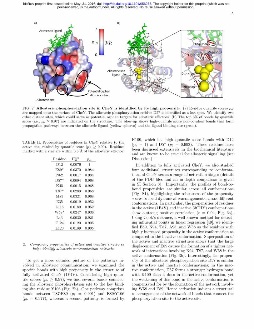
FIG. 2. Allosteric phosphorylation site in CheY is identified by its high propensity. (a) Residue quantile scores pRare mapped onto the surface of CheY. The allosteric phosphorylation residue D57 is identified as a hot-spot. We identify twoother distant sites, which could serve as potential orphan targets for allosteric effectors. (b) The top 3% of bonds by quantilescore (i.e., pb ≥ 0.97) are indicated on the structure. The blow-up shows high-quantile score non-covalent bonds that formpropagation pathways between the allosteric ligand (yellow spheres) and the ligand binding site (green).
TABLE II. Propensities of residues in CheY relative to theactive site, ranked by quantile score (pR ≥ 0.90). Residuesmarked with a star are within 3.5 A of the allosteric effector.
Residue Πact
R pR
D12 0.0076 1
E89* 0.0370 0.984
N62 0.0017 0.984
D57* 0.0094 0.968
K45 0.0015 0.968
T87* 0.0283 0.968
M85 0.0321 0.968
E35 0.0019 0.952
L116 0.0189 0.952
W58* 0.0247 0.936
L43 0.0030 0.921
F124 0.0120 0.905
L120 0.0189 0.905
2. Comparing propensities of active and inactive structureshelps identify allosteric communication networks
To get a more detailed picture of the pathways in-volved in allosteric communication, we examined thespecific bonds with high propensity in the structure offully activated CheY (1F4V). Considering high quan-tile scores (pb ≥ 0.97), we find several bonds connect-ing the allosteric phosphorylation site to the key bind-ing site residue Y106 (Fig. 2b). One pathway comprisesbonds between T87:E89 (pb = 0.991) and E89:Y106(pb = 0.977), whereas a second pathway is formed by
K109, which has high quantile score bonds with D12(pb = 1) and D57 (pb = 0.993). These residues havebeen discussed extensively in the biochemical literatureand are known to be crucial for allosteric signalling (seeDiscussion).
In addition to fully activated CheY, we also studiedfour additional structures corresponding to conforma-tions of CheY across a range of activation stages (detailsof the PDB files and an in-depth comparison is givenin SI Section 3). Importantly, the profiles of bond-to-bond propensities are similar across all conformations(Fig. S1), highlighting the robustness of the propensityscores to local dynamical rearrangements across differentconformations. In particular, the propensities of residuesin the active (1F4V) and inactive (3CHY) conformationsshow a strong positive correlation (r = 0.94, Fig. 3a).Using Cook’s distance, a well-known method for detect-ing influential points in linear regression [49], we identi-fied E89, N94, T87, A98, and W58 as the residues withhighly increased propensity in the active conformation ascompared to the inactive conformation. Superposition ofthe active and inactive structures shows that the largedisplacement of E89 causes the formation of a tighter net-work of interactions involving N94, T87, and W58 in theactive conformation (Fig. 3b). Interestingly, the propen-sity of the allosteric phosphorylation site D57 is similarin the active and inactive conformations; in the inac-tive conformation, D57 forms a stronger hydrogen bondwith K109 than it does in the active conformation, yetthe weakening of this bond in the active conformation iscompensated for by the formation of the network involv-ing W58 and E89. Hence activation induces a structuralre-arrangement of the network of bonds that connect thephosphorylation site to the active site.
peer-reviewed) is the author/funder. All rights reserved. No reuse allowed without permission. The copyright holder for this preprint (which was not. http://dx.doi.org/10.1101/056275doi: bioRxiv preprint first posted online May. 31, 2016;
6
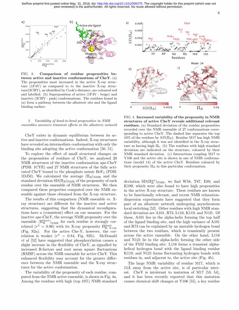
FIG. 3. Comparison of residue propensities be-
tween active and inactive conformations of CheY. (a)The propensities most increased in the active X-ray struc-ture (1F4V) as compared to to the inactive X-ray struc-ture(3CHY), as identified by Cook’s distance, are coloured redand labelled. (b) Superposition of active (1F4V - beige) andinactive (3CHY - pink) conformations. The residues found in(a) form a pathway between the allosteric site and the ligandbinding surface.
3. Variability of bond-to-bond propensities in NMRensembles uncovers transient effects in the allosteric network
CheY exists in dynamic equilibrium between its ac-tive and inactive conformations. Indeed, X-ray structureshave revealed an intermediate conformation with only thebinding site adopting the active conformation [50, 51].
To explore the effect of small structural changes onthe propensities of residues of CheY, we analysed 20NMR structures of the inactive conformation apo-CheY(PDB: 1CYE) and 27 NMR structures of the fully acti-vated CheY bound to the phosphate mimic BeF3 (PDB:1DJM). We calculated the average 〈ΠR〉NMR and thestandard deviation SD(ΠR)NMR of the propensity of eachresidue over the ensemble of NMR structures. We thencompared these properties computed over the NMR en-semble against those obtained from the X-ray structure.
The results of this comparison (NMR ensemble vs. X-ray structure) are different for the inactive and activestructures, suggesting that the dynamical reconfigura-tions have a (consistent) effect on our measure. For theinactive apo-CheY, the average NMR propensity over theensemble 〈Πinact
R 〉NMR for each residue is strongly cor-
related (r2 = 0.96) with its X-ray propensity ΠinactR, X-ray
(Fig. S2a). For the active Che-Y, however, the cor-relation is weaker (r2 = 0.84, Fig. S2b). McDonaldet al [52] have suggested that phosphorylation causes aslight increase in the flexibility of CheY, as signalled byincreased B-factors and root mean square fluctuations(RMSF) across the NMR ensemble for active CheY. Thisenhanced flexibility may account for the greater differ-ence between the NMR ensemble and the X-ray struc-tures for the active conformation.
The variability of the propensity of each residue, com-puted from the NMR active ensemble, is shown in Fig. 4a.Among the residues with high (top 10%) NMR standard
FIG. 4. Increased variability of the propensity in NMR
structures of active CheY reveals additional relevant
residues. (a) Standard deviation of the residue propensitiesrecorded over the NMR ensemble of 27 conformations corre-sponding to active CheY. The dashed line separates the top10% of the residues by SD(ΠR). Residue M17 has high NMRvariability, although it was not identified in the X-ray struc-ture as having high Πb. (b) The residues with high standarddeviation are indicated on the structure, coloured by theirNMR standard deviation. (c) Interactions coupling M17 toY106 and the active site is shown in one of NMR conforma-tions (model 14) of the active CheY. Residues coloured bytheir propensity ΠR in this particular conformation.
deviation SD(ΠactR )NMR, we find W58, T87, E89, and
K109, which were also found to have high propensitiesin the active X-ray structure. These residues are knownto be functionally relevant, and recent NMR relaxation-dispersion experiments have suggested that they formpart of an allosteric network undergoing asynchronouslocal switching [52]. Other residues with high NMR stan-dard deviation are A101, R73, L116, K119, and N121. Ofthese, A101 lies in the alpha-helix forming the top halfof the ligand binding site, and the high variance of A101and R73 can be explained by an unstable hydrogen bondbetween the two residues, which is transiently presentacross the active ensemble. On the other hand, L116and N121 lie in the alpha-helix forming the other sideof the FliM binding site: L116 forms a transient alpha-helical hydrogen bond with the ligand binding residueK119, and N121 forms fluctuating hydrogen bonds withresidues in, and adjacent to, the active site (Fig. 4b).
The large NMR variability of residue M17, which is15A away from the active site, is of particular inter-est. CheY is intolerant to mutation of M17 [53, 54],and it has been recently reported that this mutationcauses chemical shift changes at Y106 [55], a key residue
peer-reviewed) is the author/funder. All rights reserved. No reuse allowed without permission. The copyright holder for this preprint (which was not. http://dx.doi.org/10.1101/056275doi: bioRxiv preprint first posted online May. 31, 2016;

7
in the distant FliM binding site. Our analysis showsthat the propensity of M17 is higher in the active struc-ture (both NMR and X-ray) than in the inactive struc-ture: 〈Πact
M17〉NMR = 0.0173 > ΠactM17, X-ray = 0.0113 >
〈ΠinactM17 〉NMR = 0.0094 > Πinact
M17, X-ray = 0.0081. Fur-thermore, the NMR standard deviation of the propen-sity is higher in the active than in the inactive ensemble:SD(Πact
M17)NMR = 0.0032 > SD(ΠinactM17 )NMR = 0.0016.
All these results indicate that phosphorylation (i.e., acti-vation) causes transient pathways to form between M17and the active site which are not observed in the X-raystructure. By examining bonds with high propensity be-tween M17 and Y106, we visually uncover a communica-tion pathway involving residue K109 and residues in theflexible α4− β4 loop: T87, A88, and E89. Indeed, whenwe examine the individual NMR conformation in whichM17 has the highest propensity, M17 bonds directly withA88 and is indirectly connected to T87 through a hy-drogen bond with K109 (Fig. 4c). This suggests thatM17 is transiently coupled to Y106 through a network ofhydrogen bonds and hydrophobic contacts not capturedin the active X-ray structure. In general, the transientmaking-and-breaking of particular bonds in the NMR en-semble translates into highly variable propensities asso-ciated with functionally important allosteric residues.
C. Structural water molecules are crucial to the
allosteric communication network in h-Ras
The enzyme h-Ras is a GTPase involved in sig-nal transduction pertaining to cell-cycle regulation [56].Crystallographic evidence shows that calcium acetateacts as an allosteric activator in this process [57]. Bycomparing the calcium acetate-bound structure to the in-active structure, Buhrman et al have proposed a networkof hydrogen bonds, involving structural water molecules,linking the allosteric site to the catalytic residue Q61 [57].
TABLE III. Top bonds ranked by propensity quantile scorefor h-Ras (pb ≥ 0.99)
Bond Πb Distance (A) pb
Q99:HOH727 0.0051 14.8 0.9991
K117:G13 0.026 2.76 0.9983
HOH727:S65 0.0067 12.2 0.9974
R164:E49 0.0013 25.0 0.9974
I21:S17 0.019 4.83 0.9965
D47:R161 0.0015 21.6 0.9948
H27:Q25 0.0075 10.8 0.9940
V8:L56 0.0010 9.05 0.9940
R161:D47 0.0013 21.6 0.9931
I24:K42 0.0035 14.8 0.9922
Q22:A146 0.017 5.09 0.9905
We have calculated the propensities and quantile scores
of hRas (bound to substrate and allosteric activator,PDB code: 3K8Y) for two scenarios: with and withoutinclusion of structural water molecules in the graph. Inthe absence of water (Fig. 5a left), we find no bonds orresidues with high quantile scores near the allosteric bind-ing pocket. When we include the 8 molecules of struc-tural water present in the PDB file, we identify a highquantile bond between the allosteric site residue Y137and H94, and a pathway involving a structural watermolecule that connects the allosteric region to a catalyticresidue (Fig. 5b). In Table III, we show that the Q99-water and S65-water bonds involved in this pathway have1st and 3rd highest quantile scores out of the 1159 weakinteractions in the protein.
This water-mediated link between Q99 and S65 con-nects the allosteric binding pocket on helix 3 with thehelical structure known as the switch 2 region, at thebottom of which lies Q61, which has been identified as akey catalytic residue [57]. Our results thus suggest thatstructural water plays a crucial role in coupling the al-losteric effector to the catalytic residue Q61.
D. Absolute bond propensities against a reference
set from the SCOP protein database
The quantile regression scores pb in the previous sec-tions identify bonds with high propensities as comparedto other bonds which are at a similar distance from theactive site within the same protein. To assess the ab-
solute significance of bond propensities, we have assem-bled a reference set of 100 protein structures from theSCOP database [58] (see SI, Section 4), and calculatedthe propensities with respect to the active site of all465,409 weak bonds in this reference set (Fig. 6a). Be-cause the propensities are dependent on both the distancefrom the active site, d, and the total number of weak in-teractions in the protein, E, we apply quantile regressionagainst both d and E (as given by Eq. (15) in Materialsand Methods) to obtain fitted quantiles for the referenceset. The quantiles computed from this reference set canthen be used to obtain absolute bond propensity scores,denoted pref
b , for any given protein without recomputingthe regression.
We have obtained the absolute quantiles pref
b for thepropensities of the three proteins (caspase-1, CheY, andh-Ras) studied above (Fig. 6b). Reassuringly, the sig-nificant bonds are also found to be important accordingto the absolute measure, with a strong correlation be-tween quantile scores and absolute bond quantile scores(Fig. S3). Visualising the bonds with pref
b ≥ 0.99 showsthey form pathways between the active and allostericsites (Fig. 6c). These results confirm that these bonds areimportant not only relative to other bonds and residueswithin each of the respective proteins, but also in abso-lute terms when compared to the protein reference set.
peer-reviewed) is the author/funder. All rights reserved. No reuse allowed without permission. The copyright holder for this preprint (which was not. http://dx.doi.org/10.1101/056275doi: bioRxiv preprint first posted online May. 31, 2016;

8
FIG. 5. Structural water molecules are essential for the allosteric pathway in hRas. (a) Top percentile bonds bypropensity quantile score (pb ≥ 0.99) are shown on the structure: the left panel shows pathways identified without the inclusionof water molecules, and the right panel when structural water molecules are included in the graph. The structural water allowsthe formation of a pathway between the bottom of the switch 2 region and the top of helix 3, where the allosteric binding siteis situated. The crucial water molecule which connects Q99 and S65 is indicated. (b) Blow-up indicating details of the pathwayformed by Q99, a water molecule and S65, linking the allosteric pocket to the switch 2 region. The catalytic residue Q61 isshown at the bottom of switch 2.
E. Validating the propensity measure: predicting
allosteric sites in an extended set of proteins
To test the validity of our methodology, we have com-puted the bond propensities for an additional 17 proteinsknown to exhibit allostery. Ten of these proteins weretaken from a benchmark set collected by Daily et al [59]and a further 7 were obtained through an extensive liter-ature search. (Five proteins in Ref. [59] could not be usedeither due to the presence of non-standard amino-acids,to the absence of an allosteric ligand, or to a mismatchbetween the oligomeric state of the active and inactivestructures.) The details and structures of all 20 proteinsanalysed in the paper are given in the SI (Table S2 andFigure S4).
For each protein, we calculated the propensity quan-tile scores of all its bonds and residues, both intrinsic(pb, pR) and absolute (prefb ), with respect to their activesite. Again, no a priori knowledge about the allostericsite was used. Figure 7 shows the structures of the 20 pro-teins coloured according to the residue quantile score pR,with the allosteric sites marked with spheres. To validateour findings on this test set, we used the location of theallosteric site a posteriori and evaluated the significanceof the computed allosteric quantile scores according tofour statistical measures (Fig. 7a–d). See Section IVDfor a full description and definitions.
All combined, the allosteric site is detected signifi-cantly by at least one of the four measures in 19 outof 20 proteins in the test set, and is detected by three ormore of our measures in 15 out of 20 proteins in the testset. The full numerical values are given in the SI (TableS3). In practice, all statistical measures provide impor-tant and complementary information about the distribu-tion of bond propensities, and can be used in conjunctionfor the detection of allosteric sites.
III. DISCUSSION
Using a description of protein structural data in termsof an atomistic energy-weighted network with both co-valent and non-covalent bonds, we have defined a graph-theoretic measure of bond propensity and used it to iden-tify allosteric sites in proteins without prior informationas to their location. Our propensity measure identifiesbonds that are strongly coupled to the active site viacommunication pathways on the protein graph, even ifthey might be separated by large geometric distances.Allosteric sites correspond to hot spots, i.e., sites withhigh propensity to perturbations generated at the activesite, as measured by their quantile score relative to othersites in the protein that are at a similar distance fromthe active site. This finding suggests that the structuralfeatures embedded in the architecture of the protein areexploited so as to enhance the propagation of perturba-tions over long distances.
By using a representative reference set of 100 proteinsrandomly assembled from the SCOP database, we alsocomputed absolute quantile scores to further confirm thesignificance of bond propensities. One advantage of thisabsolute measure is that the quantile regression over thereference SCOP set does not need to be re-calculated,and the absolute bond quantile scores in any protein ofinterest can be obtained directly against them, thus re-duces the analysis time even further.
We have validated our method against a test set of 20allosteric proteins without using any a priori informationof their allosteric sites. We used our propensity quantilescores and a structural bootstrap to define four statisti-cal measures of significance based on the average and tailof the distribution of bond propensities in the allostericsite. The allosteric site is detected for 19/20 proteins,according to at least one statistical measure, and for
peer-reviewed) is the author/funder. All rights reserved. No reuse allowed without permission. The copyright holder for this preprint (which was not. http://dx.doi.org/10.1101/056275doi: bioRxiv preprint first posted online May. 31, 2016;

9
FIG. 6. Absolute propensities: calibration against the SCOP reference set. (a) The logarithm of the bond propensitylog(Πb) of all 465,409 weak bonds in the reference set (100 proteins from the SCOP database) plotted against d, the distancefrom their corresponding active site, and E, where E is the number of weak bonds in the corresponding protein. (b) The logpropensities log(Πb) for caspase-1 (blue), CheY (orange), and h-Ras (yellow) are plotted together with the plane defining the99th quantile fit obtained by solving the optimisation Eq. (15) against the SCOP set of bonds shown in (a). For each of thethree proteins, there are bonds lying above the 99th quantile plane. (c) The bonds above the plane in (b) have pref
b > 0.99 andare marked in red on the corresponding protein structures (active site ligand in green, allosteric ligand as yellow spheres). Thebonds thus identified play key allosteric roles, in agreement with the intrinsic results in previous sections.
15/20, according to at least three of our four statisticalmeasures. These findings indicate the robustness of thebond-to-bond propensity as a predictor of allosteric sites,which could be used to guide structure-based drug discov-ery efforts, e.g., by ranking potential binding sites basedon their allosteric potential. Our method also uncovershot spots not previously identified as allosteric sites (seeour results for CheY in Fig. 2). Hardy and Wells havediscussed the existence of ‘orphan’ or ‘serendipitous’ al-losteric sites, i.e., sites targeted by as-yet undiscoverednatural effectors or open for exploitation by novel smallmolecules [8]. The identified sites could thus provide tar-gets for mutational analysis or allosteric small-moleculeinhibition.
We have exemplified the use of atomistic propensi-ties with the detailed analysis of three proteins (caspase-
1, CheY, and h-Ras), focussing on the contribu-tion of high propensity bonds to pathways (or net-works) of weak bonds linking the active and allostericsites. The weak bond network we found in caspase-1 (E390/R286/S332/S339/N337) has been previouslytested experimentally and shown to be functionally im-portant [48]. In CheY, we found that bonds betweenT87:E89 and E89:Y106, with very high quantile scores,are key to an important pathway for transmission ofthe signal induced by phosphorylation, also consistentwith experimental evidence [50, 52, 60]. We also founda second pathway in CheY involving the bond K109:D57(3rd highest quantile score). Interestingly, mutation ofK109 abolishes chemotactic activity [53] and has beenproposed to form part of the post-phosphorylation acti-vation mechanism [61]. Our analysis of bond propensi-
peer-reviewed) is the author/funder. All rights reserved. No reuse allowed without permission. The copyright holder for this preprint (which was not. http://dx.doi.org/10.1101/056275doi: bioRxiv preprint first posted online May. 31, 2016;

10
FIG. 7. Prediction of allosteric sites based on bond-to-bond propensity for a test set of 20 allosteric proteins.
The structures of the 20 proteins in the test set (labelled by PDB code) have their residues coloured by their quantile scorepR, and the allosteric site is shown as spheres. For full details of these proteins, see Table S2 in the SI. The four statisticscomputed from our propensity are showed in the centre: (a) average residue quantile scores in the allosteric site pR,allo (red)compared to the average score of 1000 surrogate sites 〈pR,site〉surr (grey), with a 95% confidence interval for the average from abootstrap with 10000 resamples (see Section IVD1); (b) average bond quantile scores in the allosteric site against the equivalentbootstrap of 1000 surrogate sites; (c) tail of the distribution of bond propensities, i.e., proportion of allosteric site bonds withquantile scores pb,allo > 0.95. Proteins above the expected proportion of 0.05 (red line) have a larger than expected number
of bonds with high quantile scores; (d) average reference bond quantile score in the allosteric site prefb,allo. The red dotted lineindicates the expected value of 0.5, and proteins above this line have a higher than expected reference quantile score. For thenumerical values of all measures see Table S3 in the SI. The four circle code by each protein indicates whether the allostericsite is identified (filled circle) or not identified (open circle) according to each of the four measures (a)–(d). 19/20 allostericsites are identified by at least one measure, and 15/20 sites are identified by at least three of four measures.
peer-reviewed) is the author/funder. All rights reserved. No reuse allowed without permission. The copyright holder for this preprint (which was not. http://dx.doi.org/10.1101/056275doi: bioRxiv preprint first posted online May. 31, 2016;

11
ties across active/inactive conformations and NMR datafurther confirmed that K109 forms a central link in thecommunication between the phosphorylation and bind-ing sites in CheY.
Determination of protein structures from NMR solu-tion experiments results in multiple models, each con-sistent with experimentally-derived distance restraints.The resulting ‘ensemble’ of structures should be inter-preted with caution, since variation could be due to ac-tual flexibility and thermal motion during the exper-iment, or to inadequate (or under-constrained) inter-atomic distance restraints. Hence the set of NMR struc-tures is not a true thermodynamic ensemble. However,our analysis suggests that the variation within the NMRstructures can reveal functionally relevant information.For CheY, residues with highly variable propensitiesacross the NMR ensemble (E89/W58/T87/E89/K109)coincide with those forming an asynchronously switch-ing allosteric circuit after phosphorylation, as revealedby NMR relaxation-dispersion experiments [52]. We alsoidentify residue M17 as having high propensity in theNMR ensemble due to the presence of a transient networkof interactions. This may explain experiments showingthat mutation of M17 has a functional effect and causeschemical shift changes at Y106 [55].
Comparing the results across conformations indicatesthat propensities are fairly robust to local dynamic fluc-tuations, as seen by the strong correlation between activeand inactive conformations and across NMR structures(Fig. 3 and Figs. S1 and S2). As an additional confir-mation of its robustness, we show in SI (Section 6, Ta-bles S4 and S5) that the propensities, and the ensuingidentification of significant residues and bonds, are gen-erally robust to both randomness in the bond energiesand to the breakage of a large proportion of weak interac-tions. On the other hand, as discussed above, our graph-theoretic analysis shows that further information aboutresidues and bonds can be obtained by evaluating thehighest variations induced by dynamical and structuralvariations. A fuller investigation of the effect of dynam-ics on the calculated propensities using experimental data(NMR, conformational studies) and complemented withthe analysis of molecular dynamics simulations wouldthus be an interesting area for future research.
The role of structural water molecules in mediatingallosteric communication has so far received limited at-tention. In a recent study of a PDZ domain, Buchli etal. suggest that changes in water structure could beresponsible for mediating communication with remoteparts of the protein [62]. Our analysis of h-Ras foundthat structural water molecules in the protein graph arenecessary to reveal a pathway linking the allosteric andactive sites. These results and the findings of Buchli etal. suggest that novel methods to study interaction net-works between proteins and water are worth investigat-ing. However, beyond including structural water whenpresent in experimental structures (as in h-Ras here),the addition of bulk water would require the simulation
of hydration, including energy minimisation and equili-bration steps. This could constitute another directionof future research, since the computational efficiency ofour method would make it possible to analyse all-atomrepresentations of such hydrated structures.To what extent does the identification of the allosteric
site require an atomistic, chemically detailed construc-tion of the graph? To answer this question, we ap-plied our propensity measure to residue-residue interac-tion networks (RRINs), the coarse-grained residue-levelmodels used in almost all previous network analyses ofproteins. For caspase-1, we found that allosteric residuesare not found significant in RRINs (across several dif-ferent cut-off radii), whereas, on the other hand, the al-losteric site of CheY was consistently detected in both theatomistic and residue-level descriptions. This indicatesthat both coarser topological features, as well as more de-tailed chemical communication pathways can be relevantdepending on the protein; e.g., in caspase-1, the bindingof the allosteric ligand perturbs a network of strong hy-drogen bonds and salt-bridges as identified in our analy-sis. Therefore, the atomistic graph with detailed physico-chemical information can in some cases provide impor-tant features underpinning the communication featuresof the protein. The analysis of coarse-grained modelswith a variety of cut-off radii for all 20 proteins in ourallosteric test set in SI Section 7 confirm that the out-come for RRINs varies for each protein and can also bedependent on the choice of cut-off radii [38]. We wouldlike to emphasise, however, that our propensity measureis principally agnostic to the protein network model un-der analysis, thus allowing for the evaluation of distinctgraph-construction techniques (e.g., atomistic vs coarse-grained) or the use of different force-fields. Again, thiswould open another interesting avenue for future work.Finally, it is important to remark that our method is
computationally efficient. To obtain the bond-to-bondpropensities, we only need to solve a sparse linear sys-tem (Eq. 6) involving the (weighted) Laplacian of theprotein graph. As discussed in Section IVA3, recent al-gorithmic advances allow us to solve such linear systemsin almost linear time [42, 43]. Hence protein complexesof ∼ 100, 000 atoms can be run in minutes on a standarddesktop computer. We can thus maintain atomistic de-tail, yet analyse large biomolecular complexes that areintractable for traditional computational methods.
IV. MATERIALS AND METHODS
A. Mathematical derivation of the bond-to-bond
propensity
1. Fluctuations and the edge-to-edge transfer matrix of agraph
The edge-to-edge transfer matrix M was introduced inRef. [41] as a non-local edge-coupling matrix for the anal-
peer-reviewed) is the author/funder. All rights reserved. No reuse allowed without permission. The copyright holder for this preprint (which was not. http://dx.doi.org/10.1101/056275doi: bioRxiv preprint first posted online May. 31, 2016;

12
ysis of weighted undirected graphs, based on the conceptof flow redistribution. In that work, it was shown thatthe element Mji reflects the effect that an injected fluxon edge i has on the flux along edge j after the fluxesare redistributed over the whole graph when at equilib-rium. Alternatively, M can be understood as a discreteGreen’s function in the edge space of the graph. See [41]for detailed derivations and applications.In this paper, we derive a complementary interpreta-
tion of the matrix M . As shown below, the edge-to-edgetransfer matrix can be understood as describing how thefluctuations of the edge weights propagate through thegraph. This new re-interpretation underpins the work inthis paper, as it highlights the importance of M for theanalysis of bond fluctuations in biomolecules.As our starting point, consider the well-known
Langevin equation, sometimes denoted the heat kernel
equation [63, 64]:
x = −Lx+ ǫ. (1)
Formally, Eq. (1) has the same structure as the canon-ical model for scalar vibrations with nearest neighbourinteractions encoded by the matrix L [29, 30]. Alterna-tively, Eq. (1) may be considered as a model of a diffusingparticle transitioning like a random walker on the under-lying graph structure represented by L. In contrast toresidue level methods [33], the variable x is associatedwith atomic fluctuations, i.e., our graph model reflectsan atomic description that incorporates physico-chemicalinteractions derived from the three dimensional struc-ture of the protein recorded in a PDB file. The resultinggraph contains energy-weighted interactions representingbonds in the protein, including both covalent bonds andweak interactions such as hydrogen bonds, salt bridges,hydrophobic tethers, and electrostatic interactions. Fordetails of the graph construction see Section IVE and SI.The matrix L is the graph Laplacian [39, 65]:
Lij =
{
−wij , i 6= j∑
j wij , i = j,(2)
where wij is the weight of the edge between nodes(atoms) i, j. In this case, wij is the energy of the bond be-tween both atoms. Thermal background fluctuations aremodelled by ǫ, a zero mean white Gaussian noise inputvector, i.e., a (simple) heat bath acting independently onall atomic sites with covariance matrix
〈ǫi(t)ǫj(s)〉 = δ(t− s)δij , (3)
where δ stands for the Dirac delta function.Instead of focusing on the atomic (node) variables x,
we wish to study the coupling between bonds, and thusconcentrate on the bond (edge) variables of the graph:
yb = xhead(b) − xtail(b). (4)
Clearly, yb describes the difference of the node variablesat the endpoints of the associated bond b, i.e., a fluctua-
tion associated with the bond between two atoms. The
vector of bond fluctuations can be compactly representedin vector notation as:
y = BTx,
where B is the incidence matrix of the graph relatingeach edge variable to its corresponding node variables,i.e., Bbi = 1 if node i is the head of bond b; Bbi = −1 ifnode i is the tail of bond b; and Bbi = 0 otherwise.We can now calculate the cross-correlations between
edge fluctuations as:
R(τ) := E[y(t)yT (t+ τ)] =1
2BT exp(−τL)L†B, (5)
where L† is the (Moore-Penrose) pseudoinverse of theLaplacian matrix. Each entry [R(τ)]b1b2 describes how afluctuation at bond b2 is correlated with a fluctuation atbond b1 at time τ . See SI for a full derivation of Eq. (5).Biophysically, we are ultimately interested in the en-
ergy fluctuations induced by bonds on other bonds.Therefore, we multiply the correlation matrix R(τ) bythe diagonal matrix of bond energies, G = diag(wb):
M(τ) := GR(τ),
to obtain the matrix of bond-to-bond energy correlations
with delay τ . Our measure of bond-to-bond propensity isobtained from the instantaneous correlations (i.e., τ = 0)leading to the edge-to-edge transfer matrix:
M := M(0) =1
2GBTL†B. (6)
Note that the diagonal entries of M are indeed relatedto the average energy stored in the bond fluctuations:Mbb = 1
2 〈wbybyb〉 = 12 〈wb(xhead(b)−xtail(b))
2〉. Likewise,the off-diagonal entries Mb1b2 reflect how a perturba-tion at bond b2 affects another bond b1 weighted by thestrength of bond b1. Hence the influence on a strongerbond is considered to be more important. Although wehave not considered here time-delayed correlations (i.e.,as a function of τ), this is an interesting direction forfuture research.
2. Definition of the bond-to-bond propensity
To construct our measure of propensity, we only as-sume knowledge of the active site and proceed as follows.Let us consider all the ligand-protein interactions formedat the active site and compute their combined effect oneach bond b outside of the active site:
Πrawb =
∑
b′∈ ligand
|Mbb′ |. (7)
This raw propensity reflects how closely the active-site iscoupled to each individual bond. Note that the compu-tations include all the bonds in the protein (covalent and
peer-reviewed) is the author/funder. All rights reserved. No reuse allowed without permission. The copyright holder for this preprint (which was not. http://dx.doi.org/10.1101/056275doi: bioRxiv preprint first posted online May. 31, 2016;

13
non-covalent). However, in the paper we only report theeffect on weak bonds, as it is changes in weak-bondingpatterns that usually drive allosteric response in proteins.Since different proteins have different numbers of bonds,we make the measure consistent by normalising the score:
Πb =Πraw
b∑
b Πrawb
. (8)
Throughout the manuscript, the quantity Πb is referredto as the propensity of bond b; a measure of how muchedge b is affected by the interactions at the active site.The propensity of a residue is defined as the sum of the(normalised) propensities of its bonds:
ΠR =∑
b∈R
Πb. (9)
3. Computational cost of bond-to-bond propensity
The computation of the propensities is efficient. Notethat Eq. (8) requires the summation over columns ofthe M matrix corresponding to protein-ligand interac-tions. Crucially, we do not need to compute the fullpseudo-inverse L† in Eq. (6); we can instead solve asparse linear system involving the graph Laplacian. Re-cent algorithmic developments [42, 43] have made thispossible in almost linear time, O(E log2(Na)), where Eis the number of bonds (edges) and Na is the num-ber of atoms (nodes). Our method therefore is scalableto extremely large systems. Using the CombinatorialMultigrid toolbox written by Y. Koutis [66] (available athttp://www.cs.cmu.edu/~jkoutis/cmg.html) propen-sities for all the bonds in proteins with ∼ 100, 000 atomscan be run in minutes on a standard desktop computer.
B. Significance of propensities through quantile
scores
To identify bonds (and residues) with high propensi-ties relative to others at a similar distance from the activesite, we use quantile regression [44], a technique of wideuse in econometrics, ecology, and medical statistics. Incontrast to standard least squares regression, which fo-cusses on estimating a model for the conditional mean ofthe samples, quantile regression (QR) provides a methodto estimate models for conditional quantile functions.This is important for two reasons: (i) the conditionaldistributions of propensities are highly non-normal; and(ii) we are interested not in the ‘average’ bond, but inthose bonds with particularly high propensities lying inthe tails of the distribution. Once the fitted models areobtained, the quantile score of a bond pb is a measureof how high the propensity Πb is relative to other bondsin the sample which are at a similar distance from theactive site.
Although QR goes back more than 200 years, it hasonly become widely used recently, due to the availabilityof computational resources. The mathematical basis ofthe method stems from the fact the pth quantile, Qp, of adistribution is given by the solution of the following opti-misation problem: given a sample {yi}
ni=1 parametrically
dependent on m variables xi ∈ Rm with parameters β,
the estimate of the conditional pth quantile of the sampledistribution is obtained by solving
minβ
n∑
i=1
ρp(yi −Q(xi,β)), p ∈ [0, 1], (10)
where ρp(·) is the tilted absolute value function
ρp(y) =∣
∣
∣y(
p− ■(y < 0))
∣
∣
∣, (11)
and ■(·) is the indicator function. If the dependence isassumed to be linear, Q(xi,β) = β0 + βT
xi, the op-timisation can be formulated as a linear program andsolved efficiently through the simplex method to ob-
tain β ∈ Rm+1, the estimated parameters defining the
model [44].In Sections IIA–IIC, we have applied QR to the
propensities Πb of bonds within each protein so as totake into account their dependence with respect to db,the minimum distance between bond b and any bond inthe active site:
db = minb′∈active
|vb − vb′ |, (12)
where the vector vb contains the coordinates of the mid-point of bond b. Based on the observed exponential decayof Π with d, we adopt a linear model for the logarithmof the propensities and estimate the conditional quantilefunctions by solving the minimisation problem
βprot(p) = argmin(β0,β1)
protein∑
b
ρp(log(Πb)− (β0 + β1d)), (13)
where the sum runs over the weak bonds of the corre-sponding protein. From the estimated model for the pro-tein, we then calculate the quantile score of bond b atdistance db from the active site and with propensity Πb,by finding the quantile pb such that
pb = argminp∈[0,1]
∣
∣βprot
0 (p) + βprot
1 (p)db − log(Πb)∣
∣ . (14)
Similarly, in Section IID, we use QR to obtain absolute
quantile scores of bonds and residues with respect to areference set of 100 proteins from the SCOP database.In this case, the propensities are regressed against boththe distance to the active site d, and the number of non-covalent bonds in the protein, E. Since the mean propen-sity scales as E−1, we also assume a power-law depen-dency of the quantiles. Hence, we solve
βref(p) = argmin(β0,β1,β2)
SCOP∑
b
ρp(log(Πb)−(β0+β1d+β2 log(E))),
(15)
peer-reviewed) is the author/funder. All rights reserved. No reuse allowed without permission. The copyright holder for this preprint (which was not. http://dx.doi.org/10.1101/056275doi: bioRxiv preprint first posted online May. 31, 2016;
14
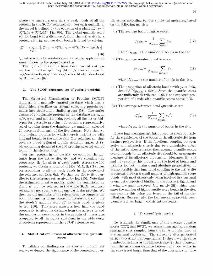
where the sum runs over all the weak bonds of all theproteins in the SCOP reference set. For each quantile p,the model is defined by the equation of a plane βref
0 (p) +βref
1 (p)d + βref
2 (p)E (Fig. 6b). The global quantile scorepref
b for bond b at a distance db from the active site in aprotein with Eb non-covalent bonds is found by solving
pref
b = argminp∈[0,1]
∣
∣βref
0 (p) + βref
1 (p)db + βref
2 (p)Eb − log(Πb)∣
∣ .
(16)Quantile scores for residues are obtained by applying thesame process to the propensities ΠR.The QR computations have been carried out us-
ing the R toolbox quantreg (http://cran.r-project.org/web/packages/quantreg/index.html) developedby R. Koenker [67].
C. The SCOP reference set of generic proteins
The Structural Classification of Proteins (SCOP)database is a manually curated database which uses ahierarchical classification scheme collecting protein do-mains into structurally similar groups [58]. The majorclasses of cytoplasmic proteins in the database are α, β,α/β, α+β, and multidomain, covering all the major fold-types for cytosolic proteins. To obtain a representativeset of proteins from the database, we randomly selected20 proteins from each of the five classes. Note that weonly include proteins for which there is a structure witha ligand bound to the active site. Our reference set thuscovers a broad region of protein structure space. A ta-ble containing details of the 100 proteins selected can befound in the electronic SI.For each protein in the dataset, we compute the dis-
tance from the active site, db, and we calculate thepropensity, Πb, for all its E weak bonds. Across the 100proteins, we obtain a total of 465409 (d,E,Πb) 3-tuplescorresponding to all the weak bonds in the proteins ofthe reference set (Fig. 6a). We then use QR to fit quan-tiles to this reference set, as given by Eq. (15). Note thatthe estimated quantile models, which are conditional ond and E, are now referred to the whole SCOP referenceset and are not specific to any one particular protein. Wethen use the quantiles of the reference set to compare thebond propensities of any protein of interest and computethe absolute quantile score pref
b for each bond, as givenby Eq. (16). This score measures how high the bondpropensity is, given its distance from the active site andthe number of weak bonds in the protein of interest, ascompared to all the bonds contained in the wide rangeof proteins represented in the SCOP reference set.
D. Statistical evaluation of allosteric site quantile
scores
To validate our findings on the allosteric protein testset, we evaluated the significance of the computed quan-
tile scores according to four statistical measures, basedon the following metrics:
(i) The average bond quantile score:
pb,site =1
Nb,site
∑
b∈site
pb, (17)
where Nb,site is the number of bonds in the site.
(ii) The average residue quantile score:
pR,site =1
NR,site
∑
R∈site
pR, (18)
where NR,site is the number of bonds in the site.
(iii) The proportion of allosteric bonds with pb > 0.95,denoted P(pb,allo > 0.95). Since the quantile scoresare uniformly distributed, 0.05 is the expected pro-portion of bonds with quantile scores above 0.95.
(iv) The average reference bond quantile score:
prefb,site =1
Nb,site
∑
b∈site
prefb , (19)
where Nb,site is the number of bonds in the site.
These four measures are introduced to check robustlyfor the significance of the bonds in the allosteric site fromdistinct perspectives. If the functional coupling betweenactive and allosteric sites is due to a cumulative effectof the entire allosteric site, then average quantile scoresover all bonds in the allosteric site should be an accuratemeasure of its allosteric propensity. Measures (i), (ii)and (iv) capture this property at the level of bonds andresidues for both intrinsic and absolute propensities. Itis also possible that functional coupling to the active siteis concentrated on a small number of high quantile scorebonds, with most others only being involved in structuralor energetic aspects of binding to the allosteric ligand andhaving low quantile scores. Our metric (iii), which mea-sures the number of high quantile score bonds in the site,can capture this behaviour based on the tail of the dis-tribution. Reassuringly, the four measures provide com-plementary, yet largely consistent outcomes.
1. Structural bootstrapping
To establish the significance of the average quantilescores pb,allo and pR,allo, we assess them against randomsurrogate sites sampled from the same protein, used asa structural bootstrap. The surrogate sites generatedsatisfy two structural constraints: 1) they have the samenumber of residues as the allosteric site; 2) their diameter(i.e., the maximum distance between any two atoms inthe site) is not larger than that of the allosteric site. The
peer-reviewed) is the author/funder. All rights reserved. No reuse allowed without permission. The copyright holder for this preprint (which was not. http://dx.doi.org/10.1101/056275doi: bioRxiv preprint first posted online May. 31, 2016;

15
algorithm for generating these sites is described in Sec-tion S5 of the SI. For each protein, we generate 1000 sur-rogate sites and calculate their quantile scores pb,site andpR,site. The average scores over the ensemble of 1000 sur-rogate sites 〈pb,site〉surr and 〈pR,site〉surr, where the anglebrackets denote the ensemble average, are then comparedagainst the average residue quantile score of the allostericsite (Figure 7a, b). A bootstrap with 10000 resampleswith replacement [68] was used to obtain 95% confidenceintervals providing statistical signficance.
2. Validation on the allosteric test set
Figure 7 (a)–(d) reports these four statistical measuresfor all 20 proteins analysed (see SI, Table S3 for the cor-responding numerical data). Our results indicate robustidentification of the allosteric sites in the test set. Thequantile score of the allosteric site is higher than that ofthe surrogate sites and above the 95% bootstrapped con-fidence interval in 14 out of 20 proteins for the residuescore, pR,allo, and for 16 out of 20 proteins for the bondscore, pb,allo (Figure 7a, b). The proteins identified byboth measures are almost coincident, with few differ-ences: Glutamate DH (1HWZ) is significant accordingto the bond score and marginally below significance ac-cording to the residue score, whereas the opposite appliesto Thrombin (1SFQ). The reason for these differenceslies with the distribution of bond scores: in some cases,allosteric sites have only a few bonds with high quan-tile scores and many other less important bonds. Whenconsidered at the level of residues, this can lead to highpR scores; yet when bonds are considered individuallythrough their pb scores, the high quantile scores are av-eraged out over the whole allosteric site.
To evaluate the presence of high scoring bonds, wecompute the proportion of bonds with high quantile scoreP(pb,allo > 0.95) in the allosteric site, as compared to theexpected proportion (0.05) above this quantile. The pro-portion of high quantile score bonds in the allosteric siteis greater than expected in 17 of the 20 proteins (Fig. 7c).Of these 17 proteins, 16 coincide with those identified us-ing the average scores reported above, and we addition-ally identify h-Ras (3K8Y). This finding confirms thatallosteric sites consistently exhibit a larger than expectednumber of bonds with a strong coupling to the active site.
Finally, we compute the average absolute quantile score
of the allosteric site prefb,allo against the SCOP reference
set (Figure 7d). The results are largely consistent withthe intrinsic measure pb,allo: in 14/20 proteins, the abso-lute quantile score is greater than the expected 0.5, i.e.
prefb,allo > 0.5. Yet some proteins (e.g., glutamate dehy-
rogenase (1HWZ), fructose 1,6-bisphosphatase (1EYI),and glycogen phosphorylase (7GPB)) have high intrin-sic quantile scores, as compared to other bonds in thesame protein, but do not score highly in absolute value,as compared to the reference SCOP ensemble. This re-
sult highlights the fact that a site need not have a highabsolute propensity, as long as its propensity is high incomparison with the rest of the protein it belongs to, sothat the ‘signal’ from the site outweighs the ‘noise’ fromthe rest of the protein. Interestingly, the lac repressor(1EFA) has an allosteric site with large absolute propen-
sity (prefb,allo = 0.60 > 0.5) but non-significant intrinsicpropensity.
E. Construction of the atomistic graph
An in-depth discussion of the construction of the graphcan be found in Refs. [39, 40], and further details aregiven in the SI, Section 2. Briefly, we use an atomisticgraph representation of a protein, where each node cor-responds to an atom and the edges represent both co-valent and non-covalent interactions, weighted by bondenergies derived from detailed atomic potentials. The co-valent bond energies are taken from standard bond disso-ciation energy tables. Non-covalent interactions includehydrogen bonds, salt bridges, hydrophobic tethers andelectrostatic interactions. Hydrogen bond energies areobtained from the DREIDING force-field [69]. Attrac-tive hydrophobic interaction energies are defined betweencarbon and sulfur atoms, according to a hydrophobic po-tential of mean force introduced by Lin et al [70]. Elec-trostatic interactions with coordination ions and ligandsare identified from the LINK entries in the PDB file, withbond energies assigned using a Coulomb potential.To compare the results between our atomistic model
and residue-level RRINs [33], we use coarse-grained net-work models obtained from the oGNM server [71]. Adetailed comparison of results obtained with atomisticnetworks and RRINs is given in the SI Section 7.We note that the main methodology (i.e., the propen-
sity measure and methods developed in Sections IVA–IVB) is independent of the construction of the graph.Users are free to construct the network using alternativepotentials (e.g., AMBER [72] or CHARMM [73]) or usingcoarse-grained networks.
V. ACKNOWLEDGMENTS
BRCA was supported by a studentship of the EP-SRC Centre for Doctoral Training under the Instituteof Chemical Biology, Imperial College London. SNYand MB acknowledge support through EPSRC grantEP/I017267/1. We thank Keith Willison for suggestingh-Ras as an example and for helpful discussions.
VI. AUTHOR CONTRIBUTIONS
BRCA, SNY and MB conceived the study. BRCA per-formed the numerical analysis and created the Figures.
peer-reviewed) is the author/funder. All rights reserved. No reuse allowed without permission. The copyright holder for this preprint (which was not. http://dx.doi.org/10.1101/056275doi: bioRxiv preprint first posted online May. 31, 2016;

16
SNY and MB supervised the study. All authors con- tributed to developing the theoretical tools. All authorswrote and reviewed the manuscript.
[1] J. Monod, J.-P. Changeux, and F. Jacob, Journal ofmolecular biology 6, 306 (1963).
[2] M. F. Perutz, Quarterly reviews of biophysics 22, 139(1989).
[3] R. Nussinov and C.-J. Tsai, Cell 153, 293 (2013).[4] H. Frauenfelder, S. G. Sligar, and P. G. Wolynes, Science
254, 1598 (1991).[5] K. Henzler-Wildman and D. Kern, Nature 450, 964
(2007).[6] B. F. Volkman, D. Lipson, D. E. Wemmer, and D. Kern,
Science Signaling 291, 2429 (2001).[7] K. Gunasekaran, B. Ma, and R. Nussinov, PROTEINS:
Structure, Function and Bioinformatics 57, 433 (2004).[8] J. A. Hardy and J. A. Wells, Current opinion in structural
biology 14, 706 (2004).[9] S. Lockless and R. Ranganathan, Science 286, 295
(1999).[10] B. J. Grant, S. Lukman, H. J. Hocker, J. Sayyah, J. H.
Brown, J. A. McCammon, and A. A. Gorfe, PLoS One6, e25711 (2011).
[11] P. Weinkam, J. Pons, and A. Sali, Proceedings of theNational Academy of Sciences 109, 4875 (2012).
[12] N. Ota and D. Agard, Journal of molecular biology 351,345 (2005).
[13] O. N. Demerdash, M. D. Daily, and J. C. Mitchell, PLoScomputational biology 5, e1000531 (2009).
[14] A. Panjkovich and X. Daura, BMC bioinformatics 13,273 (2012).
[15] G. Collier and V. Ortiz, Archives of biochemistry andbiophysics 538, 6 (2013).
[16] J. Monod, J. Wyman, and J. Changeux, Journal ofmolecular biology 12, 88 (1965).
[17] D. Koshland Jr, G. Nemethy, and D. Filmer, Biochem-istry 5, 365 (1966).
[18] V. J. Hilser, J. O. Wrabl, and H. N. Motlagh, Annualreview of biophysics 41, 585 (2012).
[19] A. del Sol, C. Tsai, B. Ma, and R. Nussinov, Structure17, 1042 (2009).
[20] P. Zhuravlev, G. Papoian, et al., Quarterly reviews ofbiophysics 43, 295 (2010).
[21] H. M. Muller-Werkmeister and J. Bredenbeck, PhysicalChemistry Chemical Physics 16, 3261 (2014).
[22] G. Li, D. Magana, and R. B. Dyer, Nature communica-tions 5 (2014).
[23] L. Martınez, A. C. Figueira, P. Webb, I. Polikarpov, andM. S. Skaf, The Journal of Physical Chemistry Letters 2,2073 (2011).
[24] N. Fujii, M. Mizuno, H. Ishikawa, and Y. Mizutani, TheJournal of Physical Chemistry Letters 5, 3269 (2014).
[25] P. H. Nguyen, P. Derreumaux, and G. Stock, The Jour-nal of Physical Chemistry B 113, 9340 (2009).
[26] R. Gnanasekaran, J. K. Agbo, and D. M. Leitner, TheJournal of chemical physics 135, 065103 (2011).
[27] Z. N. Gerek and S. B. Ozkan, PLoS computational biol-ogy 7, e1002154 (2011).
[28] C. Kaya, A. Armutlulu, S. Ekesan, and T. Haliloglu,Nucleic acids research 41, W249 (2013).
[29] T. Nakayama, K. Yakubo, and R. L. Orbach, Reviewsof modern physics 66, 381 (1994).
[30] D. M. Leitner, Annu. Rev. Phys. Chem. 59, 233 (2008).[31] A. Del Sol, H. Fujihashi, D. Amoros, and R. Nussinov,
Molecular systems biology 2 (2006).[32] A. Del Sol, M. Arauzo-Bravo, D. Amoros, R. Nussinov,
et al., Genome Biol 8, R92 (2007).[33] C. Chennubhotla and I. Bahar, PLoS computational bi-
ology 3, e172 (2007).[34] G. Amitai, A. Shemesh, E. Sitbon, M. Shklar, D. Ne-
tanely, I. Venger, and S. Pietrokovski, Journal of molec-ular biology 344, 1135 (2004).
[35] A. Ghosh and S. Vishveshwara, Proceedings of the Na-tional Academy of Sciences 104, 15711 (2007).
[36] A. Sethi, J. Eargle, A. Black, and Z. Luthey-Schulten,Proceedings of the National Academy of Sciences 106,6620 (2009).
[37] A. A. Ribeiro and V. Ortiz, Journal of Chemical Theoryand Computation 10, 1762 (2014).
[38] A. A. Ribeiro and V. Ortiz, The Journal of PhysicalChemistry B 119, 1835 (2015).
[39] A. Delmotte, E. Tate, S. Yaliraki, and M. Barahona,Physical Biology 8, 055010 (2011).
[40] B. Amor, S. Yaliraki, R. Woscholski, and M. Barahona,Molecular BioSystems 10, 2247 (2014).
[41] M. T. Schaub, J. Lehmann, S. N. Yaliraki, and M. Bara-hona, Network Science 2, 66 (2014).
[42] D. A. Spielman and S.-H. Teng, in Proceedings of thethirty-sixth annual ACM symposium on Theory of com-puting (ACM, 2004) pp. 81–90.
[43] J. A. Kelner, L. Orecchia, A. Sidford, and Z. A. Zhu, inProceedings of the forty-fifth annual ACM symposium onTheory of computing (ACM, 2013) pp. 911–920.
[44] R. Koenker, Quantile regression, 38 (Cambridge univer-sity press, 2005).
[45] Y. Wei, A. Pere, R. Koenker, and X. He, Statistics inmedicine 25, 1369 (2006).
[46] C. A. Knight and D. D. Ackerly, Ecology Letters 5, 66(2002).
[47] M. Buchinsky, Econometrica: Journal of the EconometricSociety , 405 (1994).
[48] D. Datta, J. Scheer, M. Romanowski, and J. Wells, Jour-nal of molecular biology 381, 1157 (2008).
[49] R. D. Cook, Journal of the American Statistical Associ-ation 74, 169 (1979).
[50] C. M. Dyer and F. W. Dahlquist, Journal of bacteriology188, 7354 (2006).
[51] S.-Y. Lee, H. S. Cho, J. G. Pelton, D. Yan, E. A. Berry,and D. E. Wemmer, Journal of Biological Chemistry 276,16425 (2001).
[52] L. R. McDonald, J. A. Boyer, and A. L. Lee, Structure20, 1363 (2012).
[53] R. B. Bourret, S. K. Drake, S. A. Chervitz, M. I. Simon,and J. J. Falke, Journal of Biological Chemistry 268,13089 (1993).
[54] J. G. Smith, J. A. Latiolais, G. P. Guanga, S. Citineni,R. E. Silversmith, and R. B. Bourret, Journal of bacte-
peer-reviewed) is the author/funder. All rights reserved. No reuse allowed without permission. The copyright holder for this preprint (which was not. http://dx.doi.org/10.1101/056275doi: bioRxiv preprint first posted online May. 31, 2016;

17
riology 185, 6385 (2003).[55] L. R. McDonald, M. J. Whitley, J. A. Boyer, and A. L.
Lee, Journal of molecular biology (2013).[56] F. McCormick, Molecular reproduction and development
42, 500 (1995).[57] G. Buhrman, G. Holzapfel, S. Fetics, and C. Mattos,
Proceedings of the National Academy of Sciences 107,4931 (2010).
[58] A. G. Murzin, S. E. Brenner, T. Hubbard, andC. Chothia, Journal of molecular biology 247, 536 (1995).
[59] M. Daily and J. Gray, PLoS computational biology 5,e1000293 (2009).
[60] X. Zhu, C. D. Amsler, K. Volz, and P. Matsumura, Jour-nal of bacteriology 178, 4208 (1996).
[61] L. Bellsolell, P. Cronet, M. Majolero, L. Serrano, andM. Coll, Journal of molecular biology 257, 116 (1996).
[62] B. Buchli, S. A. Waldauer, R. Walser, M. L. Donten,R. Pfister, N. Blochliger, S. Steiner, A. Caflisch, O. Zerbe,and P. Hamm, Proceedings of the National Academy ofSciences 110, 11725 (2013).
[63] F. Chung and S.-T. Yau, Journal of Combinatorial The-ory, Series A 91, 191 (2000).
[64] S. Reuveni, R. Granek, and J. Klafter, Proceedings ofthe National Academy of Sciences 107, 13696 (2010).
[65] N. Biggs, Algebraic graph theory (Cambridge universitypress, 1993).
[66] I. Koutis, G. L. Miller, and R. Peng, in Foundationsof Computer Science (FOCS), 2011 IEEE 52nd AnnualSymposium on (IEEE, 2011) pp. 590–598.
[67] R. Koenker, quantreg: Quantile Regression (2015), Rpackage version 5.19.
[68] B. Efron and R. J. Tibshirani, An introduction to thebootstrap (CRC press, 1994).
[69] S. Mayo, B. Olafson, and W. Goddard, Journal of Phys-ical Chemistry 94, 8897 (1990).
[70] M. S. Lin, N. L. Fawzi, and T. Head-Gordon, Structure15, 727 (2007).
[71] L.-W. Yang, A. Rader, X. Liu, C. J. Jursa, S. C. Chen,H. A. Karimi, and I. Bahar, Nucleic acids research 34,W24 (2006).
[72] D. Case, J. Berryman, R. Betz, D. Cerutti,T. Cheatham III, T. Darden, R. Duke, T. Giese,H. Gohlke, A. Goetz, et al., University of California, SanFrancisco (2015).
[73] B. R. Brooks, C. L. Brooks, A. D. MacKerell, L. Nilsson,R. J. Petrella, B. Roux, Y. Won, G. Archontis, C. Bartels,S. Boresch, et al., Journal of computational chemistry 30,1545 (2009).
peer-reviewed) is the author/funder. All rights reserved. No reuse allowed without permission. The copyright holder for this preprint (which was not. http://dx.doi.org/10.1101/056275doi: bioRxiv preprint first posted online May. 31, 2016;

Supplementary Information
Section 1 – Derivation of the graph-theoretical formula for edge fluctuations
We now derive in more detail Eq.(5), presented in Materials and Methods (Section IVA) in the main text.Let us consider the Langevin equation, Eq.(1) in the main text:
x = −Lx+ ǫ, (1)
where ǫ is white Gaussian noise. Without loss of generality, we may assume that the system started initiallyfrom a condition x(−∞) = 0. A standard result from linear system theory is that the solution of equation (1)is given by:
X(t) =
∫ t
−∞
exp[−L(t− s)]ǫ(s)ds. (2)
Since our input ǫ is random, X(t) is a random process, which we indicate by the upper-case notation.Likewise, the edge variables will be described by the random process:
Y(t) = BT
∫ t
−∞
exp[−L(t− s)]ǫ(s)ds, (3)
where B is the incidence matrix of the graph of the protein.The autocorrelation of the process Y(t) for τ > 0 is then
R(τ) = E[Y(t)YT (t+ τ)] = E
[∫ t+τ
−∞
∫ t
−∞
BT exp[−L(t− s)]ǫ(s)ǫ(ξ)T exp[−L(t+ τ − ξ)]TB ds dξ
]
=
∫ t+τ
−∞
∫ t
−∞
BT exp[−L(t− s)]E[
ǫ(s)ǫ(ξ)T]
exp[−L(t+ τ − ξ)]TB ds dξ
=
∫ t+τ
−∞
∫ t
−∞
BT exp[−L(t− s)] [δ(s− ξ) I] exp[−L(t+ τ − ξ)]TB ds dξ
=
∫ t
−∞
BT exp[−L(t− ξ)] exp[−L(t+ τ − ξ)]TB dξ =
∫ t
−∞
BT exp[−L(2t− 2ξ + τ)]B dξ, (4)
where we have used the fact that the noise vector ǫ is delta-correlated in time and across nodes (i.e., I = δijis the identity matrix). The last equality follows from fact that L = LT ; hence it commutes and this impliesthat exp(Lt) exp(Lt)T = exp(2Lt).
This integral can be computed using the eigendecomposition of the matrix exponential as follows:
R(τ) =
∫ t
−∞
BT exp[−L(2t− 2ξ + τ)]B dξ =
N∑
i=1
∫ t
−∞
BT e−λi(2t−2ξ+τ)vivTi B dξ
=1
N
∫ t
−∞
BT11TB dξ +N∑
i=2
∫ t
−∞
BT e−λi(2t−2ξ+τ)vivTi B dξ =
N∑
i=2
∫ t
−∞
BT e−λi(2t−2ξ+τ)vivTi B dξ
= BT
[
N∑
i=2
e−λi(τ+2t−2ξ)
2λi
∣
∣
∣
∣
t
ξ=−∞
vivTi
]
B =1
2BT
[
N∑
i=2
1
λi
e−λiτvivTi
]
B
=1
2BT
N∑
i=2
1
λi
vivTi
N∑
j=1
e−λjτvjvTj
B =1
2BTL† exp(−τL)B. (5)
Here we have used the fact that the leading eigenvector of L associated with λ1 = 0 is the vector of ones(v1 = 1), which is in the null space of BT , i.e., BT1 = 0. In the last two equations we have made use of the
orthonormality of the eigenvectors (vTi vj = δij), which implies that viv
Ti = viv
Ti
∑N
j=1 vjvTj .
1
peer-reviewed) is the author/funder. All rights reserved. No reuse allowed without permission. The copyright holder for this preprint (which was not. http://dx.doi.org/10.1101/056275doi: bioRxiv preprint first posted online May. 31, 2016;
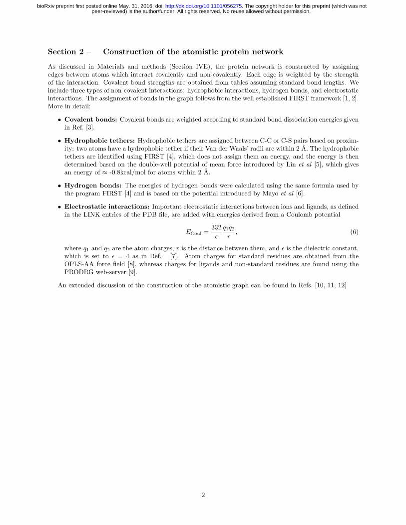
Section 2 – Construction of the atomistic protein network
As discussed in Materials and methods (Section IVE), the protein network is constructed by assigningedges between atoms which interact covalently and non-covalently. Each edge is weighted by the strengthof the interaction. Covalent bond strengths are obtained from tables assuming standard bond lengths. Weinclude three types of non-covalent interactions: hydrophobic interactions, hydrogen bonds, and electrostaticinteractions. The assignment of bonds in the graph follows from the well established FIRST framework [1, 2].More in detail:
• Covalent bonds: Covalent bonds are weighted according to standard bond dissociation energies givenin Ref. [3].
• Hydrophobic tethers: Hydrophobic tethers are assigned between C-C or C-S pairs based on proxim-ity: two atoms have a hydrophobic tether if their Van der Waals’ radii are within 2 A. The hydrophobictethers are identified using FIRST [4], which does not assign them an energy, and the energy is thendetermined based on the double-well potential of mean force introduced by Lin et al [5], which givesan energy of ≈ -0.8kcal/mol for atoms within 2 A.
• Hydrogen bonds: The energies of hydrogen bonds were calculated using the same formula used bythe program FIRST [4] and is based on the potential introduced by Mayo et al [6].
• Electrostatic interactions: Important electrostatic interactions between ions and ligands, as definedin the LINK entries of the PDB file, are added with energies derived from a Coulomb potential
ECoul =332
ǫ
q1q2r
, (6)
where q1 and q2 are the atom charges, r is the distance between them, and ǫ is the dielectric constant,which is set to ǫ = 4 as in Ref. [7]. Atom charges for standard residues are obtained from theOPLS-AA force field [8], whereas charges for ligands and non-standard residues are found using thePRODRG web-server [9].
An extended discussion of the construction of the atomistic graph can be found in Refs. [10, 11, 12]
2
peer-reviewed) is the author/funder. All rights reserved. No reuse allowed without permission. The copyright holder for this preprint (which was not. http://dx.doi.org/10.1101/056275doi: bioRxiv preprint first posted online May. 31, 2016;

Section 3 – Propensities of CheY conformations: different activation states
and NMR ensemble
In the following Table and Figures we give additional information about the active and inactive conformationsand NMR data of CheY used in Section IIB of the main text.
3.1 Active and inactive conformations of CheY
We calculated the propensities of residues for several CheY structures representing different activation states.Details of the different structures are given in Table S1 and a comparison of the perturbation propensitiesacross the different structures is shown in Figure S1. As discussed in Section IIB.2, the propensities of theresidues are strongly correlated across states. In the main text (Section IIB.2 and Figure 3), we concentrateon the comparison of 1F4V (active) against 3CHY (inactive).
Table S1: Details of X-ray structures of CheY analysed. The conformations correspond to differentstages of activation.
PDB ID Structural state Resolution
3CHY Unbound 1.7 A2CHE Bound to Mg2+ 1.8 A1FQW Bound to Mn2+ and BeFx 2.37 A2B1J Bound to FliM 2.4 A1F4V Bound to Mn2+, BeFx and FliM 2.22 A
Figure S1: Propensities in different conformations of CheY. Comparison of propensities of residuesin across different structures of CheY: unbound (3CHY); bound to Mg2+ (2CHE); bound to Mn2+ andphosphate mimic BeFx (1FQW); bound to Mn2+, BeFx and FliM (1F4V); and bound only to FliM (2B1J).The propensities of the residues are strongly correlated across states.
3
peer-reviewed) is the author/funder. All rights reserved. No reuse allowed without permission. The copyright holder for this preprint (which was not. http://dx.doi.org/10.1101/056275doi: bioRxiv preprint first posted online May. 31, 2016;

3.2 CheY structures from NMR experiments
We also calculated the perturbation propensities of residues across two ensembles of NMR structures foractive CheY (PDB ID: 1DJM; 27 structures) and inactive CheY (PDB ID: 1CYE; 20 structures). A com-parison of the average propensity of each residue (averaged across the NMR ensemble) versus its propensityin the X-ray structure is shown in Figure S2 for both the active ensemble (1DJM) and the inactive ensemble(1CYE). This data is discussed in the main text (Section IIB.3) and summarised in Figure 4.
Figure S2: Propensities computed from CheY NMR ensembles. Average propensity obtained fromall structures in an NMR ensemble of CheY against the propensity obtained from the corresponding X-raystructure for inactive (left) and active (right). The inactive ensemble contains 20 structures and the activeensemble contains 27 structures. The error bars show the standard deviation of the propensities ΠR overthe NMR ensemble. Both the variance and the deviation from the X-ray structure is greater for the activeconformation.
4
peer-reviewed) is the author/funder. All rights reserved. No reuse allowed without permission. The copyright holder for this preprint (which was not. http://dx.doi.org/10.1101/056275doi: bioRxiv preprint first posted online May. 31, 2016;

Section 4 – The protein reference set from the SCOP database and absolute
quantile scores
As discussed in Section IID, we have collected a random reference set of 100 proteins drawn from theStructural Classifiation of Proteins (SCOP) database [13]. This reference protein set is used to obtainabsolute quantile scores for the propensities, as detailed in Materials and Methods (Section IVC). Here wegive further details on the reference set and the comparison of absolute and intrinsic quantile scores.
SCOP database: Protein domains in the SCOP database are classified according to a hierarchy based onstructural similarity. Although proteins are additionally divided into superfamilies and subfamilies accordingto structural and sequence similarity, the major classes are:
1. All α: protein domains containing only alpha-helices
2. All β: protein domains containing only beta-sheets
3. Alpha and beta (α/β): protein domains containing both α-helices and β-sheets, with mainly parallelβ-sheets.
4. Alpha and beta (α + β): protein domains containing both α-helices and β-sheets, with mainly anti-parallel β-sheets.
5. Multi-domain: folds of two or more domains from different classes.
We chose 20 proteins from each of these five classes uniformly at random from all proteins in each class, yetchoosing only from structures where there is a ligand bound to the active site.
Absolute quantile scores: On this set of 100 proteins, we then identified the active site in each proteinand computed the propensity for all its bonds relative to the active site. Across the set of 100 proteins in thereference set, we have a total of 465,409 non-covalent bonds, on which we apply quantile regression to obtainabsolute quantile scores pref. In Figure S3 below, the quantile scores pb for all the bonds of the three proteinsstudied in detail in the main text (caspase-1, Che-Y, h-Ras) are plotted against their absolute quantile scorepref
b , showing a good correlation overall. In general, we observe a tighter correlation for larger proteins (e.g.,caspase-1), as a result of the QR fit being based on the number of bonds, E, which is related to the size ofthe protein.
5
peer-reviewed) is the author/funder. All rights reserved. No reuse allowed without permission. The copyright holder for this preprint (which was not. http://dx.doi.org/10.1101/056275doi: bioRxiv preprint first posted online May. 31, 2016;

Figure S3: Absolute quantile scores versus intrinsic quantile scores. The absolute quantile scorescalculated from the reference set (pref
b ) are plotted against the intrinsic quantile scores (pb) for caspase-1(blue), CheY (red), and h-Ras (yellow).
6
peer-reviewed) is the author/funder. All rights reserved. No reuse allowed without permission. The copyright holder for this preprint (which was not. http://dx.doi.org/10.1101/056275doi: bioRxiv preprint first posted online May. 31, 2016;

Section 5 – Bond-to-bond propensities of the allosteric test set
Figure S4: Allosteric test set. The structures of the 20 proteins in the allosteric test set are shown withthe active site ligand (green sticks) and allosteric site residues (yellow spheres).
5.1 Description of the allosteric test set
As discussed in the main text (Section IIE), we have constructed a test set of 20 allosteric proteins onwhich to benchmark our algorithm. Each protein in our test set has a structure with a bound active siteligand and a structure with a bound allosteric ligand. If the protein is allosterically activated then we use asingle structure in which the protein is complexed with both the activator and the active site ligand. Ref.[14] collected a test set of 15 allosteric proteins for which both active site bound and allosteric site bound
7
peer-reviewed) is the author/funder. All rights reserved. No reuse allowed without permission. The copyright holder for this preprint (which was not. http://dx.doi.org/10.1101/056275doi: bioRxiv preprint first posted online May. 31, 2016;

structures are available. We have used 10 of these proteins (the other five were found to be unsuitable forour analysis due to the presence of many non-standard amino-acids, mismatch between the oligomeric stateof the active and inactive structures, or the absence of an allosteric ligand). We have enlarged the set with afurther 10 proteins from an extensive search of the literature. The structures of the 20 proteins are shown inFigure S4, with the active site indicated by the green ligand, and the allosteric site indicated by the yellowspheres. The allosteric site is defined as any residue containing an atom within 4A of the allosteric ligand;allosteric site bonds are defined as any weak interactions formed by an allosteric residue. Full details of theproteins and allosteric site residues are shown in Table S2.
Table S2: Proteins in the allosteric test set. The active site and allosteric site bound structures foreach of the 20 test set proteins. If the protein is allosterically activated then the PDB ID for both states willbe the same. The ligand identifier is that used in the PDB file. Exceptions to this are CheY and caspase-1.As the ligand in these proteins is a peptide, the name and chain ID of the peptide is given instead.
Active Allosteric
Protein Residues PDB Ligand PDB Ligand
ATCase 2790 1D09 PAL 1RAC CTPLac repressor 658 1EFA NPF 1TLF IPTFructose-1, 6-Bisphosphatase 1344 1EYI F6P 1EYJ AMPCheY 144 1F4V FliM (D) 1F4V BEFGlutamate DH 3018 1HWZ NDP 1HWZ GTPATP Sulfurylase 3444 1I2D ADX 1M8P PPSPTP1B 299 1PTY PTR 1T48 BB3Thrombin 281 1SFQ O6G 1SFQ NAGlucokinase 449 1V4S GLC 1V4S MRKUPRTase 852 1XTT U5P 1XTU CTPPhosphoglycerate DH 1644 1YBA AKG 1PSD SER451ADP-glucose phosphorylase 1727 1YP3 ATP 1YP2 PMBCHK1 258 2BRG DFY 3JVS AGYCaspase-1 520 2HBQ z-VAD-FMK (C/F) 2FQQ F1GPDK1 278 3ORZ BI4 3ORZ 2A2Phosphofructokinase 1288 4PFK F6P 6PFK PGAGlycogen Phosphorylase 3304 7GPB PLP/SO4 7GPB SO4/AMPglcN-6-P deaminase 1604 1HOT PO4 1HOT NAG/PHSh-Ras 175 3K8Y GNP 3K8Y ACTlactate DH 1260 1LTH NAD 1LTH FBP
5.2 Summary of results on the allosteric test set
As explained in the main text (Section IIE and Materials and Methods, Section IVD), for each of the 20proteins in the test set, we analyse the propensities of all bonds with respect to the active site of the boundstructure, using the ligands shown in Fig. S4 as the source for the bond-to-bond propensity calculations.For each protein, we obtain the propensity Πb of every weak bond and its associated quantile score (pb).To establish their statistical significance, the bond quantile scores pb (and residue averaged quantile scorespR) of the allosteric site are compared against an ensemble of randomly generated surrogate sites from eachprotein. The ensemble of surrogate sites is constructed at random by picking sites that satisfy two structuralconstraints: (i) they have the same number of residues as the allosteric site; and (ii) their diameter (themaximum distance between any two atoms in the site) is no larger than that of the allosteric site. The sitesare generated using Algorithm 1 with pseudocode given below. The propensities averaged over the ensembleof surrogate sites are then used for statistical comparison with the allosteric site. We also obtain absolutepropensity scores for each bond (prefb ) by comparing against the reference SCOP ensemble of 100 proteins.These quantities are defined in the main text (Materials and Methods, Section IVD).
8
peer-reviewed) is the author/funder. All rights reserved. No reuse allowed without permission. The copyright holder for this preprint (which was not. http://dx.doi.org/10.1101/056275doi: bioRxiv preprint first posted online May. 31, 2016;

Algorithm 1 Pseudocode for surrogate site sampling
1: site ← ∅
2: while # residues in site < # residues in allosteric site do
3: choose a residue R at random4: if diameter(site ∪ R) < diameter(allosteric site) then5: site ← site ∪ R6: end if
7: end while
Using all these scores we obtain our four statistical measures of significance summarised in Table S3.These numerical results are presented also in the form of a graph in Figure 7 of the main text.
Table S3: Allosteric site quantile scores in test set proteins. The four scores described in Figure 7 ofthe main text for the test set of 20 proteins. The difference between the allosteric site average quantile scoreand the average surrogate site score for both residues and bonds are shown in bold if they are greater than0, and starred if they lie above the 95% confidence interval computed by a bootstrap with 10000 resamples.
The average reference quantile score prefb,allo is shown in bold if it is greater than 0.5 (the expected value).The proportion pb,allo > 0.95 is shown in bold if it is greater than 0.05.
Protein PDB ID pR,allo − 〈pR,site〉surr pb,allo − 〈pb,site〉surr P(pb,allo > 0.95) prefb,allo Summary
Glucokinase 1V4S 0.35∗
0.14∗
0.12 0.66
PDK1 3ORZ 0.30∗
0.030∗
0.080 0.56
ADP-glucose phosphorylase 1YP3 0.28∗
0.074∗
0.10 0.59
ATCase 1DO9 0.23∗
0.036∗
0.091 0.68
Caspase-1 2HBQ 0.15∗
0.0032∗
0.070 0.54
glcN-6-P deaminase 1HOT 0.13∗
0.031∗
0.079 0.51
PTP1B 1PTY 0.11∗
0.0088∗ 0.048 0.50
Fructose-1, 6-Bisphosphatase 1EYI 0.11∗
0.033∗
0.052 0.49Glycogen Phosphorylase 7GPB 0.11
∗
0.0027∗
0.058 0.47Chemotaxis Y 1F4V 0.096
∗
0.055∗
0.074 0.58
Phosphofructokinase 4PFK 0.092∗
0.068∗
0.16 0.54
ATP Sulfurylase 1I2D 0.091∗
0.0313∗
0.068 0.52
Phosphoglycerate DH 1YBA 0.062∗
0.076∗
0.075 0.59
Lactate DH 1LTH 0.063∗
0.024∗
0.063 0.52
UPRtase 1XTT 0.0024 -0.013 0.06 0.44Glutamate DH 1HWZ -0.015 0.039
∗
0.068 0.44h-Ras 3K8Y -0.043 -0.016 0.059 0.49Lac repressor 1EFA -0.066 -0.016 0.014 0.60
Thrombin 1SFQ -0.081 0.077∗
0.16 0.64
CHK1 2BRG -0.24 -0.15 0.0052 0.36
9
peer-reviewed) is the author/funder. All rights reserved. No reuse allowed without permission. The copyright holder for this preprint (which was not. http://dx.doi.org/10.1101/056275doi: bioRxiv preprint first posted online May. 31, 2016;

Section 6 – Robustness of the bond-to-bond propensities to random pertur-
bations of the weak interactions
Proteins are dynamic objects undergoing motions and fluctuations under the influence of the environment.Such dynamic fluctuations induce changes in the bond energies of the protein, potentially leading to thebreaking of weak bonds (hydrogen bonds, salt bridges, hydrophobic tethers). As discussed in the main textwhen studying the NMR ensemble of conformations of CheY (Section IIB.3), whilst there is considerableagreement between the results from the NMR structures and the X-ray structure (Fig. S2), the variabilityin the ensemble can reveal further information. It is also important to check that the computation ofpropensities is generally robust to the presence of such noise. To do this, we have developed two schemesto add random perturbations to our protein networks. These schemes mimic the effect of small dynamicfluctuations, without carrying out expensive molecular dynamics simulations.
Firstly, for each of the 20 proteins in our dataset, we add zero mean Gaussian noise to the edge weights(energies) of non-covalent bonds in the graph, so as to mimic the effect of thermal fluctuations. Note that weallow the bonds to break if their randomised energy becomes zero. We then recompute our quantile scoresfor the allosteric site for 10 realisations of the noisy networks generated after the addition of the Gaussianfluctuations. We do this for 3 levels of noise, i.e., we increase the standard deviation of the Gaussian from1kT=0.6 kcal/mol to 4kT=2.4 kcal/mol. The average results of these randomisations for all proteins in theallosteric test set are presented in Table S4. Our calculations show that the results are generally robustto fluctuations induced in this way: the signal at the allosteric site only drops slightly when introducingrelatively high levels of noise.
Table S4: Robustness of propensity scores to additive randomness. Mean (± standard deviation)of propensity scores pR,allo − 〈pR,site〉surr computed from randomisations of the protein networks of theallosteric test set obtained by adding Gaussian noise to the edge weights (bond energies). The noise levelvaries between 1kT and 4kT (corresponding to the standard deviation of the added Gaussian) and at eachnoise level the results were calculated from 10 randomised graphs. The difference between the allosteric siteaverage quantile score and the average surrogate site score for both residues and bonds are shown in bold ifthey are greater than 0, and starred if they lie above the 95% confidence interval computed by a bootstrapwith 10000 resamples. The unperturbed result is also shown for comparison.
PDB ID Unperturbednetwork
Gaussian noise1kT
Gaussian noise2kT
Gaussian noise4kT
1V4S 0.35∗
0.32±0.011∗
0.31±0.019∗
0.27±0.017∗
3ORZ 0.30∗
0.28±0.0087∗
0.23±0.0090∗
0.24±0.014∗
1YP3 0.28∗
0.27±0.0010∗
0.25±0.0088∗
0.17±0.016∗
1D09 0.23∗
0.22±0.0071∗
0.21±0.0024∗
0.20±0.0035∗
2HBQ 0.15∗
0.18±0.0096∗
0.20±0.0058∗
0.13±0.0097∗
1HOT 0.13∗
0.13±0.0061∗
0.098±0.024∗
0.12±0.021∗
1PTY 0.11∗
0.096±0.020∗
0.11±0.022∗
0.088±0.031∗
1EYI 0.11∗
0.13±0.0065∗
0.13±0.0022∗
0.16±0.0050∗
7GPB 0.11∗
0.096±0.018∗
0.13±0.010∗
0.14±0.015∗
1F4V 0.096∗
0.093±0.018∗
0.14±0.0097∗
0.12±0.027∗
4PFK 0.092∗
0.091±0.0052∗
0.11±0.022∗
0.12±0.0075∗
1I2D 0.091∗
0.14±0.029∗
0.14±0.030∗
0.14±0.037∗
1YBA 0.062∗
0.076±0.0034∗
0.091±0.0048∗
0.073±0.0051∗
1LTH 0.063∗
0.070±0.0099∗
0.063±0.016∗
0.039±0.019∗
1XTT 0.0024 0.0084±0.0069 0.015±0.0083 0.0077±0.0070
1HWZ -0.015 -0.0090±0.0071 -0.0028±0.0043 0.011±0.0065
3K8Y -0.043 -0.033±0.012 -0.012±0.010 -0.025±0.0221EFA -0.066 -0.047±0.0054 -0.019±0.0078 -0.0027±0.00771SFQ -0.081 -0.090±0.0089 -0.083±0.023 -0.10±0.0282BRG -0.24 -0.23±0.010 -0.24±0.013 -0.23±0.026
10
peer-reviewed) is the author/funder. All rights reserved. No reuse allowed without permission. The copyright holder for this preprint (which was not. http://dx.doi.org/10.1101/056275doi: bioRxiv preprint first posted online May. 31, 2016;

Secondly, to test a different kind of variability introduced by the environment, we have considered theeffect of breaking all bonds in our network with energy below a threshold. Starting with the originalunperturbed structure, all weak bonds below a given threshold are removed from the graph. In this way, wemimic the possibility of extended structural changes that could lead to breaking of bonds in a more globalfashion.
For each of the 20 proteins in the test set, we generate two perturbed networks obtained by bond removalof all bonds with energy below two different thresholds: 0.5 kT≃ 0.3 kcal/mol and 1kT ≃ 0.6 kcal/mol.The effect of this thresholding is extensive. For the 0.5kT threshold, we delete all hydrophobic tethers andelectrostatic interactions as well as a percentage of hydrogen bonds that ranges from 31% in 1SFQ to 44%in 1HWZ and 1LTH. For the 1kT threshold, even further hydrogen bonds are removed, corresponding toeliminating 44% of H-bonds in 1SFQ up to 57% of the H-bonds in 7GPB, 2BRG, 1LTH (in addition to allhydrophobic interactions).
The calculations of the propensity for the thresholded networks for all 20 proteins in our test set arepresented in Table S5. Our results show that, overall, the propensity of the allosteric site remains largelyrobust to such changes across all 20 proteins considered, yet with notable differences in the magnitude ofthe effect across the set. In some proteins, the signal at the allosteric site is mildly affected by bond deletion(e.g. 3ORZ, 1YP3, 2HBQ, 1HOT, 1PTY). In other cases, however, the deletion of weaker hydrogen bondshas a large effect in destroying the communication between the allosteric site and the active site (e.g. 1V4S,1D09, 1EYI, 7GPB, 1F4V). These differences could be a measure of how robust the allosteric signalling is toenergetic fluctuations in the local environment of the protein, and also provide clues as to different structuralfeatures connected with the distributed nature of allosteric signalling in the different proteins. The study ofsuch differences will be the object of future work.
Table S5: Robustness of propensity scores to deletion of weak bonds. The propensity score pR,allo−〈pR,site〉surr for networks obtained by deleting all bonds below two energy thresholds. The results are shownin bold when they are greater than 0 and starred if they lie above the 95% confidence interval computed bya bootstrap with 10000 resamples. The unperturbed score is reported also for comparison.
PDB ID Unperturbednetwork
Threshold0.5 kT
Threshold1kT
1V4S 0.35∗
0.061∗
0.049∗
3ORZ 0.30∗
0.24∗
0.25∗
1YP3 0.28∗
0.24∗
0.30∗
1D09 0.23∗
0.088∗
0.10∗
2HBQ 0.15∗
0.16∗
0.18∗
1HOT 0.13∗
0.14∗
0.17∗
1PTY 0.11∗
0.13∗
0.080∗
1EYI 0.11∗
0.026∗
0.022∗
7GPB 0.11∗
0.056∗
0.062∗
1F4V 0.096∗ -0.0010 0.0085
∗
4PFK 0.092∗
0.17∗
0.20∗
1I2D 0.091∗
0.0012 -0.0331YBA 0.062
∗
0.079∗
0.052∗
1LTH 0.063∗
0.056∗ -0.081
1XTT 0.0024 -0.016 -0.0231HWZ -0.075 -0.016 -0.203K8Y -0.043 -0.14 -0.161EFA -0.066 0.052
∗
0.051∗
1SFQ -0.081 0.073∗
0.11∗
2BRG -0.24 -0.20 -0.17
11
peer-reviewed) is the author/funder. All rights reserved. No reuse allowed without permission. The copyright holder for this preprint (which was not. http://dx.doi.org/10.1101/056275doi: bioRxiv preprint first posted online May. 31, 2016;

Section 7 – Propensities from residue-residue interaction networks
The computational efficiency of our methodology allows us to analyse all-atom networks without many ofthe restrictions on system size inherent to other methods. Proteins or protein complexes of hundreds ofthousands of atoms can be analysed in a few minutes on a standard desktop. We can thus keep atomisticdetail at the single bond level without restricting the scope of the analysis. Hence there is a less acute needto seek computational savings by obtaining coarse-grained representations of proteins at the level of residueinteractions. However, it is still instructive to consider propensity measures computed from residue-levelnetworks (RRINs) [15]. We have undertaken this comparison for all 20 proteins in our test set and reportthe results below.
As discussed in the main text (Sections IIA and IIB), in some cases (e.g., caspase-1, Fig 1b) we foundthat the additional information contained in the atomistic network leads to increased signal in the detectionof the allosteric site, whereas in other cases (e.g., CheY), RRINs already capture well the site connectivitythat reveals the presence of the allosteric site. Our analysis of the full test set (Table S6) confirms that theresults from RRINs depend on the protein analysed, and also vary substantially depending on the choiceof the cut-off distance (a tunable parameter which needs to be chosen when generating the coarse-grainedRRINs).
The coarse-grained RRINs for each of the 20 proteins in the test set were obtained by submitting thecorresponding PDB files to the oGNM server [16]. We obtained RRINs at four different cut-off radii: 6 A, 7A, 8 A and 10 A. The cut-off radius is a tunable parameter necessary to generate a RRIN from PDB files,which establishes how close two residues must be in order to be connected in the RRIN. A range of differentcut-off radii has been used throughout the literature. However, the usual radius is around 6.7-7.0 A, whichcorresponds to the first coordination shell [17].
Table S6 shows the propensity score of the allosteric site pR,allo − 〈pR,site〉surr, computed from RRINsobtained at four cut-offs (between 6A and 10A) for the 20 proteins in the allosteric test set. For comparisonpurposes, we also report the same score obtained from the all-atom network. It is important to note thatthis is just one of four scores obtained from the all-atom network, reflecting only the averaged behaviourover the residues. This score is complemented by the three other bond-based statistics, which can pick upinhomogeneities in the propensities of the bonds in the allosteric site, as given by the All-atom Summarycolumn carried over from Table S3.
Our results indicate broad consistency between RRINs and the all-atom network. However, the RRINresults vary widely depending on the choice of cut-off radius in the generation of the network. Moreoverthis variability with respect to the cut-off behaves differently for each of the proteins. As an illustration,the allosteric site of caspase-1 (2HBQ) was not found to be significant in the RRINs with cut-off radii of6 A, 7 Aand 8 A and only weakly significant for 10 A, whereas 1LTH and 2BRG are both only detected inRRINS with cut-off radius of 6 A but not for larger radii. Our results are consistent with previous studiesthat found that allosteric pathway identification in RRINs is dependent on the chosen cut-off [18]. For thedifferent cut-offs, the number of proteins with pR,allo > pR,rest varies between 11/20 (at 7, 8, and 10A) and13/20 (at 6A), and only 8/20 proteins have pR,allo > pR,rest for the RRINS at all the cut-off radii. This iscompared to 15/20 proteins for the atomistic network.
Even when the allosteric site is detected in the RRIN, the signal when using the atomistic network isconsiderably higher in a number of proteins (e.g., 1V4S, 1YP3, 7GPB, 1I2D, 2HBQ). In other cases (e.g.,1EYI, 4PFK), the RRIN directly loses the detectability of the allosteric site even if the cut-off is adjusted.This observation suggests that these are proteins where the specific chemistry of intra-protein bonds isimportant for the allosteric communication.
On the other hand, there are several other cases (e.g., 3ORZ, 1D09, 1HOT, 1PTY, 1LTH) where theRRIN can provide similar results to the atomistic network, yet still with some variability depending on thechoice of appropriate cut-off. Interestingly, there are also some proteins (specifically 1F4V, 1YBA, 3K8Yand 2BRG) in which the propensity score is higher for RRINs than for the atomistic network. In these cases,there tends to be a large heterogeneity in the propensities of the bonds in the allosteric site (see Figure 7 inthe main text) with some bonds with large negative values as well as other bonds with large positive values.Our bond statistical measures can account for some of this variability. Indeed, both 1F4V and 1YBA aredetected by all our four bond measures, and 3K8Y is picked by the measure based on the distributions of pb.Intriguingly, only 2BRG (corresponding to CHK1) cannot be detected by our bond measures. This suggests
12
peer-reviewed) is the author/funder. All rights reserved. No reuse allowed without permission. The copyright holder for this preprint (which was not. http://dx.doi.org/10.1101/056275doi: bioRxiv preprint first posted online May. 31, 2016;

other areas of future research, in which the importance of averaging at the level of pathways could be usedto enrich the findings presented here.
Figure S5: Quantile scores computed from RRINs for caspase-1 and CheY at different cut-off
radii. Surface mapping of the residue quantile scores pR of caspase-1 and CheY for RRINs generated withradii cut-offs from 6 A and 10 A. The active-site ligand is shown in green sticks and the allosteric site iscircled. The allosteric site in caspase-1 is not identified for 6, 7, and 8 A. It is identified at 10 A, but thesignal is weaker than when using an atomistic graph. In contrast, for CheY the allosteric site is identifiedas significant across the full range of cut-offs.
13
peer-reviewed) is the author/funder. All rights reserved. No reuse allowed without permission. The copyright holder for this preprint (which was not. http://dx.doi.org/10.1101/056275doi: bioRxiv preprint first posted online May. 31, 2016;

Table S6: Propensities computed from RRINs. Values of pR,allo − 〈pR,site〉surr for residue-residueinteraction networks with four cut-off radii from 6A–10A. The propensity scores are shown in bold if theyare greater than 0, and starred if they lie above the 95% confidence interval computed by a bootstrap with10000 resamples. The comparable statistic computed from the all-atom network is also presented, as well asthe summary of the four bond statistics for each protein from Table S3.
pR,allo − 〈pR,site〉surr
PDB ID All-atomsummary
All-atomnetwork
RRINcut-off = 6A
RRINcut-off = 7A
RRINcut-off = 8A
RRINcut-off = 10A
1V4S 0.35∗
0.065∗
0.010∗
0.047∗
0.16∗
3ORZ 0.30∗
0.31∗
0.34∗
0.37∗
0.22∗
1YP3 0.28∗ -0.043 0.11
∗
0.046∗
0.13∗
1D09 0.23∗
0.20∗
0.17∗
0.21∗
0.15∗
2HBQ 0.15∗ -0.079 -0.053 -0.062 0.098
∗
1HOT 0.13∗ -0.065 0.13
∗
0.18∗
0.20∗
1PTY 0.11∗
0.11∗
0.088∗
0.050∗ -0.032
1EYI 0.11∗ -0.036 -0.081 -0.098 -0.029
7GPB 0.11∗
0.048∗
0.073∗
0.047∗ -0.095
1F4V 0.096∗
0.14∗
0.11∗
0.23∗
0.071∗
4PFK 0.092∗ -0.13 -0.24 -0.19 -0.067
1I2D 0.091∗
0.034∗ -0.091 0.010
∗
0.12∗
1YBA 0.062∗
0.18∗
0.16∗
0.20∗
0.29∗
1LTH 0.063∗
0.080∗ -0.11 -0.22 -0.073
1XTT 0.0024 0.025∗ -0.017 -0.012 0.14
∗
1HWZ -0.015 0.071∗
0.041∗ -0.016 -0.0072
3K8Y -0.043 0.29∗
0.24∗
0.17∗
0.30∗
1EFA -0.066 -0.035 -0.0028 -0.064 -0.0751SFQ -0.081 -0.18 -0.19 -0.16 -0.192BRG -0.24 0.13
∗ -0.043 -0.057 -0.093
14
peer-reviewed) is the author/funder. All rights reserved. No reuse allowed without permission. The copyright holder for this preprint (which was not. http://dx.doi.org/10.1101/056275doi: bioRxiv preprint first posted online May. 31, 2016;

References
[1] D. J. Jacobs and M. F. Thorpe, “Generic rigidity percolation: the pebble game,” Physical review letters, vol. 75, no. 22,p. 4051, 1995.
[2] D. J. Jacobs and M. F. Thorpe, “Computer-implemented system for analyzing rigidity of substructures within a macro-molecule,” Jan. 11 1999. US Patent 6,014,449.
[3] J. Huheey, E. Keitler, and R. Keitler, Inorganic Chemistry, Principles of Structure and Bonding. Harper Collins CollegePublishers, New York, 1993.
[4] D. Jacobs, A. Rader, L. Kuhn, and M. Thorpe, “Protein flexibility predictions using graph theory,” Proteins: Structure,
Function, and Bioinformatics, vol. 44, no. 2, pp. 150–165, 2001.
[5] M. S. Lin, N. L. Fawzi, and T. Head-Gordon, “Hydrophobic potential of mean force as a solvation function for proteinstructure prediction,” Structure, vol. 15, no. 6, pp. 727–740, 2007.
[6] B. Dahiyat, D. Benjamin Gordon, and S. Mayo, “Automated design of the surface positions of protein helices,” Protein
Science, vol. 6, no. 6, pp. 1333–1337, 1997.
[7] M. K. Gilson and B. H. Honig, “The dielectric constant of a folded protein,” Biopolymers, vol. 25, no. 11, pp. 2097–2119,1986.
[8] W. L. Jorgensen and J. Tirado-Rives, “The OPLS [optimized potentials for liquid simulations] potential functions forproteins, energy minimizations for crystals of cyclic peptides and crambin,” Journal of the American Chemical Society,vol. 110, no. 6, pp. 1657–1666, 1988.
[9] A. W. Schuettelkopf and D. M. Van Aalten, “PRODRG: a tool for high-throughput crystallography of protein–ligandcomplexes,” Acta Crystallographica Section D: Biological Crystallography, vol. 60, no. 8, pp. 1355–1363, 2004.
[10] A. Delmotte, E. Tate, S. Yaliraki, and M. Barahona, “Protein multi-scale organization through graph partitioning androbustness analysis: application to the myosin–myosin light chain interaction,” Physical Biology, vol. 8, p. 055010, 2011.
[11] A. Delmotte, All-scale structural analysis of biomolecules through dynamical graph partitioning. PhD thesis, ImperialCollege London, 2014.
[12] B. Amor, S. Yaliraki, R. Woscholski, and M. Barahona, “Uncovering allosteric pathways in caspase-1 using Markov transientanalysis and multiscale community detection,” Molecular BioSystems, vol. 10, no. 8, pp. 2247–2258, 2014.
[13] L. L. Conte, B. Ailey, T. J. Hubbard, S. E. Brenner, A. G. Murzin, and C. Chothia, “SCOP: a structural classification ofproteins database,” Nucleic acids research, vol. 28, no. 1, pp. 257–259, 2000.
[14] M. Daily, T. Upadhyaya, and J. Gray, “Contact rearrangements form coupled networks from local motions in allostericproteins,” Proteins: Structure, Function, and Bioinformatics, vol. 71, no. 1, pp. 455–466, 2008.
[15] C. Chennubhotla and I. Bahar, “Signal propagation in proteins and relation to equilibrium fluctuations,” PLoS computa-
tional biology, vol. 3, no. 9, p. e172, 2007.
[16] L.-W. Yang, A. Rader, X. Liu, C. J. Jursa, S. C. Chen, H. A. Karimi, and I. Bahar, “oGNM: online computation ofstructural dynamics using the Gaussian Network Model,” Nucleic acids research, vol. 34, no. suppl 2, pp. W24–W31, 2006.
[17] A. R. Atilgan, P. Akan, and C. Baysal, “Small-world communication of residues and significance for protein dynamics,”Biophysical journal, vol. 86, no. 1, pp. 85–91, 2004.
[18] A. A. Ribeiro and V. Ortiz, “Determination of signaling pathways in proteins through network theory: importance of thetopology,” Journal of Chemical Theory and Computation, vol. 10, no. 4, pp. 1762–1769, 2014.
15
peer-reviewed) is the author/funder. All rights reserved. No reuse allowed without permission. The copyright holder for this preprint (which was not. http://dx.doi.org/10.1101/056275doi: bioRxiv preprint first posted online May. 31, 2016;