Embed Size (px)
Citation preview

PreciseType™ HEA Molecular BeadChip Test
Package Insert
V
2
Prec
iseT
ype
HE
A M
olec
ular
Bea
dChi
p Te
st │
190
-202
10 R
ev. B
│ M
ay 2
016
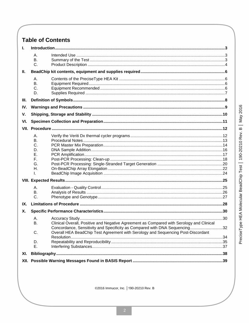
Table of Contents I. Introduction ....................................................................................................................................................... 3
A. Intended Use .................................................................................................................................... 3 B. Summary of the Test ........................................................................................................................ 3 C. Product Description .......................................................................................................................... 4
II. BeadChip kit contents, equipment and supplies required ........................................................................... 6
A. Contents of the PreciseType HEA Kit .............................................................................................. 6 B. Equipment Required ......................................................................................................................... 6 C. Equipment Recommended ............................................................................................................... 6 D. Supplies Required ............................................................................................................................ 7
III. Definition of Symbols ....................................................................................................................................... 8
IV. Warnings and Precautions .............................................................................................................................. 9
V. Shipping, Storage and Stability .................................................................................................................... 10
VI. Specimen Collection and Preparation .......................................................................................................... 11
VII. Procedure ........................................................................................................................................................ 12
A. Verify the Veriti Dx thermal cycler programs .................................................................................. 12 B. Procedural Notes ............................................................................................................................ 13 C. PCR Master Mix Preparation .......................................................................................................... 14 D. DNA Sample Addition ..................................................................................................................... 16 E. PCR Amplification ........................................................................................................................... 17 F. Post-PCR Processing: Clean-up .................................................................................................... 18 G. Post-PCR Processing: Single-Stranded Target Generation .......................................................... 20 H. On-BeadChip Array Elongation ...................................................................................................... 22 I. BeadChip Image Acquisition .......................................................................................................... 24
VIII. Expected Results ............................................................................................................................................ 25
A. Evaluation - Quality Control ............................................................................................................ 25 B. Analysis of Results ......................................................................................................................... 26 C. Phenotype and Genotype ............................................................................................................... 27
IX. Limitations of Procedure ............................................................................................................................... 28
X. Specific Performance Characteristics .......................................................................................................... 30
A. Accuracy Study ............................................................................................................................... 30 B. Clinical Overall, Positive and Negative Agreement as Compared with Serology and Clinical
Concordance, Sensitivity and Specificity as Compared with DNA Sequencing ............................. 32 C. Overall HEA BeadChip Test Agreement with Serology and Sequencing Post-Discordant
Resolution ....................................................................................................................................... 34 D. Repeatability and Reproducibility ................................................................................................... 35 E. Interfering Substances .................................................................................................................... 37
XI. Bibliography .................................................................................................................................................... 38
XII. Possible Warning Messages Found in BASIS Report ................................................................................ 39
©2016 Immucor, Inc. │190-20210 Rev. B

3
Prec
iseT
ype
HE
A M
olec
ular
Bea
dChi
p Te
st │
190
-202
10 R
ev. B
│ M
ay 2
016
I. INTRODUCTION
A. Intended Use
The HEA BeadChip Kit is an in vitro diagnostic test intended for the molecular determination of
allelic variants that predict erythrocyte antigen phenotypes in the Rh (C [RH2], c [RH4], E [RH3],
e [RH5], V [RH10], VS [RH20]); Kell (K [KEL1], k [KEL2], Kpa [KEL3], Kpb [KEL4], Jsa [KEL6],
Jsb [KEL7]); Duffy (Fya [FY1], Fyb [FY2], GATA [FY-2], Fyx [FY2W]); Kidd (Jka [JK1], Jkb
[JK2]); MNS (M [MNS1], N [MNS2], S [MNS3], s [MNS4], Uvar [MNS-3,5W], Uneg [MNS-3,-4,-5]);
Lutheran (Lua [LU1], Lub [LU2]); Dombrock (Doa [DO1], Dob [DO2], Hy [DO4], Joa [DO5]);
Landsteiner-Wiener (LWa [LW5], LWb [LW7]); Diego (Dia [DI1], Dib [DI2]); Colton (Coa
[CO1],Cob [CO2]); and Scianna (Sc1[SC1], Sc2 [SC2]) blood group systems in human genomic
DNA. The test also detects the HgbS mutation in the beta globin gene. The results from this
mutation detection are not intended for diagnosis of sickle cell disease.
B. Summary of the Test
The PreciseType HEA Molecular BeadChip Test uses the proprietary Elongation-mediated
Multiplexed Analysis of Polymorphisms (eMAP™) technology to identify the presence or absence
of the selected alleles associated with a given phenotype. After multiplex PCR amplification and
post-PCR processing using Clean-up Reagent and Lambda Exonuclease, the single-stranded
DNA strands are incubated on the BeadChip array, allowing them to anneal with the
corresponding blood-group-specific probes. An exact match between the 3’ end of the probe and
the annealed DNA triggers a subsequent elongation reaction in which the probe will be extended
through incorporation of fluorescently-labeled dNTP molecules. If there is not an exact match,
elongation does not occur. Elongation products of alleles A and B are detected simultaneously by
imaging the entire array.
In this method, each probe is attached covalently to a spectrally distinguishable bead type. A
library of individual bead types contains all of the probes of interest. The library is immobilized in
the BeadChip array, allowing for the simultaneous detection of the polymorphisms of interest.
BeadChips are read with the BioArray Array Imaging System™ (AIS™ 400), with assay results
interpreted, analyzed, and reported through the BioArray Solutions Information System (BASIS™)
software. The AIS system captures the fluorescent signal from individual beads in an image of the
entire array to determine the identity of the bead by the color of the bead and its position in the
array. It also detects the average signal intensity, coefficient of variance standard deviation of the
intensities, and number of beads measured for each type of probe. The BASIS software then
imports the raw intensity output, assesses the validity of the internal controls, and generates
assay results.
Mutations known to result in silencing (non-expression) of Duffy (Fyb) [FY2] and MNS (S) [MNS3]
antigens have been incorporated into the test.

4
Prec
iseT
ype
HE
A M
olec
ular
Bea
dChi
p Te
st │
190
-202
10 R
ev. B
│ M
ay 2
016
C. Product Description
Human erythrocyte blood group antigens – the surface markers located on the membrane of the
red blood cell – are polymorphic, inherited protein. If an antigen-negative patient receives blood
from an antigen-positive donor, it could trigger an immune reaction (alloimmunization), where the
blood recipient’s immune system develops antibodies that can attack and reject the donor RBCs.
These responses vary in degree of severity from immediate and severe to none at all [1]. Once
an alloantibody is produced, lifelong immunization occurs, even if the antibody is not detectable.
In certain medical conditions requiring frequent, chronic blood transfusion therapy, such as sickle
cell disease, autoimmune hemolytic anemia, and aplastic anemia, increased opportunity for
alloantibody product occurs. In such cases, studies demonstrate the utility of bolstering
serological phenotyping with DNA analysis to identify the presence of blood-group antigens [2][3].
Perinatal or postnatal management of hemolytic disease of the fetus and newborn (HDFN) may
be assisted by identification of human erythrocyte antigens. Minor blood group incompatibility
occurs in approximately 0.8% of pregnant women and may be associated with Kell, Kidd or Duffy
(among others). Anti-K disease may be severe due to hemolysis or erythroid suppression [4][5].
The International Society for Blood Transfusion (ISBT) Committee on Terminology for Red Cell
Surface Antigens maintains and updates a database of known alleles for 35 blood-group
systems. It provides information regarding the prevalence and significance of alleles in a pan
ethnic population as well as standards for nomenclature and terminology in transfusion medicine
[6].
Twenty-four polymorphisms associated with 35 Human Erythrocyte Antigens are included in the
PreciseType HEA Molecular BeadChip Test and are listed in the following table (Table 1) [7]. One
polymorphism in the beta-globin gene associated with hemoglobinopathies (HgbS) is also
included.

5
Prec
iseT
ype
HE
A M
olec
ular
Bea
dChi
p Te
st │
190
-202
10 R
ev. B
│ M
ay 2
016
Table 1: Genetic Markers for Red Blood Cell Antigens in the PreciseType HEA Test
Blood Group System Analyte Polymorphism ISBT Phenotype ISBT Genotype
Rh
c/C 307 C>T
RH4, RH2 RHCE*4, RHCE*2 109 Ins
e/E 676 G>C RH5, RH3 RHCE*5, RHCE*3
VS 733 C>G, 1006 G>T
RH20 RHCE*01.20.01, RHCE01.20.02, RHCE*01.20.04, RHCE*01.20.05 V RH10
Kell
K/k 698 T>C KEL1, KEL2 KEL*01, KEL*02
Jsa/Jsb 1910 C>T KEL6, KEL7 KEL*06, KEL*07
Kpa/Kpb 961 T>C KEL3, KEL4 KEL*03, KEL*04
Duffy
Fya/Fyb 125 G>A FY1, FY2 FY*01, FY*02
GATA (Silencing FY) -67 T>C** FY-2 FY*02N.01
Fyx[Fy(b+w)] 265 C>T FY2W FY*02M
Kidd Jka/Jkb 838 G>A JK1, JK2 JK*01, JK*02
MNS
M/N 59 C>T MNS1, MNS2 GYPA*01, GYPA*02
S/s 143 T>C MNS3, MNS4 GYPB*03, GYPB*04
Silencing S (Uvar, Uneg)
230C>T MNS-3, 5W, MNS-3,-4,-5
GYPB*03N.01 or GYPB*03N.02
In5 g>t GYPB*03N.03 or GYPB*03N.04
Lutheran Lua/Lub 230 A>G LU1, LU2 LU*01, LU*02
Dombrock
Doa/Dob 793 A>G DO1, DO2 DO*01, DO*02
Hy+/Hy- 323 G>T DO4 DO*04
Jo(a+)/Jo(a-) 350 C>T DO5 DO*05
Landsteiner-Wiener LWa/LWb 308 A>G LW5, LW7 LW*05, LW*07
Diego Dib/Dia 2561 C>T DI2, DI1 DI*02, DI*01
Colton Coa/Cob 134 C>T CO1, CO2 CO*01, CO*02
Scianna Sc1/Sc2 169 G>A SC1, SC2 SC*01, SC*02
** The GATA mutation listed here has been previously reported at -33 and -46 (ISBT Working Party)[8].
6
Prec
iseT
ype
HE
A M
olec
ular
Bea
dChi
p Te
st │
190
-202
10 R
ev. B
│ M
ay 2
016
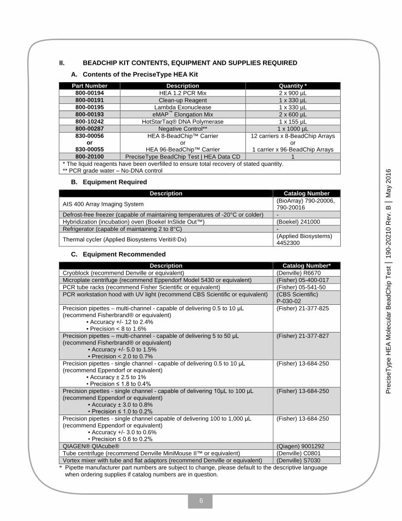
II. BEADCHIP KIT CONTENTS, EQUIPMENT AND SUPPLIES REQUIRED
A. Contents of the PreciseType HEA Kit Part Number Description Quantity *
800-00194 HEA 1.2 PCR Mix 2 x 900 µL 800-00191 Clean-up Reagent 1 x 330 µL 800-00195 Lambda Exonuclease 1 x 330 µL 800-00193 eMAP™ Elongation Mix 2 x 600 µL 800-10242 HotStarTaq® DNA Polymerase 1 x 155 µL 800-00287 Negative Control** 1 x 1000 µL 830-00056
or 830-00055
HEA 8-BeadChip™ Carrier or
HEA 96-BeadChip™ Carrier
12 carriers x 8-BeadChip Arrays or
1 carrier x 96-BeadChip Arrays 800-20100 PreciseType BeadChip Test | HEA Data CD 1
* The liquid reagents have been overfilled to ensure total recovery of stated quantity. ** PCR grade water – No-DNA control
B. Equipment Required Description Catalog Number
AIS 400 Array Imaging System (BioArray) 790-20006, 790-20016
Defrost-free freezer (capable of maintaining temperatures of -20°C or colder) - Hybridization (incubation) oven (Boekel InSlide Out™) (Boekel) 241000 Refrigerator (capable of maintaining 2 to 8°C) -
Thermal cycler (Applied Biosystems Veriti® Dx) (Applied Biosystems) 4452300
C. Equipment Recommended Description Catalog Number*
Cryoblock (recommend Denville or equivalent) (Denville) R6670 Microplate centrifuge (recommend Eppendorf Model 5430 or equivalent) (Fisher) 05-400-017 PCR tube racks (recommend Fisher Scientific or equivalent) (Fisher) 05-541-50 PCR workstation hood with UV light (recommend CBS Scientific or equivalent) (CBS Scientific)
P-030-02 Precision pipettes – multi-channel - capable of delivering 0.5 to 10 μL (recommend Fisherbrand® or equivalent)
• Accuracy +/- 12 to 2.4% • Precision < 8 to 1.6%
(Fisher) 21-377-825
Precision pipettes – multi-channel - capable of delivering 5 to 50 μL (recommend Fisherbrand® or equivalent)
• Accuracy +/- 5.0 to 1.5% • Precision < 2.0 to 0.7%
(Fisher) 21-377-827
Precision pipettes - single channel - capable of delivering 0.5 to 10 μL (recommend Eppendorf or equivalent)
• Accuracy ± 2.5 to 1% • Precision ≤ 1.8 to 0.4%
(Fisher) 13-684-250
Precision pipettes - single channel - capable of delivering 10μL to 100 μL (recommend Eppendorf or equivalent)
• Accuracy ± 3.0 to 0.8% • Precision ≤ 1.0 to 0.2%
(Fisher) 13-684-250
Precision pipettes - single channel capable of delivering 100 to 1,000 μL (recommend Eppendorf or equivalent)
• Accuracy +/- 3.0 to 0.6% • Precision ≤ 0.6 to 0.2%
(Fisher) 13-684-250
QIAGEN® QIAcube® (Qiagen) 9001292 Tube centrifuge (recommend Denville MiniMouse II™ or equivalent) (Denville) C0801 Vortex mixer with tube and flat adaptors (recommend Denville or equivalent) (Denville) S7030
* Pipette manufacturer part numbers are subject to change, please default to the descriptive language when ordering supplies if catalog numbers are in question.

7
Prec
iseT
ype
HE
A M
olec
ular
Bea
dChi
p Te
st │
190
-202
10 R
ev. B
│ M
ay 2
016
D. Supplies Required Description Catalog Number*
1.5 mL centrifuge tubes (recommend Eppendorf™ or equivalent) (Fisher) 05-402-24B 2.0 mL centrifuge tubes (recommend Eppendorf™ or equivalent) (Fisher) 05-402-24C 8-Tube strip 0.2 mL thin-wall thermal cycler tube caps (recommend Applied Biosystems MicroAmp® 8-Cap Strip or equivalent)
(Applied Biosystems) N8010535
8-Tube strip 0.2 mL thin-wall thermal cycler tubes (recommend Applied Biosystems MicroAmp® 8-Tube Strip, 0.2 mL or equivalent)
(Applied Biosystems) N8010580
96 Well, PP, clear 0.3 mL non-skirted PCR Plate – (recommend Fisherbrand® or equivalent)
(Fisher) 14230232
Water for BeadChip wash – recommend Invitrogen (Invitrogen) 10977023 Compressed/canned air, oil free (recommend Fisherbrand® or equivalent) (Fisher) 23-022523 Decontaminant (recommend Molecular BioProducts™ DNA AWAY™, or equivalent)
(Fisher) 21-236-28
DNA extraction kit (recommend QIAGEN® QlAamp® DSP DNA Blood Mini Kit or equivalent)
(Qiagen) 61104
Filtered (aerosol-resistant) disposable pipette tips covering the range 0.1 μL to 1,000 μL (recommend Eppendorf™ epTIPS™ Filtered or equivalent)
(Fisher) 05-403-14 (Fisher) 05-403-18 (Fisher) 05-403-20
PCR plate seals (recommend Applied Biosystems® MicroAmp® Clear Adhesive Film or equivalent)
(Applied Biosystems) 4306311
Multi-fold deluxe paper towels (recommend Uline brand or equivalent) (Uline) 5-7127 PreciseType™ HEA BeadCheck® Positive Control Kit (BioArray) 800-20236
* Manufacturer part numbers are subject to change, please default to the descriptive language when ordering supplies if catalog numbers are in question.
8
Prec
iseT
ype
HE
A M
olec
ular
Bea
dChi
p Te
st │
190
-202
10 R
ev. B
│ M
ay 2
016
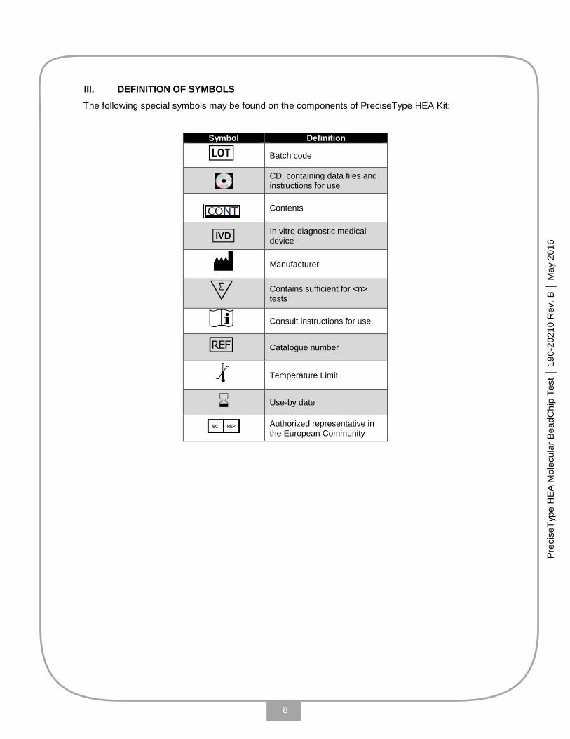
III. DEFINITION OF SYMBOLS
The following special symbols may be found on the components of PreciseType HEA Kit:
Symbol Definition
g Batch code
CD, containing data files and instructions for use
Contents
V In vitro diagnostic medical device
M Manufacturer
X Contains sufficient for <n> tests
i Consult instructions for use
h Catalogue number
l Temperature Limit
H Use-by date
P Authorized representative in the European Community

9
Prec
iseT
ype
HE
A M
olec
ular
Bea
dChi
p Te
st │
190
-202
10 R
ev. B
│ M
ay 2
016
IV. WARNINGS AND PRECAUTIONS
In samples of human origin, there is still a potential risk of infection even after DNA extraction. Handle samples using universal precautions. Use appropriate personal protective equipment throughout the test procedure, including gloves, eye protection and lab coat. In the event of contact with eyes, rinse immediately with plenty of water and seek medical advice. For additional safety information please refer to the website: http://extranet.immucor.com
Never pipette by mouth. Avoid contact of reagents and specimens with skin and mucous membranes.
Dispose of used materials in accordance with the institution’s and local regulations for disposal of potential biohazardous materials. Spillage of potentially infectious material should be cleaned and disposed of immediately in accordance with the institution’s policy and procedure for the handling and disposal of potentially biohazardous materials.
PCR technology is susceptible to contamination, especially from its own product. Aerosols of PCR amplicons that are generated during the post-PCR steps are a frequent source of contamination.
Take care to prevent excessive splashing and generation of aerosols.
Follow good laboratory practices for molecular laboratories when using the kit, including wiping of work surfaces before processing or preparing PCR samples with a freshly prepared 10% bleach (or equivalent), use of ultraviolet (UV) light in hoods or biosafety cabinets in between use, space and time separation of pre- and post-PCR activities, use of aliquoted PCR reagents, use of Positive and Negative Controls, etc.
Consistent, careful technique coupled with liberal incorporation and monitoring of controls will ensure a vigilant, proactive approach to control and monitoring of PCR contamination. (See section VII).
Operators must participate in the PreciseType HEA Molecular BeadChip Test Training Program prior to performing this assay in order to assure consistent and accurate test results.
Laboratories should validate their own cleaning procedures.
Contamination of reagents or specimens may cause erroneous results; therefore, take care to avoid contaminating this product during use. Do not use reagents if you suspect that they may have been contaminated.
Microbial contamination of reagents or specimens may lead to incorrect results.
Use the kit reagents and HEA BeadChip Carriers as supplied. Dilution or alteration may generate erroneous results.
Do not mix reagents or HEA BeadChip Carriers between different lots.
Do not use leaking or unlabeled vials.
Previously frozen samples or reagents should be mixed thoroughly and then centrifuged after thawing prior to testing. Avoid generating foam and bubbles in the samples.

10
Prec
iseT
ype
HE
A M
olec
ular
Bea
dChi
p Te
st │
190
-202
10 R
ev. B
│ M
ay 2
016
Keep all enzymes and master mixes on ice or cryoblock (2 to 8°C) during use.
Ensure that sample tubes ae properly sealed prior to amplification to prevent evaporation.
Due to inherent differences in the mechanisms of thermal cycler performance, variation in results can occur when set thermal profiles are transferred between different makes and models of thermal cycler instruments. In some cases, reaction specificity and sensitivity can be compromised, leading to the false interpretation and reporting of data. The FDA-approved PreciseType HEA IVD test requires use of the Applied Biosystems Veriti Dx thermal cycler. Immucor makes no assurance for assay performance with the use of alternate thermal cyclers and profiles, which must be validated by the user.
Samples must remain in the BeadChip reaction well during testing.
Incubation times or temperatures other than those specified may give erroneous results.
On each day of use, prior to operating the AIS 400, users must perform the Exposure Test Carrier (ETC) procedure to verify AIS performance. If the Exposure Test fails, please contact technical support for appropriate instructions. (See AIS User Manual 190-20185).
Deviation from the recommended directions for use may result in less-than-optimal product performance. Depending upon the nature and severity of the deviation assay failure (individual sample as well as run failures) and/or erroneous results may occur. For example, we have determined that use of insufficient/inactive Clean-up Reagent in the assay may result in high incidence of false Kp(a)+ calls.
The results from the mutation HgbS in the Beta Globin gene are not for diagnosis of sickle cell disease.
V. SHIPPING, STORAGE AND STABILITY
The PreciseType HEA reagents, including the PCR mix and all enzymes, are shipped on dry ice.
When the kit is received, verify that there is dry ice remaining in the package. If no ice is present,
do not use the kit and contact Technical Support. In addition, please contact Technical Support if
the vacuum-sealed HEA BeadChip Carrier pouch has been opened or damaged during transit.
Store all test reagents, including the PCR mix and all enzymes, at -20°C to -80°C in a defrost-free
freezer. Use benchtop cryoblocks when possible. When stored under these conditions and
handled correctly, unopened reagents can be used until the expiration date. Once opened, the
contents of a properly stored reagent kit may be used for six months or until the labeled expiration
date, whichever occurs earlier. When opening reagent kits, users should determine which date
would occur earlier; if the six-month use date is earlier than the labeled expiration date, record
this earlier date directly on the kit to ensure that reagents are not used beyond their expiration
date.

11
Prec
iseT
ype
HE
A M
olec
ular
Bea
dChi
p Te
st │
190
-202
10 R
ev. B
│ M
ay 2
016
Store the HEA BeadChip Carriers at 2 to 8°C until use. Unused carriers should be returned
immediately to storage at 2 to 8°C in their original packaging. HEA BeadChip Carriers cannot be
reused.
Refer to the expiration date of all kit components. Do not use beyond the expiration date. The
format of the expiry date is YYYY-MM-DD, which indicates allowable usage through the day
indicated. Components of this kit can have expiration dating that is greater than the expiration
date of the entire kit. The shortest shelf-life (i.e., earliest expiration date) of any component in the
kit will be indicated on the outermost kit label.
VI. SPECIMEN COLLECTION AND PREPARATION
Sample: Whole blood samples must be drawn into EDTA anticoagulant tubes (e.g. BD Product Numbers 366643, 368661, 367654). The PreciseType HEA assay has not been tested with cord blood or cadaveric blood. (see also section on Interfering Substances).
DNA samples should be extracted using the QlAamp DSP DNA Blood Mini Kit (QIAGEN cat# 61104) following manufacturer’s instructions for use. Use of alternative procedures requires validation by the customer.
Storage: Genomic DNA must be stored at -20°C or colder in a defrost-free freezer until use. Avoid multiple freeze/thaw cycles.
Interfering Substances: Presence of PCR inhibitors such as citrate [9], heparin [9], hemoglobin, ethanol, etc. can interfere with the PCR reaction.
DNA Quantity: A concentration ≥ 15 ng/µL of extracted genomic DNA is required for optimal performance.
12
Prec
iseT
ype
HE
A M
olec
ular
Bea
dChi
p Te
st │
190
-202
10 R
ev. B
│ M
ay 2
016
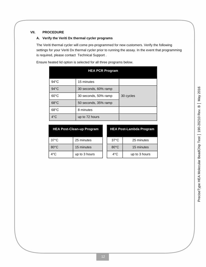
VII. PROCEDURE
A. Verify the Veriti Dx thermal cycler programs
The Veriti thermal cycler will come pre-programmed for new customers. Verify the following settings for your Veriti Dx thermal cycler prior to running the assay. In the event that programming is required, please contact Technical Support .
Ensure heated lid option is selected for all three programs below.
HEA PCR Program
94°C 15 minutes
94°C 30 seconds, 60% ramp
30 cycles 60°C 30 seconds, 50% ramp
68°C 50 seconds, 35% ramp
68°C 8 minutes
4°C up to 72 hours
HEA Post-Clean-up Program HEA Post-Lambda Program
37°C 25 minutes 37°C 25 minutes
80°C 15 minutes 80°C 15 minutes
4°C up to 3 hours 4°C up to 3 hours
13
Prec
iseT
ype
HE
A M
olec
ular
Bea
dChi
p Te
st │
190
-202
10 R
ev. B
│ M
ay 2
016
B. Procedural Notes
To reduce or eliminate the chances of carryover contamination, users should assign three (3) separate laboratory areas, including: (1) pre-PCR /set-up activities, (2) DNA Addition, and (3) post-PCR procedures.
• Steps in Section C, “PCR Master Mix Preparation,” should be performed in the pre-PCR area, within a PCR Workstation hood or clean room, using aerosol-resistant (filtered) pipette tips.
• Steps in Section D, “DNA Addition Step,” are recommended to be performed in the DNA-Addition area within a dedicated hood or dead air box, using aerosol-resistant (filtered) pipette tips.
• The remainder of the procedure after Section E, “PCR Amplification,” should be performed in the post-PCR area.
Prior to use, wipe down the processing area surfaces with 10% Bleach and/or DNA AWAY, including:
• Bench-tops and inside hood surfaces
• Supportive equipment
• All working pipettes
• Inside mini mouse centrifuge lid, tube racks and covers
• Thermal cycler and plate centrifuge surfaces
• Clean inside lid and thermal cycler plate wells using DNA Away (or equivalent) and rinsing with deionized water. Use 10% bleach solution for removing contamination from the Veriti™ instrument sample block(s); excessive use of the solution, however, can corrode the sample block(s) material.
Prior to using the hood, turn on the UV light for a minimum of 15-20 minutes.
Remove the required quantities of reagents, samples, and controls from storage and, if frozen, allow them to thaw prior to use. Return unused portions to proper storage immediately.
Use filtered tips for all pipetting steps in the procedural steps of this assay.
Multi-channel or single-channel pipettes may be used, depending upon laboratory preference. All pipettes used must be calibrated. The quantity of reagents supplied with each PreciseType test kit is sufficient for pipetting the quantities required within this procedure.
Precise pipetting of samples and reagents is required for accurate results.
Take care to mix samples and reagents adequately. Avoid foaming.
Combine working reagents just prior to use.
14
Prec
iseT
ype
HE
A M
olec
ular
Bea
dChi
p Te
st │
190
-202
10 R
ev. B
│ M
ay 2
016
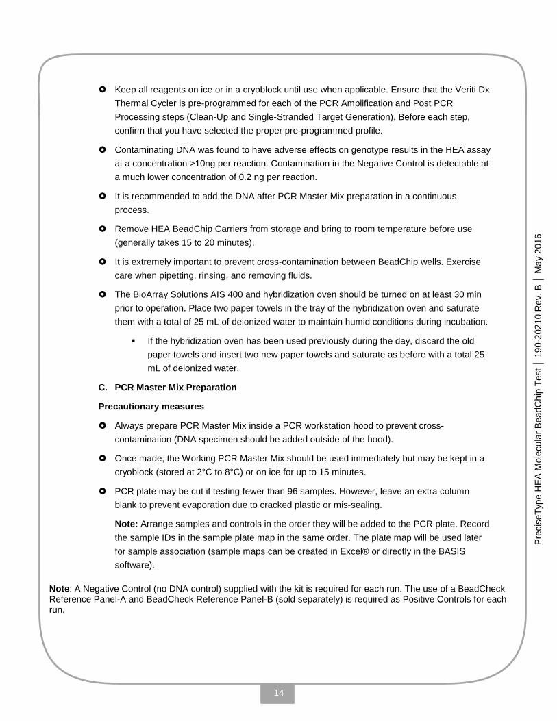
Keep all reagents on ice or in a cryoblock until use when applicable. Ensure that the Veriti Dx Thermal Cycler is pre-programmed for each of the PCR Amplification and Post PCR Processing steps (Clean-Up and Single-Stranded Target Generation). Before each step, confirm that you have selected the proper pre-programmed profile.
Contaminating DNA was found to have adverse effects on genotype results in the HEA assay at a concentration >10ng per reaction. Contamination in the Negative Control is detectable at a much lower concentration of 0.2 ng per reaction.
It is recommended to add the DNA after PCR Master Mix preparation in a continuous process.
Remove HEA BeadChip Carriers from storage and bring to room temperature before use (generally takes 15 to 20 minutes).
It is extremely important to prevent cross-contamination between BeadChip wells. Exercise care when pipetting, rinsing, and removing fluids.
The BioArray Solutions AIS 400 and hybridization oven should be turned on at least 30 min prior to operation. Place two paper towels in the tray of the hybridization oven and saturate them with a total of 25 mL of deionized water to maintain humid conditions during incubation.
If the hybridization oven has been used previously during the day, discard the old paper towels and insert two new paper towels and saturate as before with a total 25 mL of deionized water.
C. PCR Master Mix Preparation
Precautionary measures
Always prepare PCR Master Mix inside a PCR workstation hood to prevent cross-contamination (DNA specimen should be added outside of the hood).
Once made, the Working PCR Master Mix should be used immediately but may be kept in a cryoblock (stored at 2°C to 8°C) or on ice for up to 15 minutes.
PCR plate may be cut if testing fewer than 96 samples. However, leave an extra column blank to prevent evaporation due to cracked plastic or mis-sealing.
Note: Arrange samples and controls in the order they will be added to the PCR plate. Record the sample IDs in the sample plate map in the same order. The plate map will be used later for sample association (sample maps can be created in Excel® or directly in the BASIS software).
Note: A Negative Control (no DNA control) supplied with the kit is required for each run. The use of a BeadCheck Reference Panel-A and BeadCheck Reference Panel-B (sold separately) is required as Positive Controls for each run.
15
Prec
iseT
ype
HE
A M
olec
ular
Bea
dChi
p Te
st │
190
-202
10 R
ev. B
│ M
ay 2
016
Assay Procedure
1. Reagent & sample preparation
1.1 Remove PCR Mix (yellow cap) from the freezer and thaw at room temperature. Put in cryoblock (2 to 8°C) when fully thawed. Thawing the PCR Mix should take about 15 to 30 minutes depending on volume. Vortex and centrifuge briefly before use (approximately 3 to 5 seconds).
1.2 Remove HotStarTaq DNA Polymerase (orange cap) from the freezer and place in cryoblock (2 to 8°C) or on ice. Vortex and centrifuge briefly before use (approximately 3 to 5 seconds).
1.3 Bring DNA specimen to room temperature. Vortex and centrifuge briefly before use (approximately 3 to 5 seconds).
2. PCR Master Mix preparation (in PCR workstation hood or clean room)
2.1 Determine the number of samples and controls to be run.
2.2 Label a 1.5 or 2.0 mL microcentrifuge tube for PCR Master Mix.
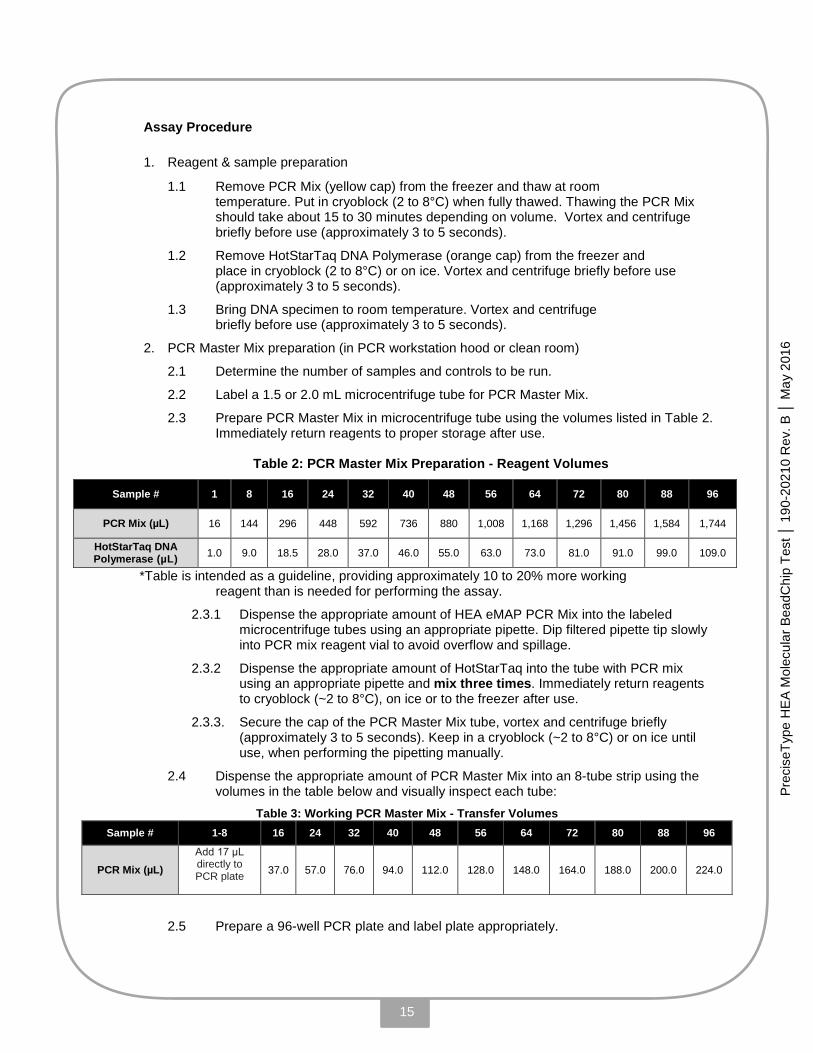
2.3 Prepare PCR Master Mix in microcentrifuge tube using the volumes listed in Table 2. Immediately return reagents to proper storage after use.
Table 2: PCR Master Mix Preparation - Reagent Volumes
Sample # 1 8 16 24 32 40 48 56 64 72 80 88 96
PCR Mix (µL) 16 144 296 448 592 736 880 1,008 1,168 1,296 1,456 1,584 1,744
HotStarTaq DNA Polymerase (µL) 1.0 9.0 18.5 28.0 37.0 46.0 55.0 63.0 73.0 81.0 91.0 99.0 109.0
*Table is intended as a guideline, providing approximately 10 to 20% more working reagent than is needed for performing the assay.
2.3.1 Dispense the appropriate amount of HEA eMAP PCR Mix into the labeled microcentrifuge tubes using an appropriate pipette. Dip filtered pipette tip slowly into PCR mix reagent vial to avoid overflow and spillage.
2.3.2 Dispense the appropriate amount of HotStarTaq into the tube with PCR mix using an appropriate pipette and mix three times. Immediately return reagents to cryoblock (~2 to 8°C), on ice or to the freezer after use.
2.3.3. Secure the cap of the PCR Master Mix tube, vortex and centrifuge briefly (approximately 3 to 5 seconds). Keep in a cryoblock (~2 to 8°C) or on ice until use, when performing the pipetting manually.
2.4 Dispense the appropriate amount of PCR Master Mix into an 8-tube strip using the volumes in the table below and visually inspect each tube:
Table 3: Working PCR Master Mix - Transfer Volumes Sample # 1-8 16 24 32 40 48 56 64 72 80 88 96
PCR Mix (µL) Add 17 μL directly to PCR plate
37.0 57.0 76.0 94.0 112.0 128.0 148.0 164.0 188.0 200.0 224.0
2.5 Prepare a 96-well PCR plate and label plate appropriately.

16
Prec
iseT
ype
HE
A M
olec
ular
Bea
dChi
p Te
st │
190
-202
10 R
ev. B
│ M
ay 2
016
2.6 Dispense 17.0 µL of PCR Master Mix into the bottom of each well of the labeled PCR plate in a cryoblock (2 to 8°C) or on ice (an appropriate pipette may be used), when performing the pipetting manually. Discard unused PCR Master Mix
D. DNA Sample Addition
Precautionary measures
Turn on the thermal cycler approximately 10 minutes prior to beginning the PCR cycle.
Begin to add DNA and controls to working PCR Master Mix outside of the hood as soon as possible, but within 15 to 20 minutes when performing the pipetting manually.
When performing the pipetting manually, keep samples and working PCR Master Mix in a cryoblock (stored at 2 to 8°C) or on ice during these steps.
Note: Verify sample order and identification on sample plate map.
Assay procedure
1. Vortex and centrifuge DNA specimen, if not already done.
2. Add 8.0 µL of required negative control (supplied in PreciseType HEA reagent kit box) to the appropriate PCR plate well using an appropriate pipette.
3. Add 8.0 µL of required positive controls (Cat. # 800-20236) into appropriate PCR plate wells using an appropriate pipette.
4. Add 8.0 µL of DNA sample into appropriate PCR plate wells using an appropriate pipette. Mix three times by pipette aspiration to ensure complete transfer of DNA.
5. Seal PCR plate securely with thermal adhesive plate seal. Ensure all wells are sealed completely to prevent evaporation.
6. Gently vortex for approximately 3-5 seconds.
7. Centrifuge briefly at approximately 1,000 rpm to bring samples to the bottom of wells.

17
Prec
iseT
ype
HE
A M
olec
ular
Bea
dChi
p Te
st │
190
-202
10 R
ev. B
│ M
ay 2
016
E. PCR Amplification
Precautionary measures
The centrifuged amplified samples and controls should be used immediately, but may be stored at −20°C (or colder) for up to 4 weeks.
Assay procedure
1. Place the PCR plate in the center of the thermal cycler.
2. Close the lid and push handle down.
3. Log into thermal cycler and select “Browse/New Methods.”
4. Select “HEA PCR” program and press “View” to verify the program is correct:
Cycles 1 30 1 1
Temp (C°) 94°C 94°C 60°C 68°C 68°C 4°C
Time 15 min 30 sec 30 sec 50 sec 8 min Until removal (no longer than
72 hours)
Ramp 100% 60% 50% 35% 100% 100%
5. Press “Run.” Reaction volume should be 25.0 µL. Enable the heated lid function with the temperature set at 105°C.
6. Press “Start Run Now” to begin the process and verify that the lid is heating and the program has initiated.
7. Remove PCR plate from the thermal cycler once the program reaches 4°C and centrifuge briefly (approximately 1,000 rpm for 5 seconds). PCR plate must be removed from the thermal cycler at 4°C within 72 hours.

18
Prec
iseT
ype
HE
A M
olec
ular
Bea
dChi
p Te
st │
190
-202
10 R
ev. B
│ M
ay 2
016
F. Post-PCR Processing: Clean-up
Precautionary measures
The steps in Section F should be performed continuously without interruption in the post-PCR area when performing the pipetting manually.
When performing the pipetting manually, the Clean-up Reagent should be added to the post-PCR product within approximately 30 minutes of removing PCR products from the thermal cycler after PCR amplification, or if frozen, within 30 minutes of thawing.
The PCR plate may be cut if testing fewer than 96 samples. However, leave an extra column blank to prevent evaporation due to cracked plastic or mis-sealing.
Clean-up Reagent and PCR product should be thawed at room temperature and kept in a cryoblock (~2 to 8°C) or on ice until use.
When transferring the post-PCR product and Clean-up Reagent into new post-PCR plates, keep in a cryoblock (stored at 2 to 8°C) or on ice during these steps when performing the pipetting manually.
The centrifuged cleaned-up samples and controls may be used immediately, or stored at -20°C (or colder) for up to 72 hours.
Assay procedure
1. Remove Clean-up Reagent (green cap) from the freezer 10-15 minutes before use and place in a cryoblock (2 to 8°C) or on ice when performing the pipetting manually.
2. Gently mix and centrifuge (approximately 1,000 rpm for 5 seconds) PCR products before use. Place in a cryoblock (2 to 8°C) or on ice when performing the pipetting manually.
3. Determine the number of samples and controls to be run.
4. Prepare a new PCR plate for post-PCR processing and label it appropriately.
5. Position the PCR Product plate in the same orientation as the Post-PCR plate in a cryoblock (2 to 8°C) when performing the pipetting manually.
6. Transfer 6.5µL of each PCR product to the bottom of the appropriate well of the new Post-PCR plate using an appropriate pipette.
7. Seal the plate with the remaining PCR products and store in the freezer at -20 to -80°C until successful completion of assay run.
8. When performing the pipetting manually, use the table below to determine the appropriate volume, then dispense Clean-up Reagent into an 8-tube-strip with an appropriate pipette and visually inspect each tube. Immediately return reagents to proper storage after use.
Table 4: Clean-up Reagent Volumes
Sample # 8 16 24 32 40 48 56 64 72 80 88 96
Clean-up Reagent (µL) Add 2 μL directly to Post-PCR
plate
6.0 9.0 12.0 14.0 16.0 18.0 20.0 22.0 24.0 26.0 28.0
9. Dispense 2.0 µL of Clean-up Reagent into each well of the post-PCR plate using an appropriate pipette. Mix each well three times by pipette aspiration.
10. After adding Clean-up Reagent to all sample wells in the Post-PCR plate, discard the 8-tube strip.

19
Prec
iseT
ype
HE
A M
olec
ular
Bea
dChi
p Te
st │
190
-202
10 R
ev. B
│ M
ay 2
016
11. Seal Post-PCR plate securely with thermal adhesive plate seal.
12. Gently vortex for approximately 3-5 seconds.
13. Briefly centrifuge at approximately 1,000 rpm to bring samples to the bottom of the wells.
14. Place the Post-PCR plate in the center of the thermal cycler.
15. Close the lid and push the handle down.
16. Log into the thermal cycler and select “Browse/New Methods.” 17. Select the HEA Clean-up program and press “View” to verify that the program is
correct:
Cycles 1 1 1
Temp (C°) 37°C 80°C 4°C
Time 25 min 15 min Until removal (no longer than 3 hours)
Ramp 100% 100% 100%
18. Press “Run.” Reaction volume should be 10.0 µL. Enable the heated lid function with the temperature set at 105°C.
19. Press “Start Run Now” to begin the process and verify that the lid is heating and the program has initiated.
20. Remove PCR plate from the thermal cycler once the program reaches 4°C and centrifuge briefly (approximately 1,000 rpm for 5 seconds). PCR plate must be removed from the thermal cycler at 4°C within 3 hours. The centrifuged amplified samples and controls should be used immediately, but may be stored at -20°C (or colder) for up to 72 hours.
20
Prec
iseT
ype
HE
A M
olec
ular
Bea
dChi
p Te
st │
190
-202
10 R
ev. B
│ M
ay 2
016
G. Post-PCR Processing: Single-Stranded Target Generation
Precautionary measures
The steps in Section G should be performed continuously and without interruption.
Lambda Exonuclease should be removed from the freezer to thaw during the incubation of the post-PCR Clean-up Reagent step in Section F. Once thawed, the Lambda Exonuclease should be kept in a cryoblock (stored at 2 to 8°C) or on ice during transfer to the post- PCR Clean-up Reagent product when performing the pipetting manually.
The post-PCR Clean-up Reagent product should be kept in a cryoblock (stored at 2 to 8°C) or on ice during these steps once thawed (if previously frozen).
Add the Lambda Exonuclease to the post- PCR Clean-up Reagent product within approximately 30 minutes of completing the clean-up process when performing the pipetting manually.
The centrifuged single-stranded samples and controls should be used immediately, but may be stored at -20°C (or colder) for up to 72 hours.
Assay procedure
1. Remove Lambda Reagent (violet cap) from the freezer 10-15 minutes before use. Thaw at room temperature.
2. Briefly mix and centrifuge Clean-up products at room temperature before use. Mix reagents with vortex mixer at mid-range speed for 3-5 seconds. Briefly spin down tubes/plates at approximately 1,000 rpm.
3. Place Post-PCR plate (“Clean-up Products”) in a cryoblock (2 to 8°C) when performing the test manually.
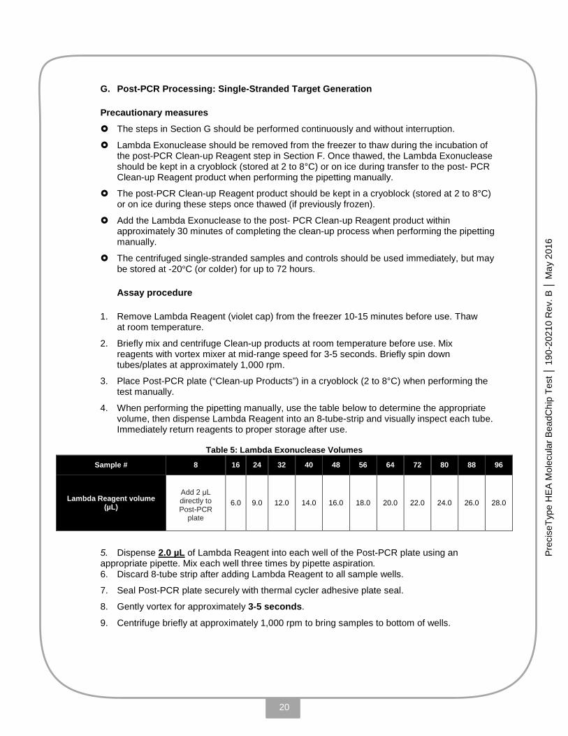
4. When performing the pipetting manually, use the table below to determine the appropriate volume, then dispense Lambda Reagent into an 8-tube-strip and visually inspect each tube. Immediately return reagents to proper storage after use.
Table 5: Lambda Exonuclease Volumes
Sample # 8 16 24 32 40 48 56 64 72 80 88 96
Lambda Reagent volume (µL)
Add 2 μL directly to Post-PCR
plate
6.0 9.0 12.0 14.0 16.0 18.0 20.0 22.0 24.0 26.0 28.0
5. Dispense 2.0 µL of Lambda Reagent into each well of the Post-PCR plate using an appropriate pipette. Mix each well three times by pipette aspiration. 6. Discard 8-tube strip after adding Lambda Reagent to all sample wells.
7. Seal Post-PCR plate securely with thermal cycler adhesive plate seal.
8. Gently vortex for approximately 3-5 seconds.
9. Centrifuge briefly at approximately 1,000 rpm to bring samples to bottom of wells.
21
Prec
iseT
ype
HE
A M
olec
ular
Bea
dChi
p Te
st │
190
-202
10 R
ev. B
│ M
ay 2
016
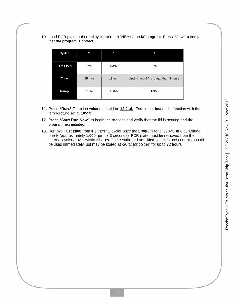
10. Load PCR plate to thermal cycler and run “HEA Lambda” program. Press “View” to verify that the program is correct:
Cycles 1 1 1
Temp (C°) 37°C 80°C 4°C
Time 25 min 15 min Until removal (no longer than 3 hours)
Ramp 100% 100% 100%
11. Press “Run.” Reaction volume should be 12.0 µL. Enable the heated lid function with the temperature set at 105°C.
12. Press “Start Run Now” to begin the process and verify that the lid is heating and the program has initiated.
13. Remove PCR plate from the thermal cycler once the program reaches 4°C and centrifuge briefly (approximately 1,000 rpm for 5 seconds). PCR plate must be removed from the thermal cycler at 4°C within 3 hours. The centrifuged amplified samples and controls should be used immediately, but may be stored at -20°C (or colder) for up to 72 hours.
22
Prec
iseT
ype
HE
A M
olec
ular
Bea
dChi
p Te
st │
190
-202
10 R
ev. B
│ M
ay 2
016
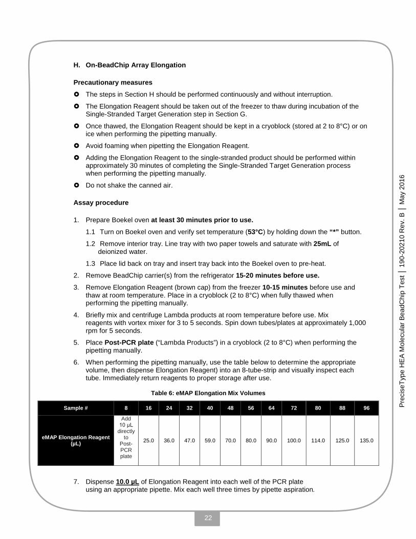
H. On-BeadChip Array Elongation
Precautionary measures
The steps in Section H should be performed continuously and without interruption.
The Elongation Reagent should be taken out of the freezer to thaw during incubation of the Single-Stranded Target Generation step in Section G.
Once thawed, the Elongation Reagent should be kept in a cryoblock (stored at 2 to 8°C) or on ice when performing the pipetting manually.
Avoid foaming when pipetting the Elongation Reagent.
Adding the Elongation Reagent to the single-stranded product should be performed within approximately 30 minutes of completing the Single-Stranded Target Generation process when performing the pipetting manually.
Do not shake the canned air.
Assay procedure
1. Prepare Boekel oven at least 30 minutes prior to use.
1.1 Turn on Boekel oven and verify set temperature (53°C) by holding down the “*” button.
1.2 Remove interior tray. Line tray with two paper towels and saturate with 25mL of deionized water.
1.3 Place lid back on tray and insert tray back into the Boekel oven to pre-heat.
2. Remove BeadChip carrier(s) from the refrigerator 15-20 minutes before use.
3. Remove Elongation Reagent (brown cap) from the freezer 10-15 minutes before use and thaw at room temperature. Place in a cryoblock (2 to 8°C) when fully thawed when performing the pipetting manually.
4. Briefly mix and centrifuge Lambda products at room temperature before use. Mix reagents with vortex mixer for 3 to 5 seconds. Spin down tubes/plates at approximately 1,000 rpm for 5 seconds.
5. Place Post-PCR plate (“Lambda Products”) in a cryoblock (2 to 8°C) when performing the pipetting manually.
6. When performing the pipetting manually, use the table below to determine the appropriate volume, then dispense Elongation Reagent) into an 8-tube-strip and visually inspect each tube. Immediately return reagents to proper storage after use.
Table 6: eMAP Elongation Mix Volumes
Sample # 8 16 24 32 40 48 56 64 72 80 88 96
eMAP Elongation Reagent (µL)
Add 10 μL
directly to
Post-PCR plate
25.0 36.0 47.0 59.0 70.0 80.0 90.0 100.0 114.0 125.0 135.0
7. Dispense 10.0 µL of Elongation Reagent into each well of the PCR plate using an appropriate pipette. Mix each well three times by pipette aspiration.

23
Prec
iseT
ype
HE
A M
olec
ular
Bea
dChi
p Te
st │
190
-202
10 R
ev. B
│ M
ay 2
016
8. Discard 8-tube strip after adding Elongation Reagent to all sample wells.
9. Remove BeadChip carrier(s) from foil pouch and number the carriers in numerical order.
10. Carefully place the BeadChip carrier(s) on the BeadChip carrier holder without touching the surface of the BeadChip.
11. Record BeadChip carrier ID(s) to the sample plate map accordingly.
12. Transfer 15.0 µL of the Elongation Reaction mixture to the corresponding BeadChip using an appropriate pipette. Visually inspect pipette tips during process and ensure that carrier orientation is the same as in the sample plate map.
13. Remove preheated tray from Boekel oven. If there is condensation on the inside of the tray cover, dry it with a lint-free tissue.
14. Carefully place BeadChip holder into Boekel oven tray, placing tray cover completely over the tray.
15. Load tray in the oven, tighten the latch securely, close the Boekel oven door, and set timer for 30 minutes. Incubate the BeadChip carriers at 53±1°C for 30 minutes. (Please note: if you intend to read the carrier immediately following elongation, turn on the BioArray AIS 400 Array Imaging System and load the CD when starting the incubation process.)
16. Remove BeadChip holder from the oven after 30 minutes.
17. Wash the elongation mixture from BeadChip surfaces using deionized water wash bottle:
17.1 Hold the BeadChip carrier so that its surface is vertical over a sink or catch basin.
17.2 Rinse each BeadChip individually for approximately 3 seconds. The water stream should be directed perpendicular to the BeadChip surface approximately one-inch away.
18. Remove excess water from BeadChip surfaces using compressed/canned air. Caution! Do not shake canned air!
19. Remove any remaining water from the back of the carrier(s) with a lint-free tissue.
20. If you cannot read the washed and dried HEA BeadChip carriers immediately following incubation, they may be stored protected from light up to 72 hours at room temperature before reading with the BioArray AIS 400 system.
24
Prec
iseT
ype
HE
A M
olec
ular
Bea
dChi
p Te
st │
190
-202
10 R
ev. B
│ M
ay 2
016
I. BeadChip Image Acquisition
Precautionary measures
Turn on the BioArray AIS 400 light source and computer at least 30 minutes prior to use.
Load PreciseType BeadChip Test | HEA Data CD once per lot.
Assay procedure
1. Open and initialize AISR program on the desktop.
2. Run the ETC (Refer to AIS User Manual 190-20185). Contact Technical Support if results are out of specifications to adjust the exposure time prior to proceeding.
3. Remove PreciseType BeadChip Test | HEA Data CD from BeadChip carrier box and load files from CD onto the computer for each new lot.
4. Read the HEA BeadChip carrier(s) using the BioArray AIS 400 Array Imaging System. Process BeadChip data using the HEA Analysis software in BASIS.
5. Properly shutdown AISR and the light source after use.
6. Continue to BASIS to perform sample association and generate BeadChip reports.

25
Prec
iseT
ype
HE
A M
olec
ular
Bea
dChi
p Te
st │
190
-202
10 R
ev. B
│ M
ay 2
016
VIII. EXPECTED RESULTS
A. Evaluation - Quality Control
The BASIS software determines run and sample validity automatically.
Run Validity: Two Positive and one Negative Control as supplied, are required for each run. The results for all controls must meet the Run Validity criteria. If any one of the controls does not meet any one criterion, the run is invalid and must be repeated.
If the results for Low Signal (LS) is <32 in the phenotype report for the Negative Control sample, it may indicate contamination by gDNA in a quantity that may impact test results. When this occurs, all sample results in the run are invalid.
The phenotype pattern of the two positive control samples must match the expected phenotype pattern. If either one of the control results do not meet any one criterion, all sample results in the run are invalid. See PreciseType BeadCheck Package Insert (P/N 190-20229) for more details.
See the BASIS AM 4G User Manual (190-20331) for more information on the BASIS software and for examples of how sample results are displayed for valid and invalid runs.
Interpretation of validity of the Positive and Negative Controls is described in Table 7: Table 7: Run Validity Criteria
Control BASIS Analysis Result Reported Interpretation Negative LS ≥ 32 Valid NC Valid Negative Control Negative LS < 32 Invalid NC Invalid Negative
Control; No results reported for all samples in the run
Positive Phenotype pattern matches pattern expected for the two positive controls from the BeadCheck kit
Valid HEA Ref-pA and
Valid HEA Ref-pB
Valid Positive Control
Positive Phenotype pattern does not match expected pattern for either one of the two positive controls from the BeadCheck kit
Invalid HEA Ref-pA and/or
Invalid HEA Ref-pB
Invalid Positive Control; No results reported for all samples in the run

26
Prec
iseT
ype
HE
A M
olec
ular
Bea
dChi
p Te
st │
190
-202
10 R
ev. B
│ M
ay 2
016
Sample Results Validity: For sample results to be valid, the phenotype results for all antigens must be valid (for a listing of causes of invalid sample results see Table 8 below). See the BASIS AM 4G User Manual (190-20331, Sections 6 and 7) for more information on the BASIS software and for examples of how valid and invalid sample results are displayed.
If a sample result shows an antigen phenotype of either Indeterminate Call (IC) or Low Signal (LS), the sample is invalid, except for S and s where LS is an expected phenotype in conjunction with U negative results. If the sample has a status of High Background (HB) or High Coefficient of Variation (CV), the sample is invalid (for a list of causes for invalid sample see Table 8 below, for a more detailed explanation see section XII Possible Warning Messages Found in BASIS Report).
Table 8: Causes for Invalid Sample Result Cause Interpretation IC ≥ 1 Indeterminate Call LS ≥ 1 Low Signal
HB High Background CV High Coefficient of Variance
For samples that are part of an invalid run or have invalid phenotype(s) themselves (sample results invalid), all antigen phenotype results are reported as No Type Determined (NTD).
B. Analysis of Results
This is a qualitative test. BASIS computes BeadChip array signal intensity data on each oligonucleotide detecting specific alleles to determine the presence or absence of each allele or the genotype result. The genotype results are then used to compute the predicted antigen phenotype results.
The HEA-analysis software in BASIS performs all calculations automatically. Please refer to the BASIS User Manual (P/N 190-20331) for more details. Expected genotype results are shown in Table 10 below (for a more detailed explanation of Ax and xB results see section XII on Troubleshooting).
For samples that are part of an invalid run or have invalid genotype(s) themselves (sample invalid), all genotype results are reported as No Type Determined (NTD).

27
Prec
iseT
ype
HE
A M
olec
ular
Bea
dChi
p Te
st │
190
-202
10 R
ev. B
│ M
ay 2
016
C. Phenotype and Genotype
For samples with valid results, the expected phenotype results are shown below in Table 9 and the expected genotype results are shown below in Table 10.
Table 9: Expected Phenotype Results
Result Reported Interpretation
+ Positive 0 Negative
(+)* Possible (C)ces haplotype (0)* Fyb variant PV Possible Variant var U variant (S silencing mutation) w Fyb Weak ++ HbS homozygous
Table 10: Expected Genotype Results
Result Reported Interpretation
AA Homozygous for A AB Heterozygous Ax Indeterminate call on B BB Homozygous for B xB Indeterminate call on A IC Indeterminate call on A and B
Note: For the RhCE-109Insert, a positive amplicon control corresponds to probe “A” and a 109-bp insertion specific probe corresponds to probe “B”, therefore:
If the RhCE-109Ins = AA, the 109-bp insertion is absent, indicating C-
If the RhCE-109Ins = AB, the 109-bp insertion is present in one of the alleles, indicating C+
• There is no RhCE-109Ins = BB since the positive control is always present
When P103 is positive (RhCE-P103S = AA, Ax, or AB), the RhCE-109Ins probe is used for prediction of C phenotype:
• If the RhCE-109Ins = AA, the phenotype = cc
• If the RhCE-109Ins = AB, the phenotype = cC
When RhCE-P103S = BB, the phenotype = CC, regardless of RhCE-109Ins status

28
Prec
iseT
ype
HE
A M
olec
ular
Bea
dChi
p Te
st │
190
-202
10 R
ev. B
│ M
ay 2
016
IX. LIMITATIONS OF PROCEDURE
False negative and/or invalid results may be generated when unanticipated rare mutation(s) affecting the primer or probe binding cause allele and/or amplicon dropout.
• Presence of RH hybrids and variant mutations in exons 2, 5 and 7 as well as introns 1, 2, 4, 5, 6, and 7 of the RHCE gene can interfere with the detection E/e and C/c antigen. Mutations in RHCE gene leading to the ceMO phenotype[12],[13], which expresses as a weak Rhe, can cause a direct suppression of the Rhe probe and may cause an invalid or false negative result . In select populations, such as Afro-Caribbean patients with sickle cell disease, the prevalence for ceMO phenotype has been reported to be up to 2 % [18].
• Presence of a rare +3g>a change in intron 5 of GYPB interferes with the detection of the S antigen and may lead to a false negative typing of the S antigen [16].
• The mutation HgbS in the beta globin gene should not be used for determination of sickle cell disease. Presence of HbSC disease interferes with the detection of the HgbS mutation in the beta globin gene mutation and may result in invalid or inaccurate HbS phenotype call (HbS (++) instead of HbS (+)). In the United States, HbSC disease has a prevalence of 0.017% among African Americans [19]. Presence of some beta thalassemia disorders may interfere with the detection of the HgbS mutation in the beta globin gene and may result in invalid HbS phenotype call.
• Presence of Mit+(GYPB 161G>A) mutation may result in an invalid or false negative typing of the S antigen. The mutation has a prevalence of 0.1% in western Europeans [6].
• Presence of a GYPB mutation (c. 137-8C>T) may result in an invalid or false negative typing of the S antigen.
False positive and/or invalid results may be generated in rare cases where a sample contains examples of molecular events that affect the blood-group antigen expression and phenotypes and the nucleotide changes associated with these events are not explicitly monitored by the assay. Examples include DNA-sequence variations including premature stop codon, SNP leading to missense change in amino acid, hybrid genes, modifying genes; changes at the RNA transcription level including alternative splicing; reduced protein expression, etc. Known phenotypes are Knull, JKnull (JKnull has a prevalence of up to 9% among Polynesians [20]), Rhnull, Rh hybrids, Kmod, Co(a-,b-), In(Lu), Lu(a-,b-) and GP hybrids. Presence of a c.179_180del (Ser60fs) mutation linked with the Fy(b) allele may change the Fy(b) antigen expression and lead to a false positive result.
The BASIS software is not designed to convert all genotype/allele combinations into phenotype calls. For example, if allele combinations that have not been widely reported in the literature are encountered, the software will display a Possible Variant (PV) result.
The HEA test uses two point mutations to predict the V and VS antigen phenotypes: 733C>G(L245V) in exon5 and 1006G>T(G336C) in exon7 of the RHCE gene
The genotype-to-phenotype-prediction conversion rules employed by the HEA test are based on the established fact that the absence of the two mutations are correlated with the absence of V and VS antigens and that the presence of the mutations lead to antigen expression.
BioArray Solutions is aware of literature [14] that point to certain genotype combinations (involving the two mutations of interest) that do not lead to a unique phenotype. This limitation only affects the V(+)VS(+) call. As per the publication [14], in a small fraction of the cases, the HEA test would report the samples as falsely positive relative to serology when giving the V(+)VS(+) call (4.2% for VS and 1.4% for V). The V(-)VS(-) call is unaffected.
In the HEA test, the presence/absence of the RhC antigen is reported based on three polymorphisms 307C>T(P103S) in Exon 2, 733C>G (L245V) in Exon 5, 1006G>T (G336C) in Exon 7 and the presence/absence of 109 bp–insert in Intron 2 of the RHCE gene.
29
Prec
iseT
ype
HE
A M
olec
ular
Bea
dChi
p Te
st │
190
-202
10 R
ev. B
│ M
ay 2
016
The (+)* call on the RHC antigen implies the possible presence of altered C antigen encoded by the (C)ces haplotype. The (C)ces haplotype comprises:
i) A hybrid RHD-CE-D allele of the RHD gene, and
ii) ces allele of the RHCE gene
The (C)ces haplotype produces weak C, normal c, weak e (also known as es), and VS (RH20)[15].
The U antigen (located on the GPB protein) is not polymorphic by itself. The expression of the U antigen is governed by changes that affect the expression of the S antigens. Specifically, the S-s- phenotype is known to be associated with the absence or weak expression of the U antigen. The HEA test monitors three mutations that inform the S-s- phenotype and can call the U(var) and U(neg) phenotype. Occasionally, a U(neg) phenotype call may not be made even if the phenotype call is S-s- due to non-specific residual intensities on the probes governing the silencing of S/s antigen.
The Fyx allele encodes an amino acid change which causes Fy(b+w) phenotype with varying degrees of weakened Fyb antigen. Licensed serological anti-Fyb reagents may not always react with such a weakened Fyb antigen [17].
30
Prec
iseT
ype
HE
A M
olec
ular
Bea
dChi
p Te
st │
190
-202
10 R
ev. B
│ M
ay 2
016
X. SPECIFIC PERFORMANCE CHARACTERISTICS
A. Accuracy Study
BioArray performed a study to demonstrate that the HEA PreciseType Test can accurately
identify the phenotypes listed using pre-selected well-characterized samples. Red-blood-cell
(RBC) antigen phenotypes were determined using two methods. The RBC antigens characterized
using serology (licensed antisera) include Dia, Fya, Fyb, M, N, S, s, Jka, Jkb, Kpa, Kpb, Lub, C, c, E,
e, K, and k (U is inferred from S/s typing). The red blood cell antigens characterized using bi-
directional sequencing (corresponding licensed antisera are not available) include Coa, Cob, Dib,
Jsa, Jsb, Lua, LWa, LWb, V, VS, Sc1, Sc2, Doa, Dob, Joa, and Hy (also included is HgbS). Samples
were selected for phenotypic diversity to cover all antigen positive statuses and all but three
antigen negative statuses (Dib, LWa and Sc1).
To assure phenotypic diversity, the accuracy included combined data (unique valid samples) from
three different studies: a genotype-detection study, the clinical study described below and the
performance evaluation conducted in Europe. Some samples or sample results that were
collected from historical sources were not available for subsequent testing for discrepancy
resolution.
In order to be accepted, all phenotypes were to meet or exceed 99% at the lower bound of the
one-sided 95% confidence interval for accuracy (defined as overall agreement with the
comparison method). All antigens met the acceptance criteria with the exception of Lub and V,
which had lower bounds of 98.46% for Lub and 98.92% for V. Subsequent testing showed
complete concordance for V on PreciseType compared with bi-directional sequencing.
Subsequent testing was not available for Lub; the discrepancy for Lub between PreciseType and
serology may be due to In(Lu) or Lu(a-b-) as described in the Limitations of Procedure section.

31
Prec
iseT
ype
HE
A M
olec
ular
Bea
dChi
p Te
st │
190
-202
10 R
ev. B
│ M
ay 2
016
Table 11: PreciseType Accuracy Study Results
Antigen Samples Percent Correct Call Lower 95% Confidence Limit
c 1147 99.91% 99.59%
C 1146 100% 99.74%
e 1383 100% 99.78%
E 1383 100% 99.78%
K 1149 100% 99.74%
k 909 99.89% 99.48%
Kpa 657 100% 99.55%
Kpb 875 100% 99.66%
Jsa 1158 100% 99.74%
Jsb 1345 100% 99.78%
Jka 1124 100% 99.73%
Jkb 1123 99.91% 99.58%
Fya 1131 99.73% 99.32%
Fyb 1130 99.82% 99.44%
M 1053 100% 99.72%
N 1052 99.81% 99.40%
S 1126 99.91% 99.58%
s 1126 100% 99.73%
Lua 1223 99.75% 99.37%
Lub 1414 99.01% 98.46%
Dia 820 100% 99.64%
Dib 820 100% 99.64%
Coa 1378 99.93% 99.66%
Cob 972 100% 99.69%
Doa 980 100% 99.69%
Dob 979 100% 99.69%
Joa 650 100% 99.54%
Hy 650 100% 99.54%
LWa 625 100% 99.52%
LWb 625 100% 99.52%
Sc1 627 100% 99.52%
Sc2 957 100% 99.69%
HbS 686 100% 99.56%
VS 649 100% 99.54%
V 843 99.53% 98.92%
U 309 100% 99.04%
32
Prec
iseT
ype
HE
A M
olec
ular
Bea
dChi
p Te
st │
190
-202
10 R
ev. B
│ M
ay 2
016
B. Clinical Overall, Positive and Negative Agreement as Compared with Serology and Clinical Concordance, Sensitivity and Specificity as Compared with DNA Sequencing
From 2011 to 2013, four laboratories across the United States conducted a study entitled
“Evaluation of the HEA BeadChip Kit in comparison to established methods for Human
Erythrocyte Antigen determination.” This study compared the typing results of the PreciseType
HEA BeadChip Test with serological and DNA sequencing methodologies. A total of 1,777
samples were tested of which 1,757 could be used for comparison, with 1,684 valid HEA
BeadChip test results, bringing the valid rate to 95.85% (1684/1757). Out of the 1,684 valid
results, 1,248 paired valid comparative results per phenotype were considered for analysis (SC1
and SC2 have 1,247 valid comparative results). Samples were selected randomly covering all
antigen-positive and all but six antigen-negative statuses (k, Kpb, Dib, Coa, LWa, and SC1
negative).
The RBC antigens characterized using serology (licensed antisera) include Dia, Fya, Fyb, M, N, S,
s, Jka, Jkb, Kpa, Kpb, Lub, C, c, E, e, K, and k (U is inferred from S/s typing). The RBC antigens
characterized using bi-directional sequencing (corresponding licensed antisera are not available)
include Coa, Cob, Dib, Jsa, Jsb, Lua, LWa, LWb, V, VS, Sc1, Sc2, Doa, Dob, Joa, and Hy (also
included is HgbS).
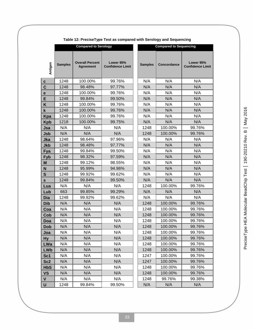
Table 12 shows all antigens tested with serological and sequencing methods. For the 18 antigens
tested with serological methods, Overall Percent Agreement ranged from 95.99% to 100.00%,
Positive Percent Agreement ranged from 98.77% to 100.00%, while the Negative Percent
Agreement ranged from 71.43% to 100.00%. For the 21 antigens tested with DNA sequencing
methods, Concordance ranged from 99.76% to 100.00%, Percent Sensitivity ranged from 98.67%
to 100.00%, while the Percent Specificity was 100.00%.
33
Prec
iseT
ype
HE
A M
olec
ular
Bea
dChi
p Te
st │
190
-202
10 R
ev. B
│ M
ay 2
016
Table 12: PreciseType Test as compared with Serology and Sequencing
Compared to Serology Compared to Sequencing A
ntig
en
Samples Overall Percent Agreement
Lower 95% Confidence Limit
Samples Concordance Lower 95% Confidence Limit
c 1248 100.00% 99.76% N/A N/A N/A C 1248 98.48% 97.77% N/A N/A N/A e 1248 100.00% 99.76% N/A N/A N/A E 1248 99.84% 99.50% N/A N/A N/A K 1248 100.00% 99.76% N/A N/A N/A k 1248 100.00% 99.76% N/A N/A N/A Kpa 1248 100.00% 99.76% N/A N/A N/A Kpb 1218 100.00% 99.75% N/A N/A N/A Jsa N/A N/A N/A 1248 100.00% 99.76% Jsb N/A N/A N/A 1248 100.00% 99.76% Jka 1248 98.64% 97.96% N/A N/A N/A Jkb 1248 98.48% 97.77% N/A N/A N/A Fya 1248 99.84% 99.50% N/A N/A N/A Fyb 1248 98.32% 97.59% N/A N/A N/A M 1248 99.12% 98.55% N/A N/A N/A N 1248 95.99% 94.96% N/A N/A N/A S 1248 99.92% 99.62% N/A N/A N/A s 1248 99.84% 99.50% N/A N/A N/A Lua N/A N/A N/A 1248 100.00% 99.76% Lub 663 99.85% 99.29% N/A N/A N/A Dia 1248 99.92% 99.62% N/A N/A N/A Dib N/A N/A N/A 1248 100.00% 99.76% Coa N/A N/A N/A 1248 100.00% 99.76% Cob N/A N/A N/A 1248 100.00% 99.76% Doa N/A N/A N/A 1248 100.00% 99.76% Dob N/A N/A N/A 1248 100.00% 99.76% Joa N/A N/A N/A 1248 100.00% 99.76% Hy N/A N/A N/A 1248 100.00% 99.76% LWa N/A N/A N/A 1248 100.00% 99.76% LWb N/A N/A N/A 1248 100.00% 99.76% Sc1 N/A N/A N/A 1247 100.00% 99.76% Sc2 N/A N/A N/A 1247 100.00% 99.76% HbS N/A N/A N/A 1248 100.00% 99.76% VS N/A N/A N/A 1248 100.00% 99.76% V N/A N/A N/A 1248 99.76% 99.38% U 1248 99.84% 99.50% N/A N/A N/A

34
Prec
iseT
ype
HE
A M
olec
ular
Bea
dChi
p Te
st │
190
-202
10 R
ev. B
│ M
ay 2
016
C. Overall HEA BeadChip Test Agreement with Serology and Sequencing Post-Discordant Resolution
In the same study mentioned in the section above, all discrepancies observed were further resolved by DNA sequence analysis. Bi-directional sequencing is considered “gold standard” – the reference method for sequence analysis. The term “reference method” refers to a well-validated analytical procedure sufficiently free of systemic or random error to make it useful for validating proposed new analytical procedures for the same analyte [21].
Antigen Number of Discordant Samples (Out of 1,248)
PreciseType Concordant with Reference Method (Bi-directional Sequencing)
Jkb 19 12 of 19 Fyb 21 20 of 21 C 19 19 of 19 Jka 17 17 of 17 M 11 11 of 11 N 50 50 of 50 Fya 2 2 of 2 E 2 2 of 2 S 1 1 of 1 s 2 2 of 2 Lub 1 (663) 1 of 1 Dia 1 1 of 1 V 3 3 of 3
Samples Discordant for Jkb with serology: • There were 19 discordant samples; all were PreciseType positive, serology negative. • Twelve samples were concordant between PreciseType and bi-directional sequencing. • Seven samples did not agree with serology or bi-directional sequencing and were
identified as Jknull (see the Limitations of Procedure section). Samples Discordant for Fyb with serology:
• There were 21 discordant samples, 20 were concordant between PreciseType and sequencing
• Fifteen were PreciseType positive, serology negative and 14 were concordant between PreciseType and bi-directional sequencing (all were Fyb weak).
• One sample was discordant between PreciseType and bi-directional sequencing and upon further investigation was found to be a novel mutation uncharacterized in literature (see the Limitations of Procedure section).
Samples Discordant for C with serology: • There were 19 discordant samples, all were PreciseType positive, serology negative; all
PreciseType results were concordant with bi-directional sequencing. Samples Discordant for Jka with serology:
• There were 17 discordant samples; all PreciseType results were concordant with bi-directional sequencing.
Samples Discordant for M with serology: • There were 11 discordant samples; all PreciseType results were concordant with bi-
directional sequencing. Samples Discordant for N with serology:
• There were 50 discordant samples; all PreciseType results were concordant with bi-directional sequencing.
All other Discrepancies (Fya, E, S, s, Lub, Dia, and V) • All other discrepancies for other antigens were found to be in concordance with bi-
directional sequencing.
35
Prec
iseT
ype
HE
A M
olec
ular
Bea
dChi
p Te
st │
190
-202
10 R
ev. B
│ M
ay 2
016
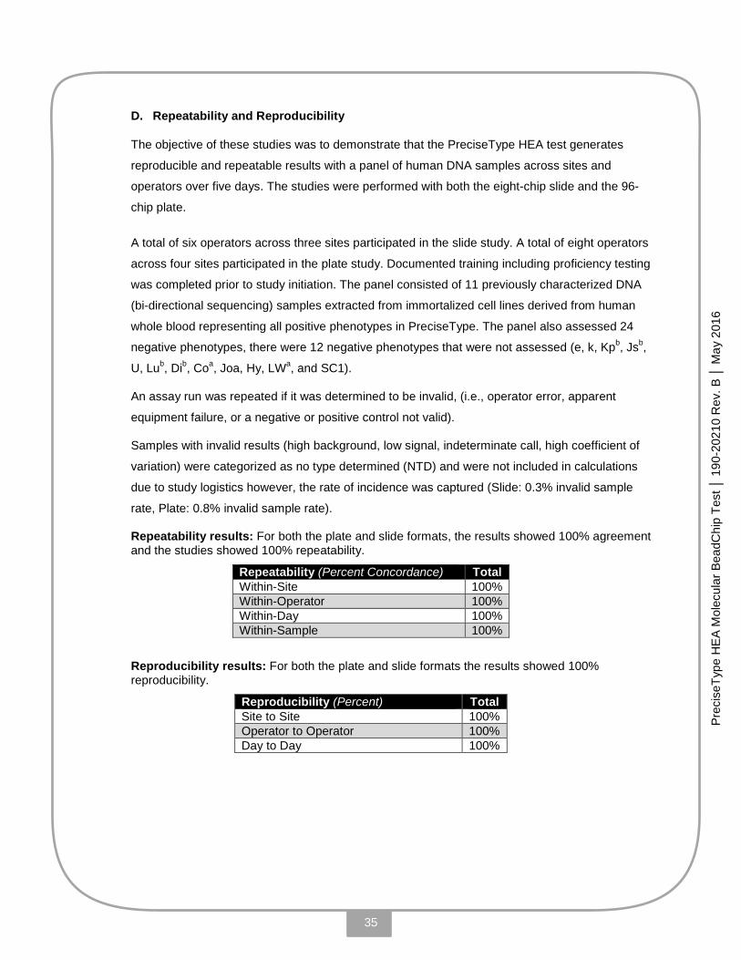
D. Repeatability and Reproducibility
The objective of these studies was to demonstrate that the PreciseType HEA test generates
reproducible and repeatable results with a panel of human DNA samples across sites and
operators over five days. The studies were performed with both the eight-chip slide and the 96-
chip plate.
A total of six operators across three sites participated in the slide study. A total of eight operators
across four sites participated in the plate study. Documented training including proficiency testing
was completed prior to study initiation. The panel consisted of 11 previously characterized DNA
(bi-directional sequencing) samples extracted from immortalized cell lines derived from human
whole blood representing all positive phenotypes in PreciseType. The panel also assessed 24
negative phenotypes, there were 12 negative phenotypes that were not assessed (e, k, Kpb, Jsb,
U, Lub, Dib, Coa, Joa, Hy, LWa, and SC1).
An assay run was repeated if it was determined to be invalid, (i.e., operator error, apparent
equipment failure, or a negative or positive control not valid).
Samples with invalid results (high background, low signal, indeterminate call, high coefficient of
variation) were categorized as no type determined (NTD) and were not included in calculations
due to study logistics however, the rate of incidence was captured (Slide: 0.3% invalid sample
rate, Plate: 0.8% invalid sample rate).
Repeatability results: For both the plate and slide formats, the results showed 100% agreement and the studies showed 100% repeatability.
Repeatability (Percent Concordance) Total Within-Site 100% Within-Operator 100% Within-Day 100% Within-Sample 100%
Reproducibility results: For both the plate and slide formats the results showed 100% reproducibility.
Reproducibility (Percent) Total Site to Site 100% Operator to Operator 100% Day to Day 100%

36
Prec
iseT
ype
HE
A M
olec
ular
Bea
dChi
p Te
st │
190
-202
10 R
ev. B
│ M
ay 2
016
Lot-to-Lot Reproducibility results: A separate lot-to-lot study was performed on a fully
characterized panel (n=22) of extracted human genomic DNA samples where the PreciseType
HEA test was performed using kits from three different lots to demonstrate the lot-to-lot
reproducibility. These 22 samples were blinded to the operator to eliminate bias and selected to
represent the broadest ranges of alleles possible that are contained in the PreciseType test. The
same operator repeated the same assays on five separate days to demonstrate the repeatability.
The results showed 100% agreement and the study shows 100% lot-to-lot repeatability and
reproducibility.
Reproducibility (Percent) Total Lot to Lot 100% Day to Day 100%
Overall Conclusion: On an antigen basis, all sample results (across all samples) were in
agreement with their expected results within operator day to day, across operators, across sites
and across lots. Therefore, we can conclude that the PreciseType HEA test is 100% repeatable
and 100% reproducible.

37
Prec
iseT
ype
HE
A M
olec
ular
Bea
dChi
p Te
st │
190
-202
10 R
ev. B
│ M
ay 2
016
E. Interfering Substances
The following substances, commonly found on skin and in blood, were not found to interfere with the PreciseType HEA test.
Microorganisms – The following organisms were tested at 10^6 CFU per mL of blood;
Bacillus subtilis, Corynebacterium diphtheria and jeikeium, Escherichia coli, Propionibacterium acnes,
Pseudomonas aeruginosa, Salmonella enterica, Staphylococcus epidermidis, haemolyticus and
aureus, Streptococcus pneumonia and mitis, Aspergillus niger, Candida albicans. Cytopathic levels of
influenza virus were also tested with no interference observed.
Exogenous Substances – Amoxicillin (2.06E+02 µmol/L), Penicillin G Potassium Salt (2.73 µg/mL)
Hydroxyurea (3.50 µg/mL), Acetaminophen (1.32E µmol/L), Ibuprofen (2.43 µmol/L), Aspirin (3.62E
µmol/L), Naproxen (2.17E µmol/L), Plavix (3.00E µmol/L), Warfarin (3.25E µmol/L), Loratadine (7.80E
µmol/L), Atorvastatin (Lipitor) (5.48E+2 µmol/L), Phenylephrine HCl (4.91E µmol/L), Nadolol (3.88E
µmol/L), Folic Acid (Vit.B) (1.50E+1 µmol/L), Ascorbic Acid (Vit.C) (3.42E µmol/L)
Endogenous substances – Pathological values of hemoglobin (up to 500g/L), bilirubin (up to 67 mg/dL), triglycerides (up to 1000mg/dL) and total protein (up to 90g/L)
38
Prec
iseT
ype
HE
A M
olec
ular
Bea
dChi
p Te
st │
190
-202
10 R
ev. B
│ M
ay 2
016
XI. BIBLIOGRAPHY
1. Klein HG, Anstee DJ. Mollison's Blood Transfusion in Clinical Medicine. 11th ed. Oxford: Blackwell Publishing; 2005.
2. Reid ME. Applications of DNA based assays in blood group antigen and antibody identification. Transfusion. 2003 Dec;43(12):1748-57.
3. Hashmi G, Shariff T, Seul M, Vissavajjhala P, HueRoye K. A flexible array format for large scale, rapid blood group DNA typing. Transfusion. 2005 May;45(5):680-8.
4. Wagner T, Resch B, Reiterer F, Gassner C, Lanzer G. Pancytopenia due to suppressed hematopoiesis in a case of fatal hemolytic disease of the newborn associated with anti-K supported by molecular K1 typing. J Pediatr Hematol Oncol. 2004 Jan;26(1):13-5.
5. Eder AF. Update on HDFN: new information on long-standing controversies. Immunohematology. 2006;22(4):188-95.
6. Reid ME, Lomas-Francis C. The Blood Group Antigen Facts Book. San Diego: Academic Press; 2003. 7. Blumenfeld OO, Patnaik SK. Allelic genes of blood group antigens: a source of human mutations and cSNPs
documented in the Blood Group Antigen Database. Human Mutation. 2004 Jan;23(1):8-16. 8. ISBT Working Party. Names for FY (ISBT 008) Blood Group Alleles. [Internet]. [cited 2012 25 07]. Available
from: http://www.isbtweb.org/fileadmin/user_upload/WP_on_Red_Cell_Immunogenetics_and/008_FY_Alleles_v2.0_110914.pdf.
9. García ME, Blanco JL, Caballero J, Gargallo-Viola D. Anticoagulants interfere with PCR used to diagnose invasive aspergillosis. J Clin Microbiol. 2002 Apr;40(4):1567-8.
10. Rios M, Hue-Roye K, Øyen R, Miller J, Reid ME. Insights into the Holley- and Joseph- phenotypes. Transfusion. 2002 Jan;42(1):52-8.
11. Scofield TL, Miller JP, Storry JR, Rios M, Reid ME. Evidence that Hy- RBCs express weak Joa antigen. Transfusion. 2004 Feb;44(2):170-2.
12. Noizat-Pirenne F, Mouro I, Le Pennec PY, Ansart-Pirenne H, Juszczak G, Patereau C, Verdier M, Babinet J, Roussel M, Rouger P, et al. Two new alleles of the RHCE gene in Black individuals: the RHce allele ceMO and the RHcE allele cEMI. Br J Haematol. 2001 Jun;113(3):672-9.
13. Noizat-Pirenne F, Lee K, Pennec PY, Simon P, Kazup P, Bachir D, Rouzaud AM, Roussel M, Juszczak G, Ménanteau C, et al. Rare RHCE phenotypes in black individuals of Afro-Caribbean origin: identification and transfusion safety. Blood. 2002 Dec 1;100(12):4223-31.
14. Daniels GL, Faas BH, Green CA, Smart E, Maaskant-van Wijk PA, Avent ND, Zondervan HA, von dem Borne AE, van der Schoot CE. The VS and V blood group polymorphisms in Africans: a serologic and molecular analysis. Transfusion. 1998 Oct;38(10):951-8.
15. Pham BN, Peyrard T, Juszczak G, Dubeaux I, Gien D, Blancher A, Cartron JP, Rouger P, Le Pennec PY. Heterogeneous molecular background of the weak C, VS+, hr B-, Hr B- phenotype in black persons. Transfusion. 2009 Mar;49(3):495-504.
16. Westhoff CM, Vege S, Copeland T, Baker E, Velliquette R, Lomas-Francis C. A new glycophorin B allele, GYPB (IVS5+3G>A), in trans to GYPB (IVS5+5G>T) is associated with discrepant prediction of S and U antigen status. Vox Sanguinis. 2012; 103 (Suppl. 1), 212.
17. Blood Grouping Reagent Anti-Fya Anti-Fyb. By indirect antiglobulin test [package insert]. Norcross, GA: Immucor, Inc; 2007
18. Westhoff CM, Vege S, Horn T, Hue-Roye K, Halter Hipsky C, Lomas-Francis C, and Reid ME. RHCE*cemo is frequently in cis to RHD*DAU0 and encodes a hrs-, hrB-, RH:61 phenotype in black persons: clinical significance. Transfusion. 2013 Nov;53(11):2983-2989
19. Wang Y, Beckwith B, Smith C, Horowitz G. Misleading glycated hemoglobin results in a patient with hemoglobin SC disease. Clinical Chemistry. 2007. 53(7):1394-5.
20.
Lin M, Yu LC. Frequencies of the JKnull (IVS5-1g>a) allele in Taiwanese, Fujian, Filipino, and Indonesian populations. Transfusion. 2008 Aug;48(8):1768.
21.
Guidance for Industry and FDA Staff: Pharmacogenetic Tests and Genetic Tests for Heritable Markers . June 19, 2007.
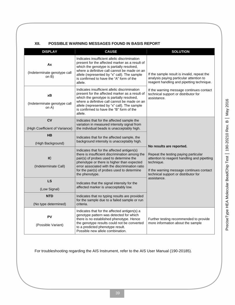
39
Prec
iseT
ype
HE
A M
olec
ular
Bea
dChi
p Te
st │
190
-202
10 R
ev. B
│ M
ay 2
016
XII. POSSIBLE WARNING MESSAGES FOUND IN BASIS REPORT
DISPLAY CAUSE SOLUTION
Ax
(Indeterminate genotype call on B)
Indicates insufficient allelic discrimination present for the affected marker as a result of which the genotype is partially resolved, where a definitive call cannot be made on an allele (represented by “x” call). The sample is confirmed to have the “A” form of the allele.
If the sample result is invalid, repeat the analysis paying particular attention to reagent handling and pipetting technique. If the warning message continues contact technical support or distributor for assistance.
xB
(Indeterminate genotype call on A)
Indicates insufficient allelic discrimination present for the affected marker as a result of which the genotype is partially resolved, where a definitive call cannot be made on an allele (represented by “x” call). The sample is confirmed to have the “B” form of the allele.
CV
(High Coefficient of Variance)
Indicates that for the affected sample the variation in measured intensity signal from the individual beads is unacceptably high.
No results are reported. Repeat the testing paying particular attention to reagent handling and pipetting technique. If the warning message continues contact technical support or distributor for assistance.
HB
(High Background)
Indicates that for the affected sample, the background intensity is unacceptably high.
IC
(Indeterminate Call)
Indicates that for the affected antigen(s) there is insufficient discrimination among the pair(s) of probes used to determine the phenotype or there is higher than expected error associated with the discrimination ratio for the pair(s) of probes used to determine the phenotype.
LS
(Low Signal)
Indicates that the signal intensity for the affected marker is unacceptably low.
NTD
(No type determined)
Indicates that no typing results are provided for the sample due to a failed sample or run criteria.
PV
(Possible Variant)
Indicates that for the affected antigen(s) a genotype pattern was detected for which there is no established phenotype. Hence the genotype results could not be converted to a predicted phenotype result. Possible new allele combination.
Further testing recommended to provide more information about the sample
For troubleshooting regarding the AIS Instrument, refer to the AIS User Manual (190-20185).
40
Prec
iseT
ype
HE
A M
olec
ular
Bea
dChi
p Te
st │
190
-202
10 R
ev. B
│ M
ay 2
016
Patents
All goods and services are sold subject to the terms and conditions of sale of BioArray Solutions Ltd. and are covered by one or more of: Patent Nos. 6,797,524; 7,427,512; 7,335,153; 7,390,676; EP1311839B1; CA2413978 (additional patents pending).
The purchase of this product grants the purchaser rights under certain Roche patents to use it solely for providing Human In Vitro Diagnostic Services. No general patent or other license of any kind other than this specific right of use from purchase is granted.
This product and its use are covered by one or more of the following patents: EP 0 820 524, US 6,150,095, 6,307,039, 6,770,751 and 7,192,707, Jap 2006 246897 and pending patents. The purchaser is licensed to practice methods and processes covered by these patents using this product for the purpose of molecular immunohematology only.
The Thermo Sequenase® DNA polymerase in this Product is covered by U.S. patent 5,614,365 and foreign equivalents owned by Harvard College and exclusively licensed to GE HEALTHCARE and U.S. patent 5,885,813 and foreign equivalents owned by GE HEALTHCARE.
Trademarks
PreciseType, BeadChip, AIS, BASIS and eMAP are trademarks, and BeadCheck is a registered trademark of BioArray Solutions Ltd.
HotStarTaq, QIAcube and QIAamp are registered trademarks of QIAGEN GmbH, Hilden, Germany, licensed to BioArray Solutions.
Veriti and GeneAmp are registered trademarks of Life Technologies Corporation.
DNA AWAY is a trademark of Molecular BioProducts, Inc.
InSlide Out is a registered trademark of Boekel Scientific
MiniMouse II is a trademark of Denville Scientific Inc.
MicroAmp is a registered trademark of Applied Biosystems
Excel is a registered trademark of Microsoft Corporation
Thermo Sequenase is a registered trademark of GE Healthcare UK Limited
Registered names, trademarks, etc. used in this document, even when not specifically marked as such, are not to be considered unprotected by law.
Manufacturer: BioArray Solutions Ltd.
35 Technology Drive, Suite 100 Warren, NJ 07059 USA
Toll-Free (+1) 866 566-8200 Fax (+1) 908 226-0800
http://www.immucor.com
To reach an Immucor Technical Support Representative, call 855.IMMUCOR (855.466.8267), option 2, or email: [email protected]