Embed Size (px)
Citation preview
1
2
PRE-LAB EXERCISES
Open the Human Anatomy Atlas app, choose Views from the top menu, and select Systems from the submenu. Then, scroll down to the Respiratory System Views.
You are responsible for the identification of all bold terms.
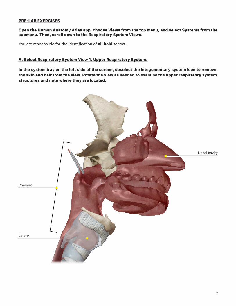
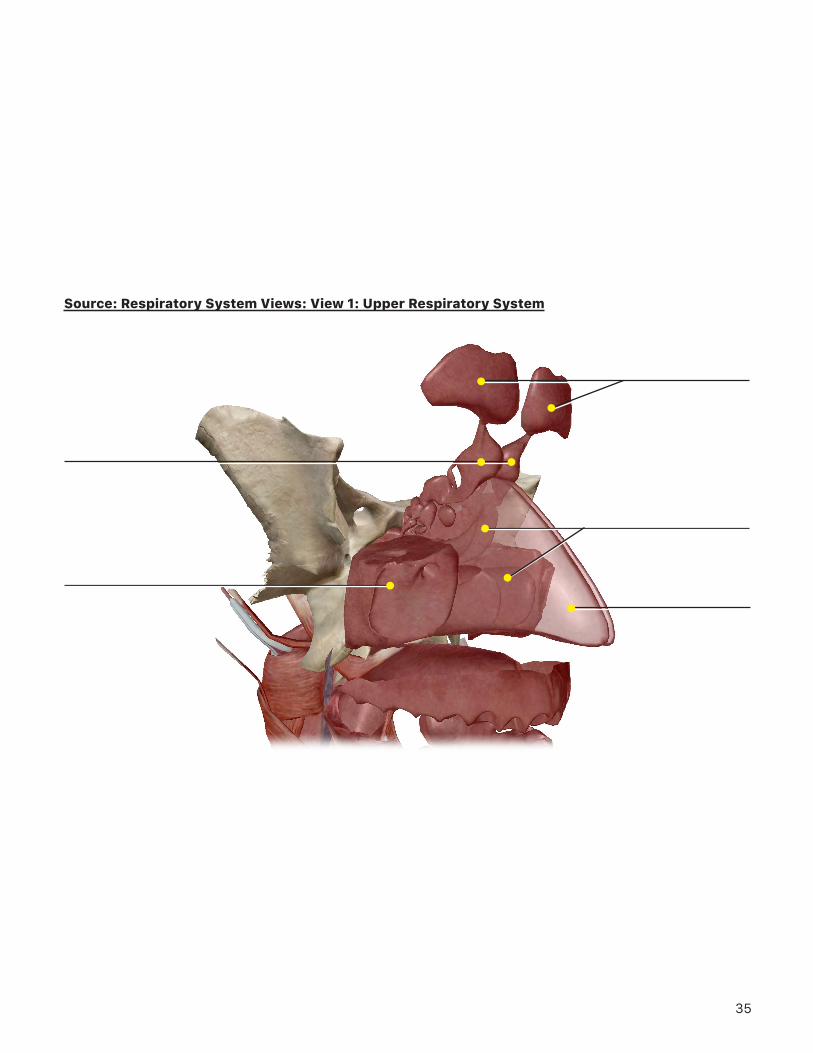
A. Select Respiratory System View 1. Upper Respiratory System.
In the system tray on the left side of the screen, deselect the integumentary system icon to remove the skin and hair from the view. Rotate the view as needed to examine the upper respiratory system structures and note where they are located.
Pharynx
Larynx
Nasal cavity
3
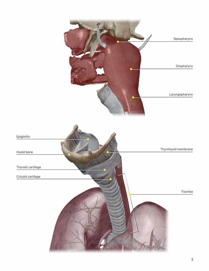
Nasopharynx
Oropharynx
Laryngopharynx
Hyoid bone
Thyroid cartilage
Cricoid cartilage
Epiglottis
Thyrohyoid membrane
Trachea
4
1. What structures comprise the upper respiratory system, from superior to inferior?
2. Select the nasal cavity and use the book icon to read about it. What are the structures and functions of the nasal cavity?
3. Examine the structure of the pharynx. What are the 3 parts of the pharynx, from superior to inferior?
4. Examine the structure of the larynx.
a. Select the thyroid cartilage and use the book icon to read about it. What is the function of this cartilage and the other laryngeal cartilages?
b. To examine the epiglottis, you must first individually select and hide the thyroid cartilage and the thyrohyoid membrane. Then, select the epiglottis and use the book icon to read about it. What is the composition and function of the epiglottis?
5
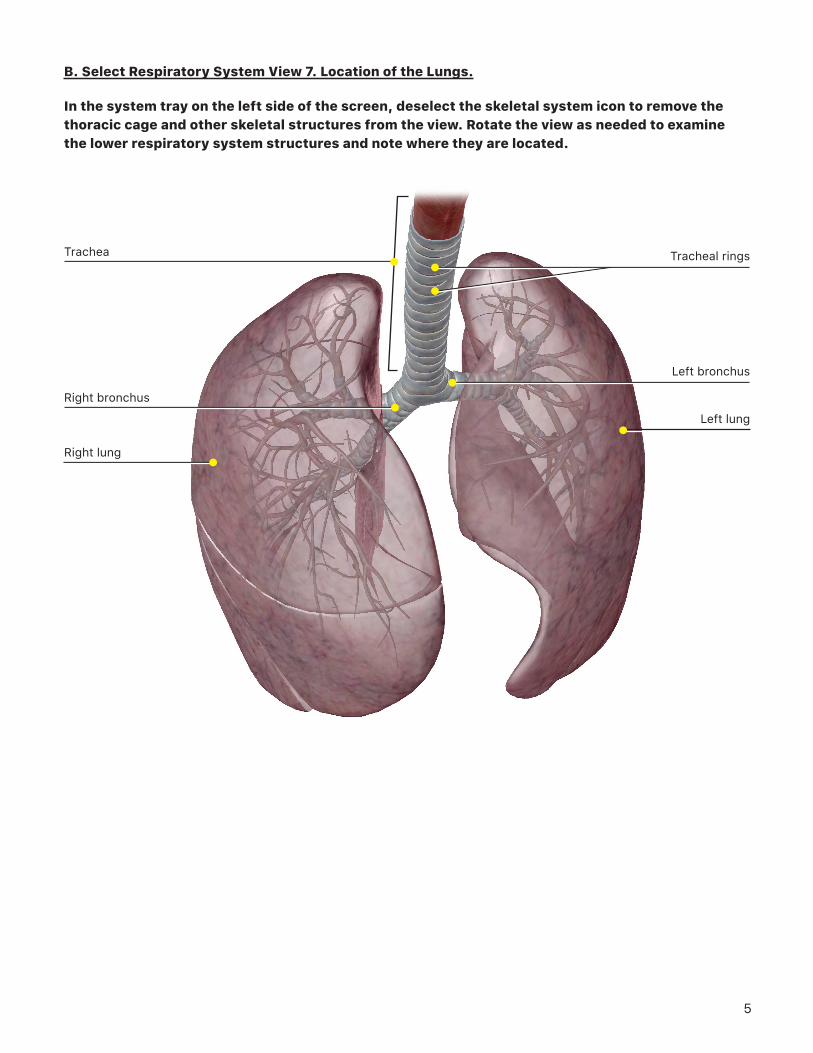
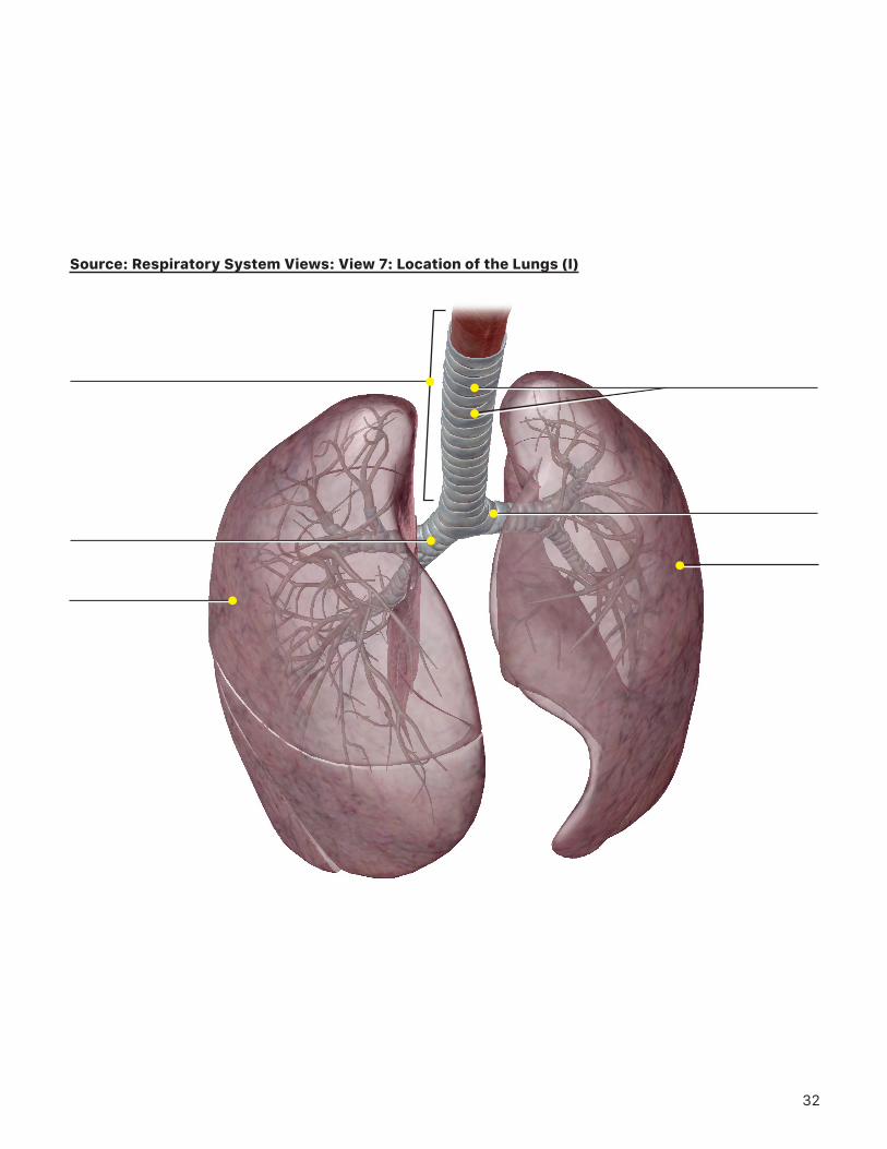
B. Select Respiratory System View 7. Location of the Lungs.
In the system tray on the left side of the screen, deselect the skeletal system icon to remove the thoracic cage and other skeletal structures from the view. Rotate the view as needed to examine the lower respiratory system structures and note where they are located.
Right lung
Right bronchus
Trachea
Left lung
Left bronchus
Tracheal rings
6
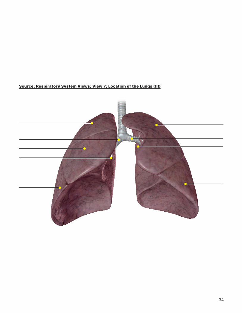
Right inferior lobe
Right hilum of lung
Right middle lobe
Right superior lobe
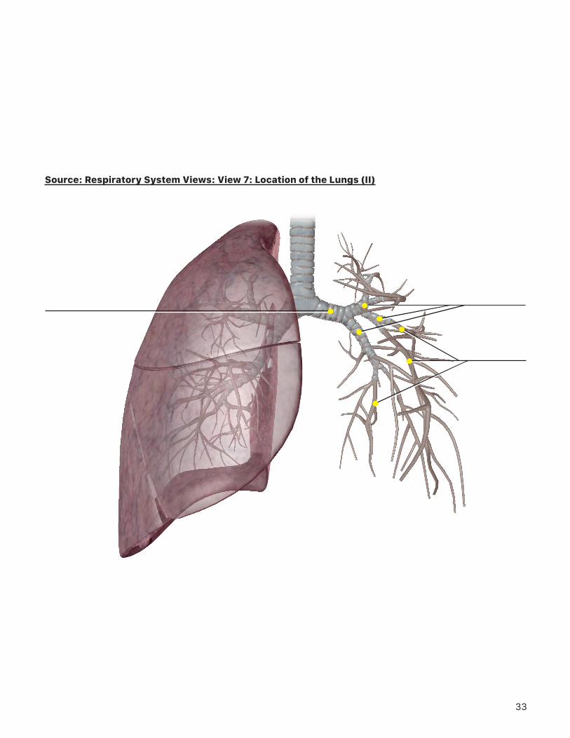
Right main bronchus
Left inferior lobe
Left superior lobe
Left main bronchus
Left hilum of the lung
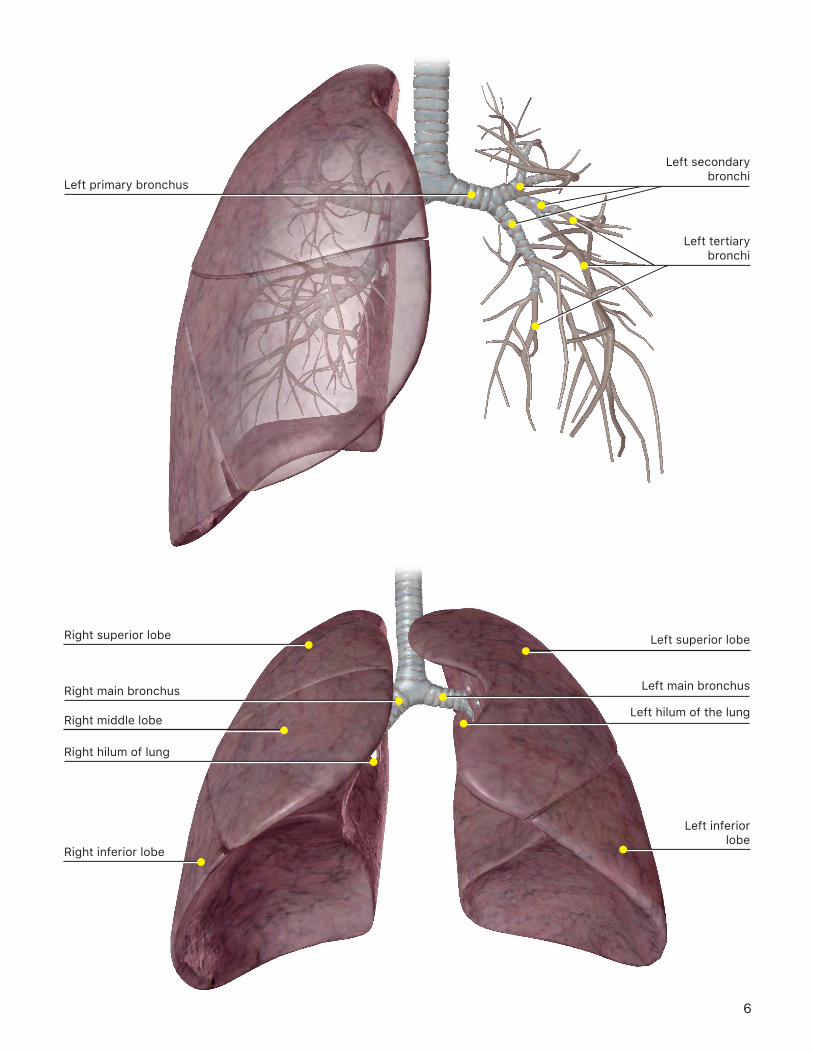
Left primary bronchus
Left tertiary bronchi
Left secondary bronchi
7
1. What structures comprise the lower respiratory system, from superior to inferior?
2. Select the trachea and use the book icon to read about it. What is the structure and function of the trachea and the tracheal rings?
3. In the system tray on the left side of the screen, deselect all the blue system icons except respiratory. Then, select and hide one of the lungs. Examine the bronchi.
a. In terms of size and structure, what is the difference between primary, secondary, and tertiary bronchi?
b. What are the tiny structures that appear at the ends of the tertiary bronchi, and what is their function?
4. In the system tray on the left side of the screen, deselect and select the respiratory system icon to refresh the respiratory structures in the view, and examine the lungs.
a. What differences do you notice between the left and right lungs?
b. What are the lines on the surface of the lungs called?
c. On the medial aspect of the lungs, what is the name of the region where the bronchi enter the lung? What else enters the lung via this region?
8
IN-LAB EXERCISES
Open the Human Anatomy Atlas app, choose Views from the top menu, and select Systems from the submenu. Then, scroll down to the Respiratory System Views.
You are responsible for the identification of all bold terms.
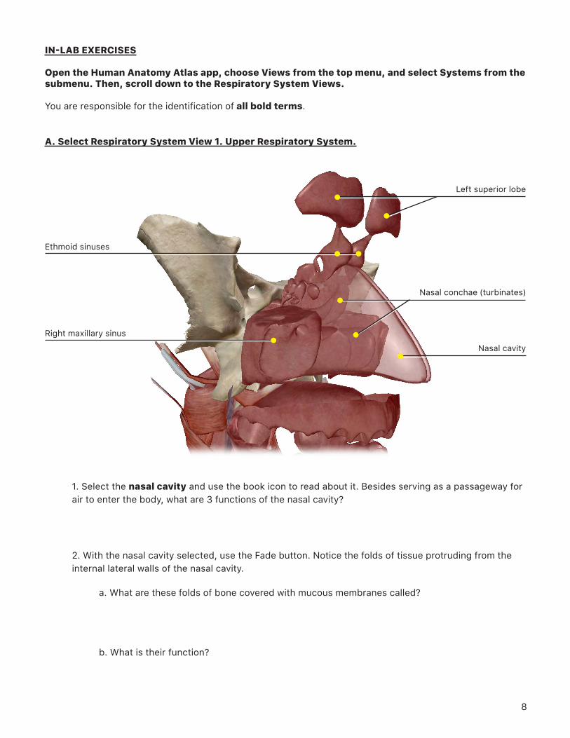
A. Select Respiratory System View 1. Upper Respiratory System.
1. Select the nasal cavity and use the book icon to read about it. Besides serving as a passageway for air to enter the body, what are 3 functions of the nasal cavity?
2. With the nasal cavity selected, use the Fade button. Notice the folds of tissue protruding from the internal lateral walls of the nasal cavity.
a. What are these folds of bone covered with mucous membranes called?
b. What is their function?
Right maxillary sinus
Ethmoid sinuses
Left superior lobe
Nasal cavity
Nasal conchae (turbinates)
9
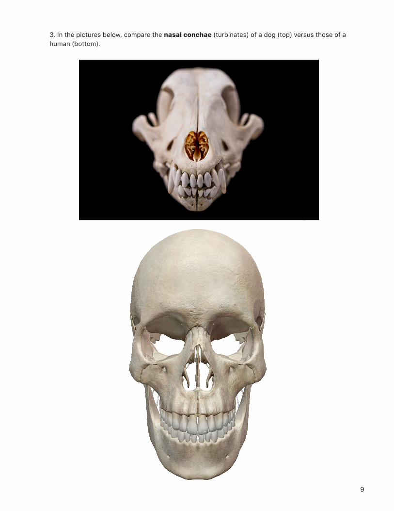
3. In the pictures below, compare the nasal conchae (turbinates) of a dog (top) versus those of a human (bottom).
10
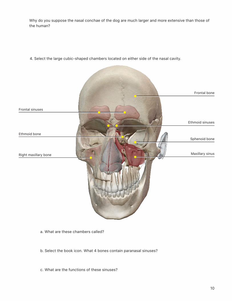
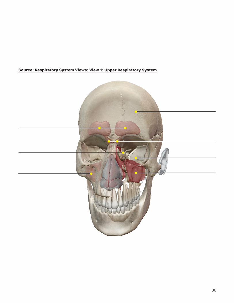
Right maxillary bone
Ethmoid bone
Frontal sinuses
Maxillary sinus
Sphenoid bone
Frontal bone
Ethmoid sinuses
Why do you suppose the nasal conchae of the dog are much larger and more extensive than those of the human?
4. Select the large cubic-shaped chambers located on either side of the nasal cavity.
a. What are these chambers called?
b. Select the book icon. What 4 bones contain paranasal sinuses?
c. What are the functions of these sinuses?
11
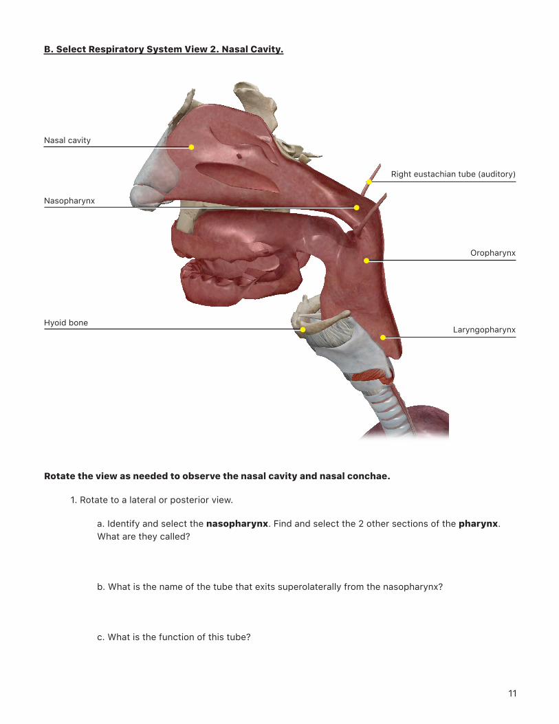
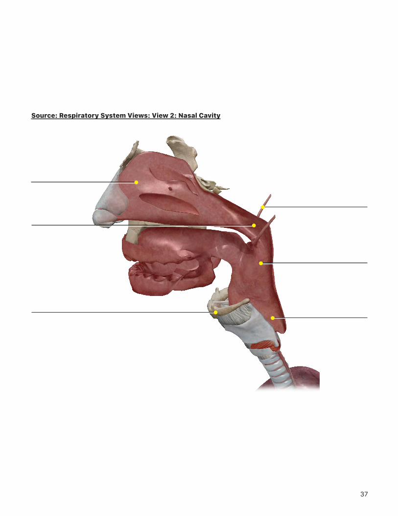
B. Select Respiratory System View 2. Nasal Cavity.
Nasal cavity
Nasopharynx
Hyoid boneLaryngopharynx
Oropharynx
Right eustachian tube (auditory)
Rotate the view as needed to observe the nasal cavity and nasal conchae.
1. Rotate to a lateral or posterior view.
a. Identify and select the nasopharynx. Find and select the 2 other sections of the pharynx. What are they called?
b. What is the name of the tube that exits superolaterally from the nasopharynx?
c. What is the function of this tube?
12
2. Rotate back to the anterior view.
a. What is the name of the bone located anterior to the laryngopharynx?
b. What is the function of this bone and what makes it unique and different from all the other bones in the body?
TIME TO PRACTICE!GO TO THE QUIZZES MENU, SCROLL DOWN TO THE RESPIRATORY SYSTEM, AND COMPLETE
QUIZZES 2 (UPPER RESPIRATORY) AND 5 (NASAL CAVITY).
13
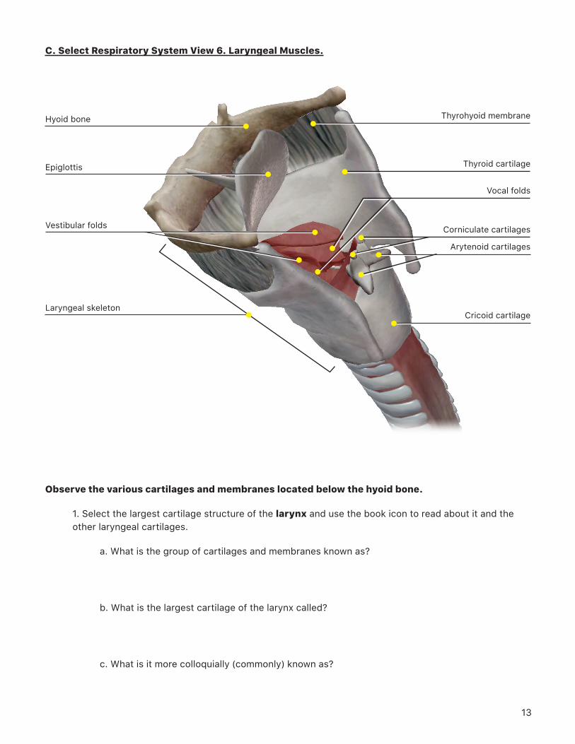
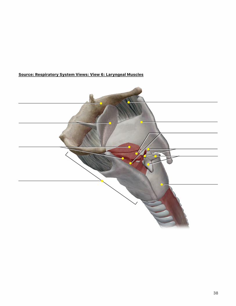
C. Select Respiratory System View 6. Laryngeal Muscles.
Observe the various cartilages and membranes located below the hyoid bone.
1. Select the largest cartilage structure of the larynx and use the book icon to read about it and the other laryngeal cartilages.
a. What is the group of cartilages and membranes known as?
b. What is the largest cartilage of the larynx called?
c. What is it more colloquially (commonly) known as?
Hyoid bone
Epiglottis
Laryngeal skeleton
Vestibular folds
Cricoid cartilage
Arytenoid cartilages
Corniculate cartilages
Thyrohyoid membrane
Thyroid cartilage
Vocal folds
14
d. What is the name of the membrane between the hyoid bone and the thyroid cartilage?
e. What is the name of the only laryngeal or tracheal cartilage that extends all the way around the larynx?
f. What is the function of the laryngeal skeleton?
g. Select the large, flap-like piece of cartilage behind the hyoid bone. What is this structure called, and what is its function?
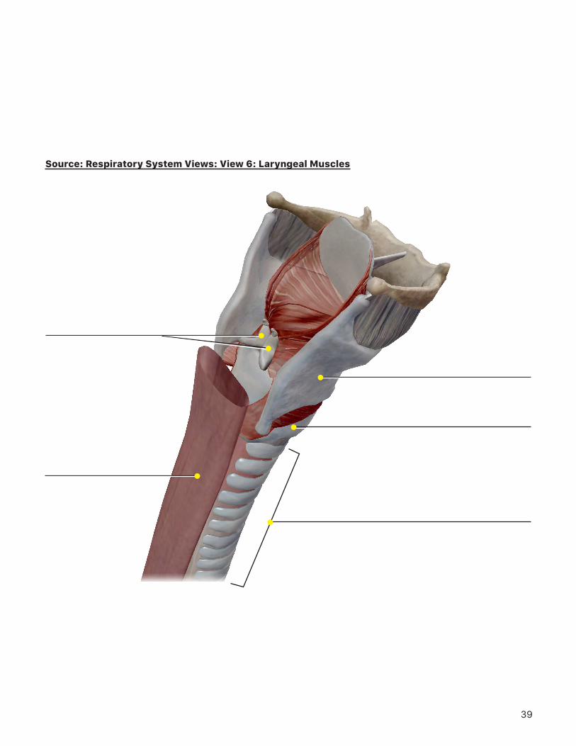
2. In the system tray on the left side of the screen, deselect the muscular system icon to remove the muscles from the view. Rotate the view to a posterior view of the larynx. Locate and select the vocal folds and use the Radius Blast button.
a. Compare the location of the vocal folds and vestibular folds. What is an alternate name for each of these structures, and what are their functions?
b. What is the name of the pyramid-shaped cartilages that sit on top of the cricoid cartilage and attach posteriorly to the vocal ligaments?
c. What are the names of the small cartilages that are located on top of the cartilages from question b?
d. Select the trachea (windpipe). Notice the cartilaginous rings located along its anterior and lateral surfaces. What is the function of these rings?
15
e. In the system tray on the left side of the screen, select the digestive system icon to add the digestive structures to the view. What is the name of the tube that is located just dorsal to the trachea? Why do you think this tube does not have cartilage rings like the trachea?
TIME TO PRACTICE!GO TO THE QUIZZES MENU, SCROLL DOWN TO THE RESPIRATORY SYSTEM,
AND COMPLETE QUIZ 7 (LARYNX).
16
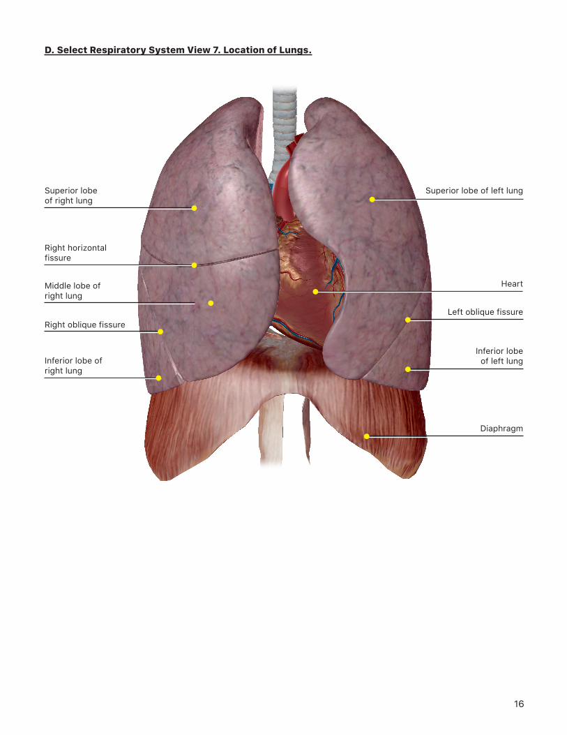
D. Select Respiratory System View 7. Location of Lungs.
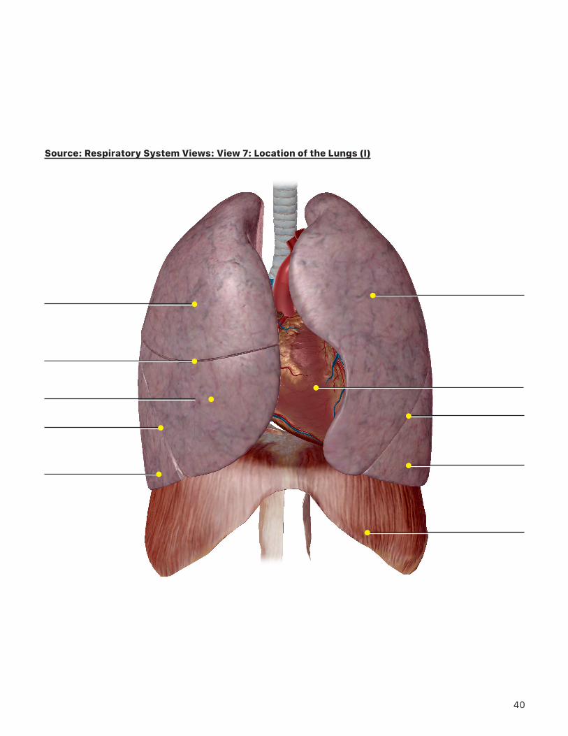
Superior lobe of right lung
Right horizontal fissure
Inferior lobe of right lung
Right oblique fissure
Middle lobe of right lung
Diaphragm
Inferior lobe of left lung
Left oblique fissure
Superior lobe of left lung
Heart
17
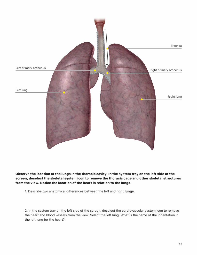
Observe the location of the lungs in the thoracic cavity. In the system tray on the left side of the screen, deselect the skeletal system icon to remove the thoracic cage and other skeletal structures from the view. Notice the location of the heart in relation to the lungs.
1. Describe two anatomical differences between the left and right lungs.
2. In the system tray on the left side of the screen, deselect the cardiovascular system icon to remove the heart and blood vessels from the view. Select the left lung. What is the name of the indentation in the left lung for the heart?
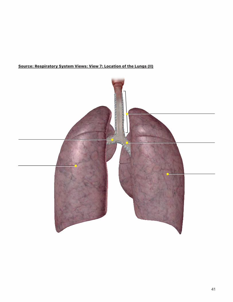
Left primary bronchus
Left lung
Right lung
Right primary bronchus
Trachea
18
3. What are the lines you see on the surface of the lungs? Name them and the lobes they separate.
4. Rotate to a posterior view of the lungs. Look at the trachea and where it bifurcates into 2 branches. What are these branches called?
5. Looking at the left and right branches, consider this question. If you accidentally aspirated (inhaled) a foreign object, which lung do you think the object would be most likely to enter and why?
19
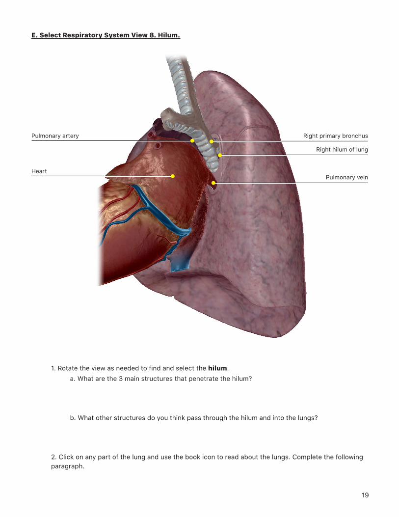
E. Select Respiratory System View 8. Hilum.
1. Rotate the view as needed to find and select the hilum. a. What are the 3 main structures that penetrate the hilum?
b. What other structures do you think pass through the hilum and into the lungs?
2. Click on any part of the lung and use the book icon to read about the lungs. Complete the following paragraph.
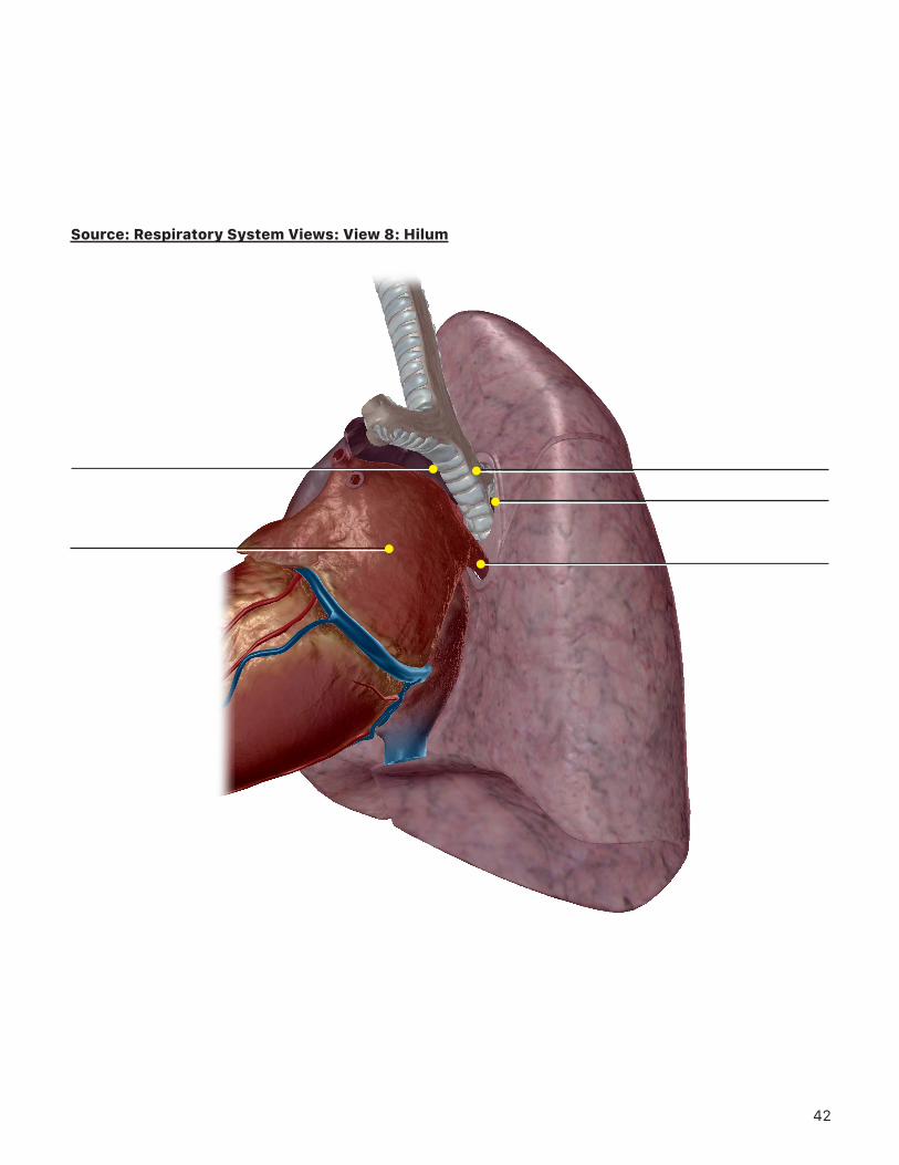
Heart
Pulmonary artery
Pulmonary vein
Right hilum of lung
Right primary bronchus
20
The ______________________________ is a double layered, serous membrane surrounding the lungs.
The space between the layers is filled with ______________________________, which serves to reduce
______________________________ against the chest wall.
3. In the system tray on the left side of the screen, deselect the circulatory system icon to hide the heart and blood vessels from the view. Select and hide the right lung. Observe the bronchial tree. What differences do you notice between the secondary (lobar) and tertiary (segmental) bronchi?
TIME TO PRACTICE!GO TO THE QUIZZES MENU, SCROLL DOWN TO THE RESPIRATORY SYSTEM, AND COMPLETE
QUIZZES 6 (TRACHEA AND BRONCHI) AND 8 (LUNGS, EXTERIOR).
21
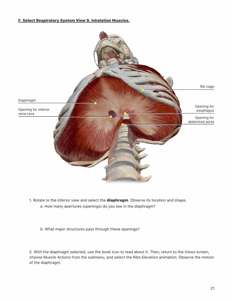
F. Select Respiratory System View 9. Inhalation Muscles.
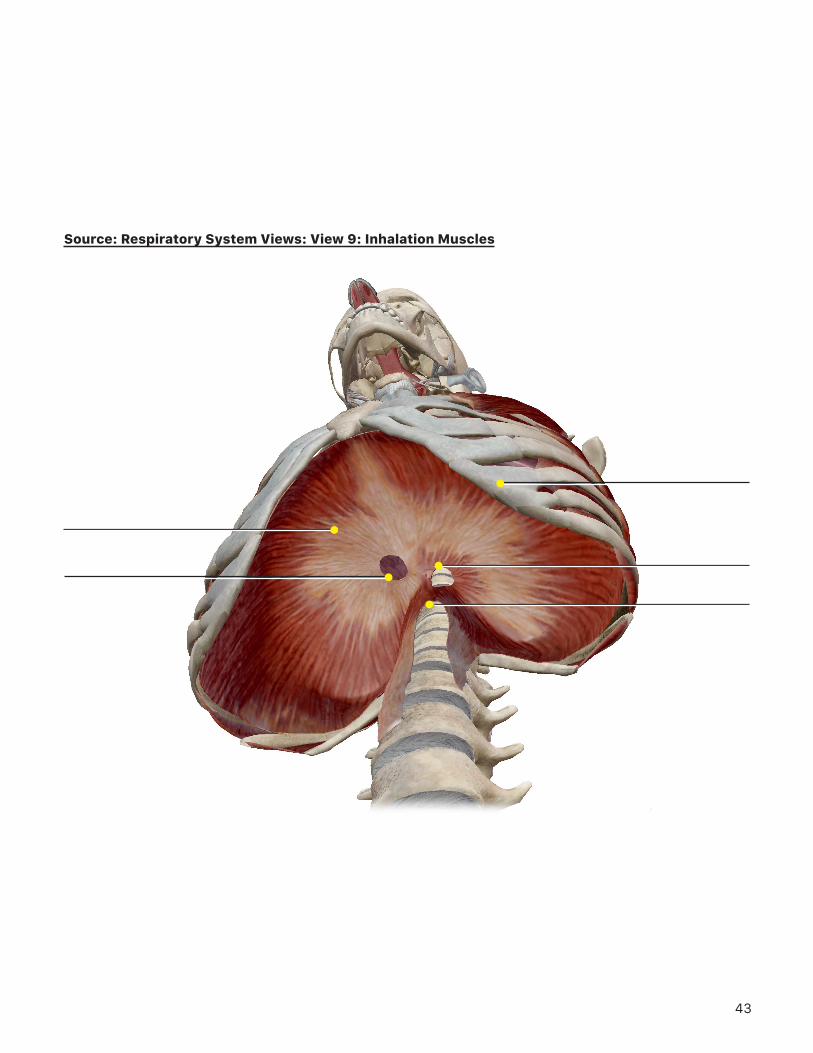
Diaphragm
Opening for inferior vena cava
Opening for esophagus
Opening for abdominal aorta
Rib cage
1. Rotate to the inferior view and select the diaphragm. Observe its location and shape.a. How many apertures (openings) do you see in the diaphragm?
b. What major structures pass through these openings?
2. With the diaphragm selected, use the book icon to read about it. Then, return to the Views screen, choose Muscle Actions from the submenu, and select the Ribs Elevation animation. Observe the motion of the diaphragm.

22
a. What happens to the dome-shaped diaphragm as it contracts? What happens to the rib cage?
b. What effect does diaphragm contraction have on the volume of the thoracic cavity and lungs?
c. What do you think happens to the pressure inside the lungs due to this volume change?
d. Do you think this will cause air to move into or out of the lungs?
e. Name 5 other muscles that may contribute to elevation of the rib cage for inhalation.
23
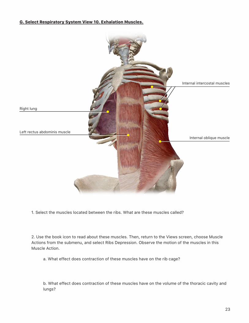
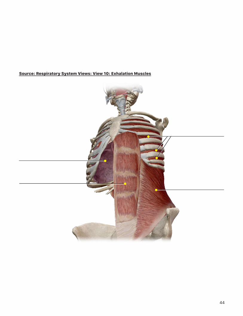
G. Select Respiratory System View 10. Exhalation Muscles.
Right lung
Left rectus abdominis muscleInternal oblique muscle
Internal intercostal muscles
1. Select the muscles located between the ribs. What are these muscles called?
2. Use the book icon to read about these muscles. Then, return to the Views screen, choose Muscle Actions from the submenu, and select Ribs Depression. Observe the motion of the muscles in this Muscle Action.
a. What effect does contraction of these muscles have on the rib cage?
b. What effect does contraction of these muscles have on the volume of the thoracic cavity and lungs?
24
c. What do you think happens to the pressure inside the lungs due to this volume change?
d. Do you think this will cause air to move into or out of the lungs?
e. What other muscles do you see contributing to depression of the rib cage?
3. Go back to Respiratory System View 10. Exhalation Muscles. Select the internal oblique muscle and use the book icon to read about it.
a. What effect does contraction of the abdominal muscles have on the abdominal viscera?
b. Do you think contraction of these muscles would aid in inhalation or exhalation? Explain your answer.
25
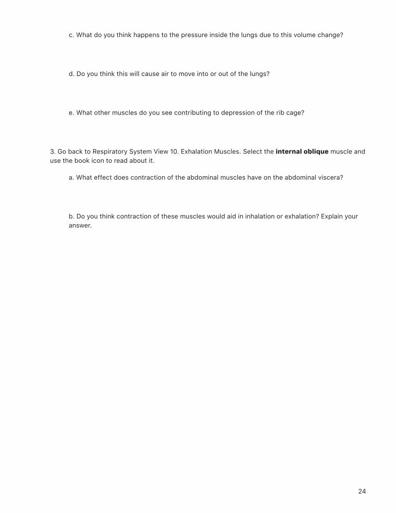
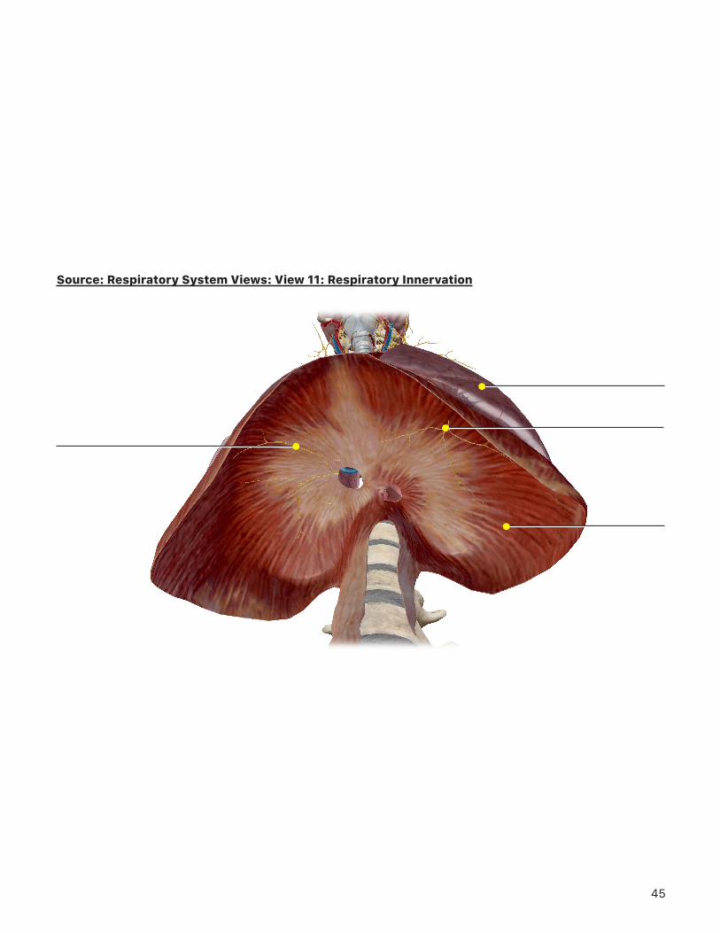
H. Select Respiratory System View 11. Respiratory Innervation.
Rotate to the inferior view. Zoom in on the diaphragm and select the nerve branches visible on the inferior surface.
1. What is the name of this nerve?
2. With the nerve still selected, rotate and zoom the view to follow the nerve back to the spinal cord. What level(s) of the spinal cord is the origin of this nerve?
3. Explain how an injury to the neck might cause someone to require an artificial respirator to breathe?
TIME TO PRACTICE! GO TO THE QUIZZES MENU, SCROLL DOWN TO THE RESPIRATORY SYSTEM, AND COMPLETE
QUIZZES 1 (OVERVIEW, RESPIRATORY) AND 3 (LOWER RESPIRATORY).
Right phrenic nerve
Diaphragm
Left phrenic nerve
Left lung
26
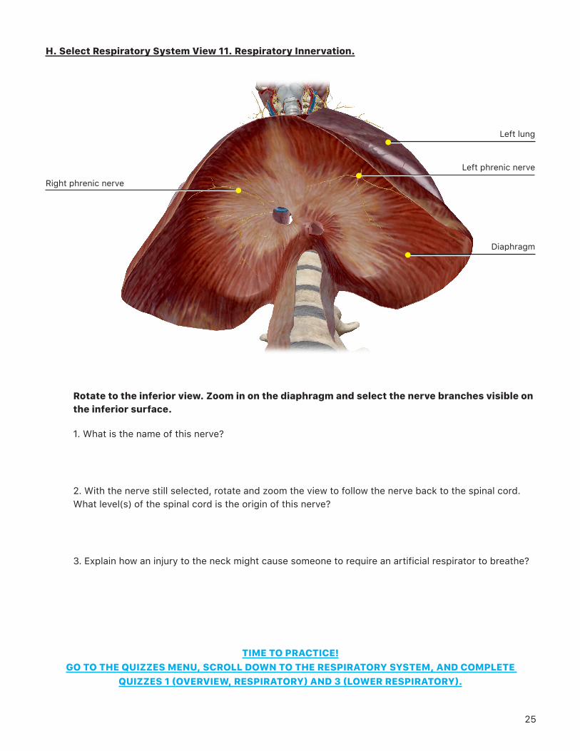
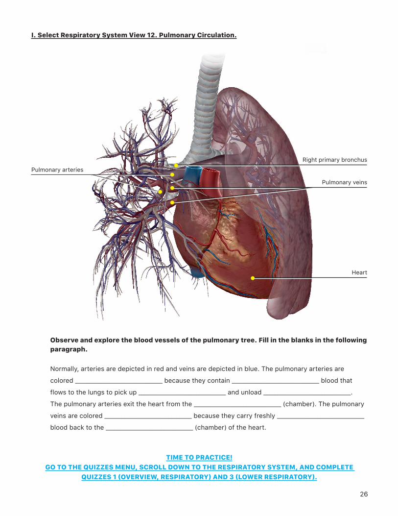
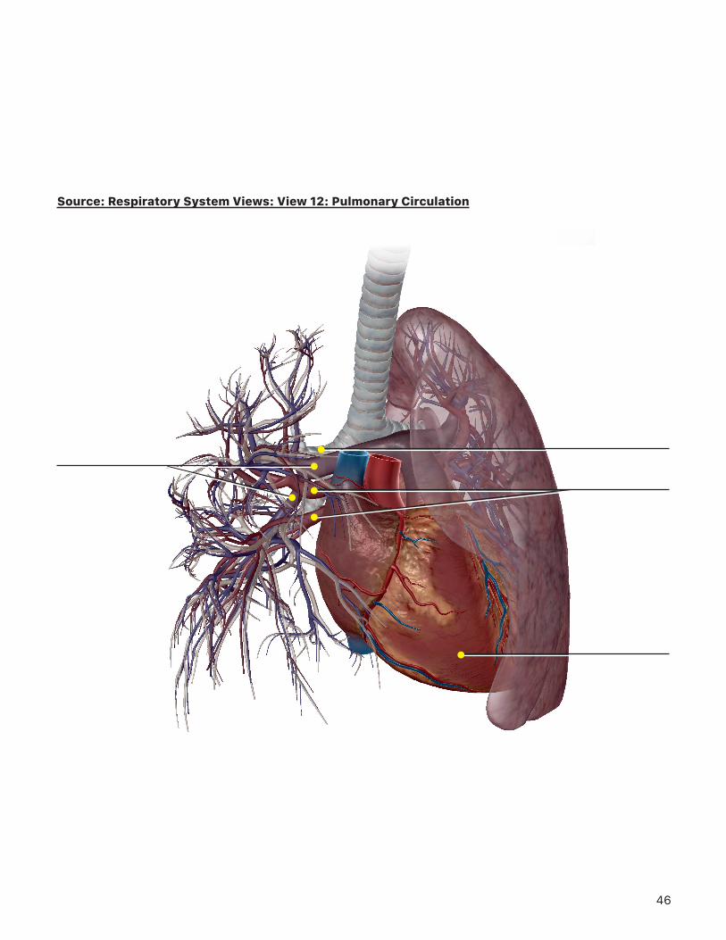
I. Select Respiratory System View 12. Pulmonary Circulation.
Observe and explore the blood vessels of the pulmonary tree. Fill in the blanks in the following paragraph.
Normally, arteries are depicted in red and veins are depicted in blue. The pulmonary arteries are
colored ______________________________ because they contain ______________________________ blood that
flows to the lungs to pick up ______________________________ and unload ______________________________.
The pulmonary arteries exit the heart from the ______________________________ (chamber). The pulmonary
veins are colored ______________________________ because they carry freshly ______________________________
blood back to the ______________________________ (chamber) of the heart.
TIME TO PRACTICE! GO TO THE QUIZZES MENU, SCROLL DOWN TO THE RESPIRATORY SYSTEM, AND COMPLETE
QUIZZES 1 (OVERVIEW, RESPIRATORY) AND 3 (LOWER RESPIRATORY).
Pulmonary arteries
Heart
Right primary bronchus
Pulmonary veins
27
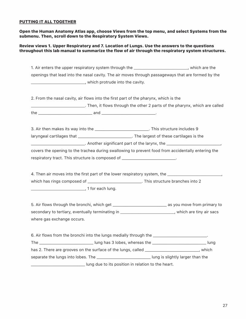
PUTTING IT ALL TOGETHER
Open the Human Anatomy Atlas app, choose Views from the top menu, and select Systems from the submenu. Then, scroll down to the Respiratory System Views.
Review views 1. Upper Respiratory and 7. Location of Lungs. Use the answers to the questions throughout this lab manual to summarize the flow of air through the respiratory system structures.
1. Air enters the upper respiratory system through the ______________________________, which are the
openings that lead into the nasal cavity. The air moves through passageways that are formed by the
______________________________, which protrude into the cavity.
2. From the nasal cavity, air flows into the first part of the pharynx, which is the
______________________________. Then, it flows through the other 2 parts of the pharynx, which are called
the ______________________________ and ______________________________.
3. Air then makes its way into the ______________________________. This structure includes 9
laryngeal cartilages that ______________________________. The largest of these cartilages is the
______________________________. Another significant part of the larynx, the ______________________________,
covers the opening to the trachea during swallowing to prevent food from accidentally entering the
respiratory tract. This structure is composed of ______________________________.
4. Then air moves into the first part of the lower respiratory system, the ______________________________,
which has rings composed of ______________________________. This structure branches into 2
______________________________, 1 for each lung.
5. Air flows through the bronchi, which get ______________________________ as you move from primary to
secondary to tertiary, eventually terminating in ______________________________, which are tiny air sacs
where gas exchange occurs.
6. Air flows from the bronchi into the lungs medially through the ______________________________.
The ______________________________ lung has 3 lobes, whereas the ______________________________ lung
has 2. There are grooves on the surface of the lungs, called ______________________________, which
separate the lungs into lobes. The ______________________________ lung is slightly larger than the
______________________________ lung due to its position in relation to the heart.
28

29
Source: Respiratory System Views: View 1: Upper Respiratory System (I)
30
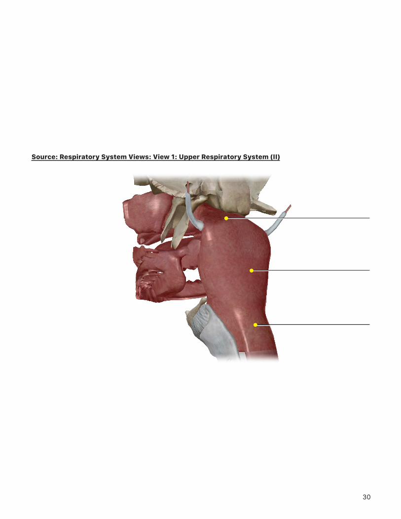
Source: Respiratory System Views: View 1: Upper Respiratory System (II)
31
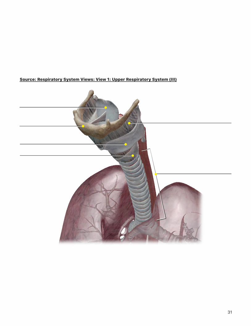
Source: Respiratory System Views: View 1: Upper Respiratory System (III)
32
Source: Respiratory System Views: View 7: Location of the Lungs (I)
33
Source: Respiratory System Views: View 7: Location of the Lungs (II)
34
Source: Respiratory System Views: View 7: Location of the Lungs (III)
35
Source: Respiratory System Views: View 1: Upper Respiratory System
36
Source: Respiratory System Views: View 1: Upper Respiratory System
37
Source: Respiratory System Views: View 2: Nasal Cavity
38
Source: Respiratory System Views: View 6: Laryngeal Muscles
39
Source: Respiratory System Views: View 6: Laryngeal Muscles
40
Source: Respiratory System Views: View 7: Location of the Lungs (I)
41
Source: Respiratory System Views: View 7: Location of the Lungs (II)
42
Source: Respiratory System Views: View 8: Hilum
43
Source: Respiratory System Views: View 9: Inhalation Muscles
44
Source: Respiratory System Views: View 10: Exhalation Muscles
45
Source: Respiratory System Views: View 11: Respiratory Innervation
46
Source: Respiratory System Views: View 12: Pulmonary Circulation