Embed Size (px)
Citation preview

Short Communication Proceedings: Impacts of Air Pollution and Climate Change on Forest Ecosystems TheScientificWorldJOURNAL (2007) 7(S1), 222–230 ISSN 1537-744X; DOI 10.1100/tsw.2007.22
*Corresponding author. ©2007 with author. Published by TheScientificWorld; www.thescientificworld.com
222
Pre-Exposure to Ozone Predisposes Oak Leaves to Attacks by Diplodia corticola and Biscogniauxia mediterranea
Elena Paoletti1,*, Naldo Anselmi2, and Antonio Franceschini3 1IPP-CNR, Via Madonna del Piano 10, I-50019 Sesto Fiorentino, Firenze, Italy; 2Dip. Protezione Piante, Univ. Tuscia, Via de Lellis, I-01100, Viterbo, Italy; 3Dip. Protezione Piante, Univ. Sassari, Via de Nicola, I-07100, Sassari, Italy
E-mail: [email protected]
Received September 27, 2006; Revised December 15, 2006; Accepted December 18, 2006; Published March 21, 2007
One-year-old cork oak (Quercus suber) and turkey oak (Q. cerris) seedlings were exposed to ozone (110 ppb, 5 h day–1, for 30 days) and were inoculated with Diplodia corticola and Biscogniauxia mediterranea, respectively, by spraying a suspension of spores on the leaves. Both fungi are endophytic and may act as weak parasites, contributing to oak decline. Ozone exposure stimulated leaf attacks after inoculation, although the physiological, visible, and structural responses of both oaks to O3 exposure were weak. In fact, steady-state gas exchange, leaf waxes, and wettability were not significantly affected by O3. In Q. cerris, O3 altered the structure of stomata, as observed by scanning microscopy, and reduced the leaf relative water content. No hyphal entry through stomata or growth towards stomata was, however, observed. Inoculations were performed in a humid chamber at low light; stomata were likely to be closed. When Q. cerris was inoculated in natural conditions, i.e., in a forest infected by B. mediterranea, seedlings pre-exposed to the enhanced O3 regime had a higher number of B. mediterranea isolates than the controls. This suggests that pre-exposure to O3 predisposed Q. cerris leaves to attacks by B. mediterranea independent of stomata. The hyphae of both fungi were able to enter the leaf through the cuticle, either by gradual in-growth into the cuticle or erosion of a hollow in the cuticle at the point of contact. The primary cause of increased leaf injury in O3-exposed seedlings appeared to be higher germination of spores than on control leaves.
Keywords: ozone, weak pathogen, oak, Quercus suber, Quercus cerris, Mediterranean ecosystems
INTRODUCTION
Forest decline results from a combination of interacting factors[1]. The terminal stage of decline often involves pathogens or pests that move from a latent to an active phase in trees under stress. Ozone (O3) is a pervasive air pollutant in most parts of the world[2]. Pathogens are believed to colonize plants weakened by O3[3]. There are extensive reports of the effects of O3 on trees, but few reports link O3 exposure with tree diseases[3].
Paoletti et al.: Ozone Predisposes Leaves to Pathogen Attacks TheScientificWorldJOURNAL (2007) 7(S1), 222–230
223
Several factors including drought, severe summer flooding, changes in agronomic practices, wood-boring insects, and fungal diseases[4] have been implicated in oak decline in Southern Europe. The role of O3 pollution is unknown. Both Biscogniauxia mediterranea and Diplodia corticola are endophytic fungi that can affect plants of different ages, inducing bark cankers or symptoms similar to those produced by a tracheomycotic disease, respectively. Diplodia corticola (formerly D. mutila) is considered to be one of the main causes of Quercus suber decline[5]. Biscogniauxia mediterranea (formerly Hypoxylon mediterraneum) has been frequently associated with oak decline in the Mediterranean area[6], mostly affecting Q. cerris[7,8].
Our objective was to clarify whether O3-exposed leaves of two Mediterranean oaks, Q. suber and Q. cerris, were more susceptible to D. corticola and B. mediterranea, respectively, and to test some O3 responses (amount of epicuticular waxes, leaf wettability, stomata structure, gas exchange, leaf water content) that may affect leaf susceptibility to fungi.
MATERIALS AND METHODS
Plant Material and Ozone Exposure
One-year-old, nursery-grown, potted, uniform-sized seedlings of Q. suber and Q. cerris were placed in controlled conditions (20–23°C, 70–80% RH, 500 µmol m–2 sec–1 PAR, 12-h photoperiod, charcoal-filtered air) for 1 week at the end of July 2001. On August 1st, seedlings were placed in exposure chambers, either in charcoal-filtered air, or to elevated O3. The elevated O3 exposure averaged 110 ppb (in the form of a square wave, 5 h day–1) for 30 days, corresponding to 10.5 ppm h AOT40. The O3 exposure was designed to be typical of peak exposures in Italy[9]. Enclosures were supplied with charcoal-filtered air and a complete air exchange was achieved every 1.5 min. A fan mixed the internal air. Ozone was generated with a Helios Italquartz generator (Model Heliozon, Milan, Italy), metered to the chambers with mass flowmeters (Model GFC171, Aalborg, NY), and monitored with an O3 monitor (Model 202, 2B Technologies, Boulder, CO). Plants were watered to field capacity once a week.
Preinoculation Measurements
Gas exchange, epicuticular waxes, leaf wettability, and water content were measured shortly after the end of O3 exposure (August 30th) and before fungal inoculation (August 31st, afternoon) on three seedlings per species and treatment. The low replication was chosen to perform all the measurements at 12:00–13:00 (gas exchange) and to reduce destructive sampling and wounding that might have favored hyphae entering the leaf (epicuticular waxes, leaf wettability, and water content). Steady-state net photosynthesis and stomatal conductance were assessed on three leaves per seedling by IRGA (CIRAS-1, PP-Systems, Herts, U.K.) that controlled temperature (25°C), leaf-to-air vapor pressure difference (2.0 ± 0.2 kPa), light (1400 μmol m–2 sec–1 PAR), and CO2 concentration (365 ppm). To extract epicuticular waxes, 1 g of fresh leaves per seedling was shaken for 10 sec with 50 ml chloroform[10]. The solution was filtered through a 0.2-µm PTFE membrane in a preweighed aluminum container, and allowed to evaporate to constant weight in a fume cupboard at room temperature. The residue weight was cross-referenced to leaf area determined with a leaf area meter AM 300 (ADC, Herts, U.K.). Wettability of the adaxial leaf surface was assessed as drop contact angle (DCA) using a bench microscope with a protractor graticule[11]. A 2-cm2 stripe was cut in the central part of each fresh leaf (one per seedling), halfway between the midvein and the margin. A 1-µl distilled water droplet was put to each leaf stripe using a 5-µl syringe, and contact angle (±1°) was immediately measured. As the sparse pubescence on the adaxial leaf surface of Q. suber induced erratic values, DCA was measured in Q. cerris only. Leaf water content was measured only in Q. cerris. Five 1-cm2 leaf discs per seedling were sampled, avoiding the midrib. Relative water content was calculated as RWC = 100*[(FW – DW)/(SW – DW)] where FW, DW, and SW are fresh, dry, and saturation weight,
Paoletti et al.: Ozone Predisposes Leaves to Pathogen Attacks TheScientificWorldJOURNAL (2007) 7(S1), 222–230
224
respectively. SW was measured after one night in Petri dishes with wet blotting paper. DW was measured after oven drying at 80°C until reaching a constant weight.
Inoculation in Controlled Conditions
Mature B. mediterranea stromata on cut Q. cerris branches were soaked with sterile deionized water and put in a humid chamber at 100% RH (measured with a hygrothermograph). At sporulation, ascospores were collected in sterile containers and combined in a single sprayer containing 1 l sterile water. The number of spores, determined with a cell-count slide, was 106 ascospores ml–1, i.e., the density that stimulates the best in vitro germination rate[7]. For inducing picnidia differentiation in D. corticola, Q. suber leaves were sterilized and put in Petri dishes containing PDA medium infected with D. corticola mycelium, at ambient light and temperature. After 30 days, picnidia were moved to sterile vials with 200 μl of sterile water. They were crushed and shaken to favor conidia dissemination. The conidia suspension obtained was adjusted at 105 conidia ml–1.
On August 31st, six control and six O3-exposed seedlings of each species were sprayed with the suspensions of B. mediterranea ascospores (Q. cerris) or D. corticola conidia (Q. suber) until upper and lower leaf blades were fully wet. Inoculated seedlings were immediately sealed in one humid chamber per pathosystem (100 × 50 × 100 cm, 100% RH, 200 μmol PAR m–2 sec–1) at 20 or 25°C, for Q. cerris or Q. suber, respectively. The temperatures were set at the optimum for germination of B. mediterranea[7] and D. corticola[12] spores, respectively. After a 2-day incubation, macroscopical and microscopical injury was observed.
Visible Injury
No visible injury was observed on O3-exposed leaves. Inoculations caused leaves to turn brownish-black, starting from edges to the midrib. The extent of such necroses was determined on ten leaf sections for each seedling with the WinDias image analysis system (Delta-T Devices, Cambridge, U.K.). Observations were carried out immediately after the 2-day incubation on September 2nd. For Q. suber, additional observations were carried out at day 2 and 5 after incubation, on September 4th and 7th, respectively.
Microscopic Observations
After incubation, three injured leaves per plant were collected. To observe the adaxial and abaxial leaf surfaces, two 0.25-cm2 squares were cut in each of the brownish and green part of the leaf, immediately coated with gold (1.5 min, Edwards S150A Sputter Coater), and observed using a scanning electron microscope (SEM 505 Philips, Eindhoven, Holland) at 5 kV. The low voltage helped to reduce damage to the fresh material. Drying or use of acetone would have altered the fungal structures. Fifty stomata per leaf portion were assigned to one of four damage classes: 0, no injury; 1, slight injury; 2, medium injury; 3, severe injury. The structural damage to stomata was quantified as SDI (Stomatal Damage Index) = [(N1 * 1) + (N2 * 2) + (N3*3)]/(N0 + N1 + N2 + N3), where N was the number of stomata in each damage class[13]. The abaxial surface of Q. suber leaves was covered by a dense pubescence, and so damage to stomata was not assessed. Ascospores, conidia, and hyphae were also observed. Quantification was impractical because of the patchy nature of infection.
Inoculation in Natural Conditions
Paoletti et al.: Ozone Predisposes Leaves to Pathogen Attacks TheScientificWorldJOURNAL (2007) 7(S1), 222–230
225
After O3 exposure (August 31st), 30 additional Q. cerris seedlings (15 control and 15 O3-exposed seedlings) were placed in a Q. cerris forest in decline because of B. mediterranea. September was dry, but October precipitation totaled 130 mm, which promoted natural fungal inoculation of the seedlings. Leaves on the seedlings were already senescent. On November 1st, fragments of outer sapwood and buds were removed from the seedlings with a sterile knife, placed in Petri dishes containing Potato Dextrose Agar, and incubated at 23°C in the dark. Fungal isolates were identified morphologically or by molecular methods, as described in Mazzaglia et al.[14].
RESULTS
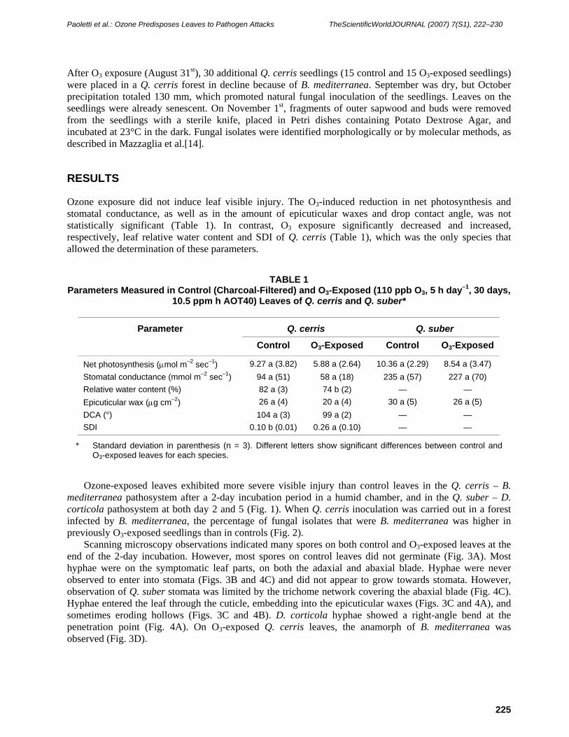
Ozone exposure did not induce leaf visible injury. The O3-induced reduction in net photosynthesis and stomatal conductance, as well as in the amount of epicuticular waxes and drop contact angle, was not statistically significant (Table 1). In contrast, O3 exposure significantly decreased and increased, respectively, leaf relative water content and SDI of Q. cerris (Table 1), which was the only species that allowed the determination of these parameters.
TABLE 1 Parameters Measured in Control (Charcoal-Filtered) and O3-Exposed (110 ppb O3, 5 h day–1, 30 days,
10.5 ppm h AOT40) Leaves of Q. cerris and Q. suber*
Parameter Q. cerris Q. suber
Control O3-Exposed Control O3-Exposed
Net photosynthesis (μmol m–2 sec–1) 9.27 a (3.82) 5.88 a (2.64) 10.36 a (2.29) 8.54 a (3.47) Stomatal conductance (mmol m–2 sec–1) 94 a (51) 58 a (18) 235 a (57) 227 a (70) Relative water content (%) 82 a (3) 74 b (2) — — Epicuticular wax (μg cm–2) 26 a (4) 20 a (4) 30 a (5) 26 a (5) DCA (°) 104 a (3) 99 a (2) — — SDI 0.10 b (0.01) 0.26 a (0.10) — —
* Standard deviation in parenthesis (n = 3). Different letters show significant differences between control and O3-exposed leaves for each species.
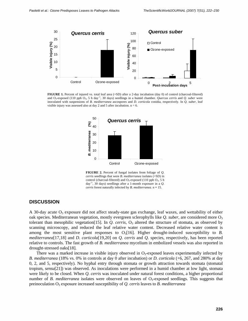
Ozone-exposed leaves exhibited more severe visible injury than control leaves in the Q. cerris – B. mediterranea pathosystem after a 2-day incubation period in a humid chamber, and in the Q. suber – D. corticola pathosystem at both day 2 and 5 (Fig. 1). When Q. cerris inoculation was carried out in a forest infected by B. mediterranea, the percentage of fungal isolates that were B. mediterranea was higher in previously O3-exposed seedlings than in controls (Fig. 2).
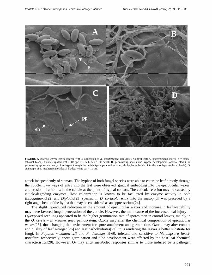
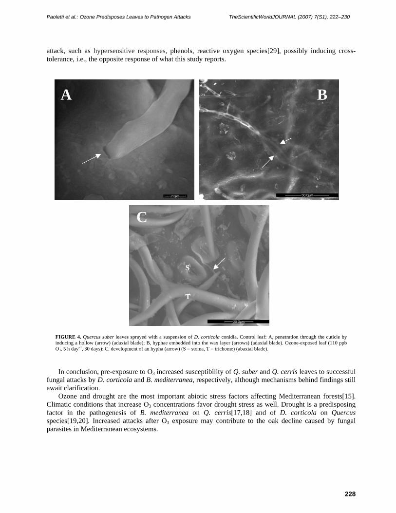
Scanning microscopy observations indicated many spores on both control and O3-exposed leaves at the end of the 2-day incubation. However, most spores on control leaves did not germinate (Fig. 3A). Most hyphae were on the symptomatic leaf parts, on both the adaxial and abaxial blade. Hyphae were never observed to enter into stomata (Figs. 3B and 4C) and did not appear to grow towards stomata. However, observation of Q. suber stomata was limited by the trichome network covering the abaxial blade (Fig. 4C). Hyphae entered the leaf through the cuticle, embedding into the epicuticular waxes (Figs. 3C and 4A), and sometimes eroding hollows (Figs. 3C and 4B). D. corticola hyphae showed a right-angle bend at the penetration point (Fig. 4A). On O3-exposed Q. cerris leaves, the anamorph of B. mediterranea was observed (Fig. 3D).
Paoletti et al.: Ozone Predisposes Leaves to Pathogen Attacks TheScientificWorldJOURNAL (2007) 7(S1), 222–230
226
Quercus suber
0
20
40
60
80
100
120
0 2 5Post-incubation days
Visi
ble
inju
ry (%
) Control
Ozone-exposed
Quercus cerris
0
5
10
15
20
25
30
Control Ozone-exposed
Visi
ble
inju
ry (%
)
FIGURE 1. Percent of injured vs. total leaf area (+SD) after a 2-day incubation (day 0) of control (charcoal-filtered) and O3-exposed (110 ppb O3, 5 h day–1, 30 days) seedlings in a humid chamber. Quercus cerris and Q. suber were inoculated with suspensions of B. mediterranea ascospores and D. corticola conidia, respectively. In Q. suber, leaf visible injury was assessed also at day 2 and 5 after incubation. n = 6.
Quercus cerris
0
10
20
30
40
50
Control Ozone-exposed
B. m
edite
rran
ea (
%)
FIGURE 2. Percent of fungal isolates from foliage of Q. cerris seedlings that were B. mediterranea isolates (+SD) in control (charcoal-filtered) and O3-exposed (110 ppb O3, 5 h day–1, 30 days) seedlings after a 1-month exposure in a Q. cerris forest naturally infected by B. mediterranea. n = 15.
DISCUSSION
A 30-day acute O3 exposure did not affect steady-state gas exchange, leaf waxes, and wettability of either oak species. Mediterranean vegetation, mostly evergreen sclerophylls like Q. suber, are considered more O3 tolerant than mesophilic vegetation[15]. In Q. cerris, O3 altered the structure of stomata, as observed by scanning microscopy, and reduced the leaf relative water content. Decreased relative water content is among the most sensitive plant responses to O3[16]. Higher drought-induced susceptibility to B. mediterranea[17,18] and D. corticola[19,20] on Q. cerris and Q. species, respectively, has been reported relative to controls. The fast growth of B. mediterranea mycelium in embolized vessels was also reported in drought-stressed oaks[18].
There was a marked increase in visible injury observed in O3-exposed leaves experimentally infected by B. mediterranea (18% vs. 0% in controls at day 0 after incubation) or D. corticola (+6, 267, and 280% at day 0, 2, and 5, respectively). No hyphal entry through stomata or growth attraction towards stomata (stomatal tropism, sensu[21]) was observed. As inoculations were performed in a humid chamber at low light, stomata were likely to be closed. When Q. cerris was inoculated under natural forest conditions, a higher proportional number of B. mediterranea isolates were observed on leaves of O3-exposed seedlings. This suggests that preinoculation O3 exposure increased susceptibility of Q. cerris leaves to B. mediterranea
Paoletti et al.: Ozone Predisposes Leaves to Pathogen Attacks TheScientificWorldJOURNAL (2007) 7(S1), 222–230
227
FIGURE 3. Quercus cerris leaves sprayed with a suspension of B. mediterranea ascospores. Control leaf: A, ungerminated spores (S = stoma) (abaxial blade). Ozone-exposed leaf (110 ppb O3, 5 h day–1, 30 days): B, germinating spores and hyphae development (abaxial blade); C, germinating spores and entry of an hypha through the cuticle (pp = penetration point; eh, hypha embedded into the wax layer) (adaxial blade); D, anamorph of B. mediterranea (adaxial blade). White bar = 10 μm.
attack independently of stomata. The hyphae of both fungal species were able to enter the leaf directly through the cuticle. Two ways of entry into the leaf were observed: gradual embedding into the epicuticular waxes, and erosion of a hollow in the cuticle at the point of hyphal contact. The cuticular erosion may be caused by cuticle-degrading enzymes. Host colonization is known to be facilitated by enzyme activity in both Biscogniauxia[22] and Diplodia[23] species. In D. corticola, entry into the mesophyll was preceded by a right-angle bend of the hypha that may be considered as an appressorium[24].
The slight O3-induced reduction in the amount of epicuticular waxes and increase in leaf wettability may have favored fungal penetration of the cuticle. However, the main cause of the increased leaf injury in O3-exposed seedlings appeared to be the higher germination rate of spores than in control leaves, mainly in the Q. cerris – B. mediterranea pathosystem. Ozone may alter the chemical composition of epicuticular waxes[25], thus changing the environment for spore attachment and germination. Ozone may alter content and quality of leaf nitrogen[26] and leaf carbohydrates[27], thus rendering the leaves a better substrate for fungi. In Populus maximowiczii and P. deltoides B-60, tolerant and sensitive to Melampsora larici-populina, respectively, spore germination and tube development were affected by the host leaf chemical characteristics[28]. However, O3 may elicit metabolic responses similar to those induced by a pathogen
A
s
DC
pp
eh
B
Paoletti et al.: Ozone Predisposes Leaves to Pathogen Attacks TheScientificWorldJOURNAL (2007) 7(S1), 222–230
228
attack, such as hypersensitive responses, phenols, reactive oxygen species[29], possibly inducing cross-tolerance, i.e., the opposite response of what this study reports.
FIGURE 4. Quercus suber leaves sprayed with a suspension of D. corticola conidia. Control leaf: A, penetration through the cuticle by inducing a hollow (arrow) (adaxial blade); B, hyphae embedded into the wax layer (arrows) (adaxial blade). Ozone-exposed leaf (110 ppb O3, 5 h day–1, 30 days): C, development of an hypha (arrow) (S = stoma, T = trichome) (abaxial blade).
In conclusion, pre-exposure to O3 increased susceptibility of Q. suber and Q. cerris leaves to successful fungal attacks by D. corticola and B. mediterranea, respectively, although mechanisms behind findings still await clarification.
Ozone and drought are the most important abiotic stress factors affecting Mediterranean forests[15]. Climatic conditions that increase O3 concentrations favor drought stress as well. Drought is a predisposing factor in the pathogenesis of B. mediterranea on Q. cerris[17,18] and of D. corticola on Quercus species[19,20]. Increased attacks after O3 exposure may contribute to the oak decline caused by fungal parasites in Mediterranean ecosystems.
A B
S
T
C
Paoletti et al.: Ozone Predisposes Leaves to Pathogen Attacks TheScientificWorldJOURNAL (2007) 7(S1), 222–230
229
ACKNOWLEDGMENT
The authors are grateful to the Italian Ministry of Education, University and Research for financial support (PRIN project: Ruolo degli endofiti fungini nel deperimento delle querce); to Giacomo Lorenzini and Cristina Nali for the help in performing ozone exposures; and to Laura Bonzi for the pictures in Fig. 4.
REFERENCES
1. Manion, P.D. (1991) Tree Disease Concepts. Prentice Hall, Englewood Cliffs, NJ. 402 p. 2. Vingarzan, R. (2004) A review of surface O3 background levels and trends. Atmos. Environ. 38, 3431–3442. 3. Manning, W.J. and von Tiedemann, A. (1995) Climate change: potential effects of increased atmospheric carbon
dioxide (CO2), ozone (O3), and ultraviolet-B (UV-B) radiation on plant diseases. Environ. Pollut. 88, 219–245. 4. Brasier, C.M. (1996) Phytophthora cinnamomi and oak decline in southern Europe. Environmental constraints
including climate change. Ann. Sci. For. 53. 347–358. 5. Franceschini, A., Corda, P., Maddau, L., and Marras, F. (1999) Manifestations de deperissement du chene-liege en
Sardaigne. Bull. OILB/SROP 22(3), 1–3. 6. Luque, J., Cohen, M., Savé, R., Biel, C., and Alvarez, I.F. (1999) Effects of three fungal pathogens on water relations,
chlorophyll fluorescence and growth of Quercus suber L. Ann. For. Sci. 56, 19–26. 7. Vannini, A., Paganini, R., and Anselmi, N. (1996a) Factors affecting discharge and germination of ascospores of
Hypoxylon mediterraneum (de Not.) Mill. Eur. J. For. Pathol. 26, 12–24. 8. Vannini, A., Valentini, R., and Luisi, N. (1996b) Impact of drought and Hypoxylon mediterraneum on oak decline in
the Mediterranean region. Ann. Sci. For. 53, 753–760. 9. Paoletti, E., de Marco, A., and Racalbuto, S. (2007) Why should we calculate complex indices of ozone exposure?
Results from Mediterranean background stations. Environ. Monit. Assess. doi 10.1007/s10661-006-9412-5. 10. Cape, J.N., Paterson, I.S., and Wolfenden, J. (1989) Regional variation in surface properties of Norway spruce and
Scots pine needles in relation to forest decline. Environ. Pollut. 58, 325–342. 11. Cape, J.N. (1983) Contact angles of water droplets on needles of Scots pine (Pinus sylvestris) growing in polluted
atmospheres. New Phytol. 93, 293–299. 12. Franceschini, A., Linaldeddu, B.T., and Marras, F. (2005) Natural infection periods of Diplodia corticola in a
declining cork oak forest. J. Plant Pathol. 87, 294–295. 13. Paoletti, E., Nourrison, G., Garrec, J.P., and Raschi, A. (1998) Modifications of the leaf surface structures of Quercus
ilex L. in open, naturally CO2-enriched environments. Plant Cell Environ. 21, 1071–1075. 14. Mazzaglia, A., Anselmi, N., Gasbarri A., and Vannini, A. (2001) Development of polymerase chain reaction assay for
the specific detection of Biscogniauxia mediterranea living as an endophyte in oak tissues. Mycol. Res. 105, 952–956. 15. Paoletti, E. (2006) Impact of ozone on Mediterranean forests: a review. Environ. Pollut. 144, 463–474. 16. Nali, C., Paoletti, E., Marabottini, R., Della Rocca, G., Lorenzini, G., Paolacci, A.R., Ciaffi, M., and Badiani, M.
(2004) Ecophysiological and biochemical strategies of response to ozone in Mediterranean broadleaf evergreen species. Atmos. Environ. 38, 2247–2257.
17. Vannini, A. and Scarascia Mugnozza, G. (1991) Water stress: a predisposing factor in the pathogenesis of Hypoxylon mediterraneum on Turkey oak. Eur. J. For. Pathol. 4, 193–202.
18. Vannini, A. and Valentini, R. (1994) Influence of water relations on Quercus cerris - Hypoxylon mediterraneum interaction: a model of drought-induced susceptibility to a weakness parasite. Tree Physiol. 14, 129–139.
19. Ragazzi, A., Moricca, S., and Della Valle, I. (1999a) Interactions between Quercus spp. and Diplodia mutila under water stress conditions. Z. Pflanzenkr. Pflanzenschutz 106, 495–500.
20. Ragazzi, A., Moricca, S., and Della Valle, I. (1999b) Water stress and the development of cankers by Diplodia mutila on Quercus robur. J. Phytopathol. 147, 425–428.
21. Manian, S. and Manibhushanrao, K. (1982) Histopathological studies in rice sheath blight disease incited by Rhizoctonia solani. Z. Pflanzenkr. Pflanzenschutz 89, 523–531.
22. Brunner, F. and Petrini, O. (1992) Taxonomy of some Xylaria species and xylariaceous endophytes by isozyme electrophoresis. Mycol. Res. 96, 723–733.
23. Bensch, M.J. and van Staden, J. (1992) Ultrastructural histopathology of infection and colonization of maize by Stenocarpella maydis (= Diplodia maydis). J. Phytopathol. 136, 312–318.
24. Mendgen, K. and Deising, H. (1993) Infection structures of fungal plant pathogens – a cytological and physiological evaluation. New Phytol. 124, 193–213.
25. Percy, K.E., Awmack, C.S., Lindroth, R.L., Kubiske, M.E., Kopper, B.J., Isebrands, J.G., Pregitzer, K.S., Hendrey, G.R., Dickson, R.E., Zak, D.R., Oksanen, E., Sober, J., Harrington, R., and Karnosky, D.F. (2002) Altered performance of forest pests under atmospheres enriched by CO2 and O3. Nature 420, 403–407.
26. Ballach, H.J., Oppenheimer, S., and Mooi, J. (1992) Reactions of cloned poplars to air pollution: premature leaf loss and investigations of the nitrogen metabolism. Z. Naturforsch. Teil C 47, 109–119.
Paoletti et al.: Ozone Predisposes Leaves to Pathogen Attacks TheScientificWorldJOURNAL (2007) 7(S1), 222–230
230
27. Kollner, B. and Krause, G.H.M. (2000) Changes in carbohydrates, leaf pigments and yield in potatoes induced by different ozone exposure regimes. Agric. Ecosyst. Environ. 78, 149–158.
28. Siwecki, R. and Werner, A. (1979) Resistance mechanism involved in the penetration and colonization of poplar leaf tissues by Melampsora rust. Phytopathol. Mediterr. 19, 27–29.
29. Yang, Y., Shah, J., and Klessig, D.F. (1997) Signal perception and transduction in defense responses. Genes Dev. 11, 1621–1639.
This article should be cited as follows:
Paoletti, E., Anselmi, N., and Franceschini, A. (2006) Pre-exposure to ozone predisposes oak leaves to attacks by Diplodia corticola and Biscogniauxia mediterranea. TheScientificWorldJOURNAL 7(S1), 222–230. DOI 10.1100/tsw.2007.22.

Submit your manuscripts athttp://www.hindawi.com
Hindawi Publishing Corporationhttp://www.hindawi.com Volume 2014
Anatomy Research International
PeptidesInternational Journal of
Hindawi Publishing Corporationhttp://www.hindawi.com Volume 2014
Hindawi Publishing Corporation http://www.hindawi.com
International Journal of
Volume 2014
Zoology
Hindawi Publishing Corporationhttp://www.hindawi.com Volume 2014
Molecular Biology International
GenomicsInternational Journal of
Hindawi Publishing Corporationhttp://www.hindawi.com Volume 2014
The Scientific World JournalHindawi Publishing Corporation http://www.hindawi.com Volume 2014
Hindawi Publishing Corporationhttp://www.hindawi.com Volume 2014
BioinformaticsAdvances in
Marine BiologyJournal of
Hindawi Publishing Corporationhttp://www.hindawi.com Volume 2014
Hindawi Publishing Corporationhttp://www.hindawi.com Volume 2014
Signal TransductionJournal of
Hindawi Publishing Corporationhttp://www.hindawi.com Volume 2014
BioMed Research International
Evolutionary BiologyInternational Journal of
Hindawi Publishing Corporationhttp://www.hindawi.com Volume 2014
Hindawi Publishing Corporationhttp://www.hindawi.com Volume 2014
Biochemistry Research International
ArchaeaHindawi Publishing Corporationhttp://www.hindawi.com Volume 2014
Hindawi Publishing Corporationhttp://www.hindawi.com Volume 2014
Genetics Research International
Hindawi Publishing Corporationhttp://www.hindawi.com Volume 2014
Advances in
Virolog y
Hindawi Publishing Corporationhttp://www.hindawi.com
Nucleic AcidsJournal of
Volume 2014
Stem CellsInternational
Hindawi Publishing Corporationhttp://www.hindawi.com Volume 2014
Hindawi Publishing Corporationhttp://www.hindawi.com Volume 2014
Enzyme Research
Hindawi Publishing Corporationhttp://www.hindawi.com Volume 2014
International Journal of
Microbiology