Embed Size (px)
Citation preview
I
Pre-eclampsia: the outcome of term pregnancies at
Rahima Moosa Mother and Child Hospital
Dr Kumesha Naidoo
Promoter: Professor EJ Buchmann
A research report submitted to the Faculty of Health Sciences, University of the
Witwatersrand, in fulfilment of the requirements for the degree of Master of Medicine in
Obstetrics and Gynaecology
MMed(O&G)
Johannesburg, April 2015

II
DECLARATION
I, Kumesha Naidoo, declare that this research report is my own work.
It is being submitted for the degree of Master of Medicine in Obstetrics and
Gynaecology at the University of the Witwatersrand, Johannesburg.
It has not been submitted before for any degree or examination at this or any other
university.
..........Kumesha Naidoo............................................
....20...........day of ........April.......... 2015
III
DEDICATION
I dedicate this research report to my wonderful parents Dharma and Devi Naidoo whose
unconditional love, support and encouragement carried me over the hurdles of this obstacle
course.
ACKNOWLEDGMENTS
I thank the following persons for making this research report possible:
Professor EJ Buchmann for his assistance, supervision and patience with the
preparation and execution of this study.
To the women of the maternity unit at Rahima Moosa Mother & Child Hospital for
agreeing to be a part of this study.
My husband Yugan Naidoo, for the support, and sacrifices of our time together to
allow me to complete this research report.
IV
TABLE OF CONTENTS
DECLARATION II
DEDICATION III
ABSTRACT VI
LIST OF ABBERVIATIONS VIII
DEFINITIONS IX
LISTS OF TABLES XI
1.0 INTRODUCTION 1
2.0. LITERATURE REVIEW 3
2.1 Epidemiology 3
2.2 Definitions and criteria for the severity of pre-eclampsia 4
2.3 Pathophysiology 5
2.3.1 Vasospasm and haemoconcentration 6
2.3.2 Haematological changes 7
2.3.3 Hepatic changes 7
2.3.4 Renal changes 8
2.3.5 Neurological changes 9
2.3.6 Pulmonary oedema 11
2.3.7 Placental complications 12
2.4 Neonatal complications 13
2.5 Clinical management 15
2.6 Mode of delivery 17
2.7 The pre-eclamptic patient at term 17
3.0 PROBLEM STATEMENT AND OBJECTIVES 22
3.1 Objectives of this study 22
4.0 METHODS 23
4.1 Study design and study population 23
4.2 Setting 23
4.3 Sample size 24
4.4 Data collection 24
4.5 Data analysis 25
4.6 Ethics 25
4.7 Funding 26
V
5.0 RESULTS 27
5.1 The prevalence of pre-eclampsia at term at RMMCH 27
5.2 Demographic and clinical factors in women with pre-eclampsia at term 27
5.3 The severity of maternal disease in term pre-eclamptic patients 29
5.4 The fetal outcome of pregnancies with pre-eclampsia at term 32
5.5 Maternal death 33
6.0 DISCUSSION 35
6.1 The prevalence of pre-eclampsia at term at RMMCH 35
6.2 Clinical factors in women with pre-eclampsia at term 36
6.3 The severity of maternal disease in term pre-eclamptic patients 36
6.4 The fetal outcome of pregnancies with pre-eclampsia at term 39
6.5 Limitations of the study 39
6.6 Conclusion 41
7.0 REFERENCES 42
Appendix I: Data collection sheet 47
Appendix II: Ethics approval letter 49
VI
ABSTRACT
Background
Pre-eclampsia and its complications remain a significant cause of maternal and perinatal
morbidity and mortality on a global level. There are few data regarding the maternal and fetal
outcome of pre-eclampsia at term. Studies suggest that poor maternal outcome is more
prevalent as one approaches term, while there are conflicting findings regarding the outcomes
of the babies born to term pre-eclamptic patients.
Objective
To determine the prevalence of pre-eclampsia in term pregnancies at Rahima Moosa Mother
and Child Hospital (RMMCH), a hospital that provides district and higher level referral
services, and to assess the severity of maternal disease in pre-eclampsia at term, as well as
fetal outcomes.
Methods
This was a prospective cross-sectional, descriptive study on women giving birth at term with
pre-eclampsia. All women were followed up until delivery. The indication for and mode of
delivery, maternal progress and complications, as well as fetal outcome, were recorded.
Results
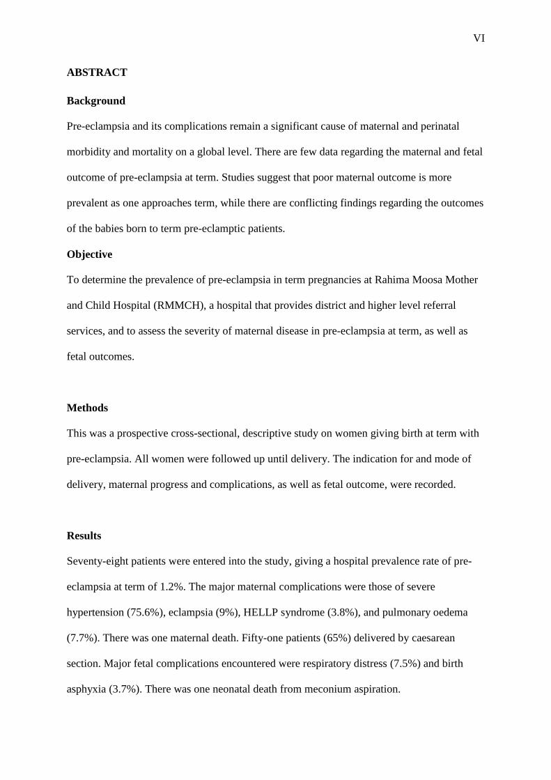
Seventy-eight patients were entered into the study, giving a hospital prevalence rate of pre-
eclampsia at term of 1.2%. The major maternal complications were those of severe
hypertension (75.6%), eclampsia (9%), HELLP syndrome (3.8%), and pulmonary oedema
(7.7%). There was one maternal death. Fifty-one patients (65%) delivered by caesarean
section. Major fetal complications encountered were respiratory distress (7.5%) and birth
asphyxia (3.7%). There was one neonatal death from meconium aspiration.

VII
Conclusion
This study revealed a significant number of patients having major maternal as well as fetal
complications of pre-eclampsia at term. Larger epidemiological studies are needed to predict
maternal and fetal outcomes in term pre-eclamptic patients.
VIII
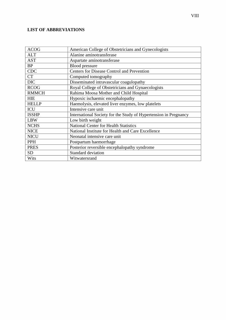
LIST OF ABBREVIATIONS
ACOG American College of Obstetricians and Gynecologists
ALT Alanine aminotransferase
AST Aspartate aminotransferase
BP Blood pressure
CDC Centers for Disease Control and Prevention
CT Computed tomography
DIC Disseminated intravascular coagulopathy
RCOG Royal College of Obstetricians and Gynaecologists
RMMCH Rahima Moosa Mother and Child Hospital
HIE Hypoxic ischaemic encephalopathy
HELLP Haemolysis, elevated liver enzymes, low platelets
ICU Intensive care unit
ISSHP International Society for the Study of Hypertension in Pregnancy
LBW Low birth weight
NCHS National Center for Health Statistics
NICE National Institute for Health and Care Excellence
NICU Neonatal intensive care unit
PPH Postpartum haemorrhage
PRES Posterior reversible encephalopathy syndrome
SD Standard deviation
Wits Witwatersrand
IX
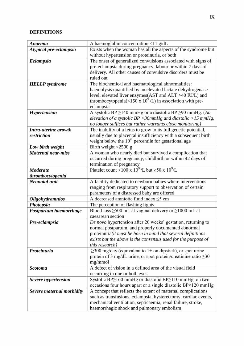
DEFINITIONS
Anaemia A haemoglobin concentration <11 g/dL
Atypical pre-eclampsia Exists when the woman has all the aspects of the syndrome but
without hypertension or proteinuria, or both
Eclampsia The onset of generalized convulsions associated with signs of
pre-eclampsia during pregnancy, labour or within 7 days of
delivery. All other causes of convulsive disorders must be
ruled out
HELLP syndrome The biochemical and haematological abnormalities:
haemolysis quantified by an elevated lactate dehydrogenase
level, elevated liver enzymes(AST and ALT >40 IU/L) and
thrombocytopenia(<150 x 109
/L) in association with pre-
eclampsia
Hypertension
A systolic BP ≥140 mmHg or a diastolic BP ≥90 mmHg. (An
elevation of a systolic BP >30mmHg and diastolic >15 mmHg,
no longer suffices but rather warrants close monitoring)
Intra-uterine growth
restriction
The inability of a fetus to grow to its full genetic potential,
usually due to placental insufficiency with a subsequent birth
weight below the 10th
percentile for gestational age
Low birth weight Birth weight <2500 g
Maternal near-miss A woman who nearly died but survived a complication that
occurred during pregnancy, childbirth or within 42 days of
termination of pregnancy
Moderate
thrombocytopenia
Platelet count <100 x 109 /L but ≥50 x 10
9/L
Neonatal unit A facility dedicated to newborn babies where interventions
ranging from respiratory support to observation of certain
parameters of a distressed baby are offered
Oligohydramnios A decreased amniotic fluid index ≤5 cm
Photopsia The perception of flashing lights
Postpartum haemorrhage Blood loss ≥500 mL at vaginal delivery or ≥1000 mL at
caesarean section
Pre-eclampsia De novo hypertension after 20 weeks’ gestation, returning to
normal postpartum, and properly documented abnormal
proteinuria(it must be born in mind that several definitions
exists but the above is the consensus used for the purpose of
this research)
Proteinuria ≥300 mg/day (equivalent to 1+ on dipstick), or spot urine
protein of 3 mg/dL urine, or spot protein/creatinine ratio ≥30
mg/mmol
Scotoma A defect of vision in a defined area of the visual field
occurring in one or both eyes
Severe hypertension Systolic BP≥160 mmHg or diastolic BP≥110 mmHg, on two
occasions four hours apart or a single diastolic BP≥120 mmHg
Severe maternal morbidity A concept that reflects the extent of maternal complications
such as transfusions, eclampsia, hysterectomy, cardiac events,
mechanical ventilation, septicaemia, renal failure, stroke,
haemorrhagic shock and pulmonary embolism
X
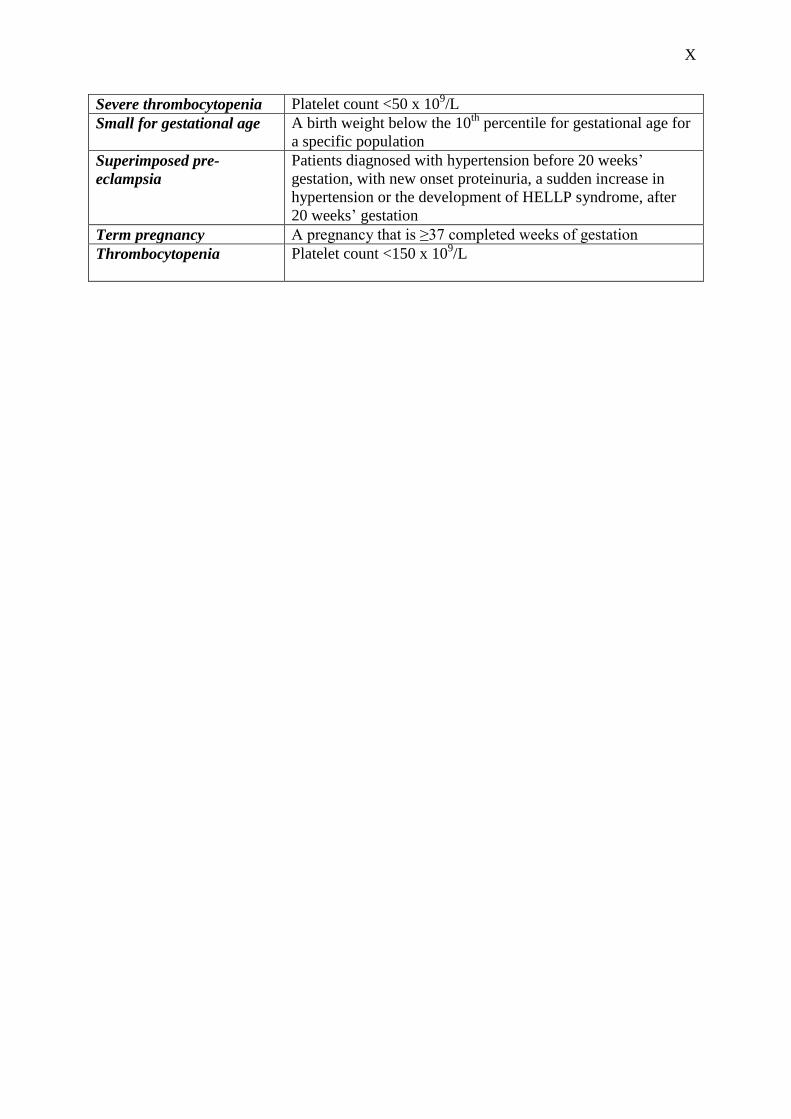
Severe thrombocytopenia Platelet count <50 x 109/L
Small for gestational age A birth weight below the 10th
percentile for gestational age for
a specific population
Superimposed pre-
eclampsia
Patients diagnosed with hypertension before 20 weeks’
gestation, with new onset proteinuria, a sudden increase in
hypertension or the development of HELLP syndrome, after
20 weeks’ gestation
Term pregnancy A pregnancy that is ≥37 completed weeks of gestation
Thrombocytopenia Platelet count <150 x 109/L
XI
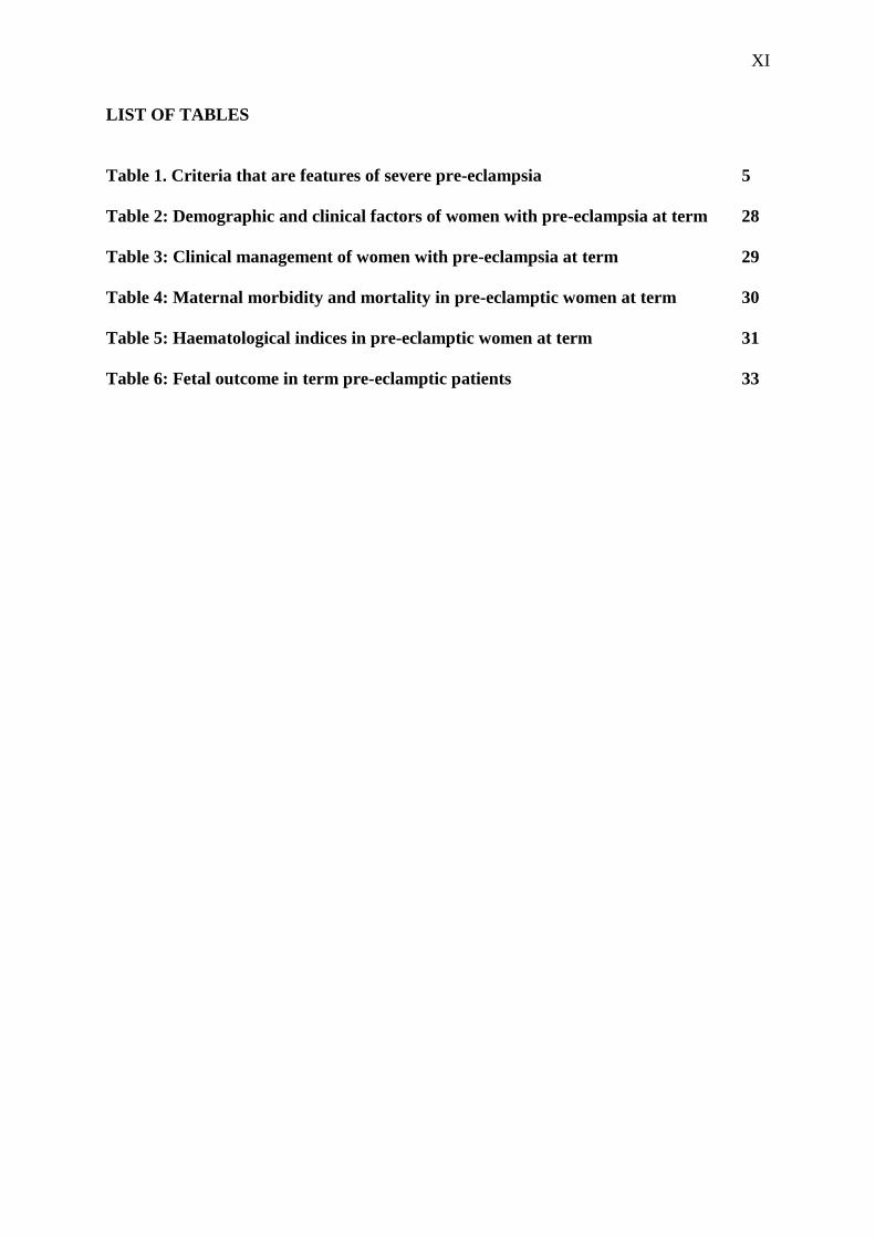
LIST OF TABLES
Table 1. Criteria that are features of severe pre-eclampsia 5
Table 2: Demographic and clinical factors of women with pre-eclampsia at term 28
Table 3: Clinical management of women with pre-eclampsia at term 29
Table 4: Maternal morbidity and mortality in pre-eclamptic women at term 30
Table 5: Haematological indices in pre-eclamptic women at term 31
Table 6: Fetal outcome in term pre-eclamptic patients 33
1
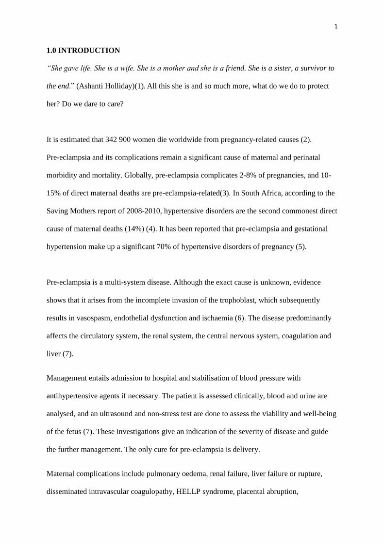
1.0 INTRODUCTION
“She gave life. She is a wife. She is a mother and she is a friend. She is a sister, a survivor to
the end.” (Ashanti Holliday)(1). All this she is and so much more, what do we do to protect
her? Do we dare to care?
It is estimated that 342 900 women die worldwide from pregnancy-related causes (2).
Pre-eclampsia and its complications remain a significant cause of maternal and perinatal
morbidity and mortality. Globally, pre-eclampsia complicates 2-8% of pregnancies, and 10-
15% of direct maternal deaths are pre-eclampsia-related(3). In South Africa, according to the
Saving Mothers report of 2008-2010, hypertensive disorders are the second commonest direct
cause of maternal deaths (14%) (4). It has been reported that pre-eclampsia and gestational
hypertension make up a significant 70% of hypertensive disorders of pregnancy (5).
Pre-eclampsia is a multi-system disease. Although the exact cause is unknown, evidence
shows that it arises from the incomplete invasion of the trophoblast, which subsequently
results in vasospasm, endothelial dysfunction and ischaemia (6). The disease predominantly
affects the circulatory system, the renal system, the central nervous system, coagulation and
liver (7).
Management entails admission to hospital and stabilisation of blood pressure with
antihypertensive agents if necessary. The patient is assessed clinically, blood and urine are
analysed, and an ultrasound and non-stress test are done to assess the viability and well-being
of the fetus (7). These investigations give an indication of the severity of disease and guide
the further management. The only cure for pre-eclampsia is delivery.
Maternal complications include pulmonary oedema, renal failure, liver failure or rupture,
disseminated intravascular coagulopathy, HELLP syndrome, placental abruption,
2
cerebrovascular accidents, eclampsia and death (7). Fetal growth impairment, placental
abruption and perinatal mortality are a consequence of ischaemia due to decreased
uteroplacental blood flow.
It has been proposed that pre-eclamptic patients who present at term may have normal to
increased utero-placental flow, sparing the fetus from the complications of placental
insufficiency that often characterises pre-eclampsia of earlier onset (8). This hypothesis
creates the need to explore the outcome of this population of pre-eclamptic patients at term.
The literature is abundant with studies looking at pre-eclampsia and comparing immediate
and delayed delivery of such patients. However, the dilemma of immediate delivery versus
delaying for maturity of the baby does not exist in the pre-eclamptic patient at ≥37 weeks’
gestation because there is consensus that delivery should be expedited in such women(7).
The aim of this study was to determine the prevalence of pre-eclampsia at term at Rahima
Moosa Mother and Child Hospital (RMMCH), a tertiary level hospital that serves the areas
around Coronationville in Johannesburg. In view of the above-mentioned hypothesis that pre-
eclamptic patients at term have a near normal utero-placental flow compared with those who
have early-onset pre-eclampsia, the researcher felt compelled to assess the maternal and fetal
outcome in this specific population of pre-eclamptic women.
3
2.0 LITERATURE REVIEW
Although the literature is overwhelmed with data on pre-eclampsia, it becomes apparent that
little work has been undertaken to show the outcome of the pre-eclamptic pregnancy at term.
Over the past 20-odd years a large amount of work has been carried out in Africa, Asia, Latin
America and the Caribbean, in which this “ongoing holocaust” is described to be killing 10-
15% of mothers(3). According to the South African Saving Mothers report of 2008-2010,
there was a small reduction in deaths due to complications of hypertension, compared to the
previous triennium (4). However, no mention was made of the gestation of pregnancy at
which these outcomes were documented.
2.1 Epidemiology
Pre-eclampsia complicates 2-8% of pregnancies worldwide (3). The prevalence varies
according to geographic regions of the world, ranging from 2.5% in Saudi Arabia to 4.3% in
Turkey, to 5-8% in the USA (6,9,10). This variation may be attributed to racial differences,
socioeconomic status and other demographic parameters such as age and parity (10). Pre-
eclampsia is described as a disease predominantly affecting nulliparous women, and women
at the extremes of their reproductive ages (<20 and >40 years) (9). The exact prevalence of
pre-eclampsia in South Africa is unknown, although a 5-year prospective study in Cape Town
reported a prevalence of 7.1% (11).
A 10 year retrospective study in Saudi Arabia showed that 42% of pre-eclamptic patients
were primigravid (9). Pregnancy, even if it is terminated early, offers some form of immunity
to pre-eclampsia in the next pregnancy (12). However, it has been shown that the protective
effect of multiparity is lost with a change of partner and also raises the concept of
primipaternity rather than primigravidity (11).

4
Extremes of maternal age, multiple pregnancy, previous pre-eclampsia, chronic hypertension,
pre-gestational diabetes, vascular and connective tissue disorder, nephropathy and
antiphospholipid syndrome are conditions that predispose a pregnant woman to develop pre-
eclampsia. In obesity, although the pathophysiology is unknown, the increased risk for
developing pre-eclampsia is postulated to be due to the oxidative stress, inflammation and
altered vascular function that have been reported in obese women (13).
African-American race or black race has been shown to be a predisposing factor (6,14).
The older literature reports cases of pre-eclampsia in close relatives, however some authors
have argued that this was owing to the commonality of the condition and could just as well be
coincidental (12). Decades later, the genetic factor still remains and has repeatedly been
shown to be a predisposing component to pre-eclampsia (6).
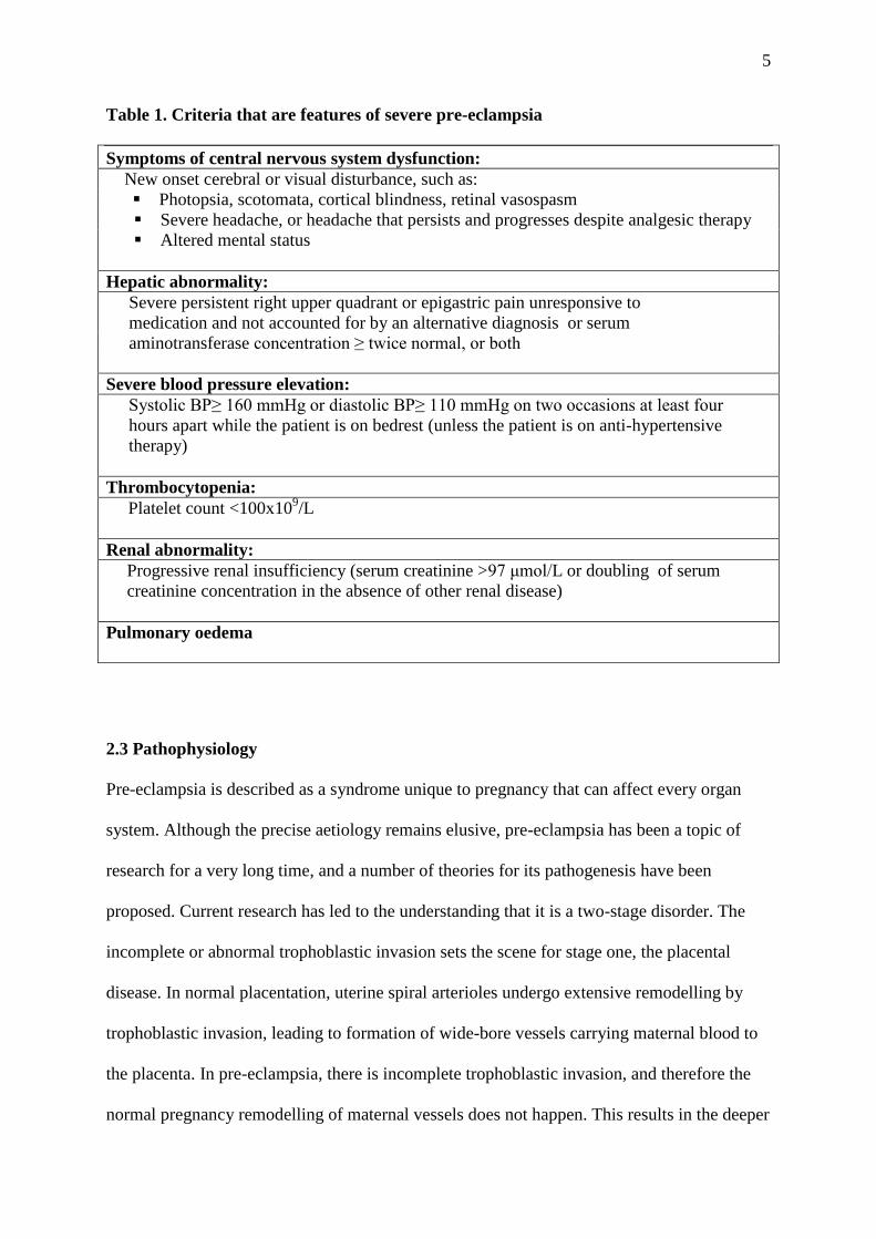
2.2 Definitions and criteria for the severity of pre-eclampsia
The importance of classifying the severity of pre-eclampsia arises when one is faced with an
ill patient who is still remote from term gestation. The definitions used in this research report
are provided in the list of definitions at the beginning of the report. In the clinical situation, a
decision on classification of a patient’s hypertensive disorder has to be made taking into
consideration the patient’s clinical condition and risks of morbidity and mortality, as well as
delivering a pre-term baby, which has its own consequences. Various classifications exist in
which there are criteria to define severe pre-eclampsia. The alternative to those criteria
defined as severe are accepted as mild or moderate as they are not specifically defined. Table
1 shows the most recent criteria as accepted by the ACOG (15).
5
Table 1. Criteria that are features of severe pre-eclampsia
Symptoms of central nervous system dysfunction:
New onset cerebral or visual disturbance, such as:
Photopsia, scotomata, cortical blindness, retinal vasospasm
Severe headache, or headache that persists and progresses despite analgesic therapy
Altered mental status
Hepatic abnormality:
Severe persistent right upper quadrant or epigastric pain unresponsive to
medication and not accounted for by an alternative diagnosis or serum
aminotransferase concentration ≥ twice normal, or both
Severe blood pressure elevation:
Systolic BP≥ 160 mmHg or diastolic BP≥ 110 mmHg on two occasions at least four
hours apart while the patient is on bedrest (unless the patient is on anti-hypertensive
therapy)
Thrombocytopenia:
Platelet count <100x109/L
Renal abnormality:
Progressive renal insufficiency (serum creatinine >97 μmol/L or doubling of serum
creatinine concentration in the absence of other renal disease)
Pulmonary oedema
2.3 Pathophysiology
Pre-eclampsia is described as a syndrome unique to pregnancy that can affect every organ
system. Although the precise aetiology remains elusive, pre-eclampsia has been a topic of
research for a very long time, and a number of theories for its pathogenesis have been
proposed. Current research has led to the understanding that it is a two-stage disorder. The
incomplete or abnormal trophoblastic invasion sets the scene for stage one, the placental
disease. In normal placentation, uterine spiral arterioles undergo extensive remodelling by
trophoblastic invasion, leading to formation of wide-bore vessels carrying maternal blood to
the placenta. In pre-eclampsia, there is incomplete trophoblastic invasion, and therefore the
normal pregnancy remodelling of maternal vessels does not happen. This results in the deeper
6
myometrial arterioles maintaining their pre-pregnancy anatomical structure, thus remaining
narrow conduits of blood flow. Their mean external diameter is half that of vessels in normal
placentas (16). The effect of these abnormally narrowed spiral arterioles is placental
hypoperfusion and hypoxia. Stage two is the maternal clinical syndrome. The placental
disease leads to a release of trophoblastic debris, apoptotic cells and anti-angiogenic factors,
which induce a maternal systemic inflammatory response and widespread endothelial
vascular damage, producing the pre-eclampsia syndrome of high blood pressure, proteinuria
and organ dysfunction (16).
2.3.1 Vasospasm and haemoconcentration
Intense vasospasm with endothelial damage and dysfunction in the maternal circulation is
caused by the interaction of various vasoactive agents such as prostacyclin (vasodilator),
thromboxane A2 (potent vasoconstrictor), nitric oxide (potent vasodilator), and endothelins
(potent vasoconstrictors) (6). In pre-eclampsia, compared with normal pregnancy,
prostacyclin production decreases, while thromboxane A2 production by platelets is
increased, with subsequent increased vascular resistance and hypertension (16). The
simultaneous endothelial cell damage results in: 1) decreased production of nitric oxide and
release of substances that promote coagulation and increase sensitivity to vasopressors; and
2) a capillary leak of blood constituents, which are deposited subendothelially. This results in
haemoconcentration, decreased colloid osmotic pressure, and contraction of the intravascular
space. As the endothelium undergoes repair after delivery, the vasoconstriction resolves, and
there is a concomitant decrease in the haematocrit levels as the blood volume increases (6).

7
2.3.2 Haematological changes
A commonly identified abnormality in pre-eclampsia is thrombocytopenia. The intensity may
vary according to the severity of the pre-eclampsia. The lower platelet count is associated
with higher maternal and fetal morbidity and mortality (17). The thrombocytopenia usually
worsens with ongoing pregnancy. After delivery, the decline in platelet count may continue
for 48-72 hours. The platelet count then increases gradually and within three to five days
should reach normal levels. Haemolysis may also occur simultaneously with the
thrombocytopenia. This is reflected by a disproportionate rise in lactate dehydrogenase in the
serum (6), and the presence of schizocytosis, spherocytosis and reticulocytosis on a
peripheral blood smear. The most sensitive objective marker of haemolysis is a reduced
serum haptoglobin level (18,19).
2.3.3 Hepatic changes
Patients with severe pre-eclampsia may have significantly altered hepatic function (6).
Alanine aminotransferase (ALT) and aspartate aminotransferase (AST) levels may be
elevated and inversely follow platelet counts. They also usually normalize within three days
after delivery. Hyperbilirubinaemia may occur especially when haemolysis is present.
Autopsy studies have found that the characteristic lesion commonly found is hepatic
infarction and periportal haemorrhage in the liver lobule periphery (16,20). Hepatic
haemorrhage may extend to form a subcapsular haematoma that presents clinically as right
upper abdominal pain. Hepatic rupture is rare but life-threatening, requiring emergency
surgical intervention (6,18).
HELLP syndrome, a variant of severe pre-eclampsia, is associated with substantial mortality
and morbidity. It diagnosis is based on laboratory abnormalities. The syndrome has been
8
reported to occur in 20% of women with severe pre-eclampsia (18). Maternal mortality
ranges from 0-4% (18). Other complications associated with HELLP syndrome include acute
renal failure in 8% of women, pulmonary oedema in 6%, and subcapsular liver haematoma in
1% (18). Due to the unfavourable cervix and early gestation found in most patients with
HELLP syndrome, caesarean delivery is commonly performed. Wound haematoma and
subsequent wound sepsis becomes a further complication in this group of patients, due to the
low platelet count (18).
2.3.4 Renal changes
In normal pregnancy, there is an increase in renal blood flow with increased glomerular
filtration and decreased serum creatinine level. However, due to the vasospasm in pre-
eclampsia this physiological change does not occur. Instead, there is decreased glomerular
filtration which, together with the haemoconcentration, leads to oliguria and a rise in serum
creatinine level. In the glomeruli, there is podocyte dysfunction and endothelial swelling, also
termed glomerular capillary endotheliosis, which causes an increase in the permeability of the
membrane with subsequent protein leakage into the urine (6).
Acute renal failure is a rare complication of pre-eclampsia and usually resolves
spontaneously after delivery (21), although dialysis may occasionally be required. More
frequently, acute tubular necrosis is precipitated by haemorrhagic shock which accompanies
the other complications mentioned above, mainly placental abruption (21). Rarely,
irreversible renal cortical necrosis develops. A five-year prospective study in the United
Kingdom showed that 0.55% of women with pre-eclampsia required dialysis based on
worsening creatinine levels (22). A prospective case series in Tygerberg Hospital, Cape
9
Town, reported that 6 out of 340 pre-eclamptic patients (1.7%) had severe renal impairment,
of whom one required dialysis (11).
2.3.5 Neurological changes
Changes to the cerebral circulation are the primary effectors of the neurological symptoms
seen in pre-eclamptic patients. Normally, by the mechanism of autoregulation, cerebral blood
flow is maintained at a constant rate despite changes in cerebral perfusion pressures. Blood
pressures above and below the autoregulatory limits result in this mechanism being lost. In
the case of an acute hypertensive episode at pressures above the autoregulatory limit, the
excessive intravascular pressure overcomes the constriction of the vascular smooth muscle
with subsequent forced dilatation of the cerebral vessels. This loss in autoregulation results in
hyperperfusion and disruption of the blood-brain barrier with resultant vasogenic oedema, as
plasma constituents leave the vasculature and expand the extra-cellular space (23). This
forms the underlying cause of eclampsia and hypertensive encephalopathy (23).
Generalised seizures occur in the presence of excessive release of excitatory
neurotransmitters, massive depolarization of network neurons, and bursts of action
potentials(16). In the pre-eclamptic patient this manifests as generalized tonic-clonic
convulsions and in the absence of other neurologic conditions is known as eclampsia.
Morbidity to both mother and fetus is increased and prolonged seizures can cause significant
brain injury and later brain dysfunction(16).
Eclampsia can occur antepartum, intrapartum or postpartum and is most common in the third
trimester with an increase in frequency as term approaches (24). Although uncommon in
Europe with a reported prevalence of 2-3 cases per 10 000 births, it occurs more frequently in
developing countries, with an estimated 16-69 cases per 10 000 births (3). Eclampsia is a

10
major cause of morbidity and mortality worldwide and accounts for more than 50 000
maternal deaths annually (25). The case fatality rate of eclampsia is 3-5% in low and middle
income countries compared to 1% in developed countries (3,5). A three-year review from an
academic hospital in Nigeria showed a prevalence of 13 per 1000 deliveries, with a case
fatality rate of 8.5% (25). A retrospective analysis done in Durban showed an increase in the
prevalence of eclampsia from 2.8 per 1000 deliveries to 6 per 1000 deliveries from 1980 to
1990. However, in the same period there was a slight decline in the case fatality rate from
12% to 9% (26). The high morbidity and mortality was attributed to low socioeconomic
status, delayed referral to tertiary institution, non-availability of transport facilities and
unbooked status of patients (25,26).
Another recently described phenomenon is the posterior reversible encephalopathy syndrome
(PRES). Although its exact cause is unclear, it is thought to occur with disrupted
autoregulation and endothelial dysfunction with or without subsequent vasogenic oedema
(27). PRES is typically associated with severe hypertension but has also been described in
patients with endothelial damage who experience rapid increases in blood pressure (28).
Insidious onset of headaches, altered levels of consciousness, visual changes and seizures,
associated with characteristic neuroimaging findings of posterior cerebral white matter
describes this clinical syndrome. Reversibility is most frequent if PRES is promptly
recognized and treated, however its effects may not always be reversible and its location may
not necessarily be confined to the posterior regions of the brain (24,27).
Intracerebral haemorrhage, resulting from severe systolic or diastolic hypertension or sudden
increases in blood pressure, has been reported to be the commonest cause of maternal
mortality from hypertensive disorders of pregnancy, irrespective of the occurrence of
11
eclampsia (29). Diastolic blood pressures above 110 mmHg and persistent systolic values
higher than 160 mmHg have been associated with significant risk of complications, which
may result in PRES, eclampsia, stroke or all three (29).
Clinically evident neurological manifestations in patients without eclampsia or stroke include
headaches, visual disturbances, and generalized hyperreflexia (24), which tend to resolve
after the administration of magnesium sulphate. Blindness is rare and may be transient (9).
The loss or disturbance of consciousness associated with eclampsia from the seizure itself,
PRES or an intra-cerebral haemorrhage, may require intensive care unit admission with
intubation and ventilation (26).
2.3.6 Pulmonary oedema
Pulmonary oedema is reported as the fourth commonest cause of maternal mortality in the
United Kingdom. A review in 2012 showed estimated rates of acute pulmonary oedema in
pregnancy of 0.08% to 0.5% (30). A five-year prospective study in the United Kingdom
showed a prevalence of pulmonary oedema of 1.2 per 10 000 deliveries (25 out of 210 631
patients) (22). The aetiology is multi-factorial, often being a combination of left ventricular
failure and non-cardiogenic pulmonary oedema. In some women, pulmonary oedema occurs
as a result of increased hydrostatic pressure and a decreased colloid osmotic pressure
resulting in fluid accumulation in the lung parenchyma, especially in the postpartum period
(29, 30). Other women may demonstrate increased capillary permeability, and in some
patients acute pulmonary oedema may be caused by iatrogenic fluid volume overload (24).
Breathing and oxygenation is subsequently compromised, manifesting as severe respiratory
distress that may require intubation. There are few studies demonstrating pulmonary oedema
in pre-eclamptic women, let alone pre-eclamptic women at term (30).

12
2.3.7 Placental complications
Due to the compromised uteroplacental perfusion following the abnormal trophoblastic
invasion, manifestations of pre-eclampsia can also be seen in the fetal-placental unit. This is a
major contributor to the increase in perinatal morbidity and mortality rates. By 16 weeks of
gestation, the vascular resistance in the uterine artery decreases markedly in normal
placentation. However, in pre-eclampsia the abnormal placentation results in the vascular
resistance remaining abnormally high, which subsequently is associated with
oligohydramnios and fetal growth restriction (16,31).
Described as one of the most significant causes of maternal morbidity and perinatal mortality,
placental abruption is defined as partial or complete separation of a normally implanted
placenta before delivery occurs (14). Bleeding then follows from the decidua basalis, between
the placenta and uterine wall. The combination of pre-eclampsia and placental abruption
results in significant maternal morbidity, with hypovolaemia, coagulopathy and renal failure
(32).
A Finnish overview in 2010 reported that the overall prevalence of placental abruption in all
pregnancies varies from 0.4-1.0% (14). The occurrence tends to decrease with advancing
gestation. One prospective case series in Tygerberg Hospital, Cape Town reported a
prevalence as high as 20.2% in women with severe pre-eclampsia between 24 and 34 weeks’
gestation (11). Some studies have shown that women with chronic hypertension have a 2.4-
fold increased risk for abruption compared to those without, which increases to 2.8-7.7-fold
with superimposed pre-eclampsia or fetal growth restriction (32). A study in Norway showed
an association of pre-eclampsia with placental abruption with an odds ratio for abruption
ranging from 1.9 in mild pre-eclampsia to 4.1 in severe pre-eclampsia (33). The literature
13
suggests that of all risk factors, hypertensive disorders ranks the highest on the list for
placental abruption.
2.4 Neonatal complications
Perinatal mortality is high in pre-eclamptic women and increases with eclampsia (3).
Together, pre-eclampsia and eclampsia account for a quarter of stillbirths and neonatal deaths
in developing countries. The perinatal mortality rate for pregnancies complicated by pre-
eclampsia is three times higher in developing countries than in higher income regions (3).
A retrospective study at Tygerberg and Paarl Hospitals, Western Cape, from 2005-2006
studied 264 women with late-onset pre-eclampsia (≥34 weeks) and showed a mean
gestational age at the time of diagnosis of 37 weeks, with a median birth weight of 2700 g.
There were no early neonatal deaths. Of the five intra-uterine deaths, all due to placental
abruption, only two occurred at ≥37 weeks. Sixty-nine babies (26%) required admission for
special neonatal care, a much greater proportion than would be expected from pregnancies at
≥34 weeks’ gestation not affected by pre-eclampsia. However, the author did not state how
many of these admissions were delivered to pre-eclamptic women at ≥37 weeks (34).
A study in the United Kingdom showed that 34.6% of 1087 pre-eclamptic women delivered
after 37 weeks (22). Serious complications were reported in 151 (13.9%) of the pre-eclamptic
patients. Of the 382 babies born after 37 weeks gestation, 14.7% required neonatal admission.
Despite the fact that the focus of the study was on general outcome and not specific to
gestational age, it does show that a significant number of babies born after 37 weeks gestation
are admitted to neonatal units. This observation has also been stated by Sibai (35) and
supports the need for more research to find out why this is the case.
14
The most important factor for survival of a healthy baby is gestational age (36). In a study
from Tygerberg Hospital comparing expectant management to immediate delivery in severe
early-onset pre-eclampsia, gestational age was the dominant consideration, with elective
delivery often decided on after 34 weeks gestation (36). Although most pregnancies were
remote from term, the study showed an increased survival rate at greater gestations, from
72% at 28 weeks to 92% at 29 weeks (36). This factor is further supported by data from the
study mentioned above from Tygerberg and Paarl Hospitals on late-onset pre-eclampsia, in
which there were no neonatal deaths (34).
A three-year prospective cohort study in Ireland from 1997-2000 on term patients confirmed
that pre-eclampsia is associated with neonatal encephalopathy (37). One third of the
encephalopathic babies, associated with hypoxaemia, were born to pre-eclamptic women at
term. The mean umbilical cord arterial pH was lower in the babies born to pre-eclamptic
mothers. The authors concluded that the fetuses of pre-eclamptic women may be hypoxaemic
resulting from poor utero-placental perfusion (37).
While much of the above data are useful, it remains evident that overall the literature lacks
studies specifically exploring the outcome of babies delivered to pre-eclamptic women at
term.
15
2.5 Clinical management
The management of pre-eclampsia is subdivided according to severity of the disease. There
is evidence that a patient with mild pre-eclampsia can be managed expectantly until fetal
viability is reached. As long as she is treatment-compliant and there is no change in her
clinical and laboratory parameters, the patient can be managed as an outpatient at a day unit,
which would exist in most facilities in developed countries. However, in South Africa,
admission to hospital is advised for all pre-eclamptic women using public service facilities
(7). There must be close maternal and fetal evaluation as life-threatening severe pre-
eclampsia can develop within a short space of time (6,21,38).
On admission to hospital, maternal evaluation consists of investigations to confirm or exclude
worsening pre-eclampsia. This includes laboratory investigations to observe haematological,
hepatic and renal function, as well as 12-24 hour urine collection for protein (6). Anti-
hypertensive treatment should commence promptly. The Royal College of Obstetricians &
Gynaecologists(RCOG) recommend emergency anti-hypertensive agents when BP ≥160/110
mmHg, and the American College of Obstetricians and Gynecologists(ACOG) recommends
treatment for diastolic blood pressure levels of ≥105-110 mmHg (6,38). The University of the
Witwatersrand (Wits) protocol adopted by academic hospitals in Johannesburg, including
RMMCH, recommends emergency treatment at a BP ≥170/110 mmHg (7). In a case of acute
severe hypertension, the aim of emergency treatment is to gradually bring down the blood
pressure so that cerebral autoregulation is not compromised (21). Decreasing the blood
pressure too quickly may cause uteroplacental hypoperfusion with fetal distress, or maternal
cerebral ischaemia. Hydralazine, labetalol, or nifedipine are used for the acute management
of hypertension, with methyldopa being the cornerstone for long term control (29,38,39).

16
It is vital that strict fluid balance is maintained. Due to the risks of fluid overload and
pulmonary oedema, total fluid input should be limited to 85 mL/hour. It must also be taken
into consideration that parenteral anti-hypertensive agents can cause a sudden drop in blood
pressure; hence, preloading with 500 mL crystalloid fluid may be of value (38).
Although there are different schools of thought regarding the use of magnesium sulphate for
prevention of seizures, the Wits protocol supports its use for patients with severe or
imminent eclampsia, as does the RCOG. Certain factors must be taken into consideration
when administering magnesium sulphate, such as awareness of toxic effects such as
hypotension, depression of reflexes, respiratory depression and cardiac arrest, for which the
antidote calcium gluconate must be readily available (7,29,38,39).
The decision to deliver the baby is multifactorial, wherein maternal and fetal wellbeing, past
obstetric history and current gestation need to be considered. By the Wits protocol and
supported by the National Institute for Health and Care Excellence (NICE) guidelines in the
United Kingdom, patients with severe pre-eclampsia are delivered at 34 weeks gestation or an
estimated fetal weight of 1.5 kg after administration of steroids for accelerating fetal lung
maturity (6,7,39). The NICE guidelines advise that delivery can also be offered to pre-
eclamptic women with mild to moderate hypertension at 34 to 36 completed weeks
considering maternal and fetal conditions, risk factors and availability of neonatal facilities.
Patients from 37 weeks and 0 days should be offered delivery within 24-48 hours (39). There
is no justification in prolonging a term pregnancy with pre-eclampsia of any severity.
Irrespective of gestational age, delivery generally needs to be expedited in women with
severe pre-eclampsia, poorly controlled BP requiring more than three agents, persistent
17
imminent eclampsia after administration of magnesium sulphate, HELLP syndrome,
eclampsia, cerebral oedema, end-organ damage, intrauterine growth restriction,
oligohydramnios, fetal distress, intra-uterine fetal death or placental abruption (6,38,39).
2.6 Mode of delivery
The operative delivery rate is increased in patients with hypertensive disorders and more so in
patients with complicated pre-eclampsia (10). However, there are retrospective studies which
have shown that induction of labour is not harmful to low birth weight infants. Vaginal
delivery can be recommended for selected patients with severe pre-eclampsia who have no
obstetric indication for caesarean section (10). However, a recent prospective study showed a
four-fold increased risk of failed induction and a two-fold increased risk of caesarean section
in pre-eclamptic women (31).
In the event of eclampsia, due to the unfavourability of the cervix and the need to deliver
timeously, the caesarean section rate increases, documented to be 84.8% in a three-year
review in Nigeria (25). There are no randomized clinical trials exploring the optimum mode
of delivery in patients with severe pre-eclampsia (6). The decision to deliver by caesarean
section should be individualized (6).
2.7 The pre-eclamptic patient at term
As stated earlier, there are few studies specifically focusing on pre-eclampsia and its
management at term, although data are available on term subgroups in studies on pre-
eclampsia at all gestations.
18
A retrospective cohort study carried out in 35 hospitals in Canada from 1991-1996 compared
the birth weights of babies born to pre-eclamptic patients, to those born to normotensive
patients from 32 weeks up to 42 weeks gestation (8). The authors looked at all babies
delivered before 37 weeks gestation, and showed that those born to the pre-eclamptic mothers
had lower birth weights compared to those born to normotensive mothers (p<0.01), after
adjusting for confounding factors (smoking, maternal age, parity, obesity, prior small-for-
gestational-age, prior large-for-gestational-age, anaemia and premature rupture of
membranes). However, the babies born at ≥37 weeks’ gestation showed no statistically
significant difference in mean birth weights when comparing both categories of patients.
Contrary to common belief, the pre-eclamptic patients who carried to beyond 42 weeks
gestation delivered babies that had a higher mean birth weight than those delivered to
normotensive mothers, but the difference was not statistically significant. The focus of the
study was the association of birth weights with gestation in pre-eclampsia, so there was no
mention of any maternal or fetal morbidity. However, it can be deduced that babies born at
term to pre-eclamptic women may have birth weights that are no different from those born at
term to women who do not have pre-eclampsia. Interestingly, a significant proportion of pre-
eclamptic patients (72%), carried uneventfully to term. Neither the gestation at the time of
diagnosis nor the severity of the disease was mentioned. However, the study questions the
hypotheses on the pathophysiology of pre-eclampsia. The findings contest the accepted
ischaemic model, which states that there is decreased uteroplacental perfusion, as these
patients, with pre-eclampsia at term, had a normal and perhaps even increased blood flow in
the majority of cases.
This outcome suggests a heterogeneity in the pathogenesis of pre-eclampsia, that pre-
eclampsia may manifest in two forms: 1) a restricted fetal growth early-onset pre-eclampsia,
where affected patients deliver before term and are more likely to have decreased
19
uteroplacental perfusion; and 2) a normal fetal growth late-onset pre-eclampsia, with patients
often presenting at and delivering at term, likely to have normal uteroplacental perfusion.
This theory is supported by a retrospective cohort analysis that was carried out in pre-
eclamptic patients in Missouri, USA (40), where ischaemic placental disease was more
frequent at pre-term than at term gestation (16% versus 5.5% respectively). Again, this
suggests different pathophysiological pathways between term and preterm pre-eclampsia and
hence a heterogeneity of the disease process. While this is a clinically evident heterogeneity,
no studies support the presence of different microvascular or molecular pathogenetic
processes between early-onset and late-onset pre-eclampsia.
A critical review in 2011 supports the above hypothesis that early- and late-onset pre-
eclampsia are two distinct diseases, characterized by different biochemical markers, risk
factors, clinical features and haemodynamic states. Early-onset pre-eclampsia is described to
have a comparatively more severe disease profile than late-onset pre-eclampsia, with a
stronger association with abnormal placentation, increased risk of multi-organ involvement,
and adverse maternal and fetal outcome. However, due to the lack of studies and reviews no
criteria have been established to define the two entities. More research is required to clarify
the differences or similarities between the two groups(41).
The differing pathophysiology in the above two groups of patients has been further
investigated and supported in a local prospective case-control study in Tygerberg Hospital
done in 2007 (42). The study showed that the placentas of patients with early-onset pre-
eclampsia weighed significantly less and had more ischaemic lesions when compared to those
of pre-eclamptic women at term (42). Although the authors described the placentas of late-
onset pre-eclamptic pregnancies as healthy, placental abruption in this group was still a
frequent occurrence (eight out of 25 cases).

20
The HYPITAT Study was a European multicentre randomised controlled trial that
investigated treatment of patients with gestational hypertension and mild pre-eclampsia at 36
to 41 weeks gestation, from 2005-2008 (43). Patients with severe pre-eclampsia were
excluded. The aim was to compare induction of labour with expectant management in
patients with mild pre-eclampsia and gestational hypertension beyond 37 weeks’ gestation.
Although there were no maternal deaths, there was a high rate of morbidity. Of 756 women,
30% developed severe hypertension, and 15 patients (2%) developed HELLP Syndrome,
10% had postpartum haemorrhage and 2 patients (0.2%) had pulmonary oedema. The
expectant management group included 11 of the 15 women who developed HELLP
syndrome, as well as both patients that developed pulmonary oedema. No patient had
placental abruption. Eighty-eight women in the induction group compared to 138 women in
the expectant management group progressed to severe disease. The study showed that
prolonging pre-eclampsia beyond 37 weeks added significant risk for maternal morbidity.
The study did not look at the outcome of pre-eclampsia at term per se, and perhaps merging
both pre-eclampsia and gestational hypertension was a weakness of this study. The
HYPITAT study data certainly show that there is a high rate of maternal morbidity in pre-
eclamptic patients at term. Morbidity rates for pre-eclampsia are likely even higher than
found in the study, because of the merging of gestational hypertension with pre-eclampsia,
and because severe disease and pre-eclampsia complications were part of the exclusion
criteria for the study.
A 10-year cross-sectional retrospective study of 685 pre-eclamptic pregnancies from Saudi
Arabia showed that 31% of stillbirths, and one out of seven neonatal deaths were in babies
who were delivered at term to pre-eclamptic patients (9). Placental abruption, renal failure,
coagulopathy and HELLP syndrome were the major maternal complications recorded. There

21
were two maternal deaths. Mild pre-eclamptic patients were managed expectantly and severe
pre-eclamptics managed by immediate delivery. About 70% of the pre-eclamptic women
delivered after 37 weeks. Whether these patients had mild pre-eclampsia and were managed
expectantly up to term, or whether they were patients who presented at term with severe
disease is unclear. Nevertheless, this is one of only a few studies where such a large number
of pre-eclamptic mothers carried to term. Unfortunately the details of the outcome of the
women at term were neither analysed nor reported on separately, and therefore cannot be
commented on with respect to morbidity and mortality of the mothers and babies(9).
22
3.0 PROBLEM STATEMENT AND OBJECTIVES
Pre-eclampsia, as part of the hypertensive disorders of pregnancy, is the second most
common cause of direct maternal mortality in South Africa. Being a disease that is exclusive
to pregnancy, it is well known that delivery is the only cure.
Conflicting findings of fetal outcomes, and potentially poor maternal outcome even as one
approaches term in pre-eclampsia, is what can be gathered from the relevant literature.
Having reviewed the literature above, it becomes evident that there is a deficiency of data
regarding the outcome of pre-eclamptic pregnancies at term. It appeared to be a feasible task
to indulge in the wealth of information that patients offer at RMMCH, to fill in some
evidence gaps regarding pre-eclampsia at term.
In a tertiary institution such as RMMCH, it was expected that patients with both mild and
severe pre-eclampsia were treated in the facility. In addition, RMMCH delivers almost all
women from its catchment area. These considerations made it feasible to study the prevalence
of pre-eclampsia in this area, in the absence of significant referral bias.
3.1 Objectives of this study
1. To determine the prevalence of pre-eclampsia at term at RMMCH
2. To describe demographic and clinical factors in women with pre-eclampsia at term
3. To assess the severity of maternal disease in term pre-eclamptic patients
4. To assess the fetal outcome of pregnancies with pre-eclampsia at term
23
4.0 METHODS
4.1 Study design and study population
This was a prospective cross-sectional, descriptive study, carried out over a period of six
months using a period sample. All women at term (≥37 weeks gestation by best estimate)
who were admitted for pre-eclampsia were entered into the study and followed up until
delivery. Patients who were admitted before term but delivered after 37 weeks were also
entered into the study. Patients who did not attend antenatal clinic (unbooked women) were
not included in the study because a true diagnosis of pre-eclampsia could not be made
without a prior blood pressure assessment.
The definition of pre-eclampsia used in this study and endorsed by the ISSHP (International
Society for the Study of Hypertension in Pregnancy) is described as the development of
hypertension with proteinuria after 20 weeks gestation, which returns to normal postpartum.
For women who booked after 20 weeks (a common occurrence in the population using
RMMCH), a normal blood pressure and absence of proteinuria that preceded the onset of pre-
eclampsia allowed classification of the patient as pre-eclamptic. Proteinuria was defined as
≥1+ on urine dipstick, or a spot protein/creatinine ratio ≥30 mg/mmol. Postpartum resolution
of the hypertension and proteinuria could not be confirmed in this study.
4.2 Setting
This study was conducted in the obstetric unit at RMMCH, a district and regional hospital
with tertiary functions, affiliated with the University of the Witwatersrand as a teaching
hospital. The hospital delivers about 1000 women each month and only rarely refers difficult
obstetric patients to other hospitals. It has only one referring midwife obstetric unit
(Discoverers Clinic), which delivers about 60 women each month. The protocol guiding care

24
at RMMCH states that all women with pre-eclampsia, irrespective of gestational age, are
admitted to hospital. Patients with mild pre-eclampsia are delivered if they reach term or if
they present at term (7).
4.3 Sample size
It was hoped that 100 women could be included in the study, to allow a precision with a 95%
confidence interval of at most 10% around observed proportion estimates.
4.4 Data collection
Every term pre-eclamptic patient admitted from 1 November 2012 to 30 April 2013 was
entered into the study. After receiving informed consent from the participating patient, the
researcher extracted the demographic and clinical data from the patient’s clinical file. The
antenatal information was entered into the study data sheet immediately, and then the notes
were followed prospectively up to delivery and postpartum. If a woman had already delivered
by the time the researcher saw her for the first time, or a woman was admitted before term
but delivered after 37 weeks, the information was entered retrospectively from the case notes.
Explanatory variables that were collected included age, parity, gravidity, and a history of
previous pre-eclampsia. The recorded details of the current pregnancy included the gestation
at, and mode of delivery, as well as the birth weight and Apgar scores of the baby. The length
of stay in hospital for the baby and the mother were also noted.
Consent from the 16 year old patient was taken after explaining the purpose of the study to
her, reassuring her that she will not be re-numerated, nor will she or her baby be exposed to
any harm, in layman’s terms in her language with the aid of a nursing staff member. An
opportunity was also allowed for her to raise any questions or concerns, and to reiterate what
25
was explained to her to reflect her understanding of the consent and her participation in the
study.
The outcome variables that were analysed were broadly divided into maternal and fetal.
Maternal variables included severe hypertension (defined as a systolic BP≥160 mmHg or
diastolic BP≥110 mmHg, on two occasions four hours apart or a single diastolic BP≥120
mmHg), eclampsia, pulmonary oedema (diagnosed clinically or radiographically), placental
abruption (clinical diagnosis and macroscopic examination of placenta) and HELLP
syndrome. Patients were classified as having HELLP syndrome by establishing the presence
of thrombocytopaenia (platelet count <100x109/L) and raised ALT or AST levels (>40 IU/L).
Fetal and neonatal outcomes were obtained from the delivery notes or the neonatal admission
file if the baby was admitted. The fetal outcome variables included stillbirth, birth weight,
Apgar scores, neonatal unit admission with reason, and neonatal death. The data collection
sheet is attached as Appendix I.
4.5 Data analysis
Descriptive statistical methods were used, including statements of summary measures such as
means, standard deviations, medians and ranges. Proportions were expressed as percentages.
Analytic statistical methods were not employed.
4.6 Ethics
This study received approval from the University of the Witwatersrand Human Resources
Research Ethics Committee, approval number M121060 (Appendix II). All participant
information was kept confidential, and data sheets did not include any unique identifiers such
as the patient’s name, birth date, hospital number or address. Hospital permission was
obtained from the Chief Executive Officer of RMMCH.

26
4.7 Funding
Data capturing was conducted by the researcher and processed with the help of the
supervisor. The researcher incurred all costs concerned.
27
5.0 RESULTS
5.1 The prevalence of pre-eclampsia at term at RMMCH
There were 6300 deliveries during the period 1 November 2012 to 30 April 2013. Seventy-
eight women were diagnosed with pre-eclampsia and delivered at term, and were recruited
for the study. Therefore, the prevalence of patients with pre-eclampsia who delivered at term
at RMMCH was 1.2%.
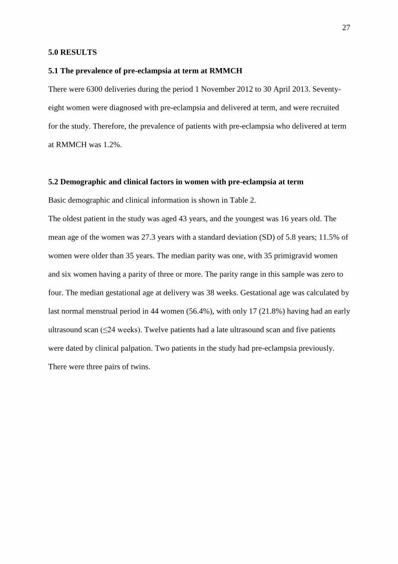
5.2 Demographic and clinical factors in women with pre-eclampsia at term
Basic demographic and clinical information is shown in Table 2.
The oldest patient in the study was aged 43 years, and the youngest was 16 years old. The
mean age of the women was 27.3 years with a standard deviation (SD) of 5.8 years; 11.5% of
women were older than 35 years. The median parity was one, with 35 primigravid women
and six women having a parity of three or more. The parity range in this sample was zero to
four. The median gestational age at delivery was 38 weeks. Gestational age was calculated by
last normal menstrual period in 44 women (56.4%), with only 17 (21.8%) having had an early
ultrasound scan (≤24 weeks). Twelve patients had a late ultrasound scan and five patients
were dated by clinical palpation. Two patients in the study had pre-eclampsia previously.
There were three pairs of twins.
28
Table 2: Demographic and clinical factors of women with pre-eclampsia at term (n=78)
No. % Age (years) ≤18 4 5.1 19-34 65 83.3 ≥35 9 11.5 Parity 0 35 44.9 1 22 28.2 2 15 19.2 ≥3 6 7.7 Antenatal
booking (weeks)
<20 22 28.2
≥20 56 71.2
Gestational age
at delivery
(weeks)
Previous
pre-eclampsia
Twin pregnancy
37
38-40
≥41
26
47
5
2
3
33.3
60.3
6.4
2.6
3.9
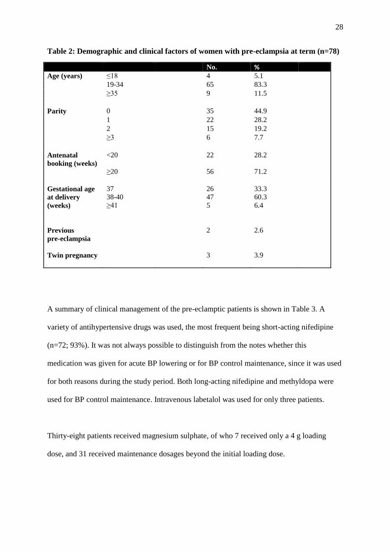
A summary of clinical management of the pre-eclamptic patients is shown in Table 3. A
variety of antihypertensive drugs was used, the most frequent being short-acting nifedipine
(n=72; 93%). It was not always possible to distinguish from the notes whether this
medication was given for acute BP lowering or for BP control maintenance, since it was used
for both reasons during the study period. Both long-acting nifedipine and methyldopa were
used for BP control maintenance. Intravenous labetalol was used for only three patients.
Thirty-eight patients received magnesium sulphate, of who 7 received only a 4 g loading
dose, and 31 received maintenance dosages beyond the initial loading dose.

29
Fifteen patients went into spontaneous labour, while 28 had labour induced for maternal
reasons, including two women who were ≥42 weeks’ gestation.
Fifty-one patients (65%) delivered by caesarean section, 52% for maternal reasons, and 48%
for fetal indications. Although there was an overlap in some patients who required operative
delivery from both maternal and fetal perspectives, the stronger indication was documented,
either maternal or fetal. There were no elective caesarean sections in this study.
Table 3: Clinical management of women with pre-eclampsia at term (n=78)
No % Anti-hypertensive drugs
Short acting nifedipine
72
92.3
Long acting nifedipine 17 21.8 MgSO4-loading 38 48.7 MgSO4-maintenance 31 39.7 Methyldopa 56 71.8 Labetalol 3 3.9 Amlodipine 1 1.3 Induction of labour 28 35.9 Caesarean section Indications: Maternal 33 52.4 Fetal 30 47.6
ICU ventilation
Blood transfusion
3
3
3.9
3.9
MgSO4=magnesium sulphate; ICU=intensive care unit
Certain indications for caesarean sections were a combination of maternal and fetal.
5.3 The severity of maternal disease in term pre-eclamptic patients
The complications of the pre-eclamptic patients are summarised in Table 4. Severe
hypertension occurred in 59 (76%) of the women. Imminent eclampsia was recorded in 29
(37%) of patients and 9% of women had eclamptic convulsions. There was some overlap
where patients had more than one complication. Six patients developed pulmonary oedema,
three of whom required intensive care and ventilation, provided in the obstetric high care area

30
as there were no beds available in the ICU. One of these patients developed pulmonary
oedema in the antenatal ward and died shortly after delivering spontaneously, and is
described in detail below.
Two patients had both eclampsia and pulmonary oedema, one of whom had to be intubated
and ventilated. There were 13 patients with isolated thrombocytopenia and three had true
HELLP syndrome by definition.
Three patients required blood transfusions for postpartum haemorrhage (PPH). One of these
patients had a placental abruption, the second patient a retained placenta and the third patient
had anaemia antenatally. They each received at least two units of red blood cells. No patient
in this study had an intra-cerebral haemorrhage.
The median number of days admitted to hospital was five, with a range of 1-11. One patient
stayed in hospital for 11 days as she was diagnosed with hyperthyroidism and was being
investigated by internal medicine specialists.
Table 4: Maternal morbidity and mortality in pre-eclamptic women at term (n=78)
Complication* No. %
Severe hypertension 59 75.6
Imminent eclampsia 29 37.2
Eclampsia 7 9.0
HELLP Syndrome 3 3.8
Pulmonary oedema 6 7.7
PPH 3 3.9
Abruptio placentae 1 1.3
Renal Failure 1 1.3
Death 1 1.3
*Patients may have more than one complication each

31
Haematological and biochemical indices were documented at their highest level during the
hospital admission and are shown in Table 4. For three patients, no blood results could be
found. Twenty-two patients (29%) were anaemic (Hb ≤11.0 g/dL). The median haemoglobin
level was 11.6 g/dL with a range of 6.2-16.3 g/dL.
Thirteen patients (17%) had low platelet counts (<150x109/L), with five patients having
moderate thrombocytopenia. None had severe thrombocytopenia. The median creatinine level
was 64.2 μmol/L, with a range of 27-153 μmol/L (a threshold of 100 mmol/L was used to
define renal dysfunction as per the institution’s protocol ). There was one patient who had
renal failure based on oliguria, but she died before she required dialysis. One patient had a
markedly elevated ALT reported as 10000 IU/L. Nine patients had abnormal AST levels
ranging from 42-643 IU/L.
Table 5: Haematological indices in pre-eclamptic women at term (n=75*)
No. % Haemoglobin (g/dL) 6.0-8.0
4
5.3
8.1-10.0 6 8.0
10.1-11.0 12 16.0
11.1-13.0 39 52.0
≥13.1 14 18.7
Platelet count (x109/L)
>150 62 82.7
100-150 8 10.7
50-100 5 6.7
<50 0 0
Creatinine levels(μmol/L)
<60 37 50.0
60-100 31 41.9
>100 6 8.1
Urea levels(mmol/L)
<4.5 65 87.8
4.5-10 9 12.2
Highest levels during admission were documented
*Blood results were unavailable for three patients
32
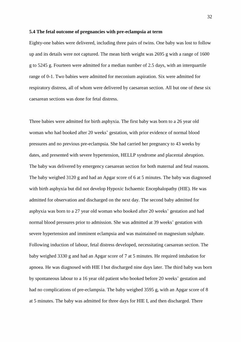
5.4 The fetal outcome of pregnancies with pre-eclampsia at term
Eighty-one babies were delivered, including three pairs of twins. One baby was lost to follow
up and its details were not captured. The mean birth weight was 2695 g with a range of 1600
g to 5245 g. Fourteen were admitted for a median number of 2.5 days, with an interquartile
range of 0-1. Two babies were admitted for meconium aspiration. Six were admitted for
respiratory distress, all of whom were delivered by caesarean section. All but one of these six
caesarean sections was done for fetal distress.
Three babies were admitted for birth asphyxia. The first baby was born to a 26 year old
woman who had booked after 20 weeks’ gestation, with prior evidence of normal blood
pressures and no previous pre-eclampsia. She had carried her pregnancy to 43 weeks by
dates, and presented with severe hypertension, HELLP syndrome and placental abruption.
The baby was delivered by emergency caesarean section for both maternal and fetal reasons.
The baby weighed 3120 g and had an Apgar score of 6 at 5 minutes. The baby was diagnosed
with birth asphyxia but did not develop Hypoxic Ischaemic Encephalopathy (HIE). He was
admitted for observation and discharged on the next day. The second baby admitted for
asphyxia was born to a 27 year old woman who booked after 20 weeks’ gestation and had
normal blood pressures prior to admission. She was admitted at 39 weeks’ gestation with
severe hypertension and imminent eclampsia and was maintained on magnesium sulphate.
Following induction of labour, fetal distress developed, necessitating caesarean section. The
baby weighed 3330 g and had an Apgar score of 7 at 5 minutes. He required intubation for
apnoea. He was diagnosed with HIE I but discharged nine days later. The third baby was born
by spontaneous labour to a 16 year old patient who booked before 20 weeks’ gestation and
had no complications of pre-eclampsia. The baby weighed 3595 g, with an Apgar score of 8
at 5 minutes. The baby was admitted for three days for HIE I, and then discharged. There
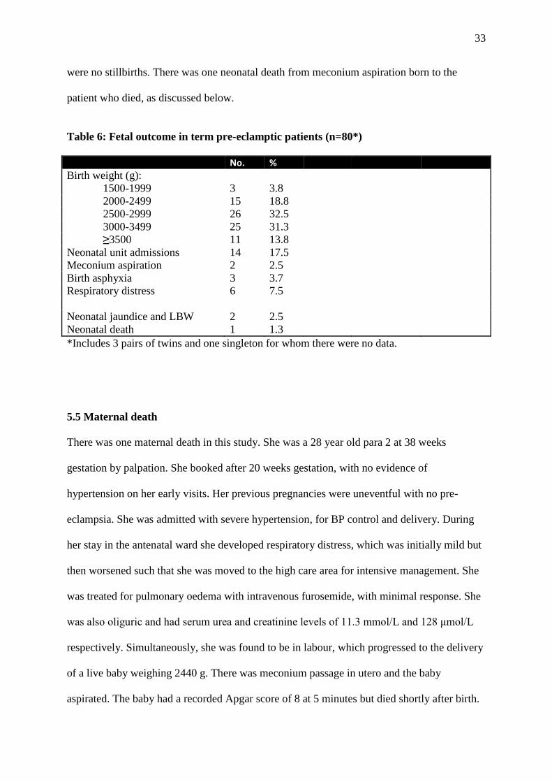
33
were no stillbirths. There was one neonatal death from meconium aspiration born to the
patient who died, as discussed below.
Table 6: Fetal outcome in term pre-eclamptic patients (n=80*)
No. %
Birth weight (g):
1500-1999
3
3.8
2000-2499 15 18.8
2500-2999 26 32.5
3000-3499 25 31.3
≥3500 11 13.8
Neonatal unit admissions 14 17.5
Meconium aspiration 2 2.5
Birth asphyxia 3 3.7
Respiratory distress 6 7.5
Neonatal jaundice and LBW 2 2.5
Neonatal death 1 1.3
*Includes 3 pairs of twins and one singleton for whom there were no data.
5.5 Maternal death
There was one maternal death in this study. She was a 28 year old para 2 at 38 weeks
gestation by palpation. She booked after 20 weeks gestation, with no evidence of
hypertension on her early visits. Her previous pregnancies were uneventful with no pre-
eclampsia. She was admitted with severe hypertension, for BP control and delivery. During
her stay in the antenatal ward she developed respiratory distress, which was initially mild but
then worsened such that she was moved to the high care area for intensive management. She
was treated for pulmonary oedema with intravenous furosemide, with minimal response. She
was also oliguric and had serum urea and creatinine levels of 11.3 mmol/L and 128 μmol/L
respectively. Simultaneously, she was found to be in labour, which progressed to the delivery
of a live baby weighing 2440 g. There was meconium passage in utero and the baby
aspirated. The baby had a recorded Apgar score of 8 at 5 minutes but died shortly after birth.

34
After delivery, the patient’s condition deteriorated to the point that she needed intubation and
ventilation for refractory pulmonary oedema. With no ICU beds available, the resuscitation
was performed in the high care area. Once she was stabilized with the support of inotropic
drugs, a CT scan of the brain was arranged, which showed cerebral oedema but no
intracranial haemorrhage. Almost 24 hours later, still on inotropes, the patient died. An
autopsy was not done.
35
6.0 DISCUSSION
This study has found that significant morbidity accompanies pre-eclampsia at term, especially
in terms of maternal complications. With seven cases of eclampsia and six cases of
pulmonary oedema including one maternal death, the risks of pre-eclampsia at term should
not be underestimated. Conversely, there appeared to be low morbidity and mortality in the
newborn infants.
6.1 The prevalence of pre-eclampsia at term at RMMCH
This study showed a prevalence of pre-eclampsia at term of 1.2%. This estimate is likely to
be close to a true prevalence because of the low likelihood of referral bias at RMMCH. The
only missing numbers from the prevalence denominator are an approximate 360 births at
Discoverers Clinic. Including these numbers in the denominator, assuming that no pre-
eclamptic women delivered at that clinic (the protocol is to transfer such women to
RMMCH), the adjusted prevalence remains 1.2% - a decrease from 1.24% to 1.17%.
The global impact of pre-eclampsia is 2-8% of pregnancies with a reported prevalence of 5-
8% in the USA (3,6). The prevalence in this study may appear low relative to the statistics
mentioned above, but it must be borne in mind that this is representative of a pre-eclamptic
population at term, and excludes pre-eclamptic women before 37 weeks’ gestation. The
statistics in the literature do not differentiate between term and pre-term pregnancies.
Considering this fact, one is led to believe that a significant proportion of our pre-eclamptic
patients make it to term or that many cases of pre-eclampsia only manifest clinically at term.

36
6.2 Clinical factors in women with pre-eclampsia at term
A variety of drug treatments was employed for treatment of hypertension. This may be
attributed to rotation of junior and trainee staff from different medical institutions in
Johannesburg, resulting in different approaches to the acute management of the hypertension
in pre-eclamptic patients at term and deviations from the protocol at RMMCH.
Unfortunately, the use of nifedipine for acute blood pressure lowering could frequently not be
distinguished from maintenance treatment of hypertension. However, the frequent use of
magnesium sulphate does indicate the clinical concern for the women that were included in
this study. Despite 48.7% of women being loaded with and 40% of patients being maintained
on magnesium sulphate , 9% complicated with eclampsia. In those patients who developed
eclampsia, the data sheet did not stipulate whether the patient had received magnesium
sulphate prior to the eclamptic episode. However, two out of seven eclamptic patients were
not documented as imminent eclampsia. From that one can deduce that the magnesium
sulphate was given as treatment of the eclampsia and not as prophylaxis. In general,
clinicians at RMMCH do not use magnesium sulphate in pre-eclampsia unless the condition
is considered severe or suggestive of imminent eclampsia. Perhaps this high incidence of
eclampsia then should prompt a wider prophylactic approach.
6.3 The severity of maternal disease in term pre-eclamptic patients
Fifty-one patients (65%) delivered by caesarean section, about half for maternal reasons, and
half for fetal indications. Although vaginal delivery is preferred for maternal considerations
in pre-eclamptic woman, the caesarean section rate was high even for maternal indications
and comparable to that experienced in Tygerberg Hospital, but similarly having a higher rate
for maternal than for fetal indications (34). Studies have shown that there is an increased risk
of failed induction and caesarean section in pre-eclamptic women (31). The study at
37
Tygerberg Hospital supported this finding by showing that 50% of their caesarean sections
were performed for failed induction of labour or fetal distress after induction of labour(34).
Short-term morbidity after caesarean section was not a significant problem, but long-term
problems such as wound haematoma (possibly related to thrombocytopaenia) and subsequent
wound sepsis could not be studied, because patients were not followed up through the
puerperium.
Severe hypertension was diagnosed in about three quarters of the patients. Imminent
eclampsia was recorded in 37%, all of whom received magnesium sulphate. Nine per cent of
the patients complicated with eclampsia. This and the high proportion of women with severe
hypertension, reflects the significant morbidity endured in pre-eclamptic patients at term. A
9% rate of eclampsia in a 1.2% proportion of cases of pre-eclampsia at term suggests an
eclampsia rate at term of one in 1000 pregnancies, much higher than the two to three cases
per 10 000 for eclampsia (at all gestational ages) quoted for developed countries (3). A
comparable eclampsia rate for term pregnancies is not available in the literature. Related to
eclampsia, intra-cerebral haemorrhage is an important cause of maternal mortality in South
Africa (29). However, there were no cases of intra-cerebral haemorrhage in this study.
Compared to a reported prevalence of 0.08-0.5% (30), 7.7% of pre-eclamptic women at term
in the current study complicated with pulmonary oedema, half of whom had to be intubated
and ventilated and required intensive care, with one death. The author speculates that perhaps
an obstetric critical care unit would have added much value to the management and outcome
of these patients, particularly the patient who developed pulmonary oedema while admitted in
the ward. The complication could have been detected earlier and promptly managed if she
was in a critical care setting. Again, extrapolating from the 1.2% prevalence of pre-eclampsia
38
at term, the prevalence of pulmonary oedema at term here would be about 1:1000
pregnancies, much higher than the 1.2 per 10 000 deliveries reported in the United Kingdome
(22). The finding is even more concerning when one remembers that the United Kingdom
rate is for pregnancies of all gestational ages and for all causes of pulmonary oedema, not
only pre-eclampsia.
In this study one patient had a placental abruption, in keeping with the national prevalence of
0.5-2% (18). However, the sample size was too small to make any conclusion about the
frequency of abruption in association with term pre-eclampsia, other than to state it was not a
common finding. From the available data on these patients, there seemed to be little evidence
of placental disease, suggesting that term pre-eclamptic patients perhaps have predominantly
maternal disease with relatively few placental and fetal adverse events, in keeping with recent
observations regarding late-onset versus early-onset pre-eclampsia(34,41,42,44). However,
growth restriction diagnosis was rarely attempted in these patients and formal screening for
small-for-gestational-age was not performed on the babies after delivery. The observed 18%
rate of low birth weight babies could suggest placental insufficiency or misclassified
gestational age.
Thirteen patients had thrombocytopenia, some of whom were managed as HELLP syndrome
based on their clinical status. This number is not included in the group of patients
documented with true HELLP syndrome. The prevalence of true HELLP syndrome by
definition was 3.8% in this study, lower than the reported prevalence of 20% in severe pre-
eclamptic patients as quoted by Curtain and Weinstein (18), but similar to the 5.2% found at
Tygerberg Hospital (11). Contrary to the haemoconcentration found in patients with pre-
eclampsia, 29.3% of women had a haemoglobin level ≤11.0 g/dL on admission. Neither the
39
type nor the cause of the anaemia was a focus of the study and therefore was not documented.
All patients who are booked at the antenatal clinics receive iron and folate. Therefore, the
question of anaemia and its association with HIV and anti-retroviral drugs, or other factors,
comes into question. However, the HIV status of the women was not recorded in the data.
6.4 The fetal outcome of pregnancies with pre-eclampsia at term
The major fetal complications encountered were meconium aspiration, from which one baby
died, respiratory distress and birth asphyxia. All babies admitted for respiratory distress were
delivered by caesarean section, of which all but one was for fetal distress. This leads one to
question whether the respiratory distress was due purely to the placental insufficiency with
metabolic acidosis related to pre-eclampsia, misclassification of gestational age, or was
related to caesarean section itself, which is known to be associated with mild respiratory
distress (45, 46) also associated with borderline maturity at 37-38 weeks (37). The neonatal
admission rate in this study was high (17.5%), similar to the 14.7% found in the United
Kingdom (22). It must yet again be remembered that the studies cited encompass all pre-
eclamptic women, whereas this study included only women at ≥37 completed weeks. As
noted by Sibai, and evidenced by this study, the majority of neonatal admissions from
hypertensive mothers are those who are delivered at term. The reasoning is unknown and
emphasizes the need for research in this area (35). The high caesarean section rate comes into
question as a possible reason, given the concern about respiratory distress mentioned above.
6.5 Limitations of the study
The small study population affected the precision of the reported estimates, and rendered it
difficult to comment on some of the findings. However, the severe outcomes in this study
such as eclampsia and pulmonary oedema are significant enough to warrant a larger

40
population-based study. The significance of the findings could perhaps have been enhanced if
the researcher compared them to a control group of term non-pre-eclamptic women and to a
group of pre-eclamptic women remote from term. The researcher relied on patients’ files for
recording of clinical observations and bedside investigations. This allowed room for error in
measurements beyond the control of the researcher, and possible misclassification of
hypertensive disorders and gestational age. Unbooked patients were excluded from this study
and therefore those patients who presented for the first time to hospital with unclassified
proteinuric hypertension or eclamptic episode were not documented. However, in the
researcher’s experience unbooked women presenting at term are a rarity at RMMCH. It was
also not possible to document resolution of hypertension after delivery, to exclude chronic
hypertensive disease misclassified as pre-eclampsia. During the study time period the
laboratory staff were upgrading their data systems, therefore results were not always readily
available. Patients were managed on whatever results were available and on their clinical
condition. Hence, patients with thrombocytopenia were often managed as HELLP syndrome,
in the absence of certainty about haemolysis. Haematocrit levels are not routinely recorded in
the patients file, and this posed a limitation to the more accurate assessment of the anaemia
and haemoconcentration observed. To the best of the researcher’s knowledge, none of the
patients entered into the study were referrals, and RMMCH did not refer any hypertensive
patients to other hospitals, and therefore this sample is likely to be representative of a pre-
eclamptic population at term. Also, in this study, the focus was on outcomes and it is unlikely
that any serious complications were missed. The likely impact of a gap in the denominator
resulting from the exclusion of Discoverers Clinic deliveries has been discussed earlier.
41
6.6 Conclusion
It seems clear that there is a tendency to poor maternal outcome as one approaches term in
pre-eclampsia. This study demonstrated that maternal complications such as eclampsia and
pulmonary oedema are common in pre-eclampsia at term. Clinical vigilance and expertise are
as important for pre-eclamptic women at term as they are for those who are pre-term. The
importance of maternal stabilization before embarking on delivery or surgery must always be
considered.
As demonstrated by this study, and suggested by the little data available in the literature, the
fetal morbidity and mortality seems to be lower in the neonate of a term pre-eclamptic
woman. This sheds more light on the theory of the heterogeneous pathophysiology of pre-
eclampsia, which may manifest as: 1) a restricted fetal growth syndrome (early-onset pre-
eclampsia), where affected patients deliver before term and decreased uteroplacental
perfusion may be evident; and 2) a normal fetal growth syndrome (late-onset pre-eclampsia)
where patients progress to term and have normal uteroplacental perfusion, as supported by
Dahlia in a critical review of both early- and late-onset pre-eclampsia(41).
There is a need for a larger epidemiological study where term pre-eclamptic patients can be
compared to pre-term pre-eclamptic patients. It may also be of value to follow these two
groups of patients through the pueperium to assess for complete resolution of the
complications or residual disease subsequent to the severe complications such as pulmonary
oedema and eclampsia.
42
7.0 REFERENCES
1. A Woman’s Worth, Inspirational Poem. Available at:
http://www.familyfriendpoems.com/poem/a-womans-worth [Accessed December 2014]
2. Hogan MC, Foreman KJ, Naghavi M, Ahn SY, Wang M, Makela SM, et al. Maternal
mortality for 181 countries, 1980–2008: a systematic analysis of progress towards
Millennium Development Goal 5. Lancet 2010;375:1579-1664.
3. Duley L. The global impact of preeclampsia and eclampsia. Semin Perinatol
2009;33:130-7.
4. Department of Health. Saving Mothers 2008-2010: Fifth report to the NCCEMD.
Available at: http://www.doh.gov.za/docs/reports/2012/savingmothersshort.pdf.
[Accessed 16th
September 2012]
5. Matthys LA, Coppage KH, Lambers DS, Barton JR, Sibai BM. Delayed pre-eclampsia:
an experience of 151 cases. Am J Obstet Gynecol 2004;190:1464-66.
6. American College of Obstetricians and Gynecologists Practice Bulletin No.33 Diagnosis
and Management of Pre-eclampsia and Eclampsia. Clinical management guidelines for
obstetrician-gynecologists. Obstet Gynecol 2002;99:159-167.
7. Buchmann E (ed). Wits Obstetrics. Johannesburg: Department of Obstetrics and
Gynaecology, University of the Witwatersrand, 2007.
8. Xiong X, Demianczuk NN, Saunders LD, Wang FL, William DF. Impact of pre-
eclampsia and gestational hypertension on birth weight by gestational age. Am J
Epidemiol 2002;155:203-9.
9. Al-Mulhim AA, Abu-Heija A, Al-Jamma F, El-Harith A. Pre-eclampsia: maternal risk
factors and perinatal outcome. Fetal Diagn Ther 2003;18:275-80.
43
10. Yucesoy G, Ozkan S, Bodur H. Maternal and perinatal outcome in pregnancies
complicated with hypertensive disorder of pregnancy: a seven year experience of a
tertiary care centre. Arch Gynecol Obstet 2005;273:43-9.
11. Hall DR, Odendaal HJ, Steyn DW, Grove D. Expectant management of early onset,
severe pre-eclampsia: maternal outcome. BJOG 2000;107:1252-7.
12. Chesley LC. History and epidemiology of pre-eclampsia-eclampsia. Clin Obstet Gynecol
1984;27:801-20.
13. Walsh SW. Obesity: a risk factor for pre-eclampsia. Trends Endocrinol Metab
2007;18:365-70.
14. Tikkanen M. Placental abruption: epidemiology, risk factors and consequences. Acta
Obstet Gynecol Scand 2011;90:140-9.
15. Hypertension in pregnancy: report of the American College of Obstetricians and
Gynecologists’ Task Force on Hypertension in Pregnancy. Obstet Gynecol 2013;1122-
1131.
16. Cunningham FG, Leveno KJ, Bloom SL, Hauth JC, Rouse DJ, Spong CY. Williams
Obstetrics. 23rd
ed. USA: McGraw-Hill: 2010.
17. Leduc L, Wheeler JM, Kirshon B, Mitchell P, Cotton DB. Coagulation profile in severe
pre-eclampsia. Obstet Gynecol 1992;79:14-18.
18. Curtain MW, Weinstein L. A review of HELLP syndrome. J Perinatol 1999;19:138-43.
19. Haram K, Svendsen E, Abildgaard U. The HELLP syndrome: clinical issues and
management. A review. BMC Pregnancy and Childbirth 2009;9:1-15.
20. Sheehan HL, Lynch JB(eds): Cerebral lesions. In: Pathology of toxaemia of pregnancy.
Baltimore, Williams & Wilkins, 1973.
21. Edmonds DK. Dewhurst’s Textbook of Obstetrics & Gynaecology. 8th
edition. Wiley-
Blackwell, 2012.

44
22. Tuffnell DJ, Jankowicz D, Lindow SW, Lyons G, Mason GC, Russell IF, et al. Outcomes
of severe pre-eclampsia/eclampsia in Yorkshire 1999-2003. BJOG 2005;112:875-80.
23. Cipolla MJ: Brief review: cerebrovascular function during pregnancy and eclampsia.
Hypertension 2007;50:14-24.
24. Pre-eclampsia: clinical features and diagnosis. 27 November 2013. Available at:
www.uptodate.com.innopac.wits.ac.za [Accessed 28 January 2014]
25. Ajida ET, Adeka BI, Jibril KA. Pregnancy outcome in eclamptics at the University of
Abuja teaching Hospital, Gwagwalada, Abuja: a 3 year review. Nig J Clin Pract
2010;13:394-8.
26. Moodley J and Daya P. Eclampsia: a continuing problem in developing countries. Int J
Gynecol Obstet 1994;44:9-14.
27. Hinchey J, Chaves C, Appignani B, Breen J, Pao L, Wang A, et al. A reversible posterior
leukoencephalopathy syndrome. N Engl Med 1996;334:494-500.
28. Eastabrook G, Brown M, Sargent I. The origins and end-organ consequence of pre-
eclampsia. Best Pract Res Clin Obstet Gynaecol 2011;25:435-447.
29. Paruk F, Moodley J. Maternal and Neonatal outcome in early- and late-onset pre-
eclampsia. Semin Neonatol 2000;5:197-207.
30. Dennis AT, Solnordal CB. Acute pulmonary oedema in pregnant women. Anaesthesia
2012;67:646-59.
31. Anumba DOC, Robson SC. Management of pre-eclampsia and haemolysis, elevated
enzymes, and low platelets syndrome. Curr Opin Obstet and Gynecol 1999;11:149-56.
32. Ananth CV, Peltier MR, Kinsler WL, Smulian JC, Vintzileos AM. Chronic hypertension
and risk of placental abruption: Is the association modified by ischemic placental disease?
Am J Obstet Gynecol 2007;197:273.e1-273.e7.
45
33. HøgbergV, Rasmussen S, Irgens LM. The effect of smoking and hypertensive disorders
on placental abruption in Norway. Acta Obstet Gynecol Scand 2007;86:304-309.
34. Kenneth L, Hall DR, Gebhardt S, Grove D. Late onset pre-eclampsia is not an innocuous
condition. Hypertens Preg 2010; 29:262-270.
35. Sibai BM. Pre-eclampsia as a cause of preterm and late preterm (near-term) births. Semin
Perinatol 2006; 30:16-9.
36. Hall DR, Odendaal HJ, Kirsten GF, Smith J, Grove D. Expectant management of early
onset, severe pre-eclampsia: Perinatal outcome. BJOG 2000;107:1258-64.
37. Impey L, Greenwood C, Sheil O, MacQuillan, Reynolds M, Redman C. The relation
between pre-eclampsia at term and neonatal encephalopathy. Arch Dis Child Fetal and
Neonatal Ed 2001; 85: F170-F172.
38. Sarris I, Bewley S, Agnihotri S. Oxford Specialty Training: Training in Obstetrics and
Gynaecology. Oxford University Press 2009.
39. Hypertension in Pregnancy: the management of hypertensive disorders during pregnancy.
NICE Clinical Guideline 2011.https://www.nice.org.uk.guidance/cg107 [Accessed July
2012]
40. Ananth CV, Smulian JC, Vintzileos AM. Ischaemic placental disease: maternal versus
fetal clinical presentations by gestational age. J Matern Fetal Neonatal Med 2010;23:887-
3.
41. Raymond D, Peterson E. A Critical Review of early-onset and late-onset preeclampsia.
Obstet Gynecol Surv 2011;66:497-506.
42. Van der Merwe J, Hall DR, Wright C, Schubert P, Grove D. Are early and late pre-
eclampsia distinct subclasses of the disease-what does the placenta reveal? Hypertens
Preg 2010;29:457-467.
46
43. Koopmans CM, Bijlenga D, Groen H, Vijgen SMC, Aarnoudse JG, Bekedam DJ, et al.
Induction of labour versus expectant monitoring for gestational hypertension or mild pre-
eclampsia after 36 weeks gestation (HYPITAT): a multicentre, open-label randomised
controlled trial. Lancet 2009; 374:379-388.
44. Kassebaum NJ, Bertozzi-Villa A, Coggeshall MS, Shackelford KA, Steiner C, Heuton
KR, et al. Global, regional, and national levels and causes of maternal mortality during
1990-2013: a systematic analysis for the Global Burden of Disease Study 2013. Lancet
2014; 384:980–1004.
45. Levine EM, Ghai V, Barton JJ, Strom C. Mode of delivery and the risk of respiratory
diseases in newborns. Obstet Gynecol 2001;97:439-442.
46. Tutdibi E, Gries K, Bucheler M, Misselwitz B, Schlosser RL, Gortner L. Impact of labor
on outcomes in transient tachypnea of the newborn: population-based study. Pediatrics
2010;125:e577-e583.
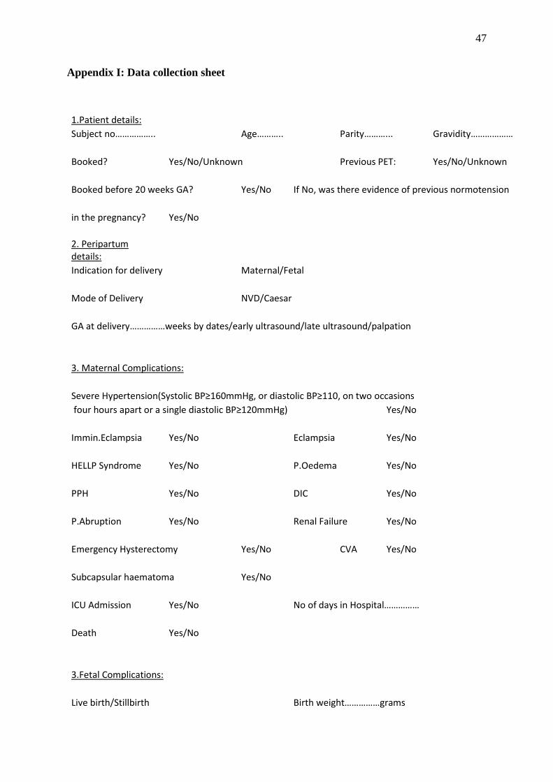
47
Appendix I: Data collection sheet
1.Patient details: Subject no……………..
Age………..
Parity………... Gravidity………………
Booked?
Yes/No/Unknown
Previous PET: Yes/No/Unknown
Booked before 20 weeks GA? Yes/No If No, was there evidence of previous normotension
in the pregnancy? Yes/No
2. Peripartum details:
Indication for delivery Maternal/Fetal
Mode of Delivery
NVD/Caesar
GA at delivery……………weeks by dates/early ultrasound/late ultrasound/palpation
3. Maternal Complications:
Severe Hypertension(Systolic BP≥160mmHg, or diastolic BP≥110, on two occasions four hours apart or a single diastolic BP≥120mmHg)
Yes/No
Immin.Eclampsia Yes/No
Eclampsia Yes/No
HELLP Syndrome Yes/No
P.Oedema Yes/No
PPH
Yes/No
DIC
Yes/No
P.Abruption Yes/No
Renal Failure Yes/No
Emergency Hysterectomy Yes/No
CVA Yes/No
Subcapsular haematoma Yes/No
ICU Admission Yes/No
No of days in Hospital……………
Death
Yes/No
3.Fetal Complications:
Live birth/Stillbirth
Birth weight……………grams
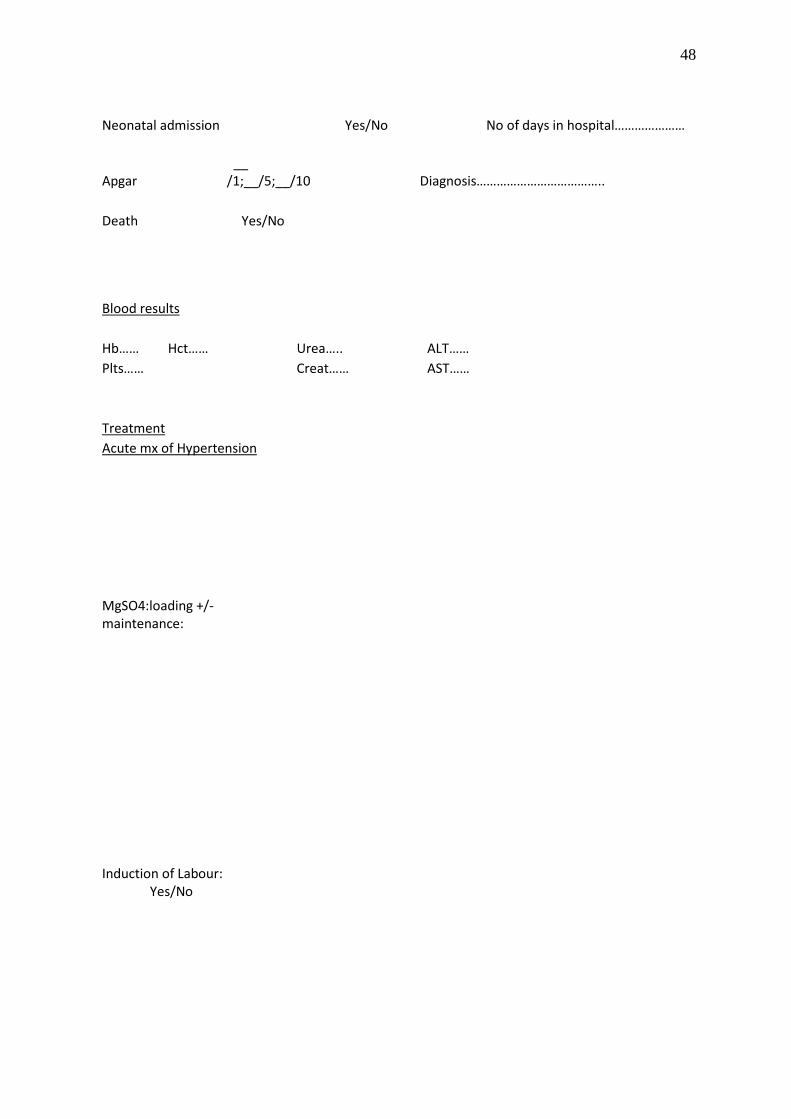
48
Neonatal admission
Yes/No
No of days in hospital…………………
Apgar
__ /1;__/5;__/10
Diagnosis………………………………..
Death
Yes/No
Blood results
Hb…… Hct……
Urea…..
ALT…… Plts……
Creat……
AST……
Treatment Acute mx of Hypertension
MgSO4:loading +/- maintenance: Induction of Labour: Yes/No
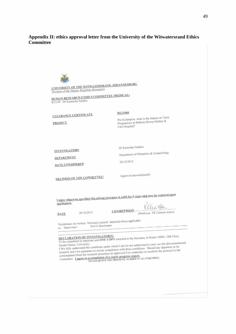
49
Appendix II: ethics approval letter from the University of the Witwatersrand Ethics
Committee